BRITISH HYPOGEOUS FUNGI BY LILIAN E. HAWKER - ASCOfrance
BRITISH HYPOGEOUS FUNGI BY LILIAN E. HAWKER - ASCOfrance
BRITISH HYPOGEOUS FUNGI BY LILIAN E. HAWKER - ASCOfrance

You also want an ePaper? Increase the reach of your titles
YUMPU automatically turns print PDFs into web optimized ePapers that Google loves.
[ 429 ]<br />
<strong>BRITISH</strong> <strong>HYPOGEOUS</strong> <strong>FUNGI</strong><br />
<strong>BY</strong> <strong>LILIAN</strong> E. <strong>HAWKER</strong><br />
Department of Botany, University of Bristol<br />
(Communicated<br />
by W. Brown, F.R.S.-Received 16 May 1953-<br />
Revised 30 July 1953)<br />
CONTENTS<br />
INTRODUCTION 430<br />
Methods of collection 430<br />
Examination and preservation of material 431<br />
Habitat 431<br />
(a) Nutrition 432<br />
(b) Soil conditions (H-ion concentration, texture and aeration)<br />
432<br />
(c) Weather 433<br />
KEY TO THE GENERA OF <strong>BRITISH</strong> <strong>HYPOGEOUS</strong> <strong>FUNGI</strong> 435<br />
PHYCOMYCETES 436<br />
Mucorales. Endogonaceae 436<br />
Systematic position of Endogone<br />
444<br />
ASCOMYCETES 444<br />
Plectascales. Elaphomycetaceae<br />
444<br />
Systematic position of Elaphomyces<br />
453<br />
Tuberales 453<br />
(I) Pseudotuberaceae 454<br />
(II) Geneaceae 459<br />
(III) Eutuberaceae 467<br />
(IV) Terfeziaceae 503<br />
Theories of relationship within the Tuberales 506<br />
BASIDIOMYCETES 507<br />
(I) Hysterangiaceae<br />
507<br />
(II) Hydnangiaceae<br />
512<br />
(III) Hymenogastraceae 523<br />
(IV) Rhizopogonaceae 536<br />
The relationship between the hypogeous Gasteromycetes and other fungi<br />
REFERENCES 542<br />
The study of hypogeous fungi has been neglected in Britain from the time of Berkeley & Broome<br />
until that of the present investigation. During the years 1948-53 some 700* collections have been<br />
made, mainly in the Bristol area, but also from other parts of England, Scotland, North Wales and<br />
Northern Ireland. These include members of the Phycomycetes (Endogone spp.), Ascomycetes<br />
(Elaphomycetaceae, Tuberales) and Basidiomycetes (Gasteromycetes). Some species were found<br />
sufficiently often to permit tentative conclusions to be drawn relating to the effect of weather and<br />
soil conditions on the production of fruit-bodies.<br />
Most of the species previously recorded in Britain have been collected and some new records<br />
made. Descriptions are given of all recorded British species, and most of these are illustrated by<br />
line drawings made from fresh material. Details of development are given for representative species<br />
and the probable relationships within the group and with other fungi are discussed.<br />
Increased to 1000 by end of 1953, later collections not listed in this paper unless of special interest.<br />
VOL. 237. B. 650. (Price 27s.) 52<br />
[Published 17 February 1954<br />
PAGE<br />
540
430<br />
<strong>LILIAN</strong> E. <strong>HAWKER</strong> ON<br />
INTRODUCTION<br />
Many fungi pass the greater part of their life cycle within the soil, but the majority of those<br />
producing large fruit-bodies either produce them above the soil surface or if they are at<br />
first subterranean they emerge before maturing. The true hypogeous fungi, however,<br />
produce relatively large fruit-bodies which complete their development beneath the surface<br />
of the soil or are at least embedded in the covering of humus or litter. None of them has any<br />
dehiscence mechanism by which the spores are shed. The latter are set free by the decay<br />
of the surrounding hyphae or are probably dispersed by soil-inhabiting invertebrates or<br />
small mammals which eat the fruit-bodies. It is not known whether the spores are capable of<br />
normal germination. The hypogeous habit is found amongst the Zygomycetes, Ascomy-<br />
cetes and Basidiomycetes, but the similarity of habitat has induced a superficial morpho-<br />
logical resemblance between the mature fruit-bodies despite the great differences in their<br />
modes of development.<br />
The early studies of European hypogeous fungi by Micheli (1729), Fries (1822), Vittadini<br />
(I83I, I842), Tulasne (I843, i844, i851), Zobel (I854), Chatin (1892) and Hesse (i89I, 1894)<br />
have been continued by Fischer (I896-I927), Bucholtz (1897-1912), Jaczewski (I909),<br />
Hollos (i9II), Soehner (1913-5I), Bataille (192I, I923), Lohwag (I924-39), Knapp (I924-52),<br />
Malen~on (I938) and others. The North American species have been studied by Harkness<br />
(1899), Lloyd (I922), Gilkey (1916, 1939), Dodge & Zeller (I918-36) and Coker & Couch<br />
(1928), and the hypogeous Gasteromycetes of South Africa (Bottomley 1948) and Australasia<br />
(Cunningham 1944) have been investigated. The hypogeous fungi of Britain, however,<br />
have been neglected since the days of Berkeley & Broome (I846-75). Only occasional<br />
references occur in the Foray Lists published by the British Mycological Society, and these<br />
are usually to two common species of Elaphomyces. The present writer became interested<br />
in hypogeous fungi as the result of the collection of specimens of Tuber puberulum by Dr P. H.<br />
Gregory at the Belfast Foray of the British Mycological Society in September 1948, and,<br />
with the aid of colleagues and students of the Department of Botany, University of Bristol,<br />
and of a few mycological colleagues elsewhere, has since made some 700 separate collections.<br />
These include most of the species previously recorded for Britain, together with one<br />
new species, a new variety and several known species not hitherto recorded as British.<br />
Much of this material was in sufficient abundance to permit developmental studies to be<br />
made. Most of the descriptions given below were compiled as a result of the examination<br />
of fresh material which was compared with herbarium specimens and checked against<br />
earlier descriptions. A few, which are indicated in the text, were based solely on a study<br />
of preserved material at the Herbarium, Royal Botanic Gardens, Kew, and at the British<br />
Museum (Natural History).<br />
Methods of collection<br />
The edible truffles are still collected in France, Italy and elsewhere on the continent<br />
of Europe with the aid of trained pigs or dogs. No such trained animals are available in<br />
this country, and search has been made by scraping away the leaf litter and loose surface<br />
soil under suitable trees with a small hand-rake. While many fruit-bodies may be missed<br />
by this method it has the advantage that the collection is not limited to mature specimens<br />
of those species whose scent is attractive to animals, but is likely to contain a representative
<strong>BRITISH</strong> <strong>HYPOGEOUS</strong> <strong>FUNGI</strong><br />
collection of these and of immature specimens or specimens of odourless varieties. Experience<br />
soon showed what type of site would be most likely to yield fruit-bodies, while it was often<br />
advantageous to investigate the neighbourhood of small holes dug by animals.<br />
Complete records were kept of the places where specimens were collected, and these<br />
included particulars of the nature and H-ion concentration of the soil, surface vegetation,<br />
if any, and the species of tree beneath which the fungus was found. Records of weather<br />
were also made. These have given some indication of the conditions favouring development<br />
of the commoner species.<br />
Examination and preservation of material<br />
A full record was made of size, colour, general external and internal appearance, spore<br />
type and size, odour, etc., of each collection in a fresh condition. The specimens were then<br />
either dried or preserved in formalin alcohol. Frequently, half of a particular collection<br />
(or fruit-body) was dried and half was pickled. For the study of development, portions<br />
of fruit-bodies were fixed and embedded in wax by a method employing butyl alcohol and<br />
avoiding the use of xylol (which causes shrinkage and distortion) based on that used by<br />
Professor Nannfeldt of the University of Uppsala, Sweden. Serial sections were cut and<br />
stained with carbol fuchsin (or safranin) and light green for the study of gross morphology<br />
or with iron alum haemotoxylon counter-stained with light green for cytological studies.<br />
Staining with gentian violet did not in general give good cytological preparations of these<br />
fungi. For the investigation of mature fruit-bodies, hand sections or occasionally freezing<br />
microtome sections were preserved in glycerine. All drawings, other than those of whole<br />
fruit-bodies, were made with the aid of the camera lucida.<br />
Habitat<br />
The large majority of the collections were made under trees, usually in woodlands but<br />
occasionally under isolated trees in parks or hedgerows. A few specimens were obtained<br />
some distance from the nearest tree, but in these examples the soil had been undisturbed<br />
for long periods and thus it is possible that the unbroken mycelium extended through it<br />
for a considerable distance. Some species were found under a number of different kinds<br />
of tree, but many were associated with one only. Beech, evergreen oak, lime and conifers<br />
(both indigenous and planted) have been the most usual tree associates, while ash and<br />
deciduous oakwoods have been relatively poor in hypogeous fungi. There is little doubt<br />
that many of these fungi are intimately associated with tree roots in the formation of<br />
ectotrophic mycorrhizal mantles. Claims have been made for a mycorrhizal association<br />
between various trees and Elaphomyces granulatus (Reess i88o), E. muricatus<br />
(Lewton-Brain<br />
1901), Tuber borchii (Mattirolo 1934), T. magnatum (Sappa 1946), Rhizopogon luteolus (Young<br />
1937) and R. roseolus (Modess I939). Fruit-bodies of various species collected by the writer<br />
have been found to be in very close association with mycorrhizae, and others have been<br />
equally closely associated with pseudomycorrhizae (Melin 1917), e.g. Endogone microcarpa.<br />
The hyphae of the true mycorrhizal mantles often show a close resemblance to those of<br />
parts of the fruit-body and its surrounding mycelium. The latter may sometimes be traced<br />
through the soil from fruit-body to root. An unusual orange mycelium which, in culture,<br />
felts together to form a sheet of hyphae with a greenish black metallic sheen, was isolated<br />
from the mantles of beech mycorrhizae and from immature fruit-bodies of Tuber excavatum.<br />
52-2<br />
431
432<br />
<strong>LILIAN</strong> E. <strong>HAWKER</strong> ON<br />
This mycelium has not been induced to produce truffles, but both isolates have caused the<br />
development of typical mycorrhizal roots on beech seedlings grown under aseptic con-<br />
ditions. Further work of this type is planned.<br />
The conditions which favour the development of fruit-bodies are not fully understood<br />
and almost certainly vary to some extent with the species. Nevertheless, it is possible to<br />
draw some general conclusions.<br />
(a) Nutrition<br />
Melin and his co-workers (Melin & Lindeberg 1939; Melin & Norkrans 1942, 1948),<br />
have shown that growth in artificial culture of certain Basidiomycetes known to be<br />
mycorrhiza-formers, including the hypogeous Rhizopogon roseolus, is stimulated by the<br />
addition to the medium of mixtures of amino-acids and of growth substances, of which<br />
vitamin B1 was the most effective. The isolate referred to above and believed to be Tuber<br />
excavatum and a culture of Hymenogaster tener behaved similarly to Melin's mycorrhizal<br />
fungi, that is, they grew better on peptone or asparagine than with nitrates or ammonium<br />
salts as sole source of nitrogen, and they were stimulated by the presence of an external<br />
supply of vitamin B1. In their natural habitats it is likely that these fungi obtain both<br />
organic nitrogen and growth substances from tree roots.<br />
Observations in the field with a number of species confirm those of De Ferry de la<br />
Bellone (I888), who found that the edible truffles, Tuber melanosporum and Tuber aestivum<br />
were most plentiful in association with mature trees in their prime and were seldom found<br />
under either very young trees or old decaying ones.* The present writer has frequently<br />
found numerous fruit-bodies round the stumps of trees felled the previous year. It is thus<br />
probable that, as with many other fungi, the hypogeous species produce their fruit-bodies<br />
only on initially well-nourished mycelia but that the onset of starvation leads to increased<br />
fructification.<br />
Some species produce their fruit-bodies only in close association with tree roots. The<br />
abundant mycelium of Hysterangium nephriticum has been observed in several localities<br />
spreading beneath the surface of the soil under beech trees. The fruit-bodies, which are<br />
produced in large numbers, occurred only within a few inches of a large root. Fruit-bodies<br />
of Tuber excavatum have been collected in quantity by following the course of a large root,<br />
while the fruit-bodies of Elaphomyces granulatus frequently grow around the roots of pine<br />
which become embedded in the 'cortex' and 'crust' of the fungus. Others produce their<br />
fruit-bodies at some distance from any large roots, and it is not always possible to trace<br />
a mycelial connexion between fructification and root.<br />
(b) Soil conditions (Hydrogen-ion concentration; texture and aeration)<br />
De Ferry de la Bellone (i888) points out that the edible truffles of France occur only on<br />
light calcareous soils and are never found in clays or in acid soils. This is also true of the<br />
majority of British species, which occur in greatest abundance in slightly alkaline soils.<br />
There are, however, a number of exceptions such as Elaphomyces granulatus, Genea hispidula,<br />
G. klotzschii and Melanogaster variegatus var. broomeianus which, while they do occur in alkaline<br />
*<br />
Elaphomyces spp. are an exception and E. muricatus was the only species found by the writer in<br />
September 1952 in the famous 'truffle walk' at Savernake Forest, an avenue of old beeches, which,<br />
according to the estate records, formerly yielded Tuber aestivum in quantity.
<strong>BRITISH</strong> <strong>HYPOGEOUS</strong> <strong>FUNGI</strong><br />
soils, are also found in acid ones. Elaphomyces granulatus is actually commoner in acid than in<br />
alkaline soils, which is not surprising, since it is most frequently associated with the Scots pine.<br />
In this connexion it is of interest that very few species and individuals have been obtained<br />
from the Chiltern beechwoods, where the surface soil is usually the acid clay with flints<br />
which overlies the chalk, compared with the large numbers from the Cotswold beechwoods<br />
where the soil is alkaline. A relatively large number of specimens have resulted from a few<br />
hurried investigations of beechwoods on chalk soil in Kent and Sussex. The texture of the<br />
soil is also important, and no specimens have been found in heavy soils although some have<br />
been found in the leaf litter overlying such soils. Species of Elaphomyces and most of the<br />
hypogeous Gasteromycetes are commonly found at the junction of the shallow layer of<br />
loose top soil with the hard pan formed by the harder subsoil. A somewhat similar habitat<br />
is provided by the hard edge of a woodland path. Fruit-bodies are frequently partially,<br />
but never completely, embedded in the hard layer. The difficulty of the expansion of the<br />
growing fruit-body in a hard soil is probably one factor in this distribution, and fruitbodies<br />
which during development encounter a hard surface such as a root or a stone are<br />
frequently much distorted and lobed. Aeration is almost certainly a more important factor,<br />
and all species are extremely susceptible to conditions of poor aeration. Even those forms,<br />
such as Tuber puberulum, which normally grow within the layer of partially decayed leaves,<br />
are not found where the leaves drift to form a thick layer. Other species, such as T. excavatum,<br />
are found only in places such as the edges of woods, artificial mounds or banks or near the<br />
top of slopes, where leaves do not accumulate. Slight disturbance of the soil may stimulate<br />
some species, presumably through improved aeration. Thus large numbers of young fruitbodies<br />
of T. puberulum were found on returning to a patch which when searched, and<br />
therefore disturbed, some months earlier had yielded only a few. Tuber aestivum, was found<br />
in quantity in the grounds of the University of Bristol under holly trees one year after the<br />
ground had been dug, after having been undisturbed for many years. No fruit-bodies<br />
were discovered at the first digging. In general, however, the soil must be relatively<br />
stable, so that steep slopes or rabbit warrens arens are seldom suitable. Some species are<br />
frequent under a thin covering of moss. Moss or herbaceous plants growing thickly<br />
over the floor of a woodland prevent the development of most species, although there<br />
are a few exceptions such as Sclerogaster compactus, Balsamia spp. and Arcangeliella<br />
stephensii which have been found among crowded roots of dog's mercury or ivy. The<br />
effect of dense undergrowth is most probably to reduce the aeration of the soil and the<br />
presence of such plants may possibly explain the absence of hypogeous fungi from<br />
deciduous oakwoods and ashwoods in the west of England. The soils of the French oakwoods,<br />
which do yield truffles, are lighter and consequently probably better aerated.<br />
(c) Weather<br />
Collections have been made over the years 1948-53. Certain woods in the Bristol area,<br />
notably three areas on the Cotswolds near Wotton-under-Edge, Gloucestershire; the<br />
Blaise Castle and Kingsweston estate in north Bristol; Abbot's Pool, Somerset and the area<br />
known as Goblin Coombe and the surrounding limestone cliffs at Cleeve, Somerset, have<br />
been visited at frequent intervals. The relative frequency of a number of species in these<br />
areas has varied considerably in different years. While the probable exhaustion of the<br />
433
434<br />
<strong>LILIAN</strong> E. <strong>HAWKER</strong> ON<br />
mycelium following the production of an unusually large crop of fruit-bodies is a factor to<br />
be considered, yet mycorrhizal forms are likely to be able to restore nutrients to the<br />
mycelium fairly rapidly. Moreover, the incidence of the fruit-bodies of particular species<br />
can be closely correlated with the prevailing conditions of temperature, rainfall and soil<br />
water content.<br />
The relation between fungus and environment depends partly on the length of the<br />
fruiting cycle of a particular species. Thus the majority of the Tuberales, together with the<br />
Phycomycetous Endogone lactiflua, produce only one batch of fruit-bodies annually, and<br />
these usually take several months or even the greater part of a year to mature. The actual<br />
initiation of fruiting is dependent upon environmental conditions and may be delayed by<br />
periods of abnormal cold or drought in the early spring. Maturation may be delayed<br />
by either excessive rainfall or drought later in the year. Such slowly developing<br />
fruit-bodies are of a relatively leathery or hard texture, and thus, while development may<br />
be delayed it is rarely entirely inhibited. The final size of mature fruit-bodies may be<br />
greatly reduced by drought during the growing period. Thus with these groups,<br />
environment influences the number of young fruit-bodies produced, the dates of initiation<br />
and maturation and the size of the mature fructifications.<br />
Mature fruit-bodies of species of Elaphomyces are usually to be found at all times of the<br />
year and independently of weather conditions. Young fruit-bodies, however, are scarce in<br />
prolonged periods of cold or drought and are abundant soon after such conditions are<br />
replaced by more favourable ones, irrespective of the time of year. Thus, in contrast to the<br />
Tuberales, the mycelium of these fungi is at all seasons potentially capable of the initiation<br />
of fruiting and only severely unfavourable conditions stop the process.<br />
The Basidiomycetous hypogeous fungi are also to be found at all times of the year if<br />
conditions of temperature and soil moisture favour the particular species. Since their<br />
development is rapid and their soft texture is such that the mature fruit-bodies are shortlived,<br />
there is usually no trace of these to be found in very cold or very dry weather.<br />
Moreover, the young fruit-bodies abort if unfavourable conditions occur during development.<br />
Drought has a more serious effect than low temperature on this group. Two large<br />
patches of mycelium of Hysterangium nephriticum were kept under observation from May<br />
1951 to October 1952. Fruit-bodies were numerous only when warm, moist conditions<br />
continued for several weeks, i.e. early summer of 1951, while a rapid alternation of wet<br />
and dry periods in the spring of 1952 caused the abortion of successive crops of young<br />
fruit-bodies. The mycelium, however, survived both cold and drought. No Basidiomycetous<br />
hypogeous fungi were found in the Bristol area during the dry summer of 1949, but these<br />
were abundant soon after the return of wet conditions in early autumn and have been<br />
found sporadically throughout the summer in subsequent wetter years.<br />
The genus Amylocarpus which is usually included in the Tuberales is excluded from this<br />
account, since the single collection of A. encephaloides made by Currey in 1858 occurred on<br />
pieces of wood washed up on the sands near Swansea, south Wales, and was not truly<br />
hypogeous. Pseudobalsamia microspora, the so-called 'truffle' of cultivated mushroom beds,<br />
is excluded, since it is not truly hypogeous and its systematic position is doubtful. Cenococcum<br />
geophilum is also excluded, since it produces hypogeous sclerotia in common with<br />
many other unrelated fungi, none of which is considered here.
<strong>BRITISH</strong> <strong>HYPOGEOUS</strong> <strong>FUNGI</strong><br />
KEY TO THE GENERA OF <strong>BRITISH</strong> <strong>HYPOGEOUS</strong> <strong>FUNGI</strong><br />
1. Fruit-bodies containing numerous thick-walled zygospores or chlamydospores (Phycomycetes)<br />
... ... ... ... ... ... ... ... Endogone, p. 436<br />
Fruit-bodies not containing zygospores or chlamydospores ... ... ... .. ... 2<br />
2. Spores produced in asci (Ascomycetes) ... .. .... .. ... ... ... 3<br />
Spores produced on basidia (Basidiomycetes) ... ... ... ... ... ... 13<br />
3. Asci globose, arranged irregularly in fertile patches soon breaking down to shed ascospores<br />
Elaphomyces, p. 445<br />
Asci globose, club-shaped or cylindrical, arranged in hymenia or<br />
breaking down to shed in fertile zones, not<br />
ascospores (Tuberales) ......<br />
... .. ... 4<br />
4. Asci in a regular hymenium or occasionally sub-hymenial ...<br />
Asci not in a regular hymenium ... ... ... ... .<br />
5. Ascospores smooth ... ... ... ......<br />
Ascospores warted or spiny ... ... .........<br />
6. Paraphyses fused to form definite epithecium ......<br />
Paraphyses longer than the asci but not fused ... ...<br />
7. Fruit-body much infolded but remaining hollow ... ... ...<br />
... 5<br />
.. ... 8<br />
Stephensia, p. 467<br />
... . ... 6<br />
Fruit-body becoming solid and chambered through intense ... Genea, p. 459<br />
. 7<br />
infolding<br />
Gyrocratera, p.<br />
8. Mature fruit-body veined ... ... ... ... ...<br />
Mature fruit-body not veined ... ... .. ... ...<br />
9. Asci club-shaped, 8-spored, spores warted ... ... ...<br />
454<br />
Hydnotrya, p. 457<br />
9 .<br />
... ... ... 10<br />
Pachyphloeus, p. 469<br />
Asci pyriform, globose or ellipsoidal, less than 8-spored, spores spiny or reticulate Tuber, p. 473<br />
10. Mature fruit-body infolded, spores reticulate ... ... ... ... Hydnobolites, p. 504<br />
Mature fruit-body chambered ... ... ... ...... ... ... ... ... 11<br />
11. Spores smooth ... ... ... ... ... ... ... ... ... Balsamia, p. 500<br />
Spores warted or spiny . .. ... ... . ... ... ... ... ... 12<br />
12. Fruit-body with a distinct obconic base, asci sub-globose ... ... ... Terfezia, p. 506<br />
Fruit-body without a sterile base, asci ellipsoidal ... ... ... Choiromyces, p. 504<br />
13. Hymenium developing over surface of complex folds of a central column or columella ... 14<br />
Hymenium lining hollow chambers ... ... ... ... ... ... ... ... 15<br />
14. Peridium well developed, spores smooth ... .. ... ... Hysterangium, p. 509<br />
Peridium absent or evanescent, spores longitudinally striate or wrinkled ... Gautieria, p. 511<br />
15. Chambers developing simultaneously throughout gleba ...<br />
Chambers not developing simultaneously throughout gleba<br />
16. Basidiospores lanceolate or fusiform<br />
Basidiospores spherical<br />
... ... ...<br />
or ellipsoidal ... ... ...<br />
17. Basidiospores colourless or almost so ... ... ...<br />
Basidiospores brown or greenish brown, black in the mass ...<br />
18. Fruit-body soft, fleshy or spongy ... ... ...<br />
Fruit-body tough with ... .<br />
... ... ...<br />
...<br />
...<br />
16<br />
21<br />
... ...<br />
... ...<br />
...<br />
...<br />
...<br />
...<br />
17<br />
18<br />
. Rhizopogon, p.<br />
relatively thick hard peridium ...<br />
19. Spores spherical, spiny ...... ... ...<br />
Spores ellipsoidal, bluntly spiny ... ... ...<br />
20. Latex tubes present<br />
Latex tubes absent<br />
... .........<br />
... ... ...<br />
...<br />
...<br />
21. Spores lanceolate or spindle-shaped, smooth, wrinkled or warted<br />
Spores spherical, dimpled or banded .........<br />
536<br />
... Melanogaster, p. 537<br />
... ... ... ... 19<br />
. Sclerogaster, p. 520<br />
... ... ... ... 20<br />
... Stephanospora, p. 519<br />
.. Arcangeliella, p. 515<br />
. Hydnangium, p. 512<br />
.. Hymenogaster, p. 523<br />
Wakefieldia, p. 521<br />
435
436<br />
<strong>LILIAN</strong> E. <strong>HAWKER</strong> ON<br />
The author has had no opportunity of examining type material of the majority of the<br />
species described. Material included in Broome's Herbarium at the British Museum<br />
(Natural History), in the Berkeley Herbarium and in some other British and European<br />
collections in the Herbarium of the Royal Botanic Gardens at Kew has been examined.<br />
Original descriptions and figures of type material have also been studied.<br />
PHYCOMYCETES<br />
MUCORALES. ENDOGONACEAE<br />
Genus ENDOGONE Link. Link (1809)<br />
REFERENCES. Bucholtz (1912), Thaxter (1922).<br />
Fruit-bodies usually hypogeous but in some species epigeous, producing numerous<br />
thick-walled isogamous or heterogamous zygospores which in some species are surrounded<br />
by a specialized hyphal sheath, and/or thick-walled asexual chlamydospores, or thin-<br />
walled sporangia, embedded in a more or less dense weft of hyphae and surrounded by<br />
a variably developed pseudoperidium.<br />
TYPE SPECIES. Endogone pisiformis Link (Link I809, 33, P1. ii, fig. 52a, b).<br />
The association within one genus of forms bearing zygospores, chlamydospores or<br />
sporangia was formerly based on a general similarity of habit and habitat. The inclusion<br />
of sporangial types, which have not been recorded for Britain, is still similarly based.<br />
Thaxter (1922) has shown that zygospores and chlamydospores may occur in the same<br />
fruit-body of the North American species E. fasciculata Thaxter. In old fruit-bodies of<br />
zygospore-producing species it is often difficult to decide whether the spores are actually<br />
zygospores or chlamydospores, since the empty gametangial cells and suspensors are the<br />
first cells to disappear when the fruit-bodies begin to disintegrate. Thaxter points out that<br />
the wall of the zygospore is continuous while that of the chlamydospore is incomplete and<br />
remains open at the point of attachment of the parent hypha. He also claims that the<br />
contents of the zygospore are denser and more regular, but this is not supported by a study<br />
of British material. It is highly probable that in some examples chlamydosporic and<br />
zygosporic specimens of the same species have been described under different specific<br />
names.<br />
KEY TO <strong>BRITISH</strong> SPECIES OF ENDOGONE<br />
1. Fruit-bodies zygosporic ... ... ...<br />
Fruit-bodies chlamydosporic ............<br />
... ... ... ...<br />
...<br />
E. lactiflua, p. 436<br />
.. ... ... 2<br />
2. Chlamydospores more than 100/6 diam.<br />
Chlamydospores less than 50/, diam. ...<br />
...<br />
...<br />
...<br />
.<br />
...<br />
....<br />
... E. macrocarpa, p. 441<br />
... E. microcarpa, p. 442<br />
ENDOGONE LACTIFLUA Berk. Berkeley (1846)<br />
REFERENCES. Tulasne (1851, 183); Zobel (I854, in Corda 6, 48-9); Berkeley (I860, 409);<br />
Cooke (1871, 637); Fischer (1897, in Rabenhorst, 2nd ed. 5, 126); Bucholtz (1910); (1912,<br />
Pls. III-VIII, figs. 59-61, P1. X, figs. 105-110).<br />
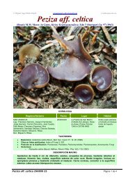
Fruit-body, general macroscopic characters. At first white, more or less spherical (figure 1 a),<br />
later tinged dingy yellow, much lobed and corrugated and finally entirely dingy and of<br />
quite irregular shape (figure 1 b, c), up to 2-5 cm diam. Surface felted, texture at first<br />
soft, becoming harder, brittle when fresh, drying leathery. Gleba, when fresh, creamy
<strong>BRITISH</strong> <strong>HYPOGEOUS</strong> <strong>FUNGI</strong><br />
white in young specimens, becoming flushed apricot yellow, then entirely pinkish orange<br />
or finally dingy cinnamon, with increasing maturity. Zygospores just detectable with<br />
naked eye as orange granules in older specimens which also yield cream to orange latex<br />
when cut. Fruit-bodies may grow around small sticks or dead leaves of conifers which thus<br />
become embedded in them. Often much eaten, sometimes by slugs, but no odour detectable.<br />
Pseudoperidium. No true peridium, but outer zone of fruit-body consists of rather more<br />
closely interwoven hyphae than those of central part. This differentiation becomes more<br />
obvious as fruit-body matures. Zygospores usually not formed in this peripheral zone but<br />
occasionally they may be quite near surface.<br />
Gleba. At first consisting of loosely interwoven hyphae without latex tubes, later more<br />
closely woven and latex tubes conspicuous. Hyphae coenocytic, diam. 5 to 7#, with<br />
numerous small nuclei and occasional septa. Latex tubes diam. 10 Q with dense contents.<br />
Development of zygospores. A fully illustrated account has been given by Bucholtz (1912).<br />
His figures show some evidence of shrinkage of the material, doubtless due to his use of<br />
xylol during processing. Examination of British material confirms Bucholtz's account with<br />
some minor differences.<br />
Paired hyphae scattered through the young fruit-body develop into progametangia.<br />
These are swollen and somewhat curved, with dense contents and numerous small nuclei<br />
(figure 1 d). They lie parallel to one another for some distance from their tips, and one is<br />
usually slightly larger than the other. Oily contents concentrate at the tips and a single<br />
nucleus in each progametangium enlarges and comes to lie in a clear area (figure 1 e),<br />
while the rest of the nuclei in the tips disintegrate. Cross-walls then form at or near the<br />
widest part of the progametangia cutting off the uninucleate gametangia from the multinucleate<br />
suspensor cells (figure if). The wall between the gametangia then breaks down,<br />
and the single nucleus from the smaller one passes into the larger one together with most<br />
or all of the oily contents (figure 1 g). The fertilized gametangium then grows out into<br />
a bud-like projection which rapidly swells to form a spherical or ellipsoidal sac into which<br />
the contents of the gametangium pass. No conclusive evidence of nuclear fusion in the<br />
gametangium was seen, although the two nuclei were seen in close association, so that it is<br />
likely that the two nuclei both pass into the sac as claimed by Bucholtz. According to his<br />
figures they remain undivided, but the British material shows numerous nuclei in the sac<br />
long before it reaches its maximum size (figure 1 h). When most of the contents of the<br />
gametangium have passed into the sac a thin wall is laid down lining it, thus delimiting<br />
the young zygospore and cutting off the nearly empty gametangium. The young zygospore<br />
continues to increase in size and the wall increases in thickness to 9 to 10 u and becomes at<br />
first pale yellow and finally deep reddish orange in colour, remaining smooth but showing<br />
two distinct layers in section (figure 1 i,j). Where the zygospores are crowded the wall<br />
may be flattened by the pressure of adjacent spores giving an angular appearance. Where<br />
such pressure is absent the spore is ellipsoidal or less often almost spherical. The contents<br />
rapidly become very dense with numerous colourless oil globules. A sheath of sterile hyphae<br />
begins to develop round the zygosporic sac at an early stage and increases as the spore<br />
matures. This sheath varies in different specimens, but may finally be up to 30,u thick and<br />
then consists of one to several layers of yellow, thick-walled hyphae encircling the zygospore<br />
in a more or less spiral manner (figure 1 i j, k). In stained sections these sheath<br />
VOL. 237, B, 53<br />
437
NO ":aNAMVH '3 NVIrIr<br />
\~.-,; :'.' % ''':-:: : .-:''-i:::::::::::::<br />
q
<strong>BRITISH</strong> <strong>HYPOGEOUS</strong> <strong>FUNGI</strong><br />
hyphae often have the appearance termed 'flammenkronen' by Bucholtz (figure 1 1), due<br />
to the projection of the hyphae out into the matrix of the fruit-body and to the affinity of<br />
their thickened walls for various stains, in contrast to the zygospore wall which remains<br />
unstained.<br />
The fruit-bodies develop slowly. Conjugating stages have not been found later than<br />
October. The gametangia continue to increase in size as the zygospore grows, but when<br />
the spore is mature they rapidly disappear. In late autumn the vegetative hyphae begin<br />
to disintegrate and thus to set free the zygospores. No record of germination of the zygospores<br />
exists and attempts to culture the fungus have consistently failed.<br />
Normally all the zygospores in any one fruit-body are at approximately the same stage,<br />
although the gametangia tend to develop first in the outer part of the fruit-body. Occasionally,<br />
however, the majority of the spores cease to develop and abort, while a few continue<br />
to develop to maturity.<br />
Bucholtz points out that the size of the mature zygospores varies considerably in different<br />
collections. This variation in size has been noted in numerous collections made recently<br />
in various parts of Britain. The size of mature zygospores varied in different fruitbodies<br />
from 80-92-100 x 60-71-80 to 140-189-200 x 120-145-150#.* No correlation<br />
could be made between size of zygospores and date or place of collection or identity of<br />
tree beneath which the fruit-bodies were found, but the differences were directly correlated<br />
with the density of distribution of the spores within the individual fruit-body. The size<br />
decreased with an increase in the number of spores per unit volume of fruit-body. Such<br />
a difference could be seen between different individuals of the same collection or even<br />
within the same specimen. There is no justification therefore for the separation of strains<br />
on the basis of spore size. All other macroscopic and microscopic characters were essentially<br />
similar in all collections made.<br />
Habitat and periodicity. The material in Broome's Herbarium on which Berkeley's original<br />
description was based was collected at Chudleigh (Devonshire) and a further collection<br />
was made in Shropshire. The Foray records of the British Mycological Society report the<br />
fungus from Thirlmere, Cumberland. During the present study collections were made in<br />
* The middle figure is the mean.<br />
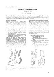
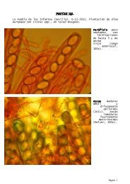
FIGURE 1. Endogone lactiflua. (a) Young fruit-body, almost spherical, x 2. (b), (c) Older fruit-bodies,<br />
showing irregular shape, x 2. (d) Young multinucleate progametangia. (e) Older progametangia,<br />
showing enlargement of single nucleus in each and concentration of oily contents in tips.<br />
(f) Uninucleate gametangia cut off from suspensor cells by transverse walls. (g) Nucleus of<br />
smaller gametangium migrating into larger one. (h) Young multinucleate zygospore with<br />
sheath hyphae beginning to develop. (d)-(h) Drawn from material stained iron alum haematoxylon<br />
and light green. (i) Nearly mature zygospore, drawn from fresh unstained material.<br />
Note oily contents of spore, 2-layered wall, enlargement of empty gametangia and suspensors<br />
and development of sheath hyphae. (j) Mature zygospore, showing fully developed hyphal<br />
sheath. Composite drawing, contents of zygospore drawn from fresh material, sheath from<br />
stained preparation. (d)-(j) x 1500. (k) Surface view of nearly mature zygospore, showing spiral<br />
arrangement of sheath hyphae. (1) Part of section of mature zygospore cut in plane parallel to<br />
that of k and showing 'flammenkrone' effect (Bucholtz I912) due to projecting folds of sheath<br />
hyphae; cf. j, which is of a specimen cut at right angles to the plane of k, I and so more or less<br />
in the plane of the enveloping hyphae. (k, 1) x 460.<br />
53-2<br />
439
440<br />
c<br />
a<br />
e<br />
d<br />
<strong>LILIAN</strong> E. <strong>HAWKER</strong> ON<br />
I<br />
b<br />
/It<br />
n<br />
9<br />
0I
<strong>BRITISH</strong> <strong>HYPOGEOUS</strong> <strong>FUNGI</strong><br />
Devonshire (Dartington Hall, 107*), Gloucestershire (Cotswold Hills, near Wotton-under-<br />
Edge, 281, 446, 455, 548, 647, 648, 649), Somerset (Cleeve, 313), Herefordshire (Haugh<br />
Wood, 470, 471), Perthshire (near Loch Tay, 407, 414, 416, 422) and Caernarvonshire<br />
(Nant-y-Garth, Bangor, 242; Bettws-y-coed, 258; Nant Heilyn, 260, 261).<br />
Mature fruit-bodies are found only in summer and early autumn. Locally abundant.<br />
The present collections were made in coniferous woods or plantations (including pine,<br />
spruce, silver fir and larch). In no case could any connexion be traced between fruitbodies<br />
of Endogone lactiflua and typical ectotrophic mycorrhizal mantles, but in most cases<br />
the tree roots in the neighbourhood of the fruit-bodies showed the typical pseudomycorrhizae<br />
described by Melin (1917). It is likely, therefore, that this fungus is a true parasite<br />
on the roots of coniferous trees.<br />
ENDOGONE MACROCARPA (Tul.), Tulasne. Tulasne (I85I, p. 182, P1. XX, fig. 1)<br />
REFERENCES. Fischer (1897, 125); Bucholtz (I912, P1. VIII, figs. 62-70; P1. IX, figs.<br />
71-4); Thaxter (I922).<br />
Syn. Glomus macrocarpus Tulasne (I845, 63).<br />
Fruit-body, general macroscopic characters. Irregularly globose or lobed, up to size of a<br />
hazelnut, usually smaller, at first pallid, later yellow, finally dingy brown, at first soft,<br />
becoming firm and usually less compact, cut surface of gleba white, then yellow, finally<br />
reddish brown, granular when mature owing to presence of large chlamydospores just<br />
visible to naked eye (figure 2a), latex absent, odourless.<br />
Pseudoperidium. No true peridium, outer zone of hyphae without chlamydospores, often<br />
enclosing soil particles, but otherwise similar to hyphal network of gleba.<br />
Gleba. Loosely interwoven, thin-walled, aseptate hyphae (10 to 30,c diam.) with characteristic<br />
wide-angled more or less dichotomous branching (figure 2 b), contents yellow, oily.<br />
Chlamydospores (140-190-230 x 120-145-180u) terminal on hyphal branches, notcrowded,<br />
at first thin-walled, hyaline, with dense, oily contents. Wall becoming thickened (5 to O1,U)<br />
and bright yellow, oil drops finally conspicuous (figure 2 c to g). Chlamydospores at different<br />
stages of development may be present in the same fruit-body, but the majority develop<br />
more or less simultaneously. No hyphal sheath develops round the spores.<br />
Habitat and periodicity. Single mature specimens were collected on two occasions (May<br />
1952, May 1953) under beech in a copse of mixed beech and larch near Wotton-under-<br />
* Figures throughout refer to the writer's collections.<br />
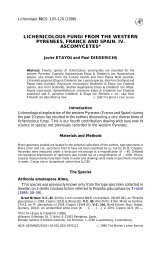
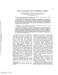
FIGURE 2. Endogone macrocarpa (a) Longitudinal section of mature fruit-body, showing granular effect<br />
due to large chlamydospores, x 2. (b) Characteristic hyphae with wide-angled dichotomous<br />
branching. (c)-(g) Stages in development of chlamydospores. (f)-(g) x 250. E. microcarpa.<br />
(h) Chlamydospores drawn to same scale for comparison with those of E. macrocarpa. (i) Fruitbody<br />
attached to stick, x 4. (j) Slender loosely woven hyphae of pseudoperidium, showing<br />
hairs which project beyond surface. (k) Portion of gleba of mature fruit-body, showing characteristic<br />
thick-walled hyphae with wide-angled dichotomous branching (cf. E. macrocarpus) and<br />
mature chlamydospores with thick walls and oily contents. Note that interior of spore is still<br />
in communication with parent hyphae. (l)-(q) Stages in development of chlamydospores.<br />
(r) Chlamydospore with unusual swelling at base. (j)-(r) x 740.<br />
441
442<br />
<strong>LILIAN</strong> E. <strong>HAWKER</strong> ON<br />
Edge, Gloucestershire (606, 722) and once from Failand, Somerset (949). A larger<br />
collection was made under yew at Kingsweston, Bristol (947). Specimens in Herb.<br />
Broome, Brit. Museum, labelled Endogone pisiformis from Somerset (Brockley Coombe,<br />
Leigh Woods) and Northamptonshire (Maidwell), are all E. macrocarpa.<br />
It is almost certain that Berkeley & Broome's (1846) description of E. pisiformis Link<br />
(see also Berkeley (i860) and Cooke (1871)) was based on these specimens of E. macrocarpa.<br />
Cooke (1871) reports thatTulasne examined this material and considered it to be E. macrocarpa.<br />
The specimen in Herb. Broome from Whitby, Yorks, labelled E. macrocarpa is, however,<br />
actually E. microcarpa. E. macrocarpa is probably relatively uncommon in Britain.<br />
The large size of the chlamydospores distinguishes it clearly from E. microcarpa.<br />
Thaxter (1922) suggests that this species may be the chlamydospore stage of E. lactiflua.<br />
This is unlikely, since the characteristic hyphae of E. macrocarpa are not seen in E. lactiflua<br />
while latex is present in the latter and absent in the former.<br />
ENDOGONE MICROCARPA (Tul.), Tulasne. Tulasne (I85I, p. 182, P1. XX, fig. 2)<br />
REFERENCES. Zobel (I854, in Corda, 6, 48); Bucholtz (I912); Thaxter (I922); Knapp<br />
(1952).<br />
Syn. Glomus microcarpus Tulasne (I844, 63).<br />
Fruit-body, general macroscopic characters. Small, seldom more than 5 mm diam., at first<br />
more or less spherical, then flattened or lobed, often closely adpressed to surface of dead<br />
leaf or stick (figure 2i). Surface felted but not visibly hairy. At first chalky white, then<br />
becoming dull straw-coloured and finally dingy yellow-brown. Cut surface of gleba homogeneous,<br />
firm, at first dingy white then ochre yellow and finally yellow-brown, always<br />
somewhat darker than the exterior, no latex present. No odour detectable.<br />
Pseudoperidium. No true peridium but outer hyphae of fruit-body slender (1 to 2# diam.),<br />
more loosely interwoven than central ones and a few of them projecting into the soil or<br />
terminating as short septate hairs (figure 2j). Small soil particles may be entangled in<br />
these loose peripheral hyphae.<br />
Gleba. Consisting of loosely interwoven hyphae (diam. 2 to 8yu), much branched,<br />
occasionally septate, bearing terminal, almost spherical chlamydospores (diam. 40-42-48<br />
x 35-38-42#) with walls 2 to 5,u thick. The hyphae show wide-angled branching similar to<br />
that of E. macrocarpa and become largely used up as the chlamydospores mature (figure 2k).<br />
Development of chlamydospores (figure 21 to q). Chlamydospores develop from swollen ends<br />
of hyphal branches. These are often in pairs (figure 2m, n), but no sign of conjugation is<br />
seen and the apparent pairing may be merely the result of the crowding of the young spores.<br />
It is possible, however, that the chlamydospores are actually parthenogenetic zygospores,<br />
and that the ability to conjugate was lost early in the evolution of the species. Figure 2r<br />
shows a hypha which has cut off a chlamydospore and is cutting off other swollen cells below<br />
it. This condition is unusual, but there is no evidence that the basal cell is of gametangial<br />
nature. The wall of the normal chlamydospore thickens and becomes yellow, but a narrow<br />
channel connecting the mature spore with the parent hypha remains open (in contrast to<br />
the zygospores of E. lactiflua). From an early stage the contents consist of large oil globules<br />
which are sufficiently uniform in size to give the appearance of spores and may account<br />
for the confusion that exists between this species and E. pisiformis. These oil drops are much
<strong>BRITISH</strong> <strong>HYPOGEOUS</strong> <strong>FUNGI</strong><br />
more crowded than those of E. macrocarpa. The majority of the chlamydospores in a<br />
particular fruit-body are usually at approximately the same stage of development, but old<br />
and young fruit-bodies may be found close together, presumably borne on the same<br />
mycelium.<br />
Habitat and periodicity. Specimens in Herb. Broome, British Museum from Credenhill<br />
Camp and Brockley Coombe (labelled Glomus macrocarpus), Whitby (labelled E. macrocarpa),<br />
Kings Cliffe (unlabelled) and Saltford (unlabelled). Numerous collections made during<br />
the present study as follows: Bristol (Stoke Bishop 484, 485; Blaise Castle, 56, 65, 66, 67,<br />
81, 280, 431, 540, 652, 655, 691; Leigh Woods, 509, 514, 516, 521, 679), Somerset (Cleeve,<br />
37, 43, 73, 76, 192, 194, 200, 312, 333, 335, 513, 557, 583, 695, 713; Orchardleigh, Frome,<br />
49, 223; Portbury, 49, 207, 215, 217, 343; Wraxall, 100, 101, 322; Brockley Coombe, 268,<br />
271, 274, 396, 491; Brockley Road, 380, 623; Abbot's Pool, Failand, 286, 528), Gloucestershire<br />
(Staunton, Forest of Dean, 462; Newark Park, 503), Herefordshire (Downton Gorge,<br />
464), Wiltshire (Savernake Forest, 364), Caernarvonshire (Vaynol Park, Bangor, 229;<br />
Bettws-y-coed, 246). The fungus has been found most frequently under yew or pine but<br />
has also occurred under various deciduous trees. It is frequently associated with pseudomycorrhizae<br />
of which it is probably the cause. Locally abundant.<br />
Fructifications of an unidentified species of Endogone in association with mycorrhizal<br />
strawberry roots have recently been described by Mosse (I953). Material has been<br />
examined by the present writer. While this fungus is undoubtedly a chlamydosporic<br />
species of Endogone, it is not attributable to any of the three species described above, nor<br />
does it closely resemble any of the species described by Thaxter (I922). The fruit-bodies<br />
are about 1 mm in diameter and contain 2 to 32 (usually 2 to 6) thick-walled yellow<br />
chlamydospores (92 to 197u diam.) embedded in a loose mass of hyphae with branching<br />
similar to that figured for E. macrocarpa and E. microcarpa (figure 2). Further description<br />
and identification must await the results of Miss Mosse's studies now in progress.<br />
Doubtful record ENDOGONE PISIFORMIS Link<br />
This was recorded by Bucknall (1878) from Hanham, Bristol, but I have been unable<br />
to trace the specimen. In view of the frequency of Endogone microcarpa in this district and<br />
the confusion between these two species it is probable that this specimen was actually<br />
E. microcarpa. The specimens in Herb. Broome British Museum, labelled E. pisiformis are<br />
all E. macrocarpa, so that Bucknall's specimen may have been this species. The record of<br />
E. pisiformis in the account of the Bangor Foray of the British Mycological Society held in<br />
September 1950 was an error and the specimen was E. microcarpa. E. pisiformis was first<br />
described by Link (1809) as a form showing normal zygospore production. Fischer (1897),<br />
writing in Rabenhorst's Kryptogamenflora, considered that E. pisiformis and E. microcarpa<br />
were synonyms but figured the latter as sporangial. Bucholtz (I912) erroneously referred<br />
to E. pisiformis as sporangial and to E. microcarpa as zygosporic. Thaxter (1922) cleared the<br />
matter up and includes only zygosporic material in E. pisiformis and reserves the name<br />
E. microcarpa for fruit-bodies forming the typical small chlamydospores. Unfortunately,<br />
Knapp (I952) has reverted to the use of the name E. pisiformis for sporangial forms.<br />
Sporangial forms have not been recorded in Britain.<br />
443
444<br />
<strong>LILIAN</strong> E. <strong>HAWKER</strong> ON<br />
Systematic position of Endogone<br />
There is little doubt that the species showing zygospore formation are advanced types<br />
of Zygomycetes. The usually aseptate nature of the ground hyphae of the fruit-body is<br />
definitely a phycomycetous character. The formation of the gametangia is not unlike the<br />
early stages of conjugation in Phycomyces, while the passage of the contents of the fertilized<br />
gametangium into a bud or vesicle in which the zygospore develops is paralleled in the<br />
Piptocephalaceae. Grouping of the paired gametangia is .seen in some members of the<br />
Entomophthorales, while the single zygospore of Mortierella is embedded in a sterile hyphal<br />
sheath resembling that of Endogone lactifua. In spite of the ascomycetous affinities shown<br />
by the relatively large fruit-body, the extra-gametangial development of the zygospores<br />
and the septation of the sheath hyphae, it is likely that Endogone represents the end of an<br />
evolutionary side branch. Nevertheless, its structure supports the hypothesis that the<br />
simple filamentous Endomycetales and such forms as Gymnoascus with simple fruit-bodies<br />
may have evolved from an ancestral Zygomycete along lines parallel to the evolution of<br />
Endogone.<br />
If the sporangial types which have been described do actually belong to the same genus,<br />
then Endogone is obviously closely related to the simple Zygomycetes. It is possible that<br />
some of the 'sporangial' stages described were actually chlamydospores containing oil<br />
drops of regular size. Cultural studies by Kanouse (1936) indicate that the sporangial<br />
forms are not related to the zygosporic ones.<br />
The chlamydospore stage remains a problem. The close resemblance of these spores to<br />
mature zygospores suggests that they may be azygospores formed parthogenetically. The<br />
production of chlamydospores is, however, a characteristic of the Zygomycetes. The<br />
usually aseptate nature of the ground hyphae of the chlamydosporic fruit-body suggests<br />
a phycomycetous derivation.<br />
The British species are only three in number and therefore not representative, but the<br />
differences in habitat and general structure are such that it is practically certain that these<br />
species are distinct. As already pointed out, the mature zygospores of E. lactiflua often show<br />
no trace of gametangia and could easily be mistaken for chlamydospores. The other two<br />
species are definitely chlamydosporic from the earliest stage. They may be distinguished<br />
from one another by the great difference in chlamydospore size.<br />
The solution of the problem of the three types of fruit-body attributed to Endogone can<br />
only be solved by careful cultural studies of a larger number of species than is available in<br />
Britain.<br />
ASCOMYCETES<br />
PLECTASCALES<br />
Asci globose, evanescent, irregularly arranged in sporocarp.<br />
ELAPHOMYCETACEAE<br />
Fruit-bodies subterranean, spore mass powdery at maturity. Dodge (1929) divides this<br />
family into two tribes: Elaphomyceteae, in which the central core is cottony in texture,<br />
and Mesophelliae, with a corky or woody core. All the British species belong to the genus<br />
Elaphomyces of the first of these tribes.
<strong>BRITISH</strong> <strong>HYPOGEOUS</strong> <strong>FUNGI</strong><br />
Genus ELAPHOMYCES Nees ex Fr. Fries (1829, p. 57)<br />
Fruit-bodies consisting of a central core or gleba surrounded by a peridium which<br />
consists of two layers, an inner true peridium and an outer layer which in many species<br />
splits into pyramidal warts. This outer layer has been termed the 'cortex'. This is an<br />
unfortunate choice, but its long usage makes it necessary to retain this term. The whole<br />
fruit-body is, in some species, surrounded by the 'crust' which consists of a layer of soil<br />
particles bound together by hyphae. No sexual organs are known, but groups of asci<br />
arise from ascogenous hyphae which develop in small knots near the periphery of the core.<br />
The asci break down at an early stage and the spores complete their development after<br />
liberation. The spore mass is finally powdery.<br />
Dodge (1929) divides the genus into two sections, subgenus Malocoderma Vitt. with a<br />
more or less fleshy cortex becoming wrinkled, but not spiny, and with spores less than<br />
151I diam., and subgenus Scleroderma Vitt. with a hard cortex and with larger spores<br />
(diam. 15 to 50/u). In view of the general acceptance of the Gasteromycete genus Scleroderma<br />
this is not a permissible use of the name. Nevertheless, the distinction between the<br />
two groups is a useful one. All the British species fall in the so-called 'scleroderma' section<br />
except Elaphomyces citrinus which is a doubtful record.<br />
TYPE SPECIES. E. granulatus Fr.<br />
KEY TO <strong>BRITISH</strong> SPECIES OF ELAPHOMrCES<br />
1. Cortex soft, ascospores less than 15/t diam. ...<br />
Cortex hard, ascospores more than 15/t diam. ...<br />
2. Cortex verrucose or echinulate ...<br />
Cortex smooth or nearly so ...<br />
...<br />
...<br />
...<br />
...<br />
...<br />
...<br />
.<br />
...<br />
...<br />
...<br />
..<br />
...<br />
E. citrinus, p. 453<br />
... ... ...... 2<br />
... ... 3<br />
... ... ... 5<br />
3. Peridium more or less homogeneous in section ...<br />
Peridium marbled in section ... ...<br />
...<br />
...<br />
...<br />
...<br />
...<br />
...<br />
...<br />
E. muricatus,<br />
... 4<br />
p. 449<br />
4. Peridium ochraceous, verrucose ...<br />
Peridium greyish, echinulate ... .<br />
...<br />
...<br />
... ...<br />
...<br />
...<br />
...<br />
...<br />
...<br />
E. granulatus,<br />
E. aculeatus,<br />
p. 445<br />
p. 451<br />
5. Peridium thin, grey-brown to russet<br />
Peridium thick, brownish black ...<br />
...<br />
...<br />
...<br />
...<br />
.......<br />
...<br />
...<br />
...<br />
E. leucosporus,<br />
E. anthracinus,<br />
p. 451<br />
p. 452<br />
ELAPHOMYCES GRANULATUS Fr., Fries (I829, p. 58)<br />
REFERENCES. Berkeley & Broome (I84I, 430, P1. XI, fig. 10); Tulasne (I841, 22, P1. I,<br />
fig. 3; P1. II, fig. 7; PI. IV, fig. 3); (I851, 109-10, PI. XIX, fig. 4); Vittadini (I842, 78,<br />
P1. III, fig. 7); Berkeley (i86o, 378); Cooke (i871, 750); Hesse (i894, 70-2, PI. XIII,<br />
figs. 1-7, P1. XXI, fig. 55); Massee (I909, 249).<br />
-Syn. ? Lycoperdon solidum Linnaeus (1737, 369).<br />
? L. cervinum Linnaeus (1753, 1183).<br />
Hypogeum cervinum Persoon (I797, 7).<br />
Hypogaeum cervinum Gray (I821, 582, P1. I).<br />
Scleroderma cervinum Persoon (I801, 156, P1. IV, fig. 2).<br />
Tuber cervinum Nees v. Esenbeck (I816, 161, P1. XV, fig. 147).<br />
Lycoperdastrum cervinum Kuntze (I891, P1. I).<br />
Elaphomyces officinalis Nees v. Esenbeck (1821-3, P1. I).<br />
Phymatium flvum Chevallier (1826, 361, P1. X, fig. 6).<br />
Elaphomyces vulgaris var. granulatus Corda (I84I, 25-6).<br />
E. cervinus Schlectendal (1824, 166); Fischer (I897, 94-5, figs. 1-4 on p. 82);<br />
Hennings (I905, 91, fig. 2); Dodge (1929); Knapp (1952).<br />
VOL. 237, B. 54<br />
445
446<br />
<strong>LILIAN</strong> E. <strong>HAWKER</strong> ON<br />
t..N;~~~~~~~~~~~~~~~~~~~~~~~~~~~~,-<br />
A 11~~~~~~~~~~~~~~~<br />
a ?~~~~~~~~~~~~~~~~~~~~~~~~4<br />
C e~~~~~~~9 C C')<br />
- C- -<br />
k s~~~~~~~~~~~~~~~~~~~~~~~~~~~~~~~~~~<br />
3 4W<br />
WI W2~~~~~W
<strong>BRITISH</strong> <strong>HYPOGEOUS</strong> <strong>FUNGI</strong><br />
This fungus has been known from the sixteenth century and was used as an aphrodisiac.<br />
The specific name was deliberately changed from cervinus to granulatus by Fries in part 1 of<br />
vol. 3 of the Systema Mycologica published in 1829. Dodge (1929) considered that the correct<br />
name was Elaphomyces cervinus (L. ex S. F. Gray) Schlect., and Knapp (I952) cites it as E.<br />
cervinus (Pers.) Schroter syn. E. granulatus Fr. By the latest definition of the Rules at the<br />
Botanical Congress at Stockholm 1950 any citation in any volume of the Systema or the<br />
Elenchus is the starting point in naming Ascomycetes. Hence, since Gray's paper is earlier<br />
than Fries's citation in vol. 3, Elaphomyces granulatus Fr. is the valid name. This name has<br />
always been used in British papers.<br />
Fruit-body, general macroscopic characters. Tough, leathery, becoming brittle with age, drying<br />
hard, at first pale yellow, becoming dingy ochraceous, usually ovoid, sometimes spherical<br />
or depressed, 2 to 4 cm. diam., covered with pyramidal concolorous warts (figure 3 a).<br />
Drying a duller colour, often becoming wrinkled, particularly when parasitized by Cordyceps<br />
capitata. Ripe fruit-bodies consist of an outer wall (cortex and peridium), tough, white<br />
in section, occasionally flushed greyish pink towards the gleba, and a central mass of<br />
purplish black spores, powdery in dry weather, moist in wet weather. In younger fruitbodies<br />
the spore mass is divided into sections by white to greyish pink sterile dissepiments<br />
(figure 3b), in very young ones gleba consists of cottony mass of hyphae.<br />
Fruit-body is embedded in a crust of soil densely impregnated with yellowish mycelium.<br />
This crust, which varies in thickness in different soils, readily breaks away, retaining an<br />
impression of the fruit-body, and often encloses mycorrhizal tree roots. Hyphae of crust<br />
are continuous with the bright yellow flocculent mycelium, which often spreads through<br />
considerable areas of soil, and closely resemble those of the hyphal mantle of the surrounding<br />
tree roots. Odour earthy.<br />
Cortex and peridium. The fruit-body wall, as seen in section, clearly consists of two layers;<br />
the outer 'cortex' which, including the warts, is up to 370,u thick and is entirely yellowbrown,<br />
and the peridium up to 1700 It thick, white, or grading to greyish pink on the inner<br />
side. Cortex consists of pyramidal warts, triangular in radial section, consisting of a central<br />
area of irregularly arranged hyphae surrounded by peripheral plates of regular, septate,<br />
thin-walled hyphae running in a direction tangential to the surface of the fruit-body, warts<br />
arising from a narrow continuous zone of irregularly interwoven hyphae which extend into<br />
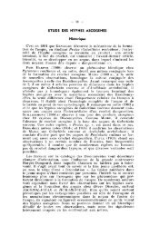
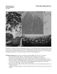
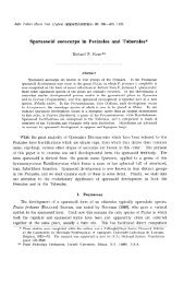
FIGURE 3. Elaphomyces granulatus (a) Whole fruit-body, nat. size. (b) Longitudinal section of nearly<br />
mature fruit-body, nat. size. (c)-(e) Stages in development of fruit-body, x 10. (c) Very young<br />
fruit-body in which differentiation of gleba (G), peridium (P) and cortex (C) is just beginning.<br />
(d) Older specimen in which differentiation of these is complete and groups of ascogenous hyphae<br />
(AH) are visible. (e) Groups of ascogenous hyphae have enlarged to form patches (AP), asci (A)<br />
are developing. (f) Ascogenous hyphae. (gl to 4) Stages in development of ascus. (h) Spores at<br />
time of liberation from ascus, showing rod-like thickening of epispore. (i) Mature spore. (f)-(i),<br />
x 740. E. muricatus. (j) Whole fruit-body, nat. size. (k) Longitudinal section of fruit-body, showing<br />
marbled peridium (P). (1) Ascogenous hyphae. (m t0 4) Stages in development of spores (2 spores<br />
aborting in m3). (n) Mature spore. (I)-(n) x 740. E. aculeatus. (o) Whole fruit-body, nat. size.<br />
(p) Longitudinal section of fruit-body, nat. size. (q) Mature spore, x 740. E. leucosporus.<br />
(r) Longitudinal section of whole fruit-body showing thin peridium (p), nat. size. (s) Mature<br />
spore, x 740. E. anthracinus. (t) Whole fruit-body, nat. size. (u) Longitudinal section of fruitbody,<br />
nat. size. (v) Ascogenous hyphae. (wI to4) Stages in development of asci and spores.<br />
(x) Mature spore. (v)-(x) x 740.<br />
54-2<br />
447
448<br />
<strong>LILIAN</strong> E. <strong>HAWKER</strong> ON<br />
the peridium. A study of young fruit-bodies suggests that the warts arise by splitting of the<br />
plates of tangential hyphae which fail to keep pace with the peripheral expansion of the<br />
peridium. The peridium consists of a firm pseudoparenchyma which is softer but less<br />
brittle than the cortex. The hyphae of the outer part, next the cortex, are closely interwoven,<br />
diameter 3,u, mostly arranged periclinally, becoming looser and tending to a radial<br />
arrangement towards the inside where the hyphae are stouter (diam. 6 I). Throughout<br />
the peridium irregularly arranged bands of hyphae occur, but these are not visible to the<br />
naked eye (cf. Elaphomyces muricatus).<br />
Asci. Globose to pyriform, 35 to 45u diam., evanescent, usually 6-spored (figure 3g4 t 4).<br />
Ascospores. Brownish black at maturity, spherical, surface divided into shallow blocks by<br />
irregular cracks, 24 to 32,c diam., wall up to 10 i thick (figure 3 i).<br />
Development offruit-body, asci and spores. At an early stage (fruit-bodies up to 5 mm diam.)<br />
only the cortex is clearly differentiated, and the densely woven peridial hyphae are not<br />
sharply divided from the loosely woven hyphae of the central core, which is often pinkish<br />
in colour (figure 3 c, d). Small knots of fine hyphae, which readily stain with aniline blue,<br />
arise near the outer edge of this core (figure 3 d) and develop rapidly to give groups of<br />
stout, coiled, much-branched, septate ascogenous hyphae with dense contents (figure 3f).<br />
These are said to be binucleate (Reess & Fisch I887; Dodge 1929). Asci soon arise from the<br />
terminal cells of the short branches of the ascogenous hyphae (figure 3 e, g1, 2). The contents<br />
of these young asci are granular with numerous oil drops. Staining with acetocarmine<br />
shows very small nuclei. Details of nuclear division could not be followed, but<br />
the young ascus contains two nuclei which probably fuse to give a uninucleate stage, and<br />
later divide by three successive divisions. At some stage in these divisions some nuclei<br />
abort so that six finally remain. The spores develop around these nuclei and soon become<br />
thick-walled, showing a peripheral arrangement of granules in rows giving a number of<br />
radial rods (figure 394, h) which, after release of the spores from the ascus, rapidly turn<br />
brown or black. Cracks developing between these rods give the spore its characteristic<br />
appearance (figure 3 i). The asci often remain in communication with the stalk cells until<br />
spore differentiation is far advanced and finally break down when the spores are about<br />
half their final size (Clemencet 1932). At this stage separate ascogenous areas can still be<br />
seen separated by bands or dissepiments of the original core hyphae (figure 3 b). As the<br />
spores mature the latter are used up and the glebal cavity finally contains only spores<br />
interspersed with a few capillitium threads.<br />
Habitat and periodicity. The records suggest that this fungus is common throughout Great<br />
Britain. It is recorded several times in British Mycological Society Foray lists and some<br />
20 to 30 separate collections, mainly in western England, were made during the present<br />
investigations as follows: Bristol (Leigh Woods, 203, 344), Gloucestershire (Forest of<br />
Dean, 460; Wotton-under-Edge, 7, 10), Somerset (Cleeve, 102; Emborough, 31, 574;<br />
Brockley Coombe, 387; Abbot's Pool, Failand, 6, 18, 55, 68, 284, 321, 522, 569, 574a, 611,<br />
617, 715; Portbury, 128, 129, 130), Devonshire (Woodbury Ring, 115; Stoke Wood,<br />
Exeter, 116), Essex (Epping Forest, 21b), Caernarvonshire (Nant-y-garth, 240, 241;<br />
Bettws-y-coed, 244, 249), Perthshire (Glen Lochay, 417) and was previously found at<br />
Mortimer Common, Hampshire. Some other recent collections from different parts of the<br />
country are preserved in the Herbarium of the Royal Botanic Gardens, Kew.
<strong>BRITISH</strong> <strong>HYPOGEOUS</strong> <strong>FUNGI</strong><br />
It is most frequently found on light acid soil or peat under Scots pine, but is also found<br />
under deciduous oak, sweet chestnut and occasionally under beech, including very old<br />
trees of each species.<br />
In a study of several collections made in different localities under different species of<br />
tree Dicker (unpublished work in this department) observed slight variations in details of<br />
structure and in spore size. One collection from a beechwood near Wotton-under-Edge,<br />
Gloucestershire, differed from the type in the more pronounced banded arrangement of<br />
the peridial hyphae and the faintly marbled appearance of the inner peridium. This may<br />
have been a hybrid between E. granulatus and the common beechwood species E. muricatus<br />
(see below). A suggestion that it might be a strain of E. asperulus was shown to be untenable<br />
when it was compared with material of that species collected by the writer in Norway.<br />
Genuine specimens of E. asperulus have not been found in Britain. Material assigned to<br />
E. granulatus, however, has proved to be very variable and more work on this species is<br />
desirable. In the present stage of our knowledge it is best to regard this as a species group<br />
covering a range of closely related forms.<br />
Fruit-bodies, in favourable localities, are usually present in large numbers at all times<br />
of the year, but initiation of young ones is inhibited by extreme cold or extreme drought.<br />
They are most frequently partially embedded in the hard pan, which underlies the surface<br />
layers of leaf litter or humus, or are pressed against a large root or stone. They are seldom<br />
present at a depth of more than 3 in. from the surface and usually occur only in welldrained<br />
situations. They are frequently eaten by soil-inhabiting invertebrates, or by<br />
rodents. The spores are probably dispersed in this way, but no attempt to germinate them<br />
has succeeded and they were even recovered apparently undamaged and still incapable<br />
of germination from the faeces of captive rabbits to which they had been fed.<br />
ELAPHOMYCES MURICATUS Fr., Fries (I829, p. 59)<br />
REFERENCES. Berkeley (I841, 430); Rabenhorst (I844, 291); Zobel (I854, 51, PI. X,<br />
fig. 97); Quelet (I873, 379); Dodge (1929).<br />
Syn. ? Lycoperdon scabrum Willdenow (1787).<br />
? Scleroderma cervinum /f scabrum Persoon (I801-8, 157).<br />
Elaphomyces variegatus Vittadini (1831, 68-9, P1. IV, fig. 4); (1843, 220); Tulasne<br />
(1841, 23, P1. I, fig. 4; P1. II, figs. 4, 11; P1. IV, fig. 1); (i85I, 108-9, P1. III,<br />
fig. 8); Berkeley (i860o, 378); Cooke (1871, 749); Reess & Fisch (1887); Hesse (1894,<br />
72-3, P1. XIII, figs. 8-16); Fischer (1897, 91); Massee (1909, 378); Dodge (1929);<br />
Ramsbottom & Balfour-Browne (1951); Knapp (1952).<br />
? Ceraunium scabrum and muricatum Wallroth (I833, 406-7).<br />
Elaphomyces vulgaris oc muricatus and y variegatus Corda (1871, 21, 27, Pls. VII<br />
and IX).<br />
E. hirtus Tulasne (I841, 23).<br />
E. scaber Schr6ter (i893, 223).<br />
This species is readily distinguished from Elaphomyces granulatus by the marbled peridium,<br />
as seen in section, smaller spores and the regular form, smaller size and darker tawny<br />
colour of the fruit-body. Dodge (I929) distinguishes E. variegatus Vitt. from E. muricatus<br />
Fries by differences in the coloration of the marbled peridium and its slightly smaller<br />
449
450<br />
<strong>LILIAN</strong> E. <strong>HAWKER</strong> ON<br />
spores, but these differences are within the range of variation in material collected during<br />
the present investigation.<br />
Fruit-body, general macroscopic characters. Tough, leathery, becoming brittle with age,<br />
drying hard, at first yellow then bright tawny orange, finally dingy yellow brown, usually<br />
spherical, up to 2 cm diam., covered with small pyramidal warts (figure 3j), often<br />
parasitized by Cordyceps ophioglossoides. General organization and development of fruitbody<br />
similar to that of Elaphomyces granulatus, from which it is distinguished by the peridium,<br />
which in section is mottled or marbled with yellowish white veins surrounding pink to<br />
chestnut brown areas, and by the smaller spores. Crust poorly developed and the<br />
surrounding flocculent yellow mycelium less conspicuous than with E. granulatus. Odour,<br />
weak, earthy.<br />
Cortex and peridium. Both thinner than in E. granulatus (ca. 200 and 500 to 2000#u<br />
respectively). Peridium marbled in section with yellowish white anastomosing veins<br />
surrounding small pink to chestnut brown areas, colour darkening generally towards<br />
centre of fruit-body, marbled effect due to large air spaces and fissures (figure 3k).<br />
Gleba. Similar to that of E. granulatus but hyphal strands which separate ascogenous<br />
areas in immature fruit-bodies are often pink or purplish fawn in colour. Spore masses<br />
bluish black or occasionally brownish black. Capillitium variable, sometimes profuse,<br />
sometimes sparse.<br />
Asci. Globose, 30 to 40/x diam., evanescent, usually 4-spored, occasionally 2-spored<br />
(figure 31, m1 to 4)<br />
Ascospores. Purplish black at maturity, spherical, 18 to 24,u diam., surface cracked<br />
into blocks to a depth of 2,u, rods making up wall in developing spores more coarsely<br />
granular than in E. granulatus, wall up to 8,u thick (figure 3n).<br />
Development. Similar to that of E. granulatus..<br />
Habitat and periodicity. Common in beechwoods in south and west England, occasionally<br />
found under other species of trees but never under conifers. It is probably equally common<br />
in other parts of Great Britain but has not been looked for so intensively. British Myco-<br />
logical Society Foray lists record it from Scotland (Forres), north Wales (Bangor) and<br />
Northern Ireland (Belfast), while its parasite Cordyceps ophioglossoides is recorded from<br />
Keswick, Aviemore, Norwich and north Wales and occurs in Windsor Park. Numerous<br />
collections were made during the present investigation as follows: Gloucestershire (Cots-<br />
wold Hills, near Wotton-under-Edge, and Dursley, 3, 8, 11, 12, 13, 45, 86, 93, 104, 170,<br />
293, 298, 307, 353, 373, 397, 398, 576, 589, 590, 597, 598, 590, 597, 598, 614, 646, 685,<br />
717, 720, 728; Michael Wood, 103; Forest of Dean, 403, 459), Somerset (Cleeve, 571;<br />
Emborough, 573; Abbot's Pool, Failand, 320, 523, 534, 618, 716; Portbury, 131, 132),<br />
Herefordshire (Downton Gorge, 468), Devonshire (Berry Pomeroy, 108), Wiltshire (Wylie<br />
Valley, 90; Savernake Forest, 360-3, 367, 680), Oxfordshire (Kingwood Common, 26),<br />
Buckinghamshire (Beaconsfield, 653), Surrey (Mickleham, 60, 92), Caernarvonshire (Vay-<br />
nol Park, Bangor, 238; Bettws-y-coed, 245, 248). Slightly alkaline soil is the most usual<br />
habitat in south-west England. The fruit-bodies are usually in the humus layer or leaf<br />
litter and in contrast to E. granulatus, are seldom found partially embedded in the under-<br />
lying hard pan.<br />
Mature fruit-bodies occur all the year round as with E. granulatus. Fruit-bodies often<br />
fail to develop spores even when not attacked by Cordyceps.
<strong>BRITISH</strong> <strong>HYPOGEOUS</strong> <strong>FUNGI</strong><br />
ELAPHOMYCES ACULEA TUS Vitt., Vittadini (I83I, 70, P1. III, fig. 12)<br />
REFERENCES. Vittadini (i842, 79); Tulasne (I841, 24-5, P1. I, fig. 5; P1. II, fig. 6;<br />
P1. III, fig. 3); (I851, 111); Fischer (I897, 98).<br />
Syn. Lycoperdastrum aculeatum Kuntze (189I).<br />
Elaphomyces rubescens Hesse (1894, 75-7, P1. XIV, figs. 1-7; P1. XXII, figs. 1-5,<br />
7, 9, 15, 18-24, 29); Fischer (I897, 97).<br />
Fruit-body, general macroscopic characters. Tough and leathery, becoming brittle with age,<br />
drying hard, spherical, 1 to 2 cm diam., cortex black or greyish black with 3 to 4 angled,<br />
pointed, dark spines embedded in thin dark crust of soil and hyphae, peridium whitish<br />
to sooty grey in section, gleba at first with numerous dissepiments which disappear at<br />
maturity to leave mass of powdery sooty-black spores (figure 3 o, p). Mycelium not visible<br />
in surrounding soil.<br />
Cortex and peridium. Cortex black in section, conical warts or spines consisting of<br />
alternating bands of fused black hyphae and plates of tangentially arranged light brown<br />
hyphae, inner layer black, pseudosclerenchymatous. Peridium whitish on outside becoming<br />
grey towards gleba, consisting of closely interwoven and fused hyphae.<br />
Asci. Pyriform 40 to 50 x 35 to 45/u, evanescent, usually 8-spored.<br />
Ascospores. Sooty-black at maturity, spherical 14 to 17# diam., surface cracked into<br />
blocks as in E. granulatus and E. muricatus (figure 3 q). Capillitium consisting of copious<br />
long grey threads.<br />
Habitat and periodicity. Under beech in calcareous woods in summer (Knapp I952).<br />
Only two collections of this fungus have been made in Britain (Synwell Hill, Wottonunder-Edge,<br />
Gloucestershire, 15, 5 September 1949, Hawker (1952) and near Dursley,<br />
Gloucestershire (760)). The description has therefore been compiled from an examination<br />
of this material and from descriptions of continental specimens. As the Gloucestershire<br />
specimens were all mature no studies of development could be made. The structure of the<br />
mature specimen is, however, essentially similar to that of other species, so that it is likely<br />
that development is also similar. Shortly after the first collection was made the wood was<br />
cut down and no specimens were found in the following summer.<br />
ELAPHOMYCES LEUCOSPORUS Vitt., Vittadini (I842, 71, P1. III, fig. 1)<br />
REFERENCES. Vittadini (I842, 215, P1. III, fig. 1); Tulasne (I85I, 104); Fischer (I897, 86);<br />
Massee (I909, 249-50).<br />
The specific name leucosporus was originally given to immature specimens in which, as<br />
in other species, the spores were still colourless or light brown. According to Massee the<br />
spores of the type material became dark in storage, and those of the single collection<br />
hitherto made in Britain, by Broome at Chudleigh, Devon, were also dark. Three mature<br />
fruit-bodies collected on separate occasions during the present investigation also had dark<br />
spores.<br />
Fruit-bodies, general macroscopic characters. Brittle, thin wall readily breaking, reddish<br />
brown in British specimens but of variable colour in continental ones, spherical 0-5 to<br />
1 cm diam., minutely papillate. Spores powdery, brownish to purplish black in mass,<br />
interspersed with fine capillitium threads (figure 3r). Crust poorly developed or absent.<br />
Odour slight.<br />
451
452<br />
<strong>LILIAN</strong> E. <strong>HAWKER</strong> ON<br />
Cortex andperidium. Together less than 1 mm thick, usually thinner, cortex dark, peridium<br />
often bright russet-red or even salmon-red in section.<br />
Gleba. Powdery mass of blue-black spores and capillitium threads at maturity.<br />
Asci. Not seen by writer, said by Fischer (I897) to be 4- to 8-spored. Disappearing before<br />
spores are mature.<br />
Ascospores. Brownish black when mature, spherical, diam. 20 to 22,u, av. 20 5u (this<br />
size is from recent collections and is slightly larger than that given for Vittadini's type<br />
material which was obviously not mature since the spores were light-coloured) (figure 3s).<br />
Habitat, distribution, periodicity. In humus under trees, originally described as under oak,<br />
one specimen in present collection under evergreen oak (Quercus Ilex) and two under<br />
beech. Broome's original collection was made at Chudleigh, Devonshire. During the<br />
present investigation a single mature fruit-body was collected from each of the following:<br />
Gloucestershire (Westridge, Wotton-under-Edge, 306), Somerset (Cleeve, 137; Wraxall,<br />
346). This is obviously an uncommon species, or at least it rarely produces fruit-bodies.<br />
The three specimens were collected in different months, suggesting that, like the commoner<br />
species, it is able to form fruit-bodies at any time of the year.<br />
ELAPHOMYCES ANTHRACINUS Vitt., Vittadini (I831, 66, P1. III, fig. 8)<br />
REFERENCES. Vittadini (i842, 72-3); (I843, 216-17); Berkeley & Broome (I846, 81);<br />
Tulasne (1851, 106, P1. XIX, fig. 5); Cooke (I87I, 749); Fischer (1897, 89-90); Massee<br />
(1909).<br />
Syn. Lycoperdastrum anthracinum, L. pyriforme Kuntze (I891, 858, P1. I).<br />
Elaphomyces pyriformis Vittadini (1842, 72); Tulasne (1851, 107, P1. III, fig. 4);<br />
Fischer (I897, 90-1).<br />
E. uliginosus Hesse (I894, 67-9, P1. XXII, figs. 8, 28, 30); Fischer (I897, 88).<br />
E.plumbeus Hesse (1894, 69-70, P1. XIV, figs. 15-18; P1. XXI, figs. 53-4; P1. XXII,<br />
fig. 6); Fischer (I897, 88).<br />
Fruit-bodies, general macroscopic characters. Hard, with carbonaceous exterior, when mature,<br />
always sooty or brownish black, globose to ovoid or depressed, often grooved, seldom more<br />
than 1 cm diam., minutely verrucose, appearing smooth and dull to naked eye (figure<br />
3 t). Crust poorly developed or absent, but floccose hyphae present in surrounding soil.<br />
These are usually dark brown and inconspicuous or occasionally bluish or greenish grey.<br />
(Dodge (1929) separates those with blue or green mycelium as Elaphomyces uliginosus and<br />
states that this form has larger spores. No such difference in spore size is shown by British<br />
material.) In section peridium is dingy white to grey, gleba dingy white, cottony in young<br />
specimens, filled powdery mass of sooty spores in mature specimens or spores may not fill<br />
gleba, leaving central hollow (figure 3 u). Spores often fail to develop.<br />
Cortex and peridium. Cortex around 220c thick, general structure of warts similar to those<br />
of E. granulatus, but valleys between them partially filled with large hyphae, so that only<br />
the tips of the warts project above the general level, inner layer of pseudoparenchyma<br />
beneath warts, all cortical hyphae dark and thick-walled (at least 1,c). Peridium about<br />
OOO,/ thick, composed of firm pseudoparenchyma of thin-walled hyphae showing banded<br />
arrangement as in E. granulatus, but homogeneous in appearance to naked eye. Outer
<strong>BRITISH</strong> <strong>HYPOGEOUS</strong> <strong>FUNGI</strong><br />
hyphae of peridium closely interwoven, narrow (2,u diam.), grading to more loosely inter-<br />
woven wider hyphae (up to 13,# diam.) on inner edge.<br />
Asci. Spherical to pyriform, 50 to 55,# diam., thin-walled, evanescent, usually<br />
8-spored (figure 3 w to 4).<br />
Ascospores. Sooty-black at maturity, spherical, 16 to 20, diam. (av. 18.2,u), slightly<br />
cracked at periphery (figure 3x). Younger spores hyaline and showing walls up to 5,#<br />
thick composed of rods as in other species described above.<br />
Development. Similar to that of E. granulatus and E. muricatus. Groups of ascogenous<br />
hyphae (figure 3v) arise near periphery of core and produce groups of asci. Dissepiments<br />
disappearing during maturation of spores and remaining as a few capillitium threads.<br />
Habitat and periodicity. This species was said to be rare by Massee (1909). A specimen in<br />
Herb. Broome, British Museum, was collected in Leigh Woods, Bristol, February 1845. In<br />
the present investigation this fungus has been found to be locally abundant in certain<br />
calcareous beechwoods of the Cotswold district of Gloucestershire (near Wotton-under-<br />
Edge and Dursley, 1, 7, 8, 10, 11, 69, 94, 105, 302, 323, 350-2, 368, 375, 575, 587, 721,<br />
727). The fruit-bodies are formed within the humus layer and have been collected at all<br />
times of the year. Disturbance of the soil does not prevent the development of fruit-bodies,<br />
since numerous collections have been made from October 1948 to 1952 under the same<br />
beech tree. It has not been found in any other area during this investigation.<br />
Doubtful record ELAPHOMYCES CITRINUS Vitt., Vittadini (1831, 65, P1. IV, fig. 16)<br />
Massee (I909) states that this species has been recorded as occurring in England but<br />
gives no reference and was unable to trace any British specimens. No British specimen<br />
exists in the herbaria at Kew or the British Museum. It can be distinguished from all<br />
confirmed British species by the persistent covering of lemon-coloured mycelium, soft<br />
thick flesh, white, tinged with green, and by the small spores (8 to 10,i diam.).<br />
Systematic position of Elaphomyces<br />
Dodge (1929) considers that two main lines of evolution may have arisen from the<br />
coremial forms of Penicillium. One, by a further development of the stipe, may have led<br />
to Onygena and the Trichocomaceae, and the other, by a 'progressive differentiation of<br />
peridium and degeneration of the stipe and sterile tissue in the centre of the fructification',<br />
may have led to the Elaphomycetaceae. A study of the structure, development and<br />
arrangement of the ascogenous hyphae and asci of Elaphomyces supports the view that this<br />
genus is a highly advanced member of the Plectascales.<br />
TUBERALES<br />
Fleshy to leathery; fruit-bodies simple with hymenium lining single cavity, opening to<br />
surface by a pore; or complex with hymenium corrugated or folded, lining irregular<br />
cavities which open to surface by one or more pores, or lining labyrinthine chambers or<br />
originally lining such cavities or chambers and later becoming separated by sterile<br />
partitions into apparently unorganized ascogenous areas. Asci cylindrical, clavate, pyri-<br />
form or globose, 8- to 1-spored. Spores smooth or variously sculptured.<br />
The mature forms of these fungi are very varied and would appear to be related only by<br />
their hypogeous habit, but, as the number of species in which the development is known<br />
VOL. 237. B. 55<br />
453
454<br />
<strong>LILIAN</strong> E. <strong>HAWKER</strong> ON<br />
increases, it becomes clear that this group is a natural one of forms resembling each other<br />
in morphology and development. Early workers subdivided it in an arbitrary manner,<br />
but developmental studies have made possible several recent attempts at a more logical<br />
arrangement. The various theories of relationship within the group will be discussed after<br />
a systematic consideration of the British species, but the most recent system of subdivisions<br />
due to Knapp (I950, revised I952) will be followed provisionally. Knapp recognizes four<br />
families: I, Pseudotuberaceae (p. 454); II, Geneaceae (p. 459); III, Eutuberaceae (p. 467);<br />
and IV, Terfeziaceae (p. 503).<br />
FAMILY I. PSEUDOTUBERACEAE<br />
Gleba non-lacunose, asci parallel in hymenium, or with a few lying in subhymenium,<br />
cylindrical or clavate, 8-spored. Two genera (Gyrocratera and Hydnotrya) are represented<br />
in Britain. Knapp also includes the non-British genera Hydnocystis, Geopora and Geoporella.<br />
Originally he also included the British genus Balsamia, but has since recommended its<br />
transfer to the Eutuberaceae as a result of further developmental studies.<br />
Genus GYROCRATERA P. Henn., Hennings (i899, 41, 8)<br />
Fruit-body simple, with single cavity opening by a pore or folded into complex chambers,<br />
asci cylindrical, 8-spored, shorter than the paraphyses, spores warted.<br />
TYPE SPECIES. Gyrocratera ploettneriana P. Henn.<br />
GYROCRATERA PLOETTNERIANA P. Henn., Hennings (I899, 41, 9)<br />
REFERENCES. Fischer (I927); Saccardo & Sydow (I902, 315); Knapp (1950).<br />
First collected in Britain by Corner in Bedfordshire 16 May and 4 December 1926, later<br />
at Bagshot Heath, Surrey, 22 September 1927. A fourth collection made by Nicholls<br />
(during present investigation) in south Devon (Dartington Hall, 12 May 1950, 106).<br />
Slight differences between material from different localities may be of varietal but not of<br />
specific order.<br />
Fruit-bodies, general macroscopic characters. Subterranean, up to 3 cm in diain., hollow,<br />
irregular in shape, increasingly folded and lobed with age, opening by one or more pores<br />
or may open out to resemble Sepultaria with scarcely any infolding of hymenium, peridium<br />
tending to split when mature, waxy firm but brittle in texture, smooth, colour variable,<br />
pale fawn, flesh-coloured, dull ochre or with distinct olive tinge, darkening when bruised<br />
(figure 4a to g). Hymenium seen in section as deep flesh-coloured to blood red streak.<br />
Odour faint, unpleasant.<br />
Peridium. About 1 000, thick, minutely hairy at first, becoming smooth, pseudoparenchymatous,<br />
consisting of an outer layer (100,u thick) of somewhat inflated coloured cells<br />
representing the fused tips of radiating hyphae and sometimes projecting as fine hairs<br />
(7 to 15pc); and an inner layer (ca. 900,u) cells of which are more or less in rows radiating<br />
from hymenium towards margin (3 to 10u thick), grading into closely interwoven subhymenial<br />
layer.<br />
Asci. Cylindrical, indehiscent, with rounded apex sometimes slightly swollen just behind<br />
apex but tapering slightly to actual tip and tapering gradually to a short stalk at the base<br />
(figure 4h), width ca. 35,u, irregular when mature, wall showing tendency to bulge round
<strong>BRITISH</strong> <strong>HYPOGEOUS</strong> <strong>FUNGI</strong><br />
spores, length up to 360y, 4- to 8-spored, mean number of spores varying in different<br />
collections, nuclear divisions often not simultaneous.<br />
Paraphyses. Colourless, straight, free from one another, slightly swollen at tip which is<br />
cut off by a septum (figure 4i), otherwise cylindrical and sparsely septate, projecting to<br />
a distance of 150#u above tips of asci.<br />
Ascospores. Uniseriate, ellipsoidal to subglobose, red-brown, covered with irregular flat,<br />
angular warts or flanges, 1-5 to 5.0,u high, which may anastomose to give a reticulum<br />
(figure 4j). Size varies in different collections (see table 1), 20-26-34x 16-21-27/U,<br />
excluding warts.<br />
Development. The fruit-body is first recognizable as a minute disk 1 mm across and 0 5 mm<br />
thick. It expands and the margin curls up at the edge, resembling first a saucer and then<br />
becoming cup-like, as in a half-opened Peziza, with a round opening (figure 4 a, b). At this<br />
stage the fruit-body varies from 3 to 8 mm, and occasionally may persist in this form until<br />
maturity. Usually, however, the inside of the cup becomes ridged and folded (figure 4e),<br />
and, in extreme examples, the folds may reach across the original simple cavity and<br />
divide it into several smaller ones, completely or incompletely separated from each other,<br />
the walls of which also show smaller wrinkles or folds (figure 4g). Simultaneously with<br />
this elaboration of the interior, tending to the obliteration of the original cavity, lobing of<br />
the growing fruit-body also occurs, often to such an extent that the mature ascophore is<br />
split nearly to the centre (figure 4f). Meanwhile the original opening may become of<br />
irregular shape (figure 4c) or may be divided into two or three separate openings, owing<br />
to the growth of the folds across it, or may become distorted and concealed in one of the<br />
grooves developing in the fruit-body. Finally, the largest fruit-bodies may show starshaped<br />
splits developing from any point on the surface and resulting in the tearing apart<br />
of sections of the fructification to reveal the hymenium.<br />
TABLE 1<br />
character Bedfordshire Devonshire Surrey<br />
shape simple or convoluted simple or convoluted simple to extremely<br />
and/or split and/or split complex<br />
colour of fruit-body pale fawn to flesh pale fawn to ochre, black fawn but with olive tinge<br />
when bruised<br />
colour of hymenium blood-red flesh flesh<br />
asci 4- to 8-spored, mostly 5- to 8-spored, never mostly 8-spored, sub-<br />
7-spored, occasionally subhymenial hymenial asci not seen<br />
subhymenial<br />
ascospores ellipsoidal, warts some- ellipsoidal, warts seldom subglobose, warts fusing<br />
times fusing to give fusing to give bands or flanges<br />
reticulate structure<br />
size of ascospores, 24-34 x 19-24,a 20-29 x 16-23/, 30-35 x 25-30/,<br />
excluding warts<br />
Not all specimens reach such a degree of complexity and the fungus may mature at any<br />
stage. Mature spore-bearing specimens may be less than 1 cm in diameter and retain the<br />
simple single chamber or they may show any degree of complexity between this and a<br />
much folded, several-chambered and split structure.<br />
Variation within the species. The differences between collections made from the three<br />
British localities are shown in table 1.<br />
55-2<br />
455
456<br />
I<br />
b<br />
<strong>LILIAN</strong> E. <strong>HAWKER</strong> ON<br />
s
<strong>BRITISH</strong> <strong>HYPOGEOUS</strong> <strong>FUNGI</strong><br />
The presence of occasional subhymenial asci in the Bedfordshire material led Ramsbottom<br />
(Fischer 1927) to give this varietal rank as Gyrocratera ploettneriana var. sabuletorum.<br />
It is not known whether subhymenial asci are present in the Surrey material, but they are<br />
entirely absent from the Devonshire material which thus conforms to the typical form.<br />
Knapp (1950) points out that these two 'varieties' approach one another closely. It is<br />
doubtful whether the distinction should be retained. The Surrey material, however, shows<br />
considerable differences in colour and, which is more important, in shape, sculpturing and<br />
size of the ascospores, and may well be a distinct variety.<br />
Both this material and the Bedfordshire specimens show an approach to Hydnotrya, the<br />
former in its extremely folded fruit-bodies and subglobose spores and the latter in the<br />
presence of subhymenial asci. The difference in the number of spores in the asci of the<br />
Bedfordshire material compared with specimens from the other localities is not of great<br />
importance, since the Devonshire material also showed some asci with less than 8 spores,<br />
often containing the remains of aborted spores, and others in which the spores did not<br />
mature simultaneously.<br />
Habitat and periodicity. Two to three inches below surface under coniferous trees, including<br />
Douglas fir and Scots pine, in sandy soil.<br />
The Bedfordshire specimens collected in December and those from Surrey (September)<br />
were more complex than the earlier collection from Bedfordshire and that from Devonshire<br />
(106), both of which were obtained in May. This suggests that, as with most members<br />
of the Tuberales, the fruit-bodies develop slowly and may continue to increase in size and<br />
complexity until late in the year.<br />
Genus HYDNOTRYA Berk. et Br., Berkeley & Broome (i846, p. 78)<br />
REFERENCE. Tulasne (I851, 127).<br />
Fruit-body subglobose, surface wrinkled and folded, hymenium lining hollow chambers<br />
or labyrinthine canals opening to surface between folds, asci forming palisade with paraphyses,<br />
or more or less embedded in subhymenial layer, cylindrical, clavate or long-ovoid,<br />
6- to 8-spored, paraphyses more or less swollen at tips, spores minutely or coarsely warted,<br />
spherical or ellipsoidal.<br />
TYPE SPECIES. Hydnotrya tulasnei Berk. et Br.<br />
HYDNOTRYA TULASNEI Berk. et Br., Berkeley & Broome (I846, p. 78)<br />
REFERENCES. Tulasne (1851, 127-8, P1. VIII, fig. 2; P1. XIV, fig. 3; P1. XXI, fig. 14);<br />
Zobel (I854, 61, P1. XV, fig. 116); Berkeley (i860, 377); Cooke (1871, 745-6); Hesse<br />
FIGURE 4. Gyrocratera ploettneriana. (a) Young fruit-body of simple shape with single pore. (b) Longitudinal<br />
section of same. (c, d) Older fruit-bodies, slightly more complex. (e) Longitudinal<br />
section of (d). (f) Old fruit-body, convoluted and split. (g) Longitudinal section of same showing<br />
complex cavity. (a)-(g) x 1. (h) Ascus, x 660. (i) Paraphyses showing slightly swollen tips,<br />
x 660. (j) Ascospore, showing thick warted epispore, x 1500. Hydnotrya tulasnei. (k) Mature<br />
fruit-body. (1) Longitudinal section of same showing infolding of margin and complex chambering<br />
ofgleba. (m) Longitudinal section of young fruit-body. (k)-(m) x 1. (n)-(p) Stages in development<br />
of ascus. (q) Uniseriate ascus. (n)-(q) x 660. (r) Paraphyses, x 660. (s) Ascospore, showing<br />
thick warted epispore, x 1500. Figures a, b,f, g are adapted from Corner's field sketches. The<br />
rest are original.<br />
457
458<br />
<strong>LILIAN</strong> E. <strong>HAWKER</strong> ON<br />
(1894, 52-3, P1. XII, fig. 4); Fischer (1897, 26, figs. 1-3 on p. 15); (1927,<br />
113); Massee<br />
(1909, 254, P1. XVII, figs. 3, 6, 14); Knapp (I950, 101-18).<br />
Syn. Hydnobolites Tulasnei Berkeley & Broome (I844, 351).<br />
Rhizopogon Tulasnei Zobel (I854, 61, P1. XV, fig. 116).<br />
Fruit-body, general macroscopic characters. Irregularly globose, often lobed, surface knobbly<br />
or wrinkled, deeply infolded and fused (figure 4k), minutely downy to scurfy when young,<br />
later smooth, waxy, soft but firm, up to 4 cm diam., red-brown to foxy red or dark<br />
flesh-coloured, discolouring to blackish red when bruised. Readily discolouring and<br />
decomposing when attacked by eelworms. Gleba lighter, cream, pink or russet, according<br />
to age, convoluted and chambered (figure 41, m). Odour musty or of mushrooms but<br />
becoming unpleasant when decomposed.<br />
Peridium. In young specimens outer peridium consisting of parallel, radially arranged<br />
elements, the projecting ends of which give downy effect, soon becoming irregularly distorted<br />
with thick, coloured walls but still showing tendency to radial arrangement (cf.<br />
Gyrocratera), 2 or 3 cells thick, merging with broad inner zone of regular fused elements,<br />
closely interwoven and not following any particular direction. The outer layer is continuous<br />
with the palisade layer lining the folds of the young gleba and resembles it in<br />
young fruit-bodies.<br />
Development of gleba. Becoming increasingly convoluted and chambered with age (figure<br />
41). Young specimens relatively simple and obviously infolded (figure 4m). Paraphyses<br />
at first septate, with equal or only slightly swollen tips (figure 4r) not fused, later some<br />
extend across canal or fold and become knotted and interwoven with those of opposite<br />
side, thus producing closed chambers. Ascogenous hyphae arise in small dense knots at base<br />
of palisade. Asci develop from these and push their way into palisade of paraphyses, but<br />
are usually rather shorter than the paraphyses, or some develop in reverse direction,<br />
extending into subhymenial layer.<br />
Asci. Usually club-shaped with spores arranged irregularly (figure 4 n top) but occasionally<br />
in old fruit-bodies, cylindrical and uniseriate (figure 4q), probably as a result of<br />
pressure due to the expansion of the hymenium, sessile when young, sometimes shortly<br />
stalked later, tip blunt, no apical pore, 150-230 x 35-70u, 8-spored.<br />
Ascospores. Spherical 20-28-35# diam. (excluding warts), epispore thickened and<br />
irregularly warted, warts 5 to 7,u deep, red-brown (figure 4s).<br />
Distribution, habitat and periodicity. Not uncommon, usually in rather light soil, or sandy<br />
soil mixed with humus up to 3 in. below surface. Recorded from number of places in<br />
Britain. During present investigation found in Hertfordshire (Panshanger Wood, 156,<br />
under oak), Caernarvonshire (Nant Heilyn, 259, under spruce) and Perthshire (Glen<br />
Lochay, 419, under beech). This fungus is obviously not associated with any particular<br />
species of tree, since European collections are also recorded from under various trees<br />
(Fischer I897; Pirk I949). It probably develops a single generation of fruit-bodies each<br />
year. These mature in late summer.<br />
Gyrocratera and Hydnotrya are obviously fairly closely related, since the general structure<br />
of the fruit-body, the form of the paraphyses and the sculpturing of the spores are very<br />
similar. H. tulasnei happens to be a particularly complex and probably advanced species,<br />
but certain non-British species such as H. carnea and the North American species H. ellipso-
<strong>BRITISH</strong> <strong>HYPOGEOUS</strong> <strong>FUNGI</strong><br />
spora (Gilkey I939, I947) are of simpler construction, showing a mode of development<br />
closely resembling that of Gyrocratera and with cylindrical uniseriate asci which are never<br />
subhymenial. Even with Hydnotrya tulasnei, however, young specimens show an approach to<br />
the structure of Gyrocratera and are obviously formed by the infolding of the margin of the<br />
fruit-body, but from several points instead of a single one. This difference is comparable<br />
to the difference between Tuber excavatum and T. dryophilum (see p. 474) in which the venae<br />
externae extend from one point on the periphery or from several respectively.<br />
Knapp (I950) considers that the Pseudotuberaceae developed from the Helvellaceae,<br />
in contrast to the other members of the Tuberales which he derives from the Pezizales. It<br />
is of interest that the spores and paraphyses of Gyrocratera resemble those of species of<br />
Sphaerosoma. Gilkey (I939) considers that the latter should be included in the Tuberales,<br />
from the other members of which it differs in the external hymenium which may be considered<br />
to have arisen through relatively rapid growth of the centre of a Pezizaceous<br />
fruit-body.<br />
FAMILY II. GENEACEAE<br />
Fruit-bodies one to several chambered, opening by one or more pores, paraphyses<br />
extending above asci and fusing to form a pseudoparenchymatous layer or epithecium<br />
covering and enclosing the asci. The British species may all be referred to the genus Genea.<br />
Genus GENEA Vitt., Vittadini (I831, p. 27)<br />
Fruit-bodies more or less globose, flattened or sometimes lobed and confluent, verrucose,<br />
usually black or brown, with or without basal tuft of hyphae, with a variously shaped pore<br />
or opening, hollow, single chamber which may be simple or wrinkled, or several chambers,<br />
asci cylindrical, 8-spored, paraphyses forming an epithecium, spores uniseriate, ellipsoidal,<br />
verrucose or echinulate.<br />
TYPE SPECIES. Genea verrucosa Vitt.<br />
KEY TO <strong>BRITISH</strong> SPECIES OF GENEA<br />
1. Mature fruit-body with single hollow chamber or confluent ... ... ... ... ... 2<br />
Mature fruit-body with the interior much infolded, tending to produce canals or chambers 3<br />
2. Ascospores covered with crowded, regular, flattened, or hemispherical angular warts<br />
G. hispidula, p. 459<br />
Ascospores with irregular blunt spines ... ... ...... ... G. verrucosa, p. 462<br />
3. Fruit-body irregularly globose, pore slit-like, ascospores sparsely covered with irregular warts<br />
G. klotzschii, p. 462<br />
Fruit-body regular, globose, pore circular, ascospores densely covered with regular, rounded<br />
papillae ... ... ... ... ... ... ... ... ... G. sphaerica, p. 465<br />
GENEA HISPIDULA (Berk. et Br.) Tul., Tulasne (I851, 121-2,<br />
P1. XII, fig. 2; P1. XIII, fig. 3)<br />
REFERENCES. Zobel (I854, 59, P1. XII, fig. 109); Cooke (I871, 748); Hesse (I894, 57-8);<br />
Fischer (I897, 20); Massee (i909, 252, P1. XVII, fig. 18); Lange (I948).<br />
Fruit-body, general macroscopic characters. Rounded, depressed, becoming wrinkled longitudinally<br />
in dry weather, opening by apical circular or slit-like pore, dark reddish brown,<br />
becoming black when dry, surface dull, at first hairy, underside remaining covered with<br />
thick-walled chestnut-coloured hairs, peridium covered small polygonal, rounded or<br />
459
460<br />
a b<br />
g<br />
c<br />
<strong>LILIAN</strong> E. <strong>HAWKER</strong> ON<br />
I
<strong>BRITISH</strong> <strong>HYPOGEOUS</strong> <strong>FUNGI</strong><br />
flattened warts, firm, fleshy, beconmng hard when dry, flesh whitish, epithecium resembling<br />
peridium but thinner, single hollow chamber lined with epithecium which may be<br />
wrinkled, particularly in dry weather (figure 5 a to g). Size 0-4-1.2 x 0-25-0-6 cm. Odour<br />
slight to strong.<br />
Peridium. 4 to 9 cells thick, consisting of globose fused cells with walls becoming thicker<br />
and darker towards outside, merging on inside with interwoven narrow hyphae of subhymenial<br />
layer (figure 5h).<br />
Asci. Cylindrical, inoperculate but often with apical circular ring or pad suggesting<br />
functionless pore (ca. 250 x 25 to 30/,), rounded blunt apex, tapering to short stalk at base,<br />
remains of crozier cell usually conspicuous, 8-spored (figure 5 i, j).<br />
Ascospores. Ellipsoidal (28-35-40 x 19-25-28u, excluding sculpturing), at first colourless,<br />
becoming dull yellow, closely covered rounded or polygonal, often flattened warts,<br />
3,u broad, 2,# deep (figure 5k). Spores uniseriate, usually end to end (figure 5j) but<br />
tending to alter position so that the long axes are at right angles to that of the ascus when<br />
the latter contracts on drying (figure 5i), ascus wall may then bulge around spores.<br />
Paraphyses (epithecium). Slender, septate, parallel but tips becoming swollen to produce<br />
row of 2 or 3 rounded, thick-walled cells, which fuse to give epithecium, walls of outer<br />
(upper) cells coloured, resembling outer cells of peridium (figure 5h).<br />
Development. At first disk-like with smooth interior and circular pore, usually pore<br />
becomes slit-like through irregular expansion of peridium and inner surface of chamber<br />
becomes wrinkled or folded but folds never fuse with opposite surface so that fruit-body<br />
always remains 1-chambered (figure 5g). There is a distinct tendency for this wrinkling<br />
to be more intense in dry conditions when it is associated with longitudinal wrinkling of<br />
the exterior of the fruit-body.<br />
Habit and periodicity. Near surface, often under moss, in beechwoods, mature fruitbodies<br />
not found until late summer or early autumn, in contrast to the other British species<br />
which are often nearly mature in May. Probably the commonest British species. Collected<br />
by Broome in Devonshire, Wiltshire and the Bristol area. During present investigation<br />
collected in Bristol (Kingsweston, 746, 812), Oxfordshire (Blenheim Park, 4, 4 a) and in<br />
Somerset (Abbot's Pool, Failand, 285, 525, 527, 700, Clapton-in-Gordano, 828). Collection<br />
812 was atypical in the light brown colour of the fruit-bodies, the unusual thickness of the<br />
epithecium and the pointed warts on the epispore. Berkeley & Broome (1846, p. 75, not<br />
i844, p. 356) describe a specimen under the name of Genea papillosa, which was actually<br />
one of G. hispidula.<br />
FIGURE 5. Genea hispidula (a) Young fruit-body with small apical pore. (b) Longitudinal section of<br />
same, showing cup-shaped simple cavity. (c) Older fruit-body with slit-like pore. (d) Longitudinal<br />
section of same, showing hymenium (H) and epithecium (E). (e) Confluent fruit-bodies.<br />
(f, g) Longitudinal section of older fruit-bodies, showing wrinkling of hymenium and epithecium.<br />
(a)-(g) x 6. (h) Part of section through fruit-body, showing peridium (P), subhymenial layer<br />
(S), hymenium (H) and epithecium (E). x 75. (i) Ascus from old fruit-body collected in dry<br />
weather, showing trace of apical pore, crozier cell, ascospores arranged with long axes at right<br />
angles to that of ascus. (j) Tip of ascus from specimen collected in damp weather, showing<br />
spores arranged end to end and no trace of apical pore. (i, j) x 740. (k) Ascospore, showing<br />
hemispherical warts, x 1870.<br />
VOL. 237. B. 56<br />
461
462<br />
<strong>LILIAN</strong> E. <strong>HAWKER</strong> ON<br />
GENEA VERRUCOSA Vitt., Vittadini (i851i 28, P1. II, fig. 7 M-P)<br />
REFERENCES. Tulasne (1843, 378); (1851, 119, P1. IV, fig. 1; P1. XII, fig. 3; P1. XIII,<br />
fig. 5); Berkeley (i860, 378); Cooke (1871, 748); Hesse (1894, 55-6); Fischer (1897, 22);<br />
Massee (I909, 251); non Klotzsch (I84I, 874).<br />
Syn. G. perlata Zobel (I854, 57, P1. XII, fig. 104).<br />
G. kunzeana Zobel (I854, 56, P1. XI, fig. 102).<br />
Fruit-body, general macroscopic characters. Globose, depressed, with single chamber, opening<br />
by circular pore, or irregular and confluent and thus with more than one chamber,<br />
interior smooth or slightly wrinkled, up to 2 cm diam., attached by basal cushion or<br />
stalk to mass of blackish brown hyphae, peridium black, shining, covered small polygonal<br />
slab-like warts, flesh greyish white, drying yellowish, epithecium well developed, resembling<br />
peridium, outer layers hard, brittle, flesh soft, drying hard (figure 6a). Odour slight to<br />
strong and unpleasant.<br />
Peridium. Outer layer (3 to 4 cells deep) of thick-walled cells rather more radially<br />
elongated than in Genea hispidula, merging into layer of thin-walled usually tangentially<br />
arranged cells, which in turn merges with small-celled subhymenial layer (figure 6b).<br />
Asci. Cylindrical (180-220x25-28,),<br />
often slightly constricted between the spores,<br />
rounded apex, sometimes with slight indication of functionless pore, tapering only slightly<br />
to base, remains of crozier cell not usually visible, 8-spored (figure 6c, d).<br />
Ascospores. Ellipsoidal, varying in size in different collections (21-28-30 x 18-20-23u),<br />
hyaline, echinulate, spines blunt, rather regularly arranged, not crowded (figure 6e).<br />
Paraphyses (epithecium). Paraphyses not showing parallel, filiform structure as in G. hispidula<br />
but fusing at level of apices of asci to form pseudoparenchymatous epithecium, outer<br />
layer of cells with thickened dark walls resembling peridium (figure 6b).<br />
Habitat and distribution. Near surface of soil or under moss in mixed woodland, mature<br />
fruit-bodies found from May until early autumn at least. Probably not uncommon.<br />
Collected by Broome in Wiltshire, Shropshire and the Bristol area. During present<br />
investigation found in Caernarvonshire (Vaynol Park, Bangor, 237) and in Somerset<br />
(Cleeve, 385). A specimen described by Berkeley & Broome (1844, p. 356, not 1846, p. 75)<br />
as G. papillosa was actually G. verrucosa.<br />
GENEA KLOTZSCHII Berk. et Br., Berkeley & Broome (1846, p. 78)<br />
REFERENCES. Tulasne (185I, 120, P1. XIII, fig. 4); Zobel, (1854, 57, P1. XI, fig. 101);<br />
Berkeley (I860, 378); Cooke (187I, 748); Hesse (I894, 56-7); Fischer (I897, 23); Massee<br />
(1909, 251-2, P1. XVII, fig. 9).<br />
This species was first described by Klotzsch (I84I, p. 874) as Genea verrucosa.<br />
Fruit-body, general macroscopic characters. Irregularly globose, or becoming more or less<br />
flattened and angular, irregularly lobed or ribbed (figure 7 a to f), 1- 0 to 1*5 cm diam.,<br />
seated on short, readily separable fleshy stalk from which brown hairs grow out into soil,<br />
black, shining, covered numerous small, more or less pyramidal warts. Apical opening at<br />
first more or less circular may remain so but more often becomes sunken in a groove or at<br />
meeting of grooves and is then slit-like, triangular or stellate according to degree of ribbing<br />
of mature fruit-body, may elongate to as much as one-third of the width of the fruit-body.<br />
Interior cavity simple in young specimens, becoming much folded in older specimens
<strong>BRITISH</strong> <strong>HYPOGEOUS</strong> <strong>FUNGI</strong><br />
forming almost, or completely, closed chambers (figure 7g to 1). Flesh white to slate-grey,<br />
much thicker and fruit-body harder than in previous two species. Odour said to be<br />
unpleasant when ripe, none in young specimens.<br />
(P), subhymenial layer (S), hymenium (H) and epithecium (E), x 100. (c) Ascus. (d) Tip of<br />
ascus, both showing traces of pore formation. (c, d) x 740. (e) Ascospore, showing sparsely<br />
echinulate epispore, x 1870.<br />
0<br />
d~<br />
00<br />
56-2<br />
463
464<br />
a<br />
9<br />
cd<br />
e<br />
k<br />
. I 1<br />
b<br />
h<br />
f<br />
j<br />
<strong>LILIAN</strong> E. <strong>HAWKER</strong> ON<br />
I<br />
FIGURE 7. Genea klotzschii. (a)-(f) Fruit-bodies of various degrees of complexity. (g)-(l)Longi-<br />
tudinal section of these, showing increasing folding of hymenium and epithecium and decreasing<br />
size of cavities. (a)-(l) x 2?. (m)-(p) Stages in development of ascus, showing persistent crozier<br />
cell, irregular development of spores and polar development of warts on the spores, x 750.<br />
(q) Mature ascospore, showing irregular angular warts, x 1870.<br />
m<br />
'6<br />
00<br />
0<br />
OQ<br />
0<br />
iv<br />
0
<strong>BRITISH</strong> <strong>HYPOGEOUS</strong> <strong>FUNGI</strong><br />
Peridium. Thicker than in G. hispidula or G. verrucosa but similarly constructed.<br />
Asci. At first club-shaped, then cylindrical (270-320 x 35-42,c), apex rounded, no trace<br />
of a pore, walls constricted between spores, tapering sharply to base with remains of<br />
square-ended hook cell clearly visible, very occasionally subhymenial, usually 8-spored<br />
but occasionally 6- or 7-spored from abortion of developing spores, usually uniseriate but<br />
occasionally one or two spores may be out of line (figure 7 m to p). Spores sometimes not<br />
formed simultaneously (figure 7 o).<br />
Ascospores. At first spherical, thin-walled, smooth, hyaline and aguttulate, becoming<br />
ellipsoidal, 25-34-45 x 20-27-32u (excluding warts), irregularly and sparsely warted with<br />
block-like or pyramidal warts, up to 4#u deep, 2-guttulate (guttules 8 to 9,u diam.) or less<br />
often 1-, 3- or more than 3-guttulate, colourless. Warts form first at poles of spore later<br />
developing over rest of surface (figure 7 q).<br />
Paraphyses (epithecium). Forming well-marked epithecial layer, thicker than in G. hispidula<br />
or G. verrucosa.<br />
Habitat andperiodicity. Up to 4 in. deep in soil or even, as at Savernake, in clay, position<br />
indicated by presence of flocculent white mycelium on surface of soil which is revealed<br />
when leaf litter and loose humus are scraped away. This feature was noted by Berkeley<br />
& Broome (I846) in their original collection at Stapleton, Bristol, and seen in collections<br />
made at Savernake Forest, Wiltshire (359, 365), and near Cleeve, Somerset (383), during<br />
the present investigation. Under beech, maturing as early as May. Probably not<br />
uncommon.<br />
A single specimen collected in June 1952 at Mickleham, Surrey (613), differed from the<br />
type in having a bright orange peridium, which rapidly became black. Immature specimens<br />
from Savernake were black when collected.<br />
GENEA SPHAERICA Tul., Tulasne (1843, p. 378)<br />
REFERENCES. Tulasne (1851, 120-1, P1. IV, fig. 2; P1. XII, fig. 1; P1. XIII, fig. 6);<br />
Hesse (1894, 54-5, P1. XII, fig. 9; P1. XVI, fig. 32); Fischer (1897, 24); Ramsbottom<br />
(1923, 197).<br />
Fruit-body, general macroscopic characters. Spherical, usually not lobed or ribbed, diam.<br />
ca. 1 cm, with short stalk arising from prominent tuft of hairs (figure 8 a); peridium black,<br />
covered with small, polygonal, flattened warts, duller in appearance than Genea klotzschii,<br />
apical pore small and circular. Interior of fruit-body much folded, cavity nearly filled<br />
(figure 8b, c). Flesh pallid, greyish white. Odour, none.<br />
Peridium. Thick, construction similar to that of other species.<br />
Asci. Clavate, becoming cylindrical (190-220x28-35u), with 8 uniseriate spores, or<br />
remaining clavate, when spores tend to become biseriate, apex bluntly rounded, no trace<br />
of pore, tapering to base in lower third of the ascus, remains of hook cell prominent,<br />
square-ended.<br />
Ascospores. Broadly ellipsoidal, colourless, size varying with collection and age, 25-30-35<br />
x 19-24-28U (excluding warts), covered with shining, hemispherical, bead-like warts of<br />
radius 2-3,c (figure 8 h, i).<br />
Paraphyses (epithecium). Septate, cylindrical, 1-5 to 2-0u thick, fusing above asci to give<br />
pseudoparenchyma as in other species.<br />
465
466<br />
2-1<br />
<strong>LILIAN</strong> E. <strong>HAWKER</strong> ON<br />
b c<br />
h.
<strong>BRITISH</strong> <strong>HYPOGEOUS</strong> <strong>FUNGI</strong><br />
Habitat and periodicity. Near surface of soil, under beech in early summer. Collected by<br />
Broome in Leigh Woods, Bristol, and found during present investigation in several beechwoods<br />
on the Cotswold Hills, near Wotton-under-Edge (389, 393, 726) and Dursley (498),<br />
Gloucestershire. Probably less common than the other British species.<br />
These four species show a progressive increase in complexity. Thus in G. hispidula and<br />
G. verrucosa the chamber of the fruit-body is comparatively simple and the asci are<br />
cylindrical, contain 8 uniseriate spores and show an indication of a vestigial apical pore.<br />
In G. verrucosa the epithecium is more fully developed than in G. hispidula but less so than<br />
in the other two species. G. klotzschii and G. sphaerica have more complex, fleshy fruitbodies<br />
with a more pronounced thickening and warting of the epithecium and a tendency<br />
to irregular development of spores, to abortion of one or more spores in the ascus, to<br />
a biseriate arrangement and occasionally to the production of subhymenial asci, while the<br />
asci have no trace of an apical pore. Some specimens of G. sphaerica approach Pachyphloeus<br />
in the solidity of the fruit-body.<br />
FAMILY III. EUTUBERACEAE<br />
Fruit-bodies veined, veins (venae externae) branching from one or more points on the<br />
peridium, in mature specimens veins usually filled by secondary growth of paraphyses.<br />
Asci cylindrical, clavate or subglobose, 8-spored or less.<br />
The British species may be referred to the genera Stephensia, Pachyphloeus, Tuber and<br />
Balsamia.<br />
Genus STEPHENSIA Tul., Tulasne (1845, p. 1433)<br />
Fruit-body subglobose or flattened, peridium felted, single glebal cavity opening by<br />
basal pore, hymenium closely folded, paraphyses not fused, asci narrow, cylindrical,<br />
8-spored, spores spherical, smooth.<br />
TYPE SPECIES. Stephensia bombycina (Vitt.) Tul.<br />
STEPHENSIA BOM<strong>BY</strong>CINA (Vitt.) Tul., Tulasne (i845, P. 1433)<br />
REFERENCES. Tulasne (I85I, 130, P1. XII, fig. 4); Berkeley (I860, 377); Cooke (I871,<br />
745); Fischer (I897, 29-30, figs. 1-4 on p. 16); Massee (I909, 254-5, P1. XVII, fig. 12).<br />
Syn. Genea bombycina Vittadini (1831, 29, P1. III, fig. 13; P1. IV, fig. 8); Berkeley &<br />
Broome (i844, 357).<br />
Fruit-body, general macroscopic characters. More or less globose, often flattened, lobed or<br />
longitudinally wrinkled, with basal pore or opening, size of fruit-body up to 2 cm, light<br />
brown or dingy yellow brown, felted with short hairs, gleba cream with deeper cream or<br />
straw-coloured veins (figure 9 a, b, c). Fleshy but firm. Odour strong and unpleasant, of<br />
drains or overripe Camembert cheese.<br />
Peridium. Consists of narrow outer brown-coloured layer (ca. 100# thick) and wider<br />
cream layer (ca. 700,u thick), hyphal elements arranged more or less parallel to one another<br />
radiating towards outside (2 to 6, diam.), in outer coloured zone cells inflated, walls<br />
467<br />
FIGURE 8. Genea spherica. (a) Fruit-body, showing regular shape, apical pore and basal mycelial tuft.<br />
(b)-(c) Longitudinal section of fruit-bodies, showing labyrinthine cavity. (a)-(c) x 21. (d)-(f)<br />
Stages in development of asci, showing persistent crozier cell and in (e) tendency to arrangement<br />
of spores in more than one row. (g) Paraphyses. (d)-(g) x 660. (A) Immature ascospore, showing<br />
large oil globule. (i) Mature ascospore, showing hemispherical warts. (h)-(i) x 1500.
468 <strong>LILIAN</strong> E. <strong>HAWKER</strong> ON<br />
0<br />
0 0<br />
'0<br />
0<br />
11<br />
d<br />
' c<br />
c<br />
a<br />
b<br />
C<br />
0 I )<br />
6 /<br />
0 0iI<br />
0 /<br />
g<br />
I<br />
FIGURE 9. Stephensia<br />
bombycina. (a) Fruit-body, showing basal pore. (b) Longitudinal section of<br />
same, showing simple venae externae. (c) Longitudinal section of immature fruit-body, showing<br />
development of venae externae by wrinkling and folding of interior surface. (a)-(c) x 2.<br />
(d) Hairs from peridium, showing characteristic incrustation. (e) Ascus. (f) Tip of old ascus,<br />
showing apical depression. (d)-(f) x 740. (g) Ascospore, x 1870.<br />
e
<strong>BRITISH</strong> <strong>HYPOGEOUS</strong> <strong>FUNGI</strong><br />
thick and coloured, hyphae terminating in hairs which derive colour from curious in-<br />
crustation or sculpturing of wall (figure 9 d). In young specimens these hairs may be one<br />
or more millimetres long, in older specimens they tend to be broken or rubbed off.<br />
Gleba. Trama consisting of loosely interwoven hyphae, becoming more densely inter-<br />
woven in subhymenial layer. Hymenium regular palisade of asci and paraphyses lining<br />
venae externae, paraphyses usually extending beyond asci but seldom entirely filling the<br />
cavities between venae externae.<br />
Asci. Cylindrical, 180-270 x 20-30,u rounded apices, occasionally showing an apical<br />
depression, but no definite indication of a vestigial pore similar to that in Genea hispidula,<br />
tapering gradually in lower half to long narrow stalk, 8-spored, uniseriate (figure 9e,f).<br />
Ascospores. Spherical, 19-21-26,u, colourless, thin-walled (figure 9g).<br />
Paraphyses. Narrow, 2-3# diam., septate, colourless.<br />
Habitat and Distribution. Under coniferous trees, in undisturbed soil, at or near surface,<br />
often partially embedded in clay or at edge of path or under moss. August to October.<br />
Reported from Devonshire, Dorset, Wiltshire and the Bristol area. Recently found at Stoke<br />
Bishop, Bristol (481, 482, 563), at or near the site of one of Broome's collections (Broome<br />
I874-6), in Somerset (Burrington 777, 789) and Gloucestershire Wotton-under-Edge 931.)<br />
Genus PACHYPHLOEUS Tul., Tulasne (I844, p. 60)<br />
Fruit-body subglobose, usually with short stalk and basal mycelial tuft, surface irregularly<br />
warted or bluntly spiny, interior compact, folded, venae externae arising from apical pore<br />
or from several points on periphery, hymenium irregular, asci clavate or almost cylindrical<br />
with a bulbous base (except in one North American species attributed to this genus,<br />
Pachyphloeus virescens Gilkey, in which the asci are said to be globose), 8 spores, irregularly<br />
arranged, spores spherical, bluntly or acutely spiny.<br />
TYPE SPECIES. P. melanoxanthus Tul.<br />
KEY TO <strong>BRITISH</strong> SPECIES OF PACHrPHLOEUS<br />
1. Spores with pointed spines ... ... ... ... ... P. melanoxanthus, p. 469.<br />
Spores with blunt spines or warts ... ... ... ... ... ... ... ... 2<br />
2. Fruit-body at first yellow then brown to black, spores with short blunt spines<br />
P. citrinus, p. 471.<br />
Fruit-body purple black, spores with short blunt warts ... P. conglomeratus, p. 471.<br />
PACHYPHLOEUS MELANOXANTHUS Tul., Tulasne (1844, p. 69)<br />
REFERENCES. Berkeley & Broome (I846, 79); Tulasne (I85I, 1P31-2, P1. IV, fig. 6;<br />
P1. XIV, fig. 4); Zobel (1854, 63); Berkeley (i86o, 377); Cooke (i871, 743); Hesse (I894,<br />
39-41, P1. XII, fig. 8; P1. XV, fig. 10; P1. XVI, figs. 20, 25); Fischer (1897, 31, figs. 1-4<br />
on p. 17); Massee (i909, 255, P1. XVII, fig. 11).<br />
Syn. Choiromyces melanoxanthum, C. viridis, Tuber melanoxanthus Berkeley & Broome (I844,<br />
359). Rhizopogon melanoxanthus Zobel (I854, P1. IV, fig. 113).<br />
Fruit-body, general macroscopic characters. More or less globose, sometimes shortly stalked,<br />
with an apical or lateral opening, size of a hazel nut, at first yellowish green, becoming<br />
black, covered polygonal warts, gleba olive-yellow or dusky, becoming black when dry,<br />
with few veins. Odour slight, not unpleasant,<br />
VOL 237. B. 57<br />
469
470<br />
<strong>LILIAN</strong> E. <strong>HAWKER</strong> ON<br />
Peridium. Pseudoparenchymatous with thin reddish or violet-brown cell walls.<br />
Asci. Clavate 80-150 x 25-45u, 8-spored, spores irregularly arranged.<br />
Ascospores. Spherical, 13 to 17u diam., hyaline to light yellow-green, rather densely<br />
covered with spines, about 3,# long (figure 10 g).<br />
No fresh material of this species seen during this investigation. Broome's material was<br />
obtained at Chudleigh (Devonshire), Bowood Park (Wiltshire) and in the Bristol area (in<br />
districts now built over or otherwise altered).<br />
f<br />
9<br />
b<br />
C<br />
(C 00 0% ~<br />
CC C)ooO%1<br />
(_"C'<br />
~~~-/ i<br />
FIGURE 10. Pachyphloeus conglomeratus. (a) Fruit-body, showing grooves and basal stalk. (b) Longitudinal<br />
section of same, showing sparsely branched venae externae. (a)-(b) x 2. (c)-(e) Stages<br />
in development of ascus, showing bulbous base and irregularly arranged ascospores, x 740.<br />
(f) Ascospore, showing small blunt regularly arranged warts. P. melanoxanthus. (g) Ascospore,<br />
showing numerous slender spines. P. citrinus. (h) Ascospore, showing small, blunt, irregularly<br />
arranged warts. (f)-(h) x 1870.<br />
d<br />
e
<strong>BRITISH</strong> <strong>HYPOGEOUS</strong> <strong>FUNGI</strong><br />
PACHYPHLOEUS CITRINUS Berk. et Br., Tulasne (1851, p. 132)<br />
REFERENCES. Berkeley (i86o, 377); Cooke (i871, 744); Hesse (I894, 41-2, P1. XVI,<br />
fig. 21); Fischer (I897, 33); Massee (1909, 255).<br />
Fruit-body, general macroscopic characters. Round or irregularly lobed with apical opening,<br />
0*8 to 1-5 cm diam., up to 1 cm high, yellow becoming brown and finally black, warted,<br />
gleba white with yellow veins (venae externae) and grey or dark venae internae, drying<br />
dark. Odour, strong and unpleasant from an early stage, of decaying seaweed or Ruta<br />
graveolens.<br />
Peridium. Pseudoparenchymatous.<br />
Asci. Clavate to cylindrical, 90-175x17-40/,, or occasionally up to 200x50u, apex<br />
usually tapering, base bulbous, 8-spored, spores irregularly arranged.<br />
Ascospores. Spherical, 13 to 15# diam., at first hyaline then yellow or yellow-brown,<br />
with irregularly arranged bluntly conical spines or warts, 15#u deep (figure 10h).<br />
Distribution. This is probably the least rare of the three British species and has been<br />
reported from Devonshire, Glamorganshire, Wiltshire, Northamptonshire, Essex and the<br />
Bristol area.<br />
No fresh mature specimens were collected during the present investigation, but two<br />
immature ones probably of this species were found under lime trees and ivy at Stoke<br />
Bishop, Bristol (473).<br />
PACHYPHLOEUS CONGLOMERATUS Berk. et Br., Berkeley & Broome (1844, p. 79)<br />
REFERENCES. Tulasne (1851, 132-3); Berkeley (1860, 377); Cooke (1871, 744); Massee<br />
(I909, 255-6).<br />
Syn. Cryptica lutea Hesse (I884, 198, Pls. VI-VIII); (I894, P1. XII, figs. 10, 11; P1. XV,<br />
figs. 1-9).<br />
Pachyphloeus luteus (Hesse) Fischer, Fischer (i897, 34, fig. 5 on p. 17).<br />
Fruit-body, general macroscopic characters. Subglobose, 1-5 to 3 cm diam., lobed and folded,<br />
with short hairy stalk, at first yellow-brown then purple-black, covered small flattened<br />
warts, yellow fibrils in the grooves of the fruit-body, firm but brittle, gleba olive-yellow,<br />
becoming black, sparsely and indistinctly veined (figure 10a, b). Odour none or slight.<br />
Peridium. Pseudoparenchymatous, walls of outer cells coloured.<br />
Gleba. Closely intertwined hyphae.<br />
Asci. Club-shaped or cylindrical, 120-180x35-45u, tapering at apex and towards<br />
bulbous base, 8-spored, spores irregularly arranged but with tendency to become uniseriate<br />
(figure 10c, d, e).<br />
Ascospores. Spherical, 18 to 20u, hyaline, then brown, sparsely covered small blunt<br />
warts (figure 10f).<br />
Distribution. Broome's material was collected from Hanham, Bristol, and a single specimen<br />
was found by the present writer in a beechwood near Wotton-under-Edge, Gloucestershire<br />
(275).<br />
All three species are uncommon, and thus no exact data for habitat and periodicity can<br />
be given. They have been reported under a number of deciduous trees, embedded in the<br />
humus, and have usually been found only in autumn.<br />
57-2<br />
471
L<br />
9<br />
e<br />
f<br />
9<br />
I i<br />
h
<strong>BRITISH</strong> <strong>HYPOGEOUS</strong> <strong>FUNGI</strong><br />
Genus TUBER Mich. ex Fr., Micheli (1729, p. 221), Fries (I823, p. 289)<br />
Fruit-bodies globose, or irregularly lobed, firm, fleshy to leathery, peridium smooth,<br />
puberulent or verrucose, gleba veined, venae externae originating at one or more points on<br />
the periphery, venae internae or trama extending between the venae externae, hymenium<br />
at first palisade-like but asci developing irregularly to give fertile bands or patches, paraphyses<br />
later filling venae externae, asci globose to pyriform, 1 to 5 or occasionally 6 spores in<br />
ascus, spores globose or elliptical, spiny or with reticulate thickening.<br />
TYPE SPECIES. Tuber aestivum Vitt., syn. T. aestiva Mich.<br />
Ramsbottom & Balfour-Browne (I95I) list fifteen British species of Tuber. From a study<br />
of material collected during the present investigation and of herbarium specimens it is<br />
considered that two of these, T. mesentericum and T. bituminatum, are merely stages in the<br />
development of T. aestivum and should not be given specific rank (see p. 497). It is also<br />
considered that T.ferrugineum and T. scleroneuron are forms of T. rufum (see p. 481). Material<br />
of T. borchii has been obtained during the present investigation and has been compared<br />
with European herbarium material at the Royal Botanic Gardens, Kew. This species<br />
was not included in the List of British Discomycetes (Ramsbottom & Balfour-Browne<br />
1951), but specimens in Broome's Herbarium at the British Museum are attributed to it<br />
and resemble the material recently collected. It was also recorded from Portbury, Somerset,<br />
by Crotch (i852). The revised number of known British species of Tuber is therefore twelve.<br />
The characteristic complex structure of the true truffles is best understood by a consideration<br />
of their mode of development. The difficulty of identifying immature specimens<br />
is no doubt responsible for the scarcity of published information. Bucholtz (1897) describes<br />
the development of T. excavatum, but since his figures of asci and spores are inaccurate his<br />
account must be followed with reserve. During the present investigation all immature<br />
fruit-bodies were preserved, the places where they were found were searched later for<br />
mature specimens, and, where possible, the immature specimens were identified by a com-<br />
parison of the peridial and tramal elements with those of mature fruit-bodies. Complete<br />
series of developing specimens were thus obtained of T. excavatum, T. dryophilum and<br />
T. puberulum. Incomplete series of other species were sufficient to show a general similarity<br />
in development.<br />
T. excavatum is one of the simplest species, since the venae externae all open from the<br />
basal cavity characteristic of this fungus. Bucholtz's (1897) account is essentially accurate.<br />
The young fruit-body consists of a small disk-like mass of hyphae which becomes corru-<br />
gated on the lower side (figure 11 a) and by an arching of the upper side soon becomes<br />
FIGURE 11. Development of Tuber excavatum. (a) Longitudinal section of very young fruit-body,<br />
showing infolding of lower surface (after Bucholtz). (b) Longitudinal section of older fruit-body,<br />
showing more complex folds (venae externae), development of palisade of paraphyses lining<br />
folds (shaded zone) and growth of some of these paraphyses across venae externae in deepest<br />
parts of folds (cross hatched). (c) Longitudinal section of older fruit-body, showing further<br />
development of venae externae, palisade and of hyphae filling venae externae, thickening of<br />
peridium and development of groups of ascogenous hyphae (large black dots) in subhymenial<br />
layer. (d) Longitudinal section of nearly mature fruit-body in which only basal cavity remains<br />
unfilled, developing asci shown as small rings (not to scale) in venae internae or tramal areas.<br />
(a)-(d) x 15. (e,f) Binucleate cells in ascogenous hyphae of fruit-body (c). (g)-(h) Young asci<br />
in binucleate stage from fruit-body (
474<br />
<strong>LILIAN</strong> E. <strong>HAWKER</strong> ON<br />
more or less spherical, with a large basal opening giving access to a hollow chamber, into<br />
which the corrugations of the inner surface extend (figure 11 b). At this stage the fruit-body<br />
is essentially similar in structure to that of a young specimen of Stephensia bombycina. The<br />
inner surface soon becomes more complex so that the cavity is reduced in relative size and<br />
becomes a mass of branched channels, the so-called 'venae externae'. A regular palisadelike<br />
layer of narrow septate paraphyses develops over the surface of the much wrinkled<br />
trama or 'venae internae' (figure 11 c). Beneath this palisade binucleate cells arise at<br />
a number of points (figure 1l1 e,f). There is some evidence that these are produced by the<br />
fusion of two tramal cells, but this could not be determined with certainty. From these<br />
binucleate cells small knots of deeply staining ascogenous hyphae develop and rapidly<br />
extend through the trama and subhymenium, finally producing masses of short-stalked,<br />
globose or ellipsoidal asci, which are quite irregularly arranged. In mature fruit-bodies the<br />
trama is almost completely replaced by the ascus masses. Meanwhile the paraphyses grow<br />
out to form a loose weft of hyphae filling the venae externae and extending into the basal<br />
cavity (figure 11 d). This spongy filling which contains numerous air spaces is responsible<br />
for the contrast in appearance between the venae externae and the more compact venae<br />
internae or tramal plates.<br />
Tuber dryophilum and T. puberulum are of more complex structure than T. excavatum, since<br />
the venae externae develop from more than one point on the periphery. In some specimens<br />
of T. puberulum the majority of the venae externae converge towards the base of the fruitbody,<br />
but in T. dryophilum there is no such distinction between those developing from the<br />
base and those those from other peripothery. parts of<br />
Immature specimens of these two species<br />
are more or less spherical masses of hyphae. The surface develops irregularly so that<br />
cavities are produced by the infolding of the margin at various points (figure 12 a). The<br />
lining of these cavities becomes wrinkled in a complex manner, and the wrinkles rapidly<br />
become so crowded that the opposite surfaces touch one another and the cavities become<br />
almost completely filled. Paraphyses grow out as in T. excavatum and interlock to complete<br />
the filling up of the original cavities and to bridge the openings where the venae externae<br />
reach the surface (figure 12b). 12 b) The asci develop from subhymenial knots of ascogenous<br />
hyphae and are irregularly arranged as in T. excavatum.<br />
The nuclei of the ascogenous hyphae and of the young asci are small and nuclear details<br />
have not been observed with certainty. Binucleate cells have been seen in young ascogenous<br />
hyphae of T. excavatum (figure 11 e, f), and young asci have also been seen in the binucleate<br />
condition (figure 11 g, h). The asci soon become uninucleate (figure ll i,j) and remain in<br />
this condition until the fruit-bodies reach almost full size. The fusion nucleus becomes<br />
much inflated and, in the resting condition, is surrounded by a distinct membrane. The<br />
chromatin is seen as small irregular pieces embedded in the layer immediately within the<br />
membrane. The central part of the nucleus is clear and contains a single large, deeply<br />
staining nucleolus (figure 15f).<br />
Greis (1939) in a study of ascus development in T. aestivum and T. brumale claims that<br />
the division of the nucleus follows a division of chromatin material and the production of<br />
a spindle within the nuclear membrane, and that the nucleolus is extruded before this<br />
segregation of chromatin takes place. No confirmation of either of these suggestions has<br />
been obtained in the present investigation. In stained microtome sections of fixed material
<strong>BRITISH</strong> <strong>HYPOGEOUS</strong> <strong>FUNGI</strong><br />
the nucleolus was occasionally seen outside the nucleus, the membrane of which appeared<br />
to be damaged. This was not seen in sections of fresh material stained with aceto-carmine.<br />
It is likely that the membrane of the inflated nucleus is readily damaged during micro-<br />
toming. No definite division stages have been observed even in material fixed in the field<br />
immediately after collection. It is concluded that the actual nuclear divisions take place<br />
very rapidly and probably occur at a definite time of the day and are thus easily missed.<br />
b<br />
475<br />
FIGURE 12. (a) Longitudinal section of young undifferentiated fruit-body of Tuber dryophilum with<br />
margin infolding at two places in plane of section. (b) Longitudinal section of older fruit-body of<br />
T. dryophilum, showing developing venae externae, lined with palisade of paraphyses (shaded<br />
zone) and entirely filled with loose hyphae (cross-hatched areas) which fan out at surface of<br />
fruit-body and fuse with peridium thus completely closing the venae externae. Contrast with<br />
young fruit-body of T. excavatum (figure 11 b) at approximately same stage of development<br />
(i.e. before the development of ascogenous hyphae) in which the venae externae are not filled.<br />
(a, b) x 20. (c)-(g) Types of venation of fruit-bodies of Tuber spp. (after Malen9on 1938).<br />
(c) = Malen.on's type 1 in which veins converge to a snall basal cushion or cavity. (d) = Type 2,<br />
in which the main venation is as in type 1, but a few subsidiary veins open to other points on the<br />
surface. (e) = Type 3, in which veins converge to larger basal area. (f) Type 4, in which<br />
main venation is as in type 3, but a few subsidiary veins open to other points on the surface.<br />
(g) = Type 5, in which there is no convergence of veins to the base, but numerous systems<br />
opening to irregularly distributed points on the surface.
476<br />
<strong>LILIAN</strong> E. <strong>HAWKER</strong> ON<br />
Asci containing two or four nuclei are frequently present, and it is of interest that these<br />
nuclei are often grouped together in the centre of the ascus (figure 15h). In view of the<br />
fact that germination of the ascospore and the subsequent development of a mycelium has<br />
never been observed with certainty,* it is not impossible that the nuclear divisions are<br />
abnormal. The unusual features described by Greis or an irregular fission of the nuclear<br />
material might then occur. The nuclei are seen to abort at any stage, giving the irregular<br />
number of spores characteristic of the genus. It is possible that some of the spores are<br />
genetically abnormal and non-viable as a result of the loss of function due to the hypogeous<br />
habit.<br />
The spores develop by the aggregation of cytoplasm around the nuclei and the secretion<br />
of a wall. The remaining cytoplasm of the ascus is used up in the formation of the<br />
intricately sculptured spore walls. As might be expected, the size of the spores is inversely<br />
correlated with the number in the ascus.<br />
All species of Tuber show considerable changes in shape, colour and other characters<br />
during development. Variation within the species, due partly to differences in environmental<br />
factors, is often considerable. It is thus difficult to formulate descriptions that will<br />
permit the ready identification of specimens at all stages of development. In fact no one<br />
character is sufficient to separate species, and a number of such characters as presence or<br />
absence of warts on the peridium, colour, texture, venation of gleba,t shape of asci,<br />
number of spores in the ascus and size, shape and sculpturing of the spores must be considered.<br />
For this reason the key given below is supplemented by a table of characters<br />
(table 2). It is certain that the confusion which still exists in the classification of this<br />
genus is partly a result of different specific names having been given to the same species<br />
according to its age or to its response to environmental variations.<br />
Several attempts to divide the genus into subgenera have been made. Thus Vittadini<br />
(1831) divides the genus into Tubera spuria and T. genuina (Eu-Tuber), and Wallroth (I833)<br />
separates off the subgenus Aschion (corresponding to Tubera spuria) to include all those,<br />
such as Tuber excavatum, which are of hard or leathery texture. The most satisfactory<br />
method is the recent one of Knapp (I950), who divides the genus into a number of groups.<br />
Most of these groups are fairly natural ones and this arrangement will be followed here,<br />
the groups being placed in approximate order of complexity. Knapp's groups are as<br />
follows, although he does not place them in this order: (1) Rufum group, p. 478, including<br />
the British species Tuber rufum, T. nitidum and the doubtful species T. ferrugineum and<br />
T. scleroneuron, characterized by a more regular arrangement of the asci, the stalked,<br />
clavate asci and the spiny ellipsoidal spores. (2) Excavatum group, p. 482, including a single<br />
British species, Tuber excavatum, and characterized by tough texture, venae externae converging<br />
to a definite basal cavity and the coarsely reticulate spores. (3) Puberulum group A,<br />
p. 485, including the British species Tuber puberulum, T. rapaeodorum, T. borchii and T. maculatum,<br />
characterized by the soft texture and globose to shortly ellipsoidal spores with deep,<br />
*<br />
Boulanger (1903) claimed to have observed the splitting of the exospore in T. melanosporum, but no<br />
mycelium developed.<br />
t Malenqon (1938) distinguishes five types of venation (figure 12c to g), according to whether the veins<br />
converge to a small area at the base or to a larger basal area, with or without subsidiary convergences to<br />
other points on the periphery or whether there is complete absence of basal convergence. These arrangements<br />
are of some use in distinguishing species, but it is not easy to distinguish types 1, 2, 3 and 4 from each<br />
other and more than one type may occur in a single collection of the same species.
0<br />
r<br />
species<br />
T. rufum<br />
T. nitidum<br />
T. excavatum<br />
T. puberulum<br />
T. rapaeodorum<br />
T. maculatum<br />
T. borchii<br />
T. dryophilum<br />
T. foetidum<br />
size of<br />
mature<br />
fruit-body<br />
(mm)<br />
10-15<br />
shape, colour,<br />
surface, texture<br />
irregularly globose,<br />
foxy-red, minutely<br />
warted, firm to hard<br />
ca. 10 globose, golden<br />
brown, shining,<br />
firm to hard<br />
colour of<br />
gleba<br />
white-+<br />
lilac-><br />
red-brown<br />
white -<br />
reddish<br />
grey<br />
10-30 irregular with basal white -><br />
cavity, dull ochre- ochre -+<br />
5-20 irregular, white,<br />
or more translucent --mottled,<br />
variable, waxy,<br />
brittle<br />
5-20 globose or lobed, dull<br />
or more yellow-brown to<br />
chocolate, smooth,<br />
firm<br />
5-20 irregular, dull<br />
ochraceous to choco-<br />
late, smooth, firm<br />
ca. 10 globose, foxy-red,<br />
minutely warted,<br />
firm<br />
T. aestivum , 30-90 irregularly warted,<br />
black, covered large<br />
warts, hard<br />
T. macro-<br />
o sporurn<br />
T. brumale<br />
20-50 globose or lobed,<br />
brown to black,<br />
small warts, fleshy<br />
ca. 20 globose, purple-black,<br />
covered large warts,<br />
firm to hard<br />
white -,<br />
pink-+<br />
purple-<br />
brown<br />
white -><br />
yellow-<br />
grey<br />
white -><br />
purple-<br />
black<br />
white -+<br />
bluish grey<br />
TABLE 2<br />
veins<br />
numerous, type 3<br />
or 4, dingy white<br />
or reddish<br />
rather few, type 1,<br />
chalk white<br />
few, type 1, cream<br />
brown, rough, leathery purple-brow:<br />
5-17 irregularly globose, white -+lilacwhite<br />
-- dingy -* grey -+<br />
mole-grey, persistently purplepuberulent,<br />
soft black<br />
10-15 globose, dingy-><br />
straw -+ mottled,<br />
smooth, firm<br />
white -+<br />
grey -+<br />
n<br />
numerous, type 2<br />
or 3, white,<br />
distinct<br />
purpleblack<br />
numerous, type 4,<br />
chalk white or<br />
cream, distinct<br />
white -<br />
claycoloured<br />
blackish-red<br />
few, type 5, chalk<br />
white<br />
white<br />
pink -><br />
chocolate<br />
few, type 1 or 2,<br />
white, later<br />
reddish<br />
white -<br />
pinkpurple-browi<br />
n<br />
numerous, type 5,<br />
dingy white<br />
fairly numerous,<br />
type 4 or 5, white,<br />
later brown<br />
numerous, type 5,<br />
white to grey<br />
mature asci<br />
ovoid, stalked<br />
ovoid, stalked<br />
globose, sessile<br />
large globose,<br />
sessile<br />
ovoid to pyriform,<br />
sessile or shortly<br />
stalked<br />
ovoid sessile<br />
ovoid to clavate,<br />
short-stalked<br />
with bulbous<br />
base<br />
globose, sessile<br />
globose sessile<br />
globose, sessile<br />
ellipsoidal, often<br />
shortly stalked<br />
numerous, type 5,<br />
white, later brown,<br />
distinct<br />
fairly numerous, ellipsoidal, sessile<br />
type 1 or 2, white,<br />
indistinct<br />
*<br />
According to number in ascus.<br />
most usual average<br />
no. of size of<br />
spores spore<br />
in ascus (it)<br />
4 20x17*<br />
to<br />
39x 24<br />
4 24x17<br />
to<br />
40 x 24<br />
2-4 31x23<br />
to<br />
51 x 31<br />
1 33x31<br />
to<br />
43 x 40<br />
I<br />
2-3 31 x23<br />
to<br />
43 x 36<br />
1-3 31 x 27<br />
to<br />
36x31<br />
2 30 x 20<br />
to<br />
43 x 29<br />
4 33 x 26<br />
to<br />
51 x 32<br />
1-3 29 x 19<br />
to<br />
40 x 26<br />
4 24x18<br />
to<br />
35 x 24<br />
2 Large, u<br />
to 80 x 4<br />
av. 60 x<br />
4 29 x 20
478<br />
<strong>LILIAN</strong> E. <strong>HAWKER</strong> ON<br />
usually small-meshed, regularly reticulate sculpturing. (4) Puberulum group B, p. 493,<br />
including the British species Tuber dryophilum and T. foetidum and differing from Puberulum<br />
group A in the long-ellipsoidal, irregularly wide-meshed reticulate spores, and (5) Aestivum<br />
group, p. 495, including the British species T. aestivum (including T. mesentericum and<br />
T. bituminatum), T. macrosporum and T. brumale and distinguished by the coarsely verrucose<br />
peridium. The members of this group show considerable differences in spore characters.<br />
It is not considered to be a natural group but is retained for convenience in identification.<br />
KEY TO <strong>BRITISH</strong> SPECIES OF TUBER<br />
1. Peridium smooth, scurfy or puberulent, light coloured, yellow, brown or reddish brown,<br />
never entirely black ... ... ... ... ... ... ... ... ... ... 2<br />
Peridium coarsely verrucose, black, purplish black or brown-black ... ... ... 10<br />
2. Spores spiny ... ... ... ... ... ... . .. ... .. ... 3<br />
Spores reticulate ... ... ... ... ... ... ... ... ... ... ... 4<br />
3. Fruit-body smooth, shining, yellow-brown, gleba cream ... ... T. nitidum, p. 479<br />
Fruit-body dull, smooth or scurfy, red-brown ... ... ... ... T. rufum, p. 478<br />
4. Fruit-body with conspicuous basal cavity ... ... ... ... T. excavatum, p. 482<br />
Fruit-body without conspicuous basal cavity ... ... ... ... ... ... ... 5<br />
5. Fruit-body puberulent throughout development ... ... T. puberulum, p. 485<br />
Fruit-body smooth or scurfy or puberulent only when young ... ... ... ... 6<br />
6. Fruit-body waxy, lobed, at first white, becoming irregularly stained yellow T. maculatum, p. 487<br />
Fruit-body firm or soft, not waxy, at first white, soon becoming ochre yellow, reddish or<br />
potato-coloured ... ... ... ... ... ... ... ... ... ... 7<br />
7. Fruit-body dingy white to ochre, often becoming cracked and blackened, gleba at first<br />
cream, finally almost black, veins always yellow-white, numerous, conspicuous, peridium<br />
sharply marked off from gleba ... ... ... ... ...T. rapaeodorum, p. 489<br />
Fruit-body reddish or potato-coloured at maturity ... ... ... ....... ... 8<br />
8. Fruit-body reddish ... ... ... ... ... ... ... T. foetidum, p. 494<br />
Fruit-body potato-coloured or golden brown, not red ... ... ... ... ... 9<br />
9. Fruit-body potato-coloured or golden brown, veins sparsely branched and from base<br />
T. borchii, p. 491<br />
Fruit-body potato to milk chocolate-coloured, veins densely branched and originating from<br />
more than one point on periphery ... ... ... ... ... T. dryophilum, p. 493<br />
10. Spores spiny ...<br />
Spores reticulate ......<br />
...<br />
... ...<br />
...<br />
...<br />
...<br />
...<br />
...<br />
...... ...<br />
... ... ...<br />
T. brumale,<br />
... ...<br />
p. 500<br />
... 11<br />
11. Spores large, up to 80,C, peridial warts irregular ...<br />
Spores never more than 50/t, peridial warts large, regular<br />
...<br />
...<br />
T. macrosporum,<br />
T. aestivum,<br />
p. 499<br />
p. 495<br />
(i) Rufum group<br />
TUBER RUFUM Pico ex Fr., Pico (I778, p. 80); Fries (I823, p. 292)<br />
REFERENCES. Vittadini (1831, 48, P1. I, fig. 1); Tulasne (1843, 380); (1851, 141-2, P1. VI,<br />
fig. 2; P1. XVIII, fig. 2); Berkeley & Broome (1844, 359); Berkeley (i860o, 376); Cooke<br />
(1871, 741); Hesse (1894, 11-12, P1. XI, figs. 10, 11; P1. XVI, fig. 4); Massee (1909, 259).<br />
Syn. Tuber suillum Bornholz (1827, 25).<br />
T. cinereum Tulasne (I844, 62).<br />
T. rufum f. typicum Fischer (I897, 57, figs. 6 and 7 on p. 19).<br />
Oogaster rufus Zobel (1854, 71, P1. XVI, fig. 123).<br />
?0. Lespiaultii Zobel (1854, 72, P1. XVI, fig. 119).
<strong>BRITISH</strong> <strong>HYPOGEOUS</strong> <strong>FUNGI</strong><br />
Fruit-body, general macroscopic characters. Globose when young, usually becoming irregularly<br />
globose or distinctly bumpy or lobed when mature, base hollow or flattened, small, seldom<br />
more than 2-0 cm diameter, peridium foxy red, becoming darker with age, surface rarely<br />
smooth, usually minutely warted or granular or minutely cracked, not unlike Balsamia<br />
(compared by De Ferry de la Bellone (i888) to a dog's nose) (figure 13a), texture firm,<br />
drying very hard, gleba at first white with cream veins, becoming flesh or with lilac or<br />
yellow tinge, or finally red-brown, veins becoming dingy white or even dark, venation<br />
usually MalenSon's type 4 or occasionally 3, arising from basal pad (figure 13 b). Odour<br />
slight or may become strong, unpleasant and phenolic in old specimens.<br />
Peridium. Usually rather thin, less than 0-5 mm, distinct from gleba, consisting of outer<br />
layer of radially arranged hyphae with large cells with thick, coloured walls, apparently<br />
fused together, and inner layer of tangentially orientated, narrow, interwoven hyphae<br />
resembling those of the trama.<br />
Gleba. Veins usually much branched, hymenium at first regular, asci arising at first<br />
within the hymenium but becoming crowded and irregularly arranged in older specimens,<br />
not developing simultaneously, hence asci at various stages are present in same fruit-body.<br />
Asci. Pyriform or clavate with definite stalk, long when young, becoming relatively<br />
shorter with age, but never entirely disappearing, bulbous base indicating origin from<br />
a crozier, 60-80 x 45-70/Q, excluding stalk, stalk 20 to 60,u long (figure 13c-f).<br />
no. of spores in ascus 1 2 3 4 5+<br />
no. of asci in counts from four collections 32 36 63 188 14<br />
Ascospores. Ellipsoidal to spindle-shaped, at first colourless, later becoming pale brown,<br />
covered with spines (figure 13g), size of spore varying with number in asci as follows:<br />
1 spore in ascus: 27-39-46 x 19-24-27u.<br />
2 spores in ascus: 18-28-39 x 17-20-26/u.<br />
3 spores in ascus: 17-23-30 x 15-18-20u.<br />
4 or more spores in ascus: 17-20-28 x 14-17-20,.<br />
Habitat and periodicity. In light soil under beech, evergreen oak or conifers, mature spores<br />
not usually present until July or August but young fruit-bodies seen as early as February.<br />
Fairly common, previously recorded from Bristol, Wiltshire, Somerset, Devonshire and<br />
Essex. Collected during present study from Bristol (Blaise Castle 27, 78, 79, 124, 345,<br />
356, 654), Somerset (Burrington Coombe, forming part of collections 424-6), Gloucester-<br />
shire (Cotswold Hills near Wotton-under-Edge and Dursley, 11, 33, 82, 84, 338, 390, 391,<br />
437, 453, 604, 637; Leighterton, 175), Sussex (Friston Forest, 392, 714), Kent (Otford,<br />
705), Caernarvonshire (Vaynol Park, Bangor, 230; Bettws-y-coed, 250).<br />
TUBER NITIDUM Vitt., Vittadini (i83I, 48, P1. II, fig. 10; P1. V, fig. 3)<br />
REFERENCES. Berkeley & Broome (I844, 359); Tulasne (185I, 142); Berkeley (I86o,<br />
376); Cooke (i87I, 741); Hesse (1894, 12-13, P1. XVI, fig. 4); Massee (i909, 260 (misprinted<br />
as T. intidum) P1. XVII, fig. 7).<br />
Syn. Rhizopogon nitidus Rabenhorst (I884, 247).<br />
Oogaster nitidus Zobel (I854, 71, P1. XV, fig. 117).<br />
T. rufum f. nitidum Fischer (I897, 59).<br />
58-2<br />
479
480 <strong>LILIAN</strong> E. <strong>HAWKER</strong> ON<br />
aj;:,?, :,; ._ ?~ r~<br />
'K?"H .~ "~' ~~, JJ \,,//'~f,<br />
f<br />
p<br />
d
<strong>BRITISH</strong> <strong>HYPOGEOUS</strong> <strong>FUNGI</strong><br />
Fruit-body, general macroscopic characters. Spherical or slightly flattened at base, small,<br />
seldom more than 1 cm diam. (figure 13h), peridium golden brown, smooth or slightly<br />
papillate but shining, not puberulent, texture firm to hard, gleba white, later becoming<br />
grey to reddish grey, veins always chalk white, few, arising from base (Malen5on's Type<br />
1 or 3), figure 13 i. Odour none.<br />
Peridium. 0-5 to 0*8 mm thick, similar in structure to Tuber rufum.<br />
Gleba. Veins fewer and less branched than in T. rufum, hymenium at first regular, asci<br />
later becoming irregularly arranged.<br />
Asci. Stalked as in T. rufum, 50-80 x 45-60,u (figure 13j to p).<br />
no. of spores in ascus 1 2 3 4 5+<br />
no. of asci in four counts from a single collection 5 8 8 19 0<br />
Ascospores. As in T. rufum, but spines more slender and more densely crowded (figure 13 q).<br />
Size: 1 spore in ascus: ca. 40 x 24,u.<br />
2 spores in ascus: 27-31-37 x 18-20-24u.<br />
3 spores in ascus: 22-25-30 x 16-19-218 .<br />
4 or more spores in ascus: 20-24-27 x 15-17-19#.<br />
Habitat and periodicity. In the present investigation recorded only twice, under beech,<br />
from Bristol and Wotton-under-Edge, Gloucestershire, (54, 486), European records from<br />
under beech, oak and ash. No mature spores seen before September, season given as August<br />
to November by Fischer (I897). Almost certainly less common than T. rufum. Recorded<br />
from Wiltshire, Gloucestershire and Somerset. The two species are obviously closely related<br />
but are distinct, differing in colour, shape, venation and in the nature of the spines on<br />
the spores. The characteristic shape and development of the asci of these species are closely<br />
similar.<br />
Ramsbottom & Balfour-Browne give also T. ferrugineum and T. scleroneuron as British<br />
records, but it is doubtful whether these are distinct species or are merely names given to<br />
stages or varieties of T. rufum. Both are said to be rare and very little material of either is<br />
available. T. ferrugineum was first described by Vittadini (1831) as a form with reticulate<br />
spores. Hesse (I894) and Fischer (1897) also record it as having reticulate spores. Massee<br />
(1909) and later MalenSon (1938) include it in the section with spiny spores. It is thus<br />
highly probable that two different organisms have been described under this name, and<br />
from the descriptions, the form with echinulate spores differs from T. rufum only in its<br />
softer texture and in having fewer veins and smaller spores. This corresponds to T. Bonetti<br />
Roumg. described by Malengon (1938). A single collection of two specimens made near<br />
Wotton-under-Edge, Gloucestershire, during the present investigation was identified by<br />
FIGURE 13. Tuber rufum. (a) Mature fruit-body, showing slightly irregular, granular surface. (b) Longi-<br />
tudinal section of same, showing venation of type 2 or 4. (a, b) x 3. (c)-(e) Stages in<br />
development of asci, showing characteristic long stalks. (f) Ascus from fruit-body with small<br />
spores possibly attributable to T. bonetti. (c)-(f) x 660. (g) Mature spore, showing blunt spines,<br />
x 1500. T. nitidum. (h) Mature fruit-body, showing smooth, shining surface. (i) Longitudinal<br />
section of same, showing venation of type 1. (A, i) x 3. (j)-(p) Stages in development of ascus,<br />
showing characteristic stalk persisting until maturity. (j)-(p) x 660. (q) Mature spore, showing<br />
numerous pointed spines, x 1500.<br />
481
482<br />
<strong>LILIAN</strong> E. <strong>HAWKER</strong> ON<br />
Miss Wakefield, of the Royal Botanic Gardens, Kew, as T. Bonetti. The differences from<br />
T. rufum are, however, so slight that it seems likely that T. Bonetti and T. ferrugineum (in<br />
the sense of Massee and Malen?on) both refer to a rather unusual variant of T. rufum.<br />
T. scleroneuron, on the other hand, is said to differ from a typical specimen of T. rufum in<br />
its firmer cartilaginous texture, its deeper red-brown colour and in the less conspicuously<br />
stalked asci. All these characters are those which might be the result of age or dry conditions,<br />
and it is unlikely that T. scleroneuron is a distinct species.<br />
T. rufum and T. nitidum are of a relatively simple organization. The convergence of all<br />
or most of the venae externae from a basal pad or hollow shows a similarity to the simple<br />
infolded fruit-body of Stephensia, while the retention of a hymenial arrangement of the asci<br />
until the development of the fruit-body is well advanced, together with the stalked, clavate<br />
asci which remain attached until mature, and perhaps also the prevalence of regularly<br />
four-spored asci, are primitive characters relating these species to the Pezizales. In one<br />
specimen of T. rufum a few cylindrical asci containing four uniseriate spores were observed.<br />
Such asci have not been seen in any other species of Tuber.<br />
(ii) Excavatum<br />
group<br />
TUBER EXCAVATUM Vitt., Vittadini (183I, p. 49, P1. 1, fig. 7)<br />
REFERENCES. Tulasne (1843, 381); (i851, 144, P1. VI, fig. 1; P1. XVII, fig. 5); Smith<br />
(1891, 11-12); Cooke (I865, 137-40); (I87I, 740); Broome (I874, 290-8). Hesse (I894,<br />
18-20, P1. XI, fig. 5-9; P1. XVI, fig. 8; P1. XIX, fig. 1-7); Fischer (i897, 55); Massee<br />
(1909, 257-8, P1. XVII, fig. 1).<br />
Syn. Aschionfuscum Wallroth (I833, 866).<br />
Rhizopogon excavatus Rabenhorst (I844, 246).<br />
Tuberfuscum Corda (I837, 25, P1. VII, fig. 298); Zobel (1854, P1. XX, fig. 142).<br />
T. (Vittadinion) montagnei Zobel (I854, 75).<br />
Fruit-body, general macroscopic characters. More or less globose, often flattened or lobed,<br />
always with a definite basal cavity (figure 14 a to d), up to 3 cm diameter, at first clear<br />
yellow, becoming dull ochraceous to potato-coloured, sometimes with a reddish tinge,<br />
surface smooth at first, then scurfy due to splitting of outer layer of peridium into minute<br />
warts, texture firm, drying very hard, gleba at first white then ochre yellow, sometimes<br />
tinged flesh pink, finally dull red to purple-black, veins few, cream to dingy yellow,<br />
radiating from basal cavity, venae internae at first conspicuous becoming indistinguishable<br />
so that young fruit-bodies appear to be more finely veined than old ones. Odour none or<br />
slight or sometimes strong and unpleasant.<br />
Peridium. Outer layer (ca. 350#u thick) of large fused hyphae with thick yellow-brown<br />
walls (diam. up to 20/, walls 1 to 2,# thick), inner layer (ca. 250,u thick) continuous with<br />
trama, of thin-walled, closely interwoven slender hyphae (1.5 to 3,u thick).<br />
Gleba. Firm, regular palisade of paraphyses at first lining venae externae, asci arising<br />
irregularly in subhymenial layer and eventually almost obscuring venae internae.<br />
Asci. Thin-walled, hyaline, more or less globose (figure 14 e), shortly stalked when young,<br />
stalk usually indistinguishable at maturity, 95-100 x 75-90,u most frequently 4-spored,
'OLS[ x 'oodsido jo IJOM:ou xejn..t Qsreoo ;sc o 'sNsods o OM1 2UImT.euoo<br />
snost aQinveju (a) *g x (p)-(v) 'Apoq-:lnij jo SuliIlds poetide pue AX.TA^O epseq ?uiMoqs 'ou-es<br />
jo uoioDs ipuipniS2uo-J (p) *AiTA^o piseq Moqis o: Mopq Luoij pM9TxA Apoq-lmjj iJaunoA (a)<br />
*nudo felUTJ;9XQ WeUDA qvLI.M OIUT AITA3o peSeq 2IUMO14S pUa eSJo UOpi9S IjUIpn2Suo-' (q) >3pJio<br />
OXl ?uipul munipluad aejnumS S2uTMOqs 'Apoq-:mnj omsEu AlsesN (v) 'uwntvavxa atqnj 'tl aun)IAo<br />
G8Fi<br />
giJ fUd SflOlJDOJAH HSIRIfI<br />
IV
484<br />
<strong>LILIAN</strong> E. <strong>HAWKER</strong> ON<br />
but average number of spores per ascus decreases with age (see table below) and varies<br />
with individual fruit-bodies and between different collections:<br />
total<br />
no. of spores in ascus 1 2 3 4 5 6 counted<br />
collected September 8 18 11 46 12 1 96<br />
collected October from same locality 36 42 46 54 8 1 187<br />
Ascospores. At first hyaline and globose, later golden brown, finally dark reddish brown<br />
and ellipsoidal. Epispore deeply reticulate (3 to 7#u deep), mesh coarse, very irregular<br />
(figure 14e), walls of reticulation 1 to 3,c thick and darker than rest of spore, inner wall of<br />
spore ca. 2,u thick. Size varying with number of spores in ascus, all measurements excluding<br />
sculpturing:<br />
1 spore in ascus: 38-51-55 x 28-31-32#.<br />
2 spores in ascus: 32-42-45 x 22-28-30#.<br />
3 spores in ascus: 25-36-43 x 24-27-30#.<br />
4 spores in ascus: 26-34-42 x 18-23-29#.<br />
5 or more spores in ascus: 22-31-40 x 16-23-29#.<br />
Habitat and periodicity. 1 to 2 in. deep in light calcareous soil in beechwoods, although<br />
one specimen was found under yew at some distance from nearest beech. European<br />
specimens collected also under oak. During present investigation usually found in light or<br />
well-drained soils such as the top of a slope or the slopes of badger earths or artificial dykes<br />
and not found in wet years. In dry warm springs young fruit-bodies have been collected as<br />
early as January, but asci are not usually formed until May or June and no spores are<br />
formed earlier than August. The fruit-bodies are not fully mature until October. Not<br />
uncommon in southern England, previously recorded from Gloucestershire, Somerset,<br />
Wiltshire and Kent and found during present investigation in beechwoods in Gloucestershire<br />
(Cotswold Hills, near Wotton-under-Edge, 4, 5, 7, 10, 11, 12, 32, 83, 98, 99, 308,<br />
325, 369, 493, 639), Somerset (Frome, 347; Leigh Woods, 918), Wiltshire (Wylie Valley,<br />
89, 119), Oxfordshire (Blenheim Park, 21), Kent (Otford, 619, 709, 711) and Surrey<br />
(Mickleham, 682).<br />
This species is a very variable one and, moreover, all the diagnostic characters alter as<br />
the fruit-body matures. Attempts to subdivide it may sometimes have been the result of<br />
the examination of specimens of different ages. According to E. Fischer (1923) the species<br />
T. fulgens Quel. is distinct from T. excavatum owing to its brighter colour and more regular<br />
reticulations of the spore. In the present investigation specimens of brighter colour than<br />
usual have been found and some of these had more regularly reticulated spores, but these<br />
were nearly always immature specimens and the differences were not sufficient to assign<br />
them to T. fulgens. Fischer subdivides T. excavatum into two subspecies typicum Fisch. and<br />
lapideum (Matt.) Fisch. on the basis of the presence or absence of peridial and tramal hyphae<br />
which stain blue with iodine, and he further subdivides each of these into var. longisporum<br />
and var. brevisporum according to the shape of the spores. No blueing of hyphae in response<br />
to iodine occurred in material collected during the present investigation and spore shape<br />
depended upon age, so that all the mature specimens would fall into Fischer's T. excavatum<br />
Vitt. s.sp. lapideum (Matt.) Fisch. var. longisporum.<br />
T'he presence of a distinct basal cavity in this species is a primitive character linking<br />
Tuber with such a form as Stephensia. The shape of the asci and loss of the ascus stalk at
<strong>BRITISH</strong> <strong>HYPOGEOUS</strong> <strong>FUNGI</strong><br />
maturity, however, may be considered to be more advanced than the Tuber rufum type of<br />
stalked ascus, while the entirely irregular arrangement of the asci is also an advanced<br />
character.<br />
(iii) Puberulum group A<br />
TUBER PUBERULUM Berk. et Br., Berkeley & Broome (I846, p. 81)<br />
REFERENCES. Tulasne (I851, 148, P1. XIX, fig. 11); Berkeley (i86o, 736); Cooke (I871,<br />
741-2); Hesse (1894, 29-30, P1. XII, figs. 15-18; P1. XV, fig. 12; P1. XVI, fig. 17); Fischer<br />
(1897, 45); Massee (I909, 258-9).<br />
Fruit-body, general macroscopic characters. At first globose, later may become lobed or<br />
irregular, up to 1*7 cm diam. but seldom more than 1 -0 cm, at first pure white, then cream<br />
or dingy yellowish grey or pinkish, finally mole grey, tinged purple or tan with lighter<br />
streaks in the grooves where venae externae reach surface, puberulent, with short hairs<br />
giving a velvety appearance, becoming smooth on surface of bumps or lobes but always<br />
remaining puberulent in grooves, texture fragile at first and waxy, later fleshy but firmer,<br />
gleba white at first, then flushed flesh-pink or lilac-grey, later purplish black, venae<br />
externae always white, numerous, much branched, arising from base and also from other<br />
points on periphery (figure 15a, b). Odour none unless parasitized and then unpleasant.<br />
Peridium. Soft, definite but very thin, composed of closely interwoven, uniform, narrow<br />
hyphae, continuous with trama, covered with groups of short pointed hairs, septate,<br />
bulbous at base (figure 16k).<br />
Gleba. At first with definite palisade-like hymenium and well-developed tramal dissepiments<br />
(venae internae), but trama largely used up in development of asci so that the<br />
whole of the gleba between the venae externae is a mass of asci embedded in a few<br />
sterile hyphae.<br />
Ascus. Thin-walled, ovoid to globose, 70 to 90,u, at first shortly stalked but stalks soon<br />
become indistinguishable and asci appear to be free within the gleba (figure 15c to i).<br />
Asci most frequently 1-spored, less often 2- or 3-spored, seldom or never more than 4-spored.<br />
no. of spores in asci 1 2 3 4 5+<br />
no. of asci 43 33 30 11 0<br />
Ascospores. Usually almost spherical but occasionally shortly ellipsoidal, at first hyaline,<br />
then pale golden brown, finally deep red-brown, maturation of spores is responsible for<br />
changes in colour of gleba with age. Epispore sculptured with deep, small-meshed, thin-<br />
walled, regular reticulations (figure 15j). Some variation in size of the meshes may be<br />
seen even within the same ascus, but this variation is less than with most other species.<br />
Size (excluding sculpturing) varies with number of spores in ascus:<br />
1 spore in ascus: 34-43-50 x 33-40-49/u.<br />
2 spores in ascus: 32-36-45 x 28-34-42u.<br />
3-4 spores in ascus: 31-33-36 x 28-31-35,.<br />
Habitat and periodicity. This is the commonest British species, widely distributed in<br />
southern England, gregarious. Over 100 separate collections have been made during the<br />
last four years, most of which have been made under beech, but some under other deciduous<br />
trees or under conifers, as follows: Bristol (Blaise Castle, 38, 278; Leigh Woods, 508),<br />
Gloucestershire (Cotswold Hills near Wotton-under-Edge and Dursley, 2, 150, 165-9,<br />
177-80, 276, 277, 282, 283, 287, 288, 290-2, 297, 300, 303, 309-11, 316, 324, 327, 328,<br />
VOL. 237. B. 59<br />
485
486<br />
at<br />
< ; ? X<br />
, . . * . . , . . s . . ;.\<br />
/..; :..-; '-.:. .,...' .... X<br />
/.: . ., :, ., .- : - ......................... ;.' : ..<br />
<strong>BRITISH</strong> <strong>HYPOGEOUS</strong> <strong>FUNGI</strong><br />
330, 436, 439, 443, 452, 456, 472, 497, 541, 542, 547, 553, 570, 577, 591, 642, 643, 656,<br />
683, 684; Leighterton, 174; Newark Park, 501, 502, 504; Culkerton, 317), Somerset<br />
(Cleeve, 30, 42, 191, 196-8, 201, 314, 381, 512, 533, 534, 554, 621, 712; Portbury, 113,<br />
114, 206, 208-10, 212, 265, 400, 423; Brockley Coombe, 269, 273, 490; Wraxall, 487, 488,<br />
641; Abbot's Pool, Failand, 526), Oxfordshire (Bagley Wood, 17), Surrey (Boxhill, 319),<br />
Caernarvonshire (Vaynol Park, Bangor, 226, 232, 235; Bettws-y-coed, 252). Usually<br />
found in considerable numbers 1 to 3 in. below surface, lying free in humus or occasionally<br />
under deeper layer of leaves, never actually embedded in clay or hard soil, occasionally<br />
in sandy or stony soil but then few and small. Favoured by damp conditions and most<br />
numerous in the wet year of 1950. Not found in acid soils. In a mild winter fruit-bodies<br />
may be found as early as February, but usually are not found until later, asci occasionally<br />
formed as early as March but spores not developed before July and in most specimens not<br />
until August, by which time peridium and gleba begin to colour. Young colourless fruitbodies<br />
with no spores may be found as late as early November, but the majority are<br />
mature by late October. Occasionally a few fruit-bodies survive until the following spring.<br />
The soft ripe fruit-body is readily attacked by eelworms and then rapidly undergoes<br />
bacterial decomposition with the development of a strong, unpleasant odour, which may<br />
account for the otherwise inexplicable confusion with T. rapaeodorum. Fruit-bodies of all<br />
ages may be attacked by the Pyrenomycetous fungus, Battarina inclusa, and then become<br />
distorted or enlarged and may be of abnormally light colour.<br />
TUBER MACULATUM Vitt., Vittadini (I83I, p. 45, P1. III, fig. 16)<br />
REFERENCES. Tulasne (1843, 380); (1851, 148-9, P1. XIX, fig. 9); Zobel (1854, 78-9,<br />
P1. XVIII, fig. 133); Hesse (1894, 26-7, P1. XVI, fig. 14; P1. XXI, figs. 1-8); Fischer<br />
(1897, 47); Ramsbottom (I923, 201).<br />
Fruit-body, general macroscopic characters. From the first very irregular, knobbly, wrinkled<br />
or bumpy (figure 16 a, b),b), size said to be up to that of a hen's egg (Fischer 1897; Malen.on<br />
I938), but British specimens all much smaller and seldom more than 2 cm in diameter, at<br />
first pure white, translucent, later becoming discoloured yellow in patches or variably<br />
coloured, reddish or spotted blue grey, surface smooth, waxy, soft, brittle, gleba at first<br />
white with a water-soaked appearance, traversed by a few chalk-white veins, later claycoloured<br />
to blackish russet but veins remaining white, venae externae few at first and<br />
sparsely branched, later more numerous, arising in the grooves at several points of the<br />
exterior (figure 16c). Odour faint, characteristic, not unpleasant, taste bitter.<br />
FIGURE 15. Tuber puberulum. (a) Nearly mature fruit-body.- (b) Longitudinal section of same, showing<br />
indistinct venation, veins numerous and arranged according to types 4 or 5. (a, b) x 4. (c)-(i)<br />
Stages in development of ascus (c, d, e and h drawn from fresh material;f, g and i from material<br />
stained iron alum haematoxylon). (c) Young binucleate ascus. (d, e) Older elongated uni-<br />
nucleate asci. (f) Ascus containing single enlarged nucleus with prominent nucleolus and<br />
peripheral network of chromatin. (g) Nucleus in which chromatin network is disintegrating<br />
(small nucleus on left is a vegetative one for comparison of size). (h) Ascus with four nuclei<br />
surrounded by dense cytoplasm. (i) Ascus with two nuclei (fresh material), showing oily nature<br />
of cytoplasm surrounding nuclei. (c)-(i) x 700. (j) Mature spore, showing regular, small-<br />
meshed reticulate thickening of epispore and nearly spherical shape of spore, x 1600. (k) Charac-<br />
teristic pointed, septate hairs arising from outer cells of peridium, x 700.<br />
59-2<br />
487
488 <strong>LILIAN</strong> E. <strong>HAWKER</strong> ON<br />
f 9<br />
b
<strong>BRITISH</strong> <strong>HYPOGEOUS</strong> <strong>FUNGI</strong><br />
Peridium. Indefinite at first, outer hyphae ending blindly to give felted appearance, soon<br />
becoming smooth, except in grooves where a few hairs may persist, peridium more clearly<br />
defined in mature specimens, 300 to 400/ thick, composed of closely interwoven thick<br />
hyphae (up to 25,u diam.), walls of outer 2 or 3 layers finally becoming thickened and<br />
slightly coloured.<br />
Gleba. Trama continuous with inner layer of peridium, asci at first rather regularly<br />
arranged, less crowded than in Tuber puberulum.<br />
Asci. Ovoid (70-110 x 50-70#) shortly stalked or sessile, 1- to 3-spored.<br />
Ascospores. Globose or broadly ellipsoidal or becoming longer ellipsoidal with age, at<br />
first hyaline, becoming yellow-brown, thick-walled, deeply reticulate, meshes regular and<br />
rather small, walls of reticulations at first delicate (figure 16h), later becoming rather<br />
thick but regular. Size variable:<br />
1 spore in ascus: 31-36-43 x 27-31-32u.<br />
2 or 3 spores in ascus: 28-31-39 x 20-27-32,.<br />
Habitat and periodicity. In Europe reported from under a number of deciduous trees,<br />
British specimens from under both deciduous and coniferous trees, 1 to 2 in. deep in<br />
humus, usually not mature until late autumn. Collected during present investigation in<br />
Gloucestershire (Wotton-under-Edge, 164, 638, 663, 690), Herefordshire (Haugh Wood,<br />
469), Somerset (Cleeve, 956), Devonshire (Stoke Wood, Exeter, 117), Sussex (Friston<br />
Forest, 568, 681), Suffolk (Barton Mills, 539) and Perthshire (Loch Tay, 413; Glen Lochay,<br />
418). These specimens closely resembled material collected by Corner in Spain and identified<br />
by Malen?on, but of the specimens in Broome's Herbarium, British Museum, only<br />
that from Herefordshire is undoubtedly this species.<br />
Specimens of T. puberulum are often much lobed and then resemble T. maculatum, but<br />
the latter is never entirely puberulent, and when young has a translucent, waxy appearance.<br />
The spores are at first nearly spherical, becoming broadly ellipsoidal, and differ from those<br />
of T. puberulum in the thicker wall and the wider mesh of the reticulations.<br />
TUBER RAPAEODORUM Tul., Tulasne (I843, p. 380, P1. XVIII, fig. 1)<br />
REFERENCES. Tulasne<br />
Massee (I909, 258).<br />
(1851, 147, P1. V, fig. 4); Hesse (1894, 28, P1. XVI, fig. 18);<br />
Fruit-body, general macroscopic characters. Ovoid to irregularly lobed, up to 1.5 cm diam.<br />
(figure 16d), dingy white to light straw-coloured, sometimes mottled reddish brown,<br />
FIGURE 16. Tuber maculatum. (a, b) Immature, lobed fruit-bodies. (c) Longitudinal section of (a)<br />
showing venation of type 2, peridium not clearly defined. T. rapaeodorum. (d) Mature fruitbody,<br />
showing irregular development of pigment and tendency to crack. (e) Longitudinal<br />
section of same, showing distinct peridium, dark gleba and venation of type 2. T. borchii.<br />
(f) Nearly mature fruit-body. (g) Longitudinal section of same, showing sparse venation of<br />
type 1. (a)-(g) x 4. (h) Mature spore of T. maculatum, showing nearly spherical shape, thick<br />
wall with delicate, regular reticulations of wider mesh than in T. puberulum (see figure 15j).<br />
(i,j) Mature spores of T. rapaeodorum, showing ellipsoidal shape, dark colour, shallow, smallmeshed,<br />
somewhat irregular reticulation. (k) Mature ascus of T. borchii, showing bulbous base<br />
and wide difference between size of mesh of reticulations on spores within same ascus. A third<br />
spore is aborting. (1) Mature spore of T. borchii, showing regular, small-meshed reticulations<br />
typical of the majority of spores of this species. (h)-(l) x 500.<br />
489
490<br />
<strong>LILIAN</strong> E. <strong>HAWKER</strong> ON<br />
finally becoming dingy brown with darker patches, at first puberulent, then smooth, dry,<br />
finally tending to develop numerous fine cracks giving a somewhat warted appearance in<br />
old specimens, firmer than Tuber puberulum but not hard, gleba white at first then becoming<br />
cream, greyish yellow or pinkish grey and finally purple-black. Peridium and venae<br />
externae conspicuous, sharply defined (in contrast to T. puberulum) and always chalky<br />
white or cream. Venae externae from base and other points on surface, fairly numerous<br />
and much branched (figure 16e). Odour none in young specimens, usually strong and<br />
unpleasant (cruciferous) in old specimens.<br />
Peridium. 600 to 900/u thick, thickness varying even within same fruit-body, distinct,<br />
composed of rounded irregular, fused elements 10 to 50,u wide. Walls of outer layers<br />
become thickened and yellow-brown.<br />
Gleba. Trama at first covered with regular palisade of paraphyses, asci developing<br />
irregularly in trama.<br />
Asci. Ovoid to pyriform (70-100 x 50-70,u) at first with conspicuous remains of crozier,<br />
then shortly stalked becoming sessile, mostly 2- or 3-spored.<br />
no. of spores in ascus 1 2 3 4 5+<br />
no. of asci 37 52 57 35 2<br />
Ascospores. Very variable, usually ellipsoidal, particularly in old specimens, reticulate<br />
network usually small and fairly regular (figure 16i), but often large or with large and<br />
small meshes on the same spore (figure 16j), deep at first but fragile and becoming shallow<br />
or collapsing when ripe, spores at first hyaline, then yellow, finally rather dark red-brown,<br />
giving dark colour to gleba. Size variable according to number of spores in ascus:<br />
1 spore in ascus: 29-43-54 x 24-36-43u.<br />
2 or more spores in ascus: 22-31-48 x 15-23-30u.<br />
Habitat andperiodicity. Collections made in present investigation in Bristol (Stoke Bishop,<br />
476), Gloucestershire (Cotswold Hills near Wotton-under-Edge, 25, 445, 447, 451, 454,<br />
458, 549-51, 650, 686-8) and Somerset (Failand, 529-31), all under larch, except one<br />
under a lime tree, 1 to 3 in. deep in needle layer or loose soil, not in acid soils, not found in<br />
wet situations or in wet seasons (cf. puberulum). Annual periodicity as in T. puberulum.<br />
Probably less common than that species.<br />
Some confusion exists in the literature between this species and T. puberulum, and the<br />
various published descriptions suggest that some of these have been made from insufficient<br />
and immature material. Fischer (1897) considers rapaeodorum to be a variety of puberulum,<br />
but Malen?on (1938) considers it to be a distinct species, which is supported by the present<br />
study. T. rapaeodorum differs from T. puberulum in its firmer texture, the evanescent nature<br />
of the hairs, in its more clearly defined peridium and in the elliptical spores with greater<br />
irregularity of spore size and sculpture. The odour of radishes claimed as characteristic<br />
by many observers occurs only in mature fruit-bodies, almost certainly as a result of<br />
bacterial attack, and is valueless as a diagnostic character.
<strong>BRITISH</strong> <strong>HYPOGEOUS</strong> <strong>FUNGI</strong><br />
TUBER BORCHII Vitt., Vittadini (I831, pp. 44-5, P1. I, fig. 3)<br />
REFERENCES. Tulasne (1851, 145-6, P1. V, fig. 1; P1. XXI, fig. 13); Hesse (I894, 24-5,<br />
P1. XVI, fig. 1); Fischer (1897, 46).<br />
Syn. Tuber albidum Pico (1788, 79).<br />
T. album Bulliard (1791-8, 80).<br />
T. mixtum Risso (I844, 567).<br />
Rhizopogon Borchii Rabenhorst (I844, 246).<br />
T. elegans Zobel (1854, P1. XXI, fig. 139; figured as T. borchianum Zobel, 77-8,<br />
P1. XIX, fig. 137).<br />
Fruit-body, general macroscopic characters. Globose or somewhat lobed, often with basal<br />
depression but no cavity (figure 16f), European specimens said to be up to size of hen's<br />
egg, British specimens never more than 2 cm diam., at first whitish then clay-coloured,<br />
golden-brown or potato-coloured to chocolate, white or light yellow in grooves, at first<br />
downy later smooth, shining when wet, hairs persisting in the folds, firm but not leathery<br />
or hard, gleba white, tinged flesh pink, becoming milk chocolate coloured and mottled,<br />
veins few, white, later discoloured reddish, venation type 1 or 2 (figure 16g). Odour<br />
earthy, or may be unpleasant when ripe.<br />
Peridium. 500 to 800,u thick, grading from large coloured spherical elements, up to 22/z<br />
diam. with walls 1 *5p thick, on the outer edge to slender, colourless, thin-walled but still<br />
spherical elements on inner side, more loosely constructed than in Tuber excavatum or<br />
T. dryophilum. Hairs on young fruit-bodies and in folds of older ones 30 to 40,u long,<br />
1-5 to 10-o thick.<br />
Gleba. Venae internae not conspicuous when ripe, paraphyses thin and septate, asci<br />
irregularly arranged.<br />
Asci. Irregularly globose, pyriform or clavate, shortly stalked with bulbous base (figure<br />
16k), usually 2-spored, less often 1- or 3-spored, occasionally 4 or more.<br />
no. of spores in ascus 1 2 3 4 5 +<br />
no. of asci 83 92 62 27 2<br />
There is a tendency for the spores to abort at a late stage in development, so that the proportion<br />
of asci with three or more spores decreases with increase in age of the specimen.<br />
Ascospores. Very variable, ellipsoidal to narrow-ellipsoidal, at first hyaline, finally redbrown,<br />
wall reticulately thickened, mesh small, shallow and regular or wide, deep and<br />
irregular, often contrasted in different spores in same ascus (figure 16k, 1), walls of network<br />
rather fine and prolonged at corners into spines, size varying to some extent with number<br />
in ascus but also independently.<br />
1 spore in ascus: 35-45-52 x 26-29-33,.<br />
2 spores in ascus: 22-34-48 x 18-22-31u.<br />
3 or more spores in ascus: 19-30-37 x 16-20-27#u.<br />
Habitat and periodicity. In humus under beech in calcareous soil, spores maturing in late<br />
summer or autumn. Collections were made in Bristol (Stoke Bishop, 475), Gloucestershire<br />
(Cotswold Hills near Wotton-under-Edge and Dursley, 157, 495, 552, 636, 645; Newark<br />
Park, 505) and Perthshire (Loch Tay, 412, 415).<br />
This species has been previously recorded in Britain only once (Crotch I852), but<br />
specimens from Chudleigh, Devonshire and Abbot's Leigh, Somerset, in the Herb.<br />
491
492 <strong>LILIAN</strong> E. <strong>HAWKER</strong> ON<br />
a
<strong>BRITISH</strong> <strong>HYPOGEOUS</strong> <strong>FUNGI</strong><br />
Broome, British Museum, are attributed to it. As it bears a close superficial resemblance<br />
to T. dryophilum specimens may have been attributed to the latter. Its few, sparsely<br />
branched venae externae, the presence of a large proportion of small-meshed regularly<br />
reticulate spores and the preponderance of 1- and 2-spored asci distinguish it clearly<br />
from T. dryophilum. The venation of the gleba and the colour of the mature fruit-bodies<br />
distinguish it from T. rapaeodorum, while the ellipsoidal spores differ markedly from the<br />
spherical ones of T. puberulum and young specimens of T. maculatum.<br />
This group is a fairly natural one. Its members are similar in the structure of the<br />
peridium, the tendency to puberulence at least in early stages, the development and shape<br />
of the asci and the predominance of regularly reticulated spores.<br />
(iv) Dryophilum group (-Knapp's puberulum group B)<br />
TUBER DRYOPHILUM Tul., Tulasne (I844, p. 62)<br />
REFERENCES. Tulasne (1851, 147, P1. V, fig. 3; P1. XIX, fig. 8); Berkeley & Broome<br />
(1846, 80); Berkeley (i86o, 376); Cooke (I87I, 742); Hesse (I894, 25-6, P1. XVI, fig. 15);<br />
Fischer (I897, 51); Massee (i909, 258).<br />
Fruit-body, general macroscopic characters. Roundish or flattened-oblong, entire, lobed,<br />
wrinkled or knobbly, often with a basal depression but no cavity (figure 17 a), up to 2 cm<br />
diam., at first light clay to potato-coloured, finally purplish chocolate colour, like Tuber<br />
borchii but duller, at first shortly puberulent, soon quite smooth but not polished, firm but<br />
soft, gleba at first cream with white venae externae, then pink to lilac grey, finally milk<br />
chocolate to purplish brown with dingy white veins, veins numerous, much branched,<br />
arising at a number of points on periphery (figure 17 b), tendency for gleba to break into<br />
oblong pieces along the veins, suggesting imperfect joining of opposite faces. Odour<br />
earthy or none.<br />
Peridium. Similar in construction to that of T. excavatum but not splitting to give granular<br />
or scurfy surface as in that species, 250 to 400# thick, outer layer of large thick-walled<br />
hyphae, walls fused, pale brown, inner layer of smaller, closely interwoven hyphae continuous<br />
with and similar to those of trama.<br />
Gleba. Very numerous and much-branched venae externae, imperfectly 'sealed' by<br />
growth of paraphyses and therefore gleba tending to split into sections, palisade of paraphyses<br />
regular at first, asci forming irregularly and filling trama.<br />
Asci. Thin-walled, hyaline, globose or ovoid, usually sessile, 50-60 x 40-50,u, usually<br />
3- or 4-spored, less often 1 or 2, seldom more than 4 (figure 17f).<br />
no. of spores in ascus 1 2 3 4 5 +<br />
no. of asci 32 51 72 151 3<br />
FIGURE 17. Tuber dryophilum. (a) Mature fruit-body. (b) Longitudinal section of same, showing<br />
venation of type 2. T. foetidum. (c, d) Nearly mature fruit-bodies, showing irregular shape and<br />
granular surface. (e) Longitudinal section of (c), showing venation of type 2 with fewer branches<br />
than in T. dryophilum. (a)-(e) x 2. (f) Nearly mature ascus of T. dryophilum. (g) Mature spore<br />
of T. dryophilum; note irregular large-meshed reticulations which are more delicate than those<br />
of T. excavatum (see figure 14 e). (h, i) Mature spores of T.foetidum, showing reticulate thickening<br />
with medium-sized, rather irregular meshes. (f)-(i) x 1870.<br />
VOL. 237. B. 6o<br />
493
494<br />
<strong>LILIAN</strong> E. <strong>HAWKER</strong> ON<br />
Ascospores. Broadly to narrowly ellipsoidal, at first hyaline, later pale golden brown,<br />
epispore deeply reticulate, meshes large and irregular with undulating walls as in T. excavatum,<br />
but walls of the meshes much more delicate and less coloured, subsidiary ridges may<br />
form within meshes of main reticulation, ridges of reticulation tending to break and<br />
collapse in old or parasitized specimens, size and shape varying with number in ascus<br />
(figure 17f, g).<br />
1 spore in ascus: 32-51-59 x 26-32-36,u.<br />
2 spores in ascus: 31-37-47 x 27-29-32u.<br />
3 spores in ascus: 28-36-46 x 24-28-36u.<br />
4 spores in ascus: 26-33-41 x 22-26-30u.<br />
Habitat and periodicity. Under beech in fairly well-drained localities, asci formed as early<br />
as May, but mature spores not present until late summer or autumn. Fairly common.<br />
Collected during present investigation in Bristol (Hanham, 565; Blaise Castle, 432, 615,<br />
651), Gloucestershire (Cotswold Hills near Wotton-under-Edge and Dursley, 34, 370, 371,<br />
377, 644), Oxfordshire (Blenheim Park, 20) and Wiltshire (Savernake Forest, 366). The<br />
material in Broome's Herbarium was collected in the Bristol area with the exception of<br />
one doubtful specimen.<br />
This species bears a superficial resemblance to T. excavatum and T. borchii but differs<br />
from the former in the absence of a basal cavity, the smooth peridium, the type of venation<br />
and the more delicate reticulations of the spore wall and from the latter in its duller surface,<br />
more numerous venae externae and the irregularly large-meshed reticulations of the<br />
spore wall.<br />
TUBER FOETIDUM Vitt., Vittadini (1831, p. 41, P1. I, fig. 8; P1. III, fig. 11)<br />
REFERENCES. Tulasne (1851, 140-1, P1. XVII, fig. 7); Zobel (1854, 80, P1. XIX, fig. 135);<br />
Fischer (1897, 49); not Massee (i909, 257).<br />
Fruit-body, general macroscopic characters. Closely resembling Tuber rufum in external and<br />
internal characters (but differing in the reticulate spores). More or less globose or may<br />
be wrinkled or lobed or indented (figure 17c, d), about 1 cm diam., tawny red-brown,<br />
smooth or minutely granular or warted, firm but softer than T. rufum, gleba at first white<br />
then grey or flesh-coloured to reddish or purplish brown, venae externae thick, branched<br />
and anastomosing, arising at a number of points on the periphery (figure 17e), whitish,<br />
becoming dingy brown and less conspicuous. Odour said to be strong of asafoetida or<br />
rancid oil, no odour in young specimens but strong in mature ones collected during present<br />
investigation.<br />
Peridium. Rather sharply marked off from gleba, outer layer of radially arranged fused<br />
hyphae with thick yellow walls, inner layer of interwoven hyphae arranged tangentially.<br />
Gleba. Large patches of irregularly arranged asci, leaving only thin tramal strands in<br />
mature specimens.<br />
Asci. Ellipsoidal, 70-90 x 65-75#, most often 3-spored:<br />
no. of spores in asci 1 2 3 4 5 +<br />
no. of asci 13 23 61 25 3
<strong>BRITISH</strong> <strong>HYPOGEOUS</strong> <strong>FUNGI</strong><br />
Ascospores. Ellipsoidal, at first hyaline then dark brown, epispore with rather shallow,<br />
reticulate thickening with medium-sized meshes, the walls of which are undulating as in<br />
T. dryophilum (figure 17 h, i). Size varying with number of spores in ascus:<br />
1 spore in ascus: 34-40-44 x 20-26-28#.<br />
2 spores in ascus: 22-31-42 x 18-22-25/u.<br />
3 or more spores in ascus: 19-29-32 x 15-19-21,.<br />
Habitat and periodicity. Collections made in this investigation, from calcareous larchwoods<br />
at Burrington Coombe, Somerset (424-6, 790, 793) and Wotton-under-Edge,<br />
Gloucestershire (902, 927). Spores not quite mature in late August. Probably not<br />
common or may have been frequently taken for T. rufum from which it differs in<br />
sculpturing of spores, shape of asci and fewer venae externae. The earlier collection<br />
made on the Mendips was at first thought to be of T. rufum but was later found to contain<br />
specimens of both species. All collections agree with the descriptions of this species given by<br />
Fischer (1897) and MalenSon (1938), but Massee's (I909) description differs in important<br />
particulars and is more applicable to T. borchii, suggesting that his specimens were of the<br />
latter species.<br />
(v) Aestivum group<br />
TUBER AESTIVUM Vitt., Vittadini (1831, pp. 38-9, P1. II, fig. 4; P1. V, fig. 11)<br />
REFERENCES. Tulasne (1843, 380); (1851, 137-8, P1. VII, fig. 3); Berkeley (i860, 376,<br />
P1. XXIII, fig. 3); Cooke (1871, 738-9); Hesse (1894, 14, P1. XI, fig. 1-4; P1. XVI, fig. 6;<br />
P1. XX); Fischer (I897, 37); Massee (I909, 256).<br />
Syn. Tubera aestiva Micheli (I729, 221).<br />
Tuber cibarium Sowerby (I797-1815, P1. 309).<br />
T. albidum Fries (I823, 291).<br />
T. mesentericum Vittadini (I83I, 40, P1. III, fig. 19); Tulasne (I843, 380).<br />
T. bituminatum Berk. et Broome (1851, 183).<br />
T. culinare var. aestiva Zobel (I854, 82, P1. XVIII, fig. 129).<br />
Fruit-body, general macroscopic characters. Irregularly globose often with hollow base, up<br />
to 9 cm diam., black or brownish or purplish black, covered slightly flattened 5- or 6-sided<br />
pyramidal warts which are often split or striate along their faces or the sides are transversely<br />
wrinkled (figure 18 a), warts 3 to 5 mm wide, 2 or 3 mm deep, peridium glossy<br />
when wet, drying duller, hard, drying very hard, gleba at first dingy white then cream,<br />
finally dingy yellow-grey or putty-coloured or occasionally tinged red or purple, very<br />
numerous much-branched veins arising from many points on surface (figure 18 b). Odour<br />
and taste pleasant except in old specimens where odour becomes phenolic. Young<br />
specimens more or less odourless, odour of mature ones nutty or of mushroom.<br />
Peridium. Consists of a thin black outer layer of more or less fused spherical cells with<br />
thick dark walls and a wide inner layer of nearly colourless, narrow, rather thin-walled<br />
hyphae, continuous with trama which is composed of similar hyphal elements. Total<br />
thickness of peridium varies considerably according to age and the position of the warts.<br />
Gleba. Whitish, becoming yellow, fawn or occasionally vinous, trama much reduced as<br />
asci develop, until in old or dry material it is seen as narrow dark lines parallel to the<br />
white venae externae. Asci irregularly arranged from the first. Flesh firm but readily<br />
breaks into small flakes along the lines of the venae externae.<br />
60-2<br />
495
496 <strong>LILIAN</strong> E. <strong>HAWKER</strong> ON<br />
b
<strong>BRITISH</strong> <strong>HYPOGEOUS</strong> <strong>FUNGI</strong><br />
Asci. At first clavate with long or short stalks and indications of crozier type of develop-<br />
ment (figure 18c to h), later globose and stalks no longer distinguishable (figure 18i),<br />
mostly 4-spored:<br />
no. of spores in ascus 1 2 3 4 5 6<br />
no. of asci 28 62 85 121 35 2<br />
Ascospores. Almost globose or broadly elliptical, at first hyaline then greenish yellowbrown.<br />
Epispore with reticulate sculpturing of irregular depth, usually rather deep, and<br />
with medium-sized, irregular mesh, often incomplete or broken (figure 18i). Spore size<br />
varying with number in ascus:<br />
1 spore in ascus: 31-35-41 x 21-24-32#.<br />
2 spores in ascus: 26-28-32 x 19-22-28#.<br />
3 spores in ascus: 20-26-31 x 18-20-24#.<br />
4 or more spores in ascus: 18-24-30 x 14-28-24#.<br />
Habitat and periodicity. Most commonly under beech and/or holly in calcareous districts,<br />
but one specimen from under isolated sycamore and another from under spruce but near<br />
to beech, usually partially embedded in surface of soil (leaving a little hollow) and<br />
covered with leaves, seldom entirely buried in soil. Readily eaten by rodents and slugs,<br />
former eat only ripe fruit-bodies, i.e. those which have developed characteristic odour.<br />
Developing May-June, spores not seen until June, not fully mature until late August,<br />
in some seasons October. Fairly common. During present investigation found in beechwoods<br />
in Gloucestershire (Cotswold Hills near Wotton-under-Edge and Dursley, 172, 182,<br />
295, 394), Somerset (Orchardleigh, Frome, 222), in the grounds of the University of<br />
Bristol (974) and Caernarvonshire (Vaynol Park, Bangor, 225, 239) and specimens<br />
received from Sussex.<br />
The species Tuber mesentericum and T. bituminatum have also been included in the list of<br />
British truffles (Ramsbottom & Balfour Brown I95I). Fischer (I897) considered these to<br />
be forms of T. aestivum, but later writers have usually given them specific rank.<br />
T. mesentericum is said to differ from T. aestivum by its slightly larger spores, the smaller<br />
size of the fruit-body, with a basal hollow, the absence of fine wrinkles on the peridial<br />
warts and the more numerous veins and darker gleba* (Hesse I894; Fischer i897; Malengon<br />
1938; Knapp 1950). Massee (gog1909), however, in describing British material claims that the<br />
spores are smaller than those of T. aestivum and that the dissepiments are less tortuous. An<br />
examination of British herbarium material attributed to T. mesentericum suggests that these<br />
specimens are in fact young fruit-bodies of T. aestivum on which Massee's description was<br />
based (figure 18j). It is thus unlikely that the records of British specimens of T. mesentericum<br />
are correct.<br />
* Since this account was completed two specimens have been found buried deeply in soil excavated by<br />
a fox. The glebas of these were unusually dark and tinged with purple. In other particulars they resembled<br />
typical specimens of T. aestivum.<br />
FIGURE 18. Tuber aestivum. (a) Mature fruit-body, showing pyramidal warts. (b) Longitudinal<br />
section of same showing numerous veins, type 5. (a, b) x 1-. (c)-(h) Stages in development<br />
of asci, showing crozier, stalk and oily contents of nearly mature asci, x 740. (i) Mature spore,<br />
showing deep, irregular, delicate, fairly wide-meshed reticulations. (j) Spore from herbarium<br />
material of T. mesentericum. (k) Spore from herbarium material of T. bituminatum. (i)-(k) x 1870.<br />
497
0.., ,I
<strong>BRITISH</strong> <strong>HYPOGEOUS</strong> <strong>FUNGI</strong><br />
499<br />
T. bituminatum is said to differ from T. aestivum in its smaller size, the presence of a basal<br />
depression from which the veins arise, the slightly elongated asci, by the occasional presence<br />
of globose spores and in particular by the bituminous smell (Fischer 1897; Malen?on I938).<br />
The spores closely resemble those of T. aestivum (figure 18k). It may be noted that De Ferry<br />
de la Bellone (i888) points out that specimens of T. melanosporum (the Perigord truffle),<br />
grown under pine acquire a resinous taste and odour, and it is not unlikely that some<br />
specimens of T. bituminatum acquired their characteristic odour in a similar way.<br />
During the present study a complete ring of fruit-bodies was found surrounding a small<br />
beech tree and obviously produced by the same mycelium. These fruit-bodies varied considerably<br />
in all the characters used to separate these three species and could be attributed<br />
to one or other according to age and size. It is likely therefore that T. mesentericum and<br />
T. bituminatum are not specifically distinct, or even worthy of varietal status (as was given<br />
them by Fischer (I897)), but are merely stages in the development of T. aestivum.<br />
TUBER MACROSPORUM Vitt., Vittadini (I831, p. 35, P1. I, fig. 5)<br />
REFERENCES. Tulasne (I851, 139-40, P1. XVII, fig. 8); Zobel (1854, 80-1, P1. XVIII,<br />
fig. 134); Berkeley (I860, 376); Cooke (I87I, 739); Hesse (I894, 23-4, P1. XII, figs. 19-21;<br />
P1. XVI, fig. 13); Fischer (1897, 41); Massee (I909, 257).<br />
Fresh material of this species has not been seen during this study. The drawings and<br />
measurements were made from herbarium material.<br />
Fruit-body, general macroscopic characters. Globose or nodulose, 2 to 5 cm diam., dull<br />
ferruginous brown or almost black, or irregularly spotted or lined, covered very small<br />
warts, often cracked, fleshy, gleba white becoming purplish brown or finally almost black,<br />
veins numerous, white, later brownish, from numerous points on periphery. Odour strong,<br />
of onions.<br />
Peridium. 1 mm thick, consisting of thick-walled interwoven hyphae, becoming pseudoparenchymatous,<br />
walls of outer elements uniformly dark.<br />
Gleba. Ascogenous patches sharply defined.<br />
Asci. Subglobose to ellipsoidal 90-140 x 70-90pu, often shortly stalked (figure 19a, b),<br />
usually 1, less often 2 or 3, occasionally 4 spores in ascus.<br />
no. of spores in ascus 1 2 3 4 5+<br />
no. of asci 23 10 6 2 0<br />
Ascospores. Long ellipsoidal, dark brown when ripe, epispore sculptured with very<br />
irregular small-meshed shallow reticulations (figure 19c), size varying with number of<br />
spores in ascus, 55-70-80 x 39-49-60,u.<br />
Distribution. Specimens in Broome's Herbarium from the Bristol area and from Elm-<br />
hurst (Wiltshire?); a specimen in the Herbarium at the Royal Botanic Gardens, Kew, was<br />
collected in Sussex.<br />
FIGURE 19. Tuber macrosporum. (a, b) Mature asci, from herbarium material mounted in KOH<br />
solution, showing short stalk in (b), x 730. (c) Mature spore, showing ellipsoidal shape, dark<br />
colour, shallow, irregular, small-meshed reticulations, x 1800. T. brumale. (d) Nearly mature<br />
fruit-body, showing flattened warts with a central depression. (e) Longitudinal section of same,<br />
showing venation of type 2. (d, e) x 2. (f)-(k) Stages in development of ascus, showing early<br />
loss of crozier cell and persistence of stalk until late stage of development, x 730. (1) Mature<br />
spore, showing slender pointed spines, x 1800.
500<br />
<strong>LILIAN</strong> E. <strong>HAWKER</strong> ON<br />
TUBER BRUMALE Vitt., Vittadini (I831, p. 37-8, P1. I, fig. 6)<br />
REFERENCES. Tulasne (I843, 380); (I85I, 135, P1. VII, fig. 2; P1. XVII, fig. 3); Berkeley<br />
(1860, 376); Cooke (1871, 740-1); Hesse (1894, 7-9, P1. XI, figs. 14-16; P1. XVI, fig. 1);<br />
Massee (1909, 259, P1. XVII, fig. 23).<br />
Syn. Tuber brumale f. typicum Fischer (1897, 42).<br />
Oogaster brumale Zobel, 73, P1. XVII, fig. 127.<br />
This species is very close to Tuber melanosporum, the Perigord truffle, and was at first<br />
thought by Berkeley (1844) to be the latter. Fischer (1897) considered T. melanosporum to<br />
be a variety of T. brumale, separated from it by its darker colour and the reddish venae<br />
externae. According to Knapp (1950) the fruit-body and the spores of the Perigord truffle<br />
are larger than those of T. brumale. Malengon (1938) points out that the spores of the<br />
former are opaque when mature and those of the latter remain translucent.<br />
Fruit-body, general macroscopic characters. More or less spherical, sometimes angular or<br />
ridged, usually with a basal depression, around 2 cm diam., dull purplish black, covered<br />
small roughly polygonal warts (1 to 2 mm diam. at base), with flattened or excavated<br />
apices (figure 19d), firm to hard, gleba at first white, becoming violet-grey or blue-grey,<br />
with a translucent, water-soaked appearance, veins moderately numerous, mainly<br />
converging to base, occasionally from other points of periphery, white, rather indistinct<br />
(figure 19e). Odour pleasant, nutty, rather like that of T. aestivum or may become<br />
strong and unpleasant.<br />
Peridium. Pseudoparenchymatous, walls of outer elements coloured.<br />
Gleba. Asci very irregularly arranged from the first.<br />
Asci. Broadly ellipsoidal (ca. 100 x 70u), at first shortly stalked, developing from short<br />
thick croziers (figure 19f to k), mostly 3- to 5-spored.<br />
no. of spores in ascus 1 2 3 4 5 6<br />
no. of asci 5 6 18 30 22 3<br />
Ascospores. Elliptical, at first hyaline, then pale brown, finally dark purple-brown, but<br />
translucent, densely covered with long (up to 4pu) pointed rather stiff spines (figure 191),<br />
size 34-29-26 x 26-20-18u, varying only-slightly with number in ascus.<br />
Habitat and periodicity. Not common, collections in 1951 and 1952 made under lime<br />
trees at Stoke Bishop, Bristol (564, 706), previously recorded from Wiltshire, Somerset and<br />
Dorset. Resembles Perigord truffle in maturing late in year, November to December.<br />
Genus BALSAMIA Vitt., Vittadini (1831, p. 30)<br />
Fruit-body nodulose, fleshy, warted, reddish brown, gleba white, chambered when<br />
mature, asci ellipsoidal, 8-spored, spores smooth, hyaline.<br />
This genus has usually been separated from the Eu-tuberaceae, and as late as 1950 was<br />
included in the Pseudotuberaceae by Knapp. Further work on the development of the<br />
fruit-body showed that it is first infolded and that the chambers develop by an irregular<br />
fusing of the opposite faces of intricate folds, and Knapp (I952) revised his classification<br />
and included Balsamia in the Eu-tuberaceae. Developmental studies during the present<br />
investigation confirm this.<br />
TYPE SPECIES. B. vulgaris Vitt.
<strong>BRITISH</strong> <strong>HYPOGEOUS</strong> <strong>FUNGI</strong><br />
KEY TO <strong>BRITISH</strong> SPECIES OF BALSAMIA.<br />
1. Spores long-ellipsoidal (av. 30 x 13.5,u), asci clavate ... ... ... B. vulgaris. p. 501<br />
Spores short-ellipsoidal (ca. 20 x 13,u) ... ... ...... ...... ... ... 2<br />
2. Chambers scattered, asci citriform ... ... ... .. ... B. platyspora, p. 501<br />
Chambers crowded, asci stalked ... ... ... ... B. fragiformis, p. 503<br />
BALSAMIA VULGARIS Vitt., Vittadini (1831, p. 30, P1. I, fig. 2; P1. V, fig. 6)<br />
REFERENCES. Tulasne (1843, 379); (I851, 123-4, PI. IV, fig. 4; PI. XV, fig. 1); Zobel<br />
(1854, 59, P1. X, fig. 99); Smith (I891, 359); Hesse (I894, 35-6, P1. XVI, fig. 29); Fischer<br />
(I897, 63, figs. 1, 2 on p. 62); Massee (I909, 252).<br />
Fruit-body, general macroscopic characters. Globose or more or less wrinkled, up to 4 cm<br />
diam., foxy red, covered with minute darker granular warts (figure 20a), brittle, gleba<br />
creamy white, becoming yellowish, at first with venae externae opening at various points,<br />
then with numerous crowded angular to curved chambers (figure 20b, c). Odour none<br />
at first, becoming strong, unpleasant, phenolic, associated with bacterial breakdown of<br />
fruit-body.<br />
Peridium. Outer layer of dark, thick-walled more or less globose or somewhat radially<br />
elongated cells, grading into inner layer of colourless thinner-walled globose hyphae.<br />
Gleba. Consists of interwoven thin-walled colourless hyphae.<br />
Asci. Clavate, stalked (figure 20d, e) 50-80 x 20-40U, 8-spored.<br />
Ascospores. Cylindrical with curved ends, 25-30-42 x 10-13-5-18u, hyaline, smooth,<br />
thin-walled with three large oil drops (figure 20f).<br />
Habitat and periodicity. In humus under beech, maturing in late summer, autumn or<br />
winter. Collected during present investigation in two beechwoods, on the Cotswold Hills,<br />
Gloucestershire (158, 689). Specimens in Broome's Herbarium, British Museum, from<br />
Devonshire and the Bristol area.<br />
BALSAMIA PLATYSPORA Berk. et Br., Berkeley & Broome (1844, p. 358)<br />
REFERENCES. Tulasne (i85I, 124-5, P1. XV, fig. 11); Berkeley (I86o, 378); Cooke<br />
(I87I, 747); Hesse (I894, 36, P1. XVI, fig. 30); Fischer (1897, 65, figs. 3 and 4 on p. 62);<br />
Massee (I909, 253, P1. XVII, fig. 13).<br />
Fruit-body, general macroscopic characters. Irregular shape, often elongated and knobbly,<br />
often cracked, usually more than 1 cm in longest diam., seldom more than 2 cm,<br />
general colour impression foxy red but due to dark chestnut warts on yellow-red ground,<br />
yellow colour more apparent in grooves of fruit-body, warts slightly larger and less densely<br />
crowded than in other two species (figure 20g), firm but brittle, gleba white with few<br />
sinuous compressed chambers, dissepiments correspondingly wide, venae externae originally<br />
fewer than in other species, becoming sealed at intervals to give chambers (figure 20h),<br />
odour none at first, then strong, unpleasant, phenolic.<br />
Peridium and gleba. Of similar elements to those described for Balsamia vulgaris.<br />
Asci. Prominent hook retained even in mature asci which are lemon-shaped, sessile,<br />
8-spored (figure 20i, j), 60-80 x 30-40u.<br />
Ascospores. Globose to ellipsoidal, size 19-22-28 x 12-13-16#, hyaline, smooth, thinwalled,<br />
single large oil globule and many smaller ones (figure 20k).<br />
VOL. 237. B.<br />
501
502<br />
d<br />
<strong>LILIAN</strong> E. <strong>HAWKER</strong> ON<br />
c<br />
nar I0t-'c,<br />
b:<br />
..<br />
b 'n O<br />
,, o '` na:bq Db(\c<br />
Ob ;?<br />
,3 ? b --ab?. .O 0, r0, ,.<br />
-36iri ;ar--.? r<br />
u<br />
D :'Zi/4co:b4 "o : 6'00o<br />
-O o de<br />
9<br />
(3<br />
o (il c?t<br />
C='<br />
.i<br />
?;;;??i;?;IJ Ps:<br />
r<br />
sk s<br />
FIGURE 20. Balsamia vulgaris. (a) Mature fruit-body, showing irregularly globose form and small<br />
warts. (b) Longitudinal section of part of (a), showing small, hollow, angular chambers. (c) Longitudinal<br />
section of immature fruit-body, showing development of chambers by infolding of<br />
margin. (a)-(c) x 2 . (d) Young clavate ascus. (e) Mature ascus with eight spores. (d, e) x 750.<br />
(f) Mature spore, showing smooth wall and three large oil drops with scattered smaller ones.<br />
x 1870. B.platyspora. (g) Mature fruit-body, showing irregular shape and small warts. (h) Longitudinal<br />
section of same, showing scattered, elongated, hollow chambers. (g, h) x 21. (i) Young<br />
ascus, showing prominent and distinctive crozier. (j) Mature ascus, showing citriform shape,<br />
Dt)0:
<strong>BRITISH</strong> <strong>HYPOGEOUS</strong> <strong>FUNGI</strong><br />
Habitat and periodicity. Under beech or other deciduous trees, spores maturing late in<br />
year. Collected during present investigation at Blaise Castle (39) and Leigh Woods (515),<br />
Bristol. Specimens in Broome's Herbarium, British Museum, from Herefordshire and<br />
Wiltshire.<br />
BALSAMIA FRAGIFORMIS (Tul.) Tulasne, Tulasne (I85I, p. 125, P1. V, fig. 3)<br />
REFERENCES. Hesse (1894, 33-4, P1. XII, fig. 12-14; P1. XVI, fig. 28; P1. XVII;<br />
P1. XVIII; P1. XXI, figs. 27-48); Fischer (I897, 64); Massee (I909, 253 (wrongly spelt<br />
as B.fagiformis)).<br />
Syn. Balsamia polysperma Tulasne (1843, 379); non Vittadini (1831, 31).<br />
Fruit-body, general macroscopic characters. Globose or flattened or much lobed, small, usually<br />
less than 1 cm diam., foxy red, densely covered minute darker warts (figure 201), firm<br />
but brittle, gleba white, numerous small hollow angular chambers (figure 20m, n). Odour<br />
none at first, strong and phenolic when mature.<br />
Peridium and gleba. Of similar elements to those described for Balsamia vulgaris.<br />
Asci. Globose or ovoid, long stalked, 8-spored (figure 200 to q), 40-65 x 30-45#,<br />
excluding stalk.<br />
Ascospores. Ellipsoidal, size 13-19-22 x 12-13-14pu, increasing with ageing of fruit-body<br />
and therefore not a good diagnostic character (different authors give widely differing<br />
measurements for this species), single large central oil drop and numerous smaller ones,<br />
smooth, thin-walled, hyaline (figure 20r).<br />
Habitat andperiodicity. In soil 1 to 3 in. deep under beech, lime, oak, etc., mature spores<br />
not earlier than September and fruit-bodies found as late as November or even<br />
December.<br />
Specimens in Broome's Herbarium, British Museum, from Batheaston and Bathford.<br />
Collected during present investigation in Bristol (Stoke Bishop, 474, 592), Gloucestershire<br />
(Cotswold Hills near Dursley, 496) and Caernarvonshire (Vaynol Park, Bangor, 224).<br />
These three species are closely similar and are most readily distinguished macroscopically<br />
by size and the arrangement of the chambers and microscopically by ascus shape.<br />
B. vulgaris is also distinguished by its longer cylindrical spores.<br />
FAMILY<br />
(IV). TERFEZIACEAE<br />
Distinguished by the chambered fruit-body, venae externae absent, or present only at<br />
very early stages, palisade of paraphyses absent or poorly developed. The British species<br />
belong to the genera Hydnobolites and Choiromyces and there is a doubtful record of Terfezia<br />
leonis.<br />
crozier and eight spores. (i,j) x 750. (k) Mature spore, showing smooth wall and large oil drop<br />
with numerous smaller ones, x 1870. B. fragiformis. (1) Mature fruit-body, showing lobing and<br />
small warts. (m) Longitudinal section of immature fruit-body, showing infolding of margin.<br />
(n) Longitudinal section of mature fruit-body, showing numerous small angular chambers.<br />
(l)-(n) x 22. (o, p) Young asci, showing crozier. (q) Mature ascus, showing stalk and eight<br />
spores, crozier has almost disappeared. (o)-(q) x 750. (r) Mature spore, showing smooth wall,<br />
large oil drop and scattered smaller ones x 1870. (s) Diagram to show infolding of margin of<br />
young fruit-body and development of palisade of paraphyses (shaded zone), x 30.<br />
6I-2<br />
503
504<br />
<strong>LILIAN</strong> E. <strong>HAWKER</strong> ON<br />
Genus HYDNOBOLITES Tul., Tulasne (1843, p. 378)<br />
Fruit-body much lobed or folded, asci irregularly arranged, 8-spored, spores reticulately<br />
thickened with angles of reticulum prolonged in spiny projections.<br />
TYPE SPECIES. Hydnobolites cerebriformis Tul.<br />
HYDNOBOLITES CEREBRIFORMIS Tul., Tulasne (1843, p. 379)<br />
REFERENCES. Berkeley & Broome (I846, 78); Tulasne (I85I, 126, P1. IV, fig. 5; P1. XIV,<br />
fig. 2); Berkeley (I860, 377-8); Cooke (I87I, 746); Hesse (1894, 45-7, P1. XII, figs. 5-7;<br />
P1. XV, fig. 11; P1. XVI, fig. 27); Fischer (I897, 71, figs. 1-3 on p. 67); Massee (I909,<br />
253-4, P1. XVII, fig. 10).<br />
Syn. Oogaster cerebriformis Cord., Zobel (I854, 60-61, P1. XVI, fig. 121).<br />
Fruit-body, general macroscopic characters. Much lobed and infolded, usually 1 to 2 cm<br />
diam., occasionally larger, at first white, then yellowish to sandy coloured, waxy, translucent,<br />
finely downy, gleba concolorous, or becoming grey, veins indistinct, few, chalky<br />
white, connecting with folds (figure 21 a tof). Odour none.<br />
Peridium. Not clearly differentiated, end cells of parallel hyphae bordering fruit-body<br />
becoming swollen, with slightly coloured walls, occasionally projecting to give downy<br />
effect of exterior.<br />
Gleba. Consisting of loosely interwoven hyphae (diam. ca. 54u), no true palisade of paraphyses<br />
but folds lined with similar hyphae to those forming 'peridium', asci scattered, in<br />
broad zones, arising from hyphae indistinguishable from those of sterile parts of fruit-body.<br />
Asci. Ovoid or broadly clavate (85-100 x 60-75,u), thin-walled, shortly stalked, remains<br />
of crozier clearly seen in situ in nearly mature asci, but stalk and hook not visible in mature<br />
unattached asci, 8-spored but occasionally 4-, 5-, 6- or 7-spored through abortion of<br />
developing spores (figure 21g to j).<br />
Ascospores. Spherical (19-20-22,u), pale greenish yellow, irregular, fragile reticulations,<br />
angles of reticulum spiny (figure 21 k).<br />
Development. At first homogeneous mass of interwoven hyphae, surface rapidly becoming<br />
infolded, and folds anastomosing to give closed slit-like chambers in mature fruit-body.<br />
Habitat andperiodicity. Usually under beech, spores not maturing until autumn. Collected<br />
during present investigation at Brockley Coombe, Somerset (270, 489), and near Wottonunder-Edge,<br />
Gloucestershire (329, 404). Herbarium specimens at British Museum and<br />
Royal Botanic Gardens, Kew, from Bristol area, Dorset and Berkshire.<br />
Genus CHOIROMYCES Vitt., Vittadini (1831, p. 50)<br />
Fruit-body irregularly knobbly, asci clavate, arranged in wavy bands, 8-spored, spores<br />
spiny.<br />
TYPE SPECIES. Choiromyces meandriformis Vitt.<br />
CHOIROMYCES MEANDRIFORMIS Vitt., Vittadini (1831, p. 51, P1. II, fig. 1)<br />
REFERENCES. Tulasne (1851, 170-1, P1. XIX, fig. 7); Zobel (1854, 68-9, P1. XIII,<br />
fig. 110 (figured as Rhizopogen meandriformis)); Berkeley (I860, 377); Cooke (I87I, 742);<br />
Hesse (1894, 37, P1. XII, fig. 22; P1. XVI, fig. 22); Fischer (1897, 75, figs. 1-4 on p. 68);<br />
Massee (I909, 261, P1. XVII, figs. 4, 5).<br />
Syn. Tuber album Sowerby (I796-I801), see Fischer (1897) for other synonyms.
<strong>BRITISH</strong> <strong>HYPOGEOUS</strong> <strong>FUNGI</strong> 505<br />
No fresh material of this species seen during present investigation, but herbarium and<br />
pickled material examined. No young specimens were available and little is known of the<br />
mode of development. The youngest fruit-body examined and figured by Bucholtz (1908)<br />
already showed the presence of closed chambers.<br />
Fruit-body, general macroscopic characters. Irregularly globose, grooved, cracked or knobbly,<br />
often 10 to 12 cm diam., drying very wrinkled, light golden brown to brown, drying<br />
ab<br />
,I<br />
c<br />
,?-<br />
d1 b c<br />
0<br />
k<br />
FIGURE 21. Hydnobolites cerebriformis. (a, b, c) Mature fruit-bodies, showing irregular, lobed and<br />
convoluted shape. (d, e,f) Longitudinal section of same, showing infolded margins and sparse<br />
venation. (a)-(f) x 2. (g)-(j) Stages in development of ascus showing persistent crozier,<br />
x750. (k) Mature spore, showing irregular reticulate thickening, x 1870. (1) Choiromyces<br />
meandriformis. Mature spore, showing irregular thick, blunt spines, x 1870.<br />
I<br />
f
506<br />
<strong>LILIAN</strong> E. <strong>HAWKER</strong> ON<br />
pale brown, smooth, soft, fleshy, drying hard, gleba white, becoming yellowish, in section<br />
showing numerous irregular wavy ochraceous lines. Odour strong, aromatic.<br />
Peridium. Not clearly differentiated.<br />
Gleba. Of interwoven thin-walled hyphae.<br />
Asci. In irregular lines, clavate (120-180 x 35-70u), 8-spored.<br />
Ascospores. Spherical (16 to 21u diam.), light yellow, irregularly covered with curved<br />
blunt spines up to 4,u long (figure 211).<br />
Habitat. Variable. Mature British herbarium specimens dated September or later, a<br />
specimen dated August was immature. Widely distributed in England.<br />
Doubtful record TERFEZIA LEONIS Tul., Tulasne (1846, p. 432, P1. XXIV, figs. 22-30)<br />
REFERENCES. Tulasne (1851, 173-5, P1. VII, fig. 5; P1. XV, fig. 3); Fischer (1897, 77,<br />
figs. 1-4 on p. 69); Massee (I909, 261-2).<br />
Syn. Tulasneina leonis Zobel (I854, p. 64-7, P1. XVI, fig. 122).<br />
Subglobose to pyriform, with short obconic base, 4 to 10 cm diam., whitish, becoming<br />
discoloured, asci large, globose or broadly ellipsoidal, 8-spored, spores spherical, coarsely<br />
nodulose, hyaline or finally slightly coloured, 19 to 25 #u diam. Odour weak.<br />
Theories of relationship within the Tuberales<br />
The hypogeous habit of the Tuberales is usually considered to be a secondary development<br />
and is assumed to be accompanied by certain degenerate characters. These are most<br />
apparent in those forms which differ the most from the cup-like Peziza type of fruit-body<br />
from which they are assumed to have developed. The chief characters in which the<br />
Tuberales show a divergence from the hypothetical Peziza-like ancestor are the wrinkling<br />
of the hymenium, leading ultimately to a compact fruit-body, the secondary growth of the<br />
paraphyses to form an epithecium which encloses the asci, the tendency to the loss of the<br />
parallel arrangement of the asci which in extreme examples are arranged quite irregularly,<br />
together with the reversion to globose asci and the increasing frequency of nuclear<br />
abortion within the ascus, giving irregular numbers of spores, occasionally in some species<br />
and constantly in others. As might be expected a particular species does not show the same<br />
degree of advancement or degeneration in all these tendencies, so that it is impossible to<br />
arrange the group in order of general complexity.<br />
The most simple form in almost every respect is Gyrocratera, and this has been the starting<br />
point for several schemes of probable development. Thus Bucholtz (1903) derived the more<br />
complex genera from Gyrocratera by way of Stephensia but considered that Gyrocratera itself<br />
might have developed from Genea from which a separate line led to Choiromyces by way of<br />
Genabea. Genea, however, is unlikely to be in the direct line of development, since the fusion<br />
of the paraphyses to form a warted epithecium is not paralleled in any other group and<br />
permits of no further development. Fischer (1910) suggests that Genea is a sideline derived<br />
from Gyrocratera, and Gilkey (I939) also considers Genea and Genabea to be the end of a line<br />
of development. MalenCon (1938), however, considers that Pseudobalsamia and Pachyphloeus<br />
are further developments in this direction in spite of the absence of a fused inner cortex or<br />
epithecium.<br />
In all attempts to derive Stephensia and Tuber from a simple Pezizaceous ancestor the<br />
difficulty of orientation of the fruit-body is encountered, since the opening in Stephensia
<strong>BRITISH</strong> <strong>HYPOGEOUS</strong> <strong>FUNGI</strong><br />
and the simpler species of Tuber such as T. excavatum is at the base. This orientation of the<br />
fruit-body is made the main basis of classification by Malengon, who divides the group into<br />
the three subgroups Superae, Inferae and Ubiquariae. Knapp's (1950) classification<br />
approaches a more natural arrangement, as it is based on the mode of development of the<br />
fruit-bodies when this is known. Further developmental studies on this group are necessary<br />
before a completely satisfactory classification is evolved.<br />
BASIDIOMYCETES<br />
(GASTEROMYCETES)<br />
The hypogeous Basidiomycetes resemble one another when mature, but a study of the<br />
ontogeny of the various species shows that they are not a homogeneous or natural group.<br />
Early systematists, e.g. Winter (I844), and even some recent ones, place them all<br />
in a single group, the Hymenogastrales (or Hymenogastraceae), as the simplest forms<br />
among the Gasteromycetes. The subsequent discovery of many new genera and species<br />
QOiX~~~~~~~~~~~~~~~~~secies<br />
~a \? ~~~b<br />
c d<br />
FIGURE 22. Diagram to show developmental types among Gasteromycetes (after Fischer).<br />
(a) Lacunar, (b) coralloid, (c) multipileate, (d) unipileate.<br />
linking members of this group with other groups of the Gasteromycetes and even<br />
with the Agaricaceae has made the retention of such heterogeneous forms in one group<br />
impossible, and various attempts at a more logical classification have been made. Lohwag<br />
(1924, I925, 1926) and Fischer (I933) consider that the Gasteromycetes show four main<br />
types of development, the lacunar, the coralloid, the multipileate and the unipileate types<br />
(figure 22). The last two are considered to be a development from the coralloid type, but<br />
the lacunar type is distinct from the rest. The British hypogeous species are readily divided<br />
into groups according to their mode of development, and it is then clear that many of<br />
them are related to other non-hypogeous groups of Basidiomycetes. Although the range of<br />
form among British species is too limited to enable them to form the basis of classification<br />
of the group, they illustrate some of the points and will here be subdivided according to<br />
their ontogeny into four families: I, Hysterangiaceae, p. 507; II, Hydnangiaceae, p. 512;<br />
III, Hymenogastraceae, p. 523; and IV, Rhizopogonaceae, p. 536.<br />
FAMILY I. HYSTERANGIACEAE'<br />
Development markedly coralloid. The British species are included in the genera Gautieria<br />
and Hysterangium.<br />
507
?;<br />
"'<br />
r\<br />
;?<br />
" :\-;2??l;b;<br />
'?"r<br />
?- -??t.r'_T.:=.?<br />
?r:l==:?.<br />
'-l-Li',J<br />
,...<br />
;C??-i-?4i-i:<br />
,i.<br />
?--<br />
'' j;s<br />
:.,;?s.f;a'9a=:t:<br />
;<br />
t<br />
;r<br />
i~~~~~c<br />
~ ~ ..<br />
I<br />
-Y<br />
4i--.C-,.-~ .-;.-<br />
I.<br />
-????- \ .?- ?<br />
t
<strong>BRITISH</strong> <strong>HYPOGEOUS</strong> <strong>FUNGI</strong><br />
Genus HYSTERANGIUM Vitt., Vittadini (I831, p. 13)<br />
REFERENCE. Zeller & Dodge (1929, 83-128).<br />
Fructifications globose to ellipsoidal, clavate or reniform, borne on rhizomorphs, peridium<br />
usually separable, gleba coralloid with central branched columella, gelatinous, basidia<br />
1- to several-spored, spores smooth, fusiform.<br />
TYPE SPECIES. Hysterangium clathroides Vitt.<br />
KEY TO <strong>BRITISH</strong> SPECIES OF HrSTERANGIUM<br />
Gleba at first white then pinkish buff, finally blue-green or olive-green, basidiospores thin-walled<br />
H. nephriticum, p. 509<br />
Gleba at first white, then milk-chocolate colour, or just tinged olive-green, basidiospores with<br />
slightly thickened walls ... ... .. ... ... .. H. thwaitesii, p. 511<br />
HYSTERANGIUM NEPHRITICUM Berk. et Br., Berkeley & Broome (I844, p. 350)<br />
REFERENCES. Tulasne (1851, 82); Berkeley (I860, 294); Cooke (1871, 358); Hesse (1891,<br />
104-5, P1. VII, fig. 25); Massee (1889, 37-8, 1, P1. I, fig. 4).<br />
Syn. Sphanchnomyces nephriticum Zobel (1854, 6, 41-2, P1. VIII, fig. 79).<br />
Fruit-body, general macroscopic characters. At first minute pyriform swellings at ends of<br />
rhizomorph branches, becoming spherical, slightly puckered near point of attachment to<br />
rhizomorph (figure 23a, b), up to 2 cm diam., at first pure white, becoming dingy claycoloured<br />
on bruising or exposure to air and light, at first downy, then smooth, peridium<br />
tough but tearing to expose soft, gelatinous gleba which is at first white then tinged pinkish<br />
buff and soon becomes blue-green or olive-green, minutely chambered, with conspicuous<br />
columella continuous with central strand of rhizomorph in young specimens, reduced to<br />
branched veins in mature ones. Odour slight or may become unpleasant.<br />
Peridium. At first with pointed hairs (figure 23 n), then smooth, 140 to 280# thick, outer<br />
zone (about five-eighths of total width) of thick-walled elements (9 to 15, diam.), inner layer<br />
of thin-walled narrow elements (6/# diam.).<br />
Gleba. Columella and dissepiments consisting of narrow hyphae (2 to 6/ diam.) embedded<br />
in a gelatinous matrix, chambers lined regular hymenium and becoming filled with spores.<br />
Basidia. Club-shaped (25-30 x 5-7u), tapering slightly to base, apices slightly rounded,<br />
two to three small knob-like projections develop at the apex and elongate to form spores<br />
(figure 23 i to 1).<br />
FIGURE 23. Hysterangium nephriticum. (a) Young fruit-bodies developing at ends of branches of<br />
rhizomorph. (b) Mature fruit-body. (a, b) x 2. (c) Longitudinal section of young fruit-body<br />
at stage shown in (a), showing peridium (P), central core or columella (C) and incipient<br />
hymenium (H) on surface of small depressions of columella, x 40. (d) Part of same section<br />
through one of the depressions of the columella, showing tendency to regular arrangements of<br />
cells to give a hymenium, and loose connecting hyphae attaching columella to peridium, x 300.<br />
(e)-(g) Stages in further development of fruit-body by increasingly complex infolding of<br />
periphery of columella. Hymenium (shown shaded) develops over surface of folds, x 12. (h) Part<br />
of section (e) at point Y, showing young basidia (B) and loose attachment of ridge of columella<br />
fold (C) to peridium (P), x 300. (i)-(l) Stages in formation of basidium. (m) Mature spore,<br />
showing claw-like remains of pedicel. (n) Hair from surface of young fruit-body. (i)-(n) x 1500.<br />
H. Thwaitesii. (o) Mature fruit-body. (p) Longitudinal section of same. (o)-(p)x2. (q)-(t)<br />
Stages in development of basidium. (u, v) Mature basidiospores. (q)-(v) x 1500,<br />
Vol, 237. B,<br />
509
510<br />
<strong>LILIAN</strong> E. <strong>HAWKER</strong> ON<br />
Basidiospores. Almost sessile, thin-walled, with homogeneous contents, elongated and<br />
tapering at both ends, with a short claw-like outgrowth at the point of attachment<br />
and with rounded or sometimes pointed apices, 15-20 x 5-6u (figure 23m), pale<br />
brown.<br />
Development. The smallest healthy fruit-body examined (figure 23a) was 2*0 mm<br />
long x 1 0 mm wide. The peridium was an extension of the outer layer of the parent<br />
rhizomorph but was slightly thicker. It consisted of loosely interwoven hyphae but was<br />
more compact than the periphery of the rhizomorph and was itself more compact towards<br />
the outside than next the central core or columella. The latter was continuous with the<br />
firm central strand of the rhizomorph and consisted of smaller elements more closely interwoven<br />
than those of the peridium. These were arranged more irregularly than the elements<br />
of the central strand of the rhizomorph which were mostly parallel to one another. At<br />
a number of points the outer edge of the columella was slightly depressed (figure 23c),<br />
indicating the beginnings of the development of folds resembling those described for other<br />
species. These depressions were lined with a definite layer of radially arranged oblong cells<br />
constituting the beginnings of the hymenium (figure 23 d). Hand sections of slightly older<br />
specimens showed that the depressions had become deeper and that the hymenial layer<br />
was more clearly defined. Serial sections of a larger fruit-body (ellipsoidal in shape and<br />
of size 3*5 x 2-0 mm) showed a definite differentiation of the peridium into a thin closely<br />
interwoven outer zone and a less compact inner zone. The columella was of denser more<br />
homogeneous structure than in the younger specimen. Deep compound wrinkles had<br />
developed at the margin of the columella (figure 23e,f). These folds had obviously<br />
developed by growth of the apices, the hyphae of which had fanned out and become firmly<br />
attached to but distinct from the peridium (figure 23 h). The hymenium was well developed<br />
over the interior of the folds up to but not including the apical region in contact with the<br />
peridium. In yet older fruit-bodies, nearly spherical in shape and of size up to 10 mm in<br />
diameter (figure 23g), the peridium was rather thicker and had lost the characteristic<br />
pointed hairs seen in young specimens. The columella occupied relatively less volume than<br />
in younger fruit-bodies, and the wrinkling of the fertile peripheral parts had become so<br />
complex that it had the appearance in section of numerous small closed chambers. Serial<br />
sections showed that these were merely pockets in the complex folds.<br />
Thus the development of the fruit-bodies of Hysterangium nephriticum is essentially similar<br />
to that of H. clathroides (Rehsteiner 1892) and H. stoloniferum var. americanum (Fitzpatrick<br />
I913) .<br />
Habitat and periodicity. Near surface under leaves in calcareous woods usually near large<br />
roots, surrounded by mass of pure white flocculent mycelium, which aggregates to form<br />
rhizomorphs which bear the fruit-bodies, mycelium almost certainly directly connected<br />
with mantles of small mycorrhizal roots. At all times of year except in very cold or very<br />
dry periods, fruit-bodies readily aborting with onset of unfavourable conditions. Fairly<br />
common. Previously recorded from Bristol area. Collected during present study in<br />
Gloucestershire (Cotswold Hills near Wotton-under-Edge, 109, 326, 582, 625) and<br />
Somerset (Brockley Coombe, 378, 399, 592, 624; Cleeve, 698). Usually under beech. The<br />
specimens from Cleeve were found under pine and differed from those under beech in the<br />
reddening of the peridium on exposure.
<strong>BRITISH</strong> <strong>HYPOGEOUS</strong> <strong>FUNGI</strong><br />
HYSTERANGIUM THWAITESII Berk. et Br., Berkeley & Broome (1848, P. 267)<br />
REFERENCES. Tulasne (1851, 82-3); Berkeley (i860, 294); Cooke (1871, 358); Hesse<br />
(1891, 105, P1. VII, figs. 20, 46); Massee (1889, 38, P1. IV, fig. 80).<br />
Syn. Splanchnomyces Thwaitesii Zobel (I854, 42).<br />
Fruit-body, general macroscopic characters, spherical or slightly irregular (figure 23o) 1 to<br />
2 cm diam., silky white, becoming rufous when bruised or exposed, peridium thin, tough,<br />
gleba soft, at first white, then colour of milk chocolate or with olive-green tinge, numerous<br />
convoluted folds filled with spores (figure 23p), columella disappearing early, and in mature<br />
specimens visible only as a few narrow branched strands, odour slight.<br />
Peridium. 160 to 1804u thick, splitting when dry, composed of interwoven colourless<br />
hyphae, 3 to 5,u diam.<br />
Gleba. With a sterile outer zone and dissepiments formed of gelatinous hyphae.<br />
Hymenium breaking down early.<br />
Basidia. Club-shaped, with blunt apex and tapering slightly to base (40-50 x 10-1 2,),<br />
2- to 4-spored. In a large collection from Stoke Wood, Exeter, the basidia were almost<br />
all 2-spored; in a single fruit-body from Cleeve, Somerset they were mostly 4-spored<br />
(figure 23 q to t).<br />
Basidiospores. On short sterigmata, ovoid, tapering to base with apex varying from blunt<br />
to pointed, 17-21 x 6-8,u, pale brown, with rather thicker walls than those of H. nephriticum<br />
(figure 23u, v).<br />
Habitat. Under leaves of deciduous trees, sometimes in rather wet situations, usually<br />
surrounded by abundant white mycelium. At all times of year except in dry or cold periods.<br />
Probably less common than H. nephriticum. Recorded from Bristol area and found during<br />
present investigation in Somerset (Cleeve, 72) and Devonshire (Stoke Wood, Exeter, 118).<br />
Genus GAUTIERIA Vitt., Vittadini (I83I, p. 25)<br />
REFERENCE. Dodge & Zeller (I934, 692); Zeller (1949).<br />
Fructifications spherical to irregular, borne on a simple or branched rhizomorph or<br />
mycelial strand, columella present in young specimens, variable in mature ones, peridium<br />
evanescent, cavities of gleba formed by infolding of periphery as in Hysterangium, basidia<br />
clavate, usually 2-spored, spores ellipsoidal to fusiform, longitudinally striate.<br />
TYPE SPECIES. Gautieria morchellaeformis Vitt.<br />
Dodge & Zeller (I934) include in this genus some species with a persistent peridium.<br />
Recent studies show that some of these are obviously more appropriately placed in other<br />
genera, and it has been suggested that the genus Gautieria should be restricted to those<br />
forms in which the peridium is absent or evanescent (Corner and Hawker I953).<br />
GAUTIERIA MORCHELLAEFORMIS Vitt., Vittadini (1831, p. 26, P1. III, fig. 6)<br />
REFERENCES. Tulasne (1851, 62); Winter (1883, 873); Hesse (I891, 109-10); Zobel (1854,<br />
34, P1. VII, fig. 62); Hawker (i952, 282, fig. 2, A-F).<br />
Fruit-body, general macroscopic characters. At first spherical to ellipsoidal, then irregularly<br />
lobed and convoluted, up to 2 cm. diam., arising from mass of grey-white mycelium and<br />
borne on much-branched poorly defined mycelial strands, white, becoming dingy yellow<br />
and finally dull brown, soft, spongy, peridium disappearing early, leaving much-infolded<br />
gleba exposed, columella prominent at first, soon almost disappearing by continued<br />
62-2<br />
511
512<br />
<strong>LILIAN</strong> E. <strong>HAWKER</strong> ON<br />
infolding of periphery to give labyrinthine chambers (figure 24a to d). Odour none at<br />
first, later strong and somewhat unpleasant.<br />
Peridium. Present only in very early stages as loose mass of inflated hyphae (figure 24e).<br />
Gleba. At first with slightly infolded margin, later complex with branched and anastomosing<br />
folds lined with hymenium which is continuous with outer sterile layer of exposed<br />
edges of folds (figure 24f).<br />
Basidia. Clavate, tapering gradually to base and slightly to flat top, 40-50 x 8-12d,<br />
bearing 2 spores on short slender pedicels (figure 24g, h).<br />
Basidiospores. Lemon-shaped, 12-18-24x 8-11-12-5,u, exospore thick, longitudinally<br />
wrinkled, dark brown (figure 24i,j).<br />
Habitat and periodicity. Collections made in present investigation were all from welldrained<br />
slopes in different parts of a beechwood, near Wotton-under-Edge, Gloucestershire<br />
(14, 110, 596) (Hawker 1952). Collections were made at various times of year after<br />
rain. Fruit-bodies shrivel and abort in dry periods. This is the only record of this fungus<br />
in Britain.<br />
Rehsteiner (1892) suggested that the Clathraceae may have originated from a form like<br />
Hysterangium. Later Fischer (I900) suggested that Protubera and Phallogaster might represent<br />
transitional forms, and Fitzpatrick (I913) proposed a series of increasing complexity thus:<br />
Gautieria -> Chamonixia -+ Hysterangium - Protubera -PIPhallogaster -- Clathraceae. The structure<br />
of the peridium, the gelatinous, greenish grey, often evil-smelling gleba, its mode of<br />
development and the shape and arrangement of the spores in Hysterangium, all suggest<br />
relationship with Clathrus. The structure of the rhizomorph of Hysterangium nephriticum<br />
resembles those of Phallus impudicus and Mutinus caninus, and is unlike that of other Gasteromycetes<br />
such as Hymenogaster tener and Lycoperdon sp. (Townsend I952). The development<br />
of the hymenial folds from the periphery of the columella links Gautieria with Hysterangium.<br />
In Bottomley's (I948) classification of the Gasteromycetes the two genera are placed close<br />
together. It is doubtful, however, whether Gautieria is more primitive, as suggested by<br />
Fitzpatrick. The spores of this genus are definitely more complex than those of Hysterangium,<br />
and the presence of a peridium is so uniform in the Gasteromycetes and it is so<br />
well developed in Hysterangium that its loss in Gautieria may well be a secondary development.<br />
This genus would then be best considered as a somewhat degenerate side line from<br />
a Hysterangium-like ancestor. The two genera would then have diverged from the common<br />
ancestor, which may have had smooth spores and a poorly developed peridium.<br />
FAMILY II. HYDNANGIACEAE<br />
Spores spherical or ellipsoidal, echinulate or verrucose. The early development of most<br />
species is unknown, since differentiation is more or less complete long before full size is<br />
attained. In those which have been studied the development is unipileate, but in some<br />
species the columella may completely disappear before the fruit-body is mature.<br />
Genus HYDNANGIUM Wallr., Wallroth (I839, p. 465)<br />
REFERENCE. Dodge & Zeller (I936).<br />
Peridium thin, membranaceous, no sterile base, chambers numerous, gyrose, spores<br />
spherical, echinulate to verrucose borne on long sterigmata.<br />
TYPE SPECIES. Hydnangium carneum Wallr.
e<br />
<strong>BRITISH</strong> <strong>HYPOGEOUS</strong> <strong>FUNGI</strong><br />
0 h 1~<br />
FIGURE 24. Gauteria morchellaeformis. (a) Longitudinal section of rhizomorph bearing very young<br />
fruit-body. Note infolding of columella and fragile peridium, x 12. (b)-(d) Fruit-bodies of<br />
increasing age, cut in half to show gradual increase in complexity of folds of columella until only<br />
a small sterile area remains at base of fruit-body. Note absence of peridium. (b) x 45, (c, d)<br />
x2-5. (e) Longitudinal section of periphery of specimen (a), showing large peridial cells (P).<br />
(J) Longitudinal section of periphery of specimen (c) showing immature hymenium (H) continuous<br />
with outer sterile layer (OL) of exposed ridge of columella. Peridial cells no longer<br />
present. (g) Part of mature hymenium. (e)-(g) x360. (h) Basidium, x750. (i,j) Basidiospores:<br />
(i) side view, (j) end view, x 1870.<br />
f<br />
d<br />
513
514<br />
<strong>LILIAN</strong> E. <strong>HAWKER</strong> ON<br />
HYDNANGIUM CARNEUM Wallr., Wallroth (1833, p. 465)<br />
REFERENCES. Tulasne (185I, 75, P1. XXI, fig. 111); Berkeley & Broome (1875);<br />
Winter (1884, 877); Massee (I889, 37, P1I. I, fig. 14); Smith (I89I, 338); Hesse (I891,<br />
82-3, P1. II, figs. 18, 19; P1. V, fig. 16).<br />
Syn. Octaviania carnea Zobel (1854, 36, P1. VII, fig. 66).<br />
Fresh material of the typical form of this species has not been seen during the present<br />
investigation.<br />
Fruit-body, general macroscopic characters. Subglobose to irregularly lobed, up to 5 or<br />
6 cm diam., at first white, downy, later flesh-coloured or reddish, smooth, peridium thin<br />
adhering to gleba, gleba pale flesh-coloured, numerous small gyrose chambers. Odour<br />
none.<br />
Basidia. Bearing two spores on long sterigmata.<br />
Basidiospores. 12 to 14#u diam., hyaline or pale pinkish brown, with slender spines<br />
2 to 3,u long.<br />
Habitat. The typical form of this species was first recorded in Britain in a tub containing<br />
a species of Eucalyptus at the Royal Botanic Gardens, Edinburgh (Berkeley &<br />
Broome 1875), and has since occurred in a similar habitat at the Royal Botanic Gardens,<br />
Kew. Mycelium was probably introduced with soil adhering to exotic plants. A specimen<br />
from Herefordshire in Broome's Herbarium, British Museum, is attributed to this genus,<br />
and is the only record of its occurrence as a wild form in Britain.<br />
A form resembling it in many particulars but differing in size and colour of fruit-body<br />
and in colour of the spores was found in a spruce plantation in North Wales during the<br />
present investigation and has been described as Hydnangium carneum var. xanthosporum<br />
(Hawker 1952).<br />
HYDNANGIUM CARNEUM Wallr. var. xanthosporum, Hawker (I952, pp. 279-81, fig. 1)<br />
Fruit-body, general macroscopic characters. Spherical to ellipsoidal, sometimes slightly wrinkled,<br />
with definite basal hollow (figure 25a), 1.0 to 1-8 cm diam. (av. 1.0 cm), cream at first,<br />
later, or on bruising, flesh-coloured, dull red and finally dark brown, peridium thin,<br />
tightly stretched but not tearing, with minute 'down' or 'bloom', soft, gleba white,<br />
becoming brown, cavities formed early, large and convoluted with thin dissepiments<br />
(figure 25b), columella absent, odour none.<br />
Peridium. Thin (ca. 50,u thick), consisting of slender hyphae (1-5 to 2,u diam.) interwoven<br />
to form tough elastic membrane but turning outwards to end in slightly swollen cells,<br />
projecting beyond the surface and collectively giving downy appearance (figure 25c).<br />
Gleba. Tramal layers of dissepiments seldom more than 20,u thick, consisting of slender<br />
hyphae (2u diam.), interwoven with a few larger elements, occasionally grouped as<br />
sphaerocysts (figure 25f), some of which contain latex but not in sufficient quantity to<br />
give visible exudation on cutting. When mature, trama tends to tear (figure 25 d, e), leading<br />
to a splitting of dissepiments giving a crumb-like appearance to gleba in section owing to<br />
the separation of the chambers. Hymenium and subhymenium forming a persistent, compact<br />
layer, subhymenium of thin-walled hyphae 1*4 to 3.5# diam. Hymenium of<br />
basidia maturing in succession, paraphyses absent (figure 25e).
<strong>BRITISH</strong> <strong>HYPOGEOUS</strong> <strong>FUNGI</strong><br />
Basidia. Clavate, widest part about one-third of length from apex, 34 x 11 5,u av. size,<br />
borne on definite stalk cell, bearing two spores on long sterigmata (figure 25g, to 4)5 occasion-<br />
ally 1- or 3-spored.<br />
Basidiospores. Spherical, 10 to 14-5u, av. 11-5 (excluding spines), at first hyaline and<br />
smooth, later pale yellow-brown, with blunt spines (ca. 3,u long), amyloid, staining black<br />
with Melzer's solution (figure 25i to 1).<br />
Habitat. Just below needle cover in dense spruce plantation, gregarious, no obvious<br />
mycelium observed. Mature specimens collected in September in Caernarvonshire<br />
(Bettws-y-coed, 247, 251, 255, 257; Vaynol Park, Bangor, 228).<br />
This variety differs from the typical form in the smaller size, more regular shape, in the<br />
colour of the fruit-body and gleba, which are flesh pink to red in the type material, and in<br />
the yellow-brown spores.<br />
Genus ARCANGELIELLA Cav., Cavara (I900, pp. 117-128)<br />
REFERENCE. Zeller & Dodge (I936, 612).<br />
Syn. Octaviania Vitt. (I83I, 15-20).<br />
Peridium tough, gleba chambered, sterile base or columella usually present, exuding<br />
latex when cut, spores spherical, echinulate or verrucose.<br />
The generic name Arcangeliella is preferred to Octaviania, since, according to Zeller &<br />
Dodge (I936), the latter was based on type material which proved to be a specimen of<br />
Melanogaster. The name Octaviania would thus become invalid. The present writer has had<br />
no opportunity of verifying this statement. The name Arcangeliella has been generally<br />
adopted by recent European workers, e.g. Soehner (I949).<br />
KEY TO <strong>BRITISH</strong> SPECIES OF ARCANGELIELLA<br />
515<br />
Fruit-body becoming purple-black or greenish blue when bruised, latex hyphae few, latex not<br />
exuding freely from cut surfaces, basidiospores coarsely echinulate ... ...<br />
A. asterosperma, p. 515<br />
Fruit-body becoming rufous on exposure, never becoming green, latex exuding copiously from<br />
cut surfaces, basidiospores with few short spines ... ... ... A. stephensii, p. 517<br />
ARCANGELIELLA ASTEROSPERMA (Vitt.) Zeller & Dodge,<br />
Zeller & Dodge (I936, p. 631)<br />
REFERENCES. Tulasne (1843, P1. XVII, fig. 21); (1851, 77-8, PI. XI, fig. 1); Berkeley<br />
& Broome (1847); Zobel (1854, 35-6, P1. VII, fig. 64); Berkeley (i860, 292); Cooke (1871,<br />
355); Winter (1884, 878, figs. 1-3 on p. 871); Massee (1889, 31, P1. I, fig. 2); Hesse (1891,<br />
72-4, PI. III, figs. 1-7; P1. V, fig. 15; P1. VI, fig. 4).<br />
Syn. Octaviania asterosperma Vitt., Vittadini (1831, p. 17, P1. III, fig. 7; P1. V, fig. 9a).<br />
Fruit-body, general macroscopic characters. Globose or sometimes irregular or slightly<br />
flattened, up to 3 cm diam., usually smaller, peridium smooth, tending to split, at first<br />
white, often slightly veined blue, becoming purple-black or greenish with age or when<br />
bruised, gleba at first white, then honey-coloured with white dissepiments, then dark claycoloured,<br />
soft, gelatinous, no obvious exudation of latex when cut, numerous chambers,<br />
sinuous but not hollow, columella or sterile base present, much reduced in old specimens<br />
(figure 25m, n). Odour pleasant, nutty, sweet.
516<br />
<strong>LILIAN</strong> E. <strong>HAWKER</strong> ON<br />
b '., X-J9<br />
U y~~~~~~~~~~~~~~~~~<br />
t<br />
It -: ." . -V .<br />
m K<br />
n,<br />
a<br />
.<br />
.<br />
~LOKO<br />
-IPO<br />
4, :<br />
Q~~~~~~<br />
`??<br />
C- K
<strong>BRITISH</strong> <strong>HYPOGEOUS</strong> <strong>FUNGI</strong><br />
Peridium. About 100,u thick, of irregularly interwoven hyphae (1 5 to 2/u diam.), running<br />
parallel to the surface to give a firm membrane but not sharply distinguished from tramal<br />
hyphae.<br />
Gleba. Tramal hyphae 1*5 to 2/i diam., in loose bundle in the plane of the dissepiment,<br />
latex hyphae few, rather wider, not conspicuous, dissepiments 100 to 150fu thick.<br />
Basidia. In regular hymenium, at first pyriform then more or less cylindrical, 40 x 101u,<br />
usually 2-spored (figure 250 to 3).<br />
Basidiospores. Globose, coarsely echinulate, pale brown, 11 to 14#u, borne on long<br />
tapering sterigmata, shed into cavity when mature (figure 25p).<br />
Habitat. Usually in humus under leaves in calcareous beechwoods, found only after wet<br />
period, mycelium said to be abundant, but not seen in collections made in three Cotswold<br />
beechwoods near Wotton-under-Edge, Gloucestershire (171, 543-5, 729), during present<br />
investigation. Fairly common, widely distributed in southern England.<br />
ARCANGELIELLA STEPHENSII (Berk. et Br.) Zeller & Dodge,<br />
Zeller & Dodge (1936, p. 413)<br />
REFERENCES. Zobel (I854, 36, P1. VII, fig. 67); Cooke (1871, 355); Massee (I889, 31-2,<br />
P1. I, fig. 3).<br />
Syn. Hydnangium stephensii Berk. et Br. (1846, 13, 352).<br />
Octaviana stephensii Tulasne (I85I, 78, P1. XXI, fig. 6).<br />
Fruit-body, general macroscopic characters. Globose, puckered at base, up to 1 cm. diam.,<br />
cream, tinged orange, later becoming rufous, smooth, soft, gleba white, exuding white<br />
latex, said by some observers to become rufous on exposure but remaining white in single<br />
fresh specimen seen (figure 25 q, r). Odour none.<br />
Peridium. Thin, ca. 100,u, of interwoven hyphae running parallel to surface, firm but not<br />
sharply marked off from gleba.<br />
Gleba. Dissepiments 75 to 150,u thick, tramal hyphae 2 to 3,/ diam., brown, irregularly<br />
arranged, loosely intertwined, latex hyphae large and conspicuous, of irregular diam.<br />
(up to 10/).<br />
Basidia. In regular hymenium, clavate, brown, 40 x 7/, bearing 2 spores on long<br />
sterigmata (figure 25 s t 3).<br />
FIGURE 25. Hydnangium carneum var. xanthosporum. (a) Mature fruit-body, showing basal depression.<br />
(b) Longitudinal section of same, showing thin peridium and numerous hollow chambers.<br />
(a, b) x 1. (c) Interwoven hyphae of outer layer of peridium ending blindly at surface and<br />
giving downy appearance of fruit-body, x 750. (d) Longitudinal section of part of periphery of<br />
mature fruit-body, showing thin peridium (P), hollow chambers (C) and breakdown of trama<br />
(T), x 40. (e) Portion of (d) enlarged, showing hymenium (H), torn tramal hyphae (T) and<br />
latex vessels (L), x 200. (f) Sphaerocysts from trama just beneath peridium, seen in tangential<br />
section, x 750. (glto 4) Developing basidia. (h) Unusual 3-spored basidium. (g)-(h) x 750.<br />
(i)-(l) Basidiospores, showing irregular blunt spines. x 1870. Arcangeliella asterosperma. (m) Mature<br />
fruit-body. (n) Longitudinal section of same, x 1-3. (01 to 3) Stages in development of basidia,<br />
x750. (p) Basidiospore, showing blunt spines, x 1870. A. stephensii. (q) Mature fruit-body.<br />
(r) Longitudinal section of same, x 11. (s to 3) Stages in development of basidia, x 750. (y-z)<br />
Basidiospores, showing short spines, x 1870.<br />
VOL. 237. B. 63<br />
51.7
518.<br />
c<br />
x<br />
P<br />
q<br />
<strong>LILIAN</strong> E. <strong>HAWKER</strong> ON<br />
6.6 06<br />
.6666<br />
6.666b<br />
6 6b6 0<br />
66 6.6.c<br />
66 "
<strong>BRITISH</strong> <strong>HYPOGEOUS</strong> <strong>FUNGI</strong><br />
Basidiospores. Subglobose or broadly elliptical, pale brown, thick epispore with few short<br />
spines, 14-5-15-5 x 11-12u, shed into cavity when mature (figure 25t).<br />
Habitat. 1 to 2 in. deep in soil, among roots of ivy, etc., often under lime trees. Found<br />
during present investigation in Leigh Woods, Bristol (510), and previously recorded from<br />
the same locality, from other places in the Bristol area and from Wraxall, Somerset.<br />
Genus STEPHANOSPORA Pat., Patouillard (I914, p. 349)<br />
Peridium thin, flocculent or lacking, basidia cylindrical, spores ovoid or spindle-shaped,<br />
echinulate with small basal collar or scar.<br />
TYPES SPECIES. Stephanospora carotaecolor.<br />
STEPHANOSPORA CAROTAECOLOR (Berk. et Br.) Pat.,.<br />
Patouillard (1914, p. 349); Zeller & Dodge (1935, p. 365)<br />
Syn. Hydnangium carotaecolor Berk. et Br. in Berkeley & Broome (1844, 13, 351). Tulasne<br />
(I851, 75-6, P1. XXI, fig. 4); Berkeley (I860, 293-4); Cooke (I871, 358); Winter<br />
(I884, 877-8, fig. on p. 871); Massee (1889, 36-7, P1. I, fig. 6).<br />
Octaviania carotaecolor Zobel (I854, 36, P1. VII, fig. 65).<br />
Fruit-body, general macroscopic characters. Irregularly globose or more often oblong, much<br />
wrinkled, about 2-0 x 1-0 cm (figure 26a), orange (colour of ripe carrot), soft and fleshy,<br />
peridium at first downy, thin or disappearing, revealing sponge-like gleba, moist when<br />
cut, trama bright orange-red, hymenium paler or white, lining large convolute chambers,<br />
sterile base lacking (figure 26 b). No mycelial attachment visible. Odour of decaying<br />
puff balls.<br />
Peridium. At first composed of loose hyphae, later disappearing.<br />
Gleba. Dissepiments 100 to 200,u thick, consisting of trama of inflated cells of irregular<br />
size up to 20,u diam. interspersed with slender hyphae, some of which extend across cavities.<br />
Basidia. In regular hymenium, at first clavate, later elongated 20-40 x 7-10,u usually<br />
2-spored with short sterigmata (figure 26c, d).<br />
Basidiospores. Pale yellow-brown, subglobose to ovoid, 11-16 x 9-1 2u (excluding spines),<br />
showing projection or scar at point of attachment but not a definite collar, sparsely<br />
echinulate, spines short (1.0 to 1-5,), conical, pointed (figure 26e-g).<br />
Habitat. On or near surface of leaf layer under trees, particularly conifers. Collected<br />
during present investigation at Bristol (Leigh Woods, 941), in Somerset (Cleeve, 836) and<br />
Sussex (Friston, 536), previous records from the Bristol area and Swanage, Dorset.<br />
carotaecolor. (a) Mature fruit-body. (b) Longitudinal section of same,<br />
showing absence of peridium and of sterile base, large convolute chambers. (a, b) x 3. (c, d)<br />
Stages in development of basidia, x 1870. (e)-(g) Basidiospores showing short conical spines,<br />
projection at point of attachment of spore to sterigma (e) or deciduous sterigma (f). (e)-(g)<br />
x 1870. Sclerogaster compactus. (h) Mature fruit-body. (i) Longitudinal section of same, showing<br />
minute chambers. (h, i) x 3. (j)-(m) Stages in development of basidia, showing irregularly<br />
arranged sessile spores of indefinite number, x 1870. (n, o) Basidiospores, x 1870. Wakefieldia<br />
macrospora. (p) Mature fruit-body. (q) Longitudinal section of same, showing numerous<br />
chambers and immature peripheral patches. (p, q) x 3. (r)-(w) Stages in development of<br />
basidia, x 1870. (x, ,y) Basidiospores, showing dimpled thickening and hyaline papillae at points<br />
of attachment to sterigmata, x 1870.<br />
FIGURE 26. Stephanospora<br />
63-2<br />
519
520)<br />
<strong>LILIAN</strong> E. <strong>HAWKER</strong> ON<br />
Genus SCLEROGASTER Hesse, Hesse (189I, p. 84)<br />
Mycelium abundant, fruit-bodies small, peridium white, fluffy, not separable from<br />
gleba, gleba yellow, columella not always present, chambers minute, basidia 4- to 8-spored,<br />
spores small, spherical, minutely verrucose or echinulate.<br />
TYPE SPECIES. Sclerogaster compactus (Tul.) Sacc.<br />
SCLEROGASTER COMPACTUS (Tul.) Sacc., Saccardo (1895, p. 170)<br />
REFERENCE. Dodge & Zeller (1936, 573).<br />
Syn. Octaviania compacta Tul. (1844, 55); (I851, 79, P1. XI, fig. 3); Massee (1889, 1, P1. I,<br />
fig. 12); Smith (I891, 337).<br />
Hydnangium compactum Quelet (I870-5).<br />
Octaviania compacta Kuntze (1891).<br />
Fruit-body, general macroscopic characters. Spherical, small, less than 1 cm diam., pure<br />
white, covered by loose flocculent mycelium, firm to hard, gleba greenish yellow, minutely<br />
chambered (figure 26h, i). Columella absent in mature specimens. Odour none.<br />
Peridium. 100 to 300u thick, not separable, outer layer of hairs 2 to 3#u thick, inner layer<br />
of inflated thin-walled cells, variable size, up to 15l diam., becoming smaller towards<br />
inside and then resembling the tramal elements.<br />
Gleba. Tramal elements similar to those of inner peridium.<br />
Basidia. At first ellipsoidal then cylindrical (10-15 x 5-8u) (figure 26j-m), evanescent,<br />
3- to 6-spored, spores shed into and filling cavity at early stage.<br />
Basidiospores. Spherical, 4 to 6,u diam., thick-walled, sparsely and minutely warted, pale<br />
yellow-brown in mass, borne on very short sterigmata (figure 26n, o).<br />
Habitat. Among roots of herbaceous plants in woods, fruit-bodies produced in large<br />
numbers from mass of white flocculent mycelium. Found during present investigation at<br />
Blaise Castle, Bristol, (62, 279) and at Cleeve, Somerset, 983, previously reported from<br />
Leigh Woods, Bristol, and Kent (Shoreham and Otford).<br />
Doubtful species SCLEROGASTER BROOMEIANUS Zeller & Dodge,<br />
Zeller & Dodge (1935, p. 370)<br />
This species is said by Dodge & Zeller (1936) to differ from last in smaller flattened fruitbodies<br />
(0.5 x 0.3 cm), separable, thicker (280#) peridium composed of prosenchymatous<br />
hyphae (3 to 4u thick), wider dissepiments consisting of slender loosely interwoven hyphae<br />
embedded in a gel, small clavate basidia (7-8 x 3-4,u), minutely and sparsely echinulate<br />
spores (5 to 7# diam.). Since Dodge & Zeller's description was compiled from dried<br />
material the appearance of the peridial and tramal hyphae and measurements of cells are<br />
probably unreliable as a guide, since the inflated cells of Sclerogaster compactus tend to<br />
collapse and become unrecognizable in dried material.<br />
Habitat. Among grass roots. Zeller & Dodge (1935) base this species on Broome's<br />
material collected near Shoreham, Kent.<br />
Doubtful species SCLEROGASTER LANATUS Hesse, Hesse (I89I, p. 85-6,<br />
P1. V, fig. 11); Dodge & Zeller (1936, p. 568)<br />
Hesse considered this species to be identical with Tulasne's Octaviania compacta, but both<br />
Soehner (1949) and Dodge & Zeller (1936) consider it to be a distinct species.
<strong>BRITISH</strong> <strong>HYPOGEOUS</strong> <strong>FUNGI</strong><br />
It is said to differ from Sclerogaster compactus in the thick (280,) 2-layered peridium of<br />
which the outer layer consists of loosely interwoven hyphae with abundant crystal deposits,<br />
readily tearing away, and the inner resembles the tramal elements, i.e. slender periclinal<br />
hyphae embedded in gel, cavities not filled with spores, wider dissepiments (35,u wide),<br />
cylindrical basidia (14 x 4,), 4- to 8-spored, and the smooth or nearly so spores 4 to 6#u<br />
diam.<br />
Habitat. Under litter layer in coniferous woods. Dodge & Zeller (1936) assign Broome's<br />
material from Otford, Kent, to this species.<br />
Excluded species SCLEROGASTER MACROSPORUS Hawker<br />
This species is now described (p. 521) as Wakefieldia macrospora (Hawker) Hawker<br />
n.comb., as a result of further studies of its development and the discovery of a related<br />
form from Malaya.<br />
The position of the genus Sclerogaster within the Hydnangiaceae is doubtful and its<br />
inclusion there rests solely on the spherical spores. Developmental studies are lacking and<br />
reports vary as to the presence or absence of a columella. The irregular number of spores<br />
on the basidium suggests a possible affinity with Rhizopogon, and it may well be that the<br />
development of the fruit-bodies is lacunose as in that genus. For the present it is perhaps<br />
best to leave Sclerogaster in the Hydnangiaceae, as suggested by Soehner (1949), to await<br />
developmental studies.<br />
There is some doubt as to the subdivision of this genus into species. Among modern<br />
authors Bataille (1923) considers that the genus contains the single species S. compactus,<br />
while Soehner (I949) and Dodge & Zeller (I936) recognize at least four species based on<br />
the thickness of the peridium and small differences in spore size.<br />
An examination of Broome's herbarium specimens and of large collections made at<br />
Blaise Castle and at Cleeve, Somerset during this investigation suggests that the British<br />
material is all S. compactus at various stages of development. The thickness of the peridium<br />
and dissepiments decreases, the peridium loses its loose outer felt of mycelium, and the size<br />
of basidia and spores and the degree of echinulation of the spores increase with age. Young<br />
specimens from the Blaise Castle material fitted Dodge & Zeller's descriptions of S. lanatus<br />
and S. broomeianus, while older specimens were typical of S. compactus. Thus it seems most<br />
likely that Broome's specimens, too, should be placed in this species.<br />
Genus WAKEFIELDIA Corner & Hawker (I953)<br />
Fruit-bodies spherical, attached to short rhizoidal fibrils or poorly developed rhizo-<br />
morphs at base, white, columella or sterile base absent in mature specimen, basidia 2- to<br />
4-spored, spores spherical, sculpturing plate-like or pitted, not echinulate.<br />
TYPE SPECIES. Wakefieldia striaespora Corner & Hawker.<br />
WAKEFIELDIA MACROSPORA (Hawker) Hawker n.comb.<br />
Syn. Sclerogaster macrosporum Hawker, Hawker (I95I, pp. 216-19).<br />
This fungus was first collected by MissJ. Fraymouth at Wotton-under-Edge, Gloucestershire,<br />
on 31 December 1949. Further collections have been made from the same wood. It<br />
521
522<br />
<strong>LILIAN</strong> E. <strong>HAWKER</strong> ON<br />
was provisionally placed in Sclerogaster (Hawker 1951), although it was pointed out that<br />
its characters did not entirely agree with those of any existing genus. Later an examination<br />
of hypogeous fungi collected by Corner in Malaya revealed a specimen which differed<br />
from S. macrosporus only in the sculpturing of the epispore. The Malayan form was<br />
described (Corner & Hawker I953) as the type species of the new genus Wakefieldia to<br />
which Sclerogaster macrosporus is transferred as Wakefieldia [macrospora (Hawker) Hawker<br />
n.comb.<br />
The youngest available material of these two species shows the chambers already well<br />
developed and no trace of a columella. In W. macrospora the chambers develop first in the<br />
centre of the fruit-body and the peripheral ones develop irregularly, so that peripheral<br />
patches of non-sporing chambers are present in young material. In the absence of early<br />
developmental stages this genus is best placed provisionally in the Hydnangiaceae on<br />
account of its spherical spores.<br />
Fruit-body, general macroscopic characters. Globose, wrinkled, attached to a poorly developed<br />
rhizomorph from which they readily become detached, leaving small basal depression 1*0<br />
to 2*5 cm diam., dingy white, smooth, peridium thin, often splitting but not separable,<br />
gleba at first pinkish purple, becoming brownish purple, cavities numerous, small, empty,<br />
developing first in centre of gleba leaving peripheral pockets of immature cavities (figure<br />
26p, q). Odour none.<br />
Peridium. Thin, entire.<br />
Gleba. Dissepiments very thin.<br />
Basidia. At first club-shaped, then cylindrical and projecting above level of hymenium,<br />
50-70 x 5-9,c, 2-spored, hyaline (figure 26 r to w).<br />
Basidiospores. At first colourless, smooth, pyriform, becoming golden brown, more or less<br />
spherical with colourless papilla at point of attachment to basidium (cf. Stephanospora), thickwalled,<br />
irregularly dimpled or pitted, 13 to 18fl diam. (figure 26x, y).<br />
Habitat. Several collections made at or near surface of soil in a calcareous beechwood<br />
near Wotton-under-Edge, Gloucestershire (51, 71, 97, 457), at all times of year.<br />
The Hydnangiaceae (excluding Sclerogaster, the position of which has already been dis-<br />
cussed) has been the subject of recent research, particularly in France. Malengon (I931)<br />
and Heim (1936, 1948) have developed an early suggestion made by Bucholtz (1903) and<br />
have shown certain striking resemblances between the genera Russula and Lactarius among<br />
agarics and the Hydnangiaceae. The spherical, spiny, often amyloid, spores and the loosely<br />
constructed fruit-body, often showing sphaerocysts and latex cells, of the typical species of<br />
Hydnangiaceae are characteristic also of the Russula and Lactarius group and transition<br />
forms between the two groups exist in the tropical species of Elasmomyces, while the family<br />
Secotiaceae (not represented in Britain) also provides a link with the agarics. It has been<br />
suggested that the hypogeous species of the Hydnangiaceae have been derived from<br />
primitive forms of Russula, and that these genera should all be grouped in the Asterosporeae<br />
of which the hypogeous forms would constitute the family Asterogastraceae. This<br />
grouping has much to recommend it as an attempt at a natural arrangement, and a study<br />
of the British species of Hydnangium, Arcangeliella and Stephanospora produces some evidence<br />
in its favour and no real objection to it. A final decision, however, must await further<br />
studies.
<strong>BRITISH</strong> <strong>HYPOGEOUS</strong> <strong>FUNGI</strong><br />
FAMILY III. HYMENOGASTRACEAE<br />
Mature fruit-body chambered, chambers developing from the periphery inwards, leaving<br />
sterile base or columella.<br />
Genus HYMENOGASTER Vitt., Vittadini (I831, pp. 20-21)<br />
REFERENCE. Dodge & Zeller (I934, 625-708).<br />
Fruit-bodies spherical to irregular, peridium usually smooth, not separable, gleba<br />
lacunose, chambers developing from periphery, usually leaving a sterile basal part, spores<br />
lanceolate to citriform, epispore smooth at first, then smooth, wrinkled or verrucose, outer<br />
sheath or utricle often present.<br />
TYPE SPECIES. Hymenogaster bulliardi Vitt.<br />
The difficulty of subdividing this large genus is responsible for the confusion which<br />
existed until the publication of Dodge & Zeller's monograph in 1934. Unfortunately,<br />
many of their descriptions were based on dried herbarium material, and, while such<br />
material yields accurate details of spore characters, their figures could have been more<br />
useful if they had been larger. Their monograph has been followed here for specific names<br />
and British records have been checked against herbarium material. Fresh material of<br />
nearly all the accepted British species has been obtained during the present investigation<br />
and the descriptions and figures have been prepared from it.<br />
The development of the fruit-body has been studied by Rehsteiner (I892) in H. decorus<br />
(said by Dodge & Zeller to be an immature form of H. olivaceus) and by Fischer (1927) in<br />
H. luteus and an unidentified species. In these three examples development of the chambers<br />
began by an infolding of the layer immediately within the peridium or by an ingrowth of<br />
tramal plates from the peridium at the top of the young fruit-body. Chamber formation<br />
523<br />
by further folding and anastomosis of the tramal plates extended into the rest of the gleba,<br />
usually leaving a small sterile pad or cushion or sometimes a more extensive branched<br />
columella at the base. Fischer (I927) considers that this type of development is characteristic<br />
of the genus as a whole, and excludes species in which the development is obviously<br />
different. Since development is known in only a few species the position is at present<br />
unsatisfactory. Moreover, Dodge & Zeller (1934) state that 'the cavities develop schizogenetically<br />
in the upper portion of the fructification leaving a large more or less<br />
hemisperical to conical sterile base which persists for a long time, occasionally to<br />
maturity'.<br />
A complete study of the development of H. luteus, H. tener and H. arenarius was made<br />
during the present investigation, and early stages of many other species were examined.<br />
In the majority of species, including H. luteus and H. tener (figure 27 a-e), development was<br />
of the type described by Rehsteiner & Fischer. In H. arenarius the cavities arose by the<br />
complex splitting of the undifferentiated young gleba and the subsequent development<br />
of a hymenial lining (figure 27 q, r). Development began just within the peridium and<br />
extended towards the centre. Thus this species is of the type described by Dodge & Zeller.<br />
It is obvious that a final definition of the genus Hymenogaster must await further developmental<br />
studies, since the structure of the mature fruit-body is independent of the exact<br />
mode of development.
524<br />
e<br />
'<br />
<strong>LILIAN</strong> E. <strong>HAWKER</strong> ON<br />
y~~~~~~~~~~~~~~~~~~~~~~~~~- ..<br />
(r ~ ~ ~ ~ ~ ~ (<br />
I ~ ~I L<br />
t ~ ~ ~ ~ ~ ~ .. -<br />
o<br />
I I f
<strong>BRITISH</strong> <strong>HYPOGEOUS</strong> <strong>FUNGI</strong> 525<br />
KEY TO <strong>BRITISH</strong> SPECIES OF HTMENOGASTER<br />
1. Epispore of mature spores smooth or nearly so ... ... ... ... ... ... 2<br />
Epispore of mature spores wrinkled or verrucose ...<br />
2. Spores sulphur yellow in mass, fusiform, 18-22 x 9-11,<br />
...<br />
...<br />
...<br />
...<br />
... ... ... 4<br />
... H. luteus, p. 526<br />
Spores pale brown or olive-brown in mass, more than 20/, long ... ... ... ... 3<br />
3. Spores pale brown 24-36 x 10-16/t, with short, blunt apiculus, long pedicel, epispore<br />
thin, covered flake-like remains of utricle ... ... ... ... H. sulcatus, p. 527<br />
Spores olive-brown 29-42 x 11-14,a, guttulate, lanceolate, short claw-like pedicel, epispore<br />
thick, immature ... ... ... H. olivaceus (H. calosporus, H. pallidus stage), p. 531<br />
4. Epispore of mature spores wrinkled more or less longitudinally ... ... ... ... 5<br />
Epispore of mature spore verrucose ... ... ... ... ... ... ... ... 11<br />
5. Mature spores with definite apiculus ... ... ... ... ... ... ... ... 6<br />
Mature spores blunt, without apiculus ... ... ... ... ... ... ... 8<br />
6. Basidia coloured, cylindrical, forked, spores dark, irregular ... ... H. citrinus, p. 529<br />
Basidia hyaline, clavate ... ... ... ... ... .. .. ... ... ... 7<br />
7. Spores light brown, rather narrow, with delicate claw-like pedicel 22-23 x 9-14,u<br />
H. vulgaris, p. 530<br />
Spores dark brown, black in mass, broad, with conspicuous claw-like pedicel, wrinkles of<br />
epispore anastomosing, almost reticulate, 16-22 x 10-15,/ ... ... H. hessei, p. 530<br />
8. Gleba of mature fruit-body olive-brown, soft, spores 20-30 x 15-20,u H. olivaceus, p. 531<br />
Gleba of mature fruit-body without greenish tinge, spores usually less than 25 x 15,a ... 9<br />
9. Spores sub-globose ... ... ... ...... ... ... H. thwaitesii, p. 532<br />
Spores elongated ... ... ...<br />
10. Spores pale brown 18-23 x 10-15/,<br />
...<br />
...<br />
....<br />
...<br />
....<br />
...<br />
....<br />
...<br />
....<br />
...<br />
.. ... ... 10<br />
H. muticus, p. 533<br />
Spores dark brown 20-25 x 11-13# ... ... . ..... ... H. griseus, p. 533<br />
11. Apiculus conical, pointed ... ... ... ... ... ... ... H. niveus, p. 536<br />
Apiculus rather blunt ... ... ... ... ... ... ... ... ... ... 12<br />
12. Spores coarsely and irregularly verrucose ... ....... ... H. arenarius, p. 534<br />
Spores regularly verrucose ... ... ... ... ... ... ... ... ... 13<br />
13. Gleba lilac, grey or grey-brown, cavities small, common, under beech or evergreen oak<br />
H. tener, p. 534<br />
Gleba yellowish brown, cavities large, rare, recorded from pots of Eucalyptus H. albus, p. 535<br />
FIGURE 27. Development of fruit-bodies of Hymenogaster spp. H. tener. (a) Longitudinal section of<br />
very young fruit-body, showing a cavity forming by stretching and tearing ofhyphae. Hymenium<br />
(shaded zone) already forming at upper margin of cavity. (b, c) Longitudinal section of slightly<br />
older fruit-bodies in which cavity is clear of torn hyphae and wrinkling of hymenium is beginning.<br />
(d) Transverse section of slightly older fruit-body. (e) Longitudinal section of older fruit-body in<br />
which hymenium is already much wrinkled and<br />
in folded. (a)-(e) x 20. (f) Mature fruit-body.<br />
(g) Longitudinal section of same, showing sterile base and elongated, more or less radially arranged<br />
chambers.. (A) Section through dissepiment, showing hymenium with basidia at various stages of<br />
development, x 350. (i)-(m) Stages of development of normal 2-spored basidia. (n) Unusual non-<br />
simultaneous development of spores. (o) 3-spored basidium. (p) 5-spored basidium. (i)-p)<br />
x 750. H. arenarius. (q) Longitudinal section of young basidium showing formation of cavities<br />
by tearing of hyphae, as in H. tener, but with hymenium forming over lower margin of cavity.<br />
(r) Longitudinal section of older fruit-body, showing sterile base and numerous cavities forming<br />
by tearing of hyphae. Tramal hyphae between cavities continuous with peridium but more<br />
loosely interwoven. (q, r) x 20.<br />
VOL. 237. B. 64
526<br />
<strong>LILIAN</strong> E. <strong>HAWKER</strong> ON<br />
The basidia in all species are well developed, clavate and hyaline (except in H. citrinus,<br />
where they are elongated and coloured), are persistent to a late stage and usually collapse<br />
only after the spores have been shed into the cavity. Most frequently they are 2-spored,<br />
but 4-spored basidia are common in some species and 5-spored, 3-spored or 1-spored ones<br />
occur occasionally. In all species, with the exception of H. citrinus, the spores are at first<br />
globose, thin-walled, colourless and smooth. In some species the spores remain smooth and<br />
light coloured until maturity, but the shape alters and they become more or less elongated<br />
longitudinally. In most species they become pale to dark brown and the epispore becomes<br />
wrinkled or verrucose, while the utricle, which at first may be an inflated transparent<br />
envelope, contracts and adheres closely to the epispore. In many species an apiculus<br />
develops as the spores grow, which may be relatively large and may be retained until<br />
maturity or may disappear or break off. Thus the spore characters alter considerably<br />
during development and, unless one is familiar with a species at all stages, it is not usually<br />
possible to identify immature specimens with certainty. This has led to a multiplicity of<br />
specific names, many of which have been based on immature material, as in H. olivaceus,<br />
the younger stages of which have been recorded as H. calosporus or H. pallidus, while the<br />
mature stage was known as H. decorus (Dodge & Zeller I934).<br />
Eleven species have been recorded from Britain, together with two doubtful records and<br />
H. albus (H. Klotzschii), which is probably not native as it is known only from pots of exotic<br />
plants in botanic gardens.<br />
Dodge & Zeller (I934) divide the genus primarily on the thickness of the peridium. This<br />
is a variable character and their key is consequently difficult to use. An attempt is made<br />
here to divide the British species mainly on the basis of the spore characters in mature<br />
specimens. This is not necessarily a natural grouping.<br />
HYMENOGASTER LUTEUS Vitt., Vittadini (183I, p. 22, P1. III,<br />
fig. 9; P1. V, fig. 7)<br />
REFERENCES. Berkeley & Broome (I844, 340); Tulasne (I85I, 65-6, P1. I, fig. 3);<br />
Berkeley (i860, 295); Cooke (1871, 360); Winter (1884, 875); Massee (1889, 43, P1. I,<br />
fig. 18); Hesse (1891, 130, P1. VII, fig. 41); Smith (1908, 493); Dodge & Zeller (1934,<br />
677, P1. XVIII, fig. 18).<br />
Syn. Splanchnomyces luteus Zobel (i854, P1. VIII, fig. 76).<br />
S. Berkeleyanus Zobel (I854, 43, P1. VIII, fig. 85).<br />
Fruit-body, general macroscopic characters. More or less globose at first, often becoming<br />
irregularly lobed, small, usually 1 cm or less but up to 17 mm diam., often borne on white<br />
rhizomorph, peridium thin, at first pure white becoming dingy yellow to brown, smooth,<br />
silky, soft but firm, consistency of cheese, gleba at first white, soon becoming greenish<br />
sulphur yellow, cavities numerous, small, soon filled spores, sterile veins frequently present<br />
radiating from sterile base (figure 28a). Odour at first none, then strong, characteristic,<br />
sweetish, reminiscent of vanillin but also phenolic and unpleasant.<br />
Peridium. 40-50u thick, of slender thick-walled hyphae.<br />
Gleba. Dissepiments thin (11 to 18,u thick) of slender hyphae similar to those of peridium,<br />
hymenium regular, lining small cavities.
<strong>BRITISH</strong> <strong>HYPOGEOUS</strong> <strong>FUNGI</strong><br />
Basidia. Hyaline, clavate, tapering to base (20-25 x 5-6,u), usually bearing 2 almost<br />
sessile spores (figure 28b, c).<br />
Basidiospores. Ovoid to fusiform, often asymmetrical or angular, usually with pointed<br />
apiculus but occasionally blunt, tapering to base, with narrow claw-like pedicel, thick-<br />
walled, smooth, 1-guttulate, greenish yellow, translucent, 18-22 x 7-11 (figure 28 d). Size<br />
given by Massee is too long. Triangular spores, as noted by Massee, not infrequent (figure<br />
28 e) and, since they are borne singly on the basidium, probably due to fusion or fascicula-<br />
tion of the normal two spores.<br />
Habitat and periodicity. Usually embedded 1 to 2 in. deep in needle layer under conifers<br />
but occasionally under deciduous trees. One large collection made in a garden path which<br />
had been undisturbed for several years and was at least 100 yards from nearest trees<br />
(Scots pines). The mycelium may have extended for this distance through the soil.<br />
Tendency to form fruit-bodies in contact with hard surface such as clay layer, stones or<br />
tree roots. Usually in calcareous districts. At all times of the year but disappearing in dry<br />
or very cold periods. Often eaten by soil invertebrates. Very common in southern<br />
England, not recorded from the north.<br />
Abundant material during present investigation from Bristol (Blaise Castle, 61, 63,<br />
430), Gloucestershire (Cotswold Hills, near Wotton-under-Edge, Dursley and Frocester,<br />
152, 163, 185, 339, 402, 579, 581, 595, 605, 628, 629, 633, 661, 664, 718, 724), Somerset<br />
(Cleeve, 41, 74, 188, 195, 382; Frome, 48, 701), Wiltshire (Wylie Valley, 88), Oxfordshire<br />
(Beacon Hill, Watlington, 47), Surrey (Mickleham, 91), Kent (Otford, 702, 703) and<br />
Caernarvonshire (Vaynol Park, Bangor, 236).<br />
HYMENOGASTER SULCATUS Hesse, Hesse (1891, p. 111-12, P1. VII, fig. 27)<br />
REFERENCES. Soehner (I923, 157); Dodge & Zeller (I934, 671).<br />
Fruit-body, general macroscopic characters. More or less globose, wrinkled, grooved or<br />
irregular, sometimes flattened, usually less than 1 cm diam., at first dingy white or<br />
yellowish, becoming dark brown, peridium somewhat flocculent when young, gleba soft<br />
and spongy but brittle, fawn to yellow-brown, chambers fairly large, sterile veins sometimes<br />
present but sterile base indistinct. Odour slight.<br />
Peridium. About 1 00,u thick, outer layer loosely interwoven, inner of compact interwoven<br />
hyphae.<br />
Gleba. Dissepiments 25 to 37,u thick, of loosely woven hyphae.<br />
Basidia. Clavate, rather long and slender, 2- or rarely 3-spored.<br />
Basidiospores. Ovoid, tapering gradually to basal claw, short thimble-shaped apiculus,<br />
pale golden yellow or straw coloured in mass, thick epispore, with flakes of torn utricle<br />
adhering giving rough effect (figure 28f), aguttulate, 24-36 x 10-1 6,.<br />
Habitat. Said to be in humus layer under oak and beech, single collection during present<br />
investigation under lime trees at Stoke Bishop, Bristol (558). Specimen in British Museum<br />
collected by Broome in North Wales and attributed by him to Hymenogaster vulgaris was<br />
identified by Dodge & Zeller (I934) as H. sulcatus.<br />
64-2<br />
527
I528<br />
<strong>LILIAN</strong> E. <strong>HAWKER</strong> ON<br />
'sf<br />
q,2<br />
U2v.-<br />
Ti<br />
wy
<strong>BRITISH</strong> <strong>HYPOGEOUS</strong> <strong>FUNGI</strong><br />
HYMENOGASTER CITRINUS Vitt., Vittadini (183I, p. 21, P1. III,<br />
fig. 2; P1. V, fig. 9b)<br />
REFERENCES. Tulasne (1843, 19, 374, P1. XVII, figs. 9-10); (1851, 69-70, P1. I, fig. 1;<br />
P1. X, fig. 3); Berkeley & Broome (1844, 346); Berkeley (i860, 296); Cooke (1871, 361);<br />
Winter (I884, 875); Massee (I889, 45, P1. I, fig. 8); Hesse (I891, 112-13, P1. VII, fig. 29);<br />
Soehner (I923, 157); Dodge & Zeller (I934, 647, P1. XVIII, fig. 9).<br />
Syn. Splanchnomyces citrinus Zobel (I854, 43-4, P1. IX, 87).<br />
S. citrus (Zobel) (1854, 43, P1. IX, 86).<br />
Hymenogaster citrus Tulasne (I854, 43).<br />
Fruit-body, general macroscopic characters. More or less globose, sometimes irregular or<br />
wrinkled, up to 4 cm but usually smaller av. ca. 1 cm, at first white, soon becoming bright<br />
golden yellow and later dingy, finally, or when bruised, rufous black, peridium silky,<br />
smooth, gleba firm but soft, brittle when dry, at first white, soon becoming greenish<br />
yellow, finally, or when bruised, reddish black, chambers variable, usually rather small,<br />
sterile base present, at first with sterile 'veins' radiating from it, sometimes attached to<br />
poorly developed rhizomorph. Odour none at first, becoming cheese-like or phenolic.<br />
Peridium. 80 to 150f# thick, outer layer of coarse loosely interwoven hyphae, inner<br />
prosenchymatous.<br />
Gleba. Dissepiments 60 to 80,# thick, of compact prosenchymatous hyphae, hymenium<br />
coloured golden brown from early stage.<br />
Basidia. At first more or less cylindrical, sometimes irregularly knobbly or twisted,<br />
golden brown, 2-spored or occasionally 1-spored, sterigmata short or sometimes very long<br />
(figure 28g1 t 4)<br />
Basidiospores. At first pyriform with blunt apex, later elongated with prominent apiculus,<br />
coloured from the first, golden brown then dark reddish brown, at first smooth and<br />
translucent but soon becoming much wrinkled and opaque, claw-like pedicel at base,<br />
14-24-46 x 17-13-19,u, but size varying in different collections, which may account for<br />
the differences in published accounts and figures of this fungus (figure 28h).<br />
Habitat and periodicity. In humus layer under various deciduous and coniferous trees in<br />
calcareous woods, at all times of year when conditions are favourable. Found during<br />
present investigation in Gloucestershire (Cotswold Hills, near Wotton-under-Edge and<br />
Dursley, 299, 331, 340, 580, 594, 626), Somerset (Cleeve, 699) and Caernarvonshire<br />
(Vaynol Park, Bangor, 231). Previously recorded from various localities in southern<br />
England.<br />
FIGURE 28. Hymenogaster luteus. (a) Longitudinal section of mature fruit-body. (b) Young basidium.<br />
(c) Mature basidium. (d) Basidiospore. (e) Abnormal triangular spore. H. sulcatus. (f) Basidiospore<br />
showing torn remains of utricle. H. citrinus. (gl to4) Stages in development of basidia.<br />
(h) Basidiospore. H. vulgaris. (i) Longitudinal section of fruit-body, showing sterile basal<br />
cushion. (j1 to 2) Young basidia. (k1 to 2) Basidiospores. H. hessei. (1) Basidiospore. H. olivaceus.<br />
(m) Mature fruit-body. (n) Longitudinal section of same. (o) Young basidiospore. (p) Mature<br />
basidiospore. H. muticus. (ql to 2) Basidiospores. H. griseus. (rl to 2) Basidiospores. H. arenarius.<br />
(s) Basidiospore seen from side. (t) Basidiospore seen from end. H. tener. (u) Basidiospore.<br />
Fruit-bodies, x 21. Basidia, x 700. Basidiospores, x 1600.<br />
529
530<br />
<strong>LILIAN</strong> E. <strong>HAWKER</strong> ON<br />
HYMENOGASTER VULGARIS Tul., Berkeley & Broome (I846, p. 74)<br />
REFERENCES. Tulasne (1851, 67-8, P1. X, fig. 10); Berkeley (i860, 296); Cooke (I871,<br />
361); Massee (I889, 44, PI. I, fig. 13 (inaccurate)); Soehner (I923, 158); Dodge & Zeller<br />
(I934, 665, P1. XVIII, fig. 39); Hawker (1953, fig. 2 G,H,I); not Hesse (I891, 114-15).<br />
Syn. Rhizopogon albus Fries (I823, 2, 293-4).<br />
Hymenogaster albus Fries (I849).<br />
Splanchnomyces Tulasneanus Zobel (I854, 43).<br />
The specimen described by Tulasne (1843, 374) as Hymenogaster griseus is thought by<br />
Dodge & Zeller (I934) to have been H. vulgaris.<br />
Fruit-body, general macroscopic characters. More or less globose, often longitudinally wrinkled<br />
and slightly flattened, sometimes lobed, basal depression often present, not attached to<br />
distinct rhizomorph, seldom more than 1-0 cm diam., usually smaller, at first white, sometimes<br />
with reddish tinge at base, soon becoming biscuit cream, dingy yellow or grey,<br />
finally clay-coloured or dull brown, soft, waxy and spongy, readily deliquescing when<br />
parasitized, peridium often splitting and tearing, gleba at first white, then mottled lilacgrey<br />
or grey-brown, finally cinnamon coloured, drying cinnamon or reddish brown,<br />
chambers variable but usually rather large, gyrose, empty, sterile cushion at base arched<br />
over basal depression (figure 28 i), but radiating sterile veins absent. Odour slight but<br />
may become unpleasant in old or parasitized specimens.<br />
Peridium. Thin, composed of compact hyphae, separable in old specimens.<br />
Gleba. Dissepiments 40 to 45,# thick, becoming gelatinous.<br />
Basidia. At first club-shaped, 30-45 x 7-10,u tapering to base, finally flat-topped, usually<br />
2-spored, occasionally 3- or 4-spored (figure 28jto 2). Sterigmata conical, short.<br />
Basidiospores. Spindle-shaped, widest part usually nearer apex, definite apiculus and<br />
small basal claw, at first smooth, hyaline and with conspicuous colourless, bladder-like<br />
utricle, later golden brown, with closely adhering wrinkled utricle, av. size 25 x ll,<br />
(figure 28 k to 2).<br />
Habitat and periodicity. In humus, usually under beech, less frequently under evergreen<br />
oak, sweet chestnut or spruce, at all times of year when conditions are suitable, common<br />
but varying greatly in frequency from year to year, usually less common following a severe<br />
winter or cold spring. Abundant material collected during present investigation in Bristol<br />
(Blaise Castle, 434, 435, 612; Stoke Bishop, 561), Gloucestershire (Cotswold Hills, near<br />
Wotton-under-Edge, 438, 444, 450, 588), Somerset (Portbury, 120, 122, 204, 213, 216,<br />
218, 219, 342, 354; Cleeve, 193, 337, 384, 586; Brockley Coombe, 272, 379), Herefordshire<br />
(Downton Gorge, 466), Caernarvonshire (Vaynol Park, Bangor, 234) and Perthshire<br />
(Loch Tay, 408, 410, 411).<br />
HYMENOGASTER HESSEI Soehner, Soehner (1923, p. 185)<br />
REFERENCES. Dodge & Zeller (I934, 673, P1. XVIII, fig. 15); Hawker (I952, 283,<br />
fig. 2J, K, L).<br />
Hesse's (i891, 114-15, PI. II, figs 14-15; P1. VII, fig. 30) description of Hymenogaster<br />
vulgaris differs widely from the type of this species, and Soehner was undoubtedly justified<br />
in giving specific rank to this fungus as H. hessei.
<strong>BRITISH</strong> <strong>HYPOGEOUS</strong> <strong>FUNGI</strong><br />
Fruit-body, general macroscopic characters. Externally indistinguishable from H. vulgaris,<br />
mature gleba becoming dark chocolate or reddish black, drying dull black, sterile base<br />
less conspicuous than in H. vulgaris except in young specimens.<br />
Peridium and gleba. Structure as in H. vulgaris.<br />
Basidia. As in H. vulgaris.<br />
Basidiospores. Broader than in H. vulgaris, apiculus less conspicuous or even lacking,<br />
basal claw wide and very prominent, mature spores very dark reddish black, deeply<br />
wrinkled, wrinkles anastomosing to give reticulate folds, av. 25 x 15/u (figure 281).<br />
Habitat and periodicity. In humus under evergreen oak or occasionally under spruce,<br />
periodicity as in H. vulgaris, rather less common. Collected during present study in Bristol<br />
(Blaise Castle, 35; Leigh Woods, 517-20), Gloucestershire (Cotswold Hills near Wottonunder-Edge,<br />
441, 449), Somerset (Cleeve, 189), Herefordshire (Downton Gorge, 467) and<br />
Caernarvonshire (Bettws-y-coed, 254). A specimen was also seen from Hertfordshire. Not<br />
previously recorded in Britain, but some of the specimens attributed to H. vulgaris may<br />
have been H. hessei.<br />
Hawker (1952) recorded the presence of H. hessei in Britain but found a close series of<br />
forms intermediate between this species and H. vulgaris, particularly in mixed woodlands.<br />
Typical material of the two species is distinct, but they are probably sufficiently close to<br />
one another to permit the development of intermediate forms through anastomosis of<br />
hyphae of neighbouring mycelia and the consequent establishment of a heterokaryotic<br />
condition. So far it has not been possible to induce the spores to germinate and so proof is<br />
lacking.<br />
HYMENOGASTER OLIVACEUS Vitt., Vittadini (1831, pp. 24-5, P1. V, fig. 9c)<br />
REFERENCES. Tulasne (1843 (2), 19, 374; (1851, 70-1); Berkeley (i860, 296); Cooke<br />
531<br />
(I871, 362); Winter (I884, 876); Massee (i889, 45-6, P1. I, figs. 15-18); Hesse (i891, 126-7);<br />
Dodge & Zeller (i934, 661-3, P1. XVIII, fig. 25).<br />
Syn. Splanchnomyces Cordaeanus, Zobel (i854, 42, PI. VIII, fig. 80).<br />
S. olivaceus Zobel (I854, 44-5, P1. XIII, fig. 107).<br />
S. Broomeanus Zobel (I854, P1. XIII, fig. 107 (nomen nudum).<br />
Hymenogaster olivaceus var. modestus Berkeley & Broome (1846, 74); Tulasne (1951, 71).<br />
H. decorus Tulasne (i843, 374); (i851, 67, P1. X, fig. 9); Cooke (I87I, 360-1);<br />
Winter (I884, 876); Massee (I889, 43-44); Hesse (i891, 115-160, P1. VII, fig. 32);<br />
Soehner (I923, 158).<br />
H. pallidus Berkeley & Broome (1846, 74); Tulasne (1851, 68-9); Cooke (I871, 361);<br />
Hesse (I891, 131).<br />
H. calosporus Tulasne (i851, 70, P1. X, fig. 4); Hesse (1891, 129-30, P1. VII, fig. 34);<br />
Soehner (I923, 158).<br />
Fruit-body, general macroscopic characters. Usually very irregular in shape, often with hyphal<br />
tuft with adhering soil at base, up to 4 cm in longest diameter, but usually not more than<br />
2-5 cm, often only 1 cm or less, peridium thin, often cracked, at first whitish, rapidly discolouring<br />
on exposure, then dingy olive brown, surface smooth but dull, at first firm then<br />
soft and spongy, gleba at first white then mottled cinnamon buff, dissepiments whitish,<br />
finally entirely dingy olive brown, often with black water-soaked decayed areas, sterile
532<br />
<strong>LILIAN</strong> E. <strong>HAWKER</strong> ON<br />
base inconspicuous except in young specimens, old specimens sometimes hollow in centre,<br />
at all ages much tunnelled and eaten by soil invertebrates (figure 29 m, n). Odour slight<br />
or may become unpleasant when parasitized.<br />
Peridium. Thin (75 to 100f thick), composed of large thin-walled hyphae.<br />
Gleba. Dissepiments 20 to 30,u thick, of slender, somewhat gelatinous, interwoven hyphae.<br />
Basidia. Slender, cylindrical 30-40 x 5-7,u, usually 2-spored, soon becoming thin and<br />
collapsing, shedding spores into cavity.<br />
Basidiospores. Arise as globose, hyaline swellings at ends of short sterigmata which may<br />
elongate later, then become fusiform or lanceolate (25-35 x 9-12,u), thick-walled, smooth<br />
with long conical apiculus and slender two-pronged claw-like pedicel, 1- or 2-guttulate,<br />
pale olive yellow, utricle closely applied, later (usually at or after shedding) becoming<br />
dark red-brown, utricle much wrinkled, apiculus less conspicuous, spores relatively<br />
broader (29-42 x 1 1-1 4) (figure 28 o, p).<br />
The striking changes in spore form and the delay in the final maturing of the dark,<br />
wrinkled stage are responsible for the confusion that existed before an examination of all<br />
available herbarium material by Dodge & Zeller (I934) showed that Hymenogaster pallidus,<br />
H. decorus, H. calosporus and H. olivaceus var. modestus are all stages of the same fungus,<br />
H. olivaceus.<br />
Habitat and periodicity. In humus layer, often as much as 3 to 4 in. deep, under deciduous<br />
or coniferous trees, at all times of year when conditions are suitable, often found in<br />
large numbers 2 to 3 weeks after the end of a dry period, disappearing rapidly after<br />
reaching maturity, very common in south-west England, collected during the present<br />
study in Bristol (Blaise Castle, 694; Hanham, 566; Stoke Bishop, 478-9, 483, 622, 708;<br />
Leigh Woods, 511), Gloucestershire (Cotswold Hills, near Wotton-under-Edge, and<br />
Dursley, 151, 154, 155, 159, 160, 176, 296, 374, 376, 401, 405, 406, 440, 448, 465, 494,<br />
499, 500, 506, 598-603, 607, 608, 610, 630-2, 640, 658, 659, 723), Somerset (Cleeve, 315,<br />
386, 556; Brockley Coombe, 267; Portbury, 266; Orchardleigh, Frome, 220, 221),<br />
Herefordshire (Downton Gorge, 465), Hertfordshire (Tring, 304-5), Caernarvonshire<br />
(Bettws-y-coed, 253) and Perthshire (Loch Tay, 421).<br />
HYMENOGASTER THWAITESII Berk. et Br., Berkeley & Broome (I846, p. 75)<br />
REFERENCES. Tulasne (I851, 71, P1. X, fig. 11); Berkeley (i86o, 297); Cooke (I871,<br />
362-3); Massee (I889, 47-8, P1. I, fig. 25); Hesse (I891, 125-6, P1. VII, fig. 46); Dodge<br />
& Zeller (I934, 669, P1. XVIII, fig. 36).<br />
No fresh material of this rare species has been collected during this investigation.<br />
Description compiled from published accounts.<br />
Fruit-body, general macroscopic characters. Globose, about the size of a pea, dingy white,<br />
stained brown, smooth, firm, peridium thin, gleba at first whitish then brown.<br />
Peridium. 110 to 120u thick, of hyaline slender hyphae.<br />
Gleba. Dissepiments thin, cavities elongated.<br />
Basidia. Cylindrical, slender, 2-spored, mixed with paraphyses.<br />
Basidiospores. Usually globose or nearly so, 11 to 13u diam., occasionally elongated<br />
(22 x 14,c), apiculus small, blunt or absent, claw-like pedicel sometimes present, wrinkled<br />
or irregularly verrucose, dark red-brown.
<strong>BRITISH</strong> <strong>HYPOGEOUS</strong> <strong>FUNGI</strong><br />
Habitat, etc. Rare, under beech or oak in autumn, reported from Portbury (near Bristol)<br />
and Tunbridge Wells.<br />
HYMENOGASTER MUTICUS Berk. et Br., Berkeley & Broome (1846, p. 267)<br />
REFERENCES. Tulasne (1851, 65, P1. X, fig. 7); Berkeley (i860, 295); Cooke (1871, 360);<br />
Massee (I889, 42-3, P1. I, fig. 20 (inaccurate)); Hesse (i891, 118-19); Dodge & Zeller<br />
(I934, 650-1, PI. XVIII, fig. 22).<br />
No specimens of this species collected during this investigation; description compiled<br />
from herbarium material and published accounts.<br />
Fruit-body, general macroscopic characters. Globose, or occasionally irregular, wrinkled<br />
when dry, ca. 1 cm diam., white at first then tinged brown, finally dark brown, drying<br />
dingy ochre, peridium separable, tending to crack, gleba yellow-brown to reddish or dark<br />
brown, cavities large and irregular, particularly towards centre, giving shrunken appearance<br />
to dried specimens.<br />
Peridium. 150 to 200# thick, homogeneous, prosenchymatous, distinct from gleba.<br />
Gleba. Dissepiments thin (20 to 40/u thick), fibrous.<br />
Basidia. Rather large, 2-spored.<br />
Basidiospores. At first hyaline, smooth, becoming pale golden to reddish brown, obtuse apex,<br />
small claw-like pedicel, covered with undulating wrinkles 15-23 x 7-11 u (figure 28q0t 2).<br />
Neither Massee's nor Dodge & Zeller's figures are typical.<br />
Habitat, etc. In humus under coniferous trees. Previously recorded from Bristol area<br />
and a specimen seen from Tring, Hertfordshire.<br />
HYMENOGASTER GRISEUS Vitt., Vittadini (I831, p. 23, P1. III, fig. 15)<br />
REFERENCES. Tulasne (i851, 69); Winter (1883, 875); Massee (1889, 48); Hesse (I89i,<br />
131-2, P1. VII, fig. 36); Dodge & Zeller (1934, 651, P1. XVIII, fig. 14); not Tulasne<br />
(1843, 374).<br />
Fruit-body, general macroscopic characters. More or less spherical or flattened and slightly<br />
lobed, 0-5 to 1-5 cm diam., at first whitish, downy, then smooth, buff-brown, firm,<br />
peridium in section light cream to buff, thick, distinct, dissepiments similar, hymenium<br />
dark, giving mottled effect, becoming entirely dark brown, chambers medium size. Odour<br />
sweet, of Convallaria.<br />
Peridium. Unusually thick (up to 1 mm), distinct from gleba but not separable, composed<br />
of densely interwoven hyphae.<br />
Gleba. Dissepiments 30 to 40# thick, of slender hyphae.<br />
Basidia. More or less cylindrical, usually 2-spored.<br />
Basidiospores. Spindle-shaped, apex slightly pointed or obtuse, basal end showing clawlike<br />
process but no true pedicel, with undulating wrinkled utricle or irregularly tuberculate,<br />
dark umber brown, 20-25 x 11-13/t (figure 2 8 r to 2).<br />
Habitat and periodicity. In humus under various trees, in calcareous woods, at any time<br />
of year but most frequent in late summer, not uncommon or locally common in western<br />
England. Collected by Broome in the Bristol area and by the present writer in Bristol<br />
(Stoke Bishop, 477) and in Gloucestershire (Cotswold Hills, near Wotton-under-Edge, 162,<br />
657, 660, 665, 725).<br />
VOL. 237. B, 65<br />
533
534<br />
<strong>LILIAN</strong> E. <strong>HAWKER</strong> ON<br />
HYMENOGASTER ARENARIUS Tul., Tulasne (i844, p. 55)<br />
REFERENCES. Tulasne (1851, 73, P1. X, fig. 2); Hesse (1891, 124-5); Soehner (1923,<br />
192-202); Dodge & Zeller (I934, 674-5).<br />
Syn. H. pusillus Berkeley & Broome (1846, 75); Tulasne (i85I, 73); Berkeley (i860, 297);<br />
Cooke (I87I, 383); Massee (I889, 48-9, PI. I, fig. 21).<br />
Fruit-body, general macroscopic characters. Subglobose, lobed or depressed, up to 1 cm<br />
diam., greyish white, mat surface, gleba white, then tan, finally greyish black, sterile base<br />
inconspicuous, chambers roughly triangular, large, giving spongy texture. Odour slight.<br />
Peridium. 200 to 320# thick, composed of thin-walled hyphae (7 to 8,u diam.), numerous<br />
crystals present.<br />
Gleba. Dissepiments up to 100,u thick, trama of large thin-walled hyphae, subhymenium<br />
wide.<br />
Basidia. Cylindrical (30-35 x 5-7/u), bearing two spores on long sterigmata.<br />
Basidiospores. Ellipsoidal to citriform, red-brown, coarsely and irregularly verrucose,<br />
symmetrical with pallid apex and pedicel, 11-18 x 8-11 u (figure 28s, t).<br />
Habitat. In light soil under trees. Not common. Collections made during present<br />
investigation in Bristol (Stoke Bishop, 560, 710; Hanham, 567; Blaise Castle, 616), and<br />
Gloucestershire (Synwell, 492). Previously recorded (as H. pusillus) from the Bristol area<br />
and Rushton, Northamptonshire.<br />
HYMENOGASTER TENER Berk. et Br., Berkeley & Broome (I844, p. 349)<br />
REFERENCES. Berkeley & Broome (I846, 75); Tulasne (I85I, 72-3, P1. I, fig. 4; P1. X,<br />
fig. 1); Berkeley (i860, 296); Cooke (187I, 362); Massee (1889, 46-7, P1. I, fig. 1; P1. IV,<br />
fig. 54); Hesse (189I, 122, P1. VII, fig. 47); Dodge & Zeller (I934, 642-3, P1. XVIII, fig. 35).<br />
Syn. Hymenogaster argenteus Tulasne (I844, 55).<br />
H. lilacinus Berkeley (I86o, 305); not Tulasne (I843, 19, 374).<br />
Splanchnomyces tener Zobel (I854, 44, P1. XII, fig. 108).<br />
S. tulasneanus Zobel (I854, 43, P1. VIII, fig. 84).<br />
Fruit-body, general macroscopic characters. Globose to lobed, often puckered at base, usually<br />
borne on well-developed white rhizomorph (figure 27f), 0 5 to 2-0 cm diam., usually about<br />
1 cm diam., at first pure white, later dingy white or sometimes becoming reddish when<br />
bruised, at first silky, soft but brittle, peridium tending to split in old specimens, gleba at<br />
first pure white then lilac to mushroom pink, grey or greyish brown when mature, colour<br />
developing first at periphery, columella at first prominent, later reduced to sterile basal<br />
cushion (figure 27g). Odour characteristic not unpleasant.<br />
Peridium. Variable thickness, outer layer of loosely woven hyphae, inner layer more<br />
compact.<br />
Gleba. Dissepiments 30 to 40,u thick, trama of hyphae arranged in plane of septum,<br />
subhymenial layer pseudoparenchymatous (figure 27h), branched columella at first, later<br />
reduced to sterile basal pad, chambers small at first, later becoming elongated and<br />
radiating from base, spores forming first at periphery.<br />
Basidia. Broadly clavate, ca. 11 x 1 9,u, 1- to 4-spored, most usually 2-spored, sterigmata<br />
short, conical (figure 27 i-p).
<strong>BRITISH</strong> <strong>HYPOGEOUS</strong> <strong>FUNGI</strong><br />
Basidiospores. Spindle-shaped to citriform, at first smooth, hyaline, with bladder-like<br />
utricle, later verrucose, yellow-brown, finally red-brown, with warts somewhat obscured<br />
at first by wrinkled utricle (figure 28u), apiculus at first clearly defined, hyaline, tending<br />
to become less conspicuous in mature spores, pedicel short, hyaline. Size very variable,<br />
but constant in a particular collection or fruit-body, typically 17-20 x 10-13/,, but often<br />
smaller 10-15 x 7-11 /. This small-spored form is similar to the typical form in most other<br />
particulars but with a tendency to red staining of peridium and to a reddish brown colour<br />
of the gleba. It agrees with Hymenogaster tener var. mutabilis (Soehner 1923 c) which has been<br />
given specific rank by Dodge & Zeller (1934) as H. mutabilis (Soehner) Zeller & Dodge.<br />
It is unfortunate that this variety has been given specific rank, since H. tener is a very<br />
variable species and this form is within the species range described by Knapp (1941). All<br />
the material (some fifty separate collections) collected during the present investigation,<br />
together with British herbarium material, falls within Knapp's (1941) concept of H. tener.<br />
Habitat and periodicity. Fruit-bodies often gregarious, borne on well-developed branched<br />
rhizomorphs, usually on surface of soil below loose layer of leaf litter and humus, not more<br />
than 2 in. deep, under a variety of deciduous trees, usually in calcareous districts, at all<br />
times of year when conditions are favourable. Previously recorded from numerous localities<br />
in south and west England. Collected during present study in Bristol (Blaise Castle, 40,<br />
64, 125, 149, 355, 427-9, 692-3; Stoke Bishop, 480, 559, 709; Leigh Woods, 572), in<br />
Gloucestershire (Cotswold Hills, near Wotton-under-Edge, 127, 153, 609, 627, 635, 719),<br />
Somerset (Cleeve, 75, 190, 199, 334, 336, 535, 553, 584, 585, 696; Portbury, 112, 121, 211)<br />
and Perthshire (Loch Tay, 409).<br />
This species may exist in a number of biologic races. The variation in spore size has<br />
already been noted above. Most of the collections in the present investigation have been<br />
made under evergreen oak or beech. Those under evergreen oak were typical specimens<br />
in which the basidia were almost always 2-spored and the spores were of the larger size.<br />
Those from under beech often showed variation in the number of spores in the basidium<br />
from 1 to 5; the spores did not always mature simultaneously on a single basidium and<br />
were, in general, of the smaller size.<br />
HYMENOGASTER ALBUS (Klotzsch.) Berk. et Br., Berkeley & Broome (I844, p. 349)<br />
REFERENCES. Dodge & Zeller (I934, 640-1, P1. XVIII, fig. 2).<br />
Syn. Rhizopogon albus Berkeley (1836, 229); not Fries (i821).<br />
Hymenangium album Klotzsch (I84I, 466).<br />
Splanchnomyces albus Zobel (I854, 40, P1. VIII, fig. 82).<br />
Hymenogaster Klotzschii Tulasne (i85i, 64-5, P1. X, fig. 12); Winter (1883, 875);<br />
Massee (I889,42, P1. I, fig. 24); Hesse (I89, 123-4, P1. II, figs. 10-13; P. VII, fig. 48).<br />
This species is almost certainly not indigenous either in Britain or in western Europe<br />
generally, since it has been found only in pots of cultivated plants, often Eucalyptus sp. in<br />
greenhouses. The British record is from J. D. Hooker's collection, October 1830 in the<br />
Botanic Garden, Glasgow.<br />
Fruit-body, general macroscopic characters. Subglobose, fibrillose at base, size of hazel nut,<br />
white, becoming yellowish, drying maize yellow, gleba at first pallid becoming rufous<br />
ochre, cavities large.<br />
65-2<br />
535
536<br />
<strong>LILIAN</strong> E. <strong>HAWKER</strong> ON<br />
Basidiospores. Broadly ellipsoid or citriform, 11-14-20 x 8-10-13u, ends obtuse, minutely<br />
verrucose.<br />
Doubtful records. (1) HYMENOGASTER NIVEUS Vitt. does not feature in any lists of<br />
British species. Dodge & Zeller (1934) refer to a specimen from England sent to Lloyd<br />
Herbarium by Broome, but no similar specimen could be found in Broome's collection at<br />
the British Museum.<br />
(2) H. LYCOPERDINEUS Vitt., listed by Smith (1908) without reference to source, not<br />
given as British by Dodge & Zeller (I934).<br />
The following species recorded in early lists have since been renamed or identified with<br />
other species: H. pallidus, H. decorus and H. olivaceus var. modestus identified with H. olivaceus<br />
(p. 531); H. pusillus, renamed H. arenarius (p. 534); H. lilacinus, identified with H. tener<br />
(p. 534); H. Klotzschzi, renamed H. albus (p. 535); a specimen referred to (Berkeley i860,<br />
p. 304) as H. populetorum found to be incorrectly named and referred to H. olivaceus (Smith<br />
I908, p. 313).<br />
FAMILY IV. RHIZOPOGONACEAE<br />
Development of gleba lacunose. The British species are included in the genera Rhizopogon<br />
and Melanogaster.<br />
Genus RHIZOPOGON Fr., Fries (1817-18; I823, p. 293)<br />
Fruit-bodies more or less globose, attached to definite rhizomorph or with fibrils or<br />
strands adpressed to the peridium, peridium continuous, gleba lacunose, number of spores<br />
on basidium variable, spores long-ellipsoidal, smooth, hyaline or light coloured.<br />
TYPE SPECIES. Rhizopogon luteolus Fr.<br />
KEY TO <strong>BRITISH</strong> SPECIES OF RHIZOPOGON<br />
Fruit-body dingy yellow to olive-brown, leathery, cracked ... ... R. luteolus, p. 536<br />
Fruit-body at first white, becoming sulphur yellow, reddening when touched, finally becoming<br />
yellow-brown<br />
R. rubescens, p. 537<br />
RHIZOPOGON LUTEOLUS Fr., Fries (I817-I8)<br />
REFERENCES. Fries (1823, 294); Vittadini (1831, 53); Tulasne (1851, 87-8, P1. I, fig. 5;<br />
P1. XI, fig. 5); Winter (1884, 880); Massee (1889, 40-1, P1. I, fig. 9); Hesse (1891, 87-9,<br />
P1. II, figs. 1-4; P1. V, figs. 5-7; P1. VII, fig. 26; P1. IX, fig. 26); Zobel (1854, 38, P1. VII,<br />
fig. 69).<br />
Fresh material not seen.<br />
Fruit-body, general macroscopic characters. Globose or irregular, approximately walnutsized,<br />
at first whitish, soon becoming honey-coloured, dingy yellow or olive-brown, clothed<br />
with numerous slender strands, peridium thick, becoming corky or leathery, often cracked,<br />
gleba at first white then, as spores ripen, dingy olive-yellow, numerous rounded chambers,<br />
at first empty, later filled spores. Odour slight at first, then strong and unpleasant.<br />
Basidia. 4- to 8-spored.<br />
Basidiospores. Narrowly ellipsoidal, smooth, 2-guttulate, at first hyaline, later pale olive,<br />
5-8 x 3,u (figure 29 a).
<strong>BRITISH</strong> <strong>HYPOGEOUS</strong> <strong>FUNGI</strong><br />
Habitat. Partially exposed, in sandy pine-woods in Scotland, not recorded from southern<br />
half of Britain, but specimen collected by J. H. Warcup at Ampthill, Buckinghamshire,<br />
Oct. 1950. Common in Scandinavia and Germany.<br />
[? v // Ar c d, d<br />
FIGURE 29. Rhizopogon<br />
4l<br />
h.2 43<br />
luteolus. (a) Basidiospores, x 1870. R. rubescens. (b) Fruit-body borne on white<br />
rhizomorph. (c) Longitudinal section of part of same. (b, c) x3. (d1t05) Basidia showing<br />
irregularly arranged spores, at first firstnally stalked, nallyx almost sessile, 750. (e) Basidiospores, x 1870.<br />
Melanogaster variegatus var. broomeianus. (f) Mature fruit-body. (g) Longitudinal section of part of<br />
same. (f, g) x 3. (h to0 4) Basidia, showing irregular arrangement and indefinite number of spores,<br />
x 750. (i) Basidium seen from above, x 750. (j) Basidiospores, showing claw-like process at point<br />
of attachment, x 1870. (k) Abnormal triangular spore, x 1870. M. ambiguus. (1) Section through<br />
part of young fruit-body, showing irregularly arranged basidia, x 300. (mI t 3) Stages in development<br />
of basidia, x 750. (n, o) Basidiospores showing claw-like process at point of attachment,<br />
x 1870.<br />
RHIZOPOGON RUBESCENS Tul., Tulasne (I844, p. 58)<br />
REFERENCES. Tulasne (185I, 89-91, P1. II, fig. 1; P1. XI, fig. 4); Berkeley (i86o, 294-5);<br />
Cooke (1871, 359); Winter (1884, 881); Massee (1889, 39, P1. 1, figs. 7, 7a); Hesse (1891,<br />
92-4).<br />
537
538<br />
<strong>LILIAN</strong> E. <strong>HAWKER</strong> ON<br />
Fruit-body, general macroscopic characters. Globose, usually less than 1 cm diam., at first<br />
pure white, borne on white rhizomorph (figure 29 b), resembling young fruit-body of<br />
Phallus impudicus but reddening when touched, later sulphur yellow or flushed red, finally<br />
becoming dingy yellow or brown, smooth, soft, peridium thin, gleba at first waxy, white,<br />
becoming gelatinous, greenish yellow or sometimes reddish, chambers numerous, minute<br />
(figure 29 c). Odour none at first, later strong and unpleasant of decaying meat or bad eggs.<br />
Peridium. Very thin.<br />
Gleba. Dissepiments thin, chambers becoming filled with spores.<br />
Basidia. Clavate 20 x 4,u, bearing variable number (usually 5) of irregularly arranged<br />
sessile spores (figure 29 d to 5), soon shedding spores and collapsing.<br />
Basidiospores. Long ellipsoidal, at first colourless then pale yellow-green, 2 or more<br />
guttules, thin-walled, 6-9 x 3-4# (figure 29 e).<br />
Habitat andperiodicity. Under pine, 1 to 3 in. deep in needle layer, in calcareous or sandy<br />
districts, found only in autumn, not common, gregarious. Reported by Massee (1889)<br />
from Chudleigh, Devon, and found during present investigation on limestone cliffs above<br />
Goblin Coombe, Cleeve, Somerset (36, 532, 697).<br />
Recently a third species of Rhizopogon has been collected near Burrington, Somerset<br />
(847, 993, 994) which does not fit any published description. The material is being<br />
investigated.<br />
Genus MELANOGASTER Corda, in Sturm's Deutschlands Flora (iii), XI, 1 (1837)<br />
Fruit-bodies more or less globose, peridium tomentose, dissepiments thick, cavities large,<br />
filled with gelatinous matrix in which basidia develop irregularly, no regular hymenium,<br />
spores usually dark coloured.<br />
TYPE SPECIES. Melanogaster variegatus Vitt.<br />
Zeller & Dodge (1936) consider that the British material represents three distinct species,<br />
two of which are rather closely related and one of which is based on a single<br />
collection.<br />
KEY TO <strong>BRITISH</strong> SPECIES OF MELANOGASTER<br />
1. Spores small, 6-8 x 3-4/t. ... ... ...<br />
Spores relatively large, more than 10/ long.<br />
M. variegatus var. broomeianus, p. 538<br />
... ... ... ... ... 2<br />
2. Spores 12-16 x 6-8,/. ...<br />
Spores 13-18 x 7-4-83. ... ... ...<br />
...<br />
...<br />
...<br />
...<br />
...<br />
...<br />
... M. ambiguus, p. 539<br />
... M. intermedius, p. 540<br />
MELA NO GAS TER VARIEGA TUS Vitt. var. BR 0 OMEIANUS Berkeley,<br />
Berkeley (I86o, p. 293)<br />
REFERENCES. Tulasne (1851, 93, P1. II, fig. 4); Winter (1884, 882-3); Massee (1889, 34,<br />
P1. I, fig. 11); Hesse (I891, 62, P1. IV, fig. 5).<br />
Syn. Melanogaster broomeianus Berk. et Br. (i844, 350); Tulasne (I843, 19, 377, P1. XVII,<br />
fig. 23); Zobel (I854, 46, P1. IX, fig. 90); Zeller & Dodge (1936, 639-55).<br />
Tuber moschatum Sowerby (I797-1815, P1. 426).
<strong>BRITISH</strong> <strong>HYPOGEOUS</strong> <strong>FUNGI</strong><br />
This fungus, known as the Red or Bath Truffle, was formerly sold in the market at Bath<br />
and was preferred to the true truffles.<br />
Fruit-body, general macroscopic characters. Irregularly globose to lobed, sometimes confluent,<br />
often flattened at top and with slight basal projection, 2 to 4 cm diam., peridium<br />
felted, often with adpressed fibrils, at first pale greenish yellow, then mustard yellow,<br />
becoming reddish brown, blackening when bruised, often cracked, gleba purplish black<br />
with numerous gelatinous cavities exuding slimy mass of spores when cut, thick whitish<br />
dissepiments (figure 29f, g). Odour none or slight, pleasant. Fruit-body rapidly decaying<br />
with development of unpleasant odour.<br />
Peridium. Variable in thickness 50 to 200pu, but usually 50 to 100,u, loose hyphae with<br />
large vesicular cells, coloured towards the exterior, continuous with trama.<br />
Gleba. Trama of subgelatinous hyaline hyphae, cavities arising by gelatinization of<br />
numerous patches, basidia developing irregularly in cavity, no true hymenium.<br />
Basidia. Clavate, sinuous, tapering to base (figure 29/h to 4, i), hyaline, 3- to 4-spored, or<br />
occasionally 5-spored, spores irregularly arranged on short sterigmata and not always<br />
developing simultaneously, basidia soon shedding spores and collapsing.<br />
Basidiospores. At first ellipsoidal and hyaline, then ovoid (6-9 x 3-4,u), olive-brown with<br />
several guttules, obtuse with small claw-like process at point of attachment (figure 29j).<br />
Triangular (probably fused) spores occasionally present (cf. Hymenogaster luteus) (figure 29 k).<br />
Habitat. At or near surface of soil, covered by leaves or humus, under beech or less<br />
frequently other deciduous trees, in both acid and alkaline soils, at all times of year when<br />
conditions are favourable, much eaten by rabbits. Collected during present study in<br />
Bristol (Leigh Woods, 507, 678), Gloucestershire (Cotswold Hills, near Wotton-under-<br />
Edge, and Dursley, 57, 181, 395, 546), Somerset (Cleeve, 202), Surrey (Mickleham, 58,<br />
59) and Caernarvonshire (Vaynol Park, Bangor, 227, 233). Specimens received from<br />
south and east England.<br />
Melanogaster variegatus var. broomeianus differs from the type in the colour of the dissepiments<br />
which are orange in the latter, and in the smaller, lighter-coloured spores. It is<br />
doubtful whether these differences are sufficient to warrant specific status (as proposed by<br />
Zeller & Dodge 1936a), particularly as one recent collection from Leigh Woods, Bristol,<br />
showed a tendency for the dissepiments to become orange with age. This was not observed<br />
in any other of the twelve separate collections made in various localities during this<br />
investigation, but the single example indicates that the colour of the dissepiments is not<br />
necessarily a constant character. The present author therefore prefers to retain this form<br />
as a variety of M. variegatus rather than as a distinct species.<br />
MELANOGASTER AMBIGUUS (Vitt.) Tul., Tulasne (I843, p. 377)<br />
539<br />
REFERENCES. Tulasne (I85I, 94-5, P1. II, fig. 5; P1. XII, fig. 5); Berkeley (i86o, 293);<br />
Cooke (I87I, 356-7); Massee (i889, 34-5, P1. I, fig. 5-5b); Hesse (I891, 62-4, PI. IV,<br />
figs. 6-9; P1. V, fig. 4; PI. VI, fig. 2); Winter (I891, 882-3); Zeller & Dodge (I936, 23,<br />
639-55).<br />
Syn. Octaviania ambigua Vittadini (1831, 18).<br />
Melanogaster Klotzschii Zobel (I854, 45-6, P1. IX, fig. 88).
540<br />
<strong>LILIAN</strong> E. <strong>HAWKER</strong> ON<br />
Fruit-body, general macroscopic characters. Globose or ellipsoidal, regular, usually not<br />
wrinkled or lobed, 1 to 3 cm diam., at first dull reddish brown, becoming potato-coloured,<br />
or even olive-brown, blotched black particularly when bruised, peridium smooth but dull<br />
surface, brown in section, finally cracking in a network to reveal gleba white at first, then<br />
with blue-black chambers separated by pure white or later dingy white dissepiments,<br />
chambers rounded with gelatinous contents, black slimy mass of spores exuding when cut.<br />
Odour at first sweetish but unpleasant, rubbery or like decaying toadstools, later strong<br />
and foetid.<br />
Peridium. Continuous with trama.<br />
Gleba. Basidia rather more regularly arranged than in M. variegatus var. broomeianus, but<br />
no continuous hymenium (figure 291).<br />
Basidia. Clavate, tapering to base 2- to 4-spored, spores borne on slender sterigmata<br />
(1 to 6,u long), irregularly arranged but usually developing simultaneously (figure 29 ml to 3).<br />
Basidiospores. At first ovoid, hyaline, then spindle-shaped with pointed apex and clawlike<br />
process at base, thick-walled, olive-brown, with one or more guttulae, 12-16 x 6-8,c<br />
(figure 29 n, o).<br />
Habitat. In humus under beech, and occasionally other trees, at all times of the year,<br />
widespread (reported from numerous places in Britain as far north as Jedburgh) but<br />
probably less common in western England than M. variegatus var. broomeianus, since only four<br />
collections from two localities, Blaise Castle, Bristol (52) and near Wotton-under-Edge,<br />
Gloucestershire (111, 372, 578), have been made during this investigation. A specimen<br />
was also received from Harpenden, Hertfordshire (537).<br />
MELANOGASTER INTERMEDIUS (Berkeley) Zeller & Dodge,<br />
Zeller & Dodge (1936, p. 639)<br />
Syn. Melanogaster variegatus var. intermedius Berkeley & Broome (I844, 354).<br />
M. ambiguus var. intermedius Tulasne (1851, 95); Berkeley (i860o, 293); Cooke (1871,<br />
357); Massee (i889, 35-6).<br />
This species is based on a single collection made by Broome in Spye Park, Wiltshire.<br />
It differs from Melanogaster ambiguus in the larger size of the spores, 18-13 x 7A4-8,u, in the<br />
shape of the spores which are obtuse, in the yellowish dissepiments and in the bright rusty<br />
colour of the fruit-body. It is again doubtful whether Zeller & Dodge (1936) were justified<br />
in giving this specimen specific rank in the absence of further collections.<br />
The relationship between the hypogeous Gasteromycetes and other fungi<br />
Most earlier writers assumed that the hypogeous Gasteromycetes were relatively primitive<br />
forms. Recently Holm (1949) produced evidence for the theory that some at least may have<br />
been derived from an Ascomycete related to Tuber. The observations of clamp connexions<br />
in certain species of Tuber (Greis 1938) support this view, and Holm further suggests that<br />
the epigeous Gasteromycetes and the Hymenomycetes may have been derived from the<br />
hypogeous forms.<br />
Several schemes have been elaborated deriving various groups of epigeous Gasteromycetes<br />
from hypogeous forms. Thus Rehsteiner (1892) proposed the following relationships:<br />
Hysterangium -> Clathraceae, Hymenogaster -> Phallaceae, Rhizopogon -> Lycoperdales.
<strong>BRITISH</strong> <strong>HYPOGEOUS</strong> <strong>FUNGI</strong><br />
The first of these was also postulated by Fischer (1908) and elaborated by Fitzpatrick<br />
(I913) and has already been discussed above (p. 512).<br />
For the second of Rehsteiner's groups, that of a relationship between Hymenogaster and<br />
the Phallaceae, there seems to be little evidence. The third suggested relationship, that of<br />
Rhizopogon and the Lycoperdales, although supported by Fischer (1927), seems to be equally<br />
unlikely (the spores of the Lycoperdales are more like those of Sclerogaster than those of<br />
any other hypogeous Gasteromycete). In neither of these groupings are spore characters<br />
or other structural features similar. Fischer (I927) further suggested that Octaviania<br />
(Arcangeliella) might have led to the Nidulariaceae. This is supported by the frequency with<br />
which the tramal hyphae disintegrate in such related forms as Hydnangium carneum var.<br />
xanthosporum as already pointed out. The spores of the Hydnangiaceae, however, are quite<br />
different from those of the Nidulariaceae while such scanty knowledge as we have of the<br />
development of the former group does not suggest affinity with the Nidulariaceae. A relationship<br />
between the latter and the Rhizopogonaceae is more likely, since the spores are not<br />
unlike and the development of both Rhizopogon and Melanogaster is definitely lacunose. The<br />
irregular development of both basidia and spores in these genera, however, suggests that<br />
they may be degenerate forms.<br />
The opposite view was taken by De Bary (i884), who considered that the Gasteromycetes<br />
as a whole were derived from the Agaricales. This view is strongly supported by Heim<br />
(1948) and other French mycologists who consider that some at least of the hypogeous<br />
forms are the end-points of a degeneration series. Thus Bucholtz (1903), Malen9on (1923)<br />
and Heim (1948) consider with some justification that the Hydnangiaceae have developed<br />
from the Russula-Lactarius group, as already discussed (p. 522). The same workers (Heim<br />
I948) have also produced evidence for a relationship between Rhizopogon and the Boletus-<br />
Paxillus group, which is further supported by the discovery of an intermediate form in<br />
North America (Malen^on 1931), while Romagnesi (I933) remarked upon the similarity<br />
between the spores of the non-British genus Richoniella and the agaric, Rhodophyllus. Heim,<br />
in several publications (summarized I948), has pointed out the relationship of Secotium<br />
both to the agarics and to certain hypogeous Gasteromycetes.<br />
Thus with the probable exception of the Hysterangiales the hypogeous Gasteromycetes<br />
are best considered as forms which have degenerated from various epigeous groups. The<br />
position of the Tuberales among the Ascomycetes is thus paralleled, to some extent, but<br />
whereas the Tuberales is a group the members of which are obviously related, the hypogeous<br />
Gasteromycetes are almost certainly a group of forms with diverse origins and<br />
showing few interrelationships.<br />
Thanks are due to the University of Bristol for a grant towards the expenses of collecting<br />
material; to Miss J. Fraymouth, Dr S. M. Mount, Dr V. 0. Nicholls, Dr B. E. J. Wheeler<br />
and other past and present members of the Department of Botany, University of Bristol,<br />
and to Miss E. Reyner for help in collecting the bulk of the specimens; to Mr P. C. K.<br />
Austwick, Mr E. J. H. Corner, Professor P. H. Gregory, Professor C. T. Ingold, Dr M. Lange,<br />
Miss B. Mosse, Professor A. Nannfeldt, Dr J. Ramsbottom and Dr D. P. Rogers who sent<br />
specimens; to the Director, Royal Botanic Gardens, Kew, for the loan of specimens and<br />
for access to herbarium material and books; to Dr G. Taylor and Mrs Balfour-Browne for<br />
VOL. 237. B.<br />
66<br />
541
542<br />
<strong>LILIAN</strong> E. <strong>HAWKER</strong> ON<br />
access to herbarium material and books at the British Museum (Natural History); to Miss<br />
J. Fraymouth, who made most of the permanent preparations used in developmental<br />
studies; to Dr S. M. Mount for assistance in checking herbarium material and literature;<br />
to Mrs Balfour-Browne, Dr G. R. Bisby, Dr R. W. G. Dennis and Miss E. M. Wakefield for<br />
advice on points of nomenclature; and in particular to Miss E. M. Wakefield, late of the<br />
Royal Botanic Gardens, Kew, for much help and encouragement, particularly in the early<br />
stages of this study, and for a critical reading of the manuscript. Thanks are also due to<br />
Mr E. J. H. Corner for permission to use diagrams and data from his field notes on<br />
Gyrocratera ploettneriana and to the Editor and publishers of the Transactions of the British<br />
Mycological Society for permission to include illustrations (forming part of figures 25 and 26)<br />
from my papers in that journal.<br />
Representative sets of material are to be deposited at the Herbaria of the Royal<br />
Botanic Gardens, Kew,,and the Department of Botany, British Museum (Natural History).<br />
REFERENCES<br />
Bambeke, C. van 1904 Sur l' evolution nucleaire et la sporulation chez Hydnangium carneum Wallr.<br />
Mem. Acad. R. Belg. 54, 1-44.<br />
Bataille, F. 192I Flore analytique et descriptive des Tuberoidees de l'Europe et de l'Afrique du<br />
Nord. Bull. Soc. mycol. Fr. 37, 155-207.<br />
Bataille, F. 1923 Flore analytique et descriptive des Hymenogastracees d'Europe. Bull. Soc. mycol.<br />
Fr. 39, 157-96.<br />
Berkeley, M. J. i86o Outlines of Britishfungology. London.<br />
Berkeley, M. J. & Broome, C. E. I837-1885 Notices of British Fungi 1841, Ann. Mag. Nat. Hist.<br />
(1), 6, 430-9; I844, ibid. (1), 13, 340-60; i848, ibid. (2), 2, 259-68; 1851, ibid. (2), 7, 176-89;<br />
1859, ibid. (3), 3, 356-77; i86i, ibid. (3), 7, 373-82; 1865, ibid. (3), 15, 312-22, 444-52; i866,<br />
ibid. (3), 18, 121-9; I879, ibid. (5), 3, 202-12.<br />
Berkeley, M. J. & Broome, C. E. I846 Notices of British hypogaeous fungi. Ann. Mag. Nat. Hist.<br />
(1), 18, 73-82.<br />
Bornholz, A. v. 1827 Della coltivazione dei Tartufi. Milan.<br />
Boudier, E. 1876 Du parasitisme probable de quelques especes du genre Elaphomyces et de la recherche<br />
de ces Tuberacees. Bull. Soc. bot. Fr. 23, 115-19.<br />
Boulanger, E. I903 Germination de l'ascospore de la truffe. Bull. Soc. mycol. Fr. 19, 26-46.<br />
Bottomley, A. M. 1948 Gasteromycetes of South Africa. Bothalia, 4, pt. ii, 473-810.<br />
Broome, C. E. i874-6 Notes on Bristol fungi. Proc. Bristol Nat. Soc. (N.S.), 1, 290-8.<br />
Bucholtz, F. i897 Zur Entwickelungsgeschichte der Tuberaceen. Ber. dtsch. bot. Ges. 15, 211-16.<br />
Bucholtz, F. I903 Zur Morphologie und Systematik der Fungi hypogaei. Ann. mycol., Berl., 1,<br />
152-74.<br />
Bucholtz, F. 1908 Zur Entwicklung des Choiromyces Fruchtkorper. Ann. mycol., Berl., 6, 539-50.<br />
Bucholtz, F. 1910 Zur Entwicklungsgeschichte des Balsamiaceen-Fruchtkorpers, nebst Bemerkungen<br />
zur Verwandtschaft der Tuberineen. Ann. mycol., Berl., 8, 121-41.<br />
Bucholtz, F. I9II Ueber die Befruchtung von Endogone lactiflua Berk. Ann. mycol., Berl., 9, 329-30.<br />
Bucholtz, F. 1912 Beitrage zur Kenntnis der Gattung Endogone Link. Beih. bot. zbl. 29 (2), 147-224,<br />
pls. 3-10.<br />
Bucknall, C. 1878 The fungi of the Bristol district. Proc. Bristol Nat. Soc. 2, 60, 348; 3, 68; 4, 58;<br />
6, 145; Index, 6, 425-75.<br />
Bulliard, P. 179I-8. Histoire des champignons de France. Paris.<br />
Cavara, F. 1900 Arcangeliella. Nuovo. G. bot. ital. 7, 117-128.<br />
Chatin, A. I892 La Truffe. Paris.
<strong>BRITISH</strong> <strong>HYPOGEOUS</strong> <strong>FUNGI</strong><br />
Chevallier, F. i826 Flore generale des environs de Paris, 1. Paris.<br />
Clemencet, M. 1932 Contribution a l etude du developpement et de l'anatomie des Ascomycetes<br />
hypoges. Les Elaphomycetacees. Le Botaniste, 24, 3-106.<br />
Coker, W. C. & Couch, J. N. i928 The Gasteromycetes of the eastern United States and Canada. 201 pp.<br />
North Carolina.<br />
Cooke, M. C. 187I Handbook of British fungi. London.<br />
Cooke, M. C. i865 Tuber excavatum Vitt. and other British truffles. J. Bot., Lond., 3, 137-40.<br />
Corda, A. C. J. I84I Iconesfungorum. Prague.<br />
Corda, A. C. J. 1854 See Zobel.<br />
Corda, A. C. J. I837 Pilze. in Sturm, Deutschlands Flora, 3. Nuremberg.<br />
Corner, E. J. H. & Hawker, L. E. 1953<br />
125-37.<br />
Hypogeous fungi from Malaya. Trans. Brit. Mycol. Soc. 36,<br />
Crotch, W. R. 1852. List of fungi found in the neighbourhood of Bristol, Bath and Taunton.<br />
Somersetsh. Archaeol. Nat. Hist. Soc. for 1852.<br />
Proc.<br />
Cunningham, G. H. I944 The Gasteromycetes of Australia and New Zealand, 15, 236 pp. Dunedin.<br />
Currey, F. 1858 On the existence of amorphous starch in a new Tuberaceous fungus. Proc. Roy. Soc.<br />
9, 119-23.<br />
De Bary, A. I844 Vergleichende Morphologie und Biologie der Pilze, Mycetozoen und Bacterien. Leipzig.<br />
De Bary, A. i863 Uber die Fruchtentwicklung der Ascomyceten. Leipzig.<br />
De Ferry de la Bellone I888 La Truffe, L8tude sur les truffes et les truffiexs. Paris.<br />
Dodge, C. W. I929 The higher Plectascales. Ann. mycol., Berl., 27, 145-84.<br />
Dodge, C. W. & Zeller, S. M. I934 Hymenogaster and related genera. Ann. Mo. Bot. Gdn, 21, 625-708.<br />
Dodge, C. W. & Zeller, S. M. 1936 Hydnangium and related genera. Ann. Mo. Bot. Gdn, 23, 565-98.<br />
Fischer, E. i896 Ueber den Parallelismus der Tuberaceen und Gastromyceten.<br />
14, 301-11.<br />
Ber. dtsch. bot. Ges.<br />
Fischer, E. i897 Tuberaceen. In Rabenhorst, Kryptogamenflora, 2nd ed. 5. Leipzig.<br />
Fischer, E. I897a Tuberineae.<br />
Abt. i, pp. 278-90. Leipzig.<br />
In Engler and Prantl, Die Natiirlichen Pflanzenfamilien, Teil 1,<br />
Fischer, E. 1900 Hymenogastrineae. In Engler and Prantl, Die Naturlichen Pflanzenfamilien, Teil 1,<br />
Abt. i, pp. 296-313. Leipzig.<br />
Fischer, E. go1900o a Bemerkungen uber die Tuberaceengattungen<br />
39, 48-51.<br />
Gyrocratera und Hydnotrya. Hedwigia,<br />
Fischer, E. I9I0 Neuese Untersuchungen uber die Verwandtschaftsverhaltnisse der Tuberineen und<br />
Helvellineen. Z. Bot. 2, 718-22.<br />
Fischer, E. 1923 Zur Systematik der Schweizerischen Trtiffein aus den Gruppen von Tuber excavatum<br />
und rufum. Verh. naturf. Ges. Basel, 35 (1), 34-50.<br />
Fischer, E. 1927 Mykologische Beitrage. No. 32. Zur Entwicklungeschichte der Fruchtkorper von<br />
Hymenogaster. No. 33. Die Tuberineen gattungen Hydnotrya und Gyrocratera und ihre gegenseitige<br />
Beziehungen. Mitt. naturf. Ges. Bern, for 1926, 99-114.<br />
Fischer, E. 194I Hymenogaster tener Berk. Schweiz. Z. Pilzk. 19, 41-2.<br />
Fitzpatrick, H. M. I913 A comparative study of the development of the fruit-body in Phallogaster,<br />
Hysterangium and Gautieria. Ann. mycol., Berl., 2, 119-49.<br />
Fries, E. M. I817-18 Symbolae Gasteromycetae. Lund.<br />
Fries, E. M. 1821-32 Systema mycologica (1821), 1, Lund; (1822-3), 2, Lund; (1829-32), 3, Greifswald.<br />
Fries, E. M. I846 Summa Vegetabilium Scandinaviae. Stockholm and Leipzig.<br />
Gilkey, H. M. I916 A revision of the Tuberales of California. Univ. Calif. Publ. Bot. 6.<br />
Gilkey, H. M. 1939 Tuberales of North America. Oregon State Monographs, 1.<br />
Gilkey, H. M. 1947 New or otherwise noteworthy species of Tuberales. Mycologia, 39, 441-52.<br />
Gray, J. E. I82I Natural arrangement of British plants. London.<br />
Greis, H. 1938 Die Enstehung der Basidiomycetenschnallen<br />
Bot. 86, 81-106.<br />
aus den Ascomycetenhaken. J. wiss.<br />
66-2<br />
543
544<br />
<strong>LILIAN</strong> E. <strong>HAWKER</strong> ON<br />
Greis, H. 1938 Die Sexualvorgange bei Tuber aestivum und T. brumale. Biol. Zbl. 58, 617-31.<br />
Greis, H. I939 Ascusentwicklung von Tuber aestivum und T. brumale. Z. Bot. 34, 129-78.<br />
Harkness, H. W. I899 Californian hypogaeous fungi. Proc. Calif. Acad. Sci. (3), 1, 241-92.<br />
Hawker, L. E. I95I Hypogaeous Fungi. I. A hypogaeous Gasteromycete, Sclerogaster macrosporus.<br />
Trans. Brit. Mycol. Soc. 34, 216-19.<br />
Hawker, L. E. 1952 Hypogaeous Fungi. II. A new variety of Hydnangium carneum Wallr. from North<br />
Wales. III. Three new British records: Gautieria morchellaeformis Vitt., Hymenogaster hessei Soehner<br />
and Elaphomyces aculeatus Vitt. Trans. Brit. Mycol. Soc. 35, 279-84.<br />
Heim, R. 1936 Sur la parente entre Lactarius et certains Gasteromycetes. C.R. Acad. Sci., Paris, 202,<br />
2101-3.<br />
Heim, R. I948<br />
161-78.<br />
Phylogeny and natural classification of macrofungi. Trans. Brit. Mycol. Soc. 30,<br />
Hennings, P. 1899 Gyrocratera, eine neue Tuberaceengattung, sowie einige neue und seltene Ascomyceten<br />
aus der Mark. Ver. Prov. Brandenberg, 41, 87-91.<br />
Hennings, P. I905 Tuberaceae in Krypt. Fl. Mark. Brandenberg, 7, 150-62.<br />
Hesse, R. I891 Hypogaeen Deutschlands. 1. Die Hymenogastreen. Marburg.<br />
Hesse, R. 1894 Hypogaeen Deutschlands. 2. Die Tuberaceen. Marburg.<br />
Hollos, L. I9I1 Fungi Hypogaei Hungariae. Budapest.<br />
Holm, L. 1949 Some aspects on the origin of the Gasteromycetes. Svensk bot. Tidskr. 43, 65-71.<br />
Jaczewski, A. L. i909 Monographie des Tuberacees de la Suisse. Bull. Herb. Boissier, 4, 591-682.<br />
Kanouse, B. B. I936 Studies of two species of Endogone in culture. Mycologia 27, 47-62.<br />
Klotzsch, J. F. I84I In Dietrich A. Flora Marchica, oder Bescheibung der in der Mark Brandenberg<br />
wildwachsunden Pflanzen. Berlin.<br />
Knapp, A. I924 Entwicklung von Balsamia platyspora Berk. Schweiz. Z. Pilzk. 17 (1), 5-8.<br />
Knapp, A. I94i Die Hypogaeen um Basel. In Erinnerung an Prof. Dr. Ed. Fischer, Bern. 7 Fortsetzung.<br />
Schweiz. Z. Pilzk. 19, 40-3.<br />
Knapp, A. 1950-52 Die europaischen Hypogaeen-Gattungen und ihre Gattungstypen. Schweiz.<br />
Z. Pilzk. (1950), 28 (3), 29-42; 28 (7), 101-16;<br />
133-148; (1952),<br />
28 (10), 153-79; (1951), 29 (4), 65-92; 29 (7),<br />
30 (3), 33-43; 30 (6) 81-92.<br />
Kuntze, C. E. 0. i89i Revisio generum Plantarum, 1. Leipzig.<br />
Lange, M. 1948 Genea hispidula Berk. En for Danmark ny hypogoeish Storsvamp. Dansk hot. Tidskr.<br />
40, 214-16.<br />
Lewton-Brain, L. I901 The mycorrhiza of Elaphomyces variegatus. Ann. Bot., Lond., 15, 521-30.<br />
Link, H. F. 1809 Observationes in Ordines Plantarum naturelles. Ges. naturf. Freunde. zu Berlin.<br />
Magazin. f.d. neuesten Entdeckungen in den gesammten Naturkunde, 3, 33.<br />
Linnaeus, C. I737 Flora Lapp. Amsterdam.<br />
Linnaeus, C. 1753 Species Plantarum, 2. Stockholm.<br />
Lloyd, C. G. I922<br />
7, 1138-43.<br />
The Hymenogastraceae. The Octaviania group. The genus Arcangeliella. Mycologia,<br />
Lohwag, H. V. 1924 Zur Stellung und Systematik der Gastromyceten. Verh. zool. bot. Ges. Wien,<br />
70, 38-55.<br />
Lohwag, H. V. I925 Zur Entwicklungsgeschichte und Morphologie der Gastromyceten. Beih.<br />
Bot. Zbl. 42, 298-9.<br />
Lohwag, H. V. 1926 Die Homologien im Fruchtk6rperbau der h6heren Pilze. Biol. gen. 2, 148-82,<br />
575-608.<br />
Lohwag, H. V. I939 Mykologische Studien. XVI. Tuberineenstudien. Ann. mycol., Berl., 37,<br />
455-504.<br />
Malen?on, G. 193I La Serie des Asterospores. Trav. crypt. dedies a Louis Mangin, pp. 377-96. Paris.<br />
Malengon, G. 1938 Les truffes Europeenes. Rev. mycol., Paris, (N.S.), 3, 1-92.<br />
Massee, G. 1889 A monograph of British Gasteromycetes. Ann. Bot., Lond., 4, 1-103.<br />
Massee, G. 1909 The structure and affinities of British Tuberaceae. Ann. Bot., Lond., 23, 243-63.
<strong>BRITISH</strong> <strong>HYPOGEOUS</strong> <strong>FUNGI</strong><br />
Mattiroto, 0. I888 Illustrazione di tre nuove speci di Tuberacee Italiane. Mem. Acad. Sci. Torino,<br />
(2), 37, 1-19.<br />
Mattirolo, 0. 1934 Rapporti simbiotici sviluppatisitra il tartufo 'bianchelto' (Tuber borchii Vittadini)<br />
et i proppi americana detti canadesi. Ann. R. Accad. Agric. Torino, 70, 3-10.<br />
Melin, E. 1917 Studier over der norrlandska myrmarkernas vegetation. Norrldndskt Handbibliolek, 7.<br />
Melin, E. & Lindeberg, G. I939 Uber den Einfluss von Aneurin und Biotin auf das Wachstum<br />
einiger Mykorrhizenpilze. Bot. Notiser, pp. 241-5.<br />
Melin, E. & Norkrans, B. 1942 Uber den Einfluss der Pyrimidin- und der Thiazol-Komponente des<br />
Aneurins auf das Wachstum von Wurzelpilzen. Svensk bot. Tidskr. 36, 271-86.<br />
Melin, E. & Norkrans, B. 1948 Amino acids and the growth of Lactarius deliciosus (L.) Fr. Physiol.<br />
Plant. 1, 176-84.<br />
Micheli, P. A. 1729 Nova Plantarum Genera. Florence.<br />
Modess, 0. 1939 Experimctelle Untersuchungen fiber Hymenomyceten und Gasteromyceten als<br />
Mykorrhizabilden bei Kiefer und Fichte. Svensk bot. Tidskr. 33, 91-3.<br />
Modess, 0. I94I Zur Kenntniss der Mykorrhizenbilden von Kiefer und Fichte. Symb. bot. Upsaliens,<br />
5 (i), 147 pp.<br />
Mosse, Barbara I953 Fructifications associated with mycorrhizal strawberry roots. Nature, Lond.,<br />
171, 974.<br />
Nees v. Esenbeck, C. G. I817 Das System der Pilze und Schwamme. Wurzberg.<br />
Nees v. Essenbeck, C. G. 1821-3 Plantae officinales. Dusseldorf.<br />
Parks, H. C. I92I Californian Hypogaeous Fungi. Tuberaceae. Mycologia, 13, 301-14.<br />
Patouillard, N. 1914 Contribution a la Flore Mycologique dujura. Bull. Soc. Persoon, C. H. 1797 Disp. Meth. Fung. Leipzig.<br />
Persoon, C. H. i8oi Synopsis Methodica Fungorum. G6ttingen.<br />
mycol. Fr. 30, 349-54.<br />
Petri, L. I902 Lo sviluppo del corpo fruttifero dell' Hydnangium carneum Wallr. Rendiconte del<br />
Congresso botanico di Palermo, Maggio, 148-51.<br />
Pico, V. 1788 Melethemata inauguralia de fungorum generatione et propagatione. Turin.<br />
Pirk, W. I949 Die Rote Truffel, Hydnotrya Tulasnei Berk. et Br. Schweiz. Z. Pilzk. 27, 21-9.<br />
Quelet, L. i872-5 Les champignons du Jura et des Vosges. Paris.<br />
Rabenhorst, L. i844 Deutschlands Kryptogamenflora, 1st ed. Leipzig: (see Fischer (i897), Winter<br />
(I884)).<br />
Ramsbottom, J. 1923 Handbook of the larger British fungi. London.<br />
Ramsbottom, J. & Balfour-Browne, F. L. 1951 List of Discomycetes recorded from the British Isles.<br />
Trans. Brit. Mycol. Soc. 34, 38-137.<br />
Reess, M. i88o Ueber den Parasitismus von Elaphomyces granulatus. S.B. phys.-med. Soz. Erlangen,<br />
12, 103-7.<br />
Reess, M. i88oa Uber den Parasitismus von Elaphomyces granulatus. Bot. Z. 38, 729-33.<br />
Reess, M. i885 Ueber Elaphomyces und sonstige Wurzelpilze. Ber. dtsch. bot. Ges. 3, 293-5.<br />
Reess, M. & Fisch, C. 1887 Untersuchungen uber Bau und Lebensgeschichte der Hirschtruffel<br />
Elaphomyces. Bibl. bot., Stuttgart, 7, 1-24.<br />
Rehsteiner, H. 1892 Beitrage zur Entwicklungsgeschichte der Fruchtkorper einiger Gasteromyceten.<br />
Bot. Z. 50, 761-71, 777-92.<br />
Risso, J. A. 1844 Fl. Nice. Nice.<br />
Romagnesi, H. 1933 Le genre Richoniella, chalnon angiocarpe de la serie des Rhodogoniospores.<br />
Bull. Soc. mycol. Fr. 49, 433-4.<br />
Saccardo, P. A. & Sydow, H. I902 Syll. Fung. 16. Padua.<br />
Sappa, F. 1946 Ricerche biologiche sul Tuber magnatum Pico. La germinazione delle spore e<br />
caratteri della micorrhiza. Nuovo G. bot. Ital. 47, 155-98.<br />
Schlectendahl, D. F. L. v. 1824 Flora Berol. 2. Cryptogamia. Berlin.<br />
Schroter, J. I893 In Cohn, Krypt. Fl. Schlesien, 3. Breslau.<br />
Smith, W. G. i891 Supplement to Berkeley's Outlines of British Fungology. London.<br />
545
546<br />
<strong>LILIAN</strong> E. <strong>HAWKER</strong> ON <strong>BRITISH</strong> <strong>HYPOGEOUS</strong> <strong>FUNGI</strong><br />
Smith, W. G. 1908 British Basidiomycetes. London.<br />
Soehner, F. 1913 Die Formenkreis von Hymenogaster tener Berk. et Br. Hedwigia. 64, 192-202.<br />
Soehner, F. 1923 Hydnangium stephensii Berk. Z. Pilzk. 2 (vii), 153-6.<br />
Soehner, F. 1923 a Berichtegungen zu der in Dr Rickens vademecum aufgeftihrten Hymenogastreen.<br />
Z. Pilzk. 2 (vii), 156-8.<br />
Soehner, F. 1923 b Ueber das Suchen von Truffein Physikalischbiologische Wachstumsbedingungen<br />
der Hypogaeen (Schattenpflanzen). Z. Pilzk. 2 (viii), 164-70.<br />
Soehner, F. I942, 1944 Tuberaceen. Studien I and II. Notizbl. bot. Gart. Mus. Berlin-Dahlem, 15<br />
(v, vii).<br />
Soehner, F. I949 Deutsche Hydnangiaceae. Z. Pilzk. (iii), 11-20.<br />
Soehner, F. I95i Tuberaceen-Studien. III. Z. Pilzk. (viii), 7-16.<br />
Sowerby, J. I797-1809 Coloured figures of English fungi or mushrooms. Plate 309. London.<br />
Stephens, H. 0. 1840 On the mycology of the neighbourhood of Bristol. Ann. Mag. Nat. Hist. 4, 246-53.<br />
Thaxter, R. 1922 A revision of the Endogoneae. Proc. Amer. Acad. Arts Sci. 57, 291-350.<br />
Townsend, B. B. 1952 Rhizomorphs. Part of Thesis for the Degree of Ph.D. in the University of<br />
Bristol.<br />
Tulasne, L. R. & Tulasne, C. 184I Observations sur le genre Elaphomyces et description de quelques<br />
especes nouvelles. Ann. Sci. nat. Bot. (2), 16, 5-29.<br />
Tulasne, L. R. & Tulasne, C. I843 Champignons hypoges de la famille des Lycoperdacees, observes<br />
dans les environs de Paris et les Departements de la Vienne et d'Indre-et-Loire. Ann. Sci. nat.<br />
Bot. (2), 19, 373-81.<br />
Tulasne, L. R. & Tulasne, C. 1844 Fungi nonnulli hypogaei novi v. minuscogniti auct. Nuovo G.<br />
bot. ital. 2 (1), 55-63.<br />
Tulasne, L. R. & Tulasne, C. 1845 Memoire pour servir a l'histoire des champignons hypoges suivi<br />
de leur monographie et accompagne de plances. C.R. Acad. Sci., Paris, 21, 1432-6.<br />
Tulasne, L. R. & Tulasne, C. I85i Fungi hypogaei. Histoire et monographie des champignons hypogds.<br />
Paris.<br />
Vittadini, C. 183I Monographia Tuberacearum. Milan.<br />
Vittadini, C. 1842 Monographie Lycoperdineum. Milan. (Also published, I843, in Mem. R. Acad.<br />
Sci. Torino, 2 (v), 216).<br />
Wallroth, K. F. W. 1833 In Dietrich, Flora Krypt. Germanica, 2. Nuremberg.<br />
Willdenow, C. L. I787 Flora Berolinensis prodromus. Berlin.<br />
Winter, G. I884 In Rabenhorst's Deutschlands Kryptogamenflora, 2nd ed. 1. Leipzig.<br />
Young, H. E. 1937 Rhizopogon luteolus, a mycorrhizal fungus of Pinus. Forestry, 2, 30-1.<br />
Zeller, S. M. I949 Keys to Gasteromycetes. Mycologia, 41, 36-58.<br />
Zeller, S. M. & Dodge, C. W. 1918 Rhizopogon in North America. Ann. Mo. Bot. Gdn, 5, 1-36.<br />
Zeller, S. M. & Dodge, C. W. 1918 a Gautieria in North America. Ann. Mo. Bot. Gdn, 5, 133-42.<br />
Zeller, S. M. & Dodge, C. W. 1929 Hysterangium in North America. Ann. Mo. Bot. Gdn, 16, 83-128.<br />
Zeller, S. M. & Dodge, C. W. 1935 New species of Hydnangiaceae. Ann. Mo. Bot. Gdn, 22, 365-73.<br />
Zeller, S. M. & Dodge, C. W. 1936 Elasmomyces, Arcangeliella and Macowanites. Ann. Mo. Bot. Gdn,<br />
23, 599-638.<br />
Zeller, S. M. & Dodge, C. W. 1936a Melanogaster. Ann. Mo. Bot. Gdn, 23, 639-55.<br />
Zobel, J. B. 1854 In Corda, Icones Fungorum, 6. Prague.