Prepared Microscope Slides in Systematic Order - lieder.de
Prepared Microscope Slides in Systematic Order - lieder.de
Prepared Microscope Slides in Systematic Order - lieder.de

Create successful ePaper yourself
Turn your PDF publications into a flip-book with our unique Google optimized e-Paper software.
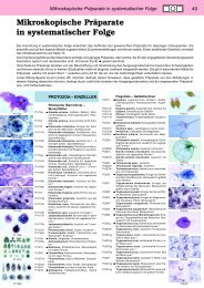
50<br />
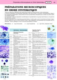
Pr237g<br />
Pr238f<br />
Pr239g<br />
Pr311f<br />
Pr315f<br />
Pr320h<br />
Pr328f<br />
Pr337f<br />
Pr338f<br />
Pr330e<br />
Pr333f<br />
<strong>Prepared</strong> <strong>Microscope</strong> <strong>Sli<strong>de</strong>s</strong> <strong>in</strong> <strong>Systematic</strong> <strong>Or<strong>de</strong>r</strong><br />
Pr2392t Leishmania donovani, smear from culture<br />
show<strong>in</strong>g Leishman and leptomonad forms *<br />
Pr2395h Leishmania donovani, promastigotes, smear<br />
from culture *<br />
Pr2396h Leishmania donovani, amastigotes, smear<br />
from tissue *<br />
Pr2397h Leishmania mexicana, promastigotes, smear<br />
from culture *<br />
Pr240f Leishmania enrietti, section through nasal<br />
abscess from Gu<strong>in</strong>ea pig. Very heavy <strong>in</strong>fection<br />
Pr2405g Crithidia fasciculata, smear from <strong>in</strong>test<strong>in</strong>e of<br />
Anopheles mosquito show<strong>in</strong>g the typical crithidia<br />
forms *<br />
Pr2378g Termite flagellates. w.m., show<strong>in</strong>g large vegetative<br />
forms *<br />
Pr251d • Silicoflagellates, various species<br />
Sporozoa<br />
Pr311f • Plasmodium falciparum, malignant tertian<br />
malaria of man, blood smear with typical r<strong>in</strong>g<br />
stages<br />
Pr3112g Plasmodium falciparum, blood smear with<br />
more gametocytes *<br />
Pr312f Plasmodium falciparum, thick diagnostic<br />
smear *<br />
Pr313h Plasmodium vivax, benign tertian malaria of<br />
man, blood smear *<br />
Pr3132h Plasmodium vivax, thick diagnostic blood<br />
smear *<br />
Pr3145h Plasmodium malariae, caus<strong>in</strong>g quartan malaria,<br />
blood smear *<br />
Pr315f • Plasmodium berghei, blood smear from experimentally<br />
<strong>in</strong>fected mouse. Very heavy <strong>in</strong>fection<br />
shows abundant parasites <strong>in</strong> different stages<br />
of <strong>de</strong>velopment<br />
Pr320h Plasmodium sp., section through <strong>in</strong>fected<br />
mosquito stomach with oocysts conta<strong>in</strong><strong>in</strong>g<br />
sporozoites *<br />
Pr321i Plasmodium sp., section through the salivary<br />
gland of <strong>in</strong>fected mosquito with sporozoites *<br />
Pr322h Plasmodium sp., exoerythrocytic stages <strong>in</strong><br />
sec. of bra<strong>in</strong> *<br />
Pr323h Plasmodium sp., exoerythrocytic stages <strong>in</strong><br />
sec. of liver *<br />
Pr3235g Malaria melanemia <strong>in</strong> human spleen, sec.<br />
show<strong>in</strong>g pigment granules <strong>in</strong> endothelium and<br />
Kupffer’s cells<br />
Pr326f Plasmodium praecox, avian malaria, blood<br />
smear<br />
Pr327f • Plasmodium gall<strong>in</strong>aceum (Proteosoma), fowl<br />
malaria, blood smear from chicken *<br />
Pr328f Plasmodium cathemerium, avian malaria,<br />
blood smear *<br />
Pr3285s Plasmodium circumflexum, smear from lung<br />
or bra<strong>in</strong> of bird show<strong>in</strong>g exoerythrocytic<br />
schizogony *<br />
Pr3287s Leukocytozoon, smear from fowl blood with<br />
parasites *<br />
Pr329s • Haemoproteus columbae, pigeon malaria,<br />
blood smear *<br />
Pr3293t Haemogregar<strong>in</strong>a, smear from frog blood with<br />
parasites *<br />
Pr337f • Babesia canis, blood smear shows heavy <strong>in</strong>fection<br />
Pr338f • Toxoplasma gondii, caus<strong>in</strong>g toxoplasmosis,<br />
tissue smear with parasites<br />
Pr3381f • Toxoplasma gondii, section of the bra<strong>in</strong> show<strong>in</strong>g<br />
cysts with parasites *<br />
Pr330e • Nosema apis, honey bee dysentery, sec. of<br />
diseased <strong>in</strong>test<strong>in</strong>e<br />
Pr331d • Monocystis lumbrici, <strong>in</strong> smear from earthworm<br />
sem<strong>in</strong>al vesicle<br />
Pr332d Monocystis lumbrici, section with parasites<br />
<strong>in</strong> situ<br />
Pr333f • Gregar<strong>in</strong>a, <strong>in</strong> smear from mealworm (Tenebrio)<br />
<strong>in</strong>test<strong>in</strong>e<br />
Pr334d Gregar<strong>in</strong>a, <strong>in</strong> section from mealworm <strong>in</strong>test<strong>in</strong>e,<br />
parasites <strong>in</strong> situ<br />
Pr335d • Eimeria stiedae, caus<strong>in</strong>g coccidiosis <strong>in</strong> rabbit,<br />
section of liver shows schizogony and all <strong>de</strong>velop<strong>in</strong>g<br />
stages<br />
Pr3352d Eimeria stiedae, coccidiosis, smear from faeces<br />
Pr336d Eimeria tenella, section of diseased chicken<br />
<strong>in</strong>test<strong>in</strong>e *<br />
Pr339f • Sarcocystis tenella, section of muscle show<strong>in</strong>g<br />
the parasites <strong>in</strong> Miescher’s tubes<br />
Pr3392f Sarcocystis tenella <strong>in</strong> heart muscle, sec.<br />
Pr3365s Myxosoma, parasite on fish gill, sec. *<br />
Ciliata (Infusoria)<br />
Pr411d • Paramecium, macro- and micronuclei sta<strong>in</strong>ed.<br />
The typical sli<strong>de</strong> for general study of this common<br />
ciliate<br />
Pr412e Paramecium, food vacuoles and nuclei doubly<br />
sta<strong>in</strong>ed<br />
Pr413e Paramecium, pellicle sta<strong>in</strong>ed after Bresslau<br />
Pr414e Paramecium, silver sta<strong>in</strong>ed to show the silver<br />
l<strong>in</strong>e or neuroformative system<br />
Pr415e Paramecium, specially prepared and sta<strong>in</strong>ed<br />
to show the trichocysts<br />
Pr416f • Paramecium, <strong>in</strong> conjugation, nuclei sta<strong>in</strong>ed *<br />
Pr417g • Paramecium, <strong>in</strong> fission, nuclei sta<strong>in</strong>ed *<br />
Pr418e Paramecium, section through many <strong>in</strong>dividuals,<br />
triply sta<strong>in</strong>ed<br />
Pr419f Paramecium, sta<strong>in</strong>ed with Feulgen reaction<br />
Pr4194e Paramecium multimicronucleatum, w.m. nuclei<br />
sta<strong>in</strong>ed. this species conta<strong>in</strong>s several micronuclei<br />
Pr4195e Paramecium aurelia, w.m. nuclei sta<strong>in</strong>ed. This<br />
species conta<strong>in</strong><strong>in</strong>g one macronucleus and two<br />
micronuclei<br />
Pr4196e Paramecium bursaria, w.m. and nuclei<br />
sta<strong>in</strong>ed, show<strong>in</strong>g symbiotic zoochlorellae <strong>in</strong><br />
endoplasm<br />
Pr422e • Vorticella, a common stalked ciliate w.m.<br />
Pr4222e Vorticella, a mar<strong>in</strong>e species, coloniate ciliate<br />
Pr421d • Stylonychia, a common ciliate w.m.<br />
Pr430e • Colpidium, a common holotrich ciliate<br />
Pr427f Spirostomum ambiguum, a ciliate with very<br />
large nucleus<br />
Pr428g Stentor, a trumpet-shaped large ciliate *<br />
Pr429e • Euplotes, a common mar<strong>in</strong>e ciliate<br />
Pr4306f Bursaria truncatella, a large fresh water ciliate<br />
*<br />
Pr4309e Blepharisma, a large ciliate with pigment granules<br />
*<br />
Pr4305e Did<strong>in</strong>ium nasutum, a small ciliate parasite on<br />
Paramecium *<br />
Pr423f Dendrocometes paradoxus, suctorial <strong>in</strong>fusoria<br />
on the gills of Gammarus *<br />
Pr424f Trichod<strong>in</strong>a domerguei, parasite liv<strong>in</strong>g on fish<br />
gills *<br />
Pr4307e • Ephelota, a stalked mar<strong>in</strong>e suctorian *<br />
Pr4311e Suctoria, mar<strong>in</strong>e species<br />
Pr425f Opal<strong>in</strong>a ranarum, smear from frog <strong>in</strong>test<strong>in</strong>e<br />
Pr426e • Opal<strong>in</strong>a ranarum, <strong>in</strong> section through frog <strong>in</strong>test<strong>in</strong>e<br />
Pr4265t Balantidium coli, human parasite, smear with<br />
trophozoites *<br />
Pr4266t Balantidium coli, smear with cysts *<br />
Pr4267t Balantidium coli, <strong>in</strong> sec. of human <strong>in</strong>test<strong>in</strong>e *<br />
Pr433f Ciliates from the rumen of cow, different species<br />
Pr435h Ciliates, specially prepared and sta<strong>in</strong>ed to<br />
show the cilia<br />
Pr440f • Mixed protozoa, many different forms are<br />
found on this sli<strong>de</strong><br />
MESOZOA<br />
Me111f Dicyema, simple animal with body and sexual<br />
cells, from smear of Sepia *<br />
PORIFERA – SPONGES<br />
Po111d • Sycon, a small mar<strong>in</strong>e sponge of the sycon<br />
type, t.s. through the body<br />
Po112f • Sycon, near med. long. sec. through body and<br />
osculum<br />
Po113d Sycon, tangential long. sec.<br />
Po114d Sycon, thick t.s. with calcareous spicules <strong>in</strong> situ<br />
Po115b • Sycon, spicules isolated, w.m.<br />
Po116f Sycon, sec. show<strong>in</strong>g stages of <strong>de</strong>velopment *<br />
Po1165e Sycon, l.s. and t.s. on one sli<strong>de</strong><br />
Po117d Grantia, a mar<strong>in</strong>e sponge of the sycon type,<br />
t.s. through the body<br />
Po118f Grantia, near median long. sec. through body<br />
and osculum<br />
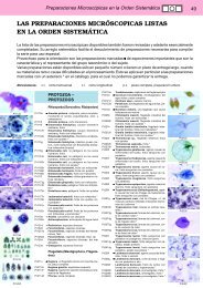
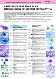
Pr339f<br />
Pr412e<br />
Pr413e<br />
Pr415e<br />
Pr416f<br />
Pr417g<br />
Pr422e<br />
Pr425f<br />
Pr4265t<br />
Me111f<br />
Po111d