Dermatology - 香港醫學組織聯會
Dermatology - 香港醫學組織聯會
Dermatology - 香港醫學組織聯會

You also want an ePaper? Increase the reach of your titles
YUMPU automatically turns print PDFs into web optimized ePapers that Google loves.
<strong>Dermatology</strong><br />
VOL.15 NO.11 NOVEMBER 2010<br />
OFFICIAL PUBLICATION FOR THE FEDERATION OF MEDICAL SOCIETIES OF HONG KONG ISSN 1812 - 1691
VOL.15 NO.11 NOVEMBER 2010<br />
for acne in this issue. In addition to the aforementioned<br />
medical dermatological diseases, we are also privileged<br />
to have Dr. Henry Chan, a renowned dermatologist<br />
with keen research interests in the field, gives us<br />
an update in the recent advancements in cosmetic<br />
dermatology. I would also like to thank Dr. Simon Ku,<br />
a very artistic dermatologist, for providing the cover<br />
shot of this issue and Dr. CK Kwan, a keen hiker for<br />
sharing some of his favourite hiking pathways in Hong<br />
Kong.<br />
I h o p e t h e s e a r t i c l e s w i l l c o n v i n c e y o u t h a t<br />
dermatological issues are indeed more than just skindeep<br />
and encourage everyone to give skin diseases<br />
their due attention and care. May I also take this<br />
opportunity to wish you all a joyful Christmas and all<br />
the best in 2011.<br />
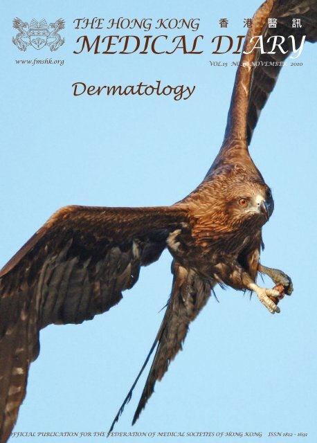
The Cover Shot<br />
Birds Photography<br />
References<br />
1.<br />
2.<br />
3.<br />
4.<br />
5.<br />
6.<br />
7.<br />
8.<br />
9.<br />
10.<br />
11.<br />
Editorial<br />
Kellett SC, Gawkrodger DJ. The psychological and emotional impact<br />
of acne and the effect of treatment with isotretinoin. Br J Dermatol<br />
1999;140:273-82.<br />
Gupta MA, Gupta AK. Depression and suicidal ideation in<br />
dermatology patients with acne, alopecia areata, atopic dermatitis and<br />
psoriasis. Br J Dermatol 1998;139:846-50.<br />
Cotterill JA, Cunliffe WJ. Suicide in dermatological patients. Br J<br />
Dermatol 1997;137:246-50.<br />
Koo JY, Smith LL. Psychologic Aspects of Acne. Ped Dermatol<br />
1991;8:185-8.<br />
Van der Meeren, Van der Schaar WW, Van der Hub CM. The<br />
psychological impact of severe acne. Cutis 1985;36:84-6.<br />
Tan JK; Psychosocial impact of acne vulgaris: evaluating the evidence.<br />
Skin Therapy Lett. 2004 Aug-Sep;9(7):1-3.<br />
Wolkenstein P, Grob JJ, Bastuji Garin S, Ruszczynski S, Roujeau JC,<br />
Revuz J. French people and skin diseases: results of a survey using a<br />
representative sample. Arch Dermatol. 2003;139(12):1614–1619.<br />
Lowell BA, Froelich CW, Federman DG, Kirsner RS. <strong>Dermatology</strong><br />
in primary care: Prevalence and patient disposition. J Am Acad<br />
Dermatol. 2001;45(2):250–255.<br />
Julian CG. <strong>Dermatology</strong> in general practice. Br J Dermatol. 1999;<br />
141(3):518–520.<br />
Elisabeth WM Verhoeven, Floor W. Kraaimaat, Chris van Weel, Peter<br />
CM van de Kerkhof, Piet Duller, Pieter GM van der Valk, Henk JM<br />
van den Hoogen, J Hans J Bor, Henk J Schers, and Andrea WM Evers:<br />
Skin Diseases in Family Medicine: Prevalence and Health Care Use.<br />
Ann Fam Med. 2008 July; 6(4): 349–354.<br />
Farah Awadalla, Daryl A Rosenbaum, Fabian Camacho, Alan B<br />
Fleischer, Steven R Feldman, Dermatologic Disease in Family<br />
Medicine. Fam Med. 2008;40(7):507-11.<br />
Birds photography is probably one of the most exciting<br />
and challenging endeavours of the photographic world.<br />
Birds, both at-rest and in-flight, are elegant and beautiful.<br />
Capturing wild birds in their natural habitats often<br />
requires big telescopic lenses like 500, 600 or even 800mm.<br />
This photo is a black kite, the commonest raptor found<br />
in HK. It was captured in one late afternoon, when the<br />
sun was shining from the side, illuminating her face and<br />
the food inside her claws. It was taken at almost eye-level<br />
with the bird flying directly towards the camera, giving it<br />
an intrusive and vivid composition. Dr. Simon Lap-shing KU<br />
MBBS(HK),<br />
MRCP(UK), FHKCP,<br />
FHKAM(MEDICINE)<br />
Specialist in <strong>Dermatology</strong><br />
3
VOL.15 NO.11 NOVEMBER 2010<br />
Practical Approach for “Eczema”<br />
Dr. KK LO<br />
FRCP, FHKCP, FHKAM (Med)<br />
Specialist in <strong>Dermatology</strong> & Venereology<br />
Eczema is the commonest diagnosis in the clinical<br />
practice for both specialists and family doctors when<br />
encountering patients complaining of skin problems.<br />
Eczema is prevalent in a local students study.<br />
It appears as an apparent simple and easy condition to<br />
deal with but indeed it is one of the conditions that our<br />
patients are often dissatisfied about the outcome of the<br />
treatments. In this brief review, I describe my approach<br />
to let family doctors having a more systemic and logical<br />
approach to the management of this common skin<br />
problem.<br />
My approach to the management of eczema follows<br />
to the convention of Western medicine. We shall<br />
be able to make the accurate diagnosis by standard<br />
clinical methods, namely: clinical history, physical<br />
examination and investigations if needed. Having<br />
an accurate diagnosis is a half-way success and the<br />
subsequent management is divided into general and<br />
specific treatments. General treatment is actually more<br />
important though very often its emphasis is skipped by<br />
both the clinician and our patients.<br />
The terms eczema and dermatitis are regarded as<br />
synonyms in clinical practice though dermatitis is<br />
used more often for exogenous causes. Eczema is<br />
derived from Greek, “ek” meaning out and “zein”<br />
to boil. Eczema defines the superficial inflammatory<br />
itchy skin problem though eczema may mean different<br />
things to different people. There are different views on<br />
eczema even among specialists and it is no wonder how<br />
confusing our clients and colleagues do in our daily<br />
practice. My approach to the diagnosis of eczema is<br />
by exclusion. The difference between a specialist and<br />
a family doctor may be just in the different length of<br />
exclusion list of differential diagnoses (see table 1) in<br />
making the diagnosis.<br />
Eczematoid rash is characterised by an erythematous<br />
scaly rash with an ill-defined border in different<br />
phases, acute, subacute and chronic. Furthermore, it<br />
is a dynamic rash in that it will gradually change its<br />
configuration, size, shape and colour with time in terms<br />
of days, weeks and months. It can remit and reappear<br />
with a turnover time from days to months. However,<br />
if a persistent (never remitted and stayed in the same<br />
location) erythematous scaly rash presents for years, a<br />
diagnosis of eczema is unlikely. Skin biopsy or referral<br />
to a specialist to exclude other serious conditions<br />
Medical Bulletin<br />
Dr. KK LO<br />
This article has been selected by the Editorial Board of the Hong Kong Medical Diary for participants in the CME programme of the Medical<br />
Council of Hong Kong (MCHK) to complete the following self-assessment questions in order to be awarded one CME credit under the programme<br />
upon returning the completed answer sheet to the Federation Secretariat on or before 30 November 2010.<br />
of papulosquamous dermatoses (Table 1) must be<br />
considered.<br />
Table 1: Differential diagnoses of eczema<br />
Common cutaneous conditions Additional cutaneous conditions<br />
considered for exclusion by considered for exclusion by<br />
family doctors<br />
specialists<br />
Various forms of Superficial<br />
fungal infections<br />
Various forms of psoriasis and<br />
psoriasiform eruptions<br />
Lichen planus and lichenoid<br />
eruptions<br />
Neoplastic conditions e.g.<br />
Mycosis fungoides, Bowen<br />
d i s e a s e , L a n g e r h a n s c e l l<br />
histiocytosis<br />
Chronic infections e.g. Hansen<br />
disease, secondary syphilis<br />
Autoimmune diseases e.g.<br />
dermatomyositis, subacute lupus<br />
erythematosus, pemphigus<br />
foliaceous<br />
Various forms of cutaneous Nutritional diseases e.g. zinc<br />
d r u g e r u p t i o n s i n c l u d i n g deficiency, essential fatty acid<br />
photodermatitis<br />
deficiency, Pellagra (niacin<br />
deficiency)<br />
Others: pityriasis lichenoides,<br />
pityriasis rosea, lichen striatus,<br />
g l u c a g o n o m a s y n d r o m e ,<br />
congenital immunodeficiency<br />
e.g. Wiskott Aldrich syndrome &<br />
DiGeorge syndrome<br />
Just making an accurate diagnosis of eczema may not be<br />
adequate professionally. We should have a very simple<br />
classification of eczema so that we may communicate<br />
with our clients more confidently though the treatment<br />
and outcome may vary little. The classification of<br />
eczema will vary from one specialist to another. Mine<br />
is a simple one (Table 2). I divide eczema into two main<br />
groups: exogenous (external factors may play a more<br />
important role) and endogenous (constitutional factor of<br />
the person is more important). The prototype examples<br />
of exogenous eczema are irritant contact eczema and<br />
allergic contact eczema. Usually, contact eczema starts<br />
in some local contact sites of our skin and a meticulous<br />
examination of the location may give the clues of<br />
allergen or irritants. Irritant contact eczema is more<br />
common and the different layman names of irritant<br />
contact eczema of hands have indicated their common<br />
occurrence in the community. A detailed history on<br />
occupation, daily hobbies and interests is essential.<br />
Allergic contact eczema is a type IV hypersensitivity<br />
condition and can be confirmed by appropriate patch<br />
tests with specific contact allergens though it may not<br />
be easily available in common clinical practice. With<br />
good substitution for avoidance of future contact with<br />
the allergens or proper protection from the irritants, the<br />
prognosis of exogenous eczema is good. However, we<br />
should remind our clients not to forget the importance<br />
of compliance to general advice and care of the skin<br />
5
6<br />
Medical Bulletin<br />
(Table 3) which is the key for a successful cure.<br />
Table 2: Simple classification of eczema<br />
Exogenous eczema Endogenous eczema<br />
Irritant Contact eczema e.g. Atopic eczema<br />
housewife dermatitis<br />
Allergic contact eczema e.g. Seborrhoeic eczema<br />
Nickel contact dermatitis<br />
Stasis eczema<br />
Discoid/Nummular eczema<br />
Asteatotic eczema<br />
Vesicular hands & feet eczema<br />
( d y s h y d r o t i c e c z e m a o r<br />
pompholyx)<br />
Table 3: General management of eczema<br />
Remarks<br />
Environmental<br />
modification<br />
Unclassified papular eczema,<br />
lichen simplex chronicus<br />
Avoid excessive bathing. Avoid all types of<br />
contact irritants including common soap,<br />
detergent, shampoo and cleansing agents.<br />
May need to change or modify job nature<br />
or posting. Use appropriate protective<br />
measures including gloves, barrier creams<br />
Use soap substitutes Many types of fragrance free emollients<br />
and liberal application and moisturisers will help skin protection<br />
of emollients especially in low humidity season. Common<br />
brand creams include aqueous cream,<br />
emulsifying ointment. Individual preference<br />
may affect choice. Proper use of moisturisers<br />
would improve the skin barrier function of<br />
eczema patients<br />
Diet The role of food avoidance, food allergy,<br />
supplements with probiotics and breast<br />
feeding becomes less clear for prevention<br />
of atopic eczema in recent studies. Defer<br />
introduction of solid food to infants may<br />
help to delay onset of atopic eczema but<br />
cannot prevent it. Normally, a balanced diet<br />
is recommended. Elimination diet for severe<br />
atopic eczema is rarely needed and should<br />
be conducted with advice of specialists;<br />
patients’ self initiated food restrictions may<br />
not be helpful to improve eczema though<br />
psychologically useful. A properly kept food<br />
diary would be a useful objective tool to find<br />
out the real problematic food<br />
Emotion stability Occasional extreme anxiety may need<br />
psychological treatment<br />
Awareness of<br />
complications of<br />
eczema e.g. secondary<br />
cutaneous bacterial<br />
infection<br />
An empirical course of Cloxacillin would be<br />
useful for occasional exacerbations of atopic<br />
eczema. Severe eczema herpeticum should<br />
be referred to specialists and commence a<br />
course of acyclovir without delay<br />
Endogenous eczema has a relatively poorer prognosis in<br />
that we cannot change our genetic make-up to prevent<br />
the relapse of eczema. Common forms of endogenous<br />
eczema encountered in our clinical practice include<br />
atopic eczema (infantile, childhood, adolescent and<br />
adult), seborrhoeic eczema, stasis eczema, vesicular<br />
hand eczema (pompholyx), nummular or discoid<br />
eczema, asteatotic or xerotic eczema and unclassified<br />
popular eczema. Personal and family history of atopy<br />
diseases (e.g. asthma, allergic rhinitis) offer hints<br />
for the diagnosis of atopic eczema. Specific areas of<br />
distribution and involvement in the body e.g. eczema<br />
involving scalp, nasolabial fold, V-of chest and flexural<br />
area will favour more on the diagnosis of seborrhoeic<br />
eczema; eczema involving lower limbs with sign of<br />
venous hypertension and insufficiency will guide you<br />
to the diagnosis of stasis eczema, coin-like nummular<br />
patches in the body and often remit and recur in the<br />
same area may point to the diagnosis of nummular<br />
eczema; generalised dry skin with fine scaling and<br />
VOL.15 NO.11 NOVEMBER 2010<br />
superficial cracks on the dry skin especially in the elderly<br />
will be an easy diagnosis for asteatotic eczema. However,<br />
some of these features may be mixed and not so clearly<br />
defined. Clinical experience and closer follow-ups for the<br />
course of the eczema will help to give the diagnosis of the<br />
very common type of endogenous eczema: unclassified<br />
popular eczema. Again, this type of eczema is diagnosed<br />
by exclusion of all other possible definite types of<br />
eczema before we label it as such. It may transform to a<br />
more definite form of eczema later on follow up visits.<br />
Lichen simplex chronicus or chronic neurodermatitis<br />
commonly appears on some typical sites such as the<br />
nape and side of neck, scrotum, vulva, eyelid and ankles.<br />
Habitual scratching of these areas results in lichenified<br />
hyperpigmented plaques with accentuation of surface<br />
markings. Potent topical steroids with occlusion may<br />
sometimes be needed to break this itch scratch vicious<br />
cycle of lichen simplex chronicus.<br />
The specific treatment for eczema is summarised in Table<br />
4. Perhaps the most difficult part in the management<br />
of eczema is to allocate adequate time to explain<br />
the disease nature and to understand the genuine<br />
concerns of our clients in the consultation. Having a<br />
good rapport with our patients, better understanding<br />
of general treatment e.g. diet advice, environmental<br />
modification, use of emollients and the proper use of<br />
topical steroids and immunosuppressive medications,<br />
the management of eczema can be optimised with<br />
better client satisfaction. Only a few types of extensive<br />
or resistant form of eczema (e.g. erythroderma in adult<br />
atopic eczema or seborrhoeic eczema) should require<br />
the long term care from specialists.<br />
Table 4: Specific treatment of eczema<br />
Remarks<br />
Topical medications Different strengths of topical steroids under<br />
doctors’ guidance and monitor (must clearly<br />
explain to patients for their proper application<br />
and their long term adverse effects) are still the<br />
mainstay of treatment; potent steroids will only<br />
be useful for short term use for exacerbations).<br />
Topical Calcineurin inhibitors (expensive but lack<br />
skin atrophy on long term use and would be good<br />
choice for applying on face or eyelids) are getting<br />
popular. Tar smells and its appearance is less<br />
acceptable to patients<br />
Oral antihistamines F o r s y m p t o m a t i c t r e a t m e n t o n l y . O r a l<br />
antihistamines suppress pruritus, allay anxiety<br />
and allow sleep with less scratch; used as<br />
adjunctive treatment<br />
Systemic medications All are with various degrees of adverse effects<br />
and should be used with caution especially<br />
on long term use. They should be closely<br />
monitored for their efficacy as well as adverse<br />
effects e.g. azathioprine, cyclosporine A. Other<br />
immunosuppressive therapies e.g. Mycophenolate<br />
mofetil, Methotrexate have been tried but they<br />
are not considered standard therapy. Long term<br />
administration of systemic steroids plays no<br />
role in the management of eczema though short<br />
term uses can alleviate exacerbations. Once<br />
commenced, it is difficult to wean off without<br />
flare ups of eczema<br />
Phototherapy Both narrow band UVB phototherapy and<br />
systemic or local PUVA are useful but the long<br />
term effects of photoageing and carcinogenic<br />
effects limit their application<br />
Others Wet-wrap is a damp layer of cotton dressing used<br />
in combination with emollients or diluted topical<br />
steroids wrapping the affected areas overnight<br />
with an outer coat of dry gauze wrapping. Its<br />
efficacy is good for small children though it is<br />
tedious for parents. Traditional Chinese herbal<br />
medicine (TCM) has been found to be useful but<br />
standardisation of TCM is problematic and it has<br />
been reported with hepatotoxicity. No clinical<br />
benefits were found in more recent studies with use<br />
of evening primrose oil. Reports of homeopathy,<br />
mind-body therapies and topical honey to treat<br />
eczema are known but evidences are weak
VOL.15 NO.11 NOVEMBER 2010<br />
References<br />
Fung WK, Lo KK: Prevalence of skin disease among school children and<br />
adolescents in a student health service center in Hong Kong. Pediatr<br />
Dermatol 2000;17:440-6.<br />
Hon KL, Wong KY, Cheung LK et al.: Efficacy and problems associated<br />
with using a wet-wrap garment for children with severe atopic dermatitis.<br />
J. Dermatol. Treat. 2007; 18(5),301-305.<br />
Heller M, Shin HT, Orlow SJ, Schaffer JV: Mycophenolate mofetil for<br />
severe childhood atopic dermatitis: experience in 14 patients. Br. J.<br />
Dermatol. 2007;157(1),127-132<br />
Medical Bulletin<br />
Zhang W, Leonard T, Bath-Hextall F et al.: Chinese herbal medicine for<br />
atopic eczema. 2005; Cochrane Database Syst. Rev. 2<br />
Greer FR, Sicherer SH, Burks AW: Effects of early nutritional interventions<br />
on the development of atopic disease in infants and children: the role<br />
of maternal dietary restriction, breastfeeding, timing of introduction<br />
of complementary foods, and hydrolyzed formulas. Pediatrics 2008;<br />
121(1),183-191.<br />
MCHK CME Programme Self-assessment Questions<br />
Please read the article entitled "Practical Approach for “Eczema” " by Dr. KK LO and complete the following<br />
self-assessment questions. Participants in the MCHK CME Programme will be awarded 1 CME credit under<br />
the Programme for returning completed answer sheets via fax (2865 0345) or by mail to the Federation<br />
Secretariat on or before 30 November 2010. Answers to questions will be provided in the next issue of The<br />
Hong Kong Medical Diary.<br />
Questions 1-10: Please answer T (true) or F (false)<br />
1. Eczema is derived from Latin with “zein” meaning to cook.<br />
2. Eczematoid rash is characterised by a persistent non-scaling erythematous rash on the skin.<br />
3. General advice and care of the skin is as important as specific treatment for the management of<br />
recalcitrant eczema.<br />
4. Stasis eczema is one form of endogenous eczema.<br />
5. Lichen simplex chronicus commonly affects the genitalia .<br />
6. Viral infection is one of the causes for exacerbations of eczema in patients with atopic eczema.<br />
7. An empirical course of Cloxacillin is not recommended for occasional flares of atopic ezema.<br />
8. Topical Calcineurin inhibitors are always better than topical steroid treatment for management of eczema.<br />
9. Long term phototherapy for treatment of severe atopic eczema is useful and safe.<br />
10. Traditional Chinese herbal medicine is not useful in management of eczema.<br />
Answers to October 2010 Issue<br />
ANSWER SHEET FOR NOVEMBER 2010<br />
Please return the completed answer sheet to the Federation Secretariat on or before 30 November 2010 for<br />
documentation. 1 CME point will be awarded for answering the MCHK CME programme (for non-specialists)<br />
self-assessment questions.<br />
Practical Approach for “Eczema”<br />
Dr. KK LO<br />
FRCP, FHKCP, FHKAM (Med)<br />
Specialist in <strong>Dermatology</strong> & Venereology<br />
1 2 3 4 5 6 7 8 9 10<br />
Name (block letters):___________________________________ HKMA No.: _________________________________<br />
HKID No.: __ __ - __ __ __ __ X X (X) HKDU No.: _________________________________<br />
Contact Tel No.:_______________________________________ CDSHK No.: ________________________________<br />
Thyroid Eye Disease: a Comprehensive Review<br />
1. F 2. F 3. T 4. T 5. F 6. F 7. F 8. T 9. T 10. F<br />
7
VOL.15 NO.11 NOVEMBER 2010<br />
Is Psoriasis a Cutaneous Disease or<br />
Systemic Disease?<br />
Dr. LY CHONG<br />
MBBS(HK), FRCP(Lond, Edin, Glasg), FHKCP, FHKAM(Med)<br />
Private Dermatologist<br />
Psoriasis is a well known chronic, non-contagious skin<br />
disorder since ancient times. The word “Psora” is a<br />
Greek word meaning “To itch”. It is notorious for its<br />
chronicity in its natural course and difficulties in the<br />
management. Although great efforts have been tried in<br />
medical researches for decades, it is still not curable and<br />
its exact aetiology remains unknown.<br />
Prevalence in Chinese<br />
In Western literature, it is well reported that 1-3% of<br />
the Caucasian population have psoriasis. Though it<br />
is a common skin disease, the prevalence reported in<br />
Chinese is lower than that of Caucasians. From the<br />
limited resources available, it is estimated that psoriasis<br />
occurs in about 0.1-0.3% of the Chinese population. 1<br />
In 1984, a nation-wide screening of psoriasis had been<br />
conducted in 24 regions (53 centres) of China, involving<br />
a coverage of 6,617,917 population. About 11,393 cases of<br />
psoriasis (0.123% among the population studied; range<br />
from 0.3%) had been reported. Interestingly,<br />
the prevalence is highest in the Northeast provinces<br />
and lowest in the Southern ones where there is more<br />
sunshine. It is also higher in cities than the villages.<br />
In Hong Kong, the prevalence is estimated to be<br />
approximately 0.3% of the total population. 2 It is the 5th<br />
leading new cases of skin disorders in public dermatology<br />
clinics, with more than 600 new cases annually.<br />
Immuno-pathogenesis of Psoriasis<br />
Despite its unknown aetiology, there have been<br />
breakthroughs in the understanding of the immunopathogenesis<br />
of psoriasis in recent years. It is now<br />
almost certain that psoriasis is a T-lymphocyte mediated<br />
inflammatory dermatosis with hyper-proliferation<br />
of keratinocytes in genetically predisposed subjects<br />
(Diagram 1). It is regarded as one form of immunemediated<br />
inflammatory diseases (IMID), which is a term<br />
designated for organ-specific diseases in which cells and<br />
cytokines of the adaptive immune system cause tissue<br />
inflammation or destruction.<br />
Until recent years, it was believed that IMID was<br />
either mediated by Th1 T-cells (which is stimulated<br />
by IL-12) or TH2 T-cells. The former subset includes<br />
psoriasis, rheumatoid arthritis, multiple sclerosis<br />
and inflammatory bowel diseases, while the latter<br />
subset includes atopic dermatitis and asthma. A new<br />
Medical Bulletin<br />
Dr. LY CHONG<br />
pathogenic concept in IMID however is developed upon<br />
the discovery of a new T-cell lineage in 2005. This new<br />
cell lineage is called Th17 T-cell, which is defined by the<br />
production of IL-17, and stimulated by IL-23. This subset<br />
likely includes psoriasis, rheumatoid arthritis, multiple<br />
sclerosis and inflammatory bowel diseases. IL-12 and IL-<br />
23 are structurally related with a common 40kD subunit<br />
(p40), which leads to the development of a new group of<br />
biologics called anti-P40 (anti-IL12/23) that blocks both<br />
the Th1 and Th17 pathways.<br />
Diagram 1. Postulated Immuno-pathogenesis of Psoriasis<br />
Co-morbidities of Psoriasis<br />
The traditional belief about psoriasis is that it is a<br />
cutaneous disease without visceral involvements, albeit<br />
10-30% of the patients have joint involvements. This<br />
concept is challenged in recent few years when more and<br />
more systemic co-morbidities had been reported. When<br />
the term “psoriasis and co-morbidities” is searched in<br />
Medline, more than 200 articles can be retrieved over<br />
the past two decades. It is one of the hottest research<br />
topics in dermatology in the past 5 years, as shown by<br />
the numerous publications from different countries and<br />
different indexed journals. The possible co-morbidities<br />
of psoriasis reported in literatures are summarised in<br />
Table 1. Three important areas which may have impacts<br />
on medical health are worth mentioning here.<br />
Cardiovascular diseases (hypertension, myocardial infarction),<br />
cerebrovascular and peripheral vascular diseases<br />
Metabolic syndrome (obesity, diabetes mellitus)<br />
Non-alcoholic fatty liver<br />
Autoimmune diseases (Crohn’s disease, ulcerative colitis, multiple sclerosis)<br />
Lymphoma, melanoma, non-melanoma skin cancers<br />
Depression, suicide<br />
Smoking, alcoholism<br />
Osteoporosis<br />
Table 1. Possible co-morbidities of psoriasis reported in literatures<br />
9
10<br />
Medical Bulletin<br />
1.Cardiovascular Diseases<br />
Gelfand JM, et al. had published an article on JAMA<br />
in 2006 concerning the risks of myocardial infarction<br />
in patients with psoriasis. 3 They had conducted a<br />
population-based cohort study using data collected by<br />
general practitioners participating in the General Practice<br />
Research Database in the United Kingdom from 1987-<br />
2002. A total of 556,995 control patients and patients with<br />
mild (n = 127,139) and severe psoriasis (n = 3,837) were<br />
studied, and controlled for traditional cardiovascular<br />
risk factors (diabetes mellitus, history of myocardial<br />
infarction (MI), hypertension, hyperlipidaemia,<br />
smoking). They found that the adjusted relative risks of<br />
MI are 1.54 (1.24–1.91) and 7.08 (3.06-16.36) respectively<br />
in mild and severe psoriasis as compared with controls.<br />
Xiao J, et al. had published another article in J Eur<br />
Acad Dermatol Venereol. in 2009 about the prevalence<br />
of myocardial infarction in patients with psoriasis in<br />
central China. Data were collected from the medical<br />
records section of five hospitals in the Mainland between<br />
1999 and 2007. 4 After adjusting for systemic therapies<br />
and other known cardiovascular risk factors in addition<br />
to age and sex, they found that the odds ratio (OR) of<br />
having an MI were 1.72 (95% CI, 1.29-2.30) and 2.01 (95%<br />
CI, 1.45-2.79) respectively in mild and severe psoriasis.<br />
Related to the cardiovascular co-morbidities, Ludwig<br />
RJ, et al. had published an article in Br J Dermatol. in<br />
2007 concerning psoriasis as a possible risk factor for the<br />
development of coronary artery calcification (CAC). 5<br />
They found a significantly increased prevalence (59.4%<br />
vs. 28.1%, P = 0.015) and severity (CAC score according<br />
to Agatston 3.7 vs. 0.0, P = 0.019) of CAC in patients<br />
with psoriasis vs. controls. Multiple linear regression<br />
calculations identified psoriasis as a likely independent<br />
risk factor for CAC.<br />
2.Metabolic Syndrome (diabetes mellitus,<br />
hypertension, hyperlipidaemia & obesity)<br />
Sommer DM, et al. had published an article about the<br />
prevalence of the metabolic syndrome in patients with<br />
moderate to severe psoriasis in Arch Dermatol Res. in<br />
2006. 6 They had investigated a total of 581 adult patients<br />
hospitalised for plaque type psoriasis as compared<br />
to 1,044 hospital-based controls. A distinct pattern<br />
of chronic disorders was found to be significantly<br />
associated with psoriasis, including type II diabetes<br />
mellitus [odds ratio (OR)=2.48], arterial hypertension (OR<br />
= 3.27), hyperlipidaemia (OR = 2.09), and coronary heart<br />
disease (OR = 1.95). The combined presence of these<br />
conditions together with obesity, known as the metabolic<br />
syndrome, was clearly more prevalent in psoriasis<br />
patients (OR = 5.29).<br />
In the cross-sectional study on association between<br />
psoriasis and the metabolic syndrome by Cohen<br />
AD, et al., published in J. <strong>Dermatology</strong> in 2008, 7 it<br />
had demonstrated that psoriasis was associated with<br />
the metabolic syndrome (OR = 1.3, 95% CI = 1.1-<br />
1.4), ischaemic heart disease (OR = 1.1, 95% CI = 1.0-<br />
1.2), diabetes mellitus (OR = 1.2, 95% CI = 1.0-1.3),<br />
hypertension (OR = 1.3, 95% CI = 1.2-1.5) and obesity<br />
(OR = 1.7, 95% CI = 1.5-1.9). This study included 16,851<br />
patients with psoriasis and 48,681 controls.<br />
3. Lympho-proliferative Diseases<br />
VOL.15 NO.11 NOVEMBER 2010<br />
Gelfand JM, et al. had studied on the risks of lymphomas<br />
in psoriasis and published in J Invest Dermatol. in 2006. 8<br />
Their study used large population-based cohort data<br />
collected from the General Practice Research Database<br />
in the United Kingdom (1988-2002), involving 153,197<br />
psoriasis patients and 765,950 control patients without<br />
psoriasis. The adjusted relative risks in Non-Hodgkin’s<br />
lymphoma, Hodgkin’s lymphoma, T-cell lymphoma<br />
and all lymphomas in severe psoriasis were 0.73, 3.18,<br />
10.75 and 1.59, indicating that there is higher risks of<br />
lymphomas in patients with psoriasis.<br />
Possible Mechanistic Links Between<br />
Psoriasis and its Co-morbidities<br />
The link between chronic inflammation and metabolic<br />
and vascular disorders is now increasingly recognised.<br />
It is postulated that proinflammatory cytokines (such as<br />
tumour necrosis factor-alpha) involved in the immunemediated<br />
or inflammatory pathway may contribute<br />
to atherogenesis, peripheral insulin resistance, and<br />
hypertension. Macrophages and adipocytes also share<br />
common features such as expression of cytokines,<br />
FABPs, and nuclear hormone receptors, which may<br />
contribute to obesity. 9<br />
Many studies had reported that various IMIDs, including<br />
psoriasis, are at higher risks of developing systemic<br />
co-morbidities. IMIDs may cause these co-morbidities<br />
through shared genetic risks, common environmental<br />
factors, or common inflammatory pathways that are coexpressed<br />
in IMIDs and target organs. 10<br />
As mentioned in previous paragraphs, psoriasis is now<br />
classified as an IMID of the skin with T-cell mediated<br />
pathogenetic pathways and involvement of various<br />
inflammatory mediators. This may similarly predispose<br />
to the increasingly reported associated co-morbidities.<br />
The potentially systemic nature of the inflammatory<br />
processes in the pathogenesis of psoriasis has thus led to<br />
the postulation that it may be considered as a systemic<br />
disease, rather than a pure cutaneous disease.<br />
Is Psoriasis Really an Independent<br />
Risk Factor for These Co-morbidities?<br />
Despite increasing reports from difficult countries<br />
supporting the association of these co-morbidities of<br />
psoriasis, there were skeptical views about their true<br />
causal relationship. Nijsten T and Wakkee M. had written<br />
an excellent and critical commentary in J Inves Dermatol.<br />
2009 Jul issue about the complexity of the association<br />
between psoriasis and its co-morbidities. 11<br />
Although these studies did involve very large data<br />
base, their designs were not without shortcomings and<br />
pitfalls. Most of them are observational studies which<br />
were not primarily designed for the detection of these<br />
co-morbidities. Diagnostic bias and detection bias were<br />
unavoidable.<br />
Moreover, many confounding factors may be involved<br />
in these co-morbidities, as illustrated in Diagram 2. For<br />
example, psoriasis itself may lead to impaired health-
VOL.15 NO.11 NOVEMBER 2010<br />
related quality of life (HRQOL) such as depression,<br />
anxiety and stress, which may result in an unhealthy<br />
life style leading to alcoholism and chronic smoking.<br />
The presence of arthropathy may lead to lack of<br />
exercise. The well known side-effects of some systemic<br />
antipsoriatic drugs may also count, such as acitretin<br />
may cause hyperlipidaemia and cyclosporine may<br />
cause hypertension. All these confounding factors<br />
may contribute to the cardiovascular diseases and<br />
metabolic syndrome. Conversely, obesity and smoking<br />
may increase the risk of developing psoriasis as<br />
reported, while drugs like lithium in treating manicdepressive<br />
illnesses and certain beta-blockers in treating<br />
cardiovascular diseases may also induce psoriasis.<br />
Despite many studies that had supported the association<br />
of these co-morbidities, there are also inconsistencies in<br />
the findings in other studies. 12<br />
Finally, although in the supporting papers, most of the<br />
reported associations had reached statistical significance<br />
after statistical analysis, as they had involved very<br />
large data base, it is essential to know that statistical<br />
associations do not equate to causal relationships, and<br />
do not always have clinical relevancy.<br />
Diagram 2. Schematic overview of possible confounding<br />
factors influencing the association between psoriasis and its<br />
co-morbidities (Modifeid from Nijsten T and Wakkee M) 11<br />
Conclusion<br />
Although there are increasing reports that psoriasis<br />
may have significant systemic co-morbidities, judging<br />
from the present evidences, causality has not yet been<br />
proven. Whether there is true causal relationship<br />
between psoriasis and these co-morbidities is still<br />
uncertain and open to debate. Upgrading psoriasis<br />
to an systemic disease obviously will have significant<br />
impacts on the management of this chronic disease,<br />
such as more aggressive treatments, routine screening<br />
of the co-morbidities, and possibly healthcare resources<br />
reallocation. Therefore, until more well proven<br />
evidences are available, it is still more appropriate to<br />
regard psoriasis as a cutaneous disease at the moment.<br />
Nonetheless, psoriasis is definitely more than skin depth<br />
in its impact on the affected patients, in view of its<br />
physical and psychosocial impairments. It also reminds<br />
Medical Bulletin<br />
dermatologists the need to manage patients holistically<br />
as a whole, rather than just focusing on their skin.<br />
References<br />
1.<br />
2.<br />
3.<br />
4.<br />
5.<br />
6.<br />
7.<br />
8.<br />
9.<br />
10.<br />
11.<br />
12.<br />
Distribution of psoriasis in China: a nation-wide screening in 1984<br />
( 全國 1984 年銀屑病流行調查報告 ). Chinese Journal of <strong>Dermatology</strong><br />
1986;19:253-61.<br />
The prevalence of psoriasis in the Mongoloid race. Yip SY. J Am Acad<br />
Dermatol. 1984;10(6):965-8.<br />
Risk of myocardial infarction in patients with psoriasis. Gelfand JM,<br />
Neimann AL, Shin DB, Wang X, Margolis DJ, Troxel AB.<br />
JAMA. 2006;296:1735-41.<br />
Prevalence of myocardial infarction in patients with psoriasis in<br />
central China. Xiao J, Chen LH, Tu YT, Deng XH, Tao J. J Eur Acad<br />
Dermatol Venereol. 2009 Nov;23(11):1311-5.<br />
Psoriasis: a possible risk factor for development of coronary artery<br />
calcification. Ludwig RJ, Herzog C, Rostock A, Ochsendorf FR, Zollner<br />
TM, Thaci D, Kaufmann R, Vogl TJ, Boehncke WH. Br J Dermatol.<br />
2007 Feb;156(2):271-6.<br />
Increased prevalence of the metabolic syndrome in patients with<br />
moderate to severe psoriasis. Sommer DM, Jenisch S, Suchan<br />
M, Christophers E, Weichenthal M. Arch Dermatol Res. 2006<br />
Dec;298(7):321-8.<br />
Association between psoriasis and the metabolic syndrome. A crosssectional<br />
study. Cohen AD, Sherf M, Vidavsky L, Vardy DA, Shapiro J,<br />
Meyerovitch J. <strong>Dermatology</strong>. 2008;216(2):152-5.<br />
Risk of Lymphoma in Psoriasis Gelfand JM, Shin DB, Neiman AL,<br />
Wang X, Margolis DJ, Troxel AB. J Invest Dermatol. 2006;126:2194-<br />
2201.<br />
Inflammation, stress and diabetes Wellen KE, Hotamisligil GS. J Clin<br />
Invest. 2005;115:1111-9.<br />
Psoriasis and systemic inflammatory diseases: potential mechanistic<br />
links between skin disease and co-morbid conditions. Davidovici BB,<br />
Sattar N, Jörg PC, et al. J Invest Dermatol. 2010;130:1785-96.<br />
Complexity of the Association Between Psoriasis and Comorbidities<br />
Nijsten T, Wakkee M. J Inves Dermatol. 2009;129:1601-3.<br />
Psoriasis may not be an independent risk factor for acute ischemic<br />
heart disease hospitalizations: Results of a large population-based<br />
Dutch cohort Wakkee M, Herings RMC, Nijsten T. J Invest Dermatol<br />
Apr; 2010;130:962-7.<br />
11
VOL.15 NO.11 NOVEMBER 2010<br />
Nail and Nail Disorders<br />
Dr. William TANG<br />
FRCP (Edin & Glasg), FHKAM (Med)<br />
Honorary Clinical Associate Professor, <strong>Dermatology</strong> Research Centre, Faculty of Medicine,<br />
The Chinese University of Hong Kong<br />
The nail unit is a dynamic complex which forms an<br />
important part of the integument. This complex<br />
consists of the nail matrix (NM), nail bed (NB),<br />
hyponychium, nail fold (NF) and the nail plate (NP).<br />
Cells of the nail matrix, under the protection of the<br />
proximal nail fold mature and keratinised to form the<br />
NP. The NM contributes to the most portion of the NP<br />
while about 10-15% is produced by the NB. As the NP<br />
grows distally, the continued addition of keratinised<br />
material makes the NP increases in thickness while it<br />
lengthens distally. There are variations in the rate of<br />
growth between different nails such that the middle<br />
finger (longest digit) grows the fastest. Finger nails<br />
grow faster than toe nails. A finger nail grows about<br />
1 cm in 3 months while a toe nail only grows about 1<br />
cm in 9 months. Nail growth can be affected by many<br />
factors (Table 1).<br />
Table 1 : Some acquired factors affecting nail growth<br />
Faster Slower<br />
Daytime Night<br />
Summer Winter<br />
Men Women<br />
Young Old<br />
Right hand (dominant) Left hand/non-dominant<br />
Minor trauma/nail biting Denervation<br />
Fingers Toes<br />
Psoriasis Finger immobilisation<br />
Thyrotoxicosis Yellow nail syndrome<br />
Pregnancy Fever<br />
Clinical Examination of Nail<br />
When examining the nails, one should examine all 20<br />
nails with the digits relaxed. Nail polish and lacquer<br />
should be removed. For a new consultation, it is better<br />
to advise the client in advance to avoid applying topical<br />
nail medicaments or cosmetics and to keep nail growing<br />
for sometime till slightly longer so that an accurate nail<br />
examination can be performed. The rest of the skin<br />
and mucous membranes and other systems should be<br />
examined for evidence of disease. Close examination of<br />
pigmented lesions and vasculature can be facilitated by<br />
the use of dermoscopy.<br />
Common investigations for nail diseases like swab<br />
for culture and sensitivity test, nail scraping and nail<br />
clipping are simple and easy to perform. A microscopic<br />
examination of the nail scraping can be done after the<br />
nail specimen has been treated with 30% potassium<br />
hydroxide. Clipped nail specimens should also be<br />
sent for fungal culture. However, when the specimens<br />
Medical Bulletin<br />
Dr. William TANG<br />
harvested are minute in amount, it is better to send all<br />
for fungal culture as mycologic yield for onychomycosis<br />
is low. 2 A higher amount of nail specimen could be<br />
obtained by curettage. Drilling the proximal border of<br />
the diseased nail to obtain nail samples where live fungi<br />
could be more abundant has been reported to give a<br />
higher yield. 3<br />
Considering the special anatomical structure of a nail<br />
in contrast to skin, investigations like plain X-rays for<br />
bone/joint abnormalities, ultrasound and MRI for soft<br />
tissue lesions should be made for accurate evaluation<br />
and confirmation of the clinical suspicion as deemed<br />
appropriate in collaboration with radiologists. The hard<br />
keratinous NP forms a natural physical barrier from a<br />
thorough clinical examination. In addition, it may also<br />
hinder delivery of topical therapy. Therefore, physicians<br />
would need reasonably good exploration skill in order to<br />
tackle the varied features seen in different nail disorders.<br />
Normal Variants, Minor Ailments and<br />
Common Nail Disorders (Table 2, Table 3)<br />
The normal nail appearance varies among individuals.<br />
Common features include length and width variation.<br />
The size of the lunula also differs among different<br />
individuals. A variety of pathological abnormalities can<br />
affect the nails but sometimes they do occur in a much<br />
milder form in otherwise normal persons. When pits<br />
affect a normal person, they are much fewer in number<br />
and usually affect only one or two nails; punctate<br />
leukonychia occurs as white spots at one or two sites of<br />
the NP, possibly attributed to minor trauma and is not<br />
significant other than cosmetic nuisance. It is noted that<br />
striae leukonychia can sometimes be hereditary and a<br />
positive family history gives the clue. Small grooves<br />
can occur on the thumb nail due to a habit-tic. One or<br />
two splinter haemorrhages under the distal NP can also<br />
be trauma-related and very often not due to systemic<br />
diseases. Old people have fine longitudinal ridges<br />
producing mild NP roughness which is aged-related.<br />
Table 2 : A simple classification of nail abnormalities<br />
Surface Configuration Consistency Soft tissue Colour Others<br />
Pits Koilonychia Brittle Paronychia Leukonychia Myxoid cyst<br />
Grooves Clubbing Hard Ragged cuticle Brown Subungual<br />
exostosis<br />
Ridges Transverse Soft Splinter Black Tumours<br />
overcurvature haemorrhage<br />
Lines Atrophic Periungual<br />
warts<br />
Yellow Onycholysis<br />
Hypertrophic Pterygium Blue-grey IGN*<br />
*IGN = Ingrowing nail<br />
Hypertrophic<br />
proximal NF<br />
Red<br />
13
14<br />
Medical Bulletin<br />
Table 3 : Common nail diseases seen in dermatological clinic<br />
VOL.15 NO.11 NOVEMBER 2010<br />
Idiopathic Infection Irritant Inflammatory Trauma Pigmentary Tumour<br />
Trachyonychia Onychomycosis Chronic<br />
paronychia<br />
Psoriatic<br />
onychopathy<br />
Subungual<br />
haematoma<br />
Longitudinal<br />
melanonychia<br />
Myxoid cyst<br />
Onycholysis Periungual wart Lichen planus IGTN*<br />
*IGTN = Ingrowing toe nail<br />
Onychomycosis<br />
Defined as a fungal infection of the nail. Represents<br />
up to 30% of diagnosed superficial fungal infections.<br />
Fungi include Trichophyton, Microsporum and<br />
Epidermophyton species. Yeasts and nondermatophytic<br />
moulds are also responsible for a minority of cases.<br />
There are four clinical types of onychomycosis: 1.<br />
Distal lateral subungual onychomycosis (DLSO):<br />
primarily involves the distal NB and the hyponychium,<br />
secondary involvement of the underside of the NP.<br />
Usually caused by T. rubrum. 2. Superficial white<br />
onychomycosis (SWO): is an invasion of NP on its<br />
surface. T. Mentagrophytes is a common pathogen. 3.<br />
Proximal subungual onychomycosis (PSO): involves the<br />
NP mainly from the proximal NF, caused by T. rubrum<br />
and more frequently affecting HIV-positive patients.<br />
4. Candida onychomycosis: commonly affecting<br />
nails of hands. If seen in chronic mucocutaneous<br />
candidiasis, it may produce massive NB hyperkeratosis<br />
and destroys the nail. Whichever the clinical types,<br />
untreated onychomycosis rarely may deteriorate to<br />
total destruction of the nail plate (total dystrophic<br />
onychomycosis).<br />
M o u l d s a c c o u n t f o r a m i n o r i t y ( < 1 0 % ) o f<br />
onychomycosis. Scopulariopsis brevicaulis infections<br />
begin at the lateral nail edge and burrow beneath<br />
the NP, producing large quantities of cheesy debris.<br />
Scytalidium hyalinum produces onychomycosis plus<br />
a moccasin-type tinea pedis. Other features may<br />
include paronychia, and transverse fractures of the<br />
proximal NP. Infections by these organisms occur<br />
more commonly when local pathology is present, e.g.,<br />
chronic paronychia, Raynaud's phenomenon, peripheral<br />
vascular disease etc. Cultures of these pathogens should<br />
be done using a medium not containing cycloheximide.<br />
Treatment efficacy of mould infections using standard<br />
oral antifungal treatment is low.<br />
The type of causative pathogens causing onychomycosis<br />
varies. In US, most tinea pedis and onychomycosis are<br />
due to T. rubrum. In a Mexico school where most people<br />
wore leather sandals, Trichosporon cutaneum, Candida<br />
spp and Trichophyton mentagrophyte accounted for<br />
most infections, whereas T. rubrum was not isolated in<br />
them. Cutaneous Scytalidium infections are common<br />
in patients from the tropics, especially the West Indies<br />
and Africa but not in Hong Kong. Onychomycosis may<br />
affect diseased nails such as psoriatic nails and the<br />
prevalence varies. It is roughly 22% compared to 13%<br />
for those with other diseases. Onychomycosis occurs<br />
more frequently in men than in women.<br />
Diagnosis of onychomycosis is confirmed when fungal<br />
hyphae are seen by microscopy or by culture. Nail<br />
specimens should be obtained from the most proximal<br />
part of the diseased area where the disease is active and<br />
fungi are still viable. Any dystrophic subungual debris<br />
should also be included. Immediate examination after<br />
soaking with 30% KOH is possible. Histopathologic<br />
examination with PAS stain enhances visualisation<br />
of fungal hyphae. Culture is done using Sabouraud<br />
agar with chloramphenicol and cycloheximide.<br />
Sensitivity of culture varies among different studies<br />
and is in general about 30% to 70%. Culture is needed<br />
for identification of genus and species. Differential<br />
diagnoses for a dystrophic nail include psoriasis, lichen<br />
planus, periungual contact dermatitis. Sometimes, the<br />
features in these conditions may not be distinguishable.<br />
A search for lesions in mucous membranes, scalp and<br />
other skin areas may give possible clues.<br />
Although onychomycosis may be asymptomatic,<br />
complications arising from non-treated cases do occur.<br />
Patients with DM or peripheral neuropathy may be<br />
at higher risks from complications. Treatment and its<br />
mode depends on many factors such as age, pregnancy,<br />
general debilitation, co-existing systemic diseases and<br />
concurrent medications, disease severity, treatment<br />
cost, and adverse drug reactions. Most traditional<br />
topical agents such as castellani’s paint, clotrimazole<br />
lotion have low efficacy especially for toe nail diseases<br />
and the clinical response could only be seen after a<br />
long period of treatment. Modern topical drugs like<br />
amorolfine and ciclopirox nail lacquers are modestly<br />
effective at moderate cost. Systemic treatment using<br />
‘modern’ antifungals is the main stay of the treatment<br />
of onychomycosis. For finger nail diseases, terbinafine is<br />
given in doses of 250 mg/day for 6-8 weeks. For toe nails<br />
12-16 weeks, Itraconazole is given in pulses. Each pulse<br />
is 200 mg bd for 1 week of each month, for 2 consecutive<br />
months for finger nail treatment and 3 to 4 months<br />
for toe nails. Fluconazole 150-300 mg once a week for<br />
6 to 12 months appears to be effective. A relapse of<br />
onychomycosis after an apparent successful treatment is<br />
not uncommon. A recent Korean study on 207 patients<br />
reported 36% of relapse. 4<br />
Table 4: Diagnosis of onychomycosis caused by dermatophytes<br />
Clinical Laboratory<br />
Primary criteria Secondary criteria*<br />
White/yellow or<br />
orange/brown patches<br />
or streaks<br />
Onycholysis Positive microscopy<br />
Subungual<br />
hyperkeratosis/debris<br />
NP thickening<br />
Positive culture for<br />
dermatophyte<br />
* Tinea pedis often occurs concomitantly with pedal onychomycosis, and T. manuum with<br />
infected finger nails<br />
Systemic antifungals are difficult to eradicate moulds<br />
such as Scopulariopsis brevicaulis and Fusarium spp. A<br />
combination of systemic antifungals and nail lacquers<br />
may give a better success rate. Nail avulsion is another<br />
option. Candida infection of nails needs topical and<br />
systemic treatments. Both itraconazole and terbinafine<br />
have been associated with adverse effects such as<br />
deranged liver functions. Liver failure is rare and may<br />
occur secondary to prolonged high dose therapy in<br />
patients with concurrent liver problem. Itraconazole<br />
may be associated with a small risk of congestive heart<br />
failure and may interact with drugs that are metabolised
VOL.15 NO.11 NOVEMBER 2010<br />
by the cytochrome P450 enzyme system. Interactions<br />
with terbinafine and tricyclic antidepressants have been<br />
reported. Monitoring of LFT is needed for systemic<br />
treatment especially for those with a history of hepatic<br />
disease or abnormal baseline LFT.<br />
Trachyonychia<br />
Trachyonychia is a condition characterised by a<br />
roughness of the nail surface, longitudinal riding, loss<br />
of lustre and grey-white discoloration affecting the<br />
nails, which become brittle and split at the free edge.<br />
Histopathologic examinations reveal features of eczema/<br />
dermatitis, lichen planus or psoriasis. It is usually a selflimiting<br />
condition. The condition is not exactly the same<br />
as Twenty-nail dystrophy which describes any disorder<br />
that causes dystrophy of all 20 nails.<br />
Table 7: Causes and association of trachyonychia<br />
Common Uncommon/rare<br />
Idiopathic (20 nail dystrophy) Ichthyosis vulgaris<br />
Alopecia areata Ectodermal dysplasia<br />
Eczematous histology Selective IgA deficiency<br />
Psoriasis Knuckle pads and dark lunulae<br />
Lichen planus Systemic amyloidosis<br />
Chemicals Pemphigus vulgaris<br />
Sarcoidosis<br />
Idiopathic trachyonychia of childhood is a benign<br />
condition that usually returns to normal within a period<br />
of time. Trachyonychia may affect about 12% of children<br />
and 3.3% of adults with alopecia areata. It occurs more<br />
frequently in alopecia totalis or alopecia universalis.<br />
Trachyonychia is mostly symptomless and patients only<br />
complain of cosmetic nuisance and sometimes brittle<br />
nails. It will not produce subsequent nail scarring.<br />
Treatment is not usually required. A mild topical steroid<br />
and moisture may be prescribed but there is no evidence<br />
to support that they hasten recovery.<br />
Onycholysis<br />
Refers to the detachment of NP from the NB. Most often<br />
the detachment occurs at the distal free margin, less<br />
commonly it occurs laterally or proximally. When the<br />
detachment extends proximally and reaches the matrix,<br />
the process is complete. The free edge may rise up like a<br />
hood, or coils upon itself like a roll of paper.<br />
The greyish white colour that appears in the onycholytic<br />
part of a nail is due to subungual air collection but the<br />
colour may vary from yellow to brown, depending on<br />
the aetiology. Onycholysis creates subungual spaces<br />
that gather dirt and keratinous debris. This area may<br />
become malodorous.<br />
In psoriasis, there is a yellowish-brown margin visible<br />
between the pink, normal nail and the white, separated<br />
area. In the oil spot variety, the NP-NB separation starts<br />
in the middle of the nail and sometimes is surrounded<br />
by a yellow margin due to accumulation of serum-like<br />
exudates containing glycoprotein, in and under the<br />
affected nails. Glycoprotein deposition is common in<br />
inflammatory and eczematous diseases affecting the NB.<br />
Onycholysis can be asymptomatic, producing<br />
Medical Bulletin<br />
cosmetic nuisance, but it can be painful at times.<br />
Transillumination of the terminal phalanx gives a good<br />
view of the affected areas. The longer the duration of<br />
onycholysis the more difficult the NP would reattach<br />
onto the NB.<br />
Table 9: Some causes of onycholysis<br />
Cause Examples Comment<br />
Idiopathic ? irritant, cosmetic, Water<br />
Systemic Hyper- hypo-thyroidism<br />
Fe deficiency<br />
Lupus erythematosus<br />
Pregnancy<br />
Cutaneous Psoriasis<br />
Blistering diseases<br />
Lichen planus<br />
Contact dermatitis<br />
NB tumour<br />
In psoriasis, the separation<br />
may start in the middle of<br />
the nail. Oil-spot sign+.<br />
Drugs Bleomycin, retinoids,<br />
captopril<br />
Photo-onycholysis C h l o r p r o m a z i n e , Photo-onycholysis may be<br />
c h l o r a m p h e n i c o l , sudden. Examine the skin.<br />
tetracyclines, Psoralen-<br />
UVA, thiazide, diuretics.<br />
Trauma Accidental, occupational,<br />
sculpture<br />
Chemicals Nail cosmetics<br />
Hydrofluoric acid.<br />
Contains formaldehyde<br />
Infection Onychomycosis<br />
Candida onycholysis<br />
Subungual wart<br />
Congenital Malalignment of the<br />
hallux nails<br />
C a n d i d a o n y c h o l y s i s<br />
exclusively affect finger<br />
nails.<br />
Pseudomonas pyocyanea<br />
produces greenish-yellow<br />
discoloration<br />
Patients with multiple nail onycholysis should be<br />
scrutinised for a systemic cause like thyrotoxicosis or<br />
iron deficiency. Bacterial swabs for culture taken from<br />
onycholytic NB and clipped nail for fungal culture for<br />
suspected superimposed bacterial or fungal infection.<br />
Treatment of onycholysis depends on the underlying<br />
cause. For chronic idiopathic cases, the prognosis is<br />
guarded. Patients should refrain from contact with<br />
irritants, chemicals and avoid local trauma. Hand<br />
protection is needed when doing wet work. Proper<br />
drying of fingers after hand washing is needed. The area<br />
can be made dry by using a hair dryer. The detached<br />
NP should be clipped away. If it is complicated by<br />
a nail infection (e.g., Pseudomonas, aspergillus, an<br />
antibacterial solution should be applied to the exposed<br />
nail bed 1-2 times a day. 2% acetic acid may also be used<br />
for Pseudomonas infections.<br />
Chronic Paronychia<br />
An inflammatory reaction of the proximal NB to<br />
irritants or allergens. It affects the finger nails that<br />
are continually exposed to a wet environment and to<br />
multiple microtrauma, favouring cuticle damage. It is<br />
more common in DM, F>M. Majority affects 30-60 year<br />
olds but sometimes seen in children who suck or bite<br />
fingers. Risk groups: bar-tenders, canteen workers, chefs<br />
and fishmongers. Often the index and middle fingers are<br />
affected for they are more likely subject to minor trauma.<br />
The condition begins as a slight swelling at PNF of one<br />
or more nails. The PNF becomes tender but less than in<br />
acute paronychia. Often the cuticle has vanished and<br />
sometimes pus may form below the nail fold. Secondary<br />
colonisation with C. albicans and/or bacteria occurs<br />
in many cases. Darkening and irregularity of the nail<br />
edges are frequent. (Darkening can be due to pigments<br />
from Pseudomonas infections).<br />
15
16<br />
Pathogenesis<br />
Medical Bulletin<br />
Although candida and bacteria may be isolated, the<br />
condition is primarily not an infection. Some postulated<br />
that foreign materials which could be derived from nonviable<br />
C. albicans had been introduced into a relatively<br />
sterile nail fold. This sets up an inflammatory reaction<br />
accounting for the chronicity of paronychia. Acute<br />
exacerbations may occur and are due to secondary<br />
bacterial infections. Cultures show various organisms<br />
such as S. aureus or S. albus, Proteus spp, Escherichia<br />
coli, and Pseudomonas pyocyanea.<br />
Treatment<br />
Chronic paronychia usually gives good response<br />
to potent topical steroids. Broad spectrum topical<br />
antifungal and antibacterial solutions can be given<br />
during exacerbations secondary to superimposed<br />
fungal/bacterial infections. Patients must be educated to<br />
keep nails dry. Wet work must be avoided or else use a<br />
pair of cotton gloves under rubber gloves. Any fiddling<br />
with the nails is detrimental to healing. The affected<br />
finger nails can be temporarily covered with a porous<br />
surgical tape for protection, but prolonged occlusion<br />
aggravates. For recalcitrant paronychia, surgical excision<br />
of the NF may help. 5 Removal of nails is not necessary<br />
except if extensive Candida infection of the NP occurs.<br />
Ingrowing Nails (IGN)<br />
The condition refers to nail growing into the surrounding<br />
soft tissue. Often the toe nails especially the big toe is<br />
affected and often the lateral and medial margins of the<br />
NP grow into the sulcus and may even form a spur like<br />
projection, resulting in pain, inflammation and secondary<br />
infections. Both the finger and toe nails can be affected<br />
and it can also be bilateral.<br />
The types of IGN occur in neonates, infants and adults<br />
appear different in their predisposing causes. In the<br />
commonest types that affect adolescents and adults,<br />
a broad rigid NP, a transverse overcurvature, tight<br />
footwear and cutting the NP following the contour of<br />
the NP instead of cutting it square are said to predispose<br />
to ingrowing. Four major types are described in adults:<br />
distal nail embedding, pincer nail, juvenile IGTN and<br />
hypertrophy of the lateral lip. Distal nail embedding<br />
occurs when a distal soft tissue wall develops after nail<br />
shedding following subungual haemorrhage or nail<br />
avulsion.<br />
Management<br />
P r e v e n t i o n : t r e a t a n y p r e d i s p o s i n g f a c t o r s<br />
(onychomycosis, hallux vulgas). Trim the NP square,<br />
rather then curving them to follow the contour of the toe.<br />
If an incipient ingrowing nail is recognised, a wisp of<br />
cotton inserted under the nail plate will often permit it<br />
to ride up and out of the sulcus. In established cases,<br />
excision of the granulation tissue, antibiotics, dressings<br />
and removal of a portion of the NP and the NM (wedge<br />
excision) are indicated in order to achieve healing and<br />
VOL.15 NO.11 NOVEMBER 2010<br />
preventing recurrences. The use of a flexible plastic tube<br />
split longitudinally inserted along the NP edge as a splint<br />
allows recovery of mild forms of IGTN. This treatment<br />
principle was first advocated by Wallace et al. 6<br />
Longitudinal Melanonychia (LM)<br />
Describes the presence of single or multiple longitudinal<br />
pigmented streaks within the nail.<br />
Causes<br />
1. Focally active NM melanocytes but normal<br />
melanocyte number.<br />
2. NM melanocyte hyperplasia but without nest<br />
formation.<br />
3. NM naevus: architecture is similar to that of skin<br />
naevus with nests arranged at dermo-epidermal<br />
junction.<br />
4. Melanoma of the nail soft tissue: affects 2-3% of<br />
melanomas in Caucasians but 15-20% in Blacks. No<br />
sex predominance. Mean age 55-60 years. Mostly<br />
found in the thumbs or great toes.<br />
Features of nail melanoma<br />
1. A spot in the NB/NM.<br />
2. LM running through the whole visible nail.<br />
3. Hutchinson’s sign: brownish black pigmentation<br />
from LM on the proximal and lateral NFs. But<br />
not exact for Bowen’s disease of nail unit,<br />
hyperpigmentation only reflects rough ‘transparent’<br />
NFs (pseudo-Hutchinson’s sign).<br />
4. Others: progressive darkening of pigmentation,<br />
nail deformity, ulceration, bleeding or tumour mass<br />
breaking through the nail.<br />
Diagnostic confirmation is histologic. About 25% of<br />
melanomas are amelanotic and may mimic pyogenic<br />
granuloma, granulation tissue or IGN. Other simulators:<br />
chronic paronychia, mycobacterial infection, mycotic<br />
onychodystrophy, and subungual haematoma.<br />
Nail unit melanoma has a poor prognosis, with up to 50%<br />
of patients dying within 5 years of the diagnosis. The 5<br />
year’s survival rates were reported from 35-50%.<br />
The ABCDEF alert for subungual melanoma 7<br />
A: Age (peak incidence being in the 5th to 7th decades<br />
of life)<br />
B: Band (pigment from brown to black, breadth of 3<br />
mm or more, irregular and blurred borders)<br />
C: Change in the nail band (rapid increase in size,<br />
failure of nail dystrophy to improve with adequate<br />
treatment)<br />
D: D i g i t m o s t c o m m o n l y i n v o l v e d<br />
(thumb>hallux>index finger, single digit >multiple<br />
digits, dominant hand)<br />
E: E x t e n s i o n o f t h e p i g m e n t t o s o f t t i s s u e<br />
(Hutchinson’s sign)<br />
F: Family or personal history of dysplastic naevus or<br />
melanoma
VOL.15 NO.11 NOVEMBER 2010<br />
Conclusion<br />
The nail apparatus is a complex integumental structure.<br />
The nail can be affected by many diseases be they<br />
primary or secondary. Treatments for some of the nail<br />
disorders are difficult. A good understanding of the nail<br />
physiology and surgical anatomy enables clinicians to<br />
deliver the best possible treatment.<br />
References<br />
1.<br />
2.<br />
3.<br />
4.<br />
5.<br />
6.<br />
7.<br />
Medical Bulletin<br />
Scher RK, Baran R. Onychomycosis in clinical practice: factors<br />
contributing to recurrence. Br J Dermatol. 2003;149 Suppl 65:5-9.<br />
Kam KM, Au WF, Wong PY, Cheung MM. Onychomycosis in Hong<br />
Kong. Int J Dermatol. 1997;36:757-61.<br />
Shemer A, Trau H, Davidovici B, Grunwald MH, Amichai B.<br />
Collection of fungi samples from nails: comparative study of curettage<br />
and drilling techniques. J Eur Acad Dermatol Venereol. 2008;22:182-5.<br />
Ko JY, Lee HE, Jae H, Oh DH, Kim JS, Yu HJ. Cure rate, duration<br />
required for complete cure and recurrence rate of onychomycosis<br />
according to clinical factors in Korean patients. Mycoses. 2010 Jul 19.<br />
[Epub ahead of print]<br />
Grover C, Bansal S, Nanda S, Reddy BS, Kumar V. En bloc excision<br />
of proximal nail fold for treatment of chronic paronychia. Dermatol<br />
Surg. 2006;32(3):393-8.<br />
Wallace WA, Milne DD, Andrew T. Gutter treatment for ingrowing<br />
toenails. Br Med J. 1979;2(6183):168-71.<br />
Levit EK, Kagen MH, Scher RK, Grossman M, Altman E. The ABC rule<br />
for clinical detection of subungual melanoma. J Am Acad Dermatol.<br />
2000;42(2 Pt 1):269-74.<br />
17
18<br />
Medical Bulletin<br />
Approach to Acne Management –<br />
A Review of the Different Medical<br />
Treatments<br />
Dr. Kingsley HN CHAN<br />
MBBS (HK), MRCP (UK), Dip Derm (Glas), FHKCP, FHKAM (Medicine)<br />
Speciatist in <strong>Dermatology</strong> & Venereology<br />
Honorary Consultant Dermatologist, Kowloon Central Cluster, Hospital Authority.<br />
Introduction<br />
A recent study has shown that the prevalence of acne<br />
among Hong Kong adolescents was at a staggering<br />
91.3% 1 and a study in Australia also found acne to<br />
be one of the most common dermatological diseases<br />
encountered by the family physicians 2 . Although<br />
acne is never life-threatening, it can nonetheless have<br />
profound psychological impacts on patients if left<br />
untreated. Psychiatric disorders including depression<br />
and anxiety disorders can develop secondary to acne 3-<br />
7 and in some cases, acne patients actually experience<br />
more severe anxiety and depression than patients with<br />
serious medical illnesses including cancer 7 . Through<br />
providing effective treatment to acne patients, not only<br />
were they physically treated, they also report significant<br />
improvements in self-esteem, affection, obsessivecompulsiveness,<br />
shame, embarrassment, body image,<br />
social assertiveness and self-confidence 8 .<br />
Pathophysiology of Acne<br />
In order to effectively manage acne, it is important<br />
to understand the pathophysiological causes of acne<br />
– (1) Increased sebum production, (2) Abnormal<br />
desquamation of follicular epithelium, (3) P. acnes<br />
proliferation and colonisation and (4) Inflammation and<br />
immune response. Treatments for acne can be classified<br />
into topical and systemic medications. Different<br />
treatments should be given to patients according to their<br />
clinical status and background health history. Table 1<br />
summarises the actions of the common acne medications<br />
on these pathophysiological factors.<br />
Table 1<br />
Treatment of Acne<br />
Topical Medications<br />
VOL.15 NO.11 NOVEMBER 2010<br />
Dr. Kingsley HN CHAN<br />
1.Topical retinoids<br />
Topical retinoids help to reduce obstruction within the<br />
follicles and are effective in the management of both<br />
non-inflammatory and inflammatory acne 9 . Different<br />
concentrations of topical retinoids are available for<br />
acne of different severity. When prescribing a topical<br />
retinoid, it is important to warn the patients that it may<br />
sometimes cause skin irritation 10 .<br />
2.Topical benzoyl peroxide<br />
Benzoyl peroxide is a bactericidal agent that has been<br />
proven effective in acne treatment. It can help to prevent<br />
or decrease the development of P acne resistance 11 .<br />
As a result, it is often used in combination with oral or<br />
topical antibiotics.<br />
3.Topical antibiotics<br />
Topical antibiotics have been shown to be effective in<br />
managing mild acne and are well tolerated 12 . However,<br />
the increase in resistance of P. acnes to topical antibiotics<br />
has limited the use of these medications as a single<br />
therapeutic agent 13 .<br />
In summary, topical mediations are effective in treating<br />
mild acne. Combinations of topical treatments are often<br />
used as studies have shown that combination therapies<br />
are better than monotherapy: Combining topical<br />
retinoids and topical antibiotics is more effective than<br />
either agent used alone 14 Combining topical antibiotics<br />
with benzoyl peroxide reduces bacterial resistance,<br />
enhances efficacy and is more effective than using either<br />
of the agents alone 15 .<br />
Systemic Medications<br />
1.Systemic antibiotics<br />
Systemic antibiotics are commonly used to treat<br />
moderate and severe acne. They help to reduce the<br />
P acnes population and decrease inflammation. The<br />
most commonly used antibiotics are tetracycline,<br />
doxycycline, and minocycline 16 . It is important not to<br />
use these medications in pregnant women as it may<br />
lead to certain birth defects and discolouration in the<br />
babies’ teeth. Patients should also be warned about<br />
the possible adverse side-effects of systemic antibiotics,<br />
such as gastric upset. In addition, doxycycline may<br />
be associated with photosensitivity and minocycline<br />
may be associated with pigment deposition in the skin,<br />
mucous membranes and teeth.
VOL.15 NO.11 NOVEMBER 2010<br />
2.Oral contraceptives<br />
Oral contraceptives have been shown to be effective in<br />
treating acne 17 . It is particularly good for patients who<br />
also need contraception. However, the clinical response<br />
is usually slow. Patients must also be warned of the<br />
possible side-effects, including breast tenderness, weight<br />
gain, melasma, etc.<br />
3.Oral isotretinoin<br />
Oral isotretinoin targets all the pathophysiological factors<br />
of acne 18 . The indications for the use of this medication<br />
include (1) severe nodular acne, (2) acne resistant to oral<br />
antibiotics and (3) acne that produces physical scarring<br />
or psychological impacts. The approved dosage is 0.5 to<br />
2.0 mg/kg/day and a course usually lasts a few months.<br />
There is a greater chance of remission if patients receive<br />
120 – 150mg/kg over the treatment course. According<br />
to the literature, 39% of patients treated with oral<br />
isotretinoin experienced no relapses after stopping the<br />
medication for 3 years. Since isotretinoin is a vitamin<br />
A derivative, it interacts with many of the biological<br />
systems of the body, and may cause side-effects including<br />
those of the mucocutaneous, musculoskeletal, ophthalmic<br />
and liver systems. Elevation of blood cholesterol as well<br />
as headaches may also occur. Changes in mood, and even<br />
suicidal tendencies have also been reported in patients<br />
taking the medication. Most of the adverse effects are<br />
temporary and resolve after the drug is discontinued.<br />
It is therefore important to watch out for these adverse<br />
effects in each consultation.<br />
In short, systemic medications are effective in treating<br />
moderate to severe acne. However, patients should<br />
always be warned about the potential side-effects before<br />
prescribing these medications.<br />
Summary<br />
As acne is one of the most commonly encountered<br />
skin diseases amongst doctors and that it is a disease<br />
that can be physically and psychologically distressing<br />
to patients, it is hoped that this review will help<br />
practitioners to identify the appropriate treatment<br />
for acne of various severity. Table 2 summarises the<br />
acne treatment algorithm suggested by the American<br />
Academy of <strong>Dermatology</strong> 20 . It can be seen that a topical<br />
retinoid alone is already effective for mild acne, whereas<br />
oral plus topical medications are needed to treat<br />
moderate to severe acne.<br />
Table 2<br />
References<br />
1.<br />
2.<br />
3.<br />
4.<br />
5.<br />
6.<br />
7.<br />
8.<br />
9.<br />
10.<br />
11.<br />
12.<br />
13.<br />
14.<br />
15.<br />
16.<br />
17.<br />
18.<br />
19.<br />
20.<br />
Medical Bulletin<br />
Yeung CK, Teo LH, Xiang LH, Chan HH. A community-based<br />
epidemiological study of acne vulgaris in Hong Kong adolescents.<br />
Acta Derm Venereol. 2002;82(2):104-107<br />
Clearihan L. Acne. Myths and Management Issues Aust Fam Physician.<br />
2001;30:1039-1044<br />
Kellett SC, Gawkrodger DJ. The psychological and emotional impact<br />
of acne and the effect of treatment with isotretinoin. Br J Dermatol<br />
1999;140:273-82.<br />
Gupta MA, Gupta AK. Depression and suicidal ideation in<br />
dermatology patients with acne, alopecia areata, atopic dermatitis and<br />
psoriasis. Br J Dermatol 1998;139:846-50.<br />
Cotterill JA, Cunliffe WJ. Suicide in dermatological patients. Br J<br />
Dermatol 1997;137:246-50.<br />
Koo JY, Smith LL. Psychologic Aspects of Acne. Ped Dermatol<br />
1991;8:185-8.<br />
Van der Meeren, Van der Schaar WW, Van der Hub CM. The<br />
psychological impact of severe acne. Cutis 1985;36:84-6.<br />
Tan JK; Psychosocial impact of acne vulgaris: evaluating the evidence.<br />
Skin Therapy Lett. 2004 Aug-Sep;9(7):1-3.<br />
Grosshans E, Marks R, Mascaro JM, Torras H, Meynadier J, Alirezai<br />
M, et al. Evaluation of clinical efficacy and safety of adapalene 0.1%<br />
gel versus tretinoin 0.025% gel in the treatment of acne vulgaris, with<br />
particular reference to the onset of action and impact on quality of life.<br />
Br J Dermatol 1998;139(suppl 52):26-33.<br />
Mills OH Jr, Berger RS. Irritation potential of a new topical tretinoin<br />
formulation and a commercially-available tretinoin formulation as<br />
measured by patch testing in human subjects. J Am Acad Dermatol<br />
1998;38:S11-6<br />
Hughes BR, Norris JF, Cunliffe WJ. A double-blind evaluation of<br />
topical isotretinoin 0.05%, benzoyl peroxide gel 5% and placebo in<br />
patients with acne. Clin Exp Dermatol 1992;17: 165-8.<br />
Shalita AR, Smith EB, Bauer E. Topical erythromycin vs clindamycin<br />
therapy for acne. A multicenter, double-blind comparison. Arch<br />
Dermatol 1984;120:351-5.<br />
Mills O Jr, Thornsberry C, Cardin CW, Smiles KA, Leyden JJ. Bacterial<br />
resistance and therapeutic outcome following three months of topical<br />
acne therapy with 2% erythromycin gel versus its vehicle. Acta Derm<br />
Venereol 2002; 82:260-5.<br />
Glass D, Boorman GC, Stables GI, Cunliffe WJ, Goode K. A placebocontrolled<br />
clinical trial to compare a gel containing a combination<br />
of isotretinoin (0.05%) and erythromycin (2%) with gels containing<br />
isotretinoin (0.05%) or erythromycin (2%) alone in the topical<br />
treatment of acne vulgaris. <strong>Dermatology</strong> 1999;199:242-7.<br />
Tschen EH, Katz HI, Jones TM, Monroe EW, Kraus SJ, Connolly MA,<br />
et al. A combination benzoyl peroxide and clindamycin topical gel<br />
compared with benzoyl peroxide, clindamycin phosphate, and vehicle<br />
in the treatment of acne vulgaris. Cutis 2001;67:165-9.<br />
Harrison PV. A comparison of doxycycline and minocycline in the<br />
treatment of acne vulgaris. Clin Exp Dermatol 1988; 13:242-4.<br />
Leyden J, Shalita A, Hordinsky M, Swinyer L, Stanczyk FZ, Weber<br />
ME. Efficacy of a low-dose oral contraceptive containing 20 microg of<br />
ethinyl estradiol and 100 microg of levonorgestrel for the treatment<br />
of moderate acne: A randomized, placebo-controlled trial. J Am Acad<br />
Dermatol 2002;47:399-409.<br />
Amichai B, Shemer A, Grunwald MH. Low-dose isotretinoin in the<br />
treatment of acne vulgaris. J Am Acad Dermatol 2006; 54:644-6.<br />
White GM, Chen W, Yao J, Wolde-Tsadik G. Recurrence rates after the<br />
first course of isotretinoin. Arch Dermatol 1998;134: 376-8.<br />
Gollnick H, Cunliffe W, Berson D et al. Management of acne: a report<br />
from a Global Alliance to Improve Outcomes in Acne. J Am Acad<br />
Dermatol 2003; 49 (Suppl. 1): S1-37.<br />
Erratum<br />
1.<br />
2.<br />
With reference to the article 'Thyroid Eye Disease: a Comprehensive<br />
Review' in the Oct 2010 Issue, the legends of Figure 3 and Figure 4 on<br />
page 6 should be:<br />
Figure 3 (A) Conjunctival injection seen around insertion of<br />
horizontal (in this case, lateral) recti muscles. (B) Exposure<br />
keratopathy and early microbial keratitis. (C) Severe conjunctival<br />
chemosis and injection during active TED<br />
Figure 4 (A) Diffuse, symmetric, tendon-sparing extraocular muscles<br />
enlargement in axial CT scan of the orbits. (B) Symmetric severe<br />
proptosis, straightening of optic nerves with mainly enlargement<br />
of fat compartment and minimal muscle involvement often seen in<br />
young female patients with TED who are typically non-smokers.<br />
With reference to the article “The Roles of Dental Professionals in the<br />
Management of Obstructive Sleep Apnoea” in the Mar 2010 Issue,<br />
The title of the author, Dr Hannah Daile CHUA, on page 4 should be:<br />
Dr. Hannah Daile CHUA<br />
DMD, MA, MDS, MOSRCS, PhD<br />
Post-doctoral Fellow, OMFS, Faculty of Dentistry, The University of<br />
Hong Kong<br />
19
VOL.15 NO.11 NOVEMBER 2010<br />
Common Superficial Fungal Infections –<br />
a Short Review<br />
Dr. King-man HO<br />
MBBS (HK), MRCP (UK), FHKCP, FHKAM (Medicine), FRCP (Glasgow, Edin),<br />
Dip Derm (London), Dip GUM (LAS)<br />
Consultant Dermatologist, Social Hygiene Service, CHP<br />
Specialist in <strong>Dermatology</strong> and Venereology<br />
Dr. Tin-sik CHENG<br />
MB, MPhil, MRCP, FHKCP, FHKAM (Medicine)<br />
Clinical Assistant Professor (Hon), <strong>Dermatology</strong> Research Centre,<br />
Faculty of Medicine, the Chinese University of Hong Kong<br />
Specialist in <strong>Dermatology</strong> and Venereology<br />
Introduction<br />
Superficial fungal infections of the skin are among<br />
the most common diseases seen in our daily practice.<br />
These infections affect the outer layers of the skin,<br />
the nails and hair. In contrary to many of the other<br />
infections affecting the other organ systems in humans,<br />
the fungi may cause dermatological conditions that<br />
do not involve tissue invasion. On the other hand, the<br />
skin surface is the habitat of some of these fungi and is<br />
liable to environmental contamination. Therefore, mere<br />
isolation of these organisms from clinical specimens<br />
taken from the skin surface is not a sine qua non of their<br />
role in the disease causation. The main groups of fungi<br />
causing superficial fungal infections are dermatophytes,<br />
yeasts and moulds.<br />
The dermatophytes that usually cause only superficial<br />
infections of the skin are grouped into three genera:<br />
Microsporum, Trichophyton, and Epidermophyton. They<br />
can be classified into three groups according to their<br />
normal habitats: 1) humans: anthropophilic species<br />
2) animals: zoophilic species 3) soil: geophilic species.<br />
Dermatophytes grow on keratin and therefore cause<br />
diseases in body sites wherein keratin is present. These<br />
sites include the skin surface, hair and nail. Presence<br />
of hyperkeratosis such as palmoplantar keratoderma<br />
predisposes to dermatophyte infections. Trichophyton<br />
rubrum is the most common cause worldwide for<br />
superficial dermatophytosis.<br />
Yeasts are not inherently pathogenic, but when the<br />
host's cellular defences, skin function, or normal flora<br />
are altered, colonisation, infection, and disease can<br />
occur. Candida is a normal inhabitant of the oropharynx,<br />
gastrointestinal tract and vagina in some people. Moist,<br />
wet conditions favour Candida overgrowth and can lead<br />
to superficial infections of the skin. Candida albicans is the<br />
most virulent of these organisms, and may cause diseases<br />
of the skin, nails, mucous membranes and viscera.<br />
The yeast Malassezia furfur, a skin commensal, can cause<br />
pityriasis versicolor and pityrosporum folliculitis. The<br />
fungus is also related to seborrhoeic dermatitis though<br />
true infection is not present and its presence on the skin<br />
is not a sufficient condition for disease expression. The<br />
presence of oil facilitates the growth of this organism.<br />
Moulds that are also referred as nondermatophyte<br />
filamentous fungi are ubiquitous in the environment<br />
but are not commonly pathogenic in normal hosts. They<br />
are however not uncommonly isolated from clinical<br />
specimens for fungal culture. Most of the time, they can<br />
Medical Bulletin<br />
Dr. King-man HO<br />
Dr. Tin-sik CHENG<br />
be regarded as innocent bystanders or contaminants.<br />
These organisms sometimes however may have roles in<br />
disease causation.<br />
Diseases Caused by Dermatophytes<br />
Species from the genera Epidermophyton, Microsporum<br />
and Trichophyton are most commonly involved. Species<br />
from the genera Epidermophyton species affect nails<br />
and skin, Microsporum species affect hair and skin<br />
while Trichophyton species affect hair, nails and skin.<br />
Dermatophyte infections are subclassified in Latin names<br />
according to the sites of skin involved, e.g. tinea faciei:<br />
face; tinea manuum: hands; tinea corporis: glabrous<br />
skin, tinea cruris: crural folds; tinea pedis: feet; tinea<br />
capitis: scalp; tinea unguium: nails. Infections involving<br />
more than one site of the integument is not uncommon.<br />
It is an axiom to look for infections of other sites when<br />
dermatophytosis is found in any one of these sites.<br />
Tinea Corporis and Variants<br />
Tinea corporis, the classic 'ringworm', is a dermatophyte<br />
infection of the glabrous skin of the trunk and extremities.<br />
Common causes are T. rubrum and T. mentagrophytes.<br />
The typical lesions are pink-to-red annular or arciform<br />
patches and plaques with scaly or vesicular borders that<br />
expand peripherally with a tendency for central clearing.<br />
Inflammatory follicular papules may be present at the<br />
active border. Sometimes, when the follicular epithelium<br />
is grossly involved resulting in folliculitis, it is known as<br />
Majocchi’s granuloma.<br />
Topical steroids are often prescribed for skin rash.<br />
Sometimes, they are wrongly prescribed for tinea. The<br />
presentations of the fungal infections are changed as<br />
the inflammatory response is decreased leading to the<br />
condition known as tinea incognito. The well defined<br />
margins and scaling may be absent while diffuse<br />
erythema and scales, papules and pustules may be found.<br />
When the face is affected, it is called tinea faciei. The<br />
typical annular lesions with peripheral scaling are<br />
frequently absent. The clinical clue is the presence of<br />
red borders which are faintly demarcated, serpiginous<br />
and involving only one side of the face.<br />
Tinea Pedis<br />
Tinea pedis occurs in four main patterns: interdigital,<br />
moccasin, ulcerative and vesiculobullous. In the<br />
interdigital type, erythema, scaling and maceration with<br />
23
24<br />
Medical Bulletin<br />
fissures are found in the web spaces, in particular, the<br />
web space between the 4th and 5th toes. This type is<br />
usually associated with T. rubrum or T. mentagrophytes.<br />
Diffuse scaling on the soles extending to the sides of the<br />
feet is found in the moccasin type, usually caused by T.<br />
rubrum. Genetic predisposition is proposed to explain<br />
the strong family history and recalcitrant nature of this<br />
type of tinea pedis.<br />
The ulcerative type, usually caused by T. mentagrophytes<br />
var. interdigitale, typically begins in the two lateral<br />
interdigital spaces and extends to the lateral dorsum<br />
and the plantar surface of the arch. The lesions of the toe<br />
webs are usually macerated and have scaling borders.<br />
Secondary bacterial infection is not uncommon which<br />
may be referred to as mixed toe web infection.<br />
In the vesiculobullous type, usually caused by T.<br />
mentagrophytes var. interdigitale, vesicular eruptions on<br />
the arch or side of the feet are found. This type may<br />
give rise to the dermatophytid reaction which is an<br />
inflammatory reaction at sites distant from the site of<br />
the associated dermatophyte infection. Pompholyx<br />
like lesions on the hands are the classic dermatophytid<br />
reaction.<br />
Tinea Manuum<br />
Tinea manuum may present as diffuse hyperkeratosis<br />
with predilection to the palmar creases of the palms and<br />
digits. White powdery scales along the palmar creases<br />
are typically seen. It may also present as annular lesions<br />
like the typical tinea corporis but on the dorsum of<br />
the hands or as pompholyx like lesions on the palmar<br />
aspect. Infection of only one hand is common and<br />
usually occurs in a patient with concomitant tinea pedis.<br />
The term ‘two feet and one hand syndrome’ is coined to<br />
describe this interesting condition. Tinea unguium of<br />
the involved hand might be observed. The typical fungi<br />
responsible for tinea manuum are the same as those for<br />
tinea pedis and tinea cruris.<br />
Tinea Cruris<br />
Flexural tinea usually only occurs in the groins and<br />
does not involve the axillae or submammary folds. The<br />
infection occurs more in males. It begins in the crural<br />
folds and may extend to the thighs, buttocks and gluteal<br />
cleft area. Scrotal infection alone is rare. Infection is<br />
nearly always from the patient’s own feet and is caused<br />
by the same organisms as those causing tinea pedis.<br />
Tinea Unguium<br />
Both dermatophytes and non-dermatophytes can<br />
cause onychomycosis. Less than 10% of cases of<br />
onychomycosis are due to yeasts or non-dermatophyte<br />
moulds, while dermatophytes account for approximately<br />
90% of cases. Toenail infections are more common<br />
than fingernail infections and are usually found along<br />
with tinea pedis. The main causative dermatophytes<br />
are T. rubrum, T. mentagrophytes and E. floccosum. The<br />
clinical presentations of onychomycosis are as follows:<br />
1) distal and lateral subungual onychomycosis (DLSO):<br />
it is the most common type, usually caused by T.<br />
rubrum. Discolouration, subungual hyperkeratosis and<br />
VOL.15 NO.11 NOVEMBER 2010<br />
distal onycholysis start at the hyponychium spreading<br />
proximally. 2) proximal subungual onychomycosis<br />
(PSO): the dermatophytes invade the nail unit under<br />
the proximal nail fold and spread distally. It is usually<br />
caused by T. rubrum and is usually associated with<br />
immunosuppressed conditions, e.g. HIV infection. 3)<br />
superficial white onychomycosis (SWO): the fungi,<br />
mainly T. mentagrophytes, directly invade the superficial<br />
layers of the nail plate but do not penetrate it leading<br />
to a white, crumbly nail surface. 4) total dystrophic<br />
onychomycosis.<br />
Tinea Capitis<br />
Tinea capitis usually occurs predominantly in<br />
prepubertal children. It can be acquired from infected<br />
puppies and kittens and by close contact with infected<br />
children. The three most common dermatophytes<br />
causing tinea capitis are Trichophyton tonsurans,<br />
Microsporum canis and Microsporum audouinii. The<br />
causative agent varies in different geographical areas.<br />
In the USA and in some cities in the UK, T. tonsurans is<br />
the most common cause. In Hong Kong, tinea capitis is<br />
usually caused by M. canis. Pet exposure is associated<br />
with infections caused by M. canis.<br />
The dermatophytes can invade hair in three patterns:<br />
ectothrix, endothrix and favus. Arthroconidia are found<br />
around the hair shaft in ectothrix infections and within<br />
the hair shaft in endothrix infections. Hyphae and air<br />
spaces are found within the hair shafts in favus. Many<br />
fungi producing a small spore ectothrix pattern and<br />
T. schoenleinii, which causes endothrix infection, will<br />
show fluorescence under Wood’s light because of the<br />
presence of pteridine.<br />
Tinea capitis can present in the following patterns:<br />
seborrhoeic pattern, black-dot pattern, kerion and favus.<br />
In the seborrhoeic pattern, dandruff-like scaling is found<br />
on the scalp. Prepubertal children presenting with<br />
suspected seborrhoeic dermatitis on the scalp should be<br />
presumed to have tinea capitis until proven otherwise.<br />
In the black dot pattern, patchy alopecia with black<br />
stumps of broken hair shaft due to breakage of hair near<br />
the scalp are found. In kerion, boggy masses covered<br />
with pustular folliculitis are found and scarring may<br />
ensue afterwards. In favus, most frequently caused by T.<br />
schoenleinii, yellow saucer-shaped adherent crusts made<br />
up of hyphae and spores occur around the hairs.<br />
Fungal culture and species identification provide<br />
additional information for patient management.<br />
Zoophilic dermatophytes, M. canis, may have an animal<br />
source and therefore the pets should be examined<br />
by veterinary surgeons for the presence of similar<br />
infections. Anthropophilic dermatophytes, T. tonsurans,<br />
should prompt the attending physician to look for<br />
infections of the household or institutional contacts<br />
or even institutional outbreaks. Mild infections and<br />
asymptomatic carriers with positive fungal culture but<br />
no clinical signs can be found in tinea capitis, especially<br />
in T. tonsurans infections. The carriers are considered<br />
infectious as they shed the fungus. Institutional<br />
outbreaks are however very uncommon in Hong Kong.
VOL.15 NO.11 NOVEMBER 2010<br />
Diseases Caused by Yeasts<br />
Candidiasis<br />
Candida species are capable of producing skin and<br />
mucous membrane infections. However, cutaneous<br />
candidosis is less common than dermatophytosis. There<br />
are about two hundred species in the genus Candida<br />
and about twenty of them are associated with human or<br />
animal infections, e.g. C. albicans, C. tropicalis, C. glabrata,<br />
C. parapsilosis, C. krusei, C. guilliermondii, with C. albicans<br />
accounting for most of the infections. 1 A decrease in<br />
the prevalence of C. albicans as a cause of infection and<br />
an increase in non-albicans Candida (NAC) such as C.<br />
glabrata, C. krusei, and C. parapsilosis was found in the<br />
last decade. It could possibly be due to the extensive<br />
use of azole drugs, such as fluconazole, which led to<br />
the emergence of those non-albicans species such as C.<br />
glabrata or C. krusei, with inherent lower sensitivity to<br />
azoles.<br />
The organisms can exist as commensal flora but become<br />
pathogenic in various predisposed conditions e.g.<br />
infancy, pregnancy, moist and occluded sites, diabetes,<br />
Cushing’s syndrome, immunosuppression, imbalance<br />
in the normal microbial flora, etc. The yeast infects<br />
only the outer layers of skin and mucous membrane in<br />
mucocutaneous candidosis.<br />
Skin and Intertriginous Infections<br />
In the skin, pustules are formed which dissect under<br />
the stratum corneum peeling it away resulting in a red,<br />
denuded, glistening surface with a long, cigarette paperlike,<br />
scaling and advancing border. Pustules rupture to<br />
form a superficial collarette of scales. The infection is<br />
usually found in the intertriginous skin folds and other<br />
moist, occluded sites, e.g. webspaces, genital area and<br />
area covered by diaper. Intertriginal candidal infections<br />
affect all flexures, e.g. the groins, axillae, finger and toe<br />
webs. The rash is red, macerated and well demarcated<br />
and surrounded by satellite papules and pustules. A<br />
fringe of moist scale might be found at the border.<br />
The rash may be sore rather than itchy. In diaper<br />
dermatitis caused by Candida, bright red plaques in the<br />
inguinal and gluteal folds and satellite pustules may be<br />
found. Candidal infection is a frequent cause of chronic<br />
paronychia, manifesting as painful periungual erythema<br />
and swellings associated with secondary nail thickening,<br />
ridging and discolouration.<br />
Infections of the Mucosae and Mucocutaneous<br />
Junction<br />
Oropharyngeal candidiasis causes white plaques and<br />
pustules on the oral mucosal surface that leave a raw,<br />
bleeding base when removed mechanically. Median<br />
rhomboid glossitis is associated with Candida infection.<br />
Candida sp may cause angular cheilitis which is<br />
predisposed in drooling and edentulous elderly patients.<br />
Candidal balanitis is more commonly found in the<br />
uncircumcised and usually presents with red patches,<br />
swelling and tiny pustules. In candidal vulvovaginitis,<br />
usually causing itchiness and soreness, a curd-like<br />
discharge, pustules, erythema and oedema of the vagina<br />
and vulva are found. Pruritus ani and macerations are<br />
usually found in perianal candidiasis.<br />
Chronic Mucocutaneous Candidiasis<br />
Medical Bulletin<br />
Chronic mucocutaneous candidiasis is a clinical entity<br />
associated with a heterogeneous group of autoimmune,<br />
immunologic and endocrinologic diseases which is<br />
characterised by recurrent or persistent superficial<br />
candidal infections due to an impaired cell-mediated<br />
immunity against Candida species. This is a clinical<br />
situation wherein true candidal infection of the nail is<br />
present.<br />
Malassezia Infections<br />
The genus Malassezia comprises a group of lipophilic<br />
yeasts that have their natural habitat on the skin of<br />
humans and different warm-blooded animals. The<br />
geographic distribution of Malassezia species is<br />
worldwide. Since the taxonomic revision in 1996,<br />
the genus Malassezia was enlarged to comprise seven<br />
different species. They are part of the normal flora<br />
of human skin and M. sympodialis is the predominant<br />
species. Pityriasis versicolor is the only human disease in<br />
which the causative role of the lipophilic yeast Malassezia<br />
is fully established. Malassezia has been implicated<br />
in several other skin diseases, including seborrhoeic<br />
dermatitis, atopic dermatitis and folliculitis. However,<br />
the role of these yeasts in these entities is controversial.<br />
Pityriasis Versicolor<br />
Pityriasis versicolor occurs most frequently in hot and<br />
humid tropical climates. However, it is also prevalent in<br />
temperate climates. The fact that Malassezia has an oil<br />
requirement for growth explains the increased incidence<br />
in adolescents and the predilection for sebum-rich areas<br />
of the skin. Pityriasis versicolor occurs when the budding<br />
yeast form transforms to the mycelial form. Various<br />
factors have been implicated, including hot and humid<br />
environment, oily skin and excessive sweating. Because<br />
this yeast is lipophilic, use of bath oils and skin lubricants<br />
may enhance disease development.<br />
The lesions consist of multiple white, pink to brown,<br />
oval to round coalescing macules and patches with<br />
mild and fine scaling mainly found on the seborrhoeic<br />
areas, in particular, the upper trunk and shoulders.<br />
Lesions can also be found on the face, scalp, antecubital<br />
fossae, submammary regions and groins. The lesions<br />
often become confluent and quite extensive. When<br />
pityriasis versicolor involves the flexural areas, it is<br />
sometimes referred to as ‘inverse’ pityriasis versicolor.<br />
Demonstration of the associated scale may require<br />
scratching of the skin surface. The fungus produces<br />
dicarboxylic acids, notably azelaic acid, that interfere with<br />
melanin synthesis leading to decreased pigmentation.<br />
Decreased tanning, due to the ability of the fungus to<br />
filter sunlight and the screening effect of tryptophandependent<br />
metabolites are other factors that explain the<br />
absence of pigmentation in exposed areas.<br />
Malassezia (Pityrosporum) Folliculitis<br />
This is characterised by inflammatory follicular papules<br />
localised predominantly on the back, chest and upper<br />
arms. These inflammatory papules are not uncommonly<br />
quite monomorphic and frank pustule formation is not<br />
common. Pruritus and the absence of comedones and<br />
facial lesions distinguish it from acne. This condition<br />
is more frequent in tropical countries and in summer<br />
in temperate regions and has been associated with<br />
25
26<br />
Medical Bulletin<br />
antibiotic treatment e.g. tetracyclines, corticosteroids<br />
and immunosuppression associated with organ<br />
transplantation. Scrapings or biopsy specimens show<br />
abundant Malassezia yeasts occluding the opening of the<br />
infected follicles. However, as the colonisation of hair<br />
follicles by Malassezia is not abnormal, the diagnosis of<br />
Malassezia folliculitis has to be confirmed by the response<br />
to antifungal therapy: topical treatment is effective in<br />
most cases, whereas others need systemic therapy with<br />
azole or triazole.<br />
Mould Infections<br />
Mould is ubiquitous in the environment, but pure mould<br />
infection of the skin is very uncommon. Superficial<br />
skin infection may mimic moccasin tinea pedis, tinea<br />
manuum, and tinea unguium.<br />
Principles of Laboratory Diagnosis of<br />
Superficial Fungal Infections<br />
Clinical diagnosis is usually good enough for the routine<br />
management of patients. If laboratory confirmation is<br />
deemed required, the following principles should be<br />
borne in mind. As cutaneous diseases associated with<br />
these fungi may or may not have genuine tissue invasion,<br />
and skin surface is the habitat of some of these fungi and<br />
the skin surface is liable to environmental contamination,<br />
mere isolation of some of these fungi from clinical<br />
specimens taken from the skin surface is not a sine qua non<br />
of their role in disease causation. The laboratory approach<br />
to these cutaneous conditions may involve answering<br />
the following 3 questions: 1) what is the purpose of<br />
performing the laboratory tests under consideration? 2)<br />
which is the most appropriate laboratory test? 3) how to<br />
interpret the laboratory results in the concerned clinical<br />
context? To better inform the laboratory microbiologists,<br />
it is prudent to provide the essential clinical information<br />
and specify the organisms of interest on the request<br />
form. The laboratory diagnostic approach will involve<br />
1) wet mount KOH examination that can be performed<br />
rapidly at the “bed-side” with or without staining (e.g. by<br />
Parker’s blue black ink, chlorazole black), 2) culture for<br />
proper species identification.<br />
The scales from active lesions produced by skin scraping<br />
can be collected and wrapped in colour paper (and put in<br />
a properly sealed container) and sent to the supporting<br />
laboratory by mail. Cleansing of the site may be required<br />
in those grossly contaminated sites such as from a “dirty”<br />
foot before performing skin scraping.<br />
Diseased hairs should be plucked (not cut) in those cases<br />
of suspected tinea capitis. Scraping of diseased scalp<br />
skin for fungal study is the recommended approach of<br />
the British Association of Dermatologists. 2 Specimen<br />
collection by cytobrush or toothbrush are alternative<br />
methods of sample collection especially in the context<br />
of outbreak investigation. 2, 3 As aforementioned, species<br />
identification in tinea capitis is preferred.<br />
Subungual hyperkeratotic material should be collected<br />
with a curette in those cases of suspected onychomycosis.<br />
Sampling should also be collected as proximal as<br />
VOL.15 NO.11 NOVEMBER 2010<br />
possible in those cases of clinical distal lateral subungual<br />
onychomycosis. Simple nail clipping of the distal<br />
diseased nails may not give the maximum yield.<br />
Repeated sampling is sometimes required to isolate the<br />
causative fungi. Two types of media, one with and the<br />
other without cycloheximide, are ideally used to culture<br />
nail samples. 4 The one with cycloheximide suppresses<br />
the growth of the non-dermatophyte filamentous fungi<br />
and hence enhances the growth of the dermatophytes.<br />
Whereas the one without allows the growth of the nondermatophyte<br />
filamentous fungi, some of which may<br />
sometimes cause skin diseases. Therefore a positive<br />
culture for non-dermatophyte filamentous fungi should<br />
be interpreted with care and correlated with the clinical<br />
features of the patient.<br />
Laboratory diagnosis of Candida infection can also be<br />
made with the help of Gram staining of relevant clinical<br />
samples. Demonstration of yeasts with pseudohyphae<br />
formation is regarded as pathognomic of tissue invasion<br />
and hence genuine clinical disease. Fungal cultures<br />
can be performed but results have to be interpreted in<br />
clinical context.<br />
As Malassezia species can be detected in many<br />
asymptomatic individuals and only cause disease<br />
when these yeasts are in certain phases of growth or<br />
metabolism, the best way to establish the diagnosis is by<br />
clinical examination (which can be assisted by Wood’s<br />
light). Diagnosis can also be made with the help of<br />
KOH wet mount examination of the scales. The classic<br />
spaghetti and meat ball pattern can be demonstrated<br />
by microscopic examination of the wet mount. Fungal<br />
culture is not indicated for the diagnosis of superficial<br />
infections caused by this yeast.<br />
Commercial kits with colour indication for positivity have<br />
been developed recently to make fungal cultures possible<br />
in the physician’s office. Molecular diagnostics are also<br />
developed to increase the sensitivity of laboratory tests<br />
for tinea unguium. 5 These technological advances will<br />
certainly facilitate the clinical management of cutaneous<br />
fungal infections in the community based clinical settings.<br />
Treatment<br />
Pharmacological treatments for superficial fungal<br />
infections can be grouped into topical and systemic.<br />
Generally speaking, imidazoles (isoconazole, tioconazole,<br />
clotrimazole) and the triazoles (itraconazole, fluconazole)<br />
are active against yeast and dermatophytes, topical<br />
allylamines (terbinafine, naftifine) and amorolfine<br />
may be fungicidal, polyenes (nystatin, amphotericin B)<br />
are active against Candida sp but not dermatophytes;<br />
systemic terbinafine albeit thought to be fungicidal to<br />
dermatophytes does not work for yeast infections. With<br />
the exception of tinea captitis and tinea unguium which<br />
(except for those cases with only mild distal disease)<br />
requires systemic treatment, topical treatment may be<br />
tried in most other superficial fungal infections. Systemic<br />
treatment may also be considered in those cases with<br />
extensive disease or significant hyperkeratosis. The<br />
treatment course for topical treatment spans from as<br />
short as 1 week to 4-6 weeks. In the real life situation,<br />
a longer course is not uncommonly required. Systemic<br />
regimes are summarised in the following table.
VOL.15 NO.11 NOVEMBER 2010<br />
References<br />
1.<br />
2.<br />
3.<br />
4.<br />
5.<br />
Segal E. Candida, still number one – what do we know and where are<br />
we going from there? Mycoses 2005;48:3–11.<br />
Higgins EM, Fuller LC, Smith CH. Guidelines for the management of<br />
tinea capitis. Br J Dermatol 2000;143:53-8.<br />
Bonifaz A, Isa-Isa R, Araiza J, et al. Cytobrush-culture method to<br />
diagnose tinea capitis. Mycopathologia 2007;163:309–13.<br />
Sobera JO, Elewski BE. Onychomycosis. In: Richard K Scher, C<br />
Ralph Daniel III. Nails: Diagnosis, Therapy, Surgery. 3rd ed. 2005.<br />
Philadelphia. Elsevier Saunders, pp126-8<br />
Robert R, Pihet M. Conventional Methods for the Diagnosis of<br />
Dermatophytosis. Mycopathologia 2008;166:295–306.<br />
Medical Bulletin<br />
Summary of systemic treatment of superficial fungal infections<br />
Disease Systemic Duration Remarks<br />
Tinea capitis Griseofulvin 500-1000 mg once daily or in divided doses as long as necessary, usually 3 to 4 Only drug approved by FDA for<br />
(adults)<br />
months in ectothrix and 2 months children<br />
15-20 mg/kg once daily or in divided doses in endothrix infections<br />
(children, under 50 kg)<br />
To take with food<br />
Terbinafine 250 mg daily (adults)<br />
2–4 weeks<br />
Superior to griseofulvin in T.<br />
62.5 mg daily (10-20 kg, over 1 year)<br />
tonsurans, similar efficacy against<br />
125 mg daily (20-40 kg)<br />
T. violaceum, less efficacious than<br />
250 mg daily (>40 kg)<br />
griseofulvin for M. canis<br />
Itraconazole 200 mg daily<br />
4–8 weeks<br />
Potential drug interactions, related to<br />
5 mg/kg daily (children)<br />
heart failure<br />
Fluconazole 200 mg daily<br />
3–4 weeks<br />
Use limited by side effects<br />
6 mg/kg daily (children)<br />
2 weeks<br />
Ketoconazole 200 – 400 mg daily<br />
5 mg/kg daily<br />
4-6 week<br />
Use limited by hepatotoxicity<br />
T. corporis Griseofulvin 500 – 1000 mg daily<br />
2-4 weeks<br />
Terbinafine 250 mg daily<br />
1-2 weeks<br />
Itraconazole 100 mg daily<br />
2 weeks<br />
200 mg daily<br />
1 week<br />
T. cruris Griseofulvin 500 -1000 mg daily<br />
2-4 weeks<br />
Terbinafine 250 mg daily<br />
2 weeks<br />
Itraconazole 100 mg daily<br />
2 weeks<br />
200 mg daily<br />
1 week<br />
T. manuum Terbinafine 250 mg daily<br />
2 weeks<br />
Itraconazole 100 mg daily<br />
30 days<br />
200 mg bd<br />
1 week<br />
200 mg daily<br />
1 week<br />
T. pedis Griseofulvin 750-1000 mg daily<br />
4 - 8 weeks<br />
Terbinafine 250 mg daily<br />
2 weeks<br />
Itraconazole 200 mg daily<br />
2 weeks<br />
200 mg bd<br />
1 week<br />
T. unguium Griseofulvin 750-1000 mg daily<br />
6 – 12 months (fingernail)<br />
12 – 18 months (toenail)<br />
FDA approved; cure rate: ~30%<br />
Terbinafine 250 mg daily<br />
6 weeks (fingernail)<br />
FDA approved; cure rate: ~80%; most<br />
12 – 16 weeks (toenail)<br />
effective in dermatophyte infections;<br />
serious side effects in less than 1% of<br />
patients; monitor LFT at 4 to 6 weeks<br />
Itraconazole 200 mg daily<br />
6 weeks (fingernails)<br />
FDA approved; effective in infections<br />
12 weeks (toenails)<br />
caused by dermatophytes, yeasts<br />
200 mg bd (pulse dosing)<br />
1 week, to repeat after 21 day and moulds; side effects diminished<br />
interval, finger nails: 2 courses; when taken as pulsed doses; monitor<br />
toenails: 3 courses<br />
LFT if used more than 1 month<br />
Fluconazole 100 – 200 mg daily<br />
Less effective in dermatophytes than<br />
150 – 400 mg/week<br />
in yeasts<br />
Ketoconazole 200 – 400 mg daily<br />
≥6-12 months<br />
More effective for Candida than<br />
dermatophytes; highest incidence of<br />
LFT abnormalities<br />
Candidiasis Fluconazole 200 mg once followed by 100 mg daily 2-3 weeks<br />
Itraconazole 100 mg daily or twice daily<br />
2 weeks<br />
Ketoconazole 200 mg daily or twice daily<br />
1-2 weeks<br />
Pityriasis Itraconazole 400 mg stat<br />
versisolor<br />
200 mg daily<br />
7 days<br />
Fluconazole 400 mg stat<br />
Ketoconazole 400 mg stat<br />
200 mg daily<br />
10 days<br />
Malassezia Itraconazole 200 mg daily; increase to 400 mg daily if discontinue when lesions resolve r e l a p s e a l m o s t a l wa y s o c c u r s<br />
folliculitis<br />
clinically indicated<br />
when treatment is withdrawn,<br />
Paediatric<br />
topical ketoconazole is indefinitely<br />
2 years: 3.3-6.6 mg/kg/d once<br />
treatment with oral medication<br />
27
VOL.15 NO.11 NOVEMBER 2010<br />
Recent Advances in Cosmetic <strong>Dermatology</strong><br />
in Asians<br />
Dr. Henry HL CHAN<br />
MD(London), PhD(HK), FRCP(London, Edinburgh, Glasgow), FHKCP, FHKAM(Medicine)<br />
Honorary Professor, LKS Faculty of Medicine, The University of Hong Kong<br />
Visiting Scientist, Wellman Center of Photomedicine, Harvard Medical School, Boston, USA<br />
Specialist in <strong>Dermatology</strong><br />
Abstract<br />
Asians differ from Caucasians as photoageing tends<br />
to present with pigmentary changes as the main issue.<br />
Furthermore, with a higher epidermal melanin context,<br />
there is a greater risk of complications especially postinflammatory<br />
hyperpigmentation and such high<br />
epidermal melanin context also acts as a competing<br />
chromophobe for the underlying target. In this review<br />
article, recent advances in the area of skin rejuvenation<br />
in Asians as well as new technologies for skin tightening<br />
and body contouring will be discussed.<br />
Introduction<br />
The global financial crisis in 2008 did have an impact<br />
to the development of medical devices in cosmetic<br />
dermatology. Many companies merged to reduce<br />
cost and some unfortunately went into bankruptcy<br />
(such as Rhytec-the company that developed<br />
plasma skin rejuvenation technology). Furthermore,<br />
most companies delayed the introduction of new<br />
technologies in such time of uncertainty which also<br />
implies that there are relatively fewer recent advances<br />
in cosmetic dermatology. Nonetheless, there are several<br />
new developments and this article will discuss such<br />
development in the treatment of acquired pigmentary<br />
disorders; fractional resurfacing; non-invasive skin<br />
tightening and body contouring.<br />
Treatment of Acquired Pigmentary<br />
Disorders<br />
Freckles and Lentigo<br />
Many lasers and light sources can be used for the<br />
treatment of freckles and lentigines. Q-switched<br />
(QS) lasers employ quality switching, a technology<br />
that involves the use of an electromagnetic switch to<br />
abruptly stop the laser from passing through the cavity<br />
and when the blockage is then suddenly removed, laser<br />
pulses with extreme short durations (in the nanosecond<br />
range) and high energy (1,000,000 W/cm 2 ) are produced.<br />
QS lasers can be most effective for the treatment of<br />
freckles and lentigines especially in light skin patients.<br />
However, previous studies indicated that postinflammatory<br />
hyperpigmentation (PIH) can occur<br />
among Asians as QS lasers produce excessive tissue<br />
response due to their high energy and therefore resulted<br />
in greater degree of inflammation which in Asians is<br />
translated to a higher risk of PIH 1,2 . To reduce such<br />
Medical Bulletin<br />
Dr. Henry HL CHAN<br />
risk, long pulsed pigment laser and intense pulsed<br />
light source have been used and lower risk of PIH can<br />
be obtained. Recently, the technique of compression<br />
window to empty the blood vessels and in doing so,<br />
reduce the risk of purpura has been advocated 3 . The<br />
purpose is to reduce haemosiderin deposition that<br />
can arise as a result of purpura post-laser treatment.<br />
Haemosiderin deposition can also contribute to the<br />
appearance of post-inflammatory hyperpigmentation in<br />
Asians. More recently, the concept of contrast between<br />
lesional and non-lesional skin in the treatment of<br />
lentigines among skin of colour is introduced. Unlike,<br />
light-skin patients whereby such contrast is great,<br />
complication is uncommon. However, for Asians or skin<br />
of colour when such contrast is low then issues occur<br />
especially when one uses a large spot size device. To<br />
avoid complications with a large spot size device, the<br />
operator has to reduce the fluence and often clinical<br />
efficacy is compromised. If the operator wishes to obtain<br />
better efficacy and push up the fluence, above threshold<br />
injury can take place leading to rather undesirable<br />
effects. Such hypothesis was validated in a recent<br />
retrospective study looking at forty Chinese subjects<br />
with lentigines treated with four different devices 4 . The<br />
long pulsed pigment laser with a compression window<br />
and small spot size achieved the greatest degree of<br />
improvement and least complication (Figure 1).<br />
Figure 1: 1month after 2 monthly treatments with long pulsed<br />
KTP 532nm laser (14J/cm2;2mm;2ms)<br />
Melasma<br />
Treatment of melasma remains the main challenge and<br />
while low fluence large spot size QS 1064nm Nd:YAG<br />
laser, otherwise known as laser toning or laser facial,<br />
had been advocated to be effective in the treatment<br />
of melasma, it is not without adverse effects. A recent<br />
article that has been accepted to be published reported<br />
14 Hong Kong women that developed punctate<br />
depigmentation as a result of frequent treatments<br />
with this laser 5 . Looking at two recent published<br />
articles from Thailand and Korea, the risk of such<br />
complication is about 10% (5 out of 47 patients) after<br />
about 6 treatment sessions when one combined the<br />
29
30<br />
Medical Bulletin<br />
results of both studies 6,7 . Clearly, laser toning should<br />
be considered as a second line therapy and only if<br />
patients are properly informed regarding potential<br />
complications.<br />
Fractional resurfacing has also been used for the<br />
treatment of melasma and the new 1927nm thulium<br />
laser is being explored as a better option for the<br />
treatment of melasma. Our own experience, however,<br />
failed to confirm such observation and further studies<br />
to determine the optimal parameters are necessary<br />
before 1927nm thulium laser can be considered to be the<br />
treatment of choice for melasma.<br />
Non-invasive Skin Tightening<br />
Non-invasive skin tightening remains a popular<br />
cosmetic procedure given the lack of down time and<br />
complications associated with such procedures. Noninvasive<br />
skin tightening involves the delivery of energy<br />
source deep into 2-4mm of the dermis inducing collagen<br />
damage. The subsequent healing response can lead to<br />
skin tightening. There are several new developments in<br />
recent years that are worthwhile to mention.<br />
Mono-polar radiofrequency has been used for noninvasive<br />
skin tightening since the last decade and is<br />
considered to be the gold standard for non-invasive skin<br />
tightening. Mono-polar radiofrequency involves the<br />
use of cooling to protect the epidermis and delivery of<br />
radiofrequency energy deep into the skin by attaching<br />
one electrode to the handpiece and the other to the<br />
trunk. The main drawback with this procedure is the<br />
pain associated which was considered to be severe<br />
among 5% of the treated subjects 8 . The third generation<br />
mono-polar radiofrequency device was designed with<br />
the intention to reduce such pain. By incorporating a<br />
vibration handpiece into the system, it reduces pain<br />
based upon the gate theory. By creating vibrations,<br />
the neurological system is confused as both the pain<br />
and vibration senses are transmitted through the same<br />
neural fibre. Another new development is to divide<br />
the radiofrequency pulses into five with cooling pulses<br />
in between and once again the intention was to fool<br />
the neurological system. To achieve greater efficacy,<br />
the radiofrequency delivery was changed to allow<br />
more even distribution. We have been using this new<br />
model for the last 12 months and in our experience, this<br />
device is more superior than the previous model and<br />
that patients are experiencing less pain without any<br />
compromise in clinical efficacy.<br />
Focused ultrasound is another new technology that<br />
obtained United States Food and Drug Administration<br />
(US FDA) approval last September for eyebrow<br />
elevation. This device utilised a see-and-treat approach<br />
whereby the probe allows imaging of the tissue before<br />
application of the focused ultrasound, the energy of<br />
which is to be translated into thermal energy, deep into<br />
the target zone 9 . In an Institutional Review Board (IRB)<br />
approved study (Western IRB, Seattle, Washington,<br />
USA), we conducted a company sponsored trial (Ulthera,<br />
Arizona, USA supported and provided indemnity)<br />
and examined thirty one Chinese patients for skin<br />
tightening. Efficacy and complications were assessed by<br />
two blinded observers as well as subjective structured<br />
VOL.15 NO.11 NOVEMBER 2010<br />
questionnaires. Our data indicated significant<br />
improvement in the lower face with transient adverse<br />
effects including occasional bruises, mild oedema and<br />
transient dermal papules 10 . While such results are<br />
promising, optimal parameters to achieve consistent<br />
clinical outcome and long term data are lacking. As a<br />
result, it cannot yet replace mono-polar radiofrequency<br />
as the gold standard for non-invasive skin tightening.<br />
Fractionated Technologies<br />
The concept of fractional photothermolysis has<br />
revolutionised the development of cosmetic technologies<br />
and many new devices are still being developed based<br />
upon this concept of inducing microscopic areas of<br />
injury with healthy tissue in between and thereby, allow<br />
rapid healing to take place.<br />
The role of 1927nm thulium laser in the treatment of<br />
melasma has already been discussed. The advantage of<br />
this laser is that it has a water absorption coefficient that<br />
is 10 times that of 1550nm allowing more superficial<br />
thermal injury with up to 70% of the skin being<br />
removed during a single session. In our experience,<br />
in combination with 1550nm Erbium laser for skin<br />
rejuvenation, the dual approach can lead to more rapid<br />
results in skin rejuvenation and therefore better patient<br />
satisfaction (Figure 2).<br />
Figure 2. Skin rejuvenation after one treatment session with<br />
dual wavelength 1550nm and 1927nm lasers<br />
In fractionated bipolar radiofrequency, multiple<br />
electrodes are mounted on the tip of the handpiece.<br />
Passage of radiofrequency creates epidermal and<br />
dermal injuries with epidermal injury occurring first<br />
and then dermal damage as a subsequent event. By<br />
alternating the parameters of the radiofrequency,<br />
sublative effects can be induced whereby there is more<br />
dermal damage with minimal epidermal disruption 11 .<br />
The advantage of sublative rejuvenation is the lower risk<br />
of post-inflammatory hyperpigmentation especially in<br />
Asians. In our study that looked at 12 acne scar subjects<br />
treated monthly with this device, our preliminary data<br />
indicated significant improvement of acne scar with PIH<br />
rate of 1.9%, which is lower than previously reported<br />
PIH rate among patients treated with non-ablative<br />
fractional resurfacing.<br />
Minimally invasive radiofrequency needles involve<br />
the insertion of radiofrequency needles into the skin<br />
and in doing so, induce significant focal thermal injury<br />
deep into the skin. Early results suggested it can cause<br />
skin tightening comparable to 37% of that obtained<br />
in a surgical facelift 12 . This is indeed impressive and<br />
the commercial device should be available in Hong<br />
Kong in October 2010. The main issue is pain and post-
VOL.15 NO.11 NOVEMBER 2010<br />
inflammatory hyperpigmentation and further study<br />
in our local population is necessary to determine its<br />
efficacy and risks.<br />
Body Contouring<br />
In August 2010, US FDA made history by approving<br />
two devices for non-invasive body contouring. Zeltiq<br />
(Zeltiq, Pleasanton, CA, USA) was approved for noninvasive<br />
fat layer reduction and Zerona (The Erchonia1<br />
LipoLaser, manufactured by Erchonia Medical, Inc.<br />
McKinney, TX, USA), for reduction of the circumference<br />
of various body parts.<br />
Cryolipolysis (Zeltiq, Pleasanton, CA, USA ) is a novel<br />
technology that utilises heat extraction to remove energy<br />
from the skin and fat layer, cooling the tissue gradually<br />
and lowering the temperature at the subcutaneous<br />
layer to 4-8 C 13 . This changes the triglyceride from<br />
liquid to solid state (triglyceride at body temperature<br />
is in a liquid state) and by maintaining such change for<br />
an hour, upon subsequent removal of the device, the<br />
triglyceride returns to the liquid state. Such alternation<br />
has been hypothesised to cause the adipocytes to go into<br />
apotosis which occurs gradually over a 2 to 6 months<br />
period. A recent study indicated a reduction of 20% of<br />
the fat layer after 2 months and 25% after 6 months 14 .<br />
Adverse effects are mild and transient including bruises<br />
due to the suction applicator, mild numbness that<br />
lasted for an average of 3 weeks and slight abdominal<br />
discomfort. Safety studies looking at the liver function<br />
and fasting lipid indicated no significant changes after<br />
the procedure and since its commercial launch in mid<br />
2009 initially as an off label use (it was approved by US<br />
FDA for treatment of cellulite before current approval<br />
for fat reduction was obtained), cryolipolysis had been<br />
shown to be effective in localised fat reduction 15 . Our<br />
own data indicated after a single treatment, 70% of the<br />
treated subjects found the results to be satisfied to very<br />
satisfied with 81% reported noticeable difference in the<br />
treated area (Figure 3).<br />
Figure 3: 2 months after treatment with Zeltiq around the<br />
abdomen and waist<br />
Low energy laser (Zerona, Erchonia Medical, Inc.<br />
McKinney, TX, USA) was granted US FDA approval<br />
for non-invasive body contouring and was approved<br />
for reduction of the circumference of various body<br />
parts. The concept derived from the fact that in an in<br />
vitro setting upon exposure to laser at 635 nm with an<br />
output of 10mW, adipocytes release 90% of triglyceride<br />
after 6 minutes of exposure 16 . The commercial device<br />
consisted of five 635nm diode lasers, 17mW output,<br />
each with a scanner. Patients then underwent a 40<br />
minutes treatment session with the laser scan to the<br />
body at a randomised manner. In a double blinded<br />
Medical Bulletin<br />
controlled placebo clinical trial that looked at 67 subjects<br />
who were randomised to receive either treatment with<br />
the device or sham treatment (same lasers but with<br />
output of only 2.5mW), after 6 treatment sessions within<br />
a two week period, there was significant reduction of<br />
total circumferences measured across the waist, hip and<br />
bilateral thighs 17 . It was such kind of data that led to its<br />
FDA approval. However, in the author’s opinion there<br />
are several issues that need to be considered before<br />
readers rush out to purchase this device. First of all, tape<br />
measurement can be very inconsistent and to achieve<br />
repeatability, is very difficult. Second of all, an in vitro<br />
experiment differs significantly from an in vivo one. For<br />
the low energy laser to penetrate through the epidermis<br />
and the dermis and then affect the subcutaneous tissue<br />
is by no means easily obtainable especially among skin<br />
of colour given the high epidermal melanin context.<br />
Unlike light skin individuals whereby an optical<br />
window exists between 600 nm to 1000nm (and light<br />
source can penetrate deeper), melanin has a much<br />
wider absorption spectrum and for skin of colour, such<br />
epidermal melanin context will prevent any such kind<br />
of optical windows to exist 18 . Finally, unlike in the US<br />
whereby the power output of a red laser pointer (635nm)<br />
is limited to 5mW, in other countries including Hong<br />
Kong, one can get high output laser pointers at a very<br />
reasonable cost. In fact, for 100mW 635nm diode laser<br />
pointers (almost 5 time the output of this device), the<br />
cost is about US$ 70. As a result, if such low energy<br />
laser is effective in non-invasive body contouring, then<br />
one can just give a powerful laser pointer to the patient<br />
to do it at home! Those particularly interested in this<br />
technology should also read the acknowledgement<br />
section (one of the most lengthy piece the author has<br />
ever come across) of the paper and draw their own<br />
conclusions regarding the study’s findings.<br />
Conclusion<br />
In conclusion, there are many new cosmetic procedures<br />
for the treatment of pigmentation, skin rejuvenation<br />
and body contouring. With the appropriate parameters,<br />
safe and effective treatments can be obtained in Asians.<br />
Careful assessment of the clinical data especially in<br />
their application to skin of colour is necessary before<br />
physicians purchase such device in their practice for<br />
clinical use.<br />
References<br />
1.<br />
2.<br />
3.<br />
4.<br />
5.<br />
6.<br />
Wang CC, Sue YM, Yang CH, Chen CK. A comparison of Q-switched<br />
alexandrite laser and intense pulsed light for the treatment of freckles<br />
and lentigines in Asian persons: a randomized, physician-blinded,<br />
split-face comparative trial. J Am Acad Dermatol 2006;54:804-10.<br />
Chan HH, Fung WK, Ying SY, Kono T. An in vivo trial comparing the<br />
use of different types of 532 nm Nd:YAG lasers in the treatment of<br />
facial lentigines in Oriental patients. Dermatol Surg 2000;26:743-9.<br />
Kono T, Chan HH, Groff WF, Sakurai H, Takeuchi M, Yamaki T,<br />
Soejima K, Nozaki M. Sakurai H, Takeuchi M, Yamaki T, Soejima K,<br />
Nozaki M. Long-pulse pulsed dye laser delivered with compression<br />
for treatment of facial lentigines. Dermatol Surg 2007;33:945-50.<br />
Ho SG, Chan HH, Chan NP, Yeung CK, Shek SY, Kono T. A<br />
retrospective comparative analysis of the management of freckles<br />
and lentigines using 595nm long pulsed dye laser, 755nm long pulsed<br />
alexandrite laser, 532 QS Nd:YAG laser and long pulsed 532nm<br />
Nd:YAG laser in oriental patients. Lasers Surg Med 2010; S22:136.<br />
Chan NP, Ho SG, Shek SY, Yeung CK, Chan HH. A case series of facial<br />
depigmentation associated with low fluence Q-switched 1064nm<br />
Nd:YAG laser for skin rejuvenation and melasma. Lasers Surg Med<br />
2010 Sep 16. [Epub ahead of print].<br />
Wattanakrai P, Mornchan R, Eimpunth S. Low-fluence Q-switched<br />
neodymium-doped yttrium aluminum garnet (1,064 nm) laser for the<br />
treatment of facial melasma in Asians. Dermatol Surg 2010; 36(1):76-<br />
87.<br />
31
32<br />
7.<br />
8.<br />
9.<br />
10.<br />
11.<br />
12.<br />
Medical Bulletin<br />
Cho SB, Kim JS, Kim MJ. Melasma treatment in Korean women using<br />
a 1064-nm Q-switched Nd:YAG laser with low pulse energy. Clin Exp<br />
Dermatol 2009; 34(8):e847-850.<br />
Dover JS, Zelickson B and 14-Physician Multispecialty Consensus<br />
Panel. Results of a Survey of 5,700 Patient Monopolar Radiofrequency<br />
Facial Skin Tightening Treatments: Assessment of a Low-Energy<br />
Multiple-Pass Technique Leading to a Clinical End Point Algorithm.<br />
Dermatol Surg 2007; 33(8):900-907.<br />
Alam M, White LE, Martin N, Whiterspoon J, Yoo S, West DP,<br />
Ultrasound tightening of facial and neck skin: a rater-blinded<br />
prospective cohort study. J Am Acad Dermatol 2010; 62: 262-9.<br />
Chan NPY, Shek SY, Yu CS, Chan HH. Safety Study of Transcutaneous<br />
Focused Ultrasound for Non-invasive Skin tightening in Asians.<br />
Lasers Surg Med 2009. S21:46.<br />
Brightman L, Goldman MP, Taub AF. Sublative rejuvenation:<br />
experience with a new fractional radiofrequency system for skin<br />
rejuvenation and repair. J Drugs Dermatol 2009;8:S9-13.<br />
Alexiades-Armenakas M, Rosenberg D, Renton B, Dover J, Arndt K.<br />
Blinded, randomized, quantitative grading comparison of minimally<br />
invasive, fractional radiofrequency and surgical face-lift to treat skin<br />
laxity. Arch Dermatol. 2010; 146: 396-405.<br />
13.<br />
14.<br />
15.<br />
16.<br />
17.<br />
18.<br />
VOL.15 NO.11 NOVEMBER 2010<br />
Manstein D, Laubach H, Watanabe K, Farinelli W, Zurakowski D,<br />
Anderson RR. Selective cryolysis: a novel method of non-invasive fat<br />
removal. Lasers Surg Med. 2008;40:595-604.<br />
Coleman SR, Sachdeva K, Egbert BM, Preciado J, Allison J. Clinical<br />
efficacy of noninvasive cryolipolysis and its effects on peripheral<br />
nerves. Aesthetic Plast Surg. 2009 ;33:482-8.<br />
Klein KB, Zelickson B, Riopelle JG, Okamoto E, Bachelor EP, Harry<br />
RS, Preciado JA. Non-invasive cryolipolysis for subcutaneous fat<br />
reduction does not affect serum lipid levels or liver function tests.<br />
Lasers Surg Med. 2009;41:785-90.<br />
Neira R, Arroyave J, Ramirez MV, Ortiz CL, Solarte E, Sequeda F,<br />
Gutierrez MI. Fat Liquefaction: Effect of Low-Level Laser Energy on<br />
Adipose Tissue Plast Reconstr Surg 2002;110:912–922<br />
Jackson RF, Dedo DD, Roche GC, Turok DI , Maloney RJ.Low-level<br />
laser therapy as a non-invasive approach for body contouring: a<br />
randomized, controlled study. Lasers Sug Med. 2009; 41:799-809.<br />
Anderson RR. and Parrish JA.. The optics of human skin. J. Invest.<br />
Dermatol. 1981; 77:13-19.<br />
Upcoming Certificate Courses of the Federation of Medical Societies of Hong Kong<br />
Date Course No Course Name Target Participants<br />
CME/CNE<br />
12 Nov 2010 -<br />
3 Dec 2010<br />
15 Nov 2010 -<br />
20 Dec 2010<br />
16 Nov 2010 -<br />
21 Dec 2010<br />
C169<br />
C167<br />
C170<br />
Certificate Course on Assessing and<br />
Managing Violent Patients/People in<br />
the General Health Care Settings<br />
Certificate Course on Sports<br />
Medicine and Emergencies<br />
Certificate Course in Obstetrics 2010<br />
Healthcare Professionals<br />
Medical and Health<br />
Professionals<br />
Medical and Health<br />
Professionals<br />
10 CNE Points / CME<br />
Accreditation in application<br />
9 CNE Points / CME<br />
Accreditation in application<br />
9 CNE Points / CME<br />
Accreditation in application
34<br />
Life Style<br />
What Do They Look Like?<br />
Dr. Chi-keung KWAN<br />
MBBS(HK), MRCP(UK), FHKCP, FHKAM(Medicine)<br />
Specialist in <strong>Dermatology</strong> & Venereology<br />
Time really flies. If you have a good memory, you should<br />
remember that I introduced to you three hiking pathways<br />
two years ago in the Medical Diary. One was the Hong<br />
Kong Trail ( 港島徑 ) from Shek O Road ( 石澳道 ) to Big<br />
Wave Bay ( 大浪灣 ). One was the MacLehose Trail ( 麥理<br />
浩徑 ) from Long Ke Wan ( 浪茄灣 ) to Pak Tam Au ( 北<br />
潭 坳 ) and the last one was the Lantau Trail ( 鳳 凰 徑 )<br />
from Ngong Ping ( 昂坪 ) to Lantau Peak ( 鳳凰山 ). Have<br />
you ever tried any one of them? This time, I bring you to<br />
see the distinctive and interesting rocks on the outlying<br />
islands of Hong Kong.<br />
What do they look like? - Rocks from Cheung Chau<br />
Cheung Chau Island ( 長洲 )<br />
Let’s start with the easiest pathway first. Cheung Chau<br />
is a very famous dumb-bell island. It is in the Southwest<br />
of Hong Kong. I am sure that most of us has been there<br />
for leisure and enjoying the delicious seafood. Besides<br />
the beautiful beaches and one Olympic champion, there<br />
are wonderful scenery and peculiar rocks. Getting off the<br />
ferry, we go to the northern part first by turning left. We<br />
take the Cheung Kwai Road (長貴路) to Tai Kwai Wan (大<br />
貴灣 ). Afterwards, we ride up a small hill following the<br />
Cheung Pak Road ( 長北路 ). In around 20-minutes’ walk,<br />
we climb a short flight of stairs on the left and will soon<br />
see the Lizard Rock ( 蜥蜴石 ) and Eagle Rock ( 麻鷹石 ).<br />
There are also other amazing rocks that we may name<br />
with imagination.<br />
We go down to Tung Wan Tsai ( 東灣仔 ) which is a small,<br />
quiet and beautiful beach. Then, walk up to Pak Kok Tsui<br />
( 北角咀 ), the highest pavilion. If the sky is clear, Lamma<br />
Island, Pok Fu Lam, QMH, and the Tsing Ma Bridge are<br />
all caught in our eyes. Going down we will reach Tung<br />
Wan ( 東 灣 ). It is a renowned beach in Cheung Chau.<br />
VOL.15 NO.11 NOVEMBER 2010<br />
Dr. Chi-keung KWAN<br />
The St. John Hospital ( 長 洲 醫 院 ) will be on the right.<br />
After passing a hotel and Kwun Yam Wan ( 觀音灣 ), we<br />
begin to climb up a small hill, around 100 metres, to a<br />
pavilion, Chi Ma Hang ( 芝麻坑 ). Not far away, we can<br />
follow the Small Great Wall ( 小長城 ) and reach the “Vase<br />
Rock” ( 花 瓶 石 ) and “Face Rock” ( 人 頭 石 ). Honestly<br />
speaking, the “Vase Rock” does not look like a vase at<br />
all to me; in contrast, the “Face Rock” is really similar<br />
to a human face. Of course, we need to look at the rock<br />
from a particular angle. We can see the eyes, eyebrows,<br />
nose and mouth clearly. We continue walking along the<br />
southern coast of Cheung Chau and can enjoy the infinite<br />
sea view. In around 45 minutes, we will see the sign to<br />
Pak Cho Wan ( 白鰽灣 ). We follow the concrete path and<br />
will reach a small rock forest. We can then walk under the<br />
huge rocks. Although the rock forest is really small and<br />
it takes only 3 minutes to walk pass, the fantastic scenery<br />
makes me feel like being in Guilin ( 桂 林 ). Last but not<br />
least, the Cheung Po Tsai Cave ( 張 保 仔 洞 ) will be at<br />
the end of the route. Most of us are very familiar with<br />
the historical background of the Cheung Po Tsai Cave.<br />
However, I do not believe Cheung Po Tsai would store<br />
the treasures and jewellery in such a small and narrow<br />
cave. Afterwards, we can walk back to the pier and enjoy<br />
the magnificent sunset. Before going back to the city,<br />
don’t forget the delicious seafood feast.<br />
What do they look like? - Rocks from Cheung Chau<br />
Poi Toi Island ( 蒲台島 )<br />
Poi Toi Island is one of the most fascinating places to<br />
view peculiar rocks in Hong Kong. It is situated at the<br />
southern part of Hong Kong and is opposite Stanley.<br />
We take a Kai To ( 街 渡 ) at the Aberdeen pier. Within<br />
around one-hour journey, we arrive at the Tai Wan Pier<br />
( 大 灣 碼 頭 ). We walk up the hill gradually along the
VOL.15 NO.11 NOVEMBER 2010<br />
Poi Toi Country Trail ( 蒲台郊遊徑 ) and then reach the<br />
demolished Mo’s House ( 巫 氏 古 屋 ). While continuing<br />
our journey, we need to pay particular attention because<br />
the coffin rock ( 棺 材 石 ) is nearby and it is very easy<br />
to be missed. The coffin rock is like a coffin floating on<br />
the trees. We then meet a large turtle ( 靈 龜 石 ) slowly<br />
climbing towards a monk( 僧人石 ). Not far away, there<br />
is a Buddha’s palm ( 佛 手 岩 ). We can see each finger<br />
clearly. We can have lunch on the Island. This pathway is<br />
easy but we need to pay attention to the Kai To schedule.<br />
Tung Ping Chau ( 東平洲 )<br />
For seeing interesting and peculiar rocks, everyone must<br />
agree that Tung Ping Chau is worth to go. Tung Ping<br />
Chau is a small crescent-shaped island. It is formed<br />
mainly by sedimentary rocks. Since she is at the far<br />
eastern part of Hong Kong facing the Pacific Ocean, there<br />
are lots of distinctive sea platforms, sea stacks and sea<br />
cliffs. We can spend one day walking around the island.<br />
It takes one and a half hour to Tung Ping Chau by ferry<br />
from Ma Liu Shui ( 馬料水 ) near the University Station.<br />
After getting off the ferry, we turn left to Kang Lau<br />
Shek ( 更 樓 石 ). The Kang Lau Shek ( 更 樓 石 ) are two<br />
unique sea stacks facing the Pacific Ocean. They are<br />
about 7 metres tall and most of us climb up to the roof<br />
of the rocks to take photos though it may have potential<br />
danger. 更樓 means the tower for security guarding. Not<br />
Life Style<br />
far away lies the Lan Kwo Shui ( 難過水 ). 難過水 means<br />
difficult to pass the water. It is one of the most typical and<br />
impressive sea wave-cut platforms in Hong Kong. It can<br />
be fully viewed during low-tide period but completely<br />
covered at high-tide. I strongly recommend you NOT<br />
to pass Lan Kwo Shui. As the name suggests, the rough<br />
sea and strong wave make it very difficult and extremely<br />
dangerous to pass. Actually, there is a pathway at the top<br />
of the cliffs of Lan Kwo Shui. We can get past by using<br />
this pathway. Walking along the pathway, Lung Lok Shui<br />
( 龍落水 ) is in front of us. Lung Lok Shui ( 龍落水 ) means<br />
a dragon going into the sea from the sky. Continuing our<br />
trip, here comes Cham Keng Chau ( 斬頸洲 ). Cham Keng<br />
( 斬 頸 ) means chopping off the neck. The name sounds<br />
horrible. The waves cut along the weakest point of the<br />
island penetrating along the landscape. Continuous<br />
erosion of the rocks leads to the collapse of the landscape<br />
forming a channel with the cliffs on both sides. People<br />
can walk through the “chop wound” of the neck to see<br />
the miracle of nature. Afterwards, we go back to the pier<br />
to enjoy the wind breezing and the beauty of the coast<br />
while waiting for the return ferry. I am sure that it must<br />
be a very fruitful geography lesson visiting Tung Ping<br />
Chau.<br />
This time, we see a lot of interesting, peculiar and<br />
distinctive rocks and landscapes. There are still many<br />
interesting landscapes in Hong Kong which are worth<br />
visiting. It is boring to read the texts. So, don’t hesitate,<br />
let’s set off now.<br />
35
36<br />
Federation News<br />
FMSHK Charity Concert for Bereaved Children<br />
Hong Kong Academy for Performing Arts<br />
Sept 19, 2010<br />
This year marks the 45th Anniversary of the Federation and<br />
our affiliated HKFMS Foundation has set up a charity fund,<br />
towards the mission of helping the bereaved children who<br />
have lost their parents through tragedy and illnesses.<br />
VOL.15 NO.11 NOVEMBER 2010<br />
On Sept 19, 2010, our Charity Concert and Launching<br />
Ceremony for the Charity Project were successfully held.<br />
We were blessed with vey encouraging response and<br />
feedback, with kind donations from many parties with hearts of gold. The evening was made most memorable,<br />
with the magnificent violin performance by our Charity Project Ambassador, Ms. Takako Nishizaki, together<br />
with the superb piano performance by Ms. Amy Sze and Mr. Phoebus Chan. We were also privileged to have<br />
wonderful performances by renowned guest performers, Dr. David Fang and Dr. Jonathan Yuen from the medical<br />
community. Delightful performances by Mr Eric Wu and the musical students of the Hong Kong Academy for<br />
Performing Arts, added to the flavour of East meets West. Above all, we were most honoured to have Professor<br />
Gabriel Leung, Under Secretary for the Food and Health Bureau, to be our officiating guest and to deliver a special<br />
performance. The programme of course was made complete by our Master of Ceremony, Ms Annie Yip Liu Onlai.<br />
We would like to acknowledge the Academy for Performing Arts as our supporting organisation, and finally<br />
millions of thanks are due to our patrons, donors, concert audiences and organising committee too for their kind<br />
sponsorship, donation, participation and effort. The Concert would not have been a success without all your<br />
support.<br />
Our next coming event for bereaved children is a recreational activity - Cathay City Visit on Nov 27, 2010. We are<br />
hopeful that around 40 bereaved children and their guardians will have the opportunity to enjoy this exciting<br />
tour. Special thanks to Cathay Pacific for their kind reception. We welcome your referral of bereaved children and<br />
youngsters who are interested to join this event. Please contact the FMSHK Secretariat on 2527-8898 for details.<br />
Further announcements of our project events will follow.
VOL.15 NO.11 NOVEMBER 2010<br />
Sunday Monday Tuesday Wednesday Thursday Friday Saturday<br />
Joint Surgical Symposium<br />
– Recurrences in Thyroid<br />
Carcinoma<br />
HKMA – Shatin Doctors<br />
Network – Management<br />
of Acne Vulgaris with<br />
HKMA New Territories<br />
West Community<br />
Network – Clinical<br />
Evidence of Quadrivalent<br />
HPV Vaccine in Adult<br />
Women ( >aged 26)<br />
FMSHK Officers’ Meeting HKMA Orchestra<br />
Rehearsal<br />
HKMA Council Meeting<br />
HKMA Trailwalker Final<br />
Briefing Session<br />
HKMA Choir Voice<br />
Training Course 2010<br />
MPS – Mastering Your New Fixed-Dose<br />
Risk<br />
Combination<br />
Treatment<br />
1 2 3 4 5 6<br />
Refresher Course for<br />
Health Care Providers<br />
2010/2011<br />
HKMA – Shatin Doctors<br />
Network – A New<br />
Frontier in Hypertensive<br />
Management<br />
HKMA Kowloon East<br />
Community Network –<br />
Joint CME Course for<br />
Health Personnel<br />
2010 on "Update<br />
on PCOS"<br />
11 12 13<br />
HKMA CME Series on<br />
Chronic Hepatitis B and<br />
its Complications (NT<br />
West District)<br />
HKMA Structured CME<br />
Programme with Hong<br />
Kong Sanatorium &<br />
Hospital Year 2010 –<br />
Common Eye<br />
Diseases of<br />
Children<br />
HKMA Orchestra Rehearsal<br />
HK Neurosurgical Society<br />
Monthly Academic Meeting –<br />
Information Technology in<br />
Neurosurgery<br />
HKMA Central, Western &<br />
Southern Community<br />
HKMA Kowloon East<br />
Community Network –<br />
Update on Rotavirus<br />
Epidemiology and Rotavirus<br />
Vaccine<br />
HKMA Choir Voice<br />
Training Course 2010<br />
HKMA Sports Day<br />
MPS – Mastering Adverse<br />
Outcomes<br />
HKMA Tennis<br />
Tournament<br />
10<br />
Network – Certificate Course<br />
on Orthopaedics<br />
(III)<br />
9<br />
HKMA Kowloon West<br />
Community Network –<br />
“Management of Chronic<br />
Cough in Children: Case<br />
Series in a Community<br />
Clinic”<br />
7 8<br />
Oxfam Trailwalker 2010 Oxfam Trailwalker 2010<br />
HKMA Hong Kong East<br />
Community Network –<br />
PPI on "Hypertension"<br />
(PENDING)<br />
Medical Diary of November<br />
HKMA CME Series on Chronic<br />
Hepatitis B and its Complications<br />
(Kowloon District)<br />
HKMA New Territories West<br />
Community Network – Lipid<br />
Disorder (PENDING)<br />
FMSHK Executive Committee<br />
and Council Meeting<br />
FMSHK and HKFMS Foundation<br />
Annual General<br />
Meeting<br />
HKMA Choir Voice<br />
Training Course 2010<br />
HKMA Certificate Course<br />
on Family Medicine 2010<br />
HKMA Tennis<br />
Tournament<br />
18 19 20<br />
14 15 16 17<br />
MPS – Mastering Adverse<br />
Outcomes<br />
HKMA Hong Kong East<br />
Community Network –<br />
Rotavirus Vaccination:<br />
Why Parents Should<br />
Consider It<br />
HKMA New Territories<br />
West Community<br />
Network – Mood<br />
(PENDING)<br />
HKMA Trailwalker<br />
Reunion Party<br />
The 3rd Hong Kong<br />
International Burns and<br />
Wound Healing<br />
Symposium<br />
MPS – Mastering Adverse<br />
Outcomes<br />
HKMA Central, Western<br />
& Southern Community<br />
Network – Certificate<br />
Course on Orthopaedics<br />
(V)<br />
HKMA Kowloon West<br />
Community Network –<br />
Sleep Apnoea Syndrome<br />
HKMA Choir Voice<br />
Training Course 2010<br />
HKMA Tennis<br />
Tournament<br />
HKMA 90th Anniversary<br />
Football Cup<br />
Oxfam Trailwalker 2010<br />
27<br />
25 26<br />
24<br />
21 22 23<br />
9th Asia-Pacific<br />
Conference on Human<br />
Genetics<br />
HKMA Choir Voice<br />
Training Course 2010<br />
29 30<br />
MPS – Mastering Adverse<br />
Outcomes<br />
HKMA Tennis<br />
Tournament<br />
The 3rd Hong Kong<br />
International Burns and<br />
Wound Healing<br />
Symposium<br />
28<br />
39