

dermatology eponyms - Our Dermatology Online Journal
dermatology eponyms - Our Dermatology Online Journal
dermatology eponyms - Our Dermatology Online Journal

Create successful ePaper yourself
Turn your PDF publications into a flip-book with our unique Google optimized e-Paper software.
<strong>Dermatology</strong> Eponyms<br />
N Dermatol <strong>Online</strong>. 2011; 2(1): 27-34<br />
ACETANILIDE SIGN<br />
DERMATOLOGY EPONYMS – PHENOMEN / SIGN –<br />
DICTIONARY (A) - CONTINUED<br />
Brzeziński Piotr 1 , Wass John 2 , White Katherine 2 , Daboul Mohamed Wael 3 ,<br />
Arlt Wiebke 4 , van den Hombergh Peter 5 , Parker Sareeta 6 ,<br />
Khamesipour Ali 7<br />
1 6th Military Support Unit, Ustka, Poland, brzezoo@wp.pl<br />
2 Addison's Disease Self-Help Group, Guildford, UK. kgwhite@addisons.org.uk<br />
3 Laboratory Medicine Specialist, Damascus, Syrian Arab Republic.<br />
idaboul@scs-net.org<br />
4 Clinical and Experimental Medicine University, of Birmingham, UK<br />
w.arlt@bham.ac.uk<br />
5 Chairman Dutch Society of Tropical Medicine and Int. Health Kolhornseweg,<br />
Hilversum, Holand p.hombergh@chello.nl<br />
6 Department of <strong>Dermatology</strong>, Emory University, Atlanta, USA. srsingh@emory.edu<br />
7 Center for Research and Training in Skin Diseases and Leprosy, Tehran University of<br />
Medical Sciences, Tehran, Iran khamesipour_ali@yahoo.com<br />
Blueness of face and lips with faintness, a sign of<br />
poisoning with acetanilide<br />
OBJAW ACETANILIDOWY<br />
Błękit zabarwienie twarzy i warg z omdleniami, jako<br />
objaw zatrucia acetanilidem.<br />
ACID BURNS SIGN<br />
Burning in the mouth and throat with vomit containing<br />
white lumps of mucosus and altered black or brown<br />
blood. Possible staining of lips, skin or clothing from the<br />
acid. A sign of poisoning with a strong mineral acid<br />
OBJAW OPARZENIA KWASEM<br />
Pieczenie w jamie ustnej i gardle, wymioty zawierające<br />
biały grudki i czarną lub brązową krew. MoŜliwe<br />
zabarwienie warg, skóry lub odzieŜy kwasem. Są to<br />
objawy zatrucia silnym kwasem mineralnym<br />
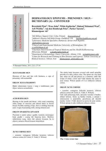
ACNE CORNEE SIGN<br />
= synonim: contagious follicular keratosis, ichtiosis<br />
sebacea cornea (H. Leloir, E. Vidal, E. Wilson).<br />
The whole body becomes covered with small spinelike<br />
growths of a dirty yellow color. The spines are very hard<br />
and, when cut off and placed in a container, rattle like<br />
scraps of metal. Leloir and Vidal shorten the name to<br />
acne cornee. Wilson speaks of it as ichthyosis sebacea<br />
cornea.<br />
OBJAW ACNE CORNEE<br />
= synonim: contagious follicular keratosis, ichtiosis<br />
sebacea cornea (H. Leloir, E. Vidal, E. Wilson).<br />
Całe ciało pokrywają małe, spiczaste wyrośla w brudno-<br />
Ŝółtym kolorze. Kolce są bardzo twarde, a odcięte i<br />
umieszczone w pojemniku, grzechoczą jak skrawki<br />
metalu. Leloir i Vidal skrócili tę nazwę do acne cornée.<br />
Wilson opisuje to jako ichthyosis sebacea cornea.<br />
Fig. 1 Acne cornee sign<br />
© N Dermatol <strong>Online</strong> 1.2011<br />
27
Fig. 2 Henri Leloir<br />
HENRI CAMILLE CHRYSOSTÔME LELOIR<br />
French dermatologist (1855-1896). Studied at Lille and<br />
Paris, obtaining his doctorate in 1881. In 1882 he became<br />
Chef de clinique at the Hôpital St.-Louis, in 1885 was<br />
appointed professor.<br />
HENRI CAMILLE CHRYSOSTÔME LELOIR<br />
Francuski dermatolog (1855-1896). Studiował w Lille i<br />
ParyŜu, uzyskując doktorat w 1881 roku. W 1882 roku<br />
został Kierownikiem Kliniki w Hôpital St Jean-Louis, w<br />
1885 został mianowany profesorem.<br />
JEAN BAPTISTE EMILE VIDAL<br />
(18 June 1825 - 16 June 1893) French dermatologist who<br />
was a native of Paris. He studied medicine in Tours and<br />
Paris, becoming médecin des hôpitaux in 1862. For much<br />
of his career he was associated with the Hôpital Saint-<br />
Louis (1867-90) in Paris. In 1883 he became a member<br />
of the Académie de Médecine. He is remembered for his<br />
investigations of lupus and skin lichenification. His name<br />
is associated with: pityriasis circinata et marginata of<br />
Vidal - a localized variant of pityriasis rosea and Vidal's<br />
disease, synonymous lichen simplex chronicus. He<br />
distinguished himself in his successful struggle to<br />
separate the contagious patients in the hospitals.<br />
28<br />
© N Dermatol <strong>Online</strong> 1.2011<br />
Fig. 3 Jean Vidal<br />
JEAN BAPTISTE EMILE VIDAL<br />
(18 czerwca 1825 - 16 czerwca 1893) francuski<br />
dermatolog, który urodził się w ParyŜu. Studiował<br />
medycynę w Tours i ParyŜu, stając się médecin des<br />
Hôpitaux w 1862 roku. Przez większość kariery<br />
związany był z Hôpital Saint-Louis (1867/90) w ParyŜu.<br />
W 1883 roku został członkiem Académie de Médecine.<br />
Pamiętany jest za swoje osiągnięcia w badaniach nad<br />
toczniem i licheniifikacją. Jego nazwisko jest związane z<br />
pityriasis circinata et marginata Vidal - zlokalizowanego<br />
wariantu rosea pityriasis i chorobą Vidala, synonim -<br />
lichen chronicus simplex. Zdobył sukces walcząc aby<br />
oddzielić pacjentów z chorobami zakaźnymi w<br />
szpitalach.<br />
WILSON WILLIAM JAMES ERASMUS<br />
Brutish dermatologist and anatomist (1809-1884). Was<br />
born in London, Bartholomew's Hospital in London. We<br />
owe to Wilson in great measure the habit of the daily<br />
bath.Died at Westgate-on-Sea in 1884.<br />
WILSON WILLIAM JAMES ERASMUS<br />
Angielski dermatolog, anatomista (1809-1884). Urodził<br />
się w Londynie, studiował w Bartholomew's Hospital w<br />
Londynie. Zawdzięczamy mu w duŜej mierze zwyczaj<br />
codziennej kąpieli. Zmarł w Westgate-on-Sea w 1884<br />
roku.<br />
ADDISON’S SIGN<br />
Characterized by bronze-like pigmentations of the skin.<br />
OBJAW ADDISONA<br />
Charakteryzuje się brązową pigmentacją skóry.<br />
ADDISON’S ORAL SIGN<br />
Fig. 4 Erasmus Wilson<br />
Hyperpigmentation of the buccal mucosa due to lack of<br />
adrenal cortical control over the pituitary secretion of<br />
melanocyte stimulating hormone.
USTNY OBJAW ADDISONA<br />
Przebarwienia błony śluzowej jamy ustnej w związku z<br />
zaburzeniem osi przysadka-kora nadnerczy i<br />
wydzielaniem przez przysadkę hormonu stymulującego<br />
melanocyty.<br />
ADDISON’S PALM SIGN<br />
Characteristic pigmentation in the creases of the hand.<br />
DŁONIOWY OBJAW ADDISONA<br />
Charakterystyczne zabarwienie w zgięciach rąk.<br />
Fig. 5 Addison disease<br />
Fig. 6 Addison’s palm sign<br />
THOMAS ADDISON<br />
English physician (1795-1860). Was the first to describe<br />
a disease of the endocrine glands and the type of anemia<br />
now known as Addison's disease. Thomas Addison was<br />
born in April 1793 at Long Benton near Newcastle-upon-<br />
Tyne. Thomas studied medicine at the University of<br />
Edinburgh and took his doctorate in medicine in 1815. In<br />
1829, in collaboration with John Morgan, he published<br />
the first work on toxicology in English.<br />
THOMAS ADDISON<br />
Angielski lekarz (1795-1860). Był pierwszym który<br />
opisał chorobę gruczołów dokrewnych z<br />
niedokrwistości, znaną obecnie jako choroba Addisona.<br />
Thomas Addison urodził się w kwietniu 1793 w Long<br />
Benton w pobliŜu Newcastle-upon-Tyne. Addison<br />
studiował medycynę na Uniwersytecie w Edynburgu i<br />
doktoryzował się w dziedzinie medycyny w 1815 roku.<br />
W 1829 roku, we współpracy z John Morgan,<br />
opublikował pierwszą pracę na temat toksykologii w<br />
języku angielskim.<br />
ALCOHOLISM BURN SIGN<br />
the characteristic burn from a cigarette occurring<br />
between the fingers after the person has fallen into a<br />
deep alcoholic sleep.<br />
OBJAW OPARZENIA SKÓRY U AKOHOLIKÓW<br />
Charakterystyczne oparzenie po papierosie występujące<br />
między palcami, kiedy osoba zapadła w głęboki sen<br />
alkoholowy.<br />
ALIBERT’S SIGN<br />
= synonym mucosis fungoides<br />
Fig. 8 Mucosis fungoides (Alibert’s sign)<br />
Fig. 7 Thomas Addison<br />
© N Dermatol <strong>Online</strong> 1.2011<br />
29
OBJAW ALIBERTA<br />
= synonim mucosis fungoides<br />
JEAN LOUIS MARC ALIBERT<br />
French dermatologist (1768-1837). In 1798, while still a<br />
student Alibert was co-founder of the Société médicale<br />
d’émulation de Paris of which he was a secretary for<br />
many years.His doctoral thesis of 1799, Dissertation sur<br />
les fièvres pernicieuses, ou ataxiques intermittentes, was<br />
such a success that it subsequently appeared in five<br />
improved and enlarged editions (1801, 1804, 1809,<br />
1820) and in 1808 was translated into English by Charles<br />
Caldwell in Philadelphia.<br />
Alibert did his work on diseases of the skin at time when<br />
Willan's system was still unknown in France.<br />
Contrary to Robert Willan, whose system was based on<br />
pathological anatomy, Alibert preferred to divide by<br />
appearance. He attempted to introduce Jussieu's<br />
classification in the classification of diseases. He divides<br />
them into families, genera, and species, and introduced a<br />
large number of new designations.<br />
He originated several terms, like the term asbestos rash.<br />
Alibert also contributed significantly to the spreading of<br />
vaccination against smallpox.<br />
JEAN LOUIS MARC ALIBERT<br />
Francuski dermatolog (1768-1837). W 1798 roku będąc<br />
jeszcze studentem Alibert był współzałoŜycielem Société<br />
d'médicale emulacji de Paris, którego był sekretarzem od<br />
wielu lat. Jego praca doktorska z 1799 roku: sur les<br />
fièvres pernicieuses, intermittentes ataxiques ou, okazała<br />
się takim sukcesem, Ŝe następnie pojawiła się w pięciu<br />
lepszych i rozszerzonych edycjach (1801, 1804, 1809,<br />
1820), a w 1808 roku została przetłumaczona na język<br />
angielski przez Charles Caldwell w Filadelfii.<br />
Alibert pisał pracę na temat chorób skóry w czasie, gdy<br />
podział chorób skóry Willana był juŜ znany we Francji.<br />
Wbrew Robertowi Willanowi, którego system opierał się<br />
na anatomii patologicznej, Alibert wolał podzielić<br />
choroby skóry ze względu na wygląd. Próbował<br />
wprowadzić własną klasyfikację chorób. Dzielił je na<br />
rodziny, rodzaje i gatunki, wprowadził wiele nowych<br />
nazw.<br />
30<br />
© N Dermatol <strong>Online</strong> 1.2011<br />
Fig. 9 Jean Alibert<br />
Od jego nazwiska pochodzi kilka określeń, takich jak<br />
określenie - wysypka azbestowa.<br />
Alibert równieŜ znacząco przyczynił się do<br />
rozprzestrzenienia się szczepienia przeciwko ospie.<br />
ALLIGATOR SIGN<br />
Martin described a remarkable variety of ichthyosis in<br />
which the skin was covered with strong hairs like the<br />
bristles of a boar. When numerous and thick the scales<br />
sometimes assumed a greenish-black hue. An example of<br />
this condition was the individual who exhibited under the<br />
name of the "alligator-boy." The skin affected in this<br />
case resembled in color and consistency that of a young<br />
alligator. Also called Martin’s sign (Martin and Taylor)<br />
Fig. 10 Alligator sign<br />
OBJAW SKÓRY ALIGATORA<br />
Martin opisał niezwykły wariant rybiej łuski, w którym<br />
skóra była pokryta twardymi jak szczecina dzika<br />
włosami. Liczne i grube zmiany czasem przybierały<br />
zielono-czarną barwę. Przykładem tego stanu była osoba,<br />
którą określono nazwą "aligator-boy". Skóra w tym<br />
przypadku miała podobny kolor i konsystencję, co skóra<br />
młodego aligatora. Zwany równieŜ objawem Martina<br />
(Martin i Taylor).<br />
AMPICILLIN MONO RASH SIGN<br />
The appearance of an irritating rash of macular and<br />
popular form seen in sensivity to ampicillin, often<br />
indicates the patient has infectious mononucleosis,<br />
because the rash appears more frequently in patients with<br />
infectious mononucleosis that been treated with<br />
ampicillin.<br />
OBJAW WYSYPKI PO AMPICYLINIE<br />
Pojawienie się plamistej wysypki przy nadwraŜliwości<br />
na ampicylinę, często występuje u pacjentów leczonych<br />
ampicyliną z powodu mononukleozy zakaźnej.
ANDERS’S SIGN<br />
=synonim adiposis tuberosa simplex (Small sensitive or<br />
painful masses of fat that occur on the abdomen or the<br />
extremities.)<br />
OBJAW ANDERSA<br />
=synonym adiposis tuberosa simplex (niewielkie<br />
wraŜliwe lub bolesne masy tłuszczu, które występują w<br />
jamie brzusznej lub na kończynach).<br />
JAMES MESCHTER ANDERS<br />
American physicaian (1854-1936). The third President of<br />
the American Society of Tropical Medicine. He was born<br />
at Fairview Village, Pennsylvania, on July 22, 1854. He<br />
received his MD degree from the University of<br />
Pennsylvania in 1877, where he later also received his<br />
PhD degree. In 1900 Anders was elected Chair of the<br />
Medical Section of the American Medical Association,<br />
and in 1908 was the President of the Inte<br />
rnational Congress on Tuberculosis. He died on August<br />
29, 1936, at the age of 82.<br />
JAMES MESCHTER ANDERS<br />
Amerykański lekarz (1854-1936). Trzeci prezes<br />
Amerykańskiego Towarzystwa Medicyny Tropikalnej.<br />
Urodził się w Fairview Village, w stanie Pensylwania, 22<br />
lipca 1854. Ukończył studia na Uniwersytecie<br />
Pensylwania w 1877 roku, gdzie później równieŜ uzyskał<br />
stopień doktora. W 1900 roku Anders został wybrany<br />
przewodniczącym Sekcji Medycznej Amerykańskiego<br />
Towarzystwa Lekarskiego, a w 1908 r. został<br />
prezydentem Międzynarodowego Kongresu na temat<br />
gruźlicy. Zmarł 29 sierpnia 1936, w wieku 82 lat.<br />
ANILINE SIGN<br />
Fig. 11 James Anders<br />
Blueness of face lips with drowsiness. A aniline oil<br />
poisoning.<br />
OBJAW ANILINOWY<br />
Błękitne usta, twarz z sennością. Jako objaw zatrucia<br />
aniliną.<br />
ANNAM SIGN<br />
=synonym: oriental boil, cutaneous leishmaniasis, Delhi<br />
boil, old world leishmaniasis, oriental sore, tropical sore,<br />
oriental boil, Bagdad boil, Delhi sore, Bombay boil, deli<br />
fever, Biskra button, furunculus Orientalis, Jericho boil,<br />
Tashkent ulcer, herpes du nil, die Orientbeule, die<br />
Aleppobeule, orientbyld, pendsjabzweer, lupus<br />
endemicus, leishmaniasis furunculosa, Bombaybuil,<br />
Bassorabuil, Cochinzweer, Bagdadbuil, Asjbadkazweer,<br />
leishmaniasis tropica, Aleppobuil, bouton du Nil, φύµα<br />
της Ανατολής, fuma tis anatolis,<br />
(Saunders 1945, E.J. Marzinowsky and Bogbow 1904).<br />
Fig. 12,13 Cutaneous leishmaniasis (L.major)<br />
Fig. 14 Cutaneous leishmaniasis (histology)<br />
© N Dermatol <strong>Online</strong> 1.2011<br />
31
OBJAW ANNAMA<br />
Patrz wyŜej.<br />
ANTRHAX SIGN<br />
A circumscribed boil with relatively little pain and an<br />
absence of pus. A sign of cutanesus anthrax.<br />
Fig. 15,16,17 Antrhax signs<br />
OBJAW “ANTRHAX”<br />
Ograniczony wrzód z towarzyszącym bólem stosunkowo<br />
niewielkich rozmiarów i bez obecności ropnego wysięku.<br />
Objaw skórnej postaci wąglika.<br />
32<br />
© N Dermatol <strong>Online</strong> 1.2011<br />
ARGYRIA SIGN<br />
A blue deposit of silver in the skin, caused by exposure<br />
to silver dusts or salts. Often appears as a gray blue haze<br />
in the white of the eye. Also known as SILVER EYE<br />
SIGN<br />
OBJAW (ARGYRII) SREBRZYCY<br />
Niebieskie złogi srebra w skórze, występujące u osób<br />
naraŜonych na pyły lub sole srebra. Często pojawia się<br />
jako szaro-niebieska mgła w białkówce oka. RównieŜ<br />
zwany jako SILVER EYE SIGN.<br />
ARMADILLO SIGN<br />
Paternal hereditary ichtyosis, morbid development of the<br />
papillae and thickening of the epidermic lamellae. Also<br />
called Pettigrew's sign (Pattigrew 1832 i Ascanius).<br />
Pettigrew mentions a man with warty elongations<br />
encasing his whole body. At the parts where friction<br />
occurred the points of the elongations were worn off.<br />
This man was called "the biped armadillo." The females<br />
had normal skins. All the members of the well-known<br />
family of Lambert had the body covered with spines.<br />
OBJAW PANCERNIKA<br />
Paternal hereditary ichtyosis,. Zwany równieŜ objawem<br />
Pattigrewsa (Pettigrew 1832 i Ascanius). Pettigrew<br />
opisał człowieka z wydłuŜonymi brdawkami,<br />
obejmującymi całe ciało. W części, w których wystąpiły<br />
tarcia punkty wydłuŜenia były zatarte. Osobę taką<br />
określono jako "pancernik dwunoŜny". Kobiety miały<br />
prawidłową skórę. Wszyscy członkowie znanej rodziny<br />
Lambert mieli ciało pokryte kolcami.<br />
ARSENIC SIGN<br />
A classic sign of chronic arsenical poisoning in which<br />
the palms and the soles of the feet have a leathery<br />
texture. Also known as LEATHERY PALM SIGN.<br />
Fig. 18 Arsenic sign
Fig. 19 Arsenic sign<br />
OBJAW ARSENOWY<br />
Klasyczne, przewlekłe zatrucie arsenem, w którym na<br />
dłoniach i podeszwach stóp występują skórzaste<br />
struktury. Znany równieŜ jako OBJAW<br />
SKÓRZASTYCH DŁONI.<br />
ARUM MACULATUM SIGN<br />
Purging, cold clammy skin, with swelling of the tongue.<br />
Indicates poisoning from arum maculatum. Also known<br />
as CUCKOO PINT SIGN.<br />
OBJAW „ARUM MACULATUM”<br />
Biegunka oraz zimna, wilgotna skóra, z obrzękiem<br />
języka. Wskazuje na zatrucie Arum maculatum<br />
(Obrazkiem Plamistym). Objaw równieŜ znany jako<br />
CUCKOO PINT SIGN.<br />
Fig. 20 Arum maculatum<br />
ASSAM’S SIGN<br />
Chloasma. Macula Gravidarum; Macula Uterina; Macula<br />
Hepatica. During pregnancy the skin can become bronze<br />
with black spots like a leopard (ASSAM 1884 and<br />
KAPOSI).<br />
OBJAW ASSAMA<br />
Fig.21 Chloasma<br />
Chloasma. Macula Gravidarum; Macula Uterina; Macula<br />
Hepatica. W czasie ciąŜy skóra moŜe stać się brązowa z<br />
czarnymi plamami, jak lampart (ASSAM w 1884 i<br />
KAPOSI).<br />
MORITZ KAPOSI<br />
Fig. 22 Moritz Kaposi<br />
(born 23 October<br />
1837 in Kaposvár,<br />
Hungary - 6 March<br />
1902 in Vienna,<br />
Austria) was an<br />
important Hungarian<br />
dermatologist,<br />
discoverer of the skin<br />
tumor that received<br />
his name (Kaposi's<br />
sarcoma). Born to a<br />
Jewish family,<br />
originally his surname<br />
was Kohn, but with<br />
his conversion to the<br />
Catholic faith he changed it to Kaposi. In 1855 Kaposi<br />
began to study medicine at the University of Vienna and<br />
attained a doctorate in 1859. In his dissertation, titled<br />
Dermatologie und Syphilis (1866) he made an important<br />
contribution to the field. Kaposi was appointed as<br />
professor at the University of Vienna in 1875, and in<br />
1881 he became member of the board of the Vienna<br />
General Hospital and director of its clinic of skin<br />
diseases. He was authored the book Lehrbuch der<br />
Hautkrankheiten (Textbook of Skin Diseases) in 1878.<br />
Kaposi’s main work, however, was Pathologie und<br />
Therapie der Hautkrankheiten in Vorlesungen für<br />
praktische Ärzte und Studierende (Pathology and<br />
Therapy of the Skin Diseases in Lectures for Practical<br />
Physicians and Students), published in 1880, which<br />
became one of the most significant books in the history<br />
of <strong>dermatology</strong>. He is credited with the description of<br />
xeroderma . pigmentosum ("Ueber Xeroderma .<br />
pigmentosum. . Medizinische . Jahrbücher, Wien, 1882:<br />
619-633"). In all, he published over 150 books and<br />
papers.<br />
MORITZ KAPOSI<br />
(ur. 23 października 1837 w Kaposvár, Węgry – zm. 6<br />
marca 1902 w Wiedniu) był znanym węgierskim<br />
© N Dermatol <strong>Online</strong> 1.2011<br />
33
dermatologiem, odkrywcą nowotworu skóry, który<br />
otrzymał jego imię (mięsak Kaposiego).<br />
Urodził się w rodzinie Ŝydowskiej, pierwotnie jego<br />
nazwisko brzmiało Kohn, ale po nawróceniu na wiarę<br />
katolicką, zmienił na Kaposi. W 1855 rozpoczął studia<br />
medyczne na Uniwersytecie w Wiedniu i otrzymał<br />
stopień doktora w 1859 roku. Swoją pracą doktorską pt<br />
Dermatologia i Kiła (1866) uczynił waŜny wkład w tej<br />
dziedzinie. Kaposi został mianowany profesorem na<br />
Uniwersytecie w Wiedniu w 1875 roku, a w 1881 roku<br />
został członkiem zarządu Vienna General Hospital i<br />
Dyrektorem Kliniki Chorób Skóry. Był autorem ksiąŜki<br />
Lehrbuch der Hautkrankheiten (podręcznik chorób<br />
skóry) z 1878 roku. Główną pracą Kaposiego, był jednak<br />
„Pathologie und der Therapie Hautkrankheiten in<br />
Vorlesungen für praktische Ärzte und Studierende”,<br />
opublikowana w 1880 roku, która stała się jedną z<br />
ksiąŜek, najbardziej znaczących w historii dermatologii.<br />
Przypisuje mu się opis xeroderma pigmentosum ("Über<br />
Xeroderma pigmentosum. Medizinische Jahrbücher,<br />
Wien, 1882: 619-633"). W sumie opublikował ponad<br />
150 ksiąŜek i artykułów.<br />
ACKNOWLEDGEMENT:<br />
Figure 18,19<br />
Dr. D. N. Guha Mazumder<br />
Director DNGM Research Foundation and<br />
Prof. & Head, Dept. of Medicine & Gastroenterology,<br />
(Retd.), Institute of Post Graduate Medical Education &<br />
Research (IPGME&R), Kolkata.<br />
Address : Kolkata -700 053, India.<br />
E-Mail : guhamazumder@yahoo.com<br />
Figure 10<br />
Dr. Vinzenz Oji<br />
Department of <strong>Dermatology</strong>, University of Münster,<br />
Münster, Germany and Interdisciplinary Center of<br />
Clinical Research, University of Münster, Münster,<br />
Germany,<br />
E-Mail : ojiv@uni-muenster.de<br />
34<br />
© N Dermatol <strong>Online</strong> 1.2011<br />
PIŚMIENNICTWO / REFERENCES:<br />
1. Morgan TN, Anderson AG: Chronic Acetanilide<br />
Poisoning. Br Med J. 1940; 2: 187-188.<br />
2. Karunadasa KP, Perera C, Kanagaratnum V, Wijerathne<br />
UP, Samarasingha I, Kannangara CK: Burns due to acid<br />
assaults in Sri Lanka. J Burn Care Res. 2010; 31: 781-785.<br />
3. Kubicz J: A case of keratosis follicularis and dyslipoidic<br />
corneal acne with alopecia in the light of associated<br />
diseases. Przegl Dermatol. 1963; 50: 527-532.<br />
4. Iniakhina AV, Senkova NV: Morrow-Brooke contagious<br />
follicular keratosis. Vestn Dermatol Venerol. 1967; 41: 81-<br />
82.<br />
5. Hadley RM: The life and works of Sir William James<br />
Erasmus Wilson 1809–84.Med Hist. 1959; 3: 215–247.<br />
6. Harbuwono DS, Edi TJ, Suyono S, Subekti I: Addison's<br />
Diseases. Acta Med Indones. 2009; 41: 36.<br />
7. Antal Z, Zhou P: Addison disease. Pediatr Rev. 200; 30:<br />
491-493.<br />
8. Lessin SR: Alibert lymphoma: renaming mycosis<br />
fungoides. Arch Dermatol. 2009; 145: 209-210.<br />
9. Morton LT: Jean Louis Marc Alibert (1768-1837): a<br />
bibliography. J Med Biogr. 1993; 1: 108-112.<br />
10. Shwayder T, Ott F: All about ichthyosis. Pediatr Clin<br />
North Am. 1991; 38: 835-857.<br />
11. Kagan BM: Ampicillin rash. West J Med. 1977; 126:<br />
333–335.<br />
12. Pauszek ME: Making a rash diagnosis: amoxicillin<br />
therapy in infectious mononucleosis. Indiana Med. 1990;<br />
83: 330-331.<br />
13. Anders JM: Adiposis tuberosa simplex. American<br />
<strong>Journal</strong> of the Medical Sciences 1908; 3: 325-332.<br />
14. No authors listed: Am J Med Scienc. 1905; 6: 11.<br />
15. Stowers JH: Case of Delhi Boil or Sore (Syn.: Oriental<br />
Sore; Aleppo Boil). Proc R Soc Med. 1920; 13: 81-83.<br />
16. Pittman HS, Kelley RM: Argyria. Am Pract Dig Treat.<br />
1947; 2: 212-214.<br />
17. Kwon HB, Lee JH, Lee SH, Lee AY, Choi JS, Ahn YS:<br />
A case of argyria following colloidal silver ingestion. Ann<br />
Dermatol. 2009; 21: 308-310.<br />
18. Nagell H:Arsenical melanosis. Med Klin. 1950; 45:<br />
1599.<br />
19. Leclerc H: Wild arum (Arum maculatum L.); its use as<br />
food. Presse Med. 1952; 60: 1514.<br />
20. Bolanca I, Bolanca Z, Kuna K, Vuković A, Tuckar N,<br />
Herman R, et al: Chloasma--the mask of pregnancy. Coll<br />
Antropol. 2008; 32: 1391-41.<br />
21 Oriel JD: Moritz Kaposi (1837-1902). Int J STD AIDS.<br />
1997; 8: 715-717.<br />
22. Leloir HCCh: Recherches cliniques et anatomopathologiques<br />
sur les affections cutanées d'origine nerveuse.<br />
Francis A. Countway Library of Medicine, Ottawa, French,<br />
1882.


