chapter 2-6 andreaeopsida, andreaeobryopsida, polytrichopsida
chapter 2-6 andreaeopsida, andreaeobryopsida, polytrichopsida
chapter 2-6 andreaeopsida, andreaeobryopsida, polytrichopsida

You also want an ePaper? Increase the reach of your titles
YUMPU automatically turns print PDFs into web optimized ePapers that Google loves.
CHAPTER 2-6<br />
ANDREAEOPSIDA,<br />
ANDREAEOBRYOPSIDA,<br />
POLYTRICHOPSIDA<br />
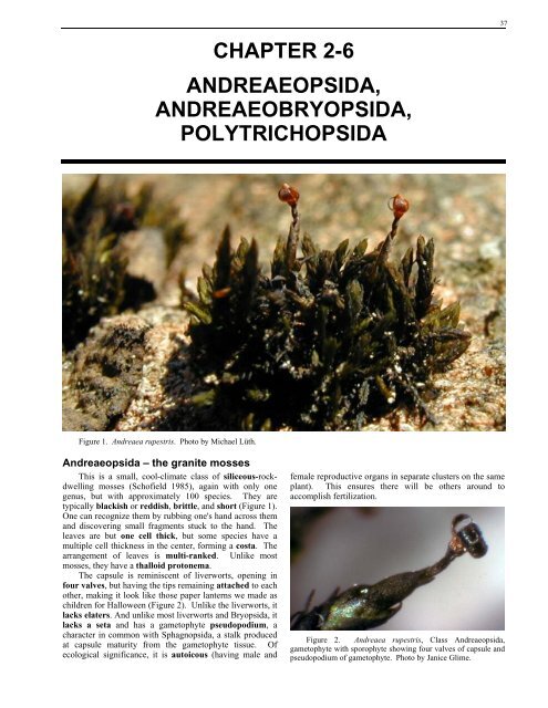
Figure 1. Andreaea rupestris. Photo by Michael Lüth.<br />
Andreaeopsida – the granite mosses<br />
This is a small, cool-climate class of siliceous-rockdwelling<br />
mosses (Schofield 1985), again with only one<br />
genus, but with approximately 100 species. They are<br />
typically blackish or reddish, brittle, and short (Figure 1).<br />
One can recognize them by rubbing one's hand across them<br />
and discovering small fragments stuck to the hand. The<br />
leaves are but one cell thick, but some species have a<br />
multiple cell thickness in the center, forming a costa. The<br />
arrangement of leaves is multi-ranked. Unlike most<br />
mosses, they have a thalloid protonema.<br />
The capsule is reminiscent of liverworts, opening in<br />
four valves, but having the tips remaining attached to each<br />
other, making it look like those paper lanterns we made as<br />
children for Halloween (Figure 2). Unlike the liverworts, it<br />
lacks elaters. And unlike most liverworts and Bryopsida, it<br />
lacks a seta and has a gametophyte pseudopodium, a<br />
character in common with Sphagnopsida, a stalk produced<br />
at capsule maturity from the gametophyte tissue. Of<br />
ecological significance, it is autoicous (having male and<br />
female reproductive organs in separate clusters on the same<br />
plant). This ensures there will be others around to<br />
accomplish fertilization.<br />
Figure 2. Andreaea rupestris, Class Andreaeopsida,<br />
gametophyte with sporophyte showing four valves of capsule and<br />
pseudopodium of gametophyte. Photo by Janice Glime.<br />
37
38 Chapter 2-6: Andreaeopsida, Andreaeobryopsida, Polytrichopsida<br />
Andreaeobryopsida<br />
This class likewise is comprised of a single genus,<br />
Andreaeobryum (Figure 3), which has been considered by<br />
most to belong to the Andreaeopsida, but recently separated<br />
in the treatment by Buck and Goffinet (2000). It differs in<br />
being dioicous (having male and female reproductive<br />
organs on separate plants) and possessing a seta. Its<br />
calyptra is larger, covering the capsule, and the capsule is<br />
valvate, but unlike the Andreaeopsida, the apex erodes, so<br />
the valves are free, not joined at the apex. The<br />
distribution is narrow, restricted to the northwestern part of<br />
Canada and adjacent Alaska, where it grows on calcareous<br />
rocks, contrasting with the acidic granite preference of<br />
Andreaea.<br />
Figure 3. Andreaeobryum macrosporum with valvate<br />
capsules. Photo from Biology 321 Course Website,<br />
http://www.botany.ubc.ca/bryophyte/LAB6b.htm.<br />
Polytrichopsida<br />
With bryophytes, the determination of primitive or<br />
advanced often depends on the generation being examined.<br />
The gametophyte may have changed considerably while<br />
some set of characters of the sporophyte remained constant.<br />
And of course, the reverse can be true. The dioicous<br />
condition (male and female reproductive organs on separate<br />
plants) that characterizes Polytrichopsida is considered to<br />
be primitive (Longton & Schuster 1983), with the<br />
monoicous condition (male and female reproductive<br />
organs on the same plant) that is so frequent in Bryopsida<br />
typically being derived by doubling of the chromosome<br />
number. Likewise, nematodontous peristome teeth<br />
(having evenly thickened walls and whole dead cells<br />
lacking eroded walls, Figure 4) of Polytrichopsida would<br />
seem to be an earlier development than the arthrodontous<br />
condition of Bryopsida.<br />
All members of the class possess an elongate<br />
sporophyte seta, supporting an operculate peristomate<br />
capsule, and a columnar columella, characters that are<br />
more advanced than in Sphagnopsida but typical in<br />
Bryopsida. Spores are produced by meiosis in a single<br />
event in sporogenous tissue that surrounds the columella<br />
(Figure 5).<br />
Figure 4. Nematodontous peristome teeth of Tetraphis<br />
pellucida (Polytrichopsida). Note the separation at the tips.<br />
Photo from Biology 321 Course Website, www.botany.ubc.ca/<br />
bryophyte/LAB6b.htm.<br />
Figure 5. Cross section of immature Polytrichum capsule<br />
showing sporogenous tissue. Photo by Janice Glime.
Figure 6. Longitudinal section of Polytrichum capsule.<br />
Photo by Janice Glime.<br />
The gametophyte is often very specialized, being<br />
characterized by stems with a central strand, reaching its<br />
peak in Polytrichaceae, with the presence of hydroids<br />
(water-conducting cells) and leptoids (sugar-conducting<br />
cells). The leaves of the class are all costate (having a<br />
midrib-like structure).<br />
Figure 7. Cross section of a Polytrichum stem showing<br />
green hydroids in center and larger leptoids surrounding them.<br />
Photo by Izawa Kawai.<br />
Chapter 2-6: Andreaeopsida, Andreaeobryopsida, Polytrichopsida 39<br />
Polytrichaceae<br />
In many ways, this family looks like a tracheophyte<br />
wanna-be. It attains a greater height than the typical moss<br />
and can even stand alone to nearly half a meter in the case<br />
of Dawsonia superba. Polytrichum commune likewise<br />
attains similar heights, but only with the support of other<br />
individuals, forming a hummock.<br />
Figure 8. Dawsonia superba from New South Wales,<br />
Australia. Photo by Janice Glime.<br />
The Polytrichaceae lead the way to complexity with<br />
their unusual leaf structure, possessing vertical lamellae<br />
(vertical tiers of cells like the pages of an open book;<br />
Figure 9) that provide an interior somewhat resembling that<br />
of a maple leaf. In fact, in the genus Polytrichum, some<br />
members have the outer portion of the blade folded over<br />
the lamellae, creating an internal chamber resembling<br />
palisade mesophyll surrounded with epidermis. The<br />
cuticle (in this case, a waxy, water-repellant covering on<br />
the outer surface of the leaf; Proctor 1979) of Polytrichum<br />
is more developed than in most other bryophytes, and<br />
Polytrichum seems to repel water from its leaves rather<br />
than to absorb it (Figure 10), a phenomenon that may<br />
prevent the spaces among the lamellae from flooding that<br />
would block access of CO2 to the chloroplasts within. Its<br />
rhizoids function not only for anchorage, but also seem to<br />
facilitate external water movement.
40 Chapter 2-6: Andreaeopsida, Andreaeobryopsida, Polytrichopsida<br />
Figure 9. Leaf cross sections of Polytrichopsida. Left: Stained section of Polytrichum leaf showing vertical lamellae. Right:<br />
Hand section of Polytrichastrum alpinum leaf showing lamellae with papillose terminal cells. Photos by Janice Glime.<br />
Figure 10. Polytrichum juniperinum with waxy leaves that<br />
roll over the lamellae. Photo by Janice Glime.<br />
In some mosses, like Polytrichum, the antheridia are in<br />
splash cups or platforms (rosette of leaves from which<br />
reproductive units such as sperm, gemmae, or spores can be<br />
splashed by raindrops; Figure 11), and when the sperm<br />
(male reproductive cells; male gametes) are mature, the<br />
antheridium swells and bursts during a rainy period. The<br />
bases of the antheridia, in taxa such as Polytrichum and<br />
Atrichum, collect fluid between the sperm tissue and the<br />
antheridial jacket (Bold et al. 1987). When the cells at the<br />
tip of the sterile jacket open, the antheridial jacket<br />
contracts. At this time, the fluid at the bottom acts as a<br />
hydraulic ram and forces the sperm out of the antheridium.<br />
Once in the open water of the splash cup, the sperm are<br />
splashed from the cup. Hopefully, some of these sperm<br />
will be splashed near the tip of a female plant (Figure 12)<br />
and will begin swimming toward the archegonium.<br />
But it appears that the sperm of Polytrichum<br />
commune, and perhaps others, may have some help in this<br />
process from another source (Harvey-Gibson & Miller<br />
Brown 1927). A variety of invertebrates visit the male<br />
splash cups once they are fertile and get the mucilage with<br />
sperm stuck on their bodies. While visiting the plants, the<br />
insects lap up the mucilage and lick the saline crystals that<br />
form on the margins of the perichaetial leaves. The same<br />
insects, bodies and limbs smeared with mucilage in which<br />
sperms were abundant and motile, likewise appear on<br />
female plants. Now, can someone show whether the red<br />
color of splash cups (Figure 11) in several members of this<br />
family have the ability to attract any dispersal agents?<br />
Figure 11. Male plants of Polytrichum juniperinum with<br />
antheridial splash cups. Photo by Janice Glime.<br />
Figure 12. Female plants of Polytrichum ohioense showing<br />
the tight leaves at the apex where archegonia are housed. To the<br />
right of the female plants, the yellow swollen tips are unopened<br />
antheridial splash cups. Photo by Janice Glime.
Figure 13. Archegonia (purple) nestled among terminal<br />
leaves of Polytrichum. Photo by Janice Glime.<br />
Figure 14. Polytrichum piliferum. Top: Young sporophyte<br />
with calyptra (old archegonium) on top. Bottom: Seta (stalk) of<br />
sporophyte with calyptra removed, showing that the capsule has<br />
not yet begun to develop. Photos by Janice Glime.<br />
Chapter 2-6: Andreaeopsida, Andreaeobryopsida, Polytrichopsida 41<br />
After fertilization, the zygote divides to form an<br />
embryo within the archegonium. Eventually this<br />
sporophyte tissue forms a foot, seta, and capsule. The<br />
capsule develops within the calyptra (Figure 15), which is<br />
the old archegonium. The calyptra is essential for normal<br />
development in most mosses, and a split on one side can<br />
cause asymmetrical development. In the case of<br />
Polytrichum, the calyptra is very hairy, earning the moss<br />
the name of hairy cap moss or goldilocks moss.<br />
Eventually the calyptra is shed, exposing the capsule.<br />
Then the operculum (lid) must come off to permit spore<br />
dispersal. In this family the capsule has 64 short teeth<br />
joined by a membrane (epiphragm) that covers the capsule<br />
like skin on a drum (Figure 16). These small spaces permit<br />
spores to escape the capsule a few at a time, providing<br />
maximum chances for some escaping under the right<br />
conditions for dispersal and establishment.<br />
Figure 15. Capsules of Polytrichum at maturity, still covered<br />
with the calyptra. Photo by Janice Glime.<br />
Figure 16. Epiphragm of Polytrichum. Photo from Biology<br />
321 Course Website,<br />
http://www.botany.ubc.ca/bryophyte/LAB6b.htm.
42 Chapter 2-6: Andreaeopsida, Andreaeobryopsida, Polytrichopsida<br />
Figure 17. Atrichum (Polytrichaceae) life cycle stages. Left: Atrichum undulatum leaves. Middle: Atrichum undulatum<br />
antheridial splash cups. Right: Atrichum angustatum capsules. Photos by Janice Glime.<br />
Tetraphidaceae<br />
Tetraphis (Figure 18), also in the Polytrichopsida,<br />
looks more like a typical moss than do other<br />
Polytrichopsida, with thin, 1-cell-thick leaves and a costa.<br />
Tetraphis is unique among mosses in having gemmae<br />
(Figure 19) arranged in splash cups at the tips of the stems<br />
when sexual reproduction is not in season, arguably a<br />
primitive remnant. These gemmae are asexual bits of plant<br />
material that can grow into a new plant. Its most unusual<br />
character is that its protonemata are not threads, but rather<br />
flaps (Figure 20). Antheridia are borne terminally on the<br />
leafy plants (Figure 21), as are the archegonia. The capsule<br />
(Figure 22) has only four long, unjoined, nematodontous<br />
teeth (Figure 22).<br />
Figure 18. Leafy gametophytes of Tetraphis pellucida with<br />
gemmae cups on top. Photo by Janice Glime.<br />
Figure 19. Gemma cup with gemmae of Tetraphis pellucida.<br />
Photo by Janice Glime.<br />
Figure 20. Protonemal flaps of Tetraphis pellucida. Photos<br />
from Biology 321 Course Website, www.botany.ubc.ca/<br />
bryophyte/LAB6b.htm.
Figure 21. Leaves and antheridia of Tetraphis pellucida.<br />
Photo from Biology 321 Course Website, www.botany.ubc.ca/<br />
bryophyte/LAB6b.htm.<br />
Figure 22. Capsules of Tetraphis pellucida. Upper: with<br />
calyptra. Lower: lacking calyptra and operculum (lid), exposing<br />
the peristome teeth. Photos by Janice Glime.<br />
Chapter 2-6: Andreaeopsida, Andreaeobryopsida, Polytrichopsida 43<br />
Buxbaumiaceae – Bug on a Stick<br />
Buxbaumia (Figure 23) is one of the strangest of all<br />
mosses. It lacks any leafy stem at all. Its archegonia and<br />
antheridia arise directly from the protonema. Hence, its<br />
capsules arise directly from this persistent protonema<br />
(Figure 23). Its capsules, although possessing teeth, more<br />
typically split across their broad, flattened surface, hence<br />
exposing the spores (Figure 24). The capsule interior is<br />
chambered and spongy, somewhat like a spongy mesophyll<br />
of Magnoliophyta. It typically occurs with tiny, black leafy<br />
liverworts such as Cephalozia.<br />
Figure 23. Unopened capsule of Buxbaumia aphylla,<br />
illustrating the flat side with a beaked operculum that has earned it<br />
the common names of bug-on-a-stick and Aladdin's lamp moss.<br />
The sporophyte originates from a protonema with no leafy<br />
gametophyte. Photo by Michael Lüth.<br />
Figure 24. Buxbaumia aphylla (Class Polytrichopsida)<br />
showing flat side of capsule peeled back to expose the spores and<br />
spongy interior. Photo by Janice Glime.
44 Chapter 2-6: Andreaeopsida, Andreaeobryopsida, Polytrichopsida<br />
Diphysciaceae<br />
Perhaps related to Buxbaumia, the moss Diphyscium<br />
(Figure 25) has a capsule of similar shape and lacks a leafy<br />
female stem, but the male plant of this genus has large,<br />
strap-shaped leaves and leads an independent and separate<br />
existence.<br />
Figure 25. Upper: Diphyscium foliosum female plants with<br />
young sessile capsules among male plants. Photo by Janice<br />
Glime. Lower: Mature female Diphyscium foliosum plants with<br />
capsules showing peristome teeth. Photo by Michael Lüth.<br />
Figure 26. Capsules and perichaetial leaves of Diphyscium<br />
foliosum. Photo by Michael Lüth.<br />
Summary<br />
Bryophyta can be considered to have six classes:<br />
Takakiopsida, Sphagnopsida, Andreaeopsida,<br />
Andreaeobryopsida, Polytrichopsida, and Bryopsida,<br />
differing most consistently in capsule structure.<br />
Gametophores of Andreaeopsida,<br />
Andreaeobryopsida, and Polytrichopsida produce<br />
archegonia and/or antheridia at the apex and the<br />
embryo develops within the archegonium.<br />
Sporophytes remain attached to the gametophyte<br />
and produce spores by meiosis. These classes, and all<br />
Bryophyta, produce spores from the sporophyte only<br />
once.<br />
Takakiopsida, Andreaeopsida, and<br />
Andreaeobryopsida have capsules that split into<br />
valves, but lack elaters. Sphagnopsida lacks valves<br />
and has an operculum that is shed at dispersal time, but<br />
lacks peristome teeth. In capsules of Polytrichopsida<br />
and Bryopsida, an operculum usually covers<br />
peristome teeth that often aid dispersal, contrasting<br />
with liverworts wherein the capsule splits into four<br />
valves with elaters that possibly facilitate spore<br />
movement. Polytrichopsida have nematodontous<br />
peristome teeth; Bryopsida have arthrodontous<br />
peristome teeth. All other classes of Bryobiotina lack<br />
peristomes. Andreaeobryopsida is dioicous (two<br />
sexes on separate plants) and possesses a seta (stalk of<br />
capsule), whereas Andreaeopsida is monoicous (both<br />
sexes on same plant) and lacks a seta.<br />
Acknowledgments<br />
I appreciate the comments and suggestions of Karla<br />
Werner, who offered a beginner's perspective. Noris<br />
Salazar Allen offered constructive criticisms on the<br />
taxonomic descriptions and helped with the proof reading.<br />
Heino Lepp alerted me to the invertebrate dispersal of<br />
sperm by his 3 September 2006 contribution to Bryonet.<br />
Literature Cited<br />
Bold, H. C., Alexopoulos, C. J., and Delevoryas, T. 1987.<br />
Morphology of Plants and Fungi. Harper & Row,<br />
Publishers, Inc., New York, NY. 912 pp.<br />
Buck, W. R. and Goffinet, B. 2000. Morphology and<br />
classification of mosses. In: Shaw, J. A. and Goffinet, B.<br />
(eds.). Bryophyte Biology. Cambridge University Press, pp.<br />
71-123.<br />
Harvey-Gibson, R. J. and Miller Brown, D. 1927. Fertilization of<br />
Bryophyta. Preliminary note. Ann. Bot. 49: 190-191.<br />
Longton, R. E. and Schuster, R. M. 1983. Reproductive biology.<br />
In: Schuster, R. M. (ed.). New Manual of Bryology, Vol. 1,<br />
pp. 386-462.<br />
Proctor, M. C. F. 1979. Surface wax on the leaves of some<br />
mosses. J. Bryol. 10: 531-538.<br />
Schofield, W. B. 1985. Introduction to Bryology. Macmillan<br />
Publishing Co., New York, 431 pp.