Blue Rubber Bleb Nevus Syndrome - Journal of Gastrointestinal and ...
Blue Rubber Bleb Nevus Syndrome - Journal of Gastrointestinal and ...
Blue Rubber Bleb Nevus Syndrome - Journal of Gastrointestinal and ...

You also want an ePaper? Increase the reach of your titles
YUMPU automatically turns print PDFs into web optimized ePapers that Google loves.
<strong>Blue</strong> <strong>Rubber</strong> <strong>Bleb</strong> Nevous <strong>Syndrome</strong><br />
CASE REPORTS<br />
<strong>Blue</strong> <strong>Rubber</strong> <strong>Bleb</strong> <strong>Nevus</strong> <strong>Syndrome</strong>: Case Report <strong>and</strong><br />
Literature Review<br />
Daniela Dobru 1 , Nicolae Seuchea 1 , Marian Dorin 2 , Valentin Careianu 3<br />
1) Gastroenterology Department. 2) 2 nd Surgery Clinic, University <strong>of</strong> Medicine <strong>and</strong> Pharmacy. 3) Imaging Department,<br />
Emergency Hospital, Târgu Mureº<br />
Abstract<br />
<strong>Blue</strong> <strong>Rubber</strong> <strong>Bleb</strong> <strong>Nevus</strong> <strong>Syndrome</strong> ( BRBNS) is a rare<br />
disorder characterized by multiple cutaneous venous<br />
malformations in the skin <strong>and</strong> gastrointestinal tract<br />
associated with intestinal hemorrhage <strong>and</strong> iron deficiency<br />
anemia. Other organs may also be involved.<br />
BRBNS has a potential for serious or fatal bleeding. The<br />
causes <strong>of</strong> this syndrome are unknown. Its most common<br />
presentation is in the form <strong>of</strong> sporadic cases, but dominant<br />
autosomal inheritance has been described. Although it was<br />
first recognized by Gascoyen in 1860, only one hundred<br />
years later did Bean further describe these lesions <strong>and</strong> coined<br />
the term BRBNS. A MEDLINE search yielded about 200 case<br />
reports published till 2003.<br />
We present a case <strong>of</strong> this syndrome diagnosed in a 16year<br />
– old patient with both upper <strong>and</strong> lower gastrointestinal<br />
bleeding. He had severe anemia <strong>and</strong> venous swellings on<br />
the trunk. Similar lesions were found in the stomach, bowel<br />
<strong>and</strong> on his foot. In addition, we review the available literature<br />
on the epidemiology, clinical features, associated conditions,<br />
diagnosis <strong>and</strong> treatment.<br />
Key words<br />
<strong>Blue</strong> rubber bleb nevus syndrome - hemorrhage - anemiavascular<br />
malformation - endoscopic therapy<br />
Rezumat<br />
<strong>Blue</strong> <strong>Rubber</strong> <strong>Bleb</strong> <strong>Nevus</strong> <strong>Syndrome</strong> (BRBNS) este o<br />
afecþiune rarã caracterizatã prin existenþa a multiple<br />
malformaþii venoase cutanate ºi a tractului gastrointestinal ,<br />
asociate cu hemoragii digestive ºi cu anemie feriprivã. Pot fi<br />
Romanian <strong>Journal</strong> <strong>of</strong> Gastroenterology<br />
September 2004 Vol.13 No.3, 237-240<br />
Address for correspondence: Dr. Daniela Dobru<br />
Spitalul Clinic Judetean<br />
de Urgenþã<br />
Departamentul Gastroenterologie<br />
Str. Gh.Marinescu nr.1<br />
Târgu Mureº, Romania<br />
afectate ºi alte organe. BRBNS este o afecþiune importantã<br />
datoritã potenþialului de sângerare masivã sau chiar fatalã.<br />
Etiologia afecþiunii este necunoscutã. Boala survine<br />
sporadic, dar a fost descrisã ºi transmiterea autosomal<br />
dominantã. Afecþiunea a fost pentru prima oarã observatã<br />
de cãtre Gascoyen în 1860, o sutã de ani mai târziu, în<br />
1958, fiind descrisã ºi denumitã de Bean. În momentul de<br />
faþã sunt descrise aproximativ 200 de cazuri, publicate pe<br />
MEDLINE.<br />
Prezentãm cazul unui tânãr de 16 ani diagnosticat în<br />
serviciul nostru cu acest sindrom, prezentând hemoragie<br />
digestivã superioarã ºi inferioarã . Pacientul prezenta anemie<br />
severã ºi tumorete vasculare tegumentare la nivelul toracelui<br />
ºi al membrelor inferioare. Aceleaºi leziuni au fost evidenþiate<br />
la nivelul stomacului ºi colonului. Articolul face o trecere în<br />
revistã a principalelor date din literatura de specialitate legate<br />
de epidemiologia , tabloul clinic , leziuni asociate , diagnostic<br />
ºi tratament.<br />
Introduction<br />
<strong>Blue</strong> rubber bleb nevus syndrome (BRBNS) is a syndrome<br />
characterized by gastrointestinal <strong>and</strong> cutaneous<br />
hemangiomas. In 1860, Gascoyen first described an<br />
association between cavernous hemangiomas <strong>of</strong> the skin<br />
<strong>and</strong> similar lesions in the gastrointestinal (GI) tract. In 1958,<br />
William Bennet Bean further described these lesions <strong>and</strong><br />
coined the term <strong>Blue</strong> <strong>Rubber</strong> <strong>Bleb</strong> <strong>Nevus</strong> <strong>Syndrome</strong> (BRBNS)<br />
(1-3).<br />
BRBNS is important because <strong>of</strong> its potential for severe<br />
or fatal bleeding.<br />
Case report<br />
We present a case <strong>of</strong> BRBNS diagnosed in a 16–year<br />
old boy. He was admitted to hospital for hematemesis, melena<br />
<strong>and</strong> severe anemia. The patient had a history <strong>of</strong> more than<br />
20 hospitalizations <strong>and</strong> blood transfusions for bleeding
238<br />
since he was 4 year old <strong>and</strong> a history <strong>of</strong> removal <strong>of</strong> gastric<br />
vascular tumors. There was no family history <strong>of</strong> the disease.<br />
At the physical examination, the patient was extremely<br />
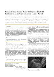
pale, tachycardic <strong>and</strong> presented a nodule <strong>of</strong> 2 cm <strong>of</strong> diameter<br />
at the sole <strong>of</strong> the left foot, in which small caliber vessels<br />
could be visualized, <strong>and</strong> another similar on the first left toe<br />
(Fig.1). The same vascular protrusions were found in the<br />
mouth (Fig.2).<br />
The blood cell count at admission revealed: Hb= 9.7 g/<br />
dl, Ht = 28 %, microcytosis , hypochromia. The leukocyte<br />
<strong>and</strong> platelet counts were normal. Before receiving a<br />
transfusion <strong>of</strong> erythrocytes, blood samples were collected<br />
<strong>and</strong> an iron serum level <strong>of</strong> 8 µmol/l was found.<br />
The upper gastrointestinal endoscopy revealed four<br />
small wine – color lesions in the stomach <strong>and</strong> a larger one in<br />
the antrum (Figs.3,4).<br />
The colonoscopy showed wine-color vascular lesions<br />
<strong>of</strong> different sizes (the largest vascular tumor was about 1.5<br />
cm) in the rectum, sigmoid, descendent <strong>and</strong> cecum, with<br />
raised , irregular surfaces, which were brittle in contact with<br />
the endoscope. In the cecum, near the ileo–cecal valve there<br />
was another larger lesion , measuring approximately 2 cm at<br />
its widest diameter (Figs.5-7).<br />
The diagnostic search for a clinical condition presenting<br />
the association <strong>of</strong> vascular tumors <strong>of</strong> the skin with severe<br />
iron deficiency anemia, the history <strong>of</strong> more than 20 episodes<br />
<strong>of</strong> melena <strong>and</strong> hematemesis, led the authors to consider the<br />
diagnosis <strong>of</strong> BRBNS.<br />
The investigation <strong>of</strong> systemic involvement was<br />
completed with an abdominal ultrasound examination, skull<br />
tomography, retinoscopy. All were normal.<br />
The therapeutic decision was difficult <strong>and</strong> was the result<br />
<strong>of</strong> working together with the surgical team. Our therapeutical<br />
schedule was: supportive therapy with octreotide to decrease<br />
blood flow, proton pump inhibitors, iron replacement <strong>and</strong><br />
blood transfusions, endoscopic sclerotherapy <strong>and</strong> argon<br />
plasma coagulation <strong>of</strong> the vascular tumors.<br />
Unfortunately, we did not have a good compliance from<br />
the patient’s family <strong>and</strong> the patient left the hospital after<br />
two sessions <strong>of</strong> sclerotherapy.<br />
Discussion<br />
BRBNS is a rare disorder with only approximately 200<br />
cases reported in the world literature. Most cases are sporadic<br />
but autosomal dominant inheritance has been reported. The<br />
disorder has not been localized to a specific chromosome or<br />
gene defect.<br />
The mortality <strong>and</strong> morbidity associated with BRBNS<br />
depend on the extent <strong>of</strong> visceral organ involvement. Most<br />
patients have a normal life span. No malignant<br />
transformation <strong>of</strong> cutaneous or visceral lesions has been<br />
reported. Some patients may have severe hemorrhage from<br />
the GI tract, sometimes fatal. Serial transfusions <strong>and</strong> periodic<br />
surveillance can improve the outcome <strong>of</strong> the disease.<br />
Lesions involving bones <strong>and</strong> joints can cause pr<strong>of</strong>ound<br />
Fig.1 Vascular protrusion on the left foot.<br />
Dobru et al<br />
discomfort <strong>and</strong> loss <strong>of</strong> function, requiring amputation in<br />
some cases. Central nervous system involvement is rare,<br />
but might be fatal (4-7).<br />
The syndrome has been reported in all races, although<br />
Caucasians appear to be most frequently affected. The<br />
disease affects males <strong>and</strong> females equally .<br />
In BRBNS, the skin <strong>and</strong> GI system are most frequently<br />
involved, with multiple vascular blebs or nodules. However,<br />
case reports have demonstrated that the central nervous<br />
system, thyroid, parotid, eyes, oral cavity, musculoskeletal<br />
system, lungs, kidney, liver, spleen <strong>and</strong> bladder may also be<br />
affected (2-5).<br />
Cutaneous lesions are <strong>of</strong>ten apparent at birth or manifest<br />
in early childhood, but late onset, beyond midlife has also<br />
been reported. GI involvement usually becomes evident<br />
during early adulthood (2,3,8 ). Histopatologic examination<br />
<strong>of</strong> lesions reveals blood-filled ectatic vessels, lined by a<br />
single layer <strong>of</strong> endothelial cells, with surrounding thin<br />
connective tissue. Dystrophic calcification may be present<br />
(4-6).<br />
Symptoms <strong>and</strong> signs vary depending on the organ<br />
system involved. Patients may report fatigue from occult<br />
blood loss. Hematemesis, melena or rectal bleeding may<br />
prompt emergency presentation, <strong>and</strong> this was also the<br />
presentation <strong>of</strong> the disease in our patient. When bones are<br />
involved, there may be complaints <strong>of</strong> joint pain or impaired<br />
ambulation. Extracutaneous lesions also may result in<br />
epistaxis, hemoptysis, hematuria or menorrhagia.<br />
Physical findings reveal either cutaneous or<br />
extracutaneous manifestations. Skin lesions are usually<br />
highly characteristic, as multiple, protuberant dark blue<br />
vascular tumors, a few millimeters to several centimeters in<br />
diameter. They have the look <strong>and</strong> feel <strong>of</strong> a rubber nipple.<br />
Lesions may be few in number or range into hundreds. The<br />
lesions are principally located on the upper limbs, trunk,<br />
perineum, but they may occur anywhere.<br />
In the GI tract, vascular malformations may occur<br />
anywhere from oral to anal mucosa, but predominate in the<br />
small bowel. In contrast to the skin lesions, the GI lesions<br />
<strong>of</strong>ten bleed. They may spontaneously rupture causing acute
<strong>Blue</strong> <strong>Rubber</strong> <strong>Bleb</strong> Nevous <strong>Syndrome</strong> 239<br />
Fig.2 Vascular protrusion in the mouth. Fig.5 Vascular protrusion in the descendent colon.<br />
Fig.3 Vascular protrusion on the posterior wall <strong>of</strong> the stomach. Fig.6 Endoscopic aspect <strong>of</strong> the sigma with two vascular<br />
protrusions.<br />
Fig.4 Giant vascular protrusion in the descendent colonum.<br />
hemorrhage <strong>and</strong> death. However, most bleedings from the<br />
GI tract are slow, minor, chronic <strong>and</strong> occult, resulting in iron<br />
deficiency anemia from ongoing loss. A case <strong>of</strong> thrombocytopenia<br />
<strong>and</strong> disseminated intravascular coagulation has been<br />
Fig.7 Endoscopic view <strong>of</strong> the ascendent colon with vascular<br />
protrusion.<br />
reported in association with BRBNS. Other complications<br />
include volvulus <strong>and</strong> bowel infarction. These diagnoses<br />
should be considered in patients with BRBNS <strong>and</strong> abdominal<br />
pain (9).
240<br />
Orthopedic manifestations include skeletal bowing,<br />
pathologic fractures, bony overgrowth <strong>and</strong> artropathy.<br />
<strong>Blue</strong> rubber bleb nevi have been reported in the skull,<br />
central nervous system, thyroid, parotid, eyes, oral cavity,<br />
lungs, pleura, pericardium, musculoskeletal system,<br />
peritoneal cavity, mesentery, kidney, liver, spleen, penis,<br />
vulva <strong>and</strong> bladder (9-11).<br />
Fecal occult blood test should be performed in order to<br />
screen for ocult blood loss from gastrointestinal lesions.<br />
Screening for iron deficiency anemia has to be performed.<br />
Presence <strong>of</strong> hematuria may be caused by lesions in the<br />
urinary bladder.<br />
Radiographic images may be useful in suspected<br />
bone or joint involvement <strong>and</strong> radiographic contrast<br />
techniques detect GI lesions but endoscopy is considered<br />
to be superior.<br />
Upper GI endoscopy is more sensitive than upper GI<br />
series <strong>and</strong> colonoscopy more useful than a barium enema.<br />
Endoscopy also provides the opportunity to treat <strong>and</strong><br />
diagnose the lesions. Magnetic resonance imaging<br />
detects extracutaneous lesions in asymptomatic family<br />
members.<br />
The treatment <strong>of</strong> GI venous malformations depends on<br />
their number, location, size <strong>and</strong> symptoms. Sometimes<br />
there are so many blebs, that complete eradication is<br />
impossible.<br />
Bleeding from GI lesions usually is managed<br />
conservatively with iron supplement <strong>and</strong> blood transfusion<br />
when necessary. Endoscopic coagulation or removal is an<br />
effective modality in case <strong>of</strong> repeated bleeding. Experience<br />
with endoscopic sclerotherapy suggests low efficiency <strong>and</strong><br />
complications may occur by the development <strong>of</strong> ulcerations<br />
<strong>and</strong> strictures. Endoscopic laser (Nd: YAG) photocoagulation<br />
<strong>and</strong> plasma argon coagulation have been used<br />
successfully for lesions in the gastrointestinal tract (4,13-<br />
18).<br />
When traditional methods fail <strong>and</strong> the vascular lesions<br />
are confined to a segment <strong>of</strong> the GI tract, resection <strong>of</strong> the<br />
involved segment <strong>of</strong> gut may be indicated (19). This<br />
approach should be used with caution because recurrence<br />
may occur after excision.<br />
Osteoarticular pathology is managed with orthopedic<br />
<strong>and</strong> supportive measures (13,20,21).<br />
BRBNS prognosis depends on the extent <strong>of</strong> visceral<br />
organ involvement. New GI lesions may continue to occur,<br />
so patients need periodic GI <strong>and</strong> hematologic follow–up.<br />
Rarely, acute GI hemorrhage or central nervous system<br />
involvement may result in death (4,14,18,20).<br />
Conclusion<br />
To the best <strong>of</strong> our knowledge, this is the first case report<br />
<strong>of</strong> BRBN syndrome published in the Romanian medical<br />
literature. Our patient has the risk <strong>of</strong> further GI hemorrhages<br />
<strong>and</strong> requires a careful follow-up.<br />
References<br />
Dobru et al<br />
1. Andersen JM: <strong>Blue</strong> rubber bleb nevus syndrom. Curr Treat<br />
Options Gastroenterol 2001; 4: 433-440<br />
2. Fleischer AB Jr, Panzer SM, Wheeler CE. <strong>Blue</strong> rubber bleb<br />
nevus syndrome in a black patient: case report. Cutis 1990; 45:<br />
103-105<br />
3. Walshe MM, Evan CD, Warrin RP. <strong>Blue</strong> rubber bleb naevus. Br<br />
Med J 1966; 2: 931-932<br />
4. Carr MM, Jamieson CG, Lal G. <strong>Blue</strong> rubber bleb nevus syndrome.<br />
Can J Surg 1996; 3: 59-62<br />
5. Bay YT, Oh CH, Kim JH, Lee CH. <strong>Blue</strong> rubber bleb nevus<br />
syndrome: endoscopic removal <strong>of</strong> gastrointestinal<br />
hemangiomas. Gastrointest Endosc 1997; 45:90-92<br />
6. Mako EK. Small–bowel hemangiomatosis in a patient with<br />
Maffucci <strong>and</strong> blue rubber bleb nevus syndromes. Am J Roentgenol<br />
1996; 166: 1499-1500<br />
7. Jennings M, Ward P, Maddocks JL. <strong>Blue</strong> rubber bleb naevus<br />
disease: an uncommon cause <strong>of</strong> gastrointestinal tract bleeding.<br />
Gut 1988; 29: 1408-1412<br />
8. Wong SH, Lau WY. <strong>Blue</strong> rubber bleb nevus syndrome. Dis Colon<br />
Rectum 1982; 25: 371-374<br />
9. Yacoub M, Gnaoul A, Abroug S, et al. The blue rubber bleb nevus<br />
(Bean <strong>Syndrome</strong>) uncommon cause <strong>of</strong> gastrointestinal bleeding.<br />
Ann Pediatr (Paris) 1993;40:157-161<br />
10. Dieckmann K, Maurage C, Faure N et al. Combined laser-steroid<br />
therapy in blue rubber bleb nevus syndrome: case report <strong>and</strong><br />
review <strong>of</strong> the literature. Eur J Pediatr Surg 1994; 4:372 – 374.<br />
11. Oranje AP. <strong>Blue</strong> rubber bleb nevus syndrome. Pediatr Dermatol<br />
1986; 3: 304–310<br />
12. H<strong>of</strong>huis WJ, Oranje AP, Bouquet J, Sinaasappel M. <strong>Blue</strong> rubber<br />
bleb naevus syndrome: report <strong>of</strong> a case with consumption<br />
coagulopathy complicated by manifest thrombosis. Eur J Pediatr<br />
1990; 149 : 526-528<br />
13. Bartoshesky LE, Bull M, Feingold M. Corticosteroid treatment<br />
<strong>of</strong> cutaneous hemangiomas: how effective? A report on 24<br />
children. Clin Pediatr 1978;17: 625-638<br />
14. Boente M del C, Cordisco MR, Frontini M del V, et al. <strong>Blue</strong><br />
rubber bleb nevus (Bean <strong>Syndrome</strong>): evolution <strong>of</strong> four cases<br />
<strong>and</strong> clinical response to pharmacologic agents. Pediatr Dermatol<br />
1999; 16: 222-227<br />
15. Arguedas MR, Wilcox CM. <strong>Blue</strong> rubber bleb nevus syndrome.<br />
Gastrointest Endosc 1999; 50: 544<br />
16. Bak YT, Oh CH , Kim JH, Lee CH. <strong>Blue</strong> rubber bleb nevus<br />
syndrome: endoscopic removal <strong>of</strong> the gastrointestinal<br />
hemangiomas. Gastrointest Endosc 1997; 45: 90-92 .<br />
17. Aihara M, Konuma Y, Okawa K, et al. <strong>Blue</strong> rubber bleb nevus<br />
syndrome with disseminated intravascular coagulation <strong>and</strong><br />
thrombocytopenia: successful treatment with high–dose intravenous<br />
gammaglobulin. Tohoku J Exp Med 1991;163:111-<br />
117.<br />
18. Moodley M, R<strong>and</strong>ial P. <strong>Blue</strong> rubber bleb nevus syndrome: case<br />
report <strong>and</strong> review <strong>of</strong> the literature. Pediatrics 1993; 93: 160-<br />
162.<br />
19. Gallo SH , Mc Clave SA. <strong>Blue</strong> rubber bleb nevus syndrome:<br />
gastrointestinal involvement <strong>and</strong> its endoscopic presentation.<br />
Gastrointest Endosc 1992; 38: 72–76.<br />
20. S<strong>and</strong>hu KS, Cohen H, Radin R, et al. <strong>Blue</strong> rubber bleb nevus<br />
syndrome presenting with recurrences. Dig Dis Sci 1987;32:<br />
214-219<br />
21. Gallmann T, Boltshauser E. <strong>Blue</strong> rubber bleb nevus syndrome<br />
with central nervous system involvement. Klin Padiatr 1987;<br />
199: 382 – 384