Phyla Cnidaria and Ctenophora, Basic Animal Development ...
Phyla Cnidaria and Ctenophora, Basic Animal Development ...
Phyla Cnidaria and Ctenophora, Basic Animal Development ...

You also want an ePaper? Increase the reach of your titles
YUMPU automatically turns print PDFs into web optimized ePapers that Google loves.
<strong>Phyla</strong> <strong>Cnidaria</strong> <strong>and</strong> <strong>Ctenophora</strong>, <strong>Basic</strong> <strong>Animal</strong> <strong>Development</strong> &<br />
Introduction to Nervous Systems 5.5<br />
Lab #5 - Biological Sciences 102 – <strong>Animal</strong> Biology<br />
Introduction to Nervous Systems: Nerve Nets in <strong>Cnidaria</strong>ns<br />
The cnidarian nerve net is an excellent example of a simple, diffuse nervous system. The nerve<br />
cells form a plexus (a network) at the base of the epidermis <strong>and</strong> the gastrodermis. There are<br />
two interconnected nerve nets, one in each tissue layer. Axons (nerve processes) end on other<br />
nerve cells at synapses or neural junctions with sensory cells or effector organs. As seen in<br />
most animals, nerve impulses are transmitted from one cell to another by release of a<br />
neurotransmitter via synaptic vesicles in the presynaptic neuron. One way transmission<br />
between neurons in higher animals occurs is because of the relative activity of the protein<br />
channels for sodium <strong>and</strong> potassium in the axons that generate the electrical signals (action<br />
potentials). In addition, synaptic vesicles are only found in the nerve cell terminal (synaptic<br />
buton). <strong>Cnidaria</strong>n nerve nets are comprised of cells that have synaptic vesicles on both<br />
sides <strong>and</strong> different ion channel properties allowing transmission across the synapse in either<br />
direction. Most nerve cells in the epidermis are multipolar (many processes) although some<br />
have bipolar neurons (two processes). In cnidarians, there can be both one-way <strong>and</strong> twoway<br />
synapses with other neurons. <strong>Cnidaria</strong>n neurons lack the myelin sheaths which we<br />
will see in other animals. This myelin is a fatty acid membrane (in fact, a cell membrane) that<br />
will increase the speed of action potentials in larger, more complex animals.<br />
There is no “central nervous system” in cnidarians where a concentration <strong>and</strong> integration<br />
between many neurons occurs as seen in other animals. In other animals there is a cerebral<br />
ganglion or brain <strong>and</strong> dorsal or ventral nerve cord which controls the activity of other<br />
peripheral nerve cells. In cnidarians, there is some organization to the nerve net. The nerves<br />
are grouped in ring nerves in the medusae of hydrozoans <strong>and</strong> in the marginal sense organs of<br />
scyphozoan medusae. In schyphozoans there is a fast conducting nervous system to<br />
coordinate swimming movements <strong>and</strong> a slower conducting system that coordinates the activity<br />
of the tentacles.<br />
Sensory cells synapse with nerve cells that have junctions with epitheliomuscular cells <strong>and</strong><br />
nematocysts. Collectively this combination of cells forms a neuromuscular system which is<br />
an important milestone in the evolution of nervous systems. In fact, versions of nerve nets<br />
(plexuses) coordinate the rhythmic contractions of the digestive systems of many other more<br />
complex invertebrate <strong>and</strong> vertebrate animals.<br />
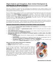
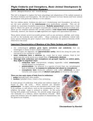
Some types of sensory cells found scattered in the<br />
cnidarian epidermis:<br />
Chemoreceptors = detect molecules/chemicals in the<br />
environment (eg. prey)<br />
mechanoreceptors or tactile receptors = responds to touch or<br />
water movement<br />
Some types of effector cells found scattered in the<br />
cnidarian epidermis:<br />
cnidocytes = have nematocysts<br />
epitheliomuscular cells = for covering <strong>and</strong> muscular<br />
contraction<br />
Near the end of the lab, your instructor will describe the<br />
basic structure of a multipolar nerve cell (neuron).<br />
Be sure to copy <strong>and</strong> label the board diagram in your<br />
notes.