Phyla Cnidaria and Ctenophora, Basic Animal Development ...
Phyla Cnidaria and Ctenophora, Basic Animal Development ...
Phyla Cnidaria and Ctenophora, Basic Animal Development ...

You also want an ePaper? Increase the reach of your titles
YUMPU automatically turns print PDFs into web optimized ePapers that Google loves.
<strong>Phyla</strong> <strong>Cnidaria</strong> <strong>and</strong> <strong>Ctenophora</strong>, <strong>Basic</strong> <strong>Animal</strong> <strong>Development</strong> &<br />
Introduction to Nervous Systems 5.1<br />
Lab #5 - Biological Sciences 102 – <strong>Animal</strong> Biology<br />
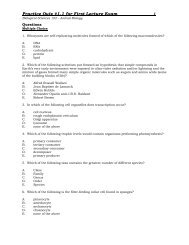
This lab is designed to explore the basic physiology <strong>and</strong> adaptations of the radiate animals in<br />
the <strong>Phyla</strong> <strong>Cnidaria</strong> <strong>and</strong> <strong>Ctenophora</strong>. We will also introduce nervous tissues <strong>and</strong> basic animal<br />
development with particular reference to the radiates.<br />
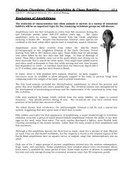
The two radiate phyla, <strong>Cnidaria</strong> (ny-dar'e-a) (= Coelenterata) <strong>and</strong> <strong>Ctenophora</strong> (te-nof'o-ra) are<br />
the most primitive of the eumetazoans (true multicellular animals). They are all radially (or<br />
sometimes biradially) symmetrical. Radially symmetrical animals have a body plan in which<br />
the parts are arranged concentrically around an oral-aboral (central) axis. These are the<br />
simplest animals that actually possess a true tissue level of organization. Generally,<br />
however, the tissues are not organized into organs with specialized functions.<br />
These phyla include several successful groups such as sea anemones, jellyfish, <strong>and</strong> corals, as<br />
well as the hydroids <strong>and</strong> comb jellies. Many cnidarians are brilliantly colored with many<br />
species forming the great tropical coral reefs that harbor the greatest diversity of life observed<br />
in the ocean<br />
Important Characteristics of Members of the <strong>Phyla</strong> <strong>Cnidaria</strong> <strong>and</strong> <strong>Ctenophora</strong><br />
two embryological primary germ layers (ectoderm <strong>and</strong> endoderm) that are homologous to<br />
those of more complex metazoans<br />
internal space for digestion, the gastrovascular cavity, which lies along the polar axis <strong>and</strong><br />
opens to the outside by a mouth<br />
some cnidarians have a skeleton (eg. coral), but in most radiates, fluid in the<br />
gastrovascular cavity serves as a simple form of hydrostatic skeleton<br />
although both cnidarians <strong>and</strong> ctenophores are grouped together as radiate phyla, they<br />
differ in important ways:<br />
o cnidarians have characteristic stinging organelles called nematocysts, usually<br />
absent in ctenophores<br />
o polymorphism- the presence in a species of more than one morphological kind<br />
of individual- is common in cnidarians but absent in ctenophores<br />
o ctenophores have distinctive adhesive cells called colloblasts on their tentacles<br />
<strong>and</strong> unique rows of ciliated comb plates not found in other phyla<br />
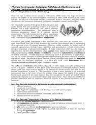
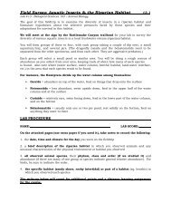
There are two main types of body form in cnidarians:<br />
1. polyp (hydroid) form, often sessile<br />
2. medusa (“jellyfish”) form, which is free-swimming<br />
In some groups of cnidarians, both polyp <strong>and</strong> medusa stages are<br />
found in their life cycle. These animals are therefore<br />
polymorphic. In others, such as sea anemones <strong>and</strong> corals, there<br />
is no medusa; in still others, such as the scyphozoans, or "true"<br />
jellyfish, the polyp stage is reduced or absent. In life cycles<br />
having both polyps <strong>and</strong> medusae, the juvenile polyp stage gives<br />
rise asexually to a medusa, which reproduces sexually. Both<br />
polyp <strong>and</strong> medusa have the diploid number of chromosomes, but<br />
the gametes are haploid.<br />
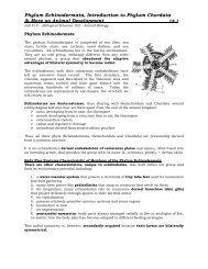
Discomedusae by Haeckel
<strong>Phyla</strong> <strong>Cnidaria</strong> <strong>and</strong> <strong>Ctenophora</strong>, <strong>Basic</strong> <strong>Animal</strong> <strong>Development</strong> &<br />
Introduction to Nervous Systems 5.2<br />
Lab #5 - Biological Sciences 102 – <strong>Animal</strong> Biology<br />
Classification of the Radiate <strong>Animal</strong>s<br />
Phylum <strong>Cnidaria</strong><br />
Class Hydrozoa (hy-dro-zo'a) (Gr. hydra, water serpent, + zoon, animal).<br />
Both polyp <strong>and</strong> medusa stages represented, although one type may be<br />
suppressed; medusa with a velum; found in fresh <strong>and</strong> marine water. The<br />
hydroids. Examples: Hydra, Obelia, Gonionemus, Tubularia, Physalia<br />
Class Scyphozoa (sy-fo-zo'a) (Gr. skyphos, cup, + zoon, animal). Solitary; medusa stage<br />
emphasized; polyp reduced or absent; enlarged mesoglea; medusa without a velum. The true<br />
jellyfish. Examples: Aurelia, Rhizostoma, Cassiopeia<br />
Class Cubozoa (ku'bo-zo'a) (Gr. kybos, a cube, + zoon, animal). Solitary; polyp stage reduced;<br />
bellshaped medusae square in cross section, with a tentacle or group of tentacles at each<br />
corner; margin without velum but with velarium; all marine. Examples: Carybdea, Chironex<br />
Class Anthozoa (an-tho-zo'a) (Gr. anthos, flower, + zoon, animal). All polyps, no medusae;<br />
gastrovascular cavity subdivided by mesenteries (septa).<br />
Subclass Hexacorallia (hek-sa-ko-ral'e-a) (Gr. hex, six, +<br />
korallion, coral) (Zoantharia). Polyp with simple, unbranched<br />
tentacles; septal arrangement hexamerous; skeleton, when<br />
present, external. Sea anemones <strong>and</strong> stony corals. Examples:<br />
Metridium, Tealia, Astrangia<br />
Subclass Ceriantipatharia (se-re-an-tip' a-tha' ri-a)<br />
(combination of Ceriantharia <strong>and</strong> Antipatharia, from type<br />
genera). With simple, unbranched tentacles; mesenteries<br />
unpaired. Tube anemones <strong>and</strong> black or thorny corals.<br />
Examples: Cerianthus, Antipathes<br />
Subclass Octocorallia (ok'to-ko-ral'e-a) (L. octo, + Gr. korallion, coral) (Alcyonaria). Polyp<br />
with eight pinnate tentacles; septal arrangement octamerous. Soft <strong>and</strong> horny corals.<br />
Examples: Gorgonia, Renilla, Alcyonium<br />
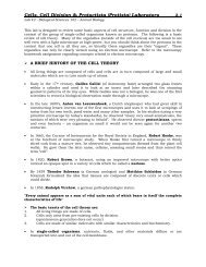
A Ctenophore (Beroe)<br />
Hydra<br />
Cross section through a<br />
hexacorallian<br />
Phylum <strong>Ctenophora</strong><br />
Class Tentaculata (ten-tak'yu-la'ta) (L. tentaculum, feeler, + ata, group suffix).<br />
With tentacles; tentacles may have sheaths into which they retract; some types<br />
flattened in oral-aboral axis for creeping; others compressed in tentacular<br />
plane to a b<strong>and</strong>-like form; in some the comb plates may be confined to the<br />
larva. Examples: Pleurobrachia, Cestum.<br />
Class Nuda (nu-da) (L. nudus, naked). Without tentacles, but flattened in<br />
tentacular plane; wide mouth <strong>and</strong> pharynx; gastrovascular canals much<br />
branched. Example: Beroe
<strong>Phyla</strong> <strong>Cnidaria</strong> <strong>and</strong> <strong>Ctenophora</strong>, <strong>Basic</strong> <strong>Animal</strong> <strong>Development</strong> &<br />
Introduction to Nervous Systems 5.3<br />
Lab #5 - Biological Sciences 102 – <strong>Animal</strong> Biology<br />
<strong>Basic</strong> <strong>Animal</strong> Life Cycles & <strong>Development</strong> (Embryology)<br />
On the chalkboard, your instructor will diagram a basic, generalized animal lifecycle to<br />
introduce the following terms related to animal life cycles <strong>and</strong> development. Your instructor<br />
will then review with you the basic life cycle of the cnidarians, Obelia sp. <strong>and</strong> Aurelia aurita as<br />
specific examples.<br />
EARLY ANIMAL DEVELOPMENT (see diagrams from board)<br />
sperm = the male gamete (haploid = N)<br />
ovum (egg) = the female gamete (haploid = N)<br />
fertilization = the species specific binding of the sperm to the egg <strong>and</strong> fusion of the<br />
sperm cell nucleus with the egg cell nucleus to create a diploid zygote<br />
zygote = fertilized egg (diploid = 2N)<br />
cleavage = the mitotic cell divisions of an animal zygote; the first cell divisions that<br />
occur to create a morula from the zygote<br />
morula = a solid ball of cells that froms from the mitotic divisions of the zygote<br />
blastula = the next embryonic stage that marks the end of early cell division during<br />
animal development; an embryo that look like a hollow ball of cells<br />
gastrula = the next embryonic stage resulting from the division of the early cells into<br />
three major tissue types during early animal development. The gastrula may have two<br />
(endoderm & ectoderm only) or three tissue layers (ectoderm, mesoderm, & endoderm)<br />
gastrulation = the process that leads to the formation of a gastrula<br />
ectoderm = the outer layer of three embryonic cell layers in a gastrula; forms the skin<br />
of the gastrula <strong>and</strong> gives rise to the epidermis <strong>and</strong> nervous system of the adult<br />
mesoderm = the middle layer of the three embryonic cell layers in a gastrula; gives rise<br />
to muscles, bones, the dermis of the skin, <strong>and</strong> many other organs in the adult<br />
endoderm = the innermost of three embryonic cell layers in a gastrula; forms the gut of<br />
the gastrula <strong>and</strong> gives rise to the innermost linings of the digestive tract <strong>and</strong> other<br />
hollow organs (e.g. lungs) in the adult<br />
larvae = an immature animal life stage that is significantly different from the adult <strong>and</strong><br />
usually incapable of sexual reproduction<br />
metamorphosis = a drastic change in form during postembryonic development from<br />
the larval stage to another form, usually the adult<br />
adult = the sexually mature form of an animal<br />
Cleavage, the earliest stage in embryonic development, consists of a succession of regular<br />
mitotic cell divisions that partition the egg into a multitude of small cells clustered together. In<br />
lower animals, cleavage is so rapid that hundreds, sometimes thous<strong>and</strong>s, of cells are produced<br />
in a matter of hours.
<strong>Phyla</strong> <strong>Cnidaria</strong> <strong>and</strong> <strong>Ctenophora</strong>, <strong>Basic</strong> <strong>Animal</strong> <strong>Development</strong> &<br />
Introduction to Nervous Systems 5.4<br />
Lab #5 - Biological Sciences 102 – <strong>Animal</strong> Biology<br />
In animals such as sponges <strong>and</strong> cnidarians, cleavage is irregular <strong>and</strong> seemingly disorganized;<br />
egg cytoplasm is partitioned r<strong>and</strong>omly into daughter cells of highly variable size <strong>and</strong> shape with<br />
no apparent relevance to future cell fates. As the metazoa evolved cleavage began to follow<br />
precise patterns <strong>and</strong> rhythms. In virtually all animal groups above the cnidarians, cleavage is<br />
regular; the egg cytoplasm is segregated into specific cells called blastomeres<br />
(Gr. blastos, bud, + meros, part) occupying discrete positions <strong>and</strong> having specific developmental<br />
fates.<br />
Patterns of regular cleavage depend greatly on amount <strong>and</strong> distribution of yolk in the egg. In<br />
eggs having a large amount of yolk, cleavage may be either complete (= holoblastic), as in<br />
amphibians, or incomplete (= meroblastic), as in birds <strong>and</strong> reptiles. In birds <strong>and</strong> reptiles with<br />
extreme telolecithal (Gr. telos, end, + lekithos, yolk) eggs, cleavage is restricted to a small disc<br />
of cytoplasm on the animal pole; this type of cleavage is called discoidal. The eggs of most<br />
insects follow another pattern of cleavage called superficial. In these the nuclei divide<br />
mitotically into hundreds or thous<strong>and</strong>s of "free" nuclei, which later migrate to the egg surface.<br />
Only then do cleavage furrows form, rapidly partitioning the cytoplasm into a superficial layer<br />
of cells.<br />
In most invertebrates, eggs have little yolk (= isolecithal ["equal-yolk"]), <strong>and</strong> cleavage is<br />
complete (holoblastic) <strong>and</strong> equal. Two major kinds of holoblastic cleavage exist: spiral <strong>and</strong><br />
radial (see text page 156, fig 8-7). The first two cleavages are the same in both kinds of eggs:<br />
the cleavage planes are along the animal-vegetal axis, producing a quartet of cells. At the third<br />
cleavage, however, these two patterns, spiral <strong>and</strong> radial, can be distinguished from each other<br />
by the geometric positioning of the cells.<br />
In radial cleavage, the third cleavage is perpendicular to the first two, yielding two quartets of<br />
cells, with the upper quartet lying directly on top of the lower. In spiral cleavage, the third<br />
cleavage planes are oblique to the polar axis <strong>and</strong> typically produce an upper quartet of smaller<br />
cells that come to lie between the furrows of the lower quartet of larger cells.<br />
There are other important differences between these two cleavage patterns. Spiral cleavage is<br />
typically mosaic, meaning that the embryo is constructed as a mosaic, with each cell fitting<br />
into its predetermined location in the larval body. If cells of the embryo are experimentally<br />
separated at this early stage, each cell will develop into partial or defective larvae because the<br />
developmental fate of each cell has already been determined. Spiral cleavage is found in<br />
several phyla, including annelids, many molluscs, some flatworms, <strong>and</strong> ribbon worms<br />
(nemerteans). All groups showing spiral cleavage belong to the grouping of animal phyla called<br />
the Protostomia, in which the embryonic blastopore forms the mouth.<br />
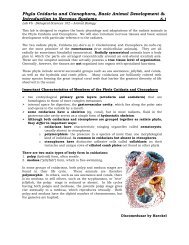
Early Embryonic <strong>Development</strong> - Cell Cleavages (mitosis)<br />
Radial cleavage is characteristically regulative<br />
because cell fate does not become fixed until after<br />
the first few cleavages. Radial cleavage is found in<br />
eggs of echinoderms <strong>and</strong> many chordates,<br />
especially protochordates, amphibians, <strong>and</strong><br />
mammals. (As mentioned earlier, eggs of birds <strong>and</strong><br />
reptiles, as well as many fishes, show discoidal<br />
cleavage.) All of these belong to the<br />
Deuterostomia, a group of phyla in which the<br />
mouth is formed from a secondary embryonic<br />
opening.
<strong>Phyla</strong> <strong>Cnidaria</strong> <strong>and</strong> <strong>Ctenophora</strong>, <strong>Basic</strong> <strong>Animal</strong> <strong>Development</strong> &<br />
Introduction to Nervous Systems 5.5<br />
Lab #5 - Biological Sciences 102 – <strong>Animal</strong> Biology<br />
Introduction to Nervous Systems: Nerve Nets in <strong>Cnidaria</strong>ns<br />
The cnidarian nerve net is an excellent example of a simple, diffuse nervous system. The nerve<br />
cells form a plexus (a network) at the base of the epidermis <strong>and</strong> the gastrodermis. There are<br />
two interconnected nerve nets, one in each tissue layer. Axons (nerve processes) end on other<br />
nerve cells at synapses or neural junctions with sensory cells or effector organs. As seen in<br />
most animals, nerve impulses are transmitted from one cell to another by release of a<br />
neurotransmitter via synaptic vesicles in the presynaptic neuron. One way transmission<br />
between neurons in higher animals occurs is because of the relative activity of the protein<br />
channels for sodium <strong>and</strong> potassium in the axons that generate the electrical signals (action<br />
potentials). In addition, synaptic vesicles are only found in the nerve cell terminal (synaptic<br />
buton). <strong>Cnidaria</strong>n nerve nets are comprised of cells that have synaptic vesicles on both<br />
sides <strong>and</strong> different ion channel properties allowing transmission across the synapse in either<br />
direction. Most nerve cells in the epidermis are multipolar (many processes) although some<br />
have bipolar neurons (two processes). In cnidarians, there can be both one-way <strong>and</strong> twoway<br />
synapses with other neurons. <strong>Cnidaria</strong>n neurons lack the myelin sheaths which we<br />
will see in other animals. This myelin is a fatty acid membrane (in fact, a cell membrane) that<br />
will increase the speed of action potentials in larger, more complex animals.<br />
There is no “central nervous system” in cnidarians where a concentration <strong>and</strong> integration<br />
between many neurons occurs as seen in other animals. In other animals there is a cerebral<br />
ganglion or brain <strong>and</strong> dorsal or ventral nerve cord which controls the activity of other<br />
peripheral nerve cells. In cnidarians, there is some organization to the nerve net. The nerves<br />
are grouped in ring nerves in the medusae of hydrozoans <strong>and</strong> in the marginal sense organs of<br />
scyphozoan medusae. In schyphozoans there is a fast conducting nervous system to<br />
coordinate swimming movements <strong>and</strong> a slower conducting system that coordinates the activity<br />
of the tentacles.<br />
Sensory cells synapse with nerve cells that have junctions with epitheliomuscular cells <strong>and</strong><br />
nematocysts. Collectively this combination of cells forms a neuromuscular system which is<br />
an important milestone in the evolution of nervous systems. In fact, versions of nerve nets<br />
(plexuses) coordinate the rhythmic contractions of the digestive systems of many other more<br />
complex invertebrate <strong>and</strong> vertebrate animals.<br />
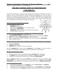
Some types of sensory cells found scattered in the<br />
cnidarian epidermis:<br />
Chemoreceptors = detect molecules/chemicals in the<br />
environment (eg. prey)<br />
mechanoreceptors or tactile receptors = responds to touch or<br />
water movement<br />
Some types of effector cells found scattered in the<br />
cnidarian epidermis:<br />
cnidocytes = have nematocysts<br />
epitheliomuscular cells = for covering <strong>and</strong> muscular<br />
contraction<br />
Near the end of the lab, your instructor will describe the<br />
basic structure of a multipolar nerve cell (neuron).<br />
Be sure to copy <strong>and</strong> label the board diagram in your<br />
notes.
<strong>Phyla</strong> <strong>Cnidaria</strong> <strong>and</strong> <strong>Ctenophora</strong>, <strong>Basic</strong> <strong>Animal</strong> <strong>Development</strong> &<br />
Introduction to Nervous Systems 5.6<br />
Lab #5 - Biological Sciences 102 – <strong>Animal</strong> Biology<br />
LAB PROCEDURE<br />
NAME: LAB SCORE:<br />
Class Hydrozoa<br />
<strong>Basic</strong> Body Plan<br />
While the dominant life stage of both Physalia <strong>and</strong> Velella exist as pneustonic (living at the airwater<br />
interface) polyp stage, Physalia is a floating colony of polyps exhibiting extensive<br />
polymorphism while the polyp stage of Velella is actually solitary.<br />
Observe the specimens <strong>and</strong>/or diagrams of the species listed below.<br />
Record the descriptive information requested at the end of the lab for each species.<br />
Aglaophenia latirostris (common name = Ostrich Plume Hydroid)<br />
Physalia physalia (common name = Portugeuese Man-of-War)<br />
Vellela velella (common name = By-the-Wind Sailor)
<strong>Phyla</strong> <strong>Cnidaria</strong> <strong>and</strong> <strong>Ctenophora</strong>, <strong>Basic</strong> <strong>Animal</strong> <strong>Development</strong> &<br />
Introduction to Nervous Systems 5.7<br />
Lab #5 - Biological Sciences 102 – <strong>Animal</strong> Biology<br />
Using the preserved, prepared slides identify the following structures of Obelia sp.<br />
Below, draw <strong>and</strong> label the Obelia sp. (polyp colony) with hydranths <strong>and</strong> gonangia<br />
with clear labels for each of the structures listed below.<br />
Use the internet <strong>and</strong> textbook to assist you in your drawings.<br />
coenosarc<br />
perisarc<br />
gonangium<br />
o medusae in gonangium<br />
hydranth<br />
o hydrotheca<br />
o mouth<br />
o tentacles
<strong>Phyla</strong> <strong>Cnidaria</strong> <strong>and</strong> <strong>Ctenophora</strong>, <strong>Basic</strong> <strong>Animal</strong> <strong>Development</strong> &<br />
Introduction to Nervous Systems 5.8<br />
Lab #5 - Biological Sciences 102 – <strong>Animal</strong> Biology<br />
Draw <strong>and</strong> CLEARLY label the Obelia sp. medusa from a prepared slide.<br />
Use the internet <strong>and</strong> textbook to assist you in your drawing.<br />
tentacles<br />
radial canals<br />
manubrium<br />
gonads<br />
How many gonads are observed in this species?<br />
Feeding Demonstration in a Hydrozoan<br />
Hydra, a Solitary Hydroid<br />
Phylum <strong>Cnidaria</strong><br />
Class Hydrozoa<br />
Order Hydroida<br />
Suborder Anthomedusae<br />
Genus Hydra, Pelmatohydra, or Chlorohydra<br />
Hydras are found in pools, quiet streams <strong>and</strong> ponds often on the underside of aquatic<br />
vegetation. Your instructor will prepare a slide of hydra for your observation. The hydra may<br />
be contracted at first <strong>and</strong> then extend. Small ciliated protozoans which are symbionts of<br />
hydras are sometimes seen gliding over the body <strong>and</strong> tentacles.<br />
Your instructor will add a drop of Daphnia or similar food to the slide. Some extracellular<br />
digestion occurs within the gastrovascular cavity via enzymes released by gl<strong>and</strong> cells. Food<br />
particles are then engulfed by cells of the gastrodermis <strong>and</strong> digestion is completed<br />
intracellularly. Undigested food is regurgitated as there is no anus.<br />
Observe <strong>and</strong> briefly describe the feeding reaction of the hydra below:
<strong>Phyla</strong> <strong>Cnidaria</strong> <strong>and</strong> <strong>Ctenophora</strong>, <strong>Basic</strong> <strong>Animal</strong> <strong>Development</strong> &<br />
Introduction to Nervous Systems 5.9<br />
Lab #5 - Biological Sciences 102 – <strong>Animal</strong> Biology<br />
Class Scyphozoa<br />
<strong>Basic</strong> Body Plans & Life Cycles<br />
Clearly label the stages of the life cycle of Aurelia aurita shown below.<br />
Use the internet <strong>and</strong> textbook to assist you in with the labels.
<strong>Phyla</strong> <strong>Cnidaria</strong> <strong>and</strong> <strong>Ctenophora</strong>, <strong>Basic</strong> <strong>Animal</strong> <strong>Development</strong> &<br />
Introduction to Nervous Systems 5.10<br />
Lab #5 - Biological Sciences 102 – <strong>Animal</strong> Biology<br />
Look at the prepared slides of the following life stages of a life cycle for Aurelia aurita.<br />
Make drawings <strong>and</strong> clearly label them to complete the diagram below.<br />
Use the internet <strong>and</strong> textbook to assist you in your drawings.<br />
scyphistoma<br />
strobila<br />
ephyra
<strong>Phyla</strong> <strong>Cnidaria</strong> <strong>and</strong> <strong>Ctenophora</strong>, <strong>Basic</strong> <strong>Animal</strong> <strong>Development</strong> &<br />
Introduction to Nervous Systems 5.11<br />
Lab #5 - Biological Sciences 102 – <strong>Animal</strong> Biology<br />
Class Cubozoa<br />
Observe the specimen <strong>and</strong>/or diagram of Carybdea sp.<br />
Record the descriptive information requested at the end of the lab for this species.<br />
Class Anthozoa<br />
Subclass Hexacorallia<br />
Observe the specimens <strong>and</strong>/or diagrams of the following species.<br />
Record the descriptive information requested at the end of the lab for these species.<br />
Balanophyllia elegans<br />
Note that the scleractinian hexacorallians (stony corals) synthesize a consolidated calcium<br />
carbonate skeleton while anemones do not.<br />
Anthopleura sola<br />
What accounts for the green color often present in this species?<br />
Feeding Demonstration in an Anthozoan<br />
As available, your instructor may feed a small piece of mussel to one of the anemones on<br />
display in the lab.<br />
Briefly describe the feeding behavior of the anemone:<br />
Cnidocytes <strong>and</strong> Examination of Nematocyst Discharge<br />
Cnidocytes are cells found in the epidermis between epitheliomuscular cells of cnidarians.<br />
Cnidocytes have nematocysts which are tiny capsules composed of material similar to chitin<br />
<strong>and</strong> contain a coiled tubular filament which is a continuation of the narrowed end of the<br />
capsule. This end of the cnidocyte is covered by the operculum. The base of the filament may<br />
have spines are barbs. The filament <strong>and</strong> barbs may contain a poison. The cnidocytes in<br />
anthozoans do not have the trigger-like cnidocil. The cnidocil is a modified cilium. In<br />
anthozoans a modified ciliary-like mechanoreceptor is involved in triggering nematocyst<br />
discharge.
<strong>Phyla</strong> <strong>Cnidaria</strong> <strong>and</strong> <strong>Ctenophora</strong>, <strong>Basic</strong> <strong>Animal</strong> <strong>Development</strong> &<br />
Introduction to Nervous Systems 5.12<br />
Lab #5 - Biological Sciences 102 – <strong>Animal</strong> Biology<br />
Discharge of nematocysts involves the following events:<br />
1. molecules released from prey may sensitize the cnidocytes, but tactile stimulation causes<br />
the nematocysts to discharge<br />
2. discharge is due to tensional forces created by a very high osmotic pressure within the<br />
nematocyst<br />
3. when triggered the high osmotic pressure causes water to rush in to the capsule.<br />
3. the build of water (increased hydrostatic pressure) forces the filament out <strong>and</strong> it turns<br />
inside out at the same time<br />
4. the barbs are then exposed <strong>and</strong>, if present, poison is injected into the prey<br />
Nematocysts of most cnidarians are not harmful to humans, but the stings of Physalia <strong>and</strong><br />
some true jellies are painful <strong>and</strong> can be dangerous – particularly if the individual is allergic to<br />
the organic poison.<br />
The instructor will remove the tip of a tentacle from Corynactis californica or similar species<br />
<strong>and</strong> prepare a slide by squashing the tip under a cover slip.<br />
Label the undischarged <strong>and</strong> sketch a discharged nematocyst below.<br />
UNDISCHARGED NEMATOCYST DISCHARGED NEMATOCYST<br />
Subclass Ceriantipatharia<br />
Order Ceriantharia (tube anemones)<br />
Observe the specimens <strong>and</strong>/or diagrams of the following species.<br />
Record the descriptive information requested at the end of the lab for these species.<br />
The available ceriantharian such as Pachycerianthus fimbriatus<br />
Subclass Octocorallia<br />
Under a dissecting scope, observe the polyp morphology of Lophogorgia chilensis or Renilla<br />
kollikeri to identify the unique features of an octocorallian polyp.<br />
Record the descriptive information requested at the end of the lab for these species.
<strong>Phyla</strong> <strong>Cnidaria</strong> <strong>and</strong> <strong>Ctenophora</strong>, <strong>Basic</strong> <strong>Animal</strong> <strong>Development</strong> &<br />
Introduction to Nervous Systems 5.13<br />
Lab #5 - Biological Sciences 102 – <strong>Animal</strong> Biology<br />
For the live specimens available for observation in the lab, record the requested<br />
information.<br />
Be sure to include descriptions of any live members of the Phylum <strong>Ctenophora</strong> if<br />
they are available.<br />
Phylum <strong>Cnidaria</strong><br />
Class Hydrozoa<br />
Scientific name: Aglaophenia latirostris<br />
Common name:<br />
General dimensions of specimen:<br />
Color of specimen:<br />
Life stage present (observed):<br />
Solitary or colonial:<br />
Benthic, planktonic or pneustonic:<br />
Unique structures or features:<br />
Draw a simple sketch to remind you of the basic structure of this species <strong>and</strong> any unique<br />
characteristics observed.<br />
Notes & observations to help you remember <strong>and</strong> distinguish this group/species:
<strong>Phyla</strong> <strong>Cnidaria</strong> <strong>and</strong> <strong>Ctenophora</strong>, <strong>Basic</strong> <strong>Animal</strong> <strong>Development</strong> &<br />
Introduction to Nervous Systems 5.14<br />
Lab #5 - Biological Sciences 102 – <strong>Animal</strong> Biology<br />
Phylum <strong>Cnidaria</strong><br />
Class Hydrozoa<br />
Scientific name: Physalia physalia<br />
Common name:<br />
General dimensions of specimen:<br />
Color of specimen:<br />
Life stage present (observed):<br />
Solitary or colonial:<br />
Benthic, planktonic or pneustonic:<br />
Unique structures or features:<br />
Draw a simple sketch to remind you of the basic structure of this species <strong>and</strong> any unique<br />
characteristics observed.<br />
Notes & observations to help you remember <strong>and</strong> distinguish this group/species:
<strong>Phyla</strong> <strong>Cnidaria</strong> <strong>and</strong> <strong>Ctenophora</strong>, <strong>Basic</strong> <strong>Animal</strong> <strong>Development</strong> &<br />
Introduction to Nervous Systems 5.15<br />
Lab #5 - Biological Sciences 102 – <strong>Animal</strong> Biology<br />
Phylum <strong>Cnidaria</strong><br />
Class Hydrozoa<br />
Scientific name: Vellela velella<br />
Common name:<br />
General dimensions of specimen:<br />
Color of specimen:<br />
Life stage present (observed):<br />
Solitary or colonial:<br />
Benthic, planktonic or pneustonic:<br />
Unique structures or features:<br />
Draw a simple sketch to remind you of the basic structure of this species <strong>and</strong> any unique<br />
characteristics observed.<br />
Notes & observations to help you remember <strong>and</strong> distinguish this group/species:
<strong>Phyla</strong> <strong>Cnidaria</strong> <strong>and</strong> <strong>Ctenophora</strong>, <strong>Basic</strong> <strong>Animal</strong> <strong>Development</strong> &<br />
Introduction to Nervous Systems 5.16<br />
Lab #5 - Biological Sciences 102 – <strong>Animal</strong> Biology<br />
Phylum <strong>Cnidaria</strong><br />
Class Scyphozoa<br />
Scientific name: Aurelia aurita<br />
Common name:<br />
General dimensions of specimen:<br />
Color of specimen:<br />
Life stage present (observed):<br />
Solitary or colonial:<br />
Benthic, planktonic or pneustonic:<br />
Unique structures or features:<br />
Draw a simple sketch to remind you of the basic structure of this species <strong>and</strong> any unique<br />
characteristics observed.<br />
Notes & observations to help you remember <strong>and</strong> distinguish this group/species:
<strong>Phyla</strong> <strong>Cnidaria</strong> <strong>and</strong> <strong>Ctenophora</strong>, <strong>Basic</strong> <strong>Animal</strong> <strong>Development</strong> &<br />
Introduction to Nervous Systems 5.17<br />
Lab #5 - Biological Sciences 102 – <strong>Animal</strong> Biology<br />
Phylum <strong>Cnidaria</strong><br />
Class Cubozoa<br />
Scientific name: Carybdea sp.<br />
Common name:<br />
General dimensions of specimen:<br />
Color of specimen:<br />
Life stage present (observed):<br />
Solitary or colonial:<br />
Benthic, planktonic or pneustonic:<br />
Unique structures or features:<br />
Draw a simple sketch to remind you of the basic structure of this species <strong>and</strong> any unique<br />
characteristics observed.<br />
Notes & observations to help you remember <strong>and</strong> distinguish this group/species:
<strong>Phyla</strong> <strong>Cnidaria</strong> <strong>and</strong> <strong>Ctenophora</strong>, <strong>Basic</strong> <strong>Animal</strong> <strong>Development</strong> &<br />
Introduction to Nervous Systems 5.18<br />
Lab #5 - Biological Sciences 102 – <strong>Animal</strong> Biology<br />
Phylum <strong>Cnidaria</strong><br />
Class Anthozoa<br />
Subclass Hexacorallia<br />
Scientific name: Balanophyllia elegans or other hexacorallian<br />
Common name:<br />
General dimensions of specimen:<br />
Color of specimen:<br />
Life stage present (observed):<br />
Solitary or colonial:<br />
Benthic, planktonic or pneustonic:<br />
Unique structures or features:<br />
Draw a simple sketch to remind you of the basic structure of this species <strong>and</strong> any unique<br />
characteristics observed.<br />
Notes & observations to help you remember <strong>and</strong> distinguish this group/species:
<strong>Phyla</strong> <strong>Cnidaria</strong> <strong>and</strong> <strong>Ctenophora</strong>, <strong>Basic</strong> <strong>Animal</strong> <strong>Development</strong> &<br />
Introduction to Nervous Systems 5.19<br />
Lab #5 - Biological Sciences 102 – <strong>Animal</strong> Biology<br />
Phylum <strong>Cnidaria</strong><br />
Class Anthozoa<br />
Subclass Hexacorallia<br />
Scientific name: Anthopleura sola<br />
Common name:<br />
General dimensions of specimen:<br />
Color of specimen:<br />
Life stage present (observed):<br />
Solitary or colonial:<br />
Benthic, planktonic or pneustonic:<br />
Unique structures or features:<br />
Draw a simple sketch to remind you of the basic structure of this species <strong>and</strong> any unique<br />
characteristics observed.<br />
Notes & observations to help you remember <strong>and</strong> distinguish this group/species:
<strong>Phyla</strong> <strong>Cnidaria</strong> <strong>and</strong> <strong>Ctenophora</strong>, <strong>Basic</strong> <strong>Animal</strong> <strong>Development</strong> &<br />
Introduction to Nervous Systems 5.20<br />
Lab #5 - Biological Sciences 102 – <strong>Animal</strong> Biology<br />
Phylum <strong>Cnidaria</strong><br />
Class Anthozoa<br />
Subclass Ceriantipatharia<br />
Order Ceriantharia<br />
Scientific name: Pachycerianthus fimbriatus<br />
Common name:<br />
General dimensions of specimen:<br />
Color of specimen:<br />
Life stage present (observed):<br />
Solitary or colonial:<br />
Benthic, planktonic or pneustonic:<br />
Unique structures or features:<br />
Draw a simple sketch to remind you of the basic structure of this species <strong>and</strong> any unique<br />
characteristics observed.<br />
Notes & observations to help you remember <strong>and</strong> distinguish this group/species:
<strong>Phyla</strong> <strong>Cnidaria</strong> <strong>and</strong> <strong>Ctenophora</strong>, <strong>Basic</strong> <strong>Animal</strong> <strong>Development</strong> &<br />
Introduction to Nervous Systems 5.21<br />
Lab #5 - Biological Sciences 102 – <strong>Animal</strong> Biology<br />
Phylum <strong>Cnidaria</strong><br />
Class Anthozoa<br />
Subclass Octocorallia<br />
Scientific name: Lophogorgia chilensis<br />
Common name:<br />
General dimensions of specimen:<br />
Color of specimen:<br />
Life stage present (observed):<br />
Solitary or colonial:<br />
Benthic, planktonic or pneustonic:<br />
Unique structures or features:<br />
Draw a simple sketch to remind you of the basic structure of this species <strong>and</strong> any unique<br />
characteristics observed.<br />
Notes & observations to help you remember <strong>and</strong> distinguish this group/species:
<strong>Phyla</strong> <strong>Cnidaria</strong> <strong>and</strong> <strong>Ctenophora</strong>, <strong>Basic</strong> <strong>Animal</strong> <strong>Development</strong> &<br />
Introduction to Nervous Systems 5.22<br />
Lab #5 - Biological Sciences 102 – <strong>Animal</strong> Biology<br />
Phylum <strong>Cnidaria</strong><br />
Class Anthozoa<br />
Subclass Octocorallia<br />
Scientific name: Renilla kollikeri or other octocorallian<br />
Common name:<br />
General dimensions of specimen:<br />
Color of specimen:<br />
Life stage present (observed):<br />
Solitary or colonial:<br />
Benthic, planktonic or pneustonic:<br />
Unique structures or features:<br />
Draw a simple sketch to remind you of the basic structure of this species <strong>and</strong> any unique<br />
characteristics observed.<br />
Notes & observations to help you remember <strong>and</strong> distinguish this group/species:
<strong>Phyla</strong> <strong>Cnidaria</strong> <strong>and</strong> <strong>Ctenophora</strong>, <strong>Basic</strong> <strong>Animal</strong> <strong>Development</strong> &<br />
Introduction to Nervous Systems 5.23<br />
Lab #5 - Biological Sciences 102 – <strong>Animal</strong> Biology<br />
Phylum <strong>Ctenophora</strong> (if available)<br />
Class:<br />
Scientific name:<br />
Common name:<br />
General dimensions of specimen:<br />
Color of specimen:<br />
Life stage present (observed):<br />
Solitary or colonial:<br />
Benthic, planktonic or pneustonic:<br />
Unique structures or features:<br />
Draw a simple sketch to remind you of the basic structure of this species <strong>and</strong> any unique<br />
characteristics observed.<br />
Notes & observations to help you remember <strong>and</strong> distinguish this group/species:
<strong>Phyla</strong> <strong>Cnidaria</strong> <strong>and</strong> <strong>Ctenophora</strong>, <strong>Basic</strong> <strong>Animal</strong> <strong>Development</strong> &<br />
Introduction to Nervous Systems 5.24<br />
Lab #5 - Biological Sciences 102 – <strong>Animal</strong> Biology<br />
LABORATORY NOTES:<br />
Actiniae by Haeckel<br />
Ernst Haeckel (1834 – 1919) was an eminent German biologist, naturalist, philosopher,<br />
physician, professor <strong>and</strong> artist who discovered, described <strong>and</strong> named thous<strong>and</strong>s of new<br />
species, mapped a genealogical tree relating all life forms, <strong>and</strong> coined many terms in biology,<br />
including phylum, phylogeny, ecology <strong>and</strong> the kingdom Protista. Haeckel promoted <strong>and</strong><br />
popularized Charles Darwin's work in Germany <strong>and</strong> developed the controversial recapitulation<br />
theory ("ontogeny recapitulates phylogeny") claiming that an individual organism's biological<br />
development, or ontogeny, parallels <strong>and</strong> summarizes its species' entire evolutionary<br />
development, or phylogeny.
<strong>Phyla</strong> <strong>Cnidaria</strong> <strong>and</strong> <strong>Ctenophora</strong>, <strong>Basic</strong> <strong>Animal</strong> <strong>Development</strong> &<br />
Introduction to Nervous Systems 5.25<br />
Lab #5 - Biological Sciences 102 – <strong>Animal</strong> Biology<br />
LABORATORY NOTES:<br />
Peromedusae by Haeckel<br />
Discomedusae by Haeckel<br />
Trachomedusae by Haeckel
<strong>Phyla</strong> <strong>Cnidaria</strong> <strong>and</strong> <strong>Ctenophora</strong>, <strong>Basic</strong> <strong>Animal</strong> <strong>Development</strong> &<br />
Introduction to Nervous Systems 5.26<br />
Lab #5 - Biological Sciences 102 – <strong>Animal</strong> Biology<br />
LABORATORY NOTES: