PDF (Lo-Res) - Smithsonian Institution Libraries
PDF (Lo-Res) - Smithsonian Institution Libraries
PDF (Lo-Res) - Smithsonian Institution Libraries

Create successful ePaper yourself
Turn your PDF publications into a flip-book with our unique Google optimized e-Paper software.
312 SMITHSONIAN CONTRIBUTIONS TO PALEOBIOLOGY<br />
20 years ago by the Polish- and Soviet-Mongolian expeditions,<br />
but the first review of their osteology was published only recently<br />
(Barsbold et al., 1990), and most of the long-known<br />
Mongolian cranial specimens still need to be thoroughly studied.<br />
New finds of oviraptorids, including an embryonic skeleton,<br />
have been reported from Mongolia (Norell et al., 1994;<br />
Dashzeveg et al., 1995).<br />
The following description is based on the skull of Oviraptor<br />
sp. (ZPAL MgD-I/95) in the collection of the Institute of Paleobiology,<br />
Polish Academy of Sciences, Warsaw, Poland. This<br />
specimen has been illustrated and interpreted in the context of<br />
broad, primarily functional, comparisons (Osmolska, 1976) but<br />
was never described in detail. Osmolska's (1976) reconstruction<br />
of the oviraptorid palate proved correct except for details<br />
of the median pterygoid contact. Barsbold (1983a, fig. 12) published<br />
an illustration of the similar palate of Oviraptor philoceratops,<br />
which is not accompanied by a description and contains<br />
a misinterpretation of the vomer as the rostral part of the<br />
pterygoid.<br />
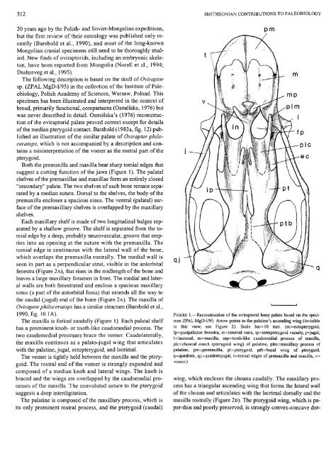
Both the premaxilla and maxilla bear sharp tomial edges that<br />
suggest a cutting function of the jaws (Figure 1). The palatal<br />
shelves of the premaxillae and maxillae form an entirely closed<br />
"secondary" palate. The two shelves of each bone remain separated<br />
by a median suture. Dorsal to the shelves, the body of the<br />
premaxilla encloses a spacious sinus. The ventral (palatal) surface<br />
of the premaxillary shelves is overlapped by the maxillary<br />
shelves.<br />
Each maxillary shelf is made of two longitudinal bulges separated<br />
by a shallow groove. The shelf is separated from the tomial<br />
edge by a deep, probably neurovascular, groove that empties<br />
into an opening at the suture with the premaxilla. The<br />
tomial edge is continuous with the lateral wall of the bone,<br />
which overlaps the premaxilla rostrally. The medial wall is<br />
seen in part as a perpendicular stmt, visible in the antorbital<br />
fenestra (Figure 2 A), that rises in the midlength of the bone and<br />
leaves a large maxillary foramen in front. The medial and lateral<br />
walls are both fenestrated and enclose a spacious maxillary<br />
sinus (a part of the antorbital fossa) that extends all the way to<br />
the caudal (jugal) end of the bone (Figure 2A). The maxilla of<br />
Oviraptorphiloceratops has a similar structure (Barsbold et al.,<br />
1990, fig. 10.1 A).<br />
The maxilla is forked caudally (Figure 1). Each palatal shelf<br />
has a prominent knob- or tooth-like caudomedial process. The<br />
two caudomedial processes brace the vomer. Caudolaterally,<br />
the maxilla continues as a palato-jugal wing that articulates<br />
with the palatine, jugal, ectopterygoid, and lacrimal.<br />
The vomer is tightly held between the maxilla and the pterygoid.<br />
The rostral end of the vomer is strongly expanded and<br />
composed of a median knob and lateral wings. The knob is<br />
braced and the wings are overlapped by the caudomedial processes<br />
of the maxilla. The convoluted suture to the pterygoid<br />
suggests a deep interdigitation.<br />
The palatine is composed of the maxillary process, which is<br />
its only prominent rostral process, and the pterygoid (caudal)<br />
qj<br />
pm<br />
mp<br />
plm<br />
1<br />
v fp<br />
A^—p 1 c<br />
W-ec<br />
FIGURE 1.—Reconstruction of the oviraptorid bony palate based on the specimen<br />
ZPAL MgD-I/95. Arrow points to the palatine's ascending wing (invisible<br />
in this view, see Figure 2). Scale bar=10 mm. (ec=ectopterygoid,<br />
fp=postpalatine fenestra, in=intemal naris, ip=interpterygoid vacuity, j=jugal,<br />
l=lacrimal, m=maxilla, mp=tooth-like caudomedial process of maxilla,<br />
plc=choanal conch (pterygoid wing) of palatine, plm=maxillary process of<br />
palatine, pm=premaxilla, pt=pterygoid, ptb=basal wing of pterygoid,<br />
q=quadrate, qj=quadratojugal, t=tomial edges of premaxilla and maxilla, v=<br />
vomer.)<br />
wing, which encloses the choana caudally. The maxillary process<br />
has a triangular ascending wing that forms the lateral wall<br />
of the choana and articulates with the lacrimal dorsally and the<br />
maxilla rostrally (Figure 2B). The pterygoid wing, which is paper-thin<br />
and poorly preserved, is strongly convex-concave dor-