PDF (Lo-Res) - Smithsonian Institution Libraries
PDF (Lo-Res) - Smithsonian Institution Libraries
PDF (Lo-Res) - Smithsonian Institution Libraries

You also want an ePaper? Increase the reach of your titles
YUMPU automatically turns print PDFs into web optimized ePapers that Google loves.
NUMBER 89 281<br />
slabs, respectively. The radius also is represented by portions<br />
of the shaft on both these slabs (Figure 3). It shows a circular<br />
cross section in the midshaft. The ulnare lies near the proximal<br />
end of the carpometacarpus. This curved element exhibits just<br />
its oval-shaped proximal portion, in which a small fossa is developed<br />
in the place of attachment of the humerocarpal ligament.<br />
The left carpometacarpus displays its ventral side and the<br />
proximal articular surface (Figures 1, 3). It has a well-developed<br />
ulnocarpal trochlea and a deep infratrochlear fossa. The<br />
carpal trochlea appears to be small and narrow, and the extensor<br />
process is poorly developed, in accord with Elzanowski<br />
(1995). The pisiform process is either not preserved or is not<br />
developed. The major metacarpal is represented by bone fragments<br />
on the main slab and by a mold on the associated slab.<br />
The minor metacarpal is represented by the most proximal part<br />
of the base and by a mold of a small portion of the shaft on the<br />
main slab. The metacarpals are completely fused at their proximal<br />
ends. The proximal shafts of both metacarpals are similar<br />
in size. The molds of all three phalanges of the major wing digit<br />
are displayed on the associated slab, with the ventral sides<br />
exposed. The proximal phalanx has a typically avian morphology,<br />
with a flat cranial surface and a thin, flat caudal plate, with<br />
two divided depressions on the ventral side. The intermediate<br />
phalanx is long and thin, and it does not show a vestigial condition.<br />
The ungual phalanx is flat, short, and slightly bowed. The<br />
intermediate and ungual phalanges form a good articular joint<br />
with each other.<br />
SOME FEATURES IN THE MORPHOLOGY OF<br />
Ambiortus AND Otogornis<br />
One of the most characteristic properties of Ambiortus dementjevi<br />
was supposed to be the amphicoelous cervical vertebrae,<br />
as I had proposed in earlier publications on this fossil<br />
(Kurochkin, 1982, 1985a, 1985b). As emphasized above, however,<br />
the eighth and tenth cervical vertebrae are now known to<br />
have heterocoelous centra. New observations also revealed a<br />
contact between the broken edges of the counterslab and associated<br />
slab. Thus, the major metacarpal, radius, and ulna in the<br />
main slab show extension on the associated slab with specimen<br />
PIN 3790-272 that provides certain confirmation of belonging<br />
to the same specimen.<br />
I have not attempted a detailed description of Otogornis<br />
genghisi, but I mention just some corrections to the original paper<br />
and the characters important for comparison with Ambiortus<br />
dementjevi.<br />
Most characters of Otogornis genghisi that are used in this<br />
paper were published in the original description by Hou (1994).<br />
In contrast to Hou's observations, however, I discovered that<br />
the deltopectoral crest is present, the transverse ligamental furrow<br />
is only expressed as a distinctive fossa, the dorsal cotyla of<br />
the proximal end of the ulna is well preserved, and the metacarpals<br />
are fused at their proximal base, although this area is very<br />
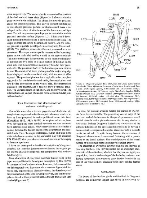
cmshed (Figures 6, 7).<br />
FIM<br />
LRD<br />
LRD<br />
DCT<br />
HAH<br />
RSC<br />
FIGURE 6.—Otogornis genghisi Hou, 1994, from the Chabu Sumu locality,<br />
Ordos Basin, China, Yijinhuoluo Formation. Cranial view; drawing made from<br />
a slide. CGR=capital groove, CRP=cranial pit, DCD=dorsal condyle,<br />
DCR=deltopectoral crest, DCT=dorsal cotyla, FIM=feather imprints, HAH=<br />
humeral articular head, LCR=left coracoid, LFU=ligamental furrow, LHM=<br />
left humerus, LRD=left radius, LUL=left ulna, OL=olecranon, OLF=<br />
olecranal fossa, RHM=right humerus, RRD=right radius, RSC=right scapula,<br />
SGR=scapular groove, TRF=tricipital fossa, VCD=ventral condyle. VTB=<br />
ventral tubercle. (Scale bar=l cm.)<br />
A wide, flat humeral articular facet in the scapula of Otogornis<br />
faces latero-cranially. The projecting ventral edge of the<br />
proximal end of the humerus in Otogornis possesses a small<br />
cranial tubercle with a pit in the center that is very similar to<br />
Ambiortus. Perhaps Otogornis is similar to Ambiortus and the<br />
Lifhornifhiformes in the specialized morphology of having a<br />
dorsoventrally compressed scapular acromion with a tubercle<br />
on its dorsal side. Despite being broken, the acromion of<br />
Otogornis shows some dorsoventral flattening with a prominence<br />
on the dorsal surface. The cranial portion of the lateral<br />
surface of the scapula bears a distinctive scapular groove.<br />
The specimen of Otogornis genghisi exhibits the imprints of<br />
two wing feathers. Hou (1994) pointed out an important characteristic<br />
of these feathers, which is that they are not tightly arranged,<br />
i.e., there is no bonding of the barbs by barbules. Ambiortus<br />
dementjevi also preserves some feather imprints in the<br />
area of the wing feathers, although these show bonded feather<br />
vanes.<br />
COMPARISON<br />
The bones of the shoulder girdle and forelimb in Otogornis<br />
genghisi are somewhat longer than those in Ambiortus de-