Sparassoid ascocarps in Pezizales and Tuberales - ASCOfrance
Sparassoid ascocarps in Pezizales and Tuberales - ASCOfrance
Sparassoid ascocarps in Pezizales and Tuberales - ASCOfrance

You also want an ePaper? Increase the reach of your titles
YUMPU automatically turns print PDFs into web optimized ePapers that Google loves.
<strong>Sparassoid</strong> <strong>ascocarps</strong> <strong>in</strong> <strong>Pezizales</strong> <strong>and</strong> <strong>Tuberales</strong> 391<br />
characterized as an "irregularly convoluted, hollow sac, externally roughened, <strong>in</strong>ternally<br />
smooth, about 2x2 <strong>in</strong>ches, <strong>and</strong> narrow<strong>in</strong>g towards the ground." McLENNAN (1961) transferred<br />
this epigean 'truffle' to Hydnotrya of the same family. On the basis of BURDSALL'S<br />
(1968) exam<strong>in</strong>ation of the type specimen, the fungus was shown to be a member of the<br />
Pezizaceae. It was necessary to rename it, as Peziza jactata BURDSALL & KORF <strong>in</strong> BURD<br />
SALL, s<strong>in</strong>ce the epithet 'convoluta' is preoccupied <strong>in</strong> Peziza by another fungus.<br />
To McALPINE'S orig<strong>in</strong>al description of the spores as "spherical or oval, slightly<br />
verrucose, about 9,um <strong>in</strong> diameter or 10 '" 11 X8.5 ,urn," McLENN AN merely added that<br />
"spore measurements of the co-type material from RODWAY'S collection are approximately<br />
12 X8 ,urn, <strong>and</strong> for the Victorian samples 12 X8.4 ,urn," <strong>and</strong> that the orig<strong>in</strong>al description<br />
"adequately fits" the Australian specimens. BURDSALL (1968) reported that the asci blue<br />
strongly <strong>in</strong> iod<strong>in</strong>e, but did not comment either on spore size or k<strong>in</strong>d of ornamentation<br />
present. My exam<strong>in</strong>ation of the type specimen (RODWAY 21, CUP 48853) shows ascospores<br />
11.3", 14 X8.1", 11.3 ,urn, <strong>and</strong> that when sta<strong>in</strong>ed <strong>in</strong> lactic acid cotton blue they are uniguttulate<br />
<strong>and</strong> very clearly reticulate (Fig. 17). I consider this k<strong>in</strong>d of convoluted, enclosed<br />
ascocarp as represent<strong>in</strong>g the <strong>in</strong>itial stage <strong>in</strong> development of a sparassoid fructification,<br />
perhaps best termed subsparassoid. The species is <strong>in</strong>cluded <strong>in</strong> this discussion primarily<br />
because the ascocarp is sufficiently sparassoid or convoluted to have been confused by at<br />
least four authors with that of the <strong>Tuberales</strong>, <strong>and</strong> scarcely recalls that of a Discomycete.<br />
A few years later, a third sparassoid species of Peziza was described by HENNINGS<br />
(1900) from Java, which he called Aleuria? sparassiformis HENN. Through the k<strong>in</strong>dness<br />
of Dr. Mien A. RIFAI of the Herbarium Bogoriense, I have been able to exam<strong>in</strong>e pickled<br />
material of the only known rema<strong>in</strong><strong>in</strong>g collection of HENNINGS' species. This was collected<br />
by RACIBORSKI, actually a year earlier than the collection by FLEISCHER which had served<br />
as HENNINGS' type material, <strong>and</strong> is the collection reported by PENZIG <strong>and</strong> SACCARDO<br />
(1902). S<strong>in</strong>ce all of HENNINGs'orig<strong>in</strong>al material is apparently lost, I hereby designate the<br />
RACIBORSKI (PENZIG, tubo 3) material, BO 10110, as the NEOTYPE of Peziza sparassiformis<br />
(HENN. <strong>in</strong> WARE.) SACC. & SYD. <strong>in</strong> SACC. The specimen may also be the same as that<br />
referred to <strong>in</strong> the Herbarium Bogoriense under a different accession number, BO 5334,<br />
though that number is no longer associated with the pickled material nor any dried specimen<br />
which can be located. Some slides <strong>and</strong> photographs serv<strong>in</strong>g as an ISONEOTYPE<br />
are on deposit <strong>in</strong> CUP 52756. The RACIBORSKI collection agrees well <strong>in</strong> nearly every<br />
respect with the description by HENNINGS, except that the paraphyses are not po<strong>in</strong>ted as<br />
illustrated by him, but swollen apically as one would expect <strong>in</strong> the genus Peziza. The<br />
fruitbodies are much smaller than <strong>in</strong> P. proteana f. sparassoides, 3.5 cm high <strong>and</strong> 2 cm broad<br />
accord<strong>in</strong>g to HENNINGS <strong>and</strong> much the same <strong>in</strong> the RACIBORSKI collection (Fig. 7: a, b).<br />
The ascospores are narrow for a Peziza, biguttulate, <strong>and</strong> completely smooth (Figs. 6 & 15).<br />
I was unable to demonstrate a blue<strong>in</strong>g reaction of the ascus apices <strong>in</strong> iod<strong>in</strong>e, but consider<strong>in</strong>g<br />
that the fungus has been preserved <strong>in</strong> a fluid for over 70 years, this is no surprize. The<br />
tissues are not <strong>in</strong> a good state of preservation, but appear to be typical of a Peziza.<br />
The fourth known sparassoid species of the Pezizaceae was described by GILKEY (1939)
39Z Richard P. KORF<br />
<strong>in</strong> the <strong>Tuberales</strong>, as Daleomyces shearii GILKEY. In my earlier report (KORF 1956), I<br />
transferred this species to Peziza as P. shearii (GILKEY) KORF. It differs widely from the<br />
three previously discussed species <strong>in</strong> its very nearly globose ascospores (Figs. 5: a, b & 16).<br />
The type material was embedded <strong>in</strong> paraff<strong>in</strong> wax, but on dissolv<strong>in</strong>g the wax from a portion<br />
of the type specimen graciously sent to me by Dr. GILKEY, I was able to demonstrate that<br />
the asci turn blue <strong>in</strong> iod<strong>in</strong>e, are operculate, <strong>and</strong> that some of the ascospores turn brownishyellow<br />
at maturity. I suspect that P. shearii is closely allied to the P. trachycarpa CURREY<br />
complex of species still need<strong>in</strong>g <strong>in</strong>tensive study. Some authors recognize a separate<br />
genus, Plicaria, for species around Peziza trachycarpa, but I f<strong>in</strong>d such a genus quite<br />
unacceptable (KORF 1961, 1972). The fact that sparassoid development is known <strong>in</strong> both<br />
'genera' re<strong>in</strong>forces my belief that these cannot be separated effectively. The excipular<br />
tissues of P. shearii conta<strong>in</strong> the same large, globose to pyriform cells which characterize<br />
the oval-spored species of Peziza.<br />
Recently I was sent material of a sparassoid Peziza by Dr. RW. LICHTWARDT of the<br />
University of Kansas, to whom it had been sent by the collector, Mrs. John BANNINGER.<br />
I assumed at first that the collection represented P. proteana f. sparassoides, but it proved<br />
to have larger, more regularly spaced, <strong>and</strong> more rounded spore mark<strong>in</strong>gs (Figs. 1 & 13: b, c)<br />
than do the type <strong>and</strong> other collections of P. proteana f. sparassoides (Figs. 4 & 14). A critical<br />
reexam<strong>in</strong>ation of all of the material at my disposal revealed to me that the type specimen<br />
of Underwoodia campbellii SACCARDO (CAMPBELL, s.n., SACCARDO Herb., PAD) has spores<br />
match<strong>in</strong>g Mrs. BANNINGER's collection (Fig. 13a). She <strong>and</strong> her gr<strong>and</strong>son then went back<br />
<strong>and</strong> collected fresh material of the fungus. She dried some accord<strong>in</strong>g to my suggestions,<br />
<strong>and</strong> preserved one specimen <strong>in</strong> alcohol (Fig. 3). This collection agreed <strong>in</strong> possess<strong>in</strong>g the<br />
same, larger warts, <strong>and</strong> is much better preserved. The ectal excipulum has the large,<br />
pyriform-globose cells (Fig. 2) which are expected <strong>in</strong> the genus Peziza. Though I earlier<br />
had considered U. campbellii to be a synonym of P. proteana f. sparassoides, the consistently<br />
different spore mark<strong>in</strong>gs have conv<strong>in</strong>ced me that these are different taxa. The spore<br />
dimensions are so nearly the same, <strong>and</strong> the differences <strong>in</strong> spore mark<strong>in</strong>gs so subtle, that I<br />
choose not to recognize SACCARDO'S taxon at specific rank, but shall treat it as a forma of<br />
Peziza proteana. Both of the new collections were made on s<strong>and</strong>y soil around <strong>and</strong> under<br />
a felled, partly burned tree, Lovers' Lane (Cemetery Road), 1/2 mile east of Solomon,<br />
Kansas. Mrs. BANNINGER'S first collection, July 1972, is deposited as CUP 52743. The<br />
second collection, Mrs. John BANNINGER <strong>and</strong> Michael Eugene BANNINGER, 17 August 1972,<br />
is on deposit as CUP 52625. I propose to call this taxon Peziza proteana (BOUD.) SEAVER<br />
forma campbellii (SACCARDO) KORF, comb. novo [basionym: Underwoodia campbellii SACC.,<br />
(as 'campbelli'), Ann. Mycol. 7: 433. 1909].<br />
The follow<strong>in</strong>g key will serve to separate the five sparassoid taxa known thus far <strong>in</strong><br />
Peziza:<br />
1. Ascospores cyl<strong>in</strong>drical (l/w=2.5",2.9), 11.0", 12.5X3.7",4.4,um, biguttulate, smooth<br />
.......................................................... P. sparassiformis<br />
I'. Ascospores broader, with cyanophilic mark<strong>in</strong>gs 2
<strong>Sparassoid</strong> <strong>ascocarps</strong> <strong>in</strong> <strong>Pezizales</strong> <strong>and</strong> <strong>Tuberales</strong> 395<br />
2. Ascospores elongate (l/w=1.6",2.0), 9.5", 11.0X4.7 ",6.2 ,urn, biguttulate 3<br />
2'. Ascospores subglobose to broadly ellipsoid, uniguttulate .4<br />
3. Ascospore mark<strong>in</strong>gs of isolated, large, rounded warts which only rarely fuse .<br />
· P. proteana f. campbellii<br />
3'. Ascospore mark<strong>in</strong>gs of more densely placed, lower warts <strong>and</strong> ridges which frequently<br />
anastomose, with larger crests or warts at the ends, appear<strong>in</strong>g sometimes apiculate<br />
· P. proteana f. sparassoides<br />
4. Ascospores subglobose (l/w=l.l '" 1.2), 13.5", 16.2X 11.0", 14.0 ,urn, mark<strong>in</strong>gs tubercules<br />
............................................................. .P. shearii<br />
4'. Ascospores broadly ellipsoid (l/w= 1.3 '" 1.4), 11.3", 14.0 X8.1", 11.3 ,urn, reticulate<br />
· P. jactata<br />
11. Helvellaceae<br />
Let us now turn our attention to the pileus <strong>in</strong> the gyrose-capped species often placed<br />
<strong>in</strong> Gyromitra <strong>and</strong> <strong>in</strong> Disc<strong>in</strong>a (some authors keep this name for cupulate species, <strong>and</strong> would<br />
use Neogyromitra <strong>and</strong> even additional generic names for those with a gyrose pileus). In<br />
the very early stage of development the pileus is a cup. This grows marg<strong>in</strong>ally, folds<br />
down aga<strong>in</strong>st the stipe, with which it may fuse, <strong>and</strong> then beg<strong>in</strong>s to proliferate, becom<strong>in</strong>g<br />
folded <strong>and</strong> eventually cerebriform. The folded hymenial condition is termed saccate, <strong>and</strong><br />
when these folds are exam<strong>in</strong>ed they do not differ fundamentally from the back-to-back<br />
excipular structure seen when section<strong>in</strong>g a typical sparassoid Discomycete. Even <strong>in</strong> some<br />
species of Helvella with a saddle-shaped pileus, portions of the pileus may fuse with each<br />
other, back-to-back, or back-to-stipe. In all these cases there rema<strong>in</strong>s enough visible,<br />
external morphology, a stipe <strong>and</strong> a pileus, that I prefer not to call such development<br />
sparassoid, <strong>and</strong> reserve the terms saccate <strong>and</strong> gyromitroid for them.<br />
Recently, at the Congress of the Mycological Society of France held <strong>in</strong> Corsica, Mr.<br />
J oseph ASTIER showed some kodachrome transparencies of a peculiar sparassoid fungus he<br />
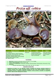
<strong>and</strong> his friend, Mr. Jean-Claude DONADINI, had collected near Marseille (Fig. 12: a, b). The<br />
fungus has broadly ellipsoid ascospores with a very large central guttule, recall<strong>in</strong>g to the<br />
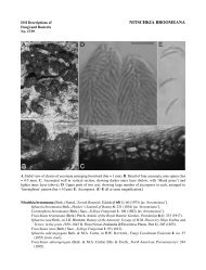
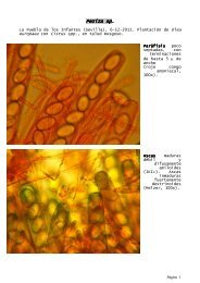
Figs. 4_12. Peziza spp. <strong>and</strong> Helvella astieri. Fig. 4. Peziza proteana f. sparassoides, holotype (PC,<br />
BOUDIER s.n.), four ascospores (a-d) show<strong>in</strong>g cyanophilic warts <strong>and</strong> crests. Fig. 5. Peziza<br />
shearii, isotype (CUP 52758), an ascospore <strong>in</strong> face view (a) show<strong>in</strong>g cyanophilic tuberculations,<br />
<strong>and</strong> (b) the same spore at a lower focal plane show<strong>in</strong>g the height of the warts. Figs. 6 & 7. Peziza<br />
sparassiformis, neotype (BO 101l0). Fig. 6. Completely smooth ascospores show<strong>in</strong>g two oil<br />
guttules. Fig. 7. Portion of an ascocarp preserved <strong>in</strong> liquid (a) <strong>in</strong> face view, <strong>and</strong> (b) viewed<br />
from the cut surface. X 2. Figs. 8-12. Helvella astieri, holotype (PC, ASTIER & DONADINI<br />
s.n.). Fig. 8. Excipular palisade of sterile cells, mounted <strong>in</strong> Bell<strong>in</strong>g's iron acetocarm<strong>in</strong>e sta<strong>in</strong>.<br />
X 700. Fig. 9. Mature ascospores with cyanophilic mark<strong>in</strong>gs, <strong>in</strong> (a) with irregular, large warts,<br />
<strong>in</strong> (b) with a partial reticulum. Fig. 10. Nearly mature ascospores after prolonged treatment<br />
<strong>in</strong> Bell<strong>in</strong>g's iron acetocarm<strong>in</strong>e sta<strong>in</strong>, one spore with three of its four nuclei visible (arrows), the<br />
fourth nucleus not <strong>in</strong> focus. X 1,050. Fig. I!. Mature ascus which has discharged its ascospores,<br />
the thrown back operculum visible (arrow). Fig. 12. <strong>Sparassoid</strong> ascocarp viewed (a) from the<br />
outside <strong>and</strong> (b) viewed from the two cut surfaces. X 2. (Figs. 4, 5, 6, 9 & II <strong>in</strong> cotton-blue lactic<br />
acid. X 1,050).
396 Richard P. KORF<br />
13<br />
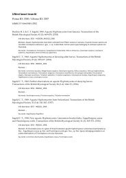
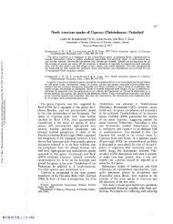
Figs. 13-19. Ascospores drawn with the aid of a Wild draw<strong>in</strong>g tube, all at X 1,700. Figs. 13-18.<br />
Mounts <strong>in</strong> cotton-blue lactic acid. Fig. 13. Peziza proteana f. campbellii, (a) from holotype<br />
(PAD, CAMPBELL, s.n.), (b) from CUP 52625, immature <strong>and</strong> mature, (c) from CUP 52743. Fig.<br />
14. Peziza proteana f. sparassoides, (a) from holotype (PC, BOUDIER, s.n.), (b) from holotype<br />
of Gyromitra phillipsii (BM= K, BECK s.n.), (c) from holotype of Daleomyces gardneri (UC, PARKS<br />
1412). Fig. 15. Peziza sparassiformis, from neotype (Ba 10110). Fig. 16. Peziza shearii,<br />
young spore <strong>in</strong> optical section show<strong>in</strong>g central guttule with two refr<strong>in</strong>gent zones, mature spore<br />
<strong>in</strong> face view, from holotype (OSC, SHEAR s.n., GILKEY 170). Fig. 17. Peziza jactata, one ascospore<br />
<strong>in</strong> optical section show<strong>in</strong>g central guttule, one spore <strong>in</strong> face view, from holotype (CUP<br />
48853, RODWAY 21). Figs. 18 & 19. Helvella astieri from holotype (PC, ASTIER & DONADINI<br />
s.n.). Fig. 18. Young spore with cyanophilic cytoplasm surround<strong>in</strong>g the large central guttule,<br />
mature <strong>and</strong> smooth spore with cytoplasm not cyanophilic, a third spore, also mature, but<br />
with large, irregular warts as seen <strong>in</strong> face view. Fig. 19. Ascospore <strong>in</strong> optical section after<br />
prolonged mount<strong>in</strong>g <strong>in</strong> BELLING'S acetocarm<strong>in</strong>e sta<strong>in</strong>, show<strong>in</strong>g location of the four nuclei <strong>in</strong><br />
relation to the central guttule.
398 Richard P. KORF<br />
Ill. Pyronemataceae, tribe Otideeae<br />
KOBAYASI (1960) erected the genus Ascosparassis for a s<strong>in</strong>gle new species, A. shimizuensis<br />
KOBAYASI, collected <strong>in</strong> Japan. He assigned his sparassoid genus to the Sclerot<strong>in</strong>iaceae,<br />
but KORF (1963) showed that it has operculate asci, <strong>and</strong> reported five additional<br />
collections from Java; he placed the genus close to Otidea FUCKEL. NANNFELDT (1966),<br />
ECKBLAD (1968), <strong>and</strong> KORF (1972, 1973) all accepted the genus as closely related to Otidea,<br />
based upon its great similarities <strong>in</strong> ascospores, paraphyses, <strong>and</strong> sterile tissues.<br />
The sparassoid fruitbodies of A. shimizuensis may be only slightly lobed <strong>and</strong> branched,<br />
as drawn by VAN OVEREEM <strong>and</strong> reproduced <strong>in</strong> KORF'S (1963) paper, or may form a much<br />
more solid, sparassoid mass as illustrated by KOBAYASI (1960). Inasmuch as I can f<strong>in</strong>d<br />
only this one character - that of fruitbody shape - to differentiate KOBAYAsI's species<br />
from the genus Otidea, I feel that to ma<strong>in</strong>ta<strong>in</strong> a separate genus for it is both unreasonable<br />
<strong>and</strong> scientifically unsound. I propose, therefore, its formal transfer, as Otidea shimizuensis<br />
(KOBAYASI) KORF, comb. novo (basionym: Ascosparassis shimizuensis KOBAYASI, Bull.<br />
Natl. Sci. Mus. 5: 45. 1960).<br />
IV. Pyronemataceae, tribe Myco1achneeae<br />
In this fourth group of the <strong>Pezizales</strong>, sparassoid development occurs <strong>in</strong> the genus<br />
Geopora HARKN. The type species of the genus, G. cooperi HARKN., orig<strong>in</strong>ally described<br />
as belong<strong>in</strong>g to the <strong>Tuberales</strong>, was shown by BURDSALL (1965) to have operculate asci, <strong>and</strong><br />
was later transferred to the <strong>Pezizales</strong> (BURDSALL 1968). Here sparassoid development<br />
differs somewhat from that we have seen previously: it occurs <strong>in</strong> a hypogean environment,<br />
<strong>and</strong> the fructification reta<strong>in</strong>s a nearly spherical shape despite the <strong>in</strong>fold<strong>in</strong>g of the ectal<br />
excipulum <strong>and</strong> the production of a highly contorted <strong>and</strong> lacunose <strong>in</strong>terior. BURDSALL<br />
recognized a second wholly hypogean species, G. clausa (TuL. & TuL.) BURDSALL, which<br />
is much smaller <strong>and</strong> shows few or no hymenial convolutions. Similarities <strong>in</strong> spores,<br />
excipular hairs, <strong>and</strong> tissues of the <strong>ascocarps</strong> led BURDSALL to place <strong>in</strong> synonymy with<br />
Geopora the younger generic name Sepultaria (COOKE) BOUD., long used <strong>in</strong> the <strong>Pezizales</strong> for<br />
species with partially buried apothecia. These, too, beg<strong>in</strong> their development as nearly<br />
closed spheres, but rupture on maturity, splitt<strong>in</strong>g <strong>in</strong>to Geastrum-like rays, to expose the<br />
hymenium <strong>and</strong> to allow for forcible ascospore discharge. A saccate or subsparassoid<br />
hymenial condition is known <strong>in</strong> at least one of the species formerly placed <strong>in</strong> Sepultaria, G.<br />
longii (SEAVER) BURDSALL & KORF <strong>in</strong> BURDSALL, as illustrated by SEAVER (1915, 1928, 1942).<br />
Most of the epigeous, or at least not completely hypogeous, species assigned to Sepultaria<br />
have not yet been transferred to Geopora. Neither Burdsall nor I have been prepared<br />
to make these transfers without proceed<strong>in</strong>g first to a monographic <strong>in</strong>vestigation of these<br />
species. We contented ourselves (BURDSALL 1968) with transferr<strong>in</strong>g only the very<br />
h<strong>and</strong>some G. sepulta (FR.) KORF & BURDSALL <strong>in</strong> BURDSALL For other species, concepts<br />
vary so widely among authors, <strong>and</strong> specific variation is so poorly understood <strong>in</strong> this genus,<br />
that transfer of other specific epithets now would be premature.
<strong>Sparassoid</strong> <strong>ascocarps</strong> <strong>in</strong> <strong>Pezizales</strong> <strong>and</strong> <strong>Tuberales</strong> 399<br />
V. Some conclusions <strong>and</strong> hypotheses<br />
on sparassoidism <strong>and</strong> the orig<strong>in</strong> of the Tubera1es<br />
The orig<strong>in</strong> of the <strong>Tuberales</strong> from Operculate <strong>Pezizales</strong> has been argued by many<br />
authors, <strong>and</strong> it is not my <strong>in</strong>tent to restate their arguments here except <strong>in</strong>sofar as it is<br />
necessary <strong>in</strong> a consideration of sparassoid development <strong>in</strong> both orders. The purported<br />
orig<strong>in</strong> of the <strong>Tuberales</strong> from Basidiomycetes seems to me too remote a possibility to consider.<br />
But that the <strong>Tuberales</strong> sensu lata may be polyphyletic I am quite prepared to accept.<br />
The unliklihood of the Elaphomycetaceae hav<strong>in</strong>g a close aff<strong>in</strong>ity with the other families<br />
of the <strong>Tuberales</strong> is clear (KORF 1973), <strong>and</strong> they may be much closer to the Eurotiales <strong>in</strong><br />
their ancestry.<br />
What actually dist<strong>in</strong>guishes the two orders, <strong>Tuberales</strong> <strong>and</strong> <strong>Pezizales</strong>? It is, <strong>in</strong> the<br />
f<strong>in</strong>al analysis, only the possession of a functional operculum <strong>in</strong> the latter, <strong>and</strong> its absence<br />
<strong>in</strong> the former. We may well question whether this is a taxonomic feature worthy of<br />
recogniz<strong>in</strong>g at the ord<strong>in</strong>al rank. That it is a fundamental biological feature cannot be<br />
denied. A fungus which depends upon air dispersal of its ascospores to reproduce effectively<br />
has, as <strong>in</strong> the <strong>Pezizales</strong>, strong evolutionary pressure to reta<strong>in</strong> those mechanisms<br />
which allow such spore dispersal. The cyl<strong>in</strong>drical ascus shape, the palisade arrangement<br />
of such asci, delimitation of an apical operculum which must split open along a predeterm<strong>in</strong>ed<br />
l<strong>in</strong>e, result<strong>in</strong>g <strong>in</strong> an ascostome of the correct size to allow passage of the squirted<br />
ascospores, capable of contract<strong>in</strong>g the right amount as each spore passes through to give<br />
an additional push to the spore projectile, ma<strong>in</strong>tenance of turgor <strong>in</strong> the epiplasm - all<br />
these are complex phenomena doubtless under the control of a multitude of genes. Muta<br />
tion of anyone of these genes would probably result <strong>in</strong> failure of the whole discharge<br />
mechanism to operate. That such deleterious gene mutations must regularly occur is<br />
clear; likewise, such non-discharg<strong>in</strong>g mutant stra<strong>in</strong>s will never get their spores <strong>in</strong>to the<br />
air, <strong>and</strong> the mutant will be self-elim<strong>in</strong>at<strong>in</strong>g. Selection pressure ma<strong>in</strong>ta<strong>in</strong>s the mechanism<br />
<strong>and</strong> elim<strong>in</strong>ates all such mutant forms for so long as that is the only way a spore can be<br />
dissem<strong>in</strong>ated.<br />
In the <strong>Tuberales</strong>, however, where asci do not discharge their spores, air dispersal of<br />
ascospores is not the means of fungal reproduction. The evidence is strong that the major<br />
role <strong>in</strong> dispersal is by animals, <strong>in</strong>sects, perhaps annelids, <strong>and</strong> above all, for the larger<br />
species, by rodents <strong>and</strong> other burrow<strong>in</strong>g animals. Moreover, the ascospores of many<br />
<strong>Tuberales</strong> are thick-walled <strong>and</strong> capable of withst<strong>and</strong><strong>in</strong>g the action of digestive juices. (I<br />
have exam<strong>in</strong>ed the stomach contents of voles <strong>in</strong> New York State which consisted almost<br />
wholly of the spores <strong>and</strong> tissues of <strong>Tuberales</strong> <strong>and</strong> of Endogonaceae). We f<strong>in</strong>d, further,<br />
that the great majority of <strong>Tuberales</strong> (<strong>and</strong> of hypogeous Basidiomycetes for that matter)<br />
emit strong odors, allow<strong>in</strong>g for the detection of subterranean fruitbodies by such animals<br />
through the fragrances they emit.<br />
When we exam<strong>in</strong>e the ascospores themselves, or sterile tissues of members of the<br />
<strong>Tuberales</strong>, we f<strong>in</strong>d many parallels with those <strong>in</strong> <strong>Pezizales</strong> at the microscopic level. To<br />
the naked eye, the "simplest" <strong>Tuberales</strong> are hollow spheres l<strong>in</strong>ed with a hymenium of asci
400 Richard P. KORF<br />
<strong>and</strong> paraphyses, closely resembl<strong>in</strong>g a still closed apothecium of, e.g., an epigean species<br />
of Geopora (Sepultaria). More complex fruitbodies are found, often <strong>in</strong> the same species<br />
or genera, where an <strong>in</strong>fold<strong>in</strong>g of the hymenium occurs <strong>in</strong> the tuberalean fruitbody, eventually<br />
result<strong>in</strong>g <strong>in</strong> a sparassoid hymenial tissue fill<strong>in</strong>g the sphere. We should also exam<strong>in</strong>e<br />
what is happen<strong>in</strong>g to two microscopic structures, the paraphyses <strong>and</strong> the asci. The<br />
former are, <strong>in</strong> the simplest forms, about as long as the asci, just as <strong>in</strong> <strong>Pezizales</strong>. Sometimes<br />
they may be much shorter than the asci (e.g., Labyr<strong>in</strong>thomyces steenisii BOEDIJN), or, far<br />
more frequently, they may be longer than the asci, often branch<strong>in</strong>g apically <strong>and</strong> form<strong>in</strong>g<br />
an epithecial tissue which may fill <strong>in</strong> the hollow cavities between adjacent hymenial folds<br />
of the cerebriform <strong>in</strong>terior of the ascocarp. Such an epithecium does not occur <strong>in</strong> the<br />
<strong>Pezizales</strong>, where it would only impede ascus discharge. The asci, on the other h<strong>and</strong>, also<br />
show marked variation from that of the <strong>Pezizales</strong>. Not only do we no longer f<strong>in</strong>d much,<br />
if any, sign of a vestigial mechanism at the apex which would delimit an operculum, but<br />
the asci <strong>in</strong> the more advanced forms lose the cyl<strong>in</strong>drical shape which is characteristic of<br />
the pezizalean ascus. Such asci assume a pyriform or even globose shape, becom<strong>in</strong>g soon<br />
displaced so that their orig<strong>in</strong>al arrangement <strong>in</strong> a dist<strong>in</strong>ct hymenium <strong>in</strong> youth may no<br />
longer be evident at maturity. The genus Tuber represents such an extreme form. Here,<br />
as <strong>in</strong> other advanced <strong>Tuberales</strong>, even spore number frequently has become reduced <strong>and</strong><br />
irregular; spore size also becomes variable among spores with<strong>in</strong> the same ascus. In the<br />
<strong>Pezizales</strong>, on the other h<strong>and</strong>, spore number is usually under rigid genetic control, s<strong>in</strong>ce<br />
spore size - which is directly correlated with spore number - must be ma<strong>in</strong>ta<strong>in</strong>ed with<strong>in</strong><br />
certa<strong>in</strong> limits if the operculum on the ascus is to function effectively.<br />
The upper surface of a smooth hymenium <strong>in</strong> the <strong>Pezizales</strong> is essentially a series of<br />
opercula ready to open <strong>and</strong> <strong>in</strong>terspersed among paraphysis apices (whose function is yet<br />
another story). This appears to be the most efficient arrangement for a member of<br />
this order. Any gross irregularity or fold<strong>in</strong>g of the hymenium, <strong>and</strong> above all a sparassoid<br />
development, can only be biologically <strong>in</strong>efficient, s<strong>in</strong>ce then a porportion of the asci will<br />
be unable to discharge their spores <strong>in</strong>to the air, but <strong>in</strong>stead will do so aga<strong>in</strong>st another<br />
hymenial surface where the spores will stick. This expla<strong>in</strong>s, I believe, why sparassoid<br />
development is so rare among <strong>Pezizales</strong>. But <strong>in</strong> a member of the <strong>Tuberales</strong>, where spore<br />
discharge is no longer a controll<strong>in</strong>g selection pressure, contortion <strong>and</strong> fold<strong>in</strong>g of the hymenium<br />
is not a disadvantage. It may, <strong>and</strong> I believe usually is, on the contrary a biological<br />
advantage, <strong>and</strong> will be selected for <strong>in</strong> the processes of natural selection. If we exam<strong>in</strong>e<br />
the simplest <strong>Tuberales</strong>, the hollow spheres with few or no hymenial contortions, we f<strong>in</strong>d<br />
that mostly these are very small fructifications. Large <strong>ascocarps</strong>, on the contrary, are<br />
almost all sparassoid <strong>in</strong>ternally. For a hypogean fruitbody to enlarge, it must develop<br />
sufficient forces to push the soil particles apart dur<strong>in</strong>g the growth phase. A hollow sphere<br />
is clearly less efficient at such a process than is a compact ball of tissues. Further, given<br />
a ball of certa<strong>in</strong> size, two features will argue for a selection towards a sparassoid rather<br />
than a hollow <strong>in</strong>terior: the solid will have more tissues capable of emitt<strong>in</strong>g an attractant<br />
odor, <strong>and</strong> the solid will be biologically more efficient <strong>in</strong> its sterile to fertile tissue ratio,
<strong>Sparassoid</strong> <strong>ascocarps</strong> <strong>in</strong> <strong>Pezizales</strong> <strong>and</strong> <strong>Tuberales</strong> 401<br />
produc<strong>in</strong>g more reproductive propagules (spores) per unit weight.<br />
From these considerations, it follows that I believe the <strong>Tuberales</strong> to represent a<br />
biological unit rather than a phylogenetic one. Several dist<strong>in</strong>ct l<strong>in</strong>es of the <strong>Pezizales</strong> have<br />
presumably given rise to members of the <strong>Tuberales</strong>, <strong>in</strong>dependently, by loss of functional<br />
opercula. At what po<strong>in</strong>t does a member of the <strong>Pezizales</strong> cease to be that, <strong>and</strong> become a<br />
truffle? That po<strong>in</strong>t is, I believe, the one at which it loses its function<strong>in</strong>g operculum <strong>and</strong><br />
successfully depends upon its new way of spore dispersal. Such a derived truffle is then<br />
free to evolve <strong>in</strong>to additional species <strong>and</strong> genera of the <strong>Tuberales</strong>.<br />
Geopora cooperi thus represents a member of the <strong>Pezizales</strong>, even though it grows below<br />
the surface of the soil. It is collected by mycologists astute enough to follow rodent<br />
digg<strong>in</strong>gs. Partially eaten <strong>ascocarps</strong> on the surface of the soil allow him to dig up others<br />
nearby, undiscovered by the squirrels or other rodents that play a role <strong>in</strong> the distribution<br />
of this fungus. Here, however, there is no evidence that the spores can or do pass through<br />
the digestive tract. On the contrary, the ma<strong>in</strong>tenance of the delicate mechanism of forcible<br />
spore discharge can only tell us that air dispersal of the spores is still the biologically significant<br />
method of reproduction <strong>in</strong> the species. The animal bites or breaks open the ascocarp,<br />
<strong>and</strong> a cloud of ascospores results as the asci (nearly) simultaneously eject their spores.<br />
Here is a prime example of a "truffle-<strong>in</strong>-the-mak<strong>in</strong>g," but it has not reached that po<strong>in</strong>t<br />
where loss of the discharge mechanism would be <strong>in</strong>significant <strong>in</strong> future evolution. When<br />
it does adopt a new method for gett<strong>in</strong>g its spores dissem<strong>in</strong>ated, it will lose that discharge<br />
mechanism rapidly by r<strong>and</strong>om mutations of the many controll<strong>in</strong>g genes, <strong>and</strong> then Geopora<br />
will <strong>in</strong>deed have given rise to a member of the <strong>Tuberales</strong>. It is <strong>in</strong>terest<strong>in</strong>g to note that<br />
this species has also, <strong>in</strong>dependently, followed the tenets set down above for development<br />
of a hypogeous fungus, i.e., it is sparassoid <strong>in</strong>teriorly as a result of selection pressures for<br />
develop<strong>in</strong>g a large sphere <strong>in</strong> a hypogean environment.<br />
All of the other sparassoid Discomycetes I have discussed <strong>in</strong> this paper differ from the<br />
hypogean species of Geopora <strong>in</strong> be<strong>in</strong>g above ground. What evolutionary advantage, <strong>in</strong>deed,<br />
can one ascribe to such epigean development by a fungus which must discharge its ascospores<br />
<strong>in</strong>to the air? I, for one, can see no such advantage, <strong>and</strong> would po<strong>in</strong>t aga<strong>in</strong> to the<br />
very obvious disadvantage, already mentioned above, that a large number of the asci will<br />
fail to function. If we exam<strong>in</strong>e aga<strong>in</strong> the case of Peziza proteana, we may f<strong>in</strong>d some<br />
clues to solve this paradox. For here is a typically cupulate species which, apparently<br />
regularly <strong>and</strong> under some unknown stimulus, produces <strong>in</strong> addition its monstrous, cabbagehead<br />
form(s). We have no data to support any logical hypothesis, but two explanations,<br />
neither far-fetched, immediately spr<strong>in</strong>g to m<strong>in</strong>d. One is that a (fairly frequent) mutational<br />
event occurs which results <strong>in</strong> the formation of a sparassoid mass <strong>in</strong>stead of a normal cup.<br />
The mutant may be somewhat self-elim<strong>in</strong>at<strong>in</strong>g, s<strong>in</strong>ce most of the ascospores bear<strong>in</strong>g such<br />
a mutant gene obviously never get <strong>in</strong>to the air, though many do. The frequency with<br />
which the sparassoid form is found <strong>in</strong> association with the normal cups suggests that a s<strong>in</strong>glegene<br />
mutation would be a perfectly likely explanation. Another, equally plausible hypothesis<br />
is that the sparassoid form develops because of an <strong>in</strong>fection, perhaps viral. We