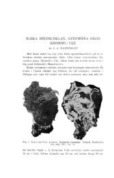
AscomyceteOrg 05-01_AscomyceteOrg
AscomyceteOrg 05-01_AscomyceteOrg
AscomyceteOrg 05-01_AscomyceteOrg

Create successful ePaper yourself
Turn your PDF publications into a flip-book with our unique Google optimized e-Paper software.
8<br />
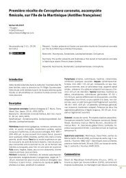
Fig. 2 – Consensus tree resulting from combined Bayesian ITS-28S LSU analysis of the Sarcosomataceae and Chorioactidaceae lineages studied.<br />
The length of the rooting branch was altered for publishing purposes. Nodes were annotated with maximum likelihood BP (above)<br />
and Bayesian PP (below). Only nodes supported (or nearly supported) by both analyses were annotated. Bold names belong to samples<br />
sequenced in the present study.<br />
of 422 bp (1.65%) specifically variable in the only available sequence<br />
of P. megalocrater. The P. milleri sample analyzed was deeply studied<br />
by CARBONE et al. (2<strong>01</strong>1a), and so was that of P. megalocrater by CAR-<br />
BONE et al. (2<strong>01</strong>1b). All collections clearly match with their respective<br />
protologues, and differ from each other in some morphological,<br />
ecological and biogeographical features. If the slight molecular differences<br />
are constant or not, it should be addressed by further sequencing<br />
of more specimens of both species. However, they seem<br />
too low to consider each clade a different species if compared with<br />
sister lineages such as P. melastoma and P. zugazae. With regards to<br />
the latter species, we believe that it deserves a small comment because<br />
our revision has revealed many common features with P. melastoma<br />
and also one previously regarded as exclusive of P. milleri. In<br />
particular, P. zugazae microscopic characters are not so different<br />
from those of P. melastoma (AGNELLO & CARBONE, 2<strong>01</strong>2); most of the<br />
features described in the protologue are confirmed whilst other are<br />
amended, such as: mature asci up to 470–490 μm long, spores<br />
Qm = 1.6, immature spores fusoid to slightly subfusoid when mature,<br />
the surface of the latter being very slightly warted (but this feature<br />
must be evaluated with more precise technique like SEM and<br />
above all on more mature spores), hymenial hairs present and av.<br />
3.5–4 μm wide, medullary excipulum crossed by brown “milleri-type”<br />
hairs (see CARBONE et al., 2<strong>01</strong>1a). So, the only really deviant features<br />
(from P. melastoma) consist in a lower Q, more elliptical spores and<br />
also a slight difference in spores size. Macroscopically, it lacks the<br />
typical orange granules on the external surface, although the picture<br />
here published and water mounts have revealed an evident covering<br />
of an encrusting pigment very similar to that one of<br />
P. melastoma. With regards to the presence of hairs in the medullary<br />
excipulum, the first author has recently studied an American col-