20 Nevus and Neurocutaneous Syndrome
20 Nevus and Neurocutaneous Syndrome
20 Nevus and Neurocutaneous Syndrome

Create successful ePaper yourself
Turn your PDF publications into a flip-book with our unique Google optimized e-Paper software.
<strong>20</strong><br />
328 <strong>20</strong> <strong>Nevus</strong> <strong>and</strong> <strong>Neurocutaneous</strong> <strong>Syndrome</strong><br />
Clinical images are available in hardcopy only.<br />
Clinical images are available in hardcopy only.<br />
Clinical images are available in hardcopy only.<br />
Clinical images are<br />
available in<br />
hardcopy only.<br />
Clinical images are available in hardcopy only.<br />
Fig. <strong>20</strong>.2-2 Nevocellular nevus.<br />
Clinical images are<br />
available in<br />
hardcopy only.<br />
Clinical features<br />
A nevocellular nevus is a flat-surfaced or verrucous macule or<br />
tumor that is brown, black, or sometimes normal skin color (Figs.<br />
<strong>20</strong>.2-1 to <strong>20</strong>.2-3). It may be accompanied by terminal hair. Nevocellular<br />
nevi are classified by size into three types. Small nevocellular<br />
nevus is commonly called mole or lentigo <strong>and</strong> varies in<br />
size from several millimeters to 1 cm in diameter. It is not present<br />
at birth but first appears between the ages of 3 <strong>and</strong> 4 <strong>and</strong><br />
gradually increases in number <strong>and</strong> size to peak at puberty. After<br />
puberty, the color often fades <strong>and</strong> the nevus is replaced by fat tissue<br />
or fibrotic tissue. When the diameter exceeds <strong>20</strong> cm, the<br />
nevus is called a giant congenital melanocytic nevus.<br />
Pathogenesis<br />
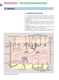
<strong>Nevus</strong> cells are derived from neural crests <strong>and</strong> proliferate<br />
abnormally, resulting in blackish-brown pigmented macules.<br />
Melanocytes <strong>and</strong> Schwann cells are derived from neural crests;<br />
however, nevus cells do not differentiate into either of these (Fig.<br />
<strong>20</strong>.3).<br />
Pathology<br />
Nevocellular nevi are classified by location of proliferation<br />
into junctional nevus, intradermal nevus <strong>and</strong> compound nevus<br />
(Fig. <strong>20</strong>.3).<br />
Diagnosis, Differential diagnosis<br />
Nevocellular nevus should be differentiated from freckles,<br />
lentigines, non-melanocytic lesions such as seborrheic keratosis,<br />
dermatofibroma, <strong>and</strong> most importantly early malignant<br />
melanoma (Chapter 22). Any pigmented lesion in adults that is<br />
growing or changing in any way should be examined carefully.<br />
When pigmentation spreads beyond the nail in the nail plate<br />
(Hutchinson’s sign), there is a high likelihood that a malignant<br />
melanoma is involved. Dermoscopic findings are important for<br />
diagnosis.<br />
Treatment<br />
Even when the dermoscopic findings are benign, follow-up is<br />
necessary. Surgical removal is the basic treatment for cases in the<br />
palms <strong>and</strong> soles, which tend to have a high likelihood of malignancy,<br />
<strong>and</strong> in cases with a relatively large congenital nevocellular<br />
nevus. Laser therapy may be conducted if there are cosmetic concerns.<br />
Excision, ablation or skin grafting may be performed on a<br />
giant congenital melanocytic nevus. When it is too large for<br />
removal, long-term follow-up may be chosen to observe for any<br />
signs of malignant melanoma.
including spindle cells, epithelioid-like cells <strong>and</strong> multinuclear<br />
cells. Dermal edema, telangiectasia, <strong>and</strong> inflammatory cell infiltration<br />
may occur. These findings resemble those of malignant<br />
melanoma; differentiation between Spitz nevus <strong>and</strong> malignant<br />
melanoma is often difficult. The basic structural pattern of nevocellular<br />
nevi is preserved in Spitz nevus: the lack of cellular atypism<br />
in the cells is important in distinguishing Spitz nevus from<br />
malignant melanoma. Homogenous nonstructural eosinophilic<br />
substances called Kamino bodies are found in the nevus cell nest<br />
in 60% of cases (Fig. <strong>20</strong>.9). Dermoscopy shows sharply circumscribed<br />
pigmented lesions with a characteristic starburst pattern<br />
at the periphery.<br />
Treatment<br />
Excision is conducted. Spitz nevus does not aggravate; however,<br />
careful differentiation from malignant melanoma is necessary.<br />
6. Dysplastic nevus<br />
Synonym: Clark nevus<br />
Dysplastic nevus occurs around puberty. A slightly elevated, a b c d e f g h i<br />
flat-topped patch or a pigmented nevus larger than 6 mm in Fig. <strong>20</strong>.9 Histopathology of Spitz nevus.<br />
diameter occurs. A light brown or black, sometimes light pink, a: Spitz nevus at low magnification. b: Spitz<br />
lesion with roughly margined pigmentation forms. Dysplastic<br />
nevus is basically a benign nevocellular nevus. When multiple<br />
familial atypical nevi <strong>and</strong> malignant melanoma occur it is called<br />
dysplastic nevus syndrome. Dysplastic nevus syndrome is autosomal<br />
dominantly inherited, <strong>and</strong> it frequently develops into<br />
malignant melanoma. Most patients are Caucasians, <strong>and</strong> there are<br />
few Asian cases. Histopathologically, many cases show compound<br />
nevi. Atypism is not usually found in the cells. Dermoscopic<br />
differentiation from superficial spreading melanoma is<br />
necessary.<br />
nevus at high magnification. Kamino bodies<br />
(arrows) are stained by eosin.<br />
7. Spotted grouped nevus<br />
Small blackish-brown pigmented macules or nodules densely<br />
aggregate in brown to light-brown patches. This nevocellular<br />
nevus occurs frequently on the trunk <strong>and</strong> less frequently at other<br />
sites.<br />
8. Balloon-cell nevus<br />
This nevus consists of balloon cells that contain clear, large<br />
cytoplasm. The cells are thought to be degenerated nevus cells.<br />
Nevertheless, balloon-cell nevus cannot be distinguished clinically<br />
from ordinary nevocellular nevus.<br />
<strong>Nevus</strong> / A. Melanocytic nevi 331<br />
a b c d e f g h<br />
Lentigo<br />
MEMO<br />
Most cases of the pigmentation that is commonly<br />
called lentigo are small nevocellular<br />
nevus. Lentigo simplex, a dermatological<br />
term, is a flat blackish-brown lesion of several<br />
millimeters in diameter caused by localized<br />
proliferation of epidermal melanocytes in<br />
which the nevus cells do not increase in number<br />
over time. Lentigo simplex is not usually<br />
present at birth, but it may appear around 3<br />
years of age. It accompanies systemic diseases<br />
(neurocutaneous syndrome) including<br />
Peutz-Jeghers syndrome, Cronkhite-Canada<br />
syndrome, <strong>and</strong> LEOPARD syndrome.<br />
Go Back to the Top To Order, Visit the Purchasing Page for Details<br />
<strong>20</strong>