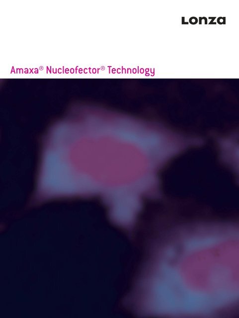
Amaxa® Nucleofector® Technology - Biocenter.hu
Amaxa® Nucleofector® Technology - Biocenter.hu
Amaxa® Nucleofector® Technology - Biocenter.hu

Create successful ePaper yourself
Turn your PDF publications into a flip-book with our unique Google optimized e-Paper software.
<strong>Amaxa®</strong> <strong>Nucleofector®</strong> <strong>Technology</strong>
<strong>Amaxa®</strong> <strong>Nucleofector®</strong> <strong>Technology</strong><br />
The <strong>Amaxa®</strong> <strong>Nucleofector®</strong> <strong>Technology</strong><br />
The application of systems biology and multidisciplinary approaches<br />
require that cells and model systems display in vivo like cellular<br />
functionality. This means that the future of cell transfection is in using<br />
primary cell types, and that transfecting these physiologically relevant<br />
cell types presents researchers with a series of technical challenges.<br />
The most physiologically relevant cell types are typically the most<br />
diffi cult to transfect using traditional methods. In contrast, when<br />
using relevant cell lines as model systems, the critical issues are to<br />
achieve reproducibly effi cient transfection with high levels of viability<br />
while matching throughput capability with the number of transfections<br />
required at each project phase – from proof of concept, through to scaleup<br />
and screening-like approaches.<br />
With <strong>Amaxa®</strong> Optimized Protocols for more than 200 primary cells and<br />
cell lines, <strong>Nucleofector®</strong> <strong>Technology</strong> is the technology of choice for<br />
leaders in the fi eld of cell biology.<br />
<strong>Nucleofector®</strong> <strong>Technology</strong> – two modular devices<br />
1<br />
■ <strong>Nucleofector®</strong> <strong>Technology</strong> delivers:<br />
<strong>Nucleofector®</strong> Device 96-well S<strong>hu</strong>ttle® Device<br />
Rapid and reliable transfection of primary cells<br />
– Simply select the cell type-specifi c <strong>Nucleofector®</strong> Kit<br />
and <strong>Amaxa®</strong> Optimized Protocol for accelerated startup<br />
and deliver your substrate straight to the nuclei of even<br />
non-dividing primary cells<br />
High effi ciency transfection of cell lines and primary cells<br />
– Get up to 99% transfection effi ciency with the unparalleled viability<br />
and unaltered functionality that make for truly informative assay<br />
Multiple savings in cell and reagent useage<br />
– 20 μl transfection volumes in 96-well format and specialized Small<br />
Cell Number (SCN) <strong>Nucleofector®</strong> Kits maximize savings by allowing<br />
as few as 1.5 x 10⁴ cells and 200 ng DNA per transfection<br />
Your choice of throughput format<br />
– Choose the modular devices you need for transfection<br />
in single cuvettes or in 96-well plates<br />
The freedom to expand your research<br />
– Explore complex systems by using the same conditions to deliver<br />
DNA, RNA, oligonucleotides, PNA, peptides, or proteins to your<br />
choice of cells or automate for high throughput
<strong>Amaxa®</strong> <strong>Nucleofector®</strong> <strong>Technology</strong><br />
Nucleofection® – Your Unique Advantage<br />
Nucleofection® is a technology based on the momentary creation<br />
of small pores in cell membranes by applying an electrical pulse.<br />
The comprehensive way in which <strong>Nucleofector®</strong> Programs and cell<br />
t ype-specifi c solutions are developed ensures that nucleic acid<br />
substrates are delivered not only to the cytoplasm, but also through<br />
the nuclear membrane and into the nucleus. Transfected cells retain<br />
excellent viability and the function of intracellular systems is highly<br />
conserved. Whatever your application, <strong>Amaxa®</strong> Transfection Specialists<br />
are available to assist you in rapidly optimizing your transfection<br />
workfl ow.<br />
Nucleic acid delivery direct to the site of action –<br />
effi ciency and fast expression<br />
A B C<br />
D<br />
Figure 1: Normal <strong>hu</strong>man dermal fi broblasts (neonatal) were trans fected with 2.5 μg<br />
TMR-labeled plasmid DNA encoding eGFP. After 2 hours, cells were fi xed with 3.5% PFA and<br />
analyzed by confocal micros copy. TMR label is shown in (A), GFP fl uorescence in (B), DAPI<br />
nuclear staining in (C) and a merge of all three fl uorescent labels in (D).<br />
<strong>Nucleofector®</strong> <strong>Technology</strong> – the superior non-viral method<br />
SSC<br />
Nucleofection® Competitor B electroporation<br />
GFP<br />
SSC<br />
30.31% 2.75%<br />
Figure 2: Transfection of the <strong>hu</strong>man natural killer cell line NKL using tradional electroporation<br />
and Nucleofection®. 5 x 10 6 NKL cells were transfected with 2.5 μg of pmaxGFP® Vector.<br />
Nucleofection®: <strong>Nucleofector®</strong> Solution V; Program O-017. Competitor B electroporation:<br />
25 mV, 96 μF. Transfection effi ciency was monitored by fl ow cytometry after 24 hours. Cells<br />
transfected by Nucleofection® show a signifi cantly better transfection effi ciency compared<br />
to cells transfected by tradional electroporation. Cell viability, as measured 18 hours after<br />
transfection was also superior using Nucleofection®.<br />
(Data courtesy of Dr. John Coligan, Laboratory of Immunogenetics, NIH/NIAID, Rockville, MD,<br />
USA. J Immunol Methods [2004] 284: 133-140.)<br />
GFP<br />
One <strong>Technology</strong> – the Broadest Choice of Cell Types<br />
Using the <strong>Nucleofector®</strong> <strong>Technology</strong>, cell lines, as well as primary cells<br />
and stem cells, can be reliably transfected at high effi ciency. Delivery<br />
of nucleic acid substrates directly into both the nucleus and cytoplasm<br />
ensures transfection effi ciencies of up to 99%. More than 200 ready-touse<br />
<strong>Amaxa®</strong> Optimized Protocols for both the <strong>Nucleofector®</strong> Device and<br />
the 96-well S<strong>hu</strong>ttle® System contain cell type-specifi c guidance and<br />
Lonza’s <strong>Amaxa®</strong> Cell Database contains user-developed protocols and<br />
data for more than 1300 cell types.<br />
Proven <strong>Technology</strong> –<br />
Unparalleled Resources and Online Support<br />
Accessing service and support for <strong>Nucleofector®</strong> <strong>Technology</strong> is easy<br />
and immediate, whether you wish to request a <strong>Nucleofector®</strong> Demonstration,<br />
upgrade your warranty, order a custom protocol, or ask for<br />
guidance from one of our PhD-level scientists.<br />
<strong>Amaxa®</strong> Optimized Protocols off er comprehensive guidance, including<br />
tips for cell sourcing, passage number, growth conditions and media,<br />
and post-transfection culture.<br />
■ www.lonza.com/cell-database<br />
■ www.lonza.com/nucleofection-citations<br />
2
<strong>Amaxa®</strong> <strong>Nucleofector®</strong> <strong>Technology</strong><br />
Two <strong>Amaxa®</strong> <strong>Nucleofector®</strong> Platforms –<br />
Fulfi lling Your Choice of Throughput<br />
Using <strong>Nucleofector®</strong> <strong>Technology</strong> allows your transfection capabilities<br />
to grow in pace with your group, to adapt to changing applications that<br />
require diff erent levels of sample throughput, to accelerate the pace<br />
of your research, or to transfect only the number of cells that fi ts your<br />
unique application.<br />
The Components of the<br />
<strong>Nucleofector®</strong> <strong>Technology</strong><br />
Proven – in Leading Labs<br />
The <strong>Nucleofector®</strong> <strong>Technology</strong> is an established and trusted transfection<br />
method in opinion-leading labs worldwide. Several thousand<br />
peer-reviewed publications illustrate the importance of Nucleofection®<br />
in <strong>hu</strong>ndreds of leading edge research applications using primary cells,<br />
such as neurons and stem cells, as well as diffi cult-to-transfect and<br />
standard cell lines.<br />
The <strong>Nucleofector®</strong> II Device and 96-well S<strong>hu</strong>ttle® System<br />
Both the <strong>Nucleofector®</strong> Device and 96-well S<strong>hu</strong>ttle® System deliver<br />
unique electrical parameters. Electrical settings are pre-programmed<br />
for each optimized cell type and can be selected using the <strong>Nucleofector®</strong><br />
Device or through the laptop PC and software supplied as part of the<br />
<strong>Nucleofector®</strong> 96-well S<strong>hu</strong>ttle® System.<br />
3<br />
<strong>Nucleofector®</strong> Kits and <strong>Nucleofector®</strong> 96-well S<strong>hu</strong>ttle® Kits<br />
Kits for <strong>Nucleofector®</strong> <strong>Technology</strong> contain specifi ed plasticware,<br />
pipettes, pmaxGFP® Vector, <strong>Nucleofector®</strong> Solution and Supplement.<br />
Each solution and supplement is individually developed for every<br />
primary cell type. For cell lines, a set of diff erent <strong>Nucleofector®</strong><br />
Solutions is available. All solutions provide a protective environment<br />
that ensures the highest transfection effi ciency and cell viability,<br />
while helping maintain physiologically relevant cellular functions.<br />
Cell Line Optimization <strong>Nucleofector®</strong> Kits provide the ideal tools to<br />
conveniently determine the optimal Nucleofection® Parameters for your<br />
cell line of interest within a single experiment.<br />
Low throughput Low to high throughput<br />
Device <strong>Nucleofector®</strong> Device 96-well S<strong>hu</strong>ttle® System<br />
Samples per run 1 1 – 96<br />
Reaction volume 100 μl 20 μl<br />
Cell number 2 x 10 5 to 2 x 10 7 5 x 10 4 to 1 x 10 6<br />
Substrate amount Oligonucleotides: 0.2 – 200 pmol (2 nM – 2 μM) Oligonucleotides: 0.04 – 40 pmol (2 nM – 2 μM)<br />
Vector DNA: 1 – 5 μg Vector DNA: 0.2 – 1 μg
<strong>Amaxa®</strong> <strong>Nucleofector®</strong> <strong>Technology</strong><br />
Substrate Flexibility –<br />
Explore the Limits of Your Imagination<br />
The fl exibility that <strong>Nucleofector®</strong> <strong>Technology</strong> allows in planning<br />
experimental projects stems from the proven capability to transfect<br />
the widest range of substrate molecules into the widest range of cell<br />
types. Imagine the fl exibility to fi rst overexpress genes of interest<br />
using DNA vectors, then to explore the regulation of the gene or genes<br />
of interest using multiple or single shRNA or siRNA substrates using<br />
the same <strong>Nucleofector®</strong> Kit or 96-well S<strong>hu</strong>ttle® Kit and Conditions.<br />
Refl ecting the focus of molecular biologists, the fl exibility of<br />
Nucleofection® now extends beyond nucleic acid substrates, to<br />
include peptides, proteins, and antibodies. This allows yet further<br />
characterization and validation of the cellular functions that interest<br />
your research group, e.g., allowing researchers to identify and<br />
characterize related signaling or protein modifi cation pathways<br />
using inhibitory peptides, before tracking active protein traffi cking,<br />
or localization of active target molecules using antibody-conjugated<br />
nano-fl uorophores or quantum-dots.<br />
<strong>Nucleofector®</strong> <strong>Technology</strong> – delivers the widest range of substrates<br />
siRNA knockdown of vimentin mRNA in <strong>hu</strong>man T-cells Knockdown of protein expression in primary neurons using shRNA<br />
Relative expression (% pulse only, control sample C)<br />
120<br />
100<br />
↑<br />
50<br />
40<br />
30<br />
20<br />
10<br />
0<br />
1 3 5 7 9 11 13 15<br />
Figure 3: Knockdown on mRNA level measured by qRT-PCR. 15 samples compared to control<br />
(C): Nucleofection pulse only set to 100%.<br />
(Data kindly provided by C. Merz, Bayer Schering Pharma AG, Berlin.)<br />
Effi cient transfection of peptides<br />
Figure 4: Jurkat cells were transfected in the presence of the <strong>Amaxa®</strong> Peptide Transfection<br />
Control 30 uM, a FAM-labeled octapeptide. Images were captured 2 hours post-transfection.<br />
(Data courtesy of Prof. Brock, NCMLS, University of Nijmegen, Netherlands.)<br />
C<br />
System Biology<br />
Peptide<br />
Antibodies<br />
Protein<br />
Conjugates<br />
Expression<br />
Protein<br />
shRNA<br />
Drug<br />
Candidates<br />
Plasmid PremiRNA<br />
Small<br />
Molecules<br />
Fluoresent<br />
conjugates<br />
Drug Discovery<br />
Figure 5: Quantitive downregulation of CDC10 protein. Rat hippocampal neurons (E17) were<br />
transfected with the shRNA vector pSuperior targeting CDC10 using the 96-well S<strong>hu</strong>ttle®<br />
System. (A) and (B) Effi cient Nucleofection® of pSuperior is shown by eGFP expression after<br />
1 day in vitro. (C) Immunostaining of CDC10 (red fl uorescence) shows reduced endogenous<br />
CDC10 protein levels in transfected neurons (green) after 4 days in vitro compared to<br />
untransfected cells (red). Western blot analysis (D) and quantifi cation (E) of CDC10<br />
downregulation.<br />
(Data courtesy of Prof. Kiebler, Medical University of Vienna, Vienna, Austria.)<br />
DNA<br />
Antisense<br />
Oligos<br />
CDC10 signal (% of Calnexin signal)<br />
RNA<br />
mRNA<br />
A B C<br />
D E<br />
CDC10 ➞<br />
Calnexin ➞<br />
siCDC10<br />
misPum2<br />
siPum2<br />
Structural Genomics<br />
100<br />
80<br />
60<br />
40<br />
20<br />
0<br />
siRNA<br />
miRNA<br />
Functional<br />
Genomics<br />
siCDC10 siPum2 misPum2<br />
4
<strong>Amaxa®</strong> <strong>Nucleofector®</strong> <strong>Technology</strong><br />
Post Transfection Functionality –<br />
Examine the Real Cellular Phenotype<br />
Allowing cells to express the true in vivo phenotype is key to generating<br />
biologically relevant data. Where many transfection methods<br />
compromise cellular functionality through their innate toxicity or<br />
by inducing immune responses, <strong>Nucleofector®</strong> <strong>Technology</strong> delivers<br />
substrates effi ciently to the nucleus and cytoplasm without disturbing<br />
cellular processes – there is no reagent incorporated into the cell<br />
membrane or any delivery particles accumulating in the cytoplasm.<br />
<strong>Nucleofector®</strong> <strong>Technology</strong> has the potential to improve your transfection<br />
applications, whether trying to uncover the functionality of cancers<br />
that cause pathology by proliferation, metastasis, or vascularisation or<br />
by transfecting stem cells for developmental or therapeutic research.<br />
<strong>Nucleofector®</strong> <strong>Technology</strong> is proven as a valuable tool to explore cellular<br />
functionality at the DNA, RNA, and protein levels.<br />
5<br />
Conserving functionality – the fi rst step to meaningful analysis<br />
A B<br />
Figure 6: Human H9 ES cells preserve pluripotency post Nucleofection®. H9 cells were<br />
transfected by Nucleofection® with the pmaxGFP® Vector. (A) Cells analyzed after 24 hours<br />
show expression of GFP (green) as well as of the pluri potency markers SSEA4 (red) and Oct4<br />
( purple). The blue signals refer to nuclear staining by DAPI. (B) The percentage of doublepositive<br />
cells (GFP/SSEA) was analyzed by fl ow cytometry.<br />
(Data kindly provided by Jennifer Moore, Rutgers University, Piscataway, USA.)<br />
OAS-1 mRNA (fold induction; 24 h)<br />
100<br />
80<br />
60<br />
40<br />
20<br />
0<br />
Untreated<br />
OAS-1expression IL-6 release<br />
Mock<br />
0.02 pmol<br />
0.2 pmol<br />
2 pmol<br />
20 pmol<br />
Untreated<br />
Figure 7: Induction of interferon responses is related to the method of substrate delivery.<br />
HeLaS3 (ATCC® CCL-2.2) were transfected with diff erent amounts of 29-mer siRNA targeting<br />
DBI (Dharmacon) – a described trigger of interferon responses (Reynolds A et al., (2006),<br />
RNA 12:988-993) – using the 96-well S<strong>hu</strong>ttle® Device or a lipid reagent. Interferon response<br />
was measured by expression of OAS-1 mRNA, determined 24 hours post transfection by<br />
QuantiGene® branched DNA assay (Panomics; normalized to GAPDH mRNA), and IL-6 release<br />
analyzed 48 hours post transfection by <strong>hu</strong>man IL-6 ELISA (Biosource).<br />
(Data generated in collaboration with Thermo Fisher Scientifi c, Dharmacon Products.)<br />
% Positive cells<br />
Mock<br />
100<br />
0.02 pmol<br />
0.2 pmol<br />
Nucleofection ® Lipofection<br />
80<br />
60<br />
40<br />
20<br />
0<br />
SSEA4 GFP SSEA4/GFP<br />
2 pmol<br />
20 pmol<br />
6000<br />
4000<br />
2000<br />
0<br />
IL-6 release (pg/ml; 48h)
<strong>Amaxa®</strong> <strong>Nucleofector®</strong> <strong>Technology</strong><br />
<strong>Nucleofector®</strong> 96-well S<strong>hu</strong>ttle® System –<br />
Flexibility and Value for Money<br />
The <strong>Nucleofector®</strong> 96-well S<strong>hu</strong>ttle® System utilizes a unique 96-well<br />
disposable Nucleocuvette® Plate. Each Nucleocuvette® Plate comprises<br />
6 modules, each with 2 columns of 8 wells. Each Nucleocuvette® Well<br />
may be individually addressed with your pre-optimized program of<br />
choice through contacts to the conductive polymer electrodes.<br />
The transfection volume of 20 μl reduces the required number of cells<br />
by 80% compared with the standard 100 μl transfection volume of the<br />
<strong>Nucleofector®</strong> Device and as few as 1 sample at a time may be easily<br />
Unparalleled performance and economy – with primary cells<br />
and cell lines<br />
Transfection efficiency %<br />
100<br />
80<br />
60<br />
40<br />
20<br />
0 B Cells, <strong>hu</strong>man<br />
Dendritic Cells,<br />
mouse, mature<br />
Macrophages <strong>hu</strong>man<br />
Dendritic Cells,<br />
mouse, immature<br />
T Cells, mouse<br />
HMEC<br />
Figure 8A: Example for the transfection of primary cells using the <strong>Nucleofector®</strong> 96-well<br />
S<strong>hu</strong>ttle®. Cells were transfected with 400 ng pmaxGFP® Vector and analyzed for maxGFP®<br />
Reporter Protein expression on a BD FACSCalibur (BD Biosciences) with HTS option 24 hours<br />
post Nucleofection®. Cell viabilities were in the range between 30% and 92% depending on the<br />
cell type.<br />
Cell Type Lonza Clonetics®<br />
Primary Cell<br />
Catalogue Number<br />
Neurons<br />
(hippo/cortical), rat<br />
NHBE<br />
Monocytes,<br />
<strong>hu</strong>man<br />
hPrEC<br />
HUVEC<br />
Chondrocytes,<br />
<strong>hu</strong>man<br />
T Cells, <strong>hu</strong>man<br />
NHDF<br />
transfected. Should optimization for your unique cell type be required, up<br />
to 96 individual programs may be tested within a single plate in less than<br />
fi ve minutes and using as few as 2 x 10 6 cells in total.<br />
The 96-well S<strong>hu</strong>ttle® System delivers fl exible throughput combined<br />
with economical processing, speed, and pre-optimized protocols for a<br />
range of both primary cells and cell lines. A choice of Service Contracts<br />
off ering levels of service to suit your needs is available to further reduce<br />
risks of project ‘downtime’.<br />
Transfection efficiency %<br />
100<br />
80<br />
60<br />
40<br />
20<br />
0 U937<br />
HL-60<br />
RAW 264.7<br />
THP-1<br />
Neuro-2a<br />
Jurkat<br />
Figure 8B: Highly effi cient transfection of various cell lines using the <strong>Nucleofector®</strong> 96-well<br />
S<strong>hu</strong>ttle®. Cell lines (ATCC®) were transfected with 400 ng pmaxGFP® Vector and analyzed for<br />
maxGFP® Reproter Protein expression on a BD FACSCalibur (BD Biosciences) with HTS option<br />
24 hours post Nucleofection®. Cell viabilities were in the range between 50% and 97% depending<br />
on the cell line.<br />
Cell Data <strong>Nucleofector®</strong> 96-well S<strong>hu</strong>ttle® Data<br />
Cells per 96-well S<strong>hu</strong>ttle®<br />
Nucleofection®<br />
(single well)<br />
Transfection<br />
Effi ciency<br />
HeLa<br />
MCF-7<br />
293<br />
HeLa S3<br />
CHO-K1<br />
CHO-S<br />
K562<br />
NIH/3T3<br />
Viability 96-well S<strong>hu</strong>ttle® Kit<br />
Catalogue Number<br />
Smooth muscle cells (AoSMC), aortic, <strong>hu</strong>man CC-2571 5 x 10⁴ 74% 92% VHPI-1004<br />
Smooth muscle cells, pulmonary artery (PASMC), <strong>hu</strong>man CC-2581 5 x 10⁴ 67% 86% VHPI-1004<br />
Smooth muscle cells, coronary artery (CASMC), <strong>hu</strong>man CC-2583 5 x 10⁴ 81% 67% VHPI-1004<br />
Fibroblasts, Dermal (NHDF), <strong>hu</strong>man (adult) CC-2511 1 x 10⁵ 96% 92% VHPD-1001<br />
Epithelial Cells, bronchial (NHBE), <strong>hu</strong>man CC-2542 7.5 x 10⁴ 54% 53% VHPK-1001<br />
Dendritic Cells (DC), Mouse, Balb-c (immat.) – 5 x 10⁴ 43% 74% VHPA-1011<br />
Epithelial cells, prostate (PrEC), <strong>hu</strong>man CC-2555 1 x 10⁵ 67% 48% VHPK-1003<br />
Chondrocytes, <strong>hu</strong>man – 2 x 10⁵ 74% 84% VHPF-1001<br />
Hepatocytes, <strong>hu</strong>man CC-2591 1 x 10⁵ 54% 69% VHPL-1001<br />
Embryonic stem cell line (H9), <strong>hu</strong>man – 2 x 10⁵ 64% 78% VHPH-5003<br />
Embryonic stem cells, mouse – 5 x 10⁴ 90% 80% VHPH-1001<br />
6
<strong>Amaxa®</strong> <strong>Nucleofector®</strong> <strong>Technology</strong><br />
96-well S<strong>hu</strong>ttle® System – Reliable High Throughput<br />
Transfection<br />
In a high throughput transfection framework, the manipulation of cells<br />
during the whole process chain is a critical issue. Stability of cell<br />
parameters such as transfection effi ciency and viability is key to<br />
statistically relevant data and valid results.<br />
Reproducibility – The Key to Success for High Throughput Transfection<br />
During the workfl ow of cell-based screening, cells are often subject to<br />
environmental changes such as oxygen partial pressure, temperature,<br />
or agitation. Each step of such demanding workfl ows must be validated<br />
to minimize the sources of error introduced at each step. In the following<br />
data, <strong>Nucleofector®</strong> 96-well S<strong>hu</strong>ttle® <strong>Technology</strong> was used to fi rst<br />
examine the impact of extended incubation in Nucleofection® Solution<br />
at room temperature and, secondly, to analyse siRNA screening assay<br />
data reproducibility.<br />
Robust transfection – suited to every workfl ow<br />
% Transfection efficiency<br />
% Viability<br />
Figure 9: Jurkat E6-1 (ATCC® TIB 152) cells were incubated in room temperature 96-well<br />
<strong>Nucleofector®</strong> Solution for 0, 2, 4, and 6 hours prior to transfection with 1 μg of pmaxGFP®<br />
Vector. Cells were analyzed 48 hours post Nucleofection®, using a BD FACSCalibur<br />
(BD Biosciences) with HTS option. Cell viability was determined using Propidium Iodide (PI)<br />
staining.<br />
7<br />
100<br />
80<br />
60<br />
40<br />
20<br />
0<br />
100<br />
80<br />
60<br />
40<br />
20<br />
0<br />
Column 1 – 3<br />
Column 5 – 6<br />
0 hours 2 hours 4 hours 6 hours<br />
Column 1 – 3<br />
Column 5 – 6<br />
0 hours 2 hours 4 hours 6 hours<br />
Reproducible transfection – making the most of your assays<br />
Median average deviation (MAD)<br />
Medain average deviation (MAD)<br />
Result from 1 screening experiment<br />
6<br />
4<br />
2<br />
0<br />
-2<br />
-4<br />
-6<br />
-8<br />
-10<br />
-12<br />
-14<br />
-16<br />
CHEK-1<br />
CHEK-1<br />
0 100 200 300 400 500 600 700 800 900<br />
Sample #<br />
Reproducible hits in 3 screens<br />
4<br />
2<br />
0<br />
-2<br />
-4<br />
-6<br />
-8<br />
-10<br />
-12<br />
-14<br />
Figure 10: Primary Screen HUVEC cells were transfected with 20 pmol of the combined<br />
Human siGENOME® siRNA Libraries for Kinases (targeting 779 genes) and Cell Cycle<br />
Regulators (targeting 111 genes). Cell viability was analyzed 72 hours post Nucleofection®.<br />
(A) Representation of robust Z-scores of cell viability measures from 1 screening experiment.<br />
(B) Robust Z-scores of the top 37 primary hits (with an |MAD| > 2) from three independent<br />
experiments.<br />
(Data generated in collaboration with Thermo Fisher Scientifi c, Dharmacon Products.)<br />
PLK-1<br />
FN3KRP<br />
NRBP<br />
STK24<br />
PRKCB1<br />
PRKAG3<br />
LMTK3<br />
RPS6KC1<br />
FGFR3<br />
FLJ13052<br />
IKBKB<br />
MAOK11<br />
FGFR1<br />
CARKL<br />
CKMT1<br />
DYRK4<br />
PLK1<br />
CENPE<br />
PIK3C2A<br />
PLK2<br />
MARK3<br />
RIOK1<br />
MST1R<br />
PKM2<br />
EDN2<br />
RPS6KL1<br />
MYC<br />
NLK<br />
PFKFB3<br />
IRAK3<br />
PYCS<br />
CHEK1<br />
CDK4<br />
RAPGEF3<br />
CNKSR1<br />
COPB2<br />
Contact Lonza today regarding your unique application or to arrange to test the <strong>Nucleofector®</strong> <strong>Technology</strong> with your cells and in your lab.<br />
A<br />
B<br />
/ /<br />
PLK-1<br />
TGFBR1<br />
TESK1<br />
LIM<br />
NEK6
<strong>Amaxa®</strong> <strong>Nucleofector®</strong> <strong>Technology</strong><br />
Ordering Information<br />
Cat. No. Description<br />
AAD-1001S <strong>Nucleofector®</strong> Device<br />
AAM-1001S 96-well S<strong>hu</strong>ttle® Device (includes the <strong>Nucleofector®</strong> Device, notebook PC and software)<br />
SBA-1001 96-well S<strong>hu</strong>ttle® Automation Package<br />
AXA-1001 96-well S<strong>hu</strong>ttle® Rack<br />
<strong>Nucleofector®</strong> Kits for primary cells<br />
VPA-1001 Human B Cell <strong>Nucleofector®</strong> Kit<br />
VPA-1002 Human T Cell <strong>Nucleofector®</strong> Kit<br />
VPA-1003 Human CD34 + Cell <strong>Nucleofector®</strong> Kit<br />
VPA-1005 Human Natural Killer Cell <strong>Nucleofector®</strong> Kit<br />
VPA-1006 Mouse T Cell <strong>Nucleofector®</strong> Kit<br />
VPA-1007 Human Monocyte <strong>Nucleofector®</strong> Kit<br />
VPA-1008 Human Macrophage <strong>Nucleofector®</strong> Kit<br />
VPA-1009 Mouse Macrophage <strong>Nucleofector®</strong> Kit<br />
VPC-1001 Human Aortic Smooth Muscle Cell <strong>Nucleofector®</strong> Kit<br />
VPD-1003 NHEM-Neo (Normal Human Epidermal Melanocyte-Neo) <strong>Nucleofector®</strong> Kit<br />
VPE-1001 Human MSC (Mesenchymal Stem Cell) <strong>Nucleofector®</strong> Kit<br />
VPE-1002 Rat Cardiomyocyte – Neonatal <strong>Nucleofector®</strong> Kit<br />
VPG-1001 Mouse Neuron <strong>Nucleofector®</strong> Kit<br />
VPG-1002 Chicken Neuron <strong>Nucleofector®</strong> Kit<br />
VPG-1003 Rat Neuron <strong>Nucleofector®</strong> Kit<br />
VPG-1004 Mouse NSC (Neural Stem Cell) <strong>Nucleofector®</strong> Kit<br />
VPG-1005 Rat NSC (Neural Stem Cell) <strong>Nucleofector®</strong> Kit<br />
VPG-1006 Mouse Astrocyte <strong>Nucleofector®</strong> Kit<br />
VPG-1007 Rat Astrocyte <strong>Nucleofector®</strong> Kit<br />
VPG-1009 Rat Oligodendrocyte <strong>Nucleofector®</strong> Kit<br />
Basic <strong>Nucleofector®</strong> Kits for primary cells<br />
VPI-1001 Basic <strong>Nucleofector®</strong> Kit for Primary Mammalian Endothelial Cells<br />
VPI-1002 Basic <strong>Nucleofector®</strong> Kit for Primary Mammalian Fibroblasts<br />
VPI-1003 Basic <strong>Nucleofector®</strong> Kit for Primary Mammalian Neurons<br />
VPI-1004 Basic <strong>Nucleofector®</strong> Kit for Primary Smooth Muscle Cells<br />
VPI-1005 Basic <strong>Nucleofector®</strong> Kit for Primary Mammalian Epithelial Cells<br />
<strong>Nucleofector®</strong> Kits for cell lines<br />
VCA-1001 Cell Line <strong>Nucleofector®</strong> Kit R<br />
VCA-1002 Cell Line <strong>Nucleofector®</strong> Kit T<br />
VCA-1003 Cell Line <strong>Nucleofector®</strong> Kit V<br />
VCA-1004 Cell Line <strong>Nucleofector®</strong> Kit C<br />
VCA-1005 Cell Line <strong>Nucleofector®</strong> Kit L<br />
VCO-1001N Cell Line Optimization <strong>Nucleofector®</strong> Kit<br />
8
<strong>Amaxa®</strong> <strong>Nucleofector®</strong> <strong>Technology</strong><br />
Ordering Information<br />
Cat.No. 96 reactions Cat.No. 960 reactions Description<br />
96-well <strong>Nucleofector®</strong> Kits for primary cells<br />
VHPA-1001 VHPA-2001 Human B Cell <strong>Nucleofector®</strong> Kits<br />
VHPA-1002 VHPA-2002 Human T Cell 96-well <strong>Nucleofector®</strong> Kits<br />
VHPA-1006 VHPA-2006 Mouse T Cell 96-well <strong>Nucleofector®</strong> Kits<br />
VHPA-1011 VHPA-2011 Mouse Dendritic Cell (Immature) 96-well <strong>Nucleofector®</strong> Kits<br />
VHPA-1012 VHPA-2012 Mouse Dendritic Cell (Mature) 96-well <strong>Nucleofector®</strong> Kits<br />
VHPB-1002 VHPB-2002 HUVEC 96-well <strong>Nucleofector®</strong> Kits<br />
VHPF-1001 VHPF-2001 Human Chondrocyte 96-well <strong>Nucleofector®</strong> Kits<br />
VHPG-1003 VHPG-2003 Rat Neuron 96-well <strong>Nucleofector®</strong> Kits<br />
VHPH-1001 VHPH-2002 Mouse ES Cell 96-well <strong>Nucleofector®</strong> Kits<br />
VHPK-1002 VHPK-2002 Human Mammary Epithelial Cell (HMEC) 96-well <strong>Nucleofector®</strong> Kits<br />
VHPK-1003 VHPK-2003 Human Prostate Epithelial Cell (hPrEC) 96-well <strong>Nucleofector®</strong> Kits<br />
Basic 96-well <strong>Nucleofector®</strong> Kits for primary cells<br />
VHPI-1002 Basic 96-well <strong>Nucleofector®</strong> Starter Kit for Primary Mammalian Fibroblasts (64 reactions)<br />
VHPI-1003 VHPI-2003 Basic 96-well <strong>Nucleofector®</strong> Kits for Primary Mammalian Neurons<br />
VHPI-1004 VHPI-2004 Basic 96-well <strong>Nucleofector®</strong> Kits for Primary Smooth Muscle Cells<br />
VHPI-1005 Basic 96-well <strong>Nucleofector®</strong> Starter Kit for Primary Mammalian Epithelial Cells (64 reactions)<br />
VHPH-5002 Basic 96-well <strong>Nucleofector®</strong> Starter Kit for Human Stem Cells (64 reactions)<br />
96-well <strong>Nucleofector®</strong> Kits for cell lines<br />
VHCA-1001 VHCA-2001 Cell Line 96-well <strong>Nucleofector®</strong> Kits SE<br />
VHCA-1002 VHCA-2002 Cell Line 96-well <strong>Nucleofector®</strong> Kits SF<br />
VHCA-1003 VHCA-2003 Cell Line 96-well <strong>Nucleofector®</strong> Kits SG<br />
VHCO-1001 Cell Line Optimization 96-well <strong>Nucleofector®</strong> Kit<br />
Related Products<br />
MycoAlert® Mycoplasma Detection Kits www.lonza.com/mycoalert<br />
ViaLight® Plus Cell Proliferation and Cytotoxicity BioAssay Kits www.lonza.com/vialight<br />
ToxiLight® Non-Destructive Cytotoxicity BioAssay Kits www.lonza.com/toxilight<br />
ApoGlow® Rapid Apoptosis Screening Kit www.lonza.com/apoglow<br />
StellARray Gene Expression System www.lonza.com/arrays<br />
For an up-to-date overview or more detailed information about <strong>Amaxa®</strong> Products please visit www.lonza.com/amaxa-productlist.<br />
9
WKB-1001_2009-01-22<br />
Contact Information<br />
North America<br />
Customer Service: 800-638-8174<br />
Scientifi c Support: 800-521-0390<br />
E-mail: scientifi c.support@lonza.com<br />
Online Ordering: www.lonza.com<br />
Europe<br />
Customer Service: 00 32 87 321 611<br />
Scientifi c Support: 00 49 221 99199 400<br />
E-mail: scientifi c.support.eu@lonza.com<br />
Online Ordering: www.lonza.com<br />
International<br />
Contact your local Lonza Distributor<br />
Customer Service: 301-898-7025, ext. 2322<br />
Fax: 301-845-8291<br />
E-mail: scientifi c.support@lonza.com<br />
International Offi ces<br />
Australia 61 3 9550 0883<br />
Austria 0800 201 538<br />
Belgium 00 32 87 321 611<br />
Brazil 55 11 2069 8800<br />
Denmark 45 43 56 74 00<br />
France 0800 91 19 81<br />
Germany 0800 182 52 87<br />
India 91 22 4342 4000<br />
Italy 0039 0363 45710<br />
Japan 81 3 5566 0612<br />
Poland 48 22 833 87 45<br />
Singapore 65 64914214<br />
Spain 34 902 531 366<br />
Sweden 020 140 4410<br />
Switzerland 0800 83 86 20<br />
The Netherlands 0800 022 4525<br />
United Kingdom 44 118 979 5234<br />
Lonza Cologne AG<br />
50829 Cologne, Germany<br />
For Research Use Only. Not for use in diagnostic procedures.<br />
The <strong>Amaxa®</strong> <strong>Nucleofector®</strong> <strong>Technology</strong>, comprising Nucleofection® Process, <strong>Nucleofector®</strong> Device,<br />
<strong>Nucleofector®</strong> Solutions, <strong>Nucleofector®</strong> 96-well S<strong>hu</strong>ttle® System and 96-well Nucleocuvette® Plates<br />
and Modules is covered by patent and/or patent pending rights owned by Lonza Cologne AG.<br />
Amaxa, Nucleofector, Nucleofection, Nucleocuvette, maxGFP and 96-well S<strong>hu</strong>ttle are either<br />
registered trademarks or trademarks of Lonza Cologne AG in Germany and/or the U.S. and/or other<br />
countries.<br />
ATCC® and the ATCC Catalog Marks are trademarks of ATCC used under License.<br />
Other product and company names mentioned herein are the trademarks of their respective owners.<br />
The information contained herein is believed to be correct and corresponds to the latest state of<br />
scientifi c and technical knowledge. However, no warranty is made, either expressed or implied,<br />
regarding its accuracy or the results to be obtained from the use of such information and no warranty<br />
is expressed or implied concerning the use of these products. The buyer assumes all risks of use and/<br />
or handling. No statement is intended or should be construed as a recommendation to infringe any<br />
existing patent.<br />
© Copyright 2009, Lonza Cologne AG. All rights reserved. SF-NucleoTech-1 11/08