apical meristem
apical meristem
apical meristem

You also want an ePaper? Increase the reach of your titles
YUMPU automatically turns print PDFs into web optimized ePapers that Google loves.
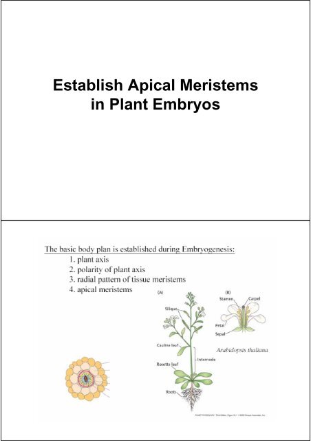
Establish Apical Meristems<br />
in Plant Embryos
Stages of Plant Embryogenesis
Uneven Cell Division Determines Polarity<br />
Mechanism of Uneven Cell Division<br />
in Plant
Radial pattern of tissue <strong>meristem</strong>s are<br />
established in a globular embryo<br />
a. protoderm epidermis<br />
b. ground <strong>meristem</strong> cortex<br />
c. procambium vascular tissues<br />
Apical growth zones, <strong>meristem</strong>s<br />
are established in the heart-shaped embryo<br />
Root Apical<br />
Meristems (RAM)<br />
remain dormant<br />
until seed<br />
germination<br />
First lateral organs<br />
are formed<br />
- embryonic<br />
leaves, cotyledons
Positional references provided by the<br />
early embryo pattern<br />
Auxin Determines Embryo Axis<br />
Formation (Positional Effect)
Cuc1 and cuc2<br />
determine the<br />
expression area<br />
for STM<br />
STM determines<br />
the area of SAM<br />
Wuc maintains the<br />
source of stem<br />
cells in SAM<br />
AS1 promote<br />
development of<br />
lateral organs<br />
Cell Fate Is Determined in<br />
L2 Layer of Meristem
Primary Growth of The Plant Body<br />
Primary growth:<br />
Apical <strong>meristem</strong>s extend roots<br />
and shoots by giving rise to the<br />
primary plant body<br />
• Primary growth produces the primary plant<br />
body - the parts of the root and shoot<br />
systems produced by <strong>apical</strong> <strong>meristem</strong>s.<br />
• An herbaceous plant and the youngest parts<br />
of a woody plant represent the primary plant<br />
body.<br />
Copyright © 2002 Pearson Education, Inc., publishing as Benjamin Cummings
Primary Growth of The Shoot<br />
Structure of The Shoot Apical Meristem
There are three <strong>apical</strong> <strong>meristem</strong>s in terminal buds<br />
• <strong>apical</strong> <strong>meristem</strong> : The <strong>apical</strong> <strong>meristem</strong> of a shoot is a<br />
dome-shaped mass of dividing cells at the terminal bud.<br />
• leaf primordia (derived from <strong>apical</strong> <strong>meristem</strong>) : Leaves<br />
arise as leaf primordia on the flanks of the <strong>apical</strong><br />
<strong>meristem</strong>.<br />
• Axillary buds (derived from <strong>apical</strong> <strong>meristem</strong>) develop<br />
from islands of <strong>meristem</strong>atic cells left by <strong>apical</strong> <strong>meristem</strong>s<br />
at the leaf primordia base.<br />
leaf primordia<br />
Axillary buds<br />
Copyright © 2002 Pearson Education, Inc., publishing as Benjamin Cummings<br />
Organization of SAM<br />
<strong>apical</strong> <strong>meristem</strong><br />
tunica – layered due to cell division planes at right angle to<br />
surface<br />
corpus – random cell division plane<br />
central zone – low rate of cell division<br />
peripheral zone – high rate of cell division
The low frequency of cell<br />
division in the central zone<br />
A pulse feeding of plants with<br />
3 H-thymidine result in 3 Hthymidine<br />
being incorporated<br />
in cells undergoing DNA<br />
synthesis and mitosis<br />
Sectioning and tissue sections<br />
are exposed x-ray films to<br />
visualize highly mitotic area<br />
(black)<br />
Interpretation:<br />
Rarely diving initial cells in the central zone.<br />
Frequently dividing daughter cells in the peripheral zone.<br />
Three Layers of Cells in SAM<br />
L1, anticlinal division epidermis<br />
L2, anticlinal divisions internal tissues<br />
L3, divisions internal tissues
Clonal analysis in chimeras – marking individual cells and<br />
tracing their progeny through successive cell divisions into<br />
the mature stages of plants<br />
chemical -(colchicine)<br />
induced polyploidy<br />
radiation-induced chloroplast<br />
pigmentation mutants.<br />
Screen functional genes that regulate formation of SAM<br />
Screening Mutants of Plants
The Genes that control SAM
CLV1,3 and WUS<br />
Maintain The Size of SAM<br />
CLV1,3 Restrict Expression<br />
of WUC at SAM
The Model of CLV/WUC Pathway<br />
Evidence<br />
1. In situ<br />
2. wus mutants lack CZ<br />
3. wus mutants have<br />
reduced/absent CLV3 expression<br />
WUS overexpression leads to<br />
enlarged domain domain of CLV3<br />
expression<br />
4. Immunolocalization<br />
5. CLV3 and CLV1 interaction<br />
leads to phosphorylation of CLV1<br />
intra cellular domain<br />
when expressed in yeast cells<br />
6. clv mutants have an expanded<br />
domain of WUS expression<br />
7. clv mutants have an expanded<br />
CZ region<br />
Apical <strong>meristem</strong>s form primary meristerms.<br />
Primary <strong>meristem</strong>s produce primary tissues
Copyright © 2002 Pearson Education, Inc., publishing as Benjamin Cummings<br />
Fig. 35.18<br />
• Unlike their central position in a root, the<br />
vascular tissue runs the length of a stem in<br />
strands called vascular bundles.<br />
– At the transition zone, the stem’s vascular<br />
bundles converge as the root’s vascular<br />
cylinder.<br />
• Each vascular bundle of the stem is<br />
surrounded by ground tissue.<br />
Copyright © 2002 Pearson Education, Inc., publishing as Benjamin Cummings
• In most dicots, the vascular bundles are arranged in a ring, with pith on the<br />
inside and cortex outside the ring.<br />
– The vascular bundles have their xylem facing the pith and their phloem facing the<br />
cortex.<br />
– Thin rays of ground tissue between the vascular bundles connect the two parts of<br />
the ground tissue system, the pith and cortex.<br />
Copyright © 2002 Pearson Education, Inc., publishing as Benjamin Cummings<br />
Primary Growth of The Leaf
Meristems of Leaves Are Derived from Shoot Apical Meristem
Knox Family Controls Formation of Leaf Initiation<br />
Determination of Area for<br />
Initiation of Leaf Primordia<br />
Meristem–leaf signalling<br />
Panel b shows a transverse view of a shoot<br />
apex.SHOOTMERISTEMLESS (STM) encodes a<br />
class I KNOX homeodomain transcription factor<br />
that is expressed throughout the SAM (purple) but<br />
is absent from leaf founder cells (indicated as a<br />
green domain in the SAM). ASYMMETRIC<br />
LEAVES1 (AS1) encodes a MYB transcription<br />
factor and AS2 is a member of the LATERAL<br />
ORGAN BOUNDARIES family of putative<br />
transcription factors. Expression of these two<br />
genes is excluded from the SAM and restricted to<br />
leaf primordia (green). Genetic analyses indicate<br />
that STM negatively regulates AS1 and AS2<br />
function in the SAM and down regulation of STM in<br />
leaves allows AS1 and AS2 expression. AS1 and<br />
AS2, in turn, negatively regulate other class I<br />
KNOX genes so that KNAT1,KNAT2 and KNAT6<br />
are ectopically expressed in the leaves of as1 and<br />
as2mutants. The additional loss of KNAT1 function<br />
in stm;as1 double mutants results in loss of a SAM,<br />
indicating that KNAT1 functions redundantly with<br />
STM in maintaining a SAM.
Determination of Leaf<br />
Polarity<br />
Compound leaves: equal to the sum of their<br />
parts?<br />
Connie Champagne and Neelima Sinha*<br />
Section of Plant Biology, University of<br />
California, 1 Shields Avenue, Davis, CA 95616,<br />
USA<br />
*Author for correspondence (e-mail:<br />
nrsinha@ucdavis.edu)<br />
Development 131, 4401-4412<br />
Determination of Axial<br />
Polarity of Leaf Primordia<br />
Leaf–<strong>meristem</strong> signalling<br />
Panel c shows a transverse view of a<br />
shoot apex.PHABULOSA (PHB) and<br />
PHAVOLUTA (PHV) encode class III<br />
homeodomain zipper (HD-ZIP)<br />
transcription factors that are expressed<br />
throughout the SAM,with high expression<br />
in rays extending from the SAM to the<br />
youngest leaf primordia, and in the<br />
adaxial domain of older leaf primordia<br />
(purple). YABBY (YAB) and KANADI<br />
(KAN) gene families encode putative<br />
transcription factors:YAB proteins contain<br />
zinc finger and HIGH MOBILITY GROUP<br />
(HMG) DOMAINS, and KAN genes<br />
belong to a larger gene family of<br />
transcriptional regulators that contains a<br />
GARP DOMAIN109.They are expressed<br />
in the abaxial domain of leaf primordia<br />
(green) and YAB members repress STM<br />
and KNAT1 expression in the leaf.
Knox 1 Determines Whether<br />
Leaves Become Compound<br />
Compound leaves: equal to the sum of their parts?<br />
Connie Champagne and Neelima Sinha*<br />
Section of Plant Biology, University of California, 1 Shields Avenue, Davis, CA 95616, USA<br />
*Author for correspondence (e-mail: nrsinha@ucdavis.edu)<br />
Development 131, 4401-4412<br />
Knox-1 Promotes Formation of<br />
Leaflets<br />
Overexpression of this gene causes the compound leaves of a<br />
tomato plant to become “supercompound”.
• The vascular tissue of a leaf is<br />
continuous with the xylem and<br />
phloem of the stem.<br />
Copyright © 2002 Pearson Education, Inc., publishing as Benjamin Cummings<br />
• Axillary buds have<br />
the potential to form<br />
branches of the shoot<br />
system at some later<br />
time.<br />
– While lateral roots<br />
originate from deep<br />
in the main root,<br />
branches of the<br />
shoot system<br />
originate from<br />
axillary buds, at the<br />
surface of a main<br />
shoot.<br />
– Because the<br />
vascular system of<br />
the stem is near the<br />
surface, branches<br />
can develop with<br />
connections to the<br />
Copyright vascular © 2002 Pearson tissue Education, Inc., publishing as Benjamin Cummings
Leaves Separated by Elongation of<br />
Internodes<br />
• Within a bud, leaf primordia are crowded close together because internodes are<br />
very short.<br />
– Most elongation of the shoot occurs by growth in length of slightly older internodes below the shoot<br />
apex.<br />
– This growth is due to cell division and cell elongation within the internode.<br />
– In some plants, including grasses, internodes continue to elongate all along the length of the shoot<br />
over a prolonged period.<br />
• These plants have <strong>meristem</strong>atic regions, called intercalary <strong>meristem</strong>s, at the base of each internode.<br />
Copyright © 2002 Pearson Education, Inc., publishing as Benjamin Cummings<br />
Fig. 35.19<br />
Copyright © 2002 Pearson Education, Inc., publishing as Benjamin Cummings<br />
MSU-DOE Plant Research Laboratory<br />
Michigan State University<br />
East Lansing, MI 48824-1312 USA<br />
Phone: (517) 353-7865<br />
Fax: (517) 353-9168<br />
e-mail: hkende@msu.edu<br />
Hans Kende<br />
Professor of Plant Biology<br />
University Distinguished Professor<br />
U.S. National Academy of Sciences<br />
German Academy of Natural Scientists<br />
Ph.D. (University of Zurich)
Primary Growth of The Root
The Structure of The Root Tip<br />
Fig. 35.14<br />
Copyright © 2002 Pearson Education, Inc., publishing as Benjamin Cummings<br />
• The root tip is covered by a<br />
thimblelike root cap, which<br />
protects the <strong>meristem</strong> as the<br />
root pushes through the<br />
abrasive soil during primary<br />
growth.<br />
– The cap also secretes a<br />
lubricating slime.<br />
– Calyptrogen: <strong>meristem</strong> for root<br />
cap<br />
Copyright © 2002 Pearson Education, Inc., publishing as Benjamin Cummings
•Growth in length is concentrated near the<br />
root’s tip, where three zones of cells at<br />
successive stages of primary growth are<br />
located.<br />
•the zone of cell division,<br />
•the zone of elongation<br />
•the zone of maturation<br />
• Root <strong>apical</strong> <strong>meristem</strong>:<br />
1. The <strong>apical</strong> <strong>meristem</strong> produces the cells of the<br />
primary <strong>meristem</strong>s and also replaces cells of<br />
the root cap that are sloughed off.<br />
2. Produce primary <strong>meristem</strong>s<br />
3. Near the middle is the quiescent center,<br />
cells that divide more slowly than other<br />
<strong>meristem</strong>atic cells.<br />
• These cells are relatively resistant to damage<br />
from radiation and toxic chemicals. They may<br />
act as a reserve that can restore the <strong>meristem</strong> if<br />
it becomes damaged.<br />
• Primary <strong>meristem</strong>s.<br />
1. Protoderm will produce dermal tissues<br />
(including root hair<br />
2. Procambium will produce vascular tissues<br />
3. ground <strong>meristem</strong> will produce ground<br />
tissues<br />
The zone of cell division<br />
Copyright © 2002 Pearson Education, Inc., publishing as Benjamin Cummings
• The zone of cell division<br />
blends into the zone of<br />
elongation where cells<br />
elongate, sometimes to<br />
more than ten times their<br />
original length.<br />
– It is this elongation of cells<br />
that is mainly responsible<br />
for pushing the root tip,<br />
including the <strong>meristem</strong>,<br />
ahead.<br />
– The <strong>meristem</strong> sustains<br />
growth by continuously<br />
adding cells to the youngest<br />
end of the zone of<br />
elongation.<br />
Copyright © 2002 Pearson Education, Inc., publishing as Benjamin Cummings<br />
• In the zone of maturation, cells begin to<br />
specialize in structure and function.<br />
– In this root region, the three tissue systems<br />
produced by primary growth complete their<br />
differentiation, their cells becoming<br />
functionally mature.<br />
Copyright © 2002 Pearson Education, Inc., publishing as Benjamin Cummings
• Three primary<br />
<strong>meristem</strong>s give rise to<br />
the three primary<br />
tissues of roots.<br />
– The epidermis<br />
develops from the<br />
dermal tissues.<br />
– The ground tissue<br />
produces the<br />
endodermis and cortex.<br />
– The vascular tissue<br />
produces the stele, the<br />
pericycle, pith, xylem,<br />
and phloem.<br />
Copyright © 2002 Pearson Education, Inc., publishing as Benjamin Cummings<br />
• The protoderm, the outermost primary <strong>meristem</strong>,<br />
produces the single cell layer of the epidermis.<br />
– Water and minerals absorbed by the plant must enter<br />
through the epidermis.<br />
– Root hairs enhance absorption by greatly increasing<br />
the surface area.<br />
Copyright © 2002 Pearson Education, Inc., publishing as Benjamin Cummings
• An established root may<br />
sprout lateral roots from the<br />
outermost layer of stele, the<br />
pericycle.<br />
– Located just inside the<br />
endodermis, the pericycle is<br />
a layer of cells that may<br />
become <strong>meristem</strong>atic and<br />
begin dividing.<br />
– Through mitosis in the<br />
pericycle, the lateral root<br />
elongates and pushes<br />
through the cortex until it<br />
emerges from the main root.<br />
– The stele of the lateral root<br />
maintains its connection to<br />
the stele of the primary root.<br />
Fig. 35.16<br />
Copyright © 2002 Pearson Education, Inc., publishing as Benjamin Cummings<br />
Fig. 35.15<br />
Copyright © 2002 Pearson Education, Inc., publishing as Benjamin Cummings
Secondary Growth of The Plant Body<br />
Copyright © 2002 Pearson Education, Inc., publishing as Benjamin Cummings<br />
• The stems and roots, but not<br />
the leaves, of most dicots<br />
increase in girth by secondary<br />
growth.<br />
– The secondary plant body<br />
consists of the tissues produced<br />
during this secondary growth in<br />
diameter.<br />
– The vascular cambium acts as<br />
a <strong>meristem</strong> for the production of<br />
secondary xylem and secondary<br />
phloem.<br />
– The cork cambium acts as a<br />
<strong>meristem</strong> for a tough thick<br />
covering for stems and roots that<br />
replaces the epidermis.<br />
Copyright © 2002 Pearson Education, Inc., publishing as Benjamin Cummings
• Vascular cambium is a cylinder of<br />
<strong>meristem</strong>atic cells that forms secondary<br />
vascular tissue.<br />
– The accumulation of this tissue over the years<br />
accounts for most of the increase in diameter<br />
of a woody plant.<br />
– Secondary xylem forms to the interior and<br />
secondary phloem to the exterior of the<br />
vascular cambium.<br />
Fig. 35.20<br />
Copyright © 2002 Pearson Education, Inc., publishing as Benjamin Cummings<br />
Fig. 35.21<br />
Copyright © 2002 Pearson Education, Inc., publishing as Benjamin Cummings<br />
lenticels
Thickening of Perennial Monocot
• While the pattern of growth and<br />
differentiation among the primary and<br />
secondary tissues of a woody shoot<br />
appears complex, there is an orderly<br />
transition of tissues that develop from the<br />
initial <strong>apical</strong> <strong>meristem</strong> of the stem.<br />
Copyright © 2002 Pearson Education, Inc., publishing as Benjamin Cummings<br />
Fig. 35.24
Phase Change<br />
Turn Vegetative into Reproductive<br />
Phase changes mark major shifts in<br />
development<br />
juvenile state mature state
Local example: Garland chrysauthemum<br />
juvenile state mature state<br />
• The leaves of juvenile versus mature shoot<br />
regions differ in shape and other features.<br />
– Once the <strong>meristem</strong> has laid down the juvenile<br />
nodes and internodes, they retain that status<br />
even as the shoot continues to elongate and<br />
the <strong>meristem</strong> eventually changes to the mature<br />
phase.<br />
Fig. 35.34<br />
Copyright © 2002 Pearson Education, Inc., publishing as Benjamin Cummings
Flower <strong>meristem</strong> identity genes<br />
form flower <strong>meristem</strong>s<br />
Fig. 35.35<br />
Copyright © 2002 Pearson Education, Inc., publishing as Benjamin Cummings<br />
Meristem Identity mutants<br />
A. lfy flower<br />
B. lfy inflorescence<br />
C. ap1 flower<br />
D. lfy ap1 inflorescence<br />
E. tfl flower & whole plant<br />
Organ identity genes regulate positional information and function<br />
in the development of the floral pattern.
• Organ identity genes code for transcription<br />
factors.<br />
– Positional information determines which organ identity<br />
genes are expressed in which particular floral-organ<br />
primordium.<br />
– In Arabidopsis, three<br />
classes of organ identity<br />
genes interact to produce<br />
the spatial pattern of floral<br />
organs by inducing the<br />
expression of those genes<br />
responsible for building<br />
an organ of specific<br />
structure and function.<br />
Copyright © 2002 Pearson Education, Inc., publishing as Benjamin Cummings<br />
Fig. 35.36