

Witmer, L. M., R. C. Ridgely, D. L. Dufeau, and M. C.
Witmer, L. M., R. C. Ridgely, D. L. Dufeau, and M. C.
Witmer, L. M., R. C. Ridgely, D. L. Dufeau, and M. C.

Create successful ePaper yourself
Turn your PDF publications into a flip-book with our unique Google optimized e-Paper software.
the tympanic branches of the glossopharyngeal<br />
(CN IX) <strong>and</strong> vagus (CN X) nerves. Killian (1890)<br />
regarded these branches as joining with the tympanic<br />
branch of the trigeminal nerve mentioned<br />
above.<br />
Just as the cranial endocast is a representation<br />
of dural form <strong>and</strong> not strictly the brain itself, the<br />
labyrinth does not record the membranous (endolymphatic)<br />
labyrinth <strong>and</strong> moreover does not truly<br />
record the ‘osseous’ labyrinth which traditionally<br />
includes the bony canals <strong>and</strong> walls; rather, it<br />
records the space just internal to the bone, <strong>and</strong><br />
hence we apply the descriptor ‘endosseous.’ The<br />
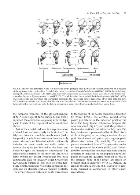
endosseous labyrinth of the inner ear (Fig. 6.5) is<br />
fairly typical for extant crocodilians (we have<br />
comparable data for Alligator, other Crocodylus,<br />
Gavialis, <strong>and</strong> numerous fossil species), with a relatively<br />
simple, triangular vestibular apparatus dorsally<br />
<strong>and</strong> an elongate cochlea (lagena) ventrally.<br />
The strongly twisted cochlea no doubt corresponds<br />
6. Using CT to Peer into the Past 73<br />
Fig. 6.5. Endosseous labyrinths of the left inner ears of the principal taxa discussed in the text, displayed on a diagram<br />
of their phylogenetic relationships. From left, the extant crocodilian Crocodylus johnstoni (OUVC 10425), the diplodocoid<br />
sauropod Diplodocus longus (CM 11161), the macronarian sauropod Camarasaurus lentus (CM 11338), the basal coelurosaurian<br />
theropod Tyrannosaurus rex (AMNH 5117), <strong>and</strong> the avian theropod (bird) Bubo virginianus (OUVC 10220).<br />
These sauropods <strong>and</strong> theropods are saurischian dinosaurs. The images are surface renderings of CT scan data. Top row:<br />
left lateral view. Middle row: dorsal view. Bottom row: caudal view. Orientations determined based on orientation of the<br />
labyrinth within the skull <strong>and</strong> with the lateral semicircular canal placed horizontally. Scale bars equal 1 cm.<br />
to the twisting of the basilar membrane described<br />
by Wever (1978). The cerebral carotid artery<br />
passes just lateral to the infl flection<br />
point of the<br />
twist. The long slender columellae (stapes) also<br />
were visualized (Fig. 6.3) <strong>and</strong> mark the position of<br />
the fenestra vestibuli (ovalis) on the labyrinth. The<br />
bony braincase is pneumatized by air-filled fidiver- ticula of the pharynx, including a median pharyngeal<br />
diverticulum <strong>and</strong> paired pharyngotympanic<br />
(Eustachian) tubes (Fig. 6.6A,B). The general<br />
pattern determined from CT is generally similar<br />
to that presented by Owen (1850) <strong>and</strong> Colbert<br />
(1946b), although the one presented here is more<br />
complete in including the long siphonial tube that<br />
passes through the quadrate bone on its way to<br />
the articular bone of the lower jaw. Based on<br />
broader studies underway by D. L. <strong>Dufeau</strong>, the<br />
cranial pneumatic system of C. johnstoni is relatively<br />
reduced in comparison to other extant<br />
crocodilians.


