sciatic nerve conduction velocity and locomotory patterns in
sciatic nerve conduction velocity and locomotory patterns in
sciatic nerve conduction velocity and locomotory patterns in

You also want an ePaper? Increase the reach of your titles
YUMPU automatically turns print PDFs into web optimized ePapers that Google loves.
Ahmad et al., The Journal of Animal & Plant Sciences, 22(4): 2012, Page: J. 878-883 Anim. Plant Sci. 22(4):2012<br />
ISSN: 1018-7081<br />
SCIATIC NERVE CONDUCTION VELOCITY AND LOCOMOTORY PATTERNS IN<br />
FROG, UROMASTIX AND RABBIT<br />
S. Ahmed 1,2 , S. Malik 2 , M. A. Azeem 3 <strong>and</strong> S. Noushad 1<br />
1. Advance Educational Institute & Research Center (AEIRC)<br />
2. Department of Physiology, University of Karachi, 75270 – Pakistan<br />
3. Department of Physiology, Faculty of Medic<strong>in</strong>e, Umm Alqura University, Makkah, K<strong>in</strong>gdom of Saudi Arabia.<br />
Correspond<strong>in</strong>g author’s email: Sadafivy@gmail.com<br />
ABSTRACT<br />
The complex <strong>in</strong>teractions among the structural features of axon, its myel<strong>in</strong> sheath <strong>and</strong> its length, determ<strong>in</strong>e the nature of<br />
<strong>nerve</strong> impulse <strong>conduction</strong>.. In addition, fibers of different diameters <strong>and</strong> degree of myel<strong>in</strong>ation determ<strong>in</strong>e the <strong>nerve</strong><br />
<strong>conduction</strong> <strong>velocity</strong> (NCV). It is therefore, hypothesized that for the same <strong>nerve</strong>, the NCV may differ <strong>in</strong> animals, due to<br />
the differences <strong>in</strong> the leg architecture <strong>and</strong> its movement dur<strong>in</strong>g locomotion. The purpose of this study was to compare<br />
right <strong>and</strong> left limb’s <strong>sciatic</strong> NCV <strong>in</strong> three different classes of animals to identify differences, if any, <strong>and</strong> to relate these<br />
differences with different types of gait locomotry pattern observed <strong>in</strong> them. The Compound Action Potential (CAP) was<br />
recorded from both the right <strong>and</strong> left isolated <strong>sciatic</strong> <strong>nerve</strong>s from adult Frog, Uromastix <strong>and</strong> Rabbit of both sexes.<br />
through Power Lab <strong>and</strong> its accessories. These CAP records were then used for the calculation of NCV, Significant<br />
difference was observed <strong>in</strong> <strong>sciatic</strong> NCV be<strong>in</strong>g greater <strong>in</strong> order of Frog>Rabbit>Uromastix. These differences were found<br />
well correlated with differences <strong>in</strong> their gait <strong>and</strong> locomotion. However, there was no significant difference among right<br />
<strong>and</strong> left <strong>sciatic</strong> <strong>nerve</strong>’s NCV for both Uromastix <strong>and</strong> Rabbit, while it was significantly different for Frog. The differences<br />
<strong>in</strong> NCV among Frog, Uromastix <strong>and</strong> Rabbit’s <strong>sciatic</strong> <strong>nerve</strong>s reflect differences <strong>in</strong> these animals for their <strong>sciatic</strong> <strong>nerve</strong><br />
diameter, leg architecture, gait <strong>and</strong> locomotion, as per their habitat.<br />
Key Words: Compound Action Potential (CAP), Nerve Conduction Velocity (NCV), Sciatic Nerve, locomotion.<br />
INTRODUCTION<br />
The <strong>conduction</strong> <strong>velocity</strong> of a myel<strong>in</strong>ated fiber<br />
depends on the complex <strong>in</strong>teractions among the structural<br />
features of the axon <strong>and</strong> its myel<strong>in</strong> sheath, <strong>and</strong> on the<br />
nature <strong>and</strong> distribution of the conductance mechanisms of<br />
the axon (Waxman, 1980). The speed of propagation for<br />
mammalian motor neurons is from 10 to 120 meters/sec,<br />
while for non-myel<strong>in</strong>ated sensory neurons it is from 5 to<br />
25 meters/sec (Kenneth, 1996). Non-myel<strong>in</strong>ated neurons<br />
fire <strong>in</strong> a cont<strong>in</strong>uous fashion, without the jumps; ion<br />
leakage allows effective closure of circuits, but it slows<br />
the rate of propagation (Kenneth, 1996). The evolution of<br />
the nervous system has been an important factor <strong>in</strong> the<br />
adaptation of animal to their environment (Abdelmelek et<br />
al., 2003). In the elongated <strong>nerve</strong>s, both the amplitude<br />
<strong>and</strong> <strong>conduction</strong> <strong>velocity</strong> of compound <strong>nerve</strong> action<br />
potential have been reported to decrease follow<strong>in</strong>g leg<br />
lengthen<strong>in</strong>g (Harumitsu et al., 2005). Body mass is an<br />
important factor regard<strong>in</strong>g locomotion, heavier animals<br />
though us<strong>in</strong>g more total energy, require less energy per<br />
unit mass to move ( Campbel <strong>and</strong> Reece, 2005).The<br />
<strong>sciatic</strong> <strong>nerve</strong>, which is the broadest <strong>nerve</strong> <strong>in</strong> the body lies<br />
deep to the gluteus maximus, superiorly. cross<strong>in</strong>g<br />
posteriorly to the obturator <strong>in</strong>ternus, gemelli <strong>and</strong> the<br />
quadratus femoris muscles (Williams et al., 1995). This<br />
<strong>nerve</strong> divides <strong>in</strong>to its tibial <strong>and</strong> common peroneal<br />
878<br />
branches approximately halfway or more down the thigh.<br />
In, approximately 12% of the cases, this <strong>nerve</strong> also<br />
branch earlier <strong>in</strong> this course, as soon as it leaves the<br />
pelvis (Moore <strong>and</strong> Dalley,, 1999). The fibers constitut<strong>in</strong>g<br />
the <strong>sciatic</strong> <strong>nerve</strong> <strong>in</strong> left leg have greater mean diameter<br />
but lower mean density than those constitut<strong>in</strong>g the right<br />
<strong>nerve</strong>. The diameter of its myel<strong>in</strong>ated fibers <strong>and</strong> the<br />
density of both the myel<strong>in</strong>ated <strong>and</strong> un-myel<strong>in</strong>ated fibers<br />
do not vary from male to female. On the other h<strong>and</strong>,<br />
diameter of the un-myel<strong>in</strong>ated axons do, s<strong>in</strong>ce the <strong>nerve</strong>s<br />
on the right side (<strong>in</strong> both sexes) have hi gher<br />
morphometric values, on average, than the contra-lateral<br />
ones ( Muglia et al’; 1995). In addition, the structural/<br />
morphological characteristics of axon <strong>and</strong> <strong>nerve</strong> itself<br />
also determ<strong>in</strong>e the impulse <strong>conduction</strong> <strong>and</strong> propagation<br />
characteristics. Further, gait <strong>and</strong> locomotion has also<br />
been mentioned to depend on the characteristics of<br />
<strong>in</strong>nervations to the muscles <strong>in</strong>volved. S<strong>in</strong>ce, <strong>in</strong>formation<br />
is not available regard<strong>in</strong>g comparison of NCV <strong>in</strong> different<br />
animal classes <strong>and</strong> species, <strong>and</strong> its relation with gait <strong>and</strong><br />
speed of locomotion, therefore, <strong>in</strong> this study, CAP has<br />
been recorded for the calculation of NCV from the<br />
isolated <strong>sciatic</strong> <strong>nerve</strong> of three animals Frog, Uromastix<br />
<strong>and</strong> Rabbit. The calculated values of NCV have been<br />
compared among these animals to expla<strong>in</strong> the differences<br />
<strong>in</strong> their gait <strong>and</strong> speed of locomotion.
Ahmad et al., J. Anim. Plant Sci. 22(4):2012<br />
MATERIALS AND METHODS<br />
Animals: 8 adult Frogs (Rana esculanta) of fresh-water<br />
or near water <strong>in</strong> damp places on l<strong>and</strong> were used. 9<br />
Uromastix that lives <strong>in</strong> dry, soft, s<strong>and</strong>y tracts with scant<br />
vegetation <strong>in</strong> burrows <strong>and</strong> 8 adult rabbits ( Oryctolagus<br />
cuniculus) lives <strong>in</strong> fields, grass-l<strong>and</strong>s <strong>and</strong> open woodl<strong>and</strong>s<br />
<strong>and</strong> <strong>in</strong> burrows underground were selected. All<br />
three animals of both sexes were selected for both right<br />
<strong>and</strong> left <strong>nerve</strong>s for dissection. Nerves from both right <strong>and</strong><br />
left sides used for record<strong>in</strong>g of NCV.<br />
Treatment Pattern: In all of the experimental animals,<br />
the <strong>sciatic</strong> <strong>nerve</strong> was exposed from its orig<strong>in</strong> (lumber<br />
vertebrae) up to knee <strong>and</strong> calf. Throughout the dissection,<br />
care was taken to prevent dry<strong>in</strong>g of exposed <strong>sciatic</strong> <strong>nerve</strong><br />
dur<strong>in</strong>g the removal of surround<strong>in</strong>g connective tissues <strong>and</strong><br />
muscles. For this purpose, respective buffer solutions all<br />
the chemicals used <strong>in</strong> the preparation of solutions were<br />
obta<strong>in</strong>ed from e. merck, germany or anala-r. For the<br />
experiments on uromastix hardwickii, similar<br />
composition of reptilian buffer was used as described by<br />
Khalil <strong>and</strong> Masseih (1954), for frog, r<strong>in</strong>ger’s solution<br />
(Franhenhauser <strong>and</strong> Widen 1965) for rabbit, Kreb’s<br />
solution ( W<strong>in</strong>egrad <strong>and</strong> Shames, 1962) was used to be<br />
dribbled over the exposed surface of <strong>nerve</strong>,<br />
<strong>in</strong>termittently. Dur<strong>in</strong>g this dissection, secondary <strong>and</strong><br />
tertiary f<strong>in</strong>e branches that were observed to arise from<br />
<strong>sciatic</strong> <strong>nerve</strong> were not traced <strong>and</strong> cut off with scissor, to<br />
follow its gross course through pelvic girdle, thigh, knee<br />
<strong>and</strong> term<strong>in</strong>ation <strong>in</strong> calf for exposure <strong>and</strong> isolation.<br />
Record<strong>in</strong>g of cap: For this purpose, isolated <strong>sciatic</strong><br />
<strong>nerve</strong> was kept <strong>in</strong> the <strong>nerve</strong> bath hav<strong>in</strong>g respective buffer<br />
solution. Care was taken to fill the bath to an extent with<br />
buffer, to avoid short circuit<strong>in</strong>g of stimulation current.<br />
The PowerLab was kept ready. The sett<strong>in</strong>gs file<br />
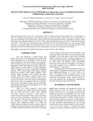
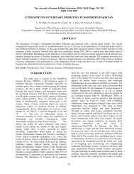
i. Left Sciatic <strong>nerve</strong> ii) Right Sciatic <strong>nerve</strong><br />
6th july<br />
6/30/2006 1:27:53.174 AM<br />
23.999 23.9995 24 24.0005 24.001 24.0015 24.002 24.0025<br />
Fig.1a Recorded trace of CAP from Sciatic <strong>nerve</strong> of Frog<br />
i. Left Sciatic <strong>nerve</strong> ii) Right Sciatic <strong>nerve</strong><br />
879<br />
associated with Power Lab experiment for NCV conta<strong>in</strong>s<br />
Chart macros that automate record<strong>in</strong>g <strong>and</strong> analysis<br />
functions. The voltage that was nearest to the one that<br />
first elicited a maximal CAP from the respective <strong>nerve</strong>s<br />
was selected. The Sett<strong>in</strong>gs used <strong>in</strong> order to have access to<br />
a pre-configured Chart Sett<strong>in</strong>gs file for experiments<br />
<strong>in</strong>cludes sampl<strong>in</strong>g rate 40k/sec. For Channel 1 <strong>and</strong> 2,<br />
Input Amplifier ranged at 50 mV. Positive checkbox <strong>and</strong><br />
Negative checkbox kept checked. Low pass filter kept off<br />
<strong>and</strong> ma<strong>in</strong> filters checkboxes were also kept unchecked.<br />
For Stimulator sett<strong>in</strong>gs, mode was kept at pulse. Its<br />
output was chosen at, ‘once only’. Marker channel kept<br />
at, ‘OFF’.<br />
Calculation of Nerve Conduction Velocity: The time<br />
difference between the two simultaneous CAPs measured<br />
from the first <strong>and</strong> second sets (pair) of record<strong>in</strong>g<br />
electrodes was measured accord<strong>in</strong>g to the procedure<br />
(PowerLab manual Model No.ML825, Serial No. 225-<br />
0815). In addition, for the distance between the two sets<br />
(pair) of record<strong>in</strong>g electrodes was also measured for the<br />
calculation of NCV by us<strong>in</strong>g the follow<strong>in</strong>g formula stated<br />
<strong>in</strong> PowerLab manual Model No.ML825, Serial No. 225-<br />
0815:<br />
Conduction <strong>velocity</strong> (meters/sec) = distance between electrodes<br />
(Cm) X __1__ time <strong>in</strong>terval between two<br />
CAPs (Sec) 100<br />
RESULTS<br />
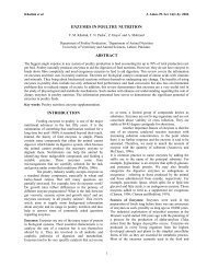
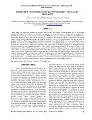
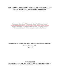
Fig. 1a, b <strong>and</strong> c, exhibits representative records<br />
of CAP recorded at two different sites of stimulation at<br />
the <strong>sciatic</strong> <strong>nerve</strong> for Frog, Uromastix <strong>and</strong> Rabbit,<br />
respectively. The distance between the latency (time of<br />
stimulation to the onset of CAP) has been found different<br />
for Uromastix when compared with Frog <strong>and</strong> Rabbit.
Ahmad et al., J. Anim. Plant Sci. 22(4):2012<br />
8/30/2006 12:20:46.258 PM<br />
19th oct-rab<br />
1.9985 1.999 1.9995 2 2.0005 2.001 2.0015 2.002 2.0025 2.003 2.0035 2.004<br />
9/14/2006 11:41:00.814 AM<br />
Fig.1b Recorded trace of CAP from Sciatic <strong>nerve</strong> of Uromastix.<br />
i. Left Sciatic <strong>nerve</strong> ii) Right Sciatic <strong>nerve</strong><br />
Copy of 29th-aug(R)<br />
1.9994 1.9996 1.9998 2 2.0002 2.0004 2.0006 2.0008 2.001 2.0012 2.0014 2.0016 2.0018 2.002<br />
The comparison of <strong>conduction</strong> <strong>velocity</strong> of left<br />
<strong>and</strong> right leg’s <strong>sciatic</strong> <strong>nerve</strong> of Frog, Uromastix <strong>and</strong><br />
Rabbit has been presented <strong>in</strong> Table 1. Accord<strong>in</strong>gly,<br />
significant difference (P0.05) between right<br />
<strong>and</strong> left <strong>sciatic</strong> <strong>nerve</strong>s of Uromastix <strong>and</strong> Rabbit.<br />
Table 1. Comparison of <strong>conduction</strong> <strong>velocity</strong> of <strong>sciatic</strong> <strong>nerve</strong> between left & right limbs of frog, uromastix & rabbit<br />
S.NO ANIMAL RIGHT<br />
NCV (m/Sec)<br />
LEFT<br />
p*<br />
1 FROG 51.371+2.011(8) 45.242+1.981(8) P0.05<br />
3 RABBIT 46.798+2.737(8) 47.94+3.168(8) P>0.05<br />
DISCUSSION<br />
Accord<strong>in</strong>g to our results, significant difference<br />
<strong>in</strong> NCV among the right <strong>and</strong> left <strong>sciatic</strong> <strong>nerve</strong>s of Frog,<br />
<strong>in</strong>dicate that the flexion <strong>and</strong> extension that occurs dur<strong>in</strong>g<br />
Frog’s jump, did not result probably <strong>in</strong> simultaneous<br />
l<strong>and</strong><strong>in</strong>g of both the legs on ground, probably due to<br />
imbalance of its body dur<strong>in</strong>g flight after jump (Rotem,<br />
2008). It is quite possible that Frogs may be hav<strong>in</strong>g<br />
preferred side or leg for l<strong>and</strong><strong>in</strong>g on to the ground at the<br />
8/30/2006 12:27:55.214 PM<br />
19th oct-rab<br />
4.9985 4.999 4.9995 5 5.0005 5.001 5.0015 5.002 5.0025 5.003 5.0035 5.004 5.0045 5.005<br />
9/14/2006 11:55:00.020 AM<br />
Copy of 29th-aug(R)<br />
2.9998 3 3.0002 3.0004 3.0006 3.0008 3.001 3.0012 3.0014 3.0016 3.0018<br />
end of each <strong>locomotory</strong> cycle. However, the nonsignificant<br />
differences observed <strong>in</strong> the NCV of right <strong>and</strong><br />
left <strong>sciatic</strong> <strong>nerve</strong>s of Uromastix <strong>and</strong> Rabbit (Table 1),<br />
<strong>in</strong>dicate non-preference <strong>in</strong> the use of right or left h<strong>in</strong>d<br />
limb <strong>in</strong> the two animals dur<strong>in</strong>g crawl<strong>in</strong>g or hopp<strong>in</strong>g<br />
movements, respectively. It is further expla<strong>in</strong>ed that<br />
dur<strong>in</strong>g locomotion either <strong>in</strong> Uromastix or Rabbit, both the<br />
limbs moves simultaneously for extension <strong>and</strong> flexion for<br />
the completion of each of the <strong>locomotory</strong> cycle. In<br />
addition, Adam <strong>and</strong> Friede (1988), collected
Ahmad et al., J. Anim. Plant Sci. 22(4):2012<br />
morphological data on Frog’s <strong>sciatic</strong> <strong>nerve</strong> which <strong>and</strong><br />
demonstrates cont<strong>in</strong>uous addition of new myel<strong>in</strong>ated <strong>and</strong><br />
non-myel<strong>in</strong>ated fibers with body growth. Adam <strong>and</strong><br />
Friede (1988) also found that along with an <strong>in</strong>crease <strong>in</strong><br />
fiber number, there is always a marked change <strong>in</strong> axon<br />
calibers <strong>and</strong> <strong>in</strong> the relative thickness of the myel<strong>in</strong><br />
sheaths as well. We assume that such changes dur<strong>in</strong>g<br />
<strong>sciatic</strong> <strong>nerve</strong> growth may be different for both the right<br />
<strong>and</strong> left legs <strong>in</strong> Frogs. Accord<strong>in</strong>g to Adam <strong>and</strong> Friede<br />
(1988), these changes are not found <strong>in</strong> mammals. We<br />
suggest that such changes <strong>in</strong> <strong>nerve</strong>s are also not<br />
associated with Uromastix (reptiles) dur<strong>in</strong>g their growth<br />
probably. Under normal circumstances, the movement of<br />
the electrical impulse down the length of a <strong>nerve</strong> is very<br />
fast, be<strong>in</strong>g 35–60m/Sec ( Rosenfalck <strong>and</strong> Rosenfalck,<br />
1975) . Accord<strong>in</strong>g to our results, NCV is highest <strong>in</strong> Frog,<br />
less <strong>in</strong> Rabbit <strong>and</strong> least <strong>in</strong> Uromastix <strong>in</strong> the order of<br />
Frog>Rabbit>Uromastix. In this connection, Waxman<br />
(1977 <strong>and</strong> 1980) stated that <strong>nerve</strong> fibers that exhibit<br />
geometrical similarity, their <strong>conduction</strong> <strong>velocity</strong> is nearly<br />
proportional to their diameter. In addition, the width of an<br />
axon varies greatly among species. The diameter of a<br />
squid axon is about 500m, lobster <strong>nerve</strong> <strong>and</strong> frog<br />
muscle axons are about 75m, <strong>and</strong> mammalian motor<br />
neurons average about 10m (Kenneth, 1996). In our<br />
op<strong>in</strong>ion, the differences <strong>in</strong> the NCV among three classes<br />
of animals, observed <strong>in</strong> the present study, also reflects the<br />
differences <strong>in</strong> their <strong>sciatic</strong> <strong>nerve</strong> diameters. Therefore, it<br />
is suggested that the diameter of <strong>sciatic</strong> <strong>nerve</strong> might be<br />
881<br />
least <strong>in</strong> Uromastix <strong>in</strong> comparison to the Frog <strong>and</strong> Rabbit<br />
<strong>and</strong> thus the obta<strong>in</strong>ed NCV is lowest <strong>in</strong> it. However, this<br />
suggestion needs confirmation through comparative<br />
morphometric studies on <strong>sciatic</strong> <strong>nerve</strong> of these animals.<br />
In addition, both the Frog <strong>and</strong> Rabbit have almost the<br />
same mode of style jump<strong>in</strong>g <strong>and</strong> hopp<strong>in</strong>g (Jordan <strong>and</strong><br />
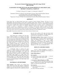
Verma 1997) . In our op<strong>in</strong>ion, the hopp<strong>in</strong>g movement of<br />
Rabbit exhibits, forward extension, then flexion, followed<br />
by backward extension <strong>in</strong> its h<strong>in</strong>d limbs, parallel to the<br />
ground (Fig. 2). While, jump<strong>in</strong>g of Frog exhibits vertical<br />
<strong>and</strong> straight jump<strong>in</strong>g movement aga<strong>in</strong>st ground, but with<br />
backward extension followed by forward flexion of its<br />
h<strong>in</strong>d limbs (Fig. 3). Hence, both the Frog <strong>and</strong> Uromastix<br />
demonstrate very fast movement dur<strong>in</strong>g locomotion. In<br />
addition, both these animals have strong h<strong>in</strong>d limbs<br />
(Jordan <strong>and</strong> Verma 1997), hav<strong>in</strong>g more fast muscles<br />
<strong>in</strong>nervated by <strong>sciatic</strong> <strong>nerve</strong>’s fast fibers, hav<strong>in</strong>g greater<br />
<strong>conduction</strong> <strong>velocity</strong>. It is the reason that the results of the<br />
present study showed non-significant difference <strong>in</strong> the<br />
NCV obta<strong>in</strong>ed from Frog <strong>and</strong> Rabbit. While, Uromastix,<br />
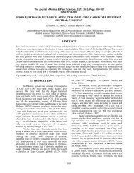
exhibits comparatively slow mode of locomotion<br />
(crawl<strong>in</strong>g), depend<strong>in</strong>g upon its gait show<strong>in</strong>g lateral<br />
movements <strong>in</strong> h<strong>in</strong>d limbs requir<strong>in</strong>g lesser force. These<br />
movements are lateral aga<strong>in</strong>st the ground (Fig. 4). In<br />
addition, the fast <strong>locomotory</strong> muscles of Uromastix are<br />
probably <strong>in</strong>nervated by small distal <strong>nerve</strong>s, hav<strong>in</strong>g slow<br />
<strong>conduction</strong> <strong>velocity</strong>. Therefore, significantly (P
Ahmad et al., J. Anim. Plant Sci. 22(4):2012<br />
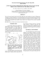
Fig. 3: Show<strong>in</strong>g photographs of Rabbit; a) sitt<strong>in</strong>g <strong>and</strong> b, c & d hop<strong>in</strong>g movements, b) forward leg extension, c) leg<br />
flexion for l<strong>and</strong><strong>in</strong>g <strong>and</strong> d) backward extension for runn<strong>in</strong>g<br />
.<br />
Fig. 4: Show<strong>in</strong>g photographs of Uromastix; a) sitt<strong>in</strong>g. (b, c & d) Lateral movements <strong>in</strong> h<strong>in</strong>d limbs. b) Slight<br />
forward extension. c) Full forward extension. d) Backward flexion for forward movement aga<strong>in</strong>st ground.<br />
A comparison of NCV between Frog <strong>and</strong> Rabbit<br />
show<strong>in</strong>g non-significant difference (P >0.05) <strong>in</strong> both the<br />
left <strong>and</strong> right <strong>sciatic</strong> <strong>nerve</strong>s (Table 2 ) is due to the fact<br />
that both of these two animals hav<strong>in</strong>g strong <strong>and</strong> long<br />
h<strong>in</strong>d limbs, <strong>and</strong> hav<strong>in</strong>g more or less similar mode of<br />
locomotion , i.e., hop or jump. Further, as suggested by<br />
Necker et al. (2000) , there is specialization <strong>in</strong> the lumbosacral<br />
vertebrae <strong>and</strong> sp<strong>in</strong>al cord <strong>in</strong> birds, which can be<br />
assumed for animals mov<strong>in</strong>g on ground. We suggest that<br />
such specialization of the lumbo-sacral vertebrae is<br />
def<strong>in</strong>itely <strong>and</strong> significantly different for Uromastix than<br />
Frog <strong>and</strong> Rabbit. Thus our results <strong>in</strong>dicate significant<br />
differences <strong>in</strong> NCV obta<strong>in</strong>ed from <strong>sciatic</strong> <strong>nerve</strong> of<br />
Uromastix to those of Frog <strong>and</strong> Rabbit (Table 2 ), as<br />
<strong>sciatic</strong> <strong>nerve</strong> character must be <strong>in</strong>fluenc<strong>in</strong>g the<br />
<strong>locomotory</strong> pattern of the <strong>in</strong>nervated <strong>locomotory</strong><br />
muscles.<br />
Conclusion: The significant difference of <strong>sciatic</strong> NCV of<br />
Uromastix <strong>in</strong> comparison with Rabbit <strong>and</strong> Frog reflects<br />
differences <strong>in</strong> these animals for their <strong>sciatic</strong> <strong>nerve</strong> course,<br />
diameter, leg architecture, gait <strong>and</strong> locomotion, as per<br />
their habitat. However, unlike Rabbit <strong>and</strong> Uromastix, the<br />
significant difference <strong>in</strong> NCV observed among right <strong>and</strong><br />
left <strong>sciatic</strong> <strong>nerve</strong>s of frog, cannot be expla<strong>in</strong>ed unless<br />
detailed morpho-metric <strong>and</strong> neurophysiological character<br />
of <strong>sciatic</strong> <strong>nerve</strong> is mapped on both the right <strong>and</strong> left sides.<br />
REFERENCES<br />
Abdelmelek, H., A. M’Chirgui, M. Ben Salem <strong>and</strong> M.<br />
Sakly (2003) Impact of evolution on the<br />
882<br />
electrical properties of <strong>sciatic</strong> <strong>nerve</strong>s:<br />
Superconductivity-like. Physical <strong>and</strong> Chemical<br />
News http://www.pcnjournal.com /volume _13_<br />
september_2003_088.htm<br />
Adam <strong>and</strong> R. L. Friede (1988).The number of frog <strong>sciatic</strong><br />
axons <strong>in</strong>creases cont<strong>in</strong>ually dur<strong>in</strong>g body growth;<br />
Anatomy <strong>and</strong> Embryology. 178(6): 537-541.<br />
Campbell, N. A. <strong>and</strong> J. B. Reece, (2005). Biology.<br />
Benjam<strong>in</strong> Cumm<strong>in</strong>gs. ISBN 0-8053-7146-X<br />
Franhenhauser, B <strong>and</strong> L. Widen (1965). Anode break<br />
excitation <strong>in</strong> de-sheathed frog <strong>nerve</strong>. J. physiol.<br />
131: 243-247.<br />
Harumitsu I., S. Takashi, A. Ichiro, H. Yuki, T. Naoto,<br />
T.mAkihito <strong>and</strong> O. Naoyuki (2005). Distribution<br />
of sodium channels dur<strong>in</strong>g <strong>nerve</strong> elongation <strong>in</strong><br />
rat peripheral <strong>nerve</strong>. J. Orthop. Sci. 10: 214–220.<br />
Jordan, E. L. <strong>and</strong> P. S. Verma (1997). Some comparative<br />
charts of vertebrate animal type. In, “Chordate<br />
Zoology”, 11th Ed; 990-1080.<br />
Kenneth, R. K. (1996). Membrane potential <strong>and</strong> action<br />
potential <strong>in</strong> electricity. In, “College Physics for<br />
Students of Biology <strong>and</strong> Chemistry”.<br />
http://www.rwc.uc.edu/koehler/biophys/contents<br />
.html<br />
Khalil, F. <strong>and</strong> G. A. Masseih (1954). Water content of<br />
desert reptiles <strong>and</strong> mammals. J. Expt. Zool. 125:<br />
407-414.<br />
Moore, K. L. <strong>and</strong> A. F. Dalley1 (1999). Cl<strong>in</strong>ically<br />
Oriented Anatomy. 4th ed. Lipp<strong>in</strong>cott Williams<br />
<strong>and</strong> Wilk<strong>in</strong>s, Philadelphia; p 558.
Ahmad et al., J. Anim. Plant Sci. 22(4):2012<br />
Muglia, U., G. Vita, R. G., Laura, C. L. R., Mammola,<br />
C.L. <strong>and</strong> G. Germana (1995). Morphometric<br />
Comparison between Contra-lateral Sciatic<br />
Nerves <strong>in</strong> the Male <strong>and</strong> Female Rabbit.<br />
Anatomia, Histologia, Embryologia. 26(2): 147<br />
– 150.<br />
Nara, T. M., P. S. Valéria, F. C. P.S., José <strong>and</strong> A. B.<br />
Amilton (2007). Recurrent laryngeal <strong>nerve</strong> postnatal<br />
development <strong>in</strong> rats. Journal of<br />
neuroscience methods. 165(1): 18-24.<br />
http://cat.<strong>in</strong>ist.fr/?aModele=afficheN<strong>and</strong>cpsidt=<br />
18955129<br />
Necker, R., A. Janssen, <strong>and</strong> A. T. Beissenhirtz (2000).<br />
Behavioral evidence of the role of lumbo-sacral<br />
anatomical specializations <strong>in</strong> pigeons <strong>in</strong><br />
ma<strong>in</strong>ta<strong>in</strong><strong>in</strong>g balance dur<strong>in</strong>g terrestrial<br />
locomotion. J. Comp. Physiol. [A], 186(4): 409-<br />
412.<br />
Rosenfalck, P., <strong>and</strong> A. Rosenfalck (1975).<br />
Electromyography-sensory <strong>and</strong> motor<br />
883<br />
<strong>conduction</strong>. F<strong>in</strong>d<strong>in</strong>gs <strong>in</strong> normal subjects. Lab of<br />
Cl<strong>in</strong>ical Neurophysiology, Copenhagen; 1-49.<br />
Rotem, A. <strong>and</strong> E. Moses (2008). Magnetic stimulation of<br />
one-dimensional neuronal cultures. Biophys J.;<br />
94:5065–5078.<br />
Waxman, S. G. (1977). Conduction <strong>in</strong> myel<strong>in</strong>ated, un -<br />
myel<strong>in</strong>ated, <strong>and</strong> de-myel<strong>in</strong>ated fibers. Arch<br />
Neurol. 34: 585-589.<br />
Waxman, S. G. (1980). Determ<strong>in</strong>ants of <strong>conduction</strong><br />
<strong>velocity</strong> <strong>in</strong> myel<strong>in</strong>ated <strong>nerve</strong> fibers Muscle<br />
Nerve. 3(2): 141-50.<br />
Williams, P. L., L. H. Bannister, M. M. L.H., Berry, P.<br />
M.M., Coll<strong>in</strong>s, M. P., Dyson, J. E. M., Dussek,<br />
<strong>and</strong> M. W. J. Ferguson (1995) Gray’s Anatomy.<br />
38th ed. Churchill Liv<strong>in</strong>gstone, Ed<strong>in</strong>burgh; p<br />
1284.<br />
W<strong>in</strong>egrad, S. <strong>and</strong> A. M. Shanes (1962) Calcium flux <strong>and</strong><br />
contractility <strong>in</strong> gu<strong>in</strong>ea pig atria. J. Gen. Physiol,<br />
345-372.