A REVISION OF THE GENUS ATELOCAUDA (UREDINALES) AND ...
A REVISION OF THE GENUS ATELOCAUDA (UREDINALES) AND ...
A REVISION OF THE GENUS ATELOCAUDA (UREDINALES) AND ...

You also want an ePaper? Increase the reach of your titles
YUMPU automatically turns print PDFs into web optimized ePapers that Google loves.
A <strong>REVISION</strong> <strong>OF</strong> <strong>THE</strong> <strong>GENUS</strong> <strong>ATELOCAUDA</strong> (<strong>UREDINALES</strong>) <strong>AND</strong> DESCRIPTION <strong>OF</strong><br />
RACOSPERMYCES GEN. NOV. FOR SOME RUSTS <strong>OF</strong> ACACIA<br />
John Walker<br />
5 Cook Street, Baulkham Hills, New South Wales 2153, Australia.<br />
Abstract<br />
The genus Atelocauda is redefined with two species, the type species A. incrustans on Lonchocarpus<br />
from Central America and A. shivasii sp. nov. on Ormosia from Queensland, Australia. Subcuticular<br />
type 7 pycnia, lobed echinulate aecial and/or uredinial urediniospores, unicellular pedicellate<br />
ornamented pigmented teliospores and hosts in Fabaceae 5. str. characterize the genus. Five Acacia<br />
rusts more recently included in Atelocauda are redisposed in Racospermyces gen. nov., and a sixth,<br />
R. tierneyi sp. nov. on Acacia harpophylla from Queensland, is described. Species of Racospermyces<br />
have subepidermal type 5 pycnia, unlobed reticulate aecial and/or uredinial urediniospores,<br />
unicellular pedicellate smooth or mainly ornamented lightly pigmented teliospores and hosts in<br />
Mimosaceae. The affinities of Atelocauda appear to lie with some Central and South American<br />
species of Dicheirinia. It is suggested that Racospermyces is Australasian in origin, associated with<br />
groups of Australasian and Pacific species of Acacia placed by some authors in the segregate genus<br />
Racosperma. Keys to the genera and species are given.<br />
Introduction<br />
Atelocauda incrustans Arthur & Cummins (1933), the type species of Atelocauda, was described from two<br />
collections made in Panama in 1920 on leaves of Lonchocarpus sp. (Fabaceae). Subcuticular, aparaphysate<br />
pycnia (type 7 of Hiratsuka & Hiratsuka 1980) and subepidermal telia producing unicellular, brown, pedicellate<br />
teliospores ornamented with block-like warts characterized the genus. No other spore stage was described.<br />
Arthur & Cummins (1933) commented that the genus resembled Uromyces in general appearance, except for<br />
the subcuticular pycnia, and that teliospore wall ornamentation was similar to that seen in Dicheirinia and<br />
Diabole. A relationship to Pileolaria, with both genera having teliospores borne singly on their pedicel, was<br />
also noted. In a discussion of species of Ravenelia and Dicheirinia on Lonchocarpus, Cummins (1937) stated<br />
that A. incrustans on the same host genus could be considered as directly derived from Dicheirinia by<br />
continued simplification'.<br />
In their summary of rust genera, Thirumalachar & Mundkur (1949) accepted Atelocauda as a valid genus but<br />
Thirumalachar & Kern (1955) reduced A. incrustans to synonymy under Pileolaria as P. incrustans (Arthur &<br />
Cummins) Thirum. & F. Kern 1955. This decision was accepted by Cummins (1959, p. 76) in his first<br />
compilation of rust genera and in his treatment of North American leguminous rusts (Qinimins 1978).<br />
Hiratsuka & Hiratsuka (1980) used the binomial P. incrustans in their list of pycnial types in the rusts.<br />
Atelocauda remained monotypic until Cummins & Hiratsuka (1983) recognized it as distinct from Pileolaria<br />
and transferred to it three Acacia rusts described originally as species of Uromyces. These were U. bicinctus<br />
McAlpine 1906 on Acacia fasciculifera Benth. from Queensland, Australia, U. digitatus G. Winter 1886 on<br />
several Acacia spp. from Australasia and Hawaii and U. koae Arthur in F. Stevens 1925 on A. koa A. Gray in<br />
Hawaii. They expanded the original description to include characters derived from the Acacia rusts i.e.<br />
subepidermal type 5 (as well as subcuticular) pycnia, and aecial uredinia and uredinia, producing spores with a<br />
reticulate wall ornamentation. Subsequently, two other Acacia rusts have been placed in Atelocauda. Ono<br />
(1984) transferred Uromyces hyalosporus Sawada 1913, described from Acacia confusa Merrill in Taiwan, to<br />
this genus and Atelocauda angustiphyllodia D.E. Gardner 1991 (as 'angustiphylloda') was described from<br />
phyllodes and witches' brooms on Acacia koa var. latifolia (Benth.) H. St John in Hawaii.<br />
There are thus now six rusts included in Atelocauda in the sense of Cummins & Hiratsuka (1983), the type<br />
species on Lonchocarpus (Fabaceae) and five species on Acacia (Mimosaceae). Hodges & Gardner (1984), Ono<br />
(1984) and Gardner (1991) have all expressed doubts about the suitability of placing the Acacia rusts in<br />
Atelocauda and the same question has interested the present author for many years. This paper presents results<br />
of investigations on the type species of Atelocauda, on a previously undescribed species of the genus on<br />
Ormosia (Fabaceae) in Australia, on the relationships of Atelocauda to the various genera suggested by Arthur<br />
& Cummins (1933) and Cummins (1937), and on the taxonomic position of the Acacia rusts.
Materials and methods<br />
Specimens studied are listed under each species. Mounts for microscopic examination were made in clear<br />
lactophenol, warmed gently to expel air and to expand the dried material and examined immediately. After<br />
scanning slides to determine the range of spore sizes present, 10 spores of each type from each specimen were<br />
measured with notes on abnormally large or small spores. Spore appendages are included in spore<br />
measurements with additional notes on the size of appendages relative to the body of the spore. Spores for SEM<br />
studies were mounted on stubs on double-sided sticky tape, gold sputter-coated in a Dynavac Minicoater<br />
SC100M and examined with a Cambridge Stereoscan 360 microscope. Herbarium abbreviations are taken from<br />
Holmgren, Keuken & Schofield (1981) and author abbreviations for fungus and plant names from Brummitt &<br />
Powell (1992). The terms and Roman numeral symbols used for rust spore states are those based primarily on<br />
morphology and used by Laundon (1967, 1973) and Savile (1968, 1988), with a qualifying term to denote<br />
function, where necessary. Thus sori that are morphologically uredinia but accompany pycnia are referred to as<br />
aecial uredinia (II 1<br />
), with the symbol II for uredinia, and the superscript 1<br />
for aecia, to denote function. A more<br />
complete discussion is given by Laundon (1967). The detailed reasons for choosing this terminology rather than<br />
the 'ontogenic' system preferred by Cummins (1959), Hiratsuka (1973), Cummins & Hiratsuka (1983) and<br />
other workers will be given elsewhere. The two terminologies are summarized by Hawksworth et al. (1995, pp.<br />
473^74). For fungal binomials, the year of publication is given after the author citation; where this is a<br />
reference listed in the reference list, the year is enclosed in brackets, otherwise no brackets are used.<br />
The position of lesions and sori on host organs is described using terms defined by Pascoe & Sutton (1986). For<br />
leguminous hosts, family concepts used in the Flora of Australia series are adopted here, with Fabaceae s. str.,<br />
Mimosaceae and Caesalpiniaceae as separate families. The identity of hosts given is that on the herbarium<br />
labels but checking of many of these is necessary, especially those in the Acacia aulacocarpa group, in the light<br />
of the recent revision by McDonald & Maslin (2000). Accurate host identification is essential in determining<br />
the precise host ranges and geographic distributions of these rusts, especially as some Acacia rust species<br />
appear to be complexes of closely related but distinct taxa, each perhaps confined to one or a small group of<br />
hosts.<br />
Taxonomy<br />
The type species of Atelocauda<br />
Two collections made in the Department Bocas del Toro, Chinguinola, Panama were listed in the original<br />
description of Atelocauda incrustans. The type collection, PUR 44631, was made in August 1920 by<br />
J.R. Johnston and the details given on the specimen label are the same as those in the original description. The<br />
other collection, PUR 44632, made by M.A. Carleton (No. 12) on 15 August 1920 has the additional locality<br />
'United Fruit Co., Farm Six' given on the specimen label. There is no evidence that the two collections are<br />
portions of the one gathering. A duplicate of PUR 44631 is also present in K. The three specimens have been<br />
examined. All show dark brown leaf lesions which penetrate the leaf thickness and occur often on either the leaf<br />
midrib or a main lateral vein. Pycnia, telia and teliospores as described originally were found on all specimens<br />
and agreed with the brief original description. Pycnia and telia were present also on a few larger lesions with<br />
whitish centers and dark borders. On PUR 44632, aecial uredinia were found surrounding pycnia on one lesion.<br />
They contained a very few teliospores characteristic of the species and many brown lobed aecial urediniospores.<br />
The lesion bearing them was on the leaf midrib and in macroscopic appearance was not different from lesions<br />
bearing pycnia and telia. Aecial urediniospores have not been found previously in this species and are described<br />
fully below.<br />
Atelocauda incrustans Arthur & Cummins, Annales Mycologici 31,41 (1933)<br />
Pileolaria incrustans (Arthur & Cummins) Thirum. & F. Kern, Bulletin of the Torrey Botanical Club 82,105<br />
(1955)<br />
Leaf lesions dark brown, both amphigenous and hologenous, 1-2 (-3) mm diam., often on the midrib or a main<br />
side vein and then elongated along the vein, to 4 mm long, thickened, bearing a mass of erumpent sori<br />
surrounding a central depressed area with minute pycnia. Pycnia (Fig. 4) subcuticular, 60-90 um diam., 30-50<br />
(-55) um high, basal layer of hyaline, cylindrical sporogenous cells to 20 um long, 2 um wide, with a hyaline<br />
peridium (Fig. 5) of elongated to hexagonal cells 6-7 x 3-5 um. Pycniospores hyaline, subglobose to somewhat<br />
angular, 2-2.5 um diam. Aecial uredinia surrounding pycnia on one leaf midrib spot, to 150 um diam.,<br />
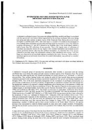
subepidermal. Aecial urediniospores (Figs 1, 9) reddish brown in a powdery mass, pedicellate, individually<br />
triangular or rarely quadrangular, 20-24 x 20-24 um, with 3, rarely 4, obtuse, apically rounded lobes making up
the body of the spore, each lobe 10-12 urn wide at a distance of 4 um below its apex, with a germ pore 2 (-3)<br />
urn diam. present in the apex of each lobe, wall to 1 um thick and covered with short hyaline spines to 1 urn<br />
high and spaced 1.5-2 um apart; hilum 4 urn wide, often with a hyaline pedicel remnant to 25 um long<br />
remaining attached. Telia surrounding pycnia, to 150 urn diam., seated in the cortex of the hypertrophied leaf<br />
spot and erumpent through the epidermis and cuticle, which remain as a marginal flap surrounding the sori,<br />
sparse marginal incurved paraphyses (Fig. 8) present, hyaline, narrowly clavate to cylindrical, 40-50 um long,<br />
4 (-7) um wide, with a uniformly thin wall or slightly thickened to 1.5-2 um at the apex, reducing the size of<br />
the lumen near the apex. Teliospores (Figs 2, 3, 6, 7,) golden brown, ellipsoidal to obovate, 22-30 x 18-22 urn,<br />
wall thin (1 um) with large, raised block-like, square, rectangular to irregular warts, often elongated along the<br />
spore, up to 2-2.5 um high, 1.5-2 um wide and, in surface view, 2-3 um across, concentrated at the spore apex<br />
but also running in lines down the spore, at their apex warts often extended into 2-4 (-6) short finger-like<br />
projections up to 1 um long (Figs 3, 7), wall uniformly 1-1.5 um thick, germ pore apical amongst apical warts,<br />
hilum 2-4 um wide, unthickened, fragment of pedicel to 30-40 um long often remaining attached.<br />
Specimens examined: Panama: Department Bocas del Toro, Chinguinola, on leaves of Lonchocarpus sp., Aug.<br />
1920, 0, III, JR. Johnston, PUR 44631, Holotype (microscope slides as DAR 69637); 0, III, duplicate of PUR<br />
44631 in K, ex IMI 65342 (microscope slide as DAR 69548); Department Bocas del Toro, United Fruit Co.,<br />
Farm Six, on leaves of Lonchocarpus sp., 15 Aug. 1920, 0, II r<br />
, III, M.A. Carlton No. 12, PUR 44632<br />
(microscope slide as DAR 72144).<br />
Atelocauda incrustans is known only from the collections made in Panama. Leaf lesions are thickened, with<br />
host cells hypertrophied and an abundant firm reddish brown substance in tissues beneath the sori. This is not<br />
present in healthy, unthickened leaf tissue. Whilst pycnia are subcuticular, other sori arise beneath the epidermis<br />
in the hypertrophied tissue. Neither aecial uredinia nor telial paraphyses have been described previously for<br />
A. incrustans. A few teliospores were present in the aecial uredinia and all spore stages appear to belong to the<br />
one species. Further collecting in Central America is needed to confirm this observation. Similar aecial<br />
urediniospores have been found in a second species of Atelocauda on Ormosia in Australia, described below.<br />
Germ pores were often difficult to detect. In aecial urediniospores, the pore at the apex of each of the two lateral<br />
lobes was usually readily seen but the third pore in the lobe seen from above was more difficult to detect. In<br />
teliospores, the apical germ pore was most easily seen between the apical tubercles in spores viewed obliquely<br />
from above. A teliospore germ pore was not mentioned in the original description by Arthur & Cummins (1933)<br />
or by Cummins (1978, as Pileolaria incrustans), although the two spores drawn in 1978 each show an apical<br />
pore. The shortly digitate apex of the teliospore warts can be observed with the light microscope but is clearly<br />
shown in SEM photographs. Paraphyses in telia were sparse and not observed in all sections. In contrast to<br />
immature teliospore pedicels, they did not stain readily in 0.1% acid fuchsin in lactophenol.<br />
An undescribed species of Atelocauda on Ormosia in Australia<br />
In August, 1992, Dr R.G. Shivas collected a leaf rust on Ormosia ormondii (F. Muell.) Merrill in North<br />
Queensland. In sending a duplicate of this rust to me for study, he commented 'The digitate teliospores<br />
resemble A digitata but the urediniospores are not those of A. digitata' (in litt. Oct. 1992). Both aecial uredinia,<br />
in association with pycnia, and solitary, unaccompanied uredinia are present. Spores present in both types of<br />
sori are brown, lobed and similar to, but distinct from, the aecial urediniospores described above for<br />
A. incrustans. The Ormosia rust is considered congeneric with A. incrustans and is described as a new species<br />
of Atelocauda.<br />
Atelocauda shivasii J. Walker sp. nov.<br />
Etymology. Roger Graham Shivas, collector, conlega, amicus.<br />
Pycnia epigena, subcuticulares, 100-130 um diam., 70-120 um alta, in gregibus 10-12 mm diam. aggregata.<br />
Sori hypogeni, subepidermales, erumpentes, ad marginem paraphysibus sparsis, (120-) 150-200 urn diam.,<br />
urediniosporas (aeciales vel urediniales) solum vel etiam teliosporas continentes. Urediniosporae aeciales<br />
pedicellatae, unicellulares, aureo-brunneae vel rubri-brunneae, rotundatae vel rhombicae lobis tribus obtusis<br />
distinctis, 28-37 x 24-28 um, echinulatae, praeter rasuram 11-16 um diam. super porum germinationem, basi<br />
poro germinatione uno. Urediniosporae urediniosporis aecialibus similes. Teliosporae pedicellatae,<br />
unicellulares, pallidae brunneae vel aureo-brunneae, infra pallidiores, late fusiformes vel late ovales, 4-6<br />
appendices apicales digitatae porum germinationem cingentes, 33^5 x 16-20 (-22) um, appendices 2-10 (—13)<br />
um longae, basim 3 urn latae, rectae, curvatae vel flexae.
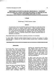
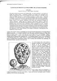
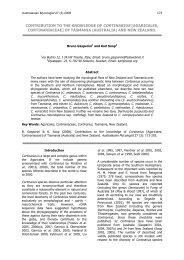
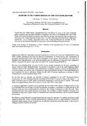
Figs 1 & 2. Atelocauda incrustans. Fig. 1. Six lobed aecial urediniospores, ex PUR 44632. Fig. 2. Two teliospores in<br />
surface view showing warts at apex and in lines on side, ex PUR 44631 (holotype). Bars: 10 um.<br />
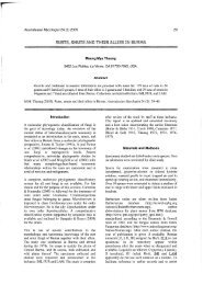
Fig. 3. Atelocauda incrustans. SEM of teliospore (partly collapsed) showing digitate warts clustered at apex and in lines on<br />
side (hilum at left of spore), ex PUR 44631 (holotype). Bar: 10 um.<br />
Holotypus hie designatus: Australia: Queensland, Horto Civiles Cape Tribulation, sectio Noah Beach, in foliis<br />
Ormosiae ormondii, 10 Aug. 1992, 0, tf, II, III, R.G. Shivas, DAR 68494 (isotypi hie designati BRIP 20529,<br />
PERTH 2595206).<br />
Leaf lesions hologenous, pale greyish brown with a darker reddish brown margin and surrounded by a yellow<br />
halo 2-3 mm wide, becoming necrotic, to 10-12 mm across or fused into larger irregular patches up to 3.5 cm<br />
long and 1.5 cm wide, or lesions absent except for pale yellow blotches. Pycnia (Fig. 14) epigenous,<br />
subcuticular but with some disruption of the underlying epidermis, grouped in the centre of the sori, 100-130<br />
um diam., 70-120 um high and protruding 40-50 um above the leaf surface, with a basal layer of sporogenous<br />
cells 20-25 um long, 2-3 um wide, pyeniospores hyaline, broadly oval, 3-4 x 2-2.5 um, marginal peridium<br />
(Fig. 15) of hyaline, septate hyphae 3—4 um wide, arranged radially around the apical pore. Sori hypogenous,<br />
arising deep in the leaf mesophyll (to a depth of 110 um below the leaf surface), erumpent, surrounded by<br />
remnants of torn epidermis and cuticle but often opening by splitting across the middle, (120-) 150-200 um<br />
diam., consisting of a basal 4-6 layers of interwoven hyphae, 18-22 um thick, which extends about half-way up
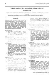
Figs 4-8. Atelocauda incrustans ex PUR 44631 (holotype). Fig. 4. Two subcuticular pycnia. Fig. 5. Fragment of pycnial<br />
peridium in surface view. Fig. 6. Two teliospores with apical and lateral warts. Fig. 7. Branched apex of warts. Fig. 8. Two<br />
paraphyses. Ban Fig. 4 = 40 urn. Fig. 5 = 20 um. Fig. 6 = 8 urn. Fig. 7 = 4 um. Fig. 8=16 um.<br />
Fig. 9. Atelocauda incrustans. Three lobed aecial urediniospores with an apical pore in each lobe, ex PUR 44632. Bar: 8<br />
um.
the sides of the sori, becoming thinner and giving rise at the margin of the sorus to a few hyaline, clavate, thinwalled<br />
paraphyses, 35-45 (-50) um long, 10-12 um thick at the rounded apex, slightly thinner below; sori<br />
either aecial uredinia (on the underside of lesions with pycnia) or uredinia (on the underside of the yellow<br />
blotches) and containing either only urediniospores or a mixture of urediniospores and teliospores. Aecial<br />
urediniospores (Figs 12, 13, 16) pedicellate, unicellular, deep golden brown to reddish brown, rounded to<br />
rhomboidal in outline, with three distinct lobes, 28-37 x<br />
24-28 um, wall 1.5 urn thick, sometimes to 2 um at the<br />
base near the hilum, closely echinulate with spines 1-1.5 um high and (1.5-) 2 (-2.5) um apart, except for one<br />
or two smooth patches (tonsures) 11-16 um diam. between the lobes above the germ pore, germ pore 1, located<br />
near the base of the spore 4-8 um above the hilum and in the basal curve of the spore wall, hilum protruding 2-<br />
4 um beyond the contour of the spore wall, sometimes a hyaline fragment of pedicel to 20 um long and 5-6 um<br />
wide remaining attached. Urediniospores similar to aecial urediniospores. Teliospores (Figs 10, 17, 19)<br />
unicellular, pedicellate, pale brown to golden brown at the apex and top third, becoming paler towards the base,<br />
broadly fusiform to broadly oval (particularly in smaller spores), with 4-6 apical digitate appendages<br />
surrounding an apical pore 4-6 um wide, spores (with appendages) measuring 33-45 x 16-20 (-22) um, wall<br />
very thin (1 um) except towards the apex where it is browner and 1.5-2 um thick, appendages 2-10 (-13) um<br />
long, 3 um wide at the base and tapering to 1.5-2 um above, with a rounded apex, straight or more commonly<br />
curved or bent, hilum 4-6 um wide, often with fragment of non-septate pedicel to 15-20 um long still attached,<br />
teliospores germinating in the sorus. Basidia (Figs 11, 18) arising from apical pore of teliospores, hyaline to<br />
pale golden yellow with granular contents, 40-45 x 8-9 um, four-celled, each cell with a tapering sterigma 10-<br />
11 um long, 3-4 um wide at the base, tapering to a small rounded tip (before basidiospore formation).<br />
Basidiospores hyaline, unicellular, oval to unequally oval to globose, 11-13 x 9-13 um.<br />
Other specimen examined: Australia: Queensland, Cape Tribulation National Park, Noah Beach section, 30<br />
July 1993, on leaves of Ormosia ormondii, 0, II 1<br />
, II, III, R.G. Shivas, BRTP 21803.<br />
Atelocauda shivasii is known from only two collections on O. ormosii from tropical Queensland. It is a fullcycled<br />
rust but apart from this, nothing is known of its seasonal life cycle. Teliospores occur in the same sori as<br />
aecial urediniospores and urediniospores. The smooth patch free of spines on the wall of the aecial<br />
urediniospores is seen readily under the light microscope. Under SEM, some spores (Fig. 13) show smooth<br />
patches on either side of a central lobe. The proportion of each spore type present varies greatly between sori<br />
from very few teliospores to predominantly teliospores. The presence of asymmetrical lobed echinulate<br />
urediniospores (aecial and Uredinial) and of appendaged unicellular pedicellate teliospores with an apical pore,<br />
together with a host in Fabaceae, relate this rust more closely to Atelocauda than to any other rust genus. The<br />
finding of aecial urediniospores in the type species of Atelocauda and in A. shivasii, and the presence of<br />
uredinia in A. shivasii, require an emendation to the generic circumscription of Atelocauda given by Arthur &<br />
Cummins (1933). Unlike the generic emendation made by Cummins & Hiratsuka (1983), the present concept of<br />
Atelocauda does not include characters drawn from the Acacia rusts. These species are discussed below.<br />
Atelocauda Arthur & Cummins emend. J. Walker<br />
Pycnia subcuticular, aparaphysate, with a peridium consisting of a single layer of septate vertical hyphae. Aecial<br />
uredinia associated with pycnia, subepidermal. Aecial urediniospores pedicellate, asymmetrical, angular and<br />
lobed, echinulate, one or more germ pores present. Uredinia not associated with pycnia, subepidermal.<br />
Urediniospores similar to aecial urediniospores. Telia subepidermal. Teliospores unicellular, pedicellate, borne<br />
singly at the pedicel apex, epispore pigmented, germ pore apical, wall ornamented especially in the upper half<br />
with tuberculate or digitate processes, often germinating in sori without a rest period. Paraphyses present at<br />
margin of sori. Known hosts in Fabaceae s. str.<br />
Relationships of Atelocauda<br />
Cmnmins (1937) suggested that, on Lonchocarpus spp. in Central and South America, there is a series of related<br />
rusts, with decreasing complexity of teliospore heads, from Ravenelia bakeriana Dietel 1908, through<br />
Dicheirinia guianensis Cummins 1937 to D. manaosensis (P. Henn.) Cummins 1935 and then D. archeri<br />
Cummins 1937. Ravenelia bakeriana has complex teliospore heads with several spores and angular lobed aecial<br />
urediniospores and urediniospores. Baxter (1968) observed occasional small 3- or 4-spored teliospore heads in<br />
R. bakeriana with modified cysts resembling the apical cells of D. guianensis. Dicheirinia guianensis and<br />
D. manaosensis both have three teliospores per head and the former has lobed aecial urediniospores and<br />
urediniospores. Dicheirinia archeri is the simplest in this series, with two teliospores per head; aecial uredinia<br />
and uredinia are not known. Cummins (1937) suggested that further simplification from D. archeri could have<br />
given rise to Atelocauda incrustans with a single teliospore. The present finding of lobed aecial urediniospores<br />
in .4. incrustans supports Cummins' (1937) hypothesis. The lobed spores of R. bakeriana and D. guianensis<br />
each have three germ pores, one located at the apex of each of the three lobes (Baxter 1968, Cummins 1937)
and they are thus similar to the aecial urediniospores of A. incrustans, described above. They are, however,<br />
much larger, those of R. bakeriana being 29^t3 x 26-35 um (Baxter 1968) and of D. guianensis 39-50 x 29-40<br />
urn (Cummins 1937).<br />
A similarity in urediniospore morphology to a species of Dicheirinia exists also with A. shivasii on Ormosia.<br />
Dicheirinia ormosiae (Arthur) Cummins 1935 occurs on O. krugii Urban in the West Indies (Dominican<br />
Republic and Puerto Rico) (Cummins 1935, Kern, Ciferri & Thurston 1933 as Puccinia ormosiae Arthur 1917,<br />
Petrak & Ciferri 1932 as P. ormosiae, Roure 1963). As noted by Cummins (1935) and observed here in two<br />
collections of D. ormosiae, urediniospores are irregularly triangular to oval, with one germ pore located just<br />
above the hilum. A smooth patch, 15 (-20) urn diam., free of echinulations, not previously reported, is present<br />
on one side of the urediniospores in their lower half (Fig. 20). This is very similar to the rhomboidal aecial<br />
urediniospores and urediniospores of A. shivasii which also have one basal germ pore associated with a smooth<br />
patch. The urediniospores of D. ormosiae measure 24-33 x 22-28 um (Cummins 1935 gives 24-32 x 20-26<br />
um) and are thus slightly smaller than those of A. shivasii.<br />
Irregularly shaped urediniospores (aecial and/or uredinial) may be characteristic for the genus Dicheirinia.<br />
Cummins (1935) described the aecial urediniospores of the generic type species, D. binata (Berk. &<br />
M.A. Curtis) Arthur 1907 as obovoid-globoid or with one side flattened'. Examination of the type specimen<br />
has shown that most spores are flattened in the vertical plane, appearing globose in face view and oval in side<br />
view (Fig. 21). There is also a smooth patch 15 (-20) um diam., free of spines or with only a few scattered<br />
spines, on one face in the lower half of the spore below the three equatorial germ pores. This has not been noted<br />
previously for this species. Asymmetrical triangular or lobed urediniospores (aecial and/or uredinial) are known<br />
in several other rusts. For example, they are common in species of Olivea Arthur (1917), Tegillum Mains (1940)<br />
(included in Olivea by Cummins & Hiratsuka 1983 and Ono & Hennen 1983) and in some species of Maravalia<br />
Arthur (1922), especially in several species formerly included in Scopella Mains (1939b, placed in Maravalia<br />
by Ono 1984).<br />
These observations on urediniospores of the two species of Atelocauda and some species of Dicheirinia provide<br />
additional evidence for a relationship between the two genera, as proposed by Arthur & Cummins (1933) and<br />
discussed by Cummins (1937).<br />
Diabole and Pileolaria were also suggested by Arthur & Cummins (1933) as possible relatives of Atelocauda,<br />
based on teliospore morphology. Diabole cubensis (Arthur & J.R. Johnson) Arthur 1922 on Mimosa in Central<br />
and South America is the only species of Diabole. It has subcuticular pycnia and teliospores consisting of a<br />
pedicel bearing 2 or 3 short apical cells each with a pair of verrucose teliospores. Aecial and uredinial stages are<br />
not known. Not enough is known about this rust to determine its possible relationships. Apart from subcuticular<br />
pycnia, it has little in common with Atelocauda on Fabaceae and is perhaps closer to some other rusts of<br />
Mimosaceae. This will be considered in more detail elsewhere under a treatment of the genus Uromycladium.<br />
Thirumalachar & Kern (1955) reduced Atelocauda to synonymy under Pileolaria. The type species,<br />
P. terebinthi Cast. 1842, and about twenty other species, occur on hosts in the Anacardiaceae, although some<br />
Acacia rusts were placed in Pileolaria by Dietel (1921, see below). Pycnia are subcuticular and teliospores<br />
globose to globose-depressed with verrucose walls. Urediniospores, when present, are commonly broadly<br />
fusiform, with longitudinal or spiral ridges or lines of warts. Leaving aside the Acacia rusts (considered below),<br />
these rusts of Anacardiaceae are morphologically quite distinct in both urediniospore and teliospore characters<br />
from Atelocauda on Fabaceae and are not considered here as closely related. Savile (1989) placed these two<br />
genera in different tribes of the Raveneliaceae, Pileolaria being grouped with Uropyxis and several other genera<br />
in the Uropyxidiae, and Atelocauda with Dicheirinia, Ravenelia and other genera in the Raveneliae. This<br />
classification is provisionally accepted here.<br />
Finally, Uredo ierensis W.T. Dale (1955) has been reported on Lonchocarpus spp. in the West Indies, Mexico,<br />
El Salvador, Guatemala and Brazil (Baxter 1968, Cummins 1978, Dale 1955, Leon Gallegos & Cummins<br />
1981). It was described by Dale (1955) with uredospores broadly ellipsoid, globoid or triangular-globoid in<br />
front view, 16-23 x 20-28 \i, more or less invaginate in side view' and with 2 or 3 approximately equatorial<br />
germ pores. Although this suggests some similarity in spore size and germ pores to the aecial urediniospores of<br />
A. incrustans, examination of portion of the type collection has shown that it is quite distinct. The taxonomic<br />
position of U. ierensis will be discussed elsewhere.
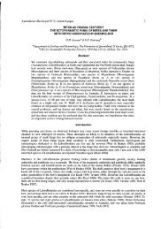
Figs 10-13. Atelocauda shivasii ex DAR 68494 (holotype). Fig. 10. Apically digitate teliospores. Fig. 11. Germinating<br />
teliospore with basidium. Fig. 12. Lobed aecial urediniospore. Fig. 13. SEM of aecial urediniospore showing smooth<br />
patches (hilum at top of spore). Bars: 10 um.<br />
Specimens examined: Dicheirinia binata (Berk. & Broome) Arthur: Nicaragua, on leaf of undetermined host<br />
[considered to be Erythrina by later workers e.g. Arthur 1925, Cummins 1978], date and collector not given, II,<br />
III, K(M) 64036, Holotype (microscope slide as DAR 72258).<br />
Dicheirinia ormosiae (Arthur) Cummins: Dominican Republic, Santo Domingo, on leaves of Ormosia krugii,<br />
3 Mar. 1930, E. Ekman 3186, II, III, IMI 101491, ex Herb. Ciferri, (microscope slide as DAR 72193);<br />
Dominica, Castle Bruce, on leaves of suspected Ormosia krugii, 2 Jan. 1972, C. Critchett, II, IMI 163804<br />
(microscope slide as DAR 72194).<br />
Uredo ierensis W.T. Dale: Trinidad, Quare Valley, on leaves of Lonchocarpus latifolius (Willd.) Humb.,<br />
Bonpl. & Kunth, 16 Dec. 1945, W.T. Dale 771, II, Isotype, PURFl 1665 (microscope slides as DAR 74665).<br />
The species of Atelocauda on Acacia, and Racospermyces gen. nov. (Uredinales)<br />
Five Acacia rusts are placed currently in Atelocauda. Four, described originally as species of Uromyces, were<br />
transferred to the genus (Cummins & Hiratsuka 1983, Ono 1984) and a fifth, A. angustiphylloda, described<br />
from Acacia koa in Hawaii (Gardner 1991). With the exception of the microcyclic A. angustiphylloda, all have<br />
aecial uredinia or uredinia or both, as well as pycnia and telia. The aecial urediniospores and urediniospores are<br />
usually similar, but not necessarily identical, variable in shape but often fusiform to oval with a raised surface<br />
reticulum and several germ pores, variously arranged but commonly in an approximately equatorial band.
Figs 14-17. Atelocauda shivasii ex DAR 68494 (holotype). Fig. 14. Subcuticular pycnium. Fig. 15. Portion of pycnial<br />
peridium in surface view. Fig. 16. Three lobed aecial urediniospores, showing smooth patch and basal pore. Fig. 17. Six<br />
teliospores, one germinating, with apical pore and appendages. Bar: Fig. 14 = 50 um. Fig. 15 = 25 um. Fig. 16 = 10 um.<br />
Fig. 17 = 20 um.
Figs 18 & 19. Atelocauda shivasii ex DAR 68494 (holotype). Fig. 18. Teliospore with basidium and one basidiospore. Fig.<br />
19. Section of telium showing marginal paraphyses. Ban 20 am.<br />
Fig. 20. Dicheirinia ormosiae. Four angular urediniospores with basal germ pore and smooth patch (a) from IMI 101491<br />
(b) from IMI 163804. Bar: 10 um.<br />
Fig. 21. Dicheirinia binata ex K(M) 64036 (holotype). Urediniospores in face and side views, with three equatorial germ<br />
pores and smooth patch. Bar: 10 um.
Teliospores are hyaline at first, pale golden brown when mature, usually darker in the upper half, from<br />
subglobose to fusiform or elongated oval, germinating through an often indistinct pore in a thin apex or in a<br />
subapical position at the side of an apically thickened wall. This germ pore is less distinct than that in the apex<br />
of teliospores of the two species of Atelocauda accepted above but much more critical work to compare the<br />
development of teliospore germ pores in these two groups is needed. Apical digitate or tuberculate appendages<br />
of varying form and extent are usually present.<br />
With their type 5 subepidermal pycnia, reticulate aecial urediniospores and urediniospores, and pale teliospores,<br />
these Acacia rusts are quite distinct from the puccinioid genus Uromyces, with type 4 pycnia (Hiratsuka &<br />
Hiratsuka 1980) and from Atelocauda s. str., as defined above. In the absence of any suitable genus to contain<br />
them, the new genus Racospermyces is described. With the exception of the generic type, the species are dealt<br />
with in chronological order of date of original description.<br />
Racospermyces J. Walker gen. nov.<br />
Etymology. Racosperma, genus plantarum ex Acacia segregatum, species hospites horum uredinalium sunt, et<br />
-myces, fungus.<br />
Pycnia subepidermalia, determinata, peridiata, aparaphysata, hymenio applanato vel parum concavo. Uredinia<br />
aeciales subepidermalia, erumpentes, pycniis concomitata. Urediniosporae aeciales pedicellatae, non-lobatae,<br />
concinnae, pariete reticulato, poris germinationibus pluribus aequatoriis vel aliter dispositis. Uredinia<br />
subepidermalia, erumpentes. Urediniosporae urediniosporis aecialis similes. Telia subepidermalia, erumpentia,<br />
saepe solida. Teliosporae unicellulares, pedicellatae, ad maturitatem pallide aureofuscae, pariete laterale tenui,<br />
paries ad apicem saepe incrassatus et plerumque appendicibus digitatis vel tuberculatis, porus germinationis<br />
praesens sed saepe obscurus, germinatio plerumque sine dormienti nunc per apicem, nunc, ubi apex incrassatus,<br />
per parietem lateralem proxime sub apice incrassato. Paraphyses saepe in soris adsunt, anguste clavatae vel<br />
cylindricae, hyalinae vel pallide flavae, interdum apice incrassato, interdum ramoso.<br />
Typus generis hie designatus: Racospermyces digitatus (G. Winter) J. Walker comb. nov.<br />
Basionym: Uromyces digitatus G. Winter, Revue de Mycologie 8,209 (1886).<br />
Pycnia subepidermal, determinate, peridiate, aparaphysate, with a flat or slightly concave hymenium (type 5,<br />
Hiratsuka & Hiratsuka 1980). Aecial uredinia subepidermal, erumpent, accompanying pycnia. Aecial<br />
urediniospores pedicellate, not lobed, symmetrical, with reticulate wall, several germ pores equatorial or<br />
otherwise arranged. Uredinia subepidermal, erumpent. Urediniospores similar to aecial urediniospores. Telia<br />
subepidermal, erumpent, often firm. Teliospores unicellular, pedicellate, pale golden brown at maturity, side<br />
wall thin, often thickened apically and commonly with digitate or tuberculate appendages, germ pore present<br />
but usually obscure, germination without a rest period, either through the apex or, when apically thickened,<br />
through the side wall below the apical thickening. Paraphyses often present in sori, narrowly clavate to<br />
cylindrical, hyaline to pale yellowish, sometimes apically thickened, occasionally branched.<br />
Racospermyces digitatus (G. Winter) J. Walker<br />
Uromyces digitatus G. Winter, Revue de Mycologie 8,209 (1886).<br />
Atelocauda<br />
(1983).<br />
digitata (G. Winter) Cummins & Y. Hirats., Illustrated Genera of Rust Fungi, revised edition, 147<br />
Melampsora phyllodiorum Berk. & Broome, The Transactions of the Linnean Society of London, ser. 2, 2, 67<br />
(1882), based on aecial uredinia.<br />
Uromyces phyllodiorum (Berk. & Broome) McAlpine, The Rusts of Australia 95 (1906).<br />
Pileolaria phyllodiorum (Berk. & Broome) Dietel, Annates Mycologici 19, 302 (1921).<br />
Uromyces phyllodii Cooke & Massee in Cooke, Grevillea 17, 70 (1889) (as i<br />
phyllodiae y<br />
uredinia.<br />
), based on aecial<br />
Pycnia mainly on hypertrophied host organs, such as bullate lesions up to 10 (-15) mm diam. and 2-4 mm high<br />
on phyllodes, smaller on leaflets of bipinnate wattles, and on small galls on twigs and pods, more rarely on<br />
small, unthickened lesions, subepidermal, pale amber but darker (often almost black) around the ostiole, 100-<br />
200 um diam., 60-70 um high, with a basal layer of sporogenous cells 20-25 um high, 2 um wide, peridium of<br />
a single layer of vertical, septate hyphae, surrounded by aecial uredinia and/or telia. Pycniospores subglobose to<br />
broadly oval, 2-4 um diam. or up to 4-5 um. Aecial uredinia with pycnia mainly on hypertrophied lesions, on<br />
some host species with pycnia occasionally on smaller unthickened lesions 3^4 x 1-2 mm, pale cinnamonbrown<br />
to pale reddish brown, singly to 250 um diam., often several fused into a soral network between the<br />
pycnia, with a cinnamon-brown granular-powdery spore mass. Aecial urediniospores pale golden yellow to
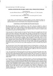
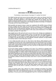
Fig. 22. Racospermyces digitatus. Variation in teliospores from six hosts. Top row: Acacia aulacocarpa s 1 BPJP 8769-<br />
A. aulacocarpa s.l. BRIP23284; A. aulacocarpa s.l. BMP 6041; A. concurrens BRIP 23071. Bottom row: A deanei BRIP<br />
14106; A. irrorata BRIP 14165; A. notabilis ex B (type; slide as DAR 30659); A. podafyriifolia BRIP 6056 Bar- 20 um
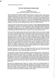
Figs 23 & 24. Racospermyces bicinctus. Fig. 23. Reticulate urediniospores with two rows of genu pores ex MEL 2061107.<br />
Fig. 24. Teliospore and urediniospores ex MEL 2061113. Bars: 10 um.<br />
golden brown, variable in shape, oval to obovate to clavate, (26-) 30-42 (-46) * (16-) 18-22 (-24) um, wall 2-<br />
4 urn thick at the sides, from unthickened to 9-12 um thick at the apex, reticulate with roughly hexagonal<br />
areolae to 2 um diam., germ pores 3-5 equatorial or rarely subequatorial, base not protruding or protruding up<br />
to 4-6 um. Uredinia unaccompanied by pycnia, on non-hypertrophied phyllode or leaf lesions 1-2 (-3) mm<br />
diam., brown, often with a thin very dark brown to almost black line-like margin or scattered or loosely<br />
clustered on unspotted areas of phyllodes or leaves, sori pale cinnamon-brown to reddish brown, to 250 um<br />
diam. singly, sometimes two or more coalescing into a larger composite sorus, granular-powdery.<br />
Urediniospores similar to aecial urediniospores but usually slightly larger, (28-) 33—45 (-55) x 16-24 (—28)<br />
um, wall 2-4 um thick at sides, from unthickened to 5-11 um thick at apex, germ pores 3-6, equatorial or rarely<br />
subequatorial, base usually protruding from slightly to 4-6 (-8) um. Telia either accompanying pycnia and then<br />
often developing in aecial uredinia, or alone, raised, single or in loose groups, often solid, pale reddish brown to<br />
reddish cinnamon, sometimes slightly glistening, often with a whitish bloom of basidia and basidiospores, to<br />
250 um diam. Teliospores (Fig. 22) at first hyaline and thin-walled, at maturity wall pale golden yellow to pale<br />
golden brown, very variable in shape, size, apical wall thickening and the development of apical appendages,<br />
often broadly fusiform to clavate, mainly (30-) 35-55 (-65) x (11-) 16-26 (-28) um but some larger forms<br />
seen, wall (1-) 1.5-2 um thick at sides, apical thickening developing as the spores mature, mainly from slight to<br />
4-16 (-18) um, some to 24 um or thicker, with (0-) 1-7 apical finger-like appendages which are straight or<br />
curved or reflexed, (2-) 4—10 (-18) x<br />
2-4 (-6) um, germinating through either an apical pore in thin apices or<br />
through a pore just below the apical thickening in other spores, pore developing just before germination.<br />
Paraphyses of variable development within and between collections, in aecial uredinia, uredinia and telia,<br />
cylindrical, thin-walled, 50-80 x 6-8 um, some thinner filaments often present.<br />
This description is a composite one, based on a series of 34 Australian collections covering 12 named Acacia<br />
spp. (nine phyllodinous and three bipinnate), three undetermined species (all phyllodinous) plus the type<br />
collections of all names listed in the synonymy. It does not include any overseas collections, or Australian<br />
collections of extreme forms, which will be dealt with elsewhere. Studies so far indicate that, as currently<br />
conceived, R. digitatus is a complex of several closely related taxa, differing in spore morphology, life cycles,<br />
host range and probably geographic distribution. Some of the variation seen in teliospore morphology on six<br />
host species is shown in Fig. 22. The synonymy given above is also a composite one, as there is no certainty at<br />
present that the aecial uredinial fungi described as Melainpsora phyllodiorum and Uromyces phyllodii belong to<br />
the same taxon as Uromyces digitatus G. Winter s. str. The type collection of U. digitatus is in poor condition<br />
but shows non-hypertrophied phyllode spots bearing true urediniospores and teliospores. The relationship of the<br />
fungus described by Winter (1886) on A. notabile to recent collections on this and other Acacia spp. requires<br />
further investigation. Work on all these problems is in progress and results will form the subject of a future<br />
paper dealing with variation within R. digitatus s. lat. These investigations will include study of the fungi from<br />
outside Australia currently placed within R. digitatus e.g. the rust of A. koa in Hawaii (Hodges & Gardner 1984)<br />
and those causing damage to several Acacia spp. in Papua New Guinea, China and other countries of South-
East Asia (Old, See, Sharma & Yuan 2000). Only a selected list of specimens examined is given here until<br />
studies on the host ranges and distributions of these R. digitatus variants are completed.<br />
Some specimens examined: Australia: South Australia, near Gawler, on phyllodes of Acacia notabilis<br />
F. Muell., 1 July 1885, II, III, J.G.O. Tepper, Holotype of Uromyces digitatus G. Winter, two fragments in B<br />
(microscope slides as DAR 29789 (no rust), DAR 30659): Queensland, Brisbane, on phyllodes of Acacia sp.,<br />
no date given, 0, II 1<br />
, EI (few, immature), F.M. Bailey 269, Holotype of Melampsora phyllodiorum, K<br />
(microscope slides as DAR 28715); duplicate, Isotype, as VPRI 5779 (microscope slides as DAR 74663);<br />
Queensland, Brisbane, on phyllodes of Acacia sp., no date given, 0, II 1<br />
, F.M. Bailey 643, Isotype of Uromyces<br />
phyllodii (as 'phyllodiae'), VPRI 5781 (microscope slides as DAR 74664); Queensland, Brisbane hilly country,<br />
on phyllodes of A. sp. aff. A. aulacocarpa Cunn. ex Benth., April 1911, II, HI, E. Jarvis, BRIP 6041<br />
(microscope slides as DAR 72323); Queensland, Marcus Beach, on phyllodes of A aulacocarpa, 17 Aug. 1972,<br />
II, HI, J.L. Alcorn 72/114, BRIP 8769 (microscope slides as DAR 72333); Queensland, near Mission Beach, on<br />
phyllodes of A. aulacocarpa, 5 Apr. 1995, 0, II, III, I. Hood, BRIP 23284 (microscope slides as DAR 72340);<br />
Queensland, Indooroopilly, on phyllodes of A. concurrens Pedley, 30 Sept. 1996, II, III, D.E. Shaw Q 1413a,<br />
BRIP 23071 (microscope slides as DAR 74659); Queensland, Indooroopilly, on leaves, twigs and pods of<br />
A. deanei (R. Baker) Welch, Coombs & McGlynn, 30 Aug. 1983, 0, JJ 1<br />
, II, III (microscope slides as DAR<br />
74654); New South Wales, Batemans Bay, 18 Mile Peg Road, on twigs, leaves and pods of A. irrorata Sieber ex<br />
Sprengel subsp. irrorata, 14 May 2000, 0, II 1<br />
, III, J.A. Simpson, DAR 72322; Queensland, Brisbane, on<br />
phyllodes of A. podalyriifolia<br />
slides as DAR 72324).<br />
Cunn. ex D. Don., 19 June 1930, II, III, R.J. McAllister, BRIP 6056 (microscope<br />
Racospermyces bicinctus (McAlpine) J. Walker comb. nov.<br />
Basionym: Uromyces bicinctus McAlpine, The Rusts of Australia 93 (1906).<br />
Pileolaria bicincta (McAlpine) Dietel, Annates Mycologici 19, 302 (1921).<br />
Atelocauda bicincta (McAlpine) Cummins & Y. Hirats., Illustrated Genera of Rust Fungi, revised edition, 147<br />
(1983).<br />
Lesions on pods and phyllodes, from 2-3 mm to 10 mm wide, grey with a thin, dark brown margin, either<br />
amphigenous or hologenous on phyllodes. Pycnia on both pod and phyllode lesions, subepidermal, amber in<br />
colour, 110-120 um wide, 60 urn high, with a basal layer of sporogenous cells 20-25 urn high, 2 um wide,<br />
peridium of a single layer of vertical hyphae; pycniospores broadly oval, hyaline, 2-2.5 x 1-1.5 um. Aecial<br />
uredinia in association with pycnia, either surrounding them or on the opposite side of the phyllode, to 250 um<br />
diam., subepidermal. Aecial urediniospores golden brown, mainly obovate, with a finely reticulate surface, 24-<br />
39 (-44) x (13.5-) 15.5-22 (-26) um, germ pores indistinct but at least 3-5 present in one row. Uredinia on<br />
pods and phyllodes, subepidermal, to 2-3 mm diam., surrounded by a rim of torn host epidermis and cuticle.<br />
Urediniospores (Figs 23-26) pale cinnamon in mass, pale golden brown singly, narrowly clavate to narrowly<br />
(occasionally broadly) obovate or cylindrical, straight or rarely slightly curved, sometimes slightly constricted<br />
in the middle, (24-) 28-33 x (11-) 13-15 (-16) um, an occasional spore to 20 um wide seen, wall 2-2.5 um<br />
thick at the sides, sometimes thickened to 3-4 um at the apex, with a finely reticulate surface which is coarser<br />
towards the apex of the spore, germ pores most commonly in two rows each of (3 or) 4 pores, rarely 5 in one<br />
row, usually one row in the top half of the spore and the other in the lower half, but sometimes one row almost<br />
equatorial and the other depressed, a few spores with only one equatorial band of pores and rarely pores in two<br />
closely intermingling rows, giving the impression of several scattered pores, germ pores 1.5-2 urn diam.,<br />
without a cap, hilum unthickened, not protruding, often concave, 4-5 um wide. Teliospores (Figs 24, 25, 27)<br />
sparse, usually borne in the same sori as urediniospores, very pale yellowish at the base and sides, golden brown<br />
in the apical third, obovate to broadly clavate to almost rectangular, 26-44 x 18-22 um, wall thin (1.5-2 um) at<br />
the sides, thickened to 7-11 um at the apex which usually has from (1-) 2-5 (-6) short digitations from 2-6 (-<br />
8) Jim long, 2-3 um wide at the base and tapering to a rounded apex, no germ pore detected, hilum 4 um wide,<br />
often with a fragment of pedicel to 6-8 urn long attached.<br />
This is a rare species of Racospermyces, represented only by specimens collected in the 1800s (earliest in 1860)<br />
on Acacia fasciculifera in the Rockhampton district of Queensland. The two collections in VPRI, which were<br />
the basis of McAlpine's (1906) original description, were taken from botanical specimens of the host in MEL.<br />
No other collections of this rust have been found in BRIP or the Queensland Forestry Herbarium. Examination<br />
of the two collections in VPRI and of several botanical sheets of A. fasciculifera in MEL collected at<br />
Rockhampton in the 1800s has revealed the presence of spore stages additional to the uredinia and telia<br />
described by McAlpine (1906). Of eleven botanical specimens of A. fasciculifera in MEL examined, the rust<br />
was found sparingly on four. As well as uredinia and telia, pycnia and aecial uredinia were observed for the first<br />
time. Lesions were very sparse and badly damaged and, due to flaking of layers of host tissue, some pycnia on
these old lesions appeared almost superficial. In the very few aecial urediniospores observed, only a single<br />
equatorial row of germ pores could be seen, thus differing from the urediniospores which characteristically have<br />
two rows of pores. However, fresh collections with abundant spores are needed to determine if this is a true<br />
difference between the two spore stages. Of the known species, R. bicinctus is most similar to the newly<br />
described R. tierneyi; both have short, compact teliospores much thickened at the apex and with apical<br />
digitations. These are less numerous and shorter in R. bicinctus than in R. tierneyi. They also have<br />
urediniospores of similar size but those of R. bicinctus have the germ pores in two rows whereas the 4-8 pores<br />
of R. tierneyi are arranged in one equatorial band.<br />
There are two collections in the McAlpine herbarium that McAlpine examined but neither is marked 'type' nor<br />
gives any indication (such as 'n. sp.') that McAlpine considered it as type. In the original description, he gave<br />
the following details: 'On phyllodes and pods of Acacia fasciculifera F.v.M. Queensland - Rockhampton, 1867<br />
(from host plant in the National Herbarium, Melbourne)'. Both VPRI 5751 and VPRI 5752 are portions of<br />
collections from plants in MEL but only one (VPRI 5751) bears the date ' 1867' and it is also the only one of the<br />
two that shows any rust. It is thus chosen as the lectotype of the name Uromyces bicinctus McAlpine. The<br />
collections in MEL from which VPRI 5751 and VPRI 5752 were taken are not known. Of the thirteen<br />
collections of A. fasciculifera from the mid-1800s examined, three (MEL 2061105, 2061109 and 2061110)<br />
were dated 1867 but rust was not found on them.<br />
Lectotypus hie designatus: Australia: Queensland, Rockhampton, on phyllodes and pods of Acacia<br />
fasciculifera Benth., 1867, II, III, G.H. Robinson (from specimen in National Herbarium), VPRI 5751 (formerly<br />
as 539/05) (microscope slide as DAR 72203, isolectotype).<br />
Other specimens examined: Australia: Queensland (all on phyllodes and pods of A. fasciculifera),<br />
Rockhampton, no date, no rust found, G.H. Robinson (from a botanical collection in MEL), VPRI 5752<br />
(formerly 538/05) (microscope slide as DAR 72202); Rockhampton, 71860, H, M.A. Thozet, MEL 2061107<br />
(microscope slide as DAR 72198); Rockhampton, no date, 0, dubious II 1<br />
, badly eroded, M.A. Thozet, MEL<br />
2061111 (microscope slide as DAR 72199); Rockhampton, no date, 0, II 1<br />
, P.O'Shanesy, MEL 2061112<br />
(microscope slide as DAR 72200); Rockhampton, no date, II, HI, J. Dallachy, MEL 2061113 (microscope slide<br />
as DAR 72201).<br />
Racospermyces hyalosporus (Sawada) J. Walker comb. nov.<br />
Basionym: Uromyces hyalosporus Sawada, The Botanical Magazine (Tokyo) 27 (No. 313-324), 19 (1913).<br />
Maravalia hyalospora (Sawada) Dietel, Annales Mycologici 22,270 (1924).<br />
Poliotelium hyalosporum (Sawada) Mains, Bulletin of the Torrey Botanical Club 66, 175 (1939) (as<br />
'hyalospora').<br />
Atelocauda hyalospora (Sawada) Ono, Mycologia 76,909 (1984).<br />
Phyllode lesions from minute (0.25-1 mm diam.) (Fig. 31) to large bullate lesions to 5 mm diam., slightly<br />
raised on the upper surface and concave on the lower, often clustered into larger groups which twist and distort<br />
the phyllode. Pycnia (Figs 31, 33) present on most lesions, only on upper surface, subepidermal, 110-150 (-<br />
160) um diam., 110-150 umhigh, with a basal layer of sporogenous cells 20-25 um high, 2 um wide, peridium<br />
(Fig. 34) of vertical septate hyphae with rectangular cells 4-8 x 3-4 um. Pycniospores subglobose to broadly<br />
oval, 3-4 um diam. Aecial uredinia (Fig. 31) subepidermal, later erumpent, 300-400 um diam., pale reddish<br />
brown, mainly epigenous and arising between the pycnia on the outer half of the lesion, occasionally<br />
amphigneous, containing both aecial urediniospores and teliospores. Aecial urediniospores (Figs 28, 35)<br />
pedicellate, golden yellow, commonly fusiform to broadly fusiform to oval or occasionally narrowly clavate,<br />
(42-) 46-70 (-75) x (20-) 22-28 (-29) um, wall 2.5-3.0 um thick at the sides, often slightly thicker in the<br />
equatorial region, a minority of spores to 4-6 (-7) um thick at the apex, finely reticulate with areolae to 1 um<br />
across, randomly arranged, germ pores 4-5 (-6), prominent, 2-2.5 urn diam., often in a slight depression and in<br />
an equatorial band, hilum 2.5-6.5 um diam., depending on spore shape and degree of basal narrowing. Uredinia<br />
not common, epigenous, less commonly amphigenous or hologenous, on minute lesions or on unspotted<br />
phyllode tissue, small, 0.25-0.5 mm diam., slightly raised, usually slightly elongated along the phyllode, single<br />
or commonly in elongated clusters up to 5 x 1 mm of 5-15 sori running along the phyllode between the veins,<br />
containing both urediniospores and teliospores. Urediniospores (Figs 29, 32) similar to aecial urediniospores<br />
but tending to be longer and thinner, (57-) 61-75 x 22-26 um, commonly thickened to (4-) 5-7 um at the apex<br />
which is often narrowed and broadly papillate. Telia developing in aecial and uredinial uredinia, 0.25-0.5 mm<br />
diam. singly, often several clustered. Teliospores (Figs 30, 37) at first hyaline, later pale golden yellow, oval to<br />
ovate or broadly obovate, apex broadly rounded or occasionally narrowed, 44-50 x 15-24 um, borne
Figs 25 & 26. Racospermyces bicinctus. Fig. 25. Two urediniospores showing two rows of germ pores and two teliospores<br />
ex VPRI 5751 (lectotype). Fig. 26. Five urediniospores showing variation in shape and germ pore arrangement ex MEL<br />
2061107. Bar: 10 um.<br />
Fig. 27. Racospermyces bicinctus. Two teliospores ex MEL 2061113. Bar: 10 um.
Figs 28-30. Racospermyces hyalosporus. Fig. 28. Four reticulate aecial urediniospores ex DAR 65442. Fig. 29. Three<br />
reticulate urediniospores ex DAR 65443. Fig. 30. Two teliospores ex DAR 65442. Bars: 10 um<br />
Fig. 31. Racospermyces hyalosporus. Very small, young, bullate phyllode lesion with central pycnia and marginal aecial<br />
uredinia ex DAR 65442. Bar: 0.5 mm.
sympodially from a basal sporogenous cell, pedicel thick, 10-12 um wide in young spores, elongating to 50-60<br />
um long and 4-6 um wide in mature spores, breaking and a fragment up to 30^10 um long often remaining<br />
attached, wall 1-2 um thick at sides, much thinner across 4-6 um of the apex through which germination<br />
occurs. Remnants of shrivelled basidia often present with teliospores. Paraphyses (Fig. 36) common in aecial<br />
uredinia, uredinia and telia, hyaline to faintly tinted yellowish, 90-110 um long, either narrow cylindrical and<br />
6-8 um wide or more commonly clavate and 10-18 um wide in their upper half and tapering to a broadly<br />
rounded apex, wall thin 1-2 um, sometimes slightly thicker at the apex.<br />
Sawada (1913) described this rust from phyllodes, pods and shoots of Acacia confusa Merrill in Taiwan<br />
(formerly Formosa). On the same host, it has been recorded from Japan (Honshu, Okinawa, Ogasawara)<br />
(Hiratsuka et al. 1992) and continental China (Cummins & Ling 1950, as Poliotelium hyalosporum). The centre<br />
of distribution of Acacia confusa is the Philippines but as far as can be determined, the rust has not been<br />
recorded on it there. It is the only known host. In Taiwan, Sawada (1913) and Hirane (1934) reported serious<br />
deformation of young shoots, especially of young trees in nurseries, with tubercular lesions on phyllodes. These<br />
often coalesce into large masses, deforming infected host organs. Sawada (1913) described only uredinia and<br />
telia but Hirane (1934) found pycnia on the tubercular lesions in association with both aecial uredinia and telia.<br />
He also reported on phyllodes smaller uredinia and telia not associated with pycnia. The rust is thus a fullcycled<br />
species, with pycnia, aecial uredinia, uredinia and telia. The most recent description is that given by<br />
Hiratsuka et al. (1992, p. 374) which agrees in essentials with that given above, except that they do not mention<br />
uredinia. In the three collections examined, sori unassociated with pycnia were not common, being found only<br />
in DAR 65443. These sori contained mainly teliospores but some urediniospores were also present.<br />
Urediniospores were slightly longer and usually slightly narrower at the base than aecial urediniospores.<br />
Sawada (1913) listed over thirty localities in Taiwan from which the rust had been studied, but no type<br />
collection was designated. It will thus be necessary to select a lectotype from whatever Sawada collections are<br />
available. Attempts to borrow them for the present investigation have been unsuccessful.<br />
Racospermyces is the fifth genus to which this rust has been assigned. Uromyces, a puccinioid rust with type 4<br />
pycnia, is unsuitable. Dietel (1921) considered placing U. hyalosporus in Pileolaria, to which he transferred<br />
several other Acacia rusts but later (Dietel 1924) disposed it in Maravalia, because of its similarity in teliospore<br />
characteristics with the type species, M. pallida Arthur & Thaxt. in Arthur 1922 on Pithecellobium latifolium<br />
(L.) Benth. (Mimosaceae) from Trinidad. In his conspectus of Uredinales, Dietel (1928) retained<br />
U. hyalosporus in Maravalia, which he considered a genus of four species, all on Mimosaceae. Mains (1939a)<br />
revised Maravalia and retained in it only species whose teliospores germinated by apical prolongation of the<br />
spore, without an evident germ pore. As he considered that teliospores of U. hyalosporus showed evidence of an<br />
apical pore, he transferred this rust to Poliotelium (a segregate genus from Uromyces based on a life cycle<br />
without uredinia, erected by Sydow 1922). However, as Ono (1984) noted, U. hyalosporus must be excluded<br />
from Poliotelium as the generic type species, P. iresines (Lagerh.) Syd. 1922 has type 4 pycnia and is a species<br />
of Uromyces, U. iresines Lagerh. (see also Laundon 1965). Ono (1984) also showed that U. hyalosporus differs<br />
in its reticulate aecial urediniospores from the 31 species he accepted in Maravalia and, with some reservations,<br />
and because of the inclusion of other Acacia rusts by Cummins & Hiratsuka (1983), he placed U. hyalosporus<br />
into Atelocauda. The reasons for excluding it from this genus have been fully detailed here.<br />
Specimens examined: Japan: Okinawa, Ishigaki Island, Institute of Tropical Agriculture, on phyllodes of<br />
Acacia confusa Merrill, 10 Sept. 1983, 0, II 1<br />
, III, Y. Ono & J.F. Hennen 1285, DAR 65441 (duplicate of IBA<br />
2826); Iriomote Island, Shirahama, Taketomi-machi, on A. confusa, 12 Sept. 1983, 0, II 1<br />
, III, Y. Ono &<br />
J.F. Hennen 1300, DAR 65442 (duplicate of IBA 2842); Okinawa Island, Nago, on .4. confusa, 7 Feb. 1985, 0,<br />
II 1<br />
, II, IE, Y. Ono 1607, DAR 65443 (duplicate of IBA 3079).<br />
Racospermyces koae (Arthur) J. Walker comb. nov.<br />
Basionym: Uromyces koae Arthur in F. Stevens, Bernice P. Bishop Museum Bulletin 19, 118 (1925).<br />
Atelocauda koae (Arthur) Cummins & Y. Hirats., Illustrated Genera of Rust Fungi, revised edition, 147 (1983).<br />
Racospermyces koae is known only from Hawaii on Acacia koa var. koa and var. latifolia. It has been studied<br />
comprehensively and its life cycle clarified by Gardner (1978, 1981), Gardner & Hodges (1985), Gardner,<br />
Miller & Kuhlman (1979) and Hodges & Gardner (1984). Although both urediniospores and teliospores were<br />
described originally for R. koae (Stevens 1925), the urediniospores described in Stevens (1925) are now known<br />
to be the aecial urediniospores of R. digitatus (Hodges & Gardner 1984). Racospermyces koae has pycnia,<br />
aecial urediniospores and teliospores in its life cycle and no true urediniospores have so far been discovered. It<br />
infects juvenile leaves, phyllodes and shoots, on which it causes severe distortion and is common on young
plants (Hodges & Gardner 1984). A full description is given by Hodges & Gardner (1984) and the spore surface<br />
morphology of R. koae and some other Acacia rusts is compared by Gardner & Hodges (1985).<br />
Specimens examined: United States of America: Hawaii, Oahu, Tantalus, on phyllodes of Acacia koa A. Gray,<br />
no date given, III, North, PUR F2888, Holotype (microscope slide as DAR 30666).<br />
Racospermyces angustiphyllodius (D.E. Gardner) J. Walker comb. nov.<br />
Basionym: Atelocauda angustiphyllodia D.E. Gardner, Mycologia 83, 650 (1991) (as 'angustiphylloda').<br />
This rust is also known only from Hawaii, on Acacia koa var. latifolia, on which it is associated with witches'<br />
broom development. It is fully described and illustrated by Gardner (1991). Phyllodes of brooms are reduced in<br />
size and brooms may be up to 1 m or more in length (Gardner 1991). The rust is considered by Gardner (1991)<br />
to be a microcyclic form of R. digitatus, producing only pycnia and telia on distorted phyllodes and branches.<br />
From ther description and illustration, its teliospores are virtually indistinguishable from those produced by<br />
R. digitatus in Hawaii. Gardner considered that this microcyclic rust merited recognition as a separate species.<br />
Its behaviour in the field is distinctive, being limited to A. koa var. latifolia, endemic to the island of Hawaii, on<br />
which it produces much larger witches' brooms than R. digitatus on the same host. So far, no specimens have<br />
been seen and it will be considered in more detail in current studies of the R. digitatus complex.<br />
The original spelling of the specific epithet has been changed. The generic name Atelocauda is feminine. The<br />
original feminine epithet 'angustiphylloda' (narrow-phylloded) denotes the much narrowed phyllodes of the<br />
witches' brooms produced by this rust. As this epithet is derived from 'angustus' (narrow) and 'phyllodium'<br />
(phyllode), its termination is altered and the spelling becomes 'angustiphyllodia', by analogy with ' angustifolia'<br />
(narrow-leaved) and in accord with Art. 32.5 of the ICBN (Greuter et al. 2000). When transferred to the<br />
masculine Racospermyces, this becomes 'angustiphyllodius'.<br />
Fig. 32. Racospermyces hyalosporus. Two urediniospores from small sori unaccompanied by pycnia ex DAR 65443. Bar:<br />
10 um.
Figs 33-37 (page 22). Racospermyces hyalosporus, all ex DAR 65441. Fig. 33. Subepidermal pycnium. Fig. 34. Fragment of<br />
pycnial peridium in surface view. Fig. 35. Three aecial urediniospores. Fig. 36. Paraphyses and basal cells. Fig. 37. Four<br />
teliospores, one showing sympodial development from basal cell. Bar: Fig. 33 = 30 um. Fig. 34 = 12 um. Figs 35-37 = 10<br />
um.<br />
Racospermyces tierneyi J. Walker & R.G. Shivas sp. nov.<br />
Etymology: John William Tiemey, 1952-1991, uredinalium Acaciae collector et studens queenslandensis, beatus.<br />
Pycnia non visa. Sori amphigeni, subepidermales, ferruginei, parum elongati secus phyllodium, 0.5 mm longi,<br />
0.25 mm lati singulatim, saepe 3-5 sori laxe aggregati, in phyllodiis super aream 2 cm longam et 2 cm latam<br />
dispersi, urediniosporas et teliosporas continentes. Paraphyses non visae. Urediniosporae formae variabilis,<br />
cylindraceae, fusiformes vel late fusiformes, obovatae vel interdum clavatae, aureae vel pallide fermgineae, 26-<br />
33 (-36) x 13-17 (-18) um, pariete 2-2.5 um crasso ad latera, 4.5-5.0 um ad apicem, reticulata cum areolis 1-<br />
1.5 um latis, pori germinationes (4-) 5-8 aequatorii, hilum 4-5 um latum, leniter vel manifesto protrudens.<br />
Teliosporae subglobosae, ovatae vel obovatae, ferrugineae, ad apicem fuscatae, 27-37 (-41) x 20-23 (-26) urn,<br />
pariete ad latera tenuissimo 1 um, ad apicem multo incrassato 7-13 um, teliosporae ad apicem 4-12 appendices<br />
digitiformes 2-8 um longas ferentes, porus germinationis unus in pariete subapicale, pedicellus ad 50-55 um<br />
longus cum fragmentum breve vel 40 um longum saepae remanens affixum. Teliosporae in soro saepe<br />
germinantes basidia et basidiosporas producentes.<br />
Holotypus hie designatus: Australia: Queensland, in regione Tambo, Castlevale, in phyllodiis Acaciae<br />
harpophyllae F. Muell. ex Benth., 6 June 2000, II, III, G. Pegg, DAR 72315 (isotypus hie designatus BRIP<br />
27071).<br />
Pycnia not seen. Sori amphigenous, subepidermal, light reddish brown, slightly elongated along the phyllode, to<br />
0.5 mm long and 0.25 mm wide singly, often 3-5 sori loosely clustered, sori scattered over an area roughly 2^2<br />
cm, containing urediniospores and teliospores. Paraphyses not seen. Urediniospores (Figs 38, 40) variable in<br />
shape, cylindrical, fusiform to broadly fusiform, obovate to sometimes clavate, golden to pale reddish brown, 26-<br />
33 (-36) x 13-17 (-18) um, with wall 2-2.5 um thick at sides, 4.5-5.0 um at apex, reticulate with areolae 1-1.5<br />
um wide, germ pores (4-) 5-8, equatorial, hilum 4-5 um wide, slightly or prominently protruding. Teliospores<br />
(Figs 38, 39, 41) subglobose, ovate to obovate, light reddish brown, darker at the apex, 27-37 (-41) x 20-23 (-<br />
26) um, with wall thin 1 um at sides, much thickened to 7-13 um at apex, bearing 4-12 apical digitate<br />
appendages 2-8 um long, a single germ pore in the subapical wall just below the apical thickening, pedicel to<br />
50-55 um long, with a short fragment or up to 40 um long remaining attached. Teliospores often germinating in<br />
the sorus, producing basidia and basidiospores, which are shrivelled in the one specimen examined.<br />
Figs 38-39. Racospermyces tierneyi, all ex DAR 72315 (holotype). Fig. 38. Urediniospores and slightly longer teliospores.<br />
Fig. 39. Teliospores showing apical appendages and one with germ pore in earliest stages of germination (arrow). Bar: 10 um.
Figs 40 & 41. Racospermyces tierneyi, all ex DAR 72315 (holotype). Fig. 40. Four urediniospores. Fig. 41. Four<br />
teliospores. Bar: 12 um.<br />
Racospermyces tierneyi is known so far only from the type specimen. It is quite distinct from the other species<br />
of Racospermyces in its short narrow urediniospores with 4-8 equatorial germ pores and in the squat, strongly<br />
apically thickened teliospores with several apical appendages. The sori are often restricted laterally by the close<br />
longitudinal veins of the phyllode and in width usually fill only one or two neighbouring interveinal spaces.<br />
They arise from a plectenchymatous stromatic mass that extends up to 200 um deep into the phyllode tissue and<br />
the long pedicels of the teliospores fan out from the top of the sorus carrying the teliospores well above the<br />
surface of the phyllode. It is most similar to R. bicinctus in teliospore characters but differs from this species in<br />
the single equatorial row of several germ pores in its urediniospores.<br />
Discussion<br />
Cummins & Hiratsuka (1983) stated that 'Teliospores are the most important spore state in generic distinctions'.<br />
Atelocauda and Racospermyces cannot be distinguished by their teliospores alone. Although no discussion of<br />
reasons was given, their similarity in teliospore characters was obviously one of the factors influencing<br />
Cummins & Hiratsuka (1983) to place all these fungi together in the genus Atelocauda. Teliospores of<br />
A. incrustans have a tuberculate ornamentation very similar to that of teliospores of R. koae (compare Fig. 3 in<br />
the present paper with Fig. 7 of Gardner & Hodges 1985) and, in the absence of other spore states, the striking<br />
similarity of the digitate teliospores of A. shivasii and those of the R. digitatus complex would suggest strongly<br />
that they belonged to the same genus. However, it is felt that, in establishing sound generic distinctions in the<br />
Uredinales, all spore states together with the life cycles, host ranges and distributions of the rusts should be
taken into account. In the present case, it is considered that the similarity in teliospores does not constitute a<br />
sound reason for placing these fungi in the one genus. The cumulative differences in urediniospores (aecial and<br />
uredinial) and in pycnial type, together with the different host families (subfamilies of some authors) are<br />
sufficient to regard the two groups of rusts as distinct genera.<br />
Treating these rusts in this way gives rise to certain biogeographical hypotheses:<br />
(i) The genus Atelocauda is of Central American origin and in the same group (?clade) as the genus<br />
Dicheirinia with several species in the region. Both the type species of Atelocauda, A. incrustans and some<br />
species of Dicheirinia occur on the genus Lonchocarpus and the possible connection between them suggested<br />
by Arthur & Cummins (1933) and Cummins (1937) has been dealt with above. A similar resemblance in<br />
urediniospore characters between the Australian A. shivasii and the Central American D. ormosiae, both on<br />
Ormosia, has been shown here. The occurrence of A. shivasii on Ormosia in Queensland is considered another<br />
example of the relationship between the floras and the fungi of the Central and South American and<br />
Australasian regions detailed earlier by Walker (1996).<br />
(ii) Racospermyces is confined to hosts in the group of Acacia species accepted by Pedley (1986,<br />
1989) (but not by some other authors, see Maslin 1989) as belonging to the segregate genus Racosperma Mart.<br />
Racospermyces has probably arisen in the Australasian region from rust ancestors of African origin, arriving<br />
with ancestraUcac/a spp. of the Racosperma group (Pedley 1986, Walker 1996). All species of Racospermyces<br />
occur on phyllodinous hosts, with the exception of some members of the R. digitatus complex which infect<br />
several bipinnate wattles. Species such as Racospermyces hyalosporus and R. koae have developed on Acacia<br />
(Racosperma group) dispersed in the Asian and Pacific area.<br />
(iii) The six species of Racospermyces are all variations on the R. digitatus theme and originated from<br />
it. As currently conceived, R. digitatus is a complex of related taxa, with varying morphologies, life cycles and<br />
host ranges. It is in an active state of differentiation and, as a complex, has the largest number of recorded hosts<br />
(over 30 species) of any Racospermyces. The three species, R. angustiphyllodius, R. bicinctus and R. tierneyi,<br />
each have only one known host and teliospores with apical thickening and varying development of digitate<br />
appendages. They are probably R. digitatus variants that have become relatively recently recognisable as<br />
distinct species on their particular hosts. In Hawaii, Hodges & Gardner (1984) considered that R. koae arose<br />
from a macrocyclic form of R. digitatus, which they suggested as the original colonizing species from which all<br />
the Hawaiian rusts of endemic Acacia spp. have evolved. Racospermyces hyalosporus differs from the other<br />
species in having non-ornamented teliospores, not thickened apically. However, its mature teliospores closely<br />
resemble immature thin-walled, non-ornamented teliospores of the ^?. digitatus complex, some of whose<br />
variants produce mature teliospores with none or very few short appendages. Production of pycnia and<br />
reticulate aecial uredinia on bullate lesions distorting host organs is similar in both species. It is suggested that<br />
R. hyalosporus is an R. digitatus derivative, which has retained pale, thin-walled teliospores, posssibly as a<br />
beneficial adaptation to moist tropical conditions (see Savile 1980).<br />
(iv) Cummins & Hiratsuka (1983, 1984) placed Atelocauda (including the Acacia rusts here disposed<br />
in Racospermyces) with Pileolaria and Uromycladium in the Pileolariaceae. Dicheirinia was included with<br />
several other genera in Raveneliaceae. As shown above, Atelocauda in the strict sense defined here shows no<br />
similarity to Pileolaria but is close to Dicheirinia and should be included in the same family. It is thus<br />
redisposed here in Raveneliaceae. The familial position of the various species of Racospermyces will be<br />
considered later together with a detailed treatment of the genus Uromycladium.<br />
With modern DNA techniques, it should be possible to test aspects of these hypotheses and uncover further<br />
evidence to help determine the precise relationships of the genera and species discussed here.<br />
Teliospore germ pores in Atelocauda and Racospermyces<br />
Preliminary observations on herbarium specimens indicate differences in teliospore germ pores in these two<br />
genera. In the two species of Atelocauda, the germ pore in mature spores is distinct, well-developed, penetrates<br />
the spore wall and is similar to that seen in many other rusts, such as species of Uromyces and Puccinia. In<br />
mature teliospores of Racospermyces spp., a germ pore in ungerminated spores is either not obvious or is<br />
recognised only by a small bulge of cytoplasm into the spore wall at the germination site. The pore appears to<br />
form shortly before germination, in the wall just to one side of the apical thickening or, in R. hyalosporus,<br />
through the thin apex. The basidial initial develops by penetration through the spore wall and not by apical<br />
elongation of the spore into a basidium. Much more work with fresh material and spores germinated<br />
experimentally is needed to categorise accurately any differences in germ pores between the two genera.
Keys<br />
Key to genera Atelocauda and Racospermyces<br />
In the key to rust genera provided by Cummins & Hiratsuka (1983), these two genera fall into Section VIII.<br />
With the inclusion of Racospermyces, their key needs to be modified as follows from step 16:<br />
16 Teliospores depressed-globoid, verrucose or reticulate Pileolaria<br />
16 Teliospores subglobose to broadly ellipsoidal or longer, smooth or with digitate appendages or blocklike<br />
warts 17<br />
17 Urediniospores (aecial and uredinial) triangular to lobed or irregular, with echinulate walls, and one or<br />
more germ pores variously arranged. Pycnia type 7. Known hosts in Fabaceae s. str. .Atelocauda<br />
17 Urediniospores (aecial and uredinial) regularly oval, ovate, obovate to fusiform, with reticulate walls, and<br />
several germ pores usually in one or two rows. Pycnia type 5. Known hosts in<br />
Mimosaceae Racospermyces<br />
Key to species of Atelocauda<br />
1 Teliospores with block-like or shortly digitate tubercles, at apex and in a few rows down the side. Aecial<br />
urediniospores lobed, with three germ pores, one in apex of each lobe. Wall completely echinulate. On<br />
Lonchocarpus, Central America. 0, II 1<br />
, III A. incrustans<br />
1 Teliospores without tubercles but with 4-6 digitate apical appendages. Aecial and uredinial urediniospores<br />
longitudinally lobed with one basal germ pore. Wall partly echinulate, with smooth patch 11-16 urn diam.<br />
above germ pore. On Ormosia, Australia (Queensland). 0, II 1<br />
, II, III .A. shivasii<br />
Key to species of Racospermyces<br />
1 Teliospores without ornamentation or appendages, apex unthickened; urediniospores (aecial and uredinial)<br />
fusiform, often over 60 um long. On Acacia confusa, China, Japan, Taiwan. 0, II 1<br />
, II, III Jt. hyalosporus<br />
1 Teliospores with tuberculate ornamentation or digitate appendages, apex often thickened; urediniospores<br />
(aecial and uredinial), when present, variable in shape, ellipsoidal, oval to ovate or obovate, fusiform,<br />
rarely to 50 um long 2<br />
2 Teliospores ornamented with several short tubercles at apex and extending in a few vertical rows down<br />
most spores. On Acacia koa, Hawaiian Islands. 0, II 1<br />
, III R. koae<br />
2 Teliospores with digitate appendages apical to subapical, not extending in vertical rows down the<br />
spore 3<br />
3 Teliospores mainly with length:breadth less than 2:1, apex much thickened, urediniospores (aecial and<br />
uredinial) only slightly shorter than teliospores 4<br />
3 Teliospores mainly with length:breadfh 2:1 to 3:1 or more, apex slightly to much thickened; urediniospores<br />
(aecial and uredinial), when present, generally much shorter than teliospores 5<br />
4 Urediniospore germ pores mostly in two distinct rows of (3 or) 4 pores each. On Acacia fasciculifera,<br />
Australia (Queensland). 0, II 1<br />
, II, III R. bicinctus<br />
4 Urediniospore germ pores mostly in one equatorial row of (5-) 6-8 pores. On Acacia harpophylla,<br />
Australia (Queensland). II, III R. tierneyi<br />
5 Urediniospores (aecial and uredinial) present, on spots, tuberculate swellings, galls and compact witches'<br />
brooms. On Acacia spp., widespread in Australasia, South-East Asia and the Hawaiian Islands. 0, II 1<br />
, II, HJ<br />
R. digitatus s. lat.<br />
5 Urediniospores (aecial and uredinial) not known. Pycnia and telia on elongated witches' brooms and<br />
phyllode spots. On Acacia koa var. latifolia, Hawaiian Islands (Hawaii, Gardner 1991). 0, III<br />
: R. angustiphyllodius
Acknowledgements<br />
Whilst much of this work has been completed recently, many notes have been built up gradually from<br />
observations over the past forty years and many people too numerous to name have assisted in this. I am<br />
grateful to the current Curators and Collection Managers of the following herbaria for lending specimens in<br />
their care and especially to the past and present colleagues named who have always helped generously in my<br />
work: Dr B. Hein (B), Dr R.G. Shivas (BRIP), Dr M.J. Priest (DAR), Dr B.C. Sutton
Hiratsuka, Y. (1973). The nuclear cycle and the terminology of spore states in Uredinales. Mycologia 65, 432-<br />
443.<br />
Hiratsuka, Y. & Hiratsuka, N. (1980). Morphology of spermogonia and taxonomy of rust fungi. Reports of the<br />
Tottori Mycological Institute (Japan) No. 18,257-268.<br />
Hiratsuka, N., Sato, S., Katsuya, K., Kakishima, M., Hiratsuka, Y., Kaneko, S., Ono, Y., Sato, T., Harada, Y.,<br />
Hiratsuka, T. &Nakayama, K. (1992). The Rust Flora of Japan. Tsukuba Shuppankai, Ibaraki.<br />
Hodges, C.S. Jr & Gardner, D.E. (1984). Hawaiian forest fungi. IV. Rusts on endemic Acacia species.<br />
Mycologia 76,332-349.<br />
Holmgren, P.K., Keuken, W. & Schofield, E.K. (1981). Index Herbariorum. Part I. The Herbaria of the World,<br />
7th edn. Bohn, Scheltema & Holkema, Utrecht.<br />
Kern, F.D., Ciferri, R. & Thurston, H.W. Jr. (1933). The rust-flora of the Dominican Republic. Annates<br />
Mycologici 31,1-40.<br />
Laundon, G.F. (1965). Rust fungi. Ill: on Alangiaceae, Amaranthaceae and Amaryllidaceae. Commonwealth<br />
Mycological Institute Mycological Papers No. 102,1-52.<br />
Laundon, G.F. (1967). Terminology in the rust fungi. Transactions of the British Mycological Society 50, 189-<br />
194.<br />
Laundon, G.F. (1973). Uredinales. In The Fungi. An Advanced Treatise Vol IVB (G.C. Ainsworth, F.K.<br />
Sparrow & A.S. Sussman, eds) Chapter 15: 247-279. Academic Press, New York & London.<br />
Leon Gallegos, H. & Cummins, G.B. (1981). Uredinales (Royas) de Mexico. Vol. II. Secretaria de Agriculture<br />
y Recursos Hidraulicos, Culiacan, Sinaloa, Mexico.<br />
Mains, E.B. (1939a). Studies in the Uredinales, the genus Maravalia. Bulletin of the Torrey Botanical Club 66,<br />
173-179.<br />
Mains, E.B. (1939b). Scopella gen. nov. of the Pucciniaceae. Annates Mycologici 37, 57-60.<br />
Mains, E.B. (1940). Tegillum, a new genus of the Uredinales. Bulletin of the Torrey Botanical Club 67, 705-<br />
709.<br />
Maslin, B. (1989). Wattle become of Acacia"? Australian Systematic Botany Society Newsletter 58,1—13.<br />
McAlpine, D. (1906). The Rusts of Australia. Their Structure, Nature and Classification. Robert S. Brain,<br />
Government Printer, Melbourne.<br />
McDonald, M.W. & Maslin, B.R. (2000). Taxonomic revision of the salwoods: Acacia aulacocarpa Cunn. ex<br />
Benth. and its allies (Leguminosae: Mimosoideae: section Juliflorae). Australian Systematic Botany 13,21—<br />
78.<br />
Old, K.M., See, L.S., Sharma, J.K. & Yuan, Z.Q. (2000). A Manual of Diseases of Tropical Acacias in<br />
Australia, South-East Asia and India. Centre for International Forestry Research, Jakarta.<br />
Ono, Y. (1984). A monograph of Maravalia (Uredinales). Mycologia 76, 892-911.<br />
Ono, Y. & Hennen, J.F. (1983). Taxonomy of the chaconiaceous genera (Uredinales). Transactions of the<br />
Mycological Society of Japan 24,369-402.<br />
Pascoe, I.G. & Sutton, B.C. (1986). Terms for the disposition of lesions and fungal structures on laminar leaves<br />
and phyllodes. Australasian Plant Pathology 15,78-80.<br />
Pedley, L. (1986). Derivation and dispersal of Acacia (Leguminosae), with particular reference to Australia, and<br />
the recognition of Senegalia and Racosperma. Botanical Journal of theLinnean Society 92,219-254.<br />
Pedley, L. (1989). 'Racosperma' again. Australian Systematic Botany Society Newsletter 59,1-2.<br />
Petrak, F. & Ciferri, R. (1932). Fungi dominicani. II. Annales Mycologici 30, 149-353.<br />
Roure, L.A. (1963). The rusts of Puerto Rico. University of Puerto Rico Agricultural Experiment Station<br />
Technical Paper No. 35,1-145.<br />
Savile, D.B.O. (1968). The case against 'uredium'. Mycologia 60,459-464.<br />
Savile, D.B.O. (1980). Ecology, convergent evolution and classification in Uredinales. Reports of the Tottori<br />
Mycological Institute (Japan) No. 18,275-281.<br />
Savile, D.B.O. (1988). Terminology of spore states in Uredinales. Mycotaxon 33,387-389.<br />
Savile, D.B.O. (1989). Raveneliaceae revisited. Canadian Journal of Botany 67,2983-2994.<br />
Sawada, K. (1913). Uromyces hyalosporus Sawada sp. nov. causing the disease to the shoots of Acacia confusa<br />
Merrill. The Botanical Magazine (Tokyo) 27, 16-20.<br />
Stevens, F.L. (1925). Hawaiian fungi. Bernice P. Bishop Museum Bulletin No. 19, i-ii, 1-189.<br />
Sydow, H. (1922). Weitere Mitteilungen zur Umgrenzung der Gattungen bei den Uredineen. Annales<br />
Mycologici 20,109-125.<br />
Thirumalachar, M.J. & Kern, F.D. (1955). The rust genera Allotelium, Atelocauda, Coinostelium and<br />
Monosporidium. Bulletin of the Torrey Botanical Club 82, 102-107.<br />
Tlrirumalachar, M.J. & Mundkur, B.B. (1949). Genera of rusts I. Indian Phytopathology 2, 65-101.<br />
Walker, J. (1996). Biogeography of fungi with special reference to Australia. Fungi of Australia 1A, 263-320.<br />
Winter, G. (1886). Fungi australienses. Revue Mycologique (Toulouse) 8,207-213.