radiological outcome in ankylosing spondylitis - Rheumatology
radiological outcome in ankylosing spondylitis - Rheumatology
radiological outcome in ankylosing spondylitis - Rheumatology

Create successful ePaper yourself
Turn your PDF publications into a flip-book with our unique Google optimized e-Paper software.
374 BRITISH JOURNAL OF RHEUMATOLOGY VOL. 35 NO. 4<br />
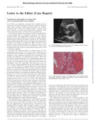
FIG. 1.—Radiographic scor<strong>in</strong>g system (see the text for details).<br />
performed <strong>in</strong> the previous 12 months. All corners of<br />
each vertebra between the lower border of T12 and the<br />
upper border of SI are exam<strong>in</strong>ed and scored 1 for<br />
erosion, squar<strong>in</strong>g or sclerosis, 2 for syndesmophyte<br />
formation, and 3 for total bony bridg<strong>in</strong>g, giv<strong>in</strong>g a<br />
maximum possible score of 72 (see Fig. 1). In ~ 10%<br />
of films, the upper sacrum is poorly shown on the<br />
lateral lumbar sp<strong>in</strong>e view and a lateral sacral film is<br />
needed <strong>in</strong> order not to compromise the analysis.<br />
Follow<strong>in</strong>g a period of tra<strong>in</strong><strong>in</strong>g, the available X-rays<br />
from the orig<strong>in</strong>al study and all the latest X-rays were<br />
scored <strong>in</strong>dependently by a rheumatologjst and a<br />
radiologist, but for the purpose of this study we<br />
subsequently exam<strong>in</strong>ed each X-ray jo<strong>in</strong>tly to agree on<br />
the f<strong>in</strong>al score. The effective radiation dose of a lateral<br />
lumbar sp<strong>in</strong>e X-ray taken at 80 kVp is 0.205 mSv.<br />
Forty-two patients had had quantitative sacroiliac<br />
sc<strong>in</strong>tigraphy us<strong>in</strong>g 450 mBq Tc-99m-labelled methylose<br />
diphosphonate (MDP) <strong>in</strong> a previous study to assess the<br />
role of isotope scann<strong>in</strong>g <strong>in</strong> the management of AS [11].<br />
Ratios were calculated, <strong>in</strong>clud<strong>in</strong>g sacroiliac: soft tissue<br />
(SIJ:ST) and sacrum:ST.<br />
Statistical analyses were performed with the Number<br />
Cruncher Statistical System 5.01. Spearman's cor-<br />
relation was used to <strong>in</strong>vestigate the relationship of<br />
SASSS with other variables.<br />
RESULTS<br />
We have def<strong>in</strong>ed <strong>in</strong>itial results as time (t) = 0 and the<br />
latest results as t = 1.<br />
Five patients failed to attend the whole study and<br />
three more were reclassified as diffuse idiopathic<br />
skeletal hyperostosis, so 53 patients with def<strong>in</strong>ite AS<br />
were fully assessed. The median age of patients at<br />
follow-up was 47.5 yr (range 32-66) and the median<br />
duration of history was 20 yr (range 9-48). The median<br />
study follow-up time was 9 yr.<br />
Thirty-two patients had def<strong>in</strong>ite syndesmophytes and<br />
21 had no sign of syndesmophytes.<br />
Change <strong>in</strong> SASSS over time<br />
The median total sp<strong>in</strong>e score at / = 0 was 5.5 (range<br />
0-52); 9 yr later the median score was 28.5 (range 0-72)<br />
(P < 0.0001).<br />
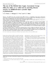
Figure 2 shows the <strong>in</strong>ter<strong>in</strong>dividual variation <strong>in</strong><br />
change <strong>in</strong> sp<strong>in</strong>e score, and <strong>in</strong> particular shows that<br />
while <strong>in</strong> some patients there is little change, others<br />
show considerable progression of <strong>radiological</strong> changes<br />
dur<strong>in</strong>g the study period. By choos<strong>in</strong>g an arbitrary value<br />
of 12 to separate the patients <strong>in</strong>to slow and fast<br />
progressors, we were unable to demonstrate any<br />
variable that could predict <strong>outcome</strong>.<br />
Change <strong>in</strong> sp<strong>in</strong>e score was not significantly related to<br />
cl<strong>in</strong>ic attendance, length of history, age of patient or<br />
age at diagnosis.<br />
Relationship between SASSS and cl<strong>in</strong>ical measurements<br />
SASSS was significantly correlated with chest<br />
expansion, occiput-wall distance, f<strong>in</strong>ger-floor distance,<br />
72 -<br />
TIME<br />
FK}. 2.—Change <strong>in</strong> Stoke AS Sp<strong>in</strong>e Score <strong>in</strong> patients with AS<br />
over 9 yr.<br />
Downloaded from<br />
http://rheumatology.oxfordjournals.org/<br />
by guest on March 29, 2013