A Summary of Tissue Lesions in Aquatic Animals Induced ... - NOAA
A Summary of Tissue Lesions in Aquatic Animals Induced ... - NOAA
A Summary of Tissue Lesions in Aquatic Animals Induced ... - NOAA

You also want an ePaper? Increase the reach of your titles
YUMPU automatically turns print PDFs into web optimized ePapers that Google loves.
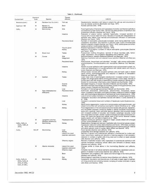
Exposure<br />
Contam<strong>in</strong>ant route Species<br />
Ammonia (coni.) W Ra<strong>in</strong>bow trout try (cont.)<br />
Cadmium 109 W Medaka fry<br />
(Oryzias latipes)<br />
CdCl, W Mummichog<br />
CdCI,; CdCl, &<br />
CuC!,; CdCl, &<br />
ZnCl,<br />
CuCl,<br />
CuCI,; CuCl, &<br />
ZnCI,; CuCl, &<br />
ZnCl, & CdCI,;<br />
CuCl, & CdCl,<br />
w<br />
w<br />
IP<br />
w<br />
w<br />
w<br />
w<br />
W& IP<br />
W<br />
W<br />
Brook trout<br />
Cunner<br />
Goldfish<br />
Sapo (Halobatrachus<br />
didactylus)<br />
Spot<br />
Langost<strong>in</strong>o (shrimp)<br />
(Penaeus kerathurus)<br />
Mummichog<br />
Mummichog<br />
Atlantic silverside<br />
Mummichog<br />
Gills<br />
Table 2.-Conllnued.<br />
<strong>Tissue</strong>s<br />
affected<br />
Yolk sac<br />
Bra<strong>in</strong><br />
Intest<strong>in</strong>e<br />
Peripheral blood<br />
Thyroid gland<br />
Kidney<br />
Testes<br />
GillS<br />
Integument<br />
Peripheral blood<br />
Intest<strong>in</strong>e<br />
Kidney<br />
Testes<br />
Ovaries<br />
Kidney<br />
Liver<br />
Peripheral blood<br />
Intest<strong>in</strong>e<br />
Liver<br />
Kidney<br />
Kidney<br />
Hepatopancreas<br />
Kidney<br />
Liver<br />
Kidney<br />
Lateral l<strong>in</strong>e canal<br />
(cephalic extension)<br />
Olfactory organs<br />
Lateral l<strong>in</strong>e canal<br />
(cephalic exlension)<br />
Olfactory organ<br />
Bra<strong>in</strong><br />
Eye<br />
Kidney<br />
<strong>Lesions</strong><br />
Developmental retardation with failure to absorb the yolk sac and occurrence <strong>of</strong><br />
blue sac disease syndrome (Burkhalter and Kaya, 1977).<br />
Nuclear pyknosis (Aoki, 1978).<br />
Focal hypertrophy <strong>of</strong> filaments and hyperplasia <strong>of</strong> lamellar and filament epithelium<br />
accompanied by high mitotic <strong>in</strong>dex, necrosis and desquamation <strong>of</strong> epithelium and<br />
lymphocyte <strong>in</strong>filtration (Gardner and Yevich, 1970).<br />
Distension <strong>of</strong> anterior portion, epithelial hypertrophy, <strong>in</strong>creased secretion <strong>of</strong><br />
mucous with mucoid casts from anus; necrosis <strong>of</strong> mucosa with desquamation <strong>of</strong><br />
epithelial cells; edema, focal necrosis and lymphocytic <strong>in</strong>filtration <strong>of</strong> submucosa<br />
(Gardner and Yevich, 1970).<br />
Numbers <strong>of</strong> eos<strong>in</strong>ophilic granulocytes <strong>in</strong>creased, some hav<strong>in</strong>g deformed nuclei,<br />
cytoplasmic vacuolation, and reduction <strong>in</strong> granular mass; nuclei <strong>of</strong> thrombocytes<br />
became irregularly shaped (Gardner and Yevich, 1970); poikilocytosis and anisocytosis<br />
primarily <strong>in</strong> erythrocytes (Gardner, 1975).<br />
Possible hyperplasia <strong>of</strong> follicles? (Gardner, 1975).<br />
Reduction <strong>of</strong> pronephric numbers <strong>of</strong> mature eos<strong>in</strong>ophilic granulocytes (Gardner<br />
and Yevich, 1970).<br />
Purple·brown mottl<strong>in</strong>g throughout, necrosis <strong>of</strong> tubular boundary cells, hemorrhage,<br />
vasodilation, and congestion (Sangalang and O'Halioran, 1972).<br />
Equivocal lesions, possibly artefactual (Newman and MacLean, 1974).<br />
Swell<strong>in</strong>g <strong>of</strong> epithelial cells and reduced mucus secretion (Newman and MacLean,<br />
1974).<br />
Poikilocytosis, karyorrhexis and abundant "smudge" cells among erythrocytes;<br />
lymphocytopenia, thrombocytopenia and neutrophilia (Newman and MacLean,<br />
1974).<br />
Swollen mucosal epithelium wrth hypertrophied nuclei and prom<strong>in</strong>ent nucleoli; necrosis<br />
and desquamation <strong>of</strong> mucosal epithelium with cellular debris and mucus <strong>in</strong><br />
lumen (Newman and MacLean, 1974).<br />
Diffuse tubular necrosis with sloughed epithelial cells and hyal<strong>in</strong> casts <strong>in</strong> dilated<br />
tubule lum<strong>in</strong>a; erythrophagocytosis and reduction or absence <strong>of</strong> hemosider<strong>in</strong><br />
(Newman and MacLean, 1974).<br />
Lower mean <strong>in</strong>dex <strong>of</strong> spermatogenic development; <strong>in</strong>creased numbers <strong>of</strong> macrophages<br />
with phagocytized debris form<strong>in</strong>g granulomas <strong>in</strong> some cases; necrosis <strong>of</strong><br />
primary germ cells with atrophy <strong>of</strong> sem<strong>in</strong>iferous tubules result<strong>in</strong>g <strong>in</strong> fibrosis and <strong>in</strong>·<br />
filtration <strong>of</strong> mononuclear <strong>in</strong>flammatory cells (Tafanelli and Summerfelt, 1975).<br />
Decreased frequency <strong>of</strong> oocyte maturation (Tafanelli and Summerfelt, 1975).<br />
Abundant <strong>in</strong>terstitial macrophage accumulation usually form<strong>in</strong>g granulomas, some<br />
tubular atrophy (Tafanelli and Summerfelt, 1975).<br />
Formation <strong>of</strong> macrophage granulomas (Tafanelli and Summerfelt, 1975).<br />
Erythrocyte anisocytosis, poikilocytosis, anisochroms;a, microcytosis, karyorrhexis,<br />
cyroplasmic vacuolation and hyperchromasia, presence <strong>of</strong> fusiform shaped<br />
cells, and morphological alterations <strong>of</strong> red blood cell nuclei (Gutierrez et aI., 1978).<br />
Loss <strong>of</strong> normal nuclear orientation <strong>in</strong> mucosal epithelium, nuclear hyperchromasia,<br />
cytoplasmic vacuolation and epithelial necrosis with debris <strong>in</strong> lumen (Gutierrez et<br />
al.,1978).<br />
Increase <strong>in</strong> connective tissue and numbers <strong>of</strong> hepatocyte nuclei (Gutierrez et aI.,<br />
1978).<br />
Renal tubular degeneration, nuclei not <strong>in</strong> normal position and hyperchromatic, and<br />
dilated lum<strong>in</strong>a filled with amorphous eos<strong>in</strong>ophilic material (Guilterez et al.. 1978).<br />
Focal degeneration <strong>of</strong> first and second proximal tubules with granular, basophilic,<br />
and vacuolated cytoplasm conta<strong>in</strong><strong>in</strong>g swollen or dense contracted mitochondria,<br />
some with a granular matrix and focal electron densities; third proximal tubules<br />
with <strong>in</strong>creased numbers <strong>of</strong> vacuoles, lipid droplets, autophagic vacuoles, nuclei<br />
with marg<strong>in</strong>ated chromat<strong>in</strong>, swollen nuclear envelopes and basal membranes con·<br />
torted <strong>in</strong>to myel<strong>in</strong>-like figures and cellular casts <strong>in</strong> the lum<strong>in</strong>a; Bowman's space<br />
slightly swollen with cell debris (Hawk<strong>in</strong>s et aI., 1980).<br />
Digestive diverticular epithelium reduced <strong>in</strong> height, tubules dilated with enlarged<br />
circular lum<strong>in</strong>a (Establier et aI., 1978c).<br />
Degenerative changes and necrosis <strong>in</strong> epithelial cells <strong>of</strong> proximal tubules conta<strong>in</strong>·<br />
<strong>in</strong>g granular casts and nuclear debris (Gardner and Yevich, 1970; Eisler and Gardner,<br />
1973); necrotic tubule epithelium appear<strong>in</strong>g as p<strong>in</strong>k sta<strong>in</strong><strong>in</strong>g granular material<br />
with either pyknotic nuclei or nuclear debris (Eisler and Gardner, 1973).<br />
Focal necrosis (Gardner and LaRoche, 1973).<br />
Unspecified renal damage (Gardner and LaRoche, 1973).<br />
Necrosis <strong>of</strong> sensory and sustentacular epithelium characterized by nuclear pyk·<br />
nosis, karyorrhexis, swell<strong>in</strong>g and reduction <strong>of</strong> sta<strong>in</strong><strong>in</strong>g capacity <strong>of</strong> cell cytoplasm;<br />
reduction <strong>in</strong> numbers <strong>of</strong> goblet cells l<strong>in</strong><strong>in</strong>g canal and no effect on basal cuboidal<br />
cells (Gardner and LaRoche, 1973).<br />
Necrosis <strong>of</strong> epithelial receptor cells, hyperplasia <strong>of</strong> sustentacular epithelium wrth<br />
cyst·like formations conta<strong>in</strong><strong>in</strong>g cellular debris and remnants <strong>of</strong> sensory tissue<br />
(Gardner and LaRoche, 1973).<br />
Changes similar to those above <strong>in</strong> the mummichog (Gardner and LaRoche,<br />
1973).<br />
Complete necrosis <strong>of</strong> all cellular elements accompanied by vasodilation and congestion<br />
<strong>of</strong> vessels <strong>in</strong> submucosa (Gardner and LaRoche, 1973).<br />
Vasodilation and congestion with rupture and hemorrhage <strong>of</strong> the menix primitiva<br />
(Gardner and LaRoche, 1973).<br />
Hemorrhag<strong>in</strong>g <strong>of</strong> periorbital connective tissue (Gardner and LaRoche, 1973).<br />
Cytoplasmic vacuolation and discont<strong>in</strong>uity <strong>of</strong> cell walls <strong>in</strong> basal region <strong>of</strong> epithelium<br />
l<strong>in</strong><strong>in</strong>g proximal and collect<strong>in</strong>g tubules; clump<strong>in</strong>g <strong>of</strong> nuclear chromat<strong>in</strong> <strong>in</strong> same<br />
cells (Eisler and Gardner, 1973).<br />
December 1982,44(12) 9