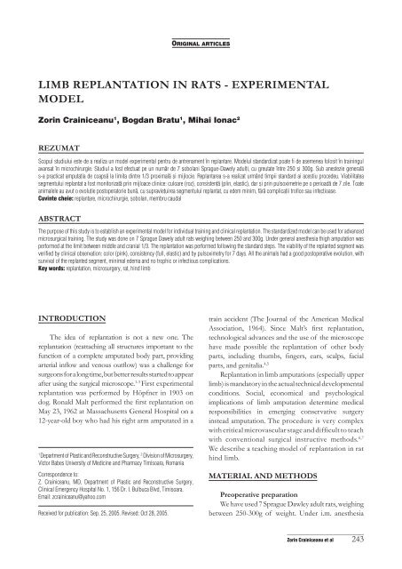
limb replantation in rats - experimental model - Timisoara Medical ...
limb replantation in rats - experimental model - Timisoara Medical ...
limb replantation in rats - experimental model - Timisoara Medical ...

Create successful ePaper yourself
Turn your PDF publications into a flip-book with our unique Google optimized e-Paper software.
LIMB REPLANTATION IN RATS - EXPERIMENTAL<br />
MODEL<br />
Zor<strong>in</strong> Cra<strong>in</strong>iceanu 1 , Bogdan Bratu 1 , Mihai Ionac 2<br />
REZUMAT<br />
Scopul studiului este de a realiza un <strong>model</strong> <strong>experimental</strong> pentru de antrenament n replantare. Modelul standardizat poate fi de asemenea folosit n tra<strong>in</strong><strong>in</strong>gul<br />
avansat n microchirurgie. Studiul a fost efectuat pe un num\r de 7 [obolani Sprague-Dawely adul]i, cu greutate ntre 250 [i 300g. Sub anestezie general\<br />
s-a practicat amputa]ia de coaps\ la limita d<strong>in</strong>tre 1/3 proximal\ [i mijlocie. Replantarea s-a realizat urmnd timpii standard ai acestiu procedeu. Viabilitatea<br />
segmentului replantat a fost monitorizat\ pr<strong>in</strong> mijloace cl<strong>in</strong>ice: culoare (roz), consisten]\ (pl<strong>in</strong>, elastic), dar [i pr<strong>in</strong> pulsoximetrie pe o perioad\ de 7 zile. Toate<br />
animalele au avut o evolu]ie postoperatorie bun\, cu supravie]uirea segmentului replantat, cu edem m<strong>in</strong>im, f\r\ complica]ii trofice sau <strong>in</strong>fec]ioase.<br />
Cuv<strong>in</strong>te cheie: replantare, microchirurgie, [obolan, membru caudal<br />
ABSTRAcT<br />
The purpose of this study is to establish an <strong>experimental</strong> <strong>model</strong> for <strong>in</strong>dividual tra<strong>in</strong><strong>in</strong>g and cl<strong>in</strong>ical <strong>replantation</strong>. The standardized <strong>model</strong> can be used for advanced<br />
microsurgical tra<strong>in</strong><strong>in</strong>g. The study was done on 7 Sprague Dawely adult <strong>rats</strong> weigh<strong>in</strong>g between 250 and 300g. Under general anesthesia thigh amputation was<br />
performed at the limit between middle and cranial 1/3. The <strong>replantation</strong> was performed follow<strong>in</strong>g the standard steps. The viability of the replanted segment was<br />
verified by cl<strong>in</strong>ical observation: color (p<strong>in</strong>k), consistency (full, elastic) and by pulsoximetry for 7 days. All the animals had a good postoperative evolution, with<br />
survival of the replanted segment, m<strong>in</strong>imal edema and no trophic or <strong>in</strong>fectious complications.<br />
Key words: <strong>replantation</strong>, microsurgery, rat, h<strong>in</strong>d <strong>limb</strong><br />
INTRODUcTION<br />
The idea of <strong>replantation</strong> is not a new one. The<br />
<strong>replantation</strong> (reattach<strong>in</strong>g all structures important to the<br />
function of a complete amputated body part, provid<strong>in</strong>g<br />
arterial <strong>in</strong>flow and venous outflow) was a challenge for<br />
surgeons for a long time, but better results started to appear<br />
after us<strong>in</strong>g the surgical microscope. 1-3 First <strong>experimental</strong><br />
<strong>replantation</strong> was performed by Höpfner <strong>in</strong> 1903 on<br />
dog. Ronald Malt performed the first <strong>replantation</strong> on<br />
May 23, 1962 at Massachusetts General Hospital on a<br />
12-year-old boy who had his right arm amputated <strong>in</strong> a<br />
1 Department of Plastic and Reconstructive Surgery, 2 Division of Microsurgery,<br />
Victor Babes University of Medic<strong>in</strong>e and Pharmacy <strong>Timisoara</strong>, Romania<br />
Correspondence to:<br />
Z. Cra<strong>in</strong>iceanu, MD, Department of Plastic and Reconstructive Surgery,<br />
Cl<strong>in</strong>ical Emergency Hospital No. 1, 156 Dr. I. Bulbuca Blvd, <strong>Timisoara</strong>.<br />
Email: zcra<strong>in</strong>iceanu@yahoo.com<br />
Received for publication: Sep. 25, 2005. Revised: Oct 28, 2005.<br />
0RIGINAL ARTICLES<br />
tra<strong>in</strong> accident (The Journal of the American <strong>Medical</strong><br />
Association, 1964). S<strong>in</strong>ce Malt’s first <strong>replantation</strong>,<br />
technological advances and the use of the microscope<br />
have made possible the <strong>replantation</strong> of other body<br />
parts, <strong>in</strong>clud<strong>in</strong>g thumbs, f<strong>in</strong>gers, ears, scalps, facial<br />
parts, and genitalia. 4,5<br />
Replantation <strong>in</strong> <strong>limb</strong> amputations (especially upper<br />
<strong>limb</strong>) is mandatory <strong>in</strong> the actual technical developmental<br />
conditions. Social, economical and psychological<br />
implications of <strong>limb</strong> amputation determ<strong>in</strong>e medical<br />
responsibilities <strong>in</strong> emerg<strong>in</strong>g conservative surgery<br />
<strong>in</strong>stead amputation. The procedure is very complex<br />
with critical microvascular stage and difficult to teach<br />
with conventional surgical <strong>in</strong>structive methods. 6,7<br />
We describe a teach<strong>in</strong>g <strong>model</strong> of <strong>replantation</strong> <strong>in</strong> rat<br />
h<strong>in</strong>d <strong>limb</strong>.<br />
MATERIAL AND METHODS<br />
Preoperative preparation<br />
We have used 7 Sprague Dawley adult <strong>rats</strong>, weigh<strong>in</strong>g<br />
between 250-300g of weight. Under i.m. anesthesia<br />
_____________________________<br />
Zor<strong>in</strong> Cra<strong>in</strong>iceanu et al 243
(xylaz<strong>in</strong>e and ketam<strong>in</strong>e) the medial and lateral thigh<br />
sides were shaved. The rat was positioned <strong>in</strong> decubitus<br />
with the posterior <strong>limb</strong>s immobilized <strong>in</strong> extension with<br />
elastic bands. The anterior <strong>limb</strong>s were free <strong>in</strong> order to<br />
permit respiratory movements. Wet swabs were used<br />
to protect the subcutaneous tissue and the exposed<br />
organs dur<strong>in</strong>g surgery.<br />
Together with the usual microsurgical <strong>in</strong>struments,<br />
for <strong>replantation</strong> we used a few osteosynthesis tools:<br />
portable motor drill, an 18G needle and a steel 3-0<br />
suture for cerclage.<br />
Anatomy<br />
The proximal segment of the h<strong>in</strong>d <strong>limb</strong> <strong>in</strong> rat<br />
<strong>in</strong>cludes: 8<br />
- skeleton, represented by the femoral bone;<br />
- the muscles of the thigh, grouped <strong>in</strong>to anterior<br />
femoral muscles, medial femoral muscles, muscles of<br />
the gluteal region and posterior femoral muscles;<br />
- the vessels: femoral artery, femoral ve<strong>in</strong> and their<br />
branches;<br />
- the peripheral nerves: femoral, obturator and<br />
sciatic nerves and their branches. (Fig. 1)<br />
Surface landmarks<br />
The limit between the middle and the proximal third<br />
of the thigh were marked, us<strong>in</strong>g as surface landmarks<br />
the <strong>in</strong>gu<strong>in</strong>al flexion fold and the knee jo<strong>in</strong>t. We drew<br />
a transverse curve l<strong>in</strong>e with its concavity towards the<br />
cranial extremity. This <strong>in</strong>cision l<strong>in</strong>e creates a cranial sk<strong>in</strong><br />
and subcutaneous flap based on the <strong>in</strong>ferior epigastric<br />
artery that will protect the anastomosis.<br />
Amputation<br />
The sk<strong>in</strong> of the medial and lateral aspect of the<br />
thigh was <strong>in</strong>cised, on the previously marked l<strong>in</strong>e. The<br />
dissection began by prepar<strong>in</strong>g the elements of the<br />
vasculo-nervous femoral bundle. The muscular branches<br />
of the femoral artery and ve<strong>in</strong> were ligated, to obta<strong>in</strong> a<br />
greater mobility of the vessels to be anastomosed. Once<br />
prepared, they can be easily protected while cutt<strong>in</strong>g the<br />
muscle and perform<strong>in</strong>g osteotomy. We cut the gluteus<br />
maximus muscle on the lateral aspect of the thigh,<br />
dissected and cut the sciatic nerve. H<strong>in</strong>d muscles were<br />
sectioned, carefully protect<strong>in</strong>g, protect<strong>in</strong>g carefully the<br />
vasculo-nervous pedicle; the bleed<strong>in</strong>g was controlled<br />
with the bipolar. (Fig. 2)<br />
Prepar<strong>in</strong>g of the femoral vessels for the future<br />
anastomosis started before cutt<strong>in</strong>g them. First<br />
microclamp was put on the ve<strong>in</strong> <strong>in</strong> a proximal<br />
position, thus caus<strong>in</strong>g its dilatation, and performed<br />
adventicectomy <strong>in</strong> good conditions. Then we clamped<br />
the distal artery, obta<strong>in</strong><strong>in</strong>g a partially dilated artery, and<br />
perform also the adventitial stripp<strong>in</strong>g. Now we cut the<br />
vasculo-nervous femoral bundle.<br />
_____________________________<br />
244 TMJ 2005, Vol. 55, No. 3<br />
Figure 1. Surgical anatomy<br />
Figure 2. Vessels and nerves disection<br />
The distal segment is now <strong>in</strong> ischemia, and the<br />
time until revascularisation should not exceed 60<br />
m<strong>in</strong>utes from this moment. We cleaned the femur for<br />
about 1cm and cut it with the circular saw, shorten<strong>in</strong>g<br />
it by 0.5 cm. Bone shorten<strong>in</strong>g will allow perform<strong>in</strong>g<br />
vascular anastomosis without any tension. (Fig. 3)<br />
Bone fixation<br />
We washed the femoral medullar channel with<br />
sal<strong>in</strong>e. We adjusted a fragment of suitable length<br />
from the 18G needle and use it for centro-medullar<br />
osteosynthesis. We performed a hole <strong>in</strong> frontal plane<br />
<strong>in</strong>to the distal and proximal femoral segments and put<br />
the cerclage wire. We put the needle <strong>in</strong>to the medullar<br />
channel and made a knot of the wire. A stable bone<br />
coaptation was obta<strong>in</strong>ed, without permitt<strong>in</strong>g bone<br />
fragments movements to put the anastomosis at risk.<br />
(Fig. 4)
Figure 3. The aspect of the amputated segment and stump<br />
Figure 4. Bone fixation<br />
Muscle and nerve suture<br />
The muscles were sutured with absorbable 4-0<br />
sutures, start<strong>in</strong>g on the medial aspect of the thigh: vastus<br />
medialis, adductors, semimembranosus, semitend<strong>in</strong>osus,<br />
gracilis and than on the lateral aspect: biceps femoris,<br />
caudofemoris and partially the gluteus medius.<br />
This is the best moment for sciatic nerve coaptation.<br />
Epiper<strong>in</strong>eural suture with a 10-0 stitch is good enough <strong>in</strong><br />
this exercise. Then we cont<strong>in</strong>ued sutur<strong>in</strong>g on the lateral<br />
aspect with gluteus medius, gluteus maximus and tensor<br />
fasciae latae. F<strong>in</strong>ish the muscle suture on the <strong>in</strong>ternal<br />
aspect: rectus femoris, vastus lateralis and <strong>in</strong>termedius. All<br />
muscles are now sutured. We performed coaptation of<br />
the femoral nerve, also us<strong>in</strong>g a 10-0 stitch. If the time runs<br />
up, we first performed the suture of the femoral vessels.<br />
Vascular suture<br />
The vascular step of the surgery started with the<br />
end to end artery anastomosis, which is situated <strong>in</strong> the<br />
distal plane, then suture the ve<strong>in</strong> by separate stitches. 9<br />
10-0 nylon suture was used. The clamps were removed<br />
<strong>in</strong> the follow<strong>in</strong>g order: first the proximally clamp of<br />
the ve<strong>in</strong>, second the distally clamp of the ve<strong>in</strong>, third<br />
the distally clamp of the artery and last the proximally<br />
clamp of the artery. (Fig. 5)<br />
After perform<strong>in</strong>g the patency tests, we covered the<br />
anastomosis with the <strong>in</strong>gu<strong>in</strong>al fat pad irrigated by the<br />
<strong>in</strong>ferior epigastric vessels and suture the sk<strong>in</strong>.<br />
Figure 5. Microsurgical sutures<br />
Sk<strong>in</strong> suture<br />
The sk<strong>in</strong> suture started on the <strong>in</strong>ternal aspect of<br />
the thigh, with separate non-absorbable 4-0 stitches.<br />
We checked the viability of the replanted segment by<br />
<strong>in</strong>spect<strong>in</strong>g the color (p<strong>in</strong>k), the turgor (full, elastic) and<br />
by pulsoxymetry. (Fig. 6)<br />
Figure 6. Pulsoxymetry monitorisation<br />
_____________________________<br />
Zor<strong>in</strong> Cra<strong>in</strong>iceanu et al 245
Postoperative care<br />
No immobilization of the h<strong>in</strong>d leg or other special<br />
treatment is necessary. For a longer term follow-up, the<br />
rat should be protected from hurt<strong>in</strong>g his anesthetized<br />
leg by us<strong>in</strong>g a plastic collar.<br />
RESULTS<br />
All the animals had a good postoperative evolution,<br />
with the survival of the replanted segment, m<strong>in</strong>imal<br />
edema and no trofic or <strong>in</strong>fectious complications.<br />
DIScUSSIONS<br />
S<strong>in</strong>ce the first successful digit <strong>replantation</strong> with<br />
microvascular repair (reported by Komatsu and Tamai,<br />
1968), the laboratory microsurgical tra<strong>in</strong><strong>in</strong>g importance<br />
was obvious, because of surgical procedure complexity<br />
and critical microvascular stage be<strong>in</strong>g difficult to teach<br />
us<strong>in</strong>g conventional surgical <strong>in</strong>structive methods. 7,10<br />
Most <strong>model</strong>s from literature use similar technique.<br />
Some authors describe techniques very similar with<br />
the cl<strong>in</strong>ical approach, first detach<strong>in</strong>g the <strong>limb</strong> by clear<br />
cut amputation and than debridement, dissection of<br />
the vessels, nerves, muscle and bone, bone fixation,<br />
muscle suture, vessels and nerve repair sk<strong>in</strong> suture. 6,8<br />
We consider that nerve and vessels dissection and<br />
clamp<strong>in</strong>g before amputation is mandatory for limited<br />
bleed<strong>in</strong>g and ischemic time shorten<strong>in</strong>g. Also, the bone<br />
fixation by p<strong>in</strong>e and <strong>in</strong>traosseous wir<strong>in</strong>g will provide<br />
better bone alignment and stability than us<strong>in</strong>g p<strong>in</strong>e<br />
only.<br />
The rat is very convenient animal for teach<strong>in</strong>g<br />
microsurgery. They are relatively easy to obta<strong>in</strong>,<br />
manipulate and anesthetize and the size of their vessels<br />
is equivalent to digital vessels. They also provide real<br />
<strong>limb</strong> structures handl<strong>in</strong>g (bone, muscles, etc.), different<br />
from other <strong>model</strong>s. 11,12,14 Our <strong>model</strong> can be used for<br />
junior surgeons tra<strong>in</strong><strong>in</strong>g to improve their <strong>in</strong>struction<br />
and practice and avoid many possible pitfalls. This<br />
_____________________________<br />
246 TMJ 2005, Vol. 55, No. 3<br />
<strong>model</strong> teaches also microsurgical technique and <strong>limb</strong><br />
structure handl<strong>in</strong>g but also surgical steps <strong>in</strong> <strong>replantation</strong><br />
and postoperative manage of the replanted segment.<br />
cONcLUSIONS<br />
Consider<strong>in</strong>g <strong>limb</strong> <strong>replantation</strong> a very important<br />
surgery, with high risk level, the laboratory tra<strong>in</strong><strong>in</strong>g<br />
is a primordial step to avoid pitfalls, achieve good<br />
survival rate and functional results. Our <strong>model</strong> of<br />
rat h<strong>in</strong>d <strong>replantation</strong> <strong>in</strong>sures all surgical steps of the<br />
<strong>replantation</strong>. It can be considered the ideal <strong>experimental</strong><br />
<strong>model</strong> for tra<strong>in</strong><strong>in</strong>g <strong>in</strong> <strong>replantation</strong> to improve cl<strong>in</strong>ical<br />
proficiency.<br />
REFERENcES<br />
1. Hopfner E. Ueber Gefassnaht, Gefasstransplantationen und Replantation<br />
von Amputierten Extremitaten. Arch Kl<strong>in</strong> Chir 1903;70:417-71.<br />
2. Malt R, Mc Khann C. Replantation of severed arms. J Amer Med Assoc<br />
1964;189:716-22.<br />
3. Biemer E. Def<strong>in</strong>itions and classifications <strong>in</strong> <strong>replantation</strong> surgery. Brit J<br />
Plast Surg 1980;33:164-8.<br />
4. Biemer E, Duspiva W. Rec Micro Surg 1982;56-58.<br />
5. Jones NF. Replantation <strong>in</strong> the Upper Extremity, Ch. 82, Grabb and<br />
Smith’s Plastic Sugery. 5th Ed., Lipp<strong>in</strong>cott-Raven, 1997<br />
6. Ashur H, Owen ED. Reimplantation of completely amputated rat <strong>limb</strong>.<br />
Inter Surg 1979;64(3):45-50.<br />
7. Ad-El DD, Harper A, Hoffman LA. Digital <strong>replantation</strong> teach<strong>in</strong>g <strong>model</strong><br />
<strong>in</strong> <strong>rats</strong>. Microsurg 2000;20:42-44.<br />
8. Greene EC. Anatomy of the rat. New York and London, Hafner Pub,<br />
1963.<br />
9. Lee S. Manual of Microsurgery. CRC Press Inc, 2000, p. 94-7.<br />
10. Kmatsu S, Tamai S. Successful <strong>replantation</strong> of a completely cut of<br />
thumb. Plast Rec Surg 1968;42:374.<br />
11. Wang H, Gu Y, Dong Z. Rat –tail <strong>replantation</strong> <strong>model</strong>. J Rec Microsurg<br />
1999;15(3):203-6.<br />
12. Amara B, Fernandez JJ, Newl<strong>in</strong> L, et al. Transfer of the rat tail: an<br />
<strong>experimental</strong> <strong>model</strong> for free osteocutaneous transfer. J Rec Microsurg<br />
1998;14(5):359-62.<br />
13. Zhang F, Ch<strong>in</strong> BT, Ho PR, et al. Rat tail <strong>replantation</strong> as a tra<strong>in</strong><strong>in</strong>g<br />
<strong>model</strong> for microvascular procedures of digit <strong>replantation</strong>, Microsurg<br />
1998;18:364-7.<br />
14. Nicher HP, Morgan LS, Horowitz GH, et al. A digit repantation<br />
<strong>model</strong>. Microsurg 1985;6:70-2.