bone Medullary cavity Diaphysis Distal epiphysis
bone Medullary cavity Diaphysis Distal epiphysis
bone Medullary cavity Diaphysis Distal epiphysis

Create successful ePaper yourself
Turn your PDF publications into a flip-book with our unique Google optimized e-Paper software.
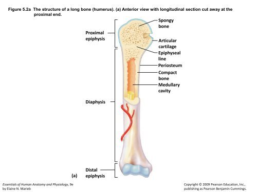
Figure 5.2a The structure of a long <strong>bone</strong> (humerus). (a) Anterior view with longitudinal section cut away at the<br />
proximal end.<br />
Essentials of Human Anatomy and Physiology, 9e<br />
by Elaine N. Marieb<br />
(a)<br />
Spongy<br />
<strong>bone</strong><br />
Proximal<br />
<strong>epiphysis</strong> Articular<br />
cartilage<br />
Epiphyseal<br />
line<br />
Periosteum<br />
Compact<br />
<strong>bone</strong><br />
<strong>Medullary</strong><br />
<strong>cavity</strong><br />
<strong>Diaphysis</strong><br />
<strong>Distal</strong><br />
<strong>epiphysis</strong><br />
Copyright © 2009 Pearson Education, Inc.,<br />
publishing as Pearson Benjamin Cummings.
Figure 5.3a Microscopic structure of compact <strong>bone</strong>. (a).<br />
Perforating<br />
fibers<br />
Compact<br />
<strong>bone</strong><br />
Periosteal<br />
blood vessel<br />
Periosteum<br />
(a)<br />
Osteon<br />
(Haversian system)<br />
Lamellae<br />
Essentials of Human Anatomy and Physiology, 9e<br />
by Elaine N. Marieb<br />
Central (Haversian) canal<br />
Perforating (Volkmann’s) canal<br />
Blood vessel<br />
Blood vessel continues<br />
into medullary <strong>cavity</strong><br />
containing marrow<br />
Spongy <strong>bone</strong><br />
Copyright © 2009 Pearson Education, Inc.,<br />
publishing as Pearson Benjamin Cummings.
Figure 5.3b Microscopic structure of compact <strong>bone</strong>. (b).<br />
Essentials of Human Anatomy and Physiology, 9e<br />
by Elaine N. Marieb<br />
Lamella<br />
Osteocyte<br />
(b)<br />
Lacuna<br />
Canaliculus<br />
Central<br />
(Haversian) canal<br />
Copyright © 2009 Pearson Education, Inc.,<br />
publishing as Pearson Benjamin Cummings.
Figure 5.6a The human skeleton. (a) Anterior view.<br />
Essentials of Human Anatomy and Physiology, 9e<br />
by Elaine N. Marieb<br />
Vertebral<br />
column<br />
Skull<br />
Thoracic cage<br />
(ribs and<br />
sternum)<br />
(a) Anterior view<br />
Sacrum<br />
Cranium<br />
Facial <strong>bone</strong>s<br />
Clavicle<br />
Scapula<br />
Sternum<br />
Rib<br />
Humerus<br />
Vertebra<br />
Radius<br />
Ulna<br />
Carpals<br />
Phalanges<br />
Metacarpals<br />
Femur<br />
Patella<br />
Tibia<br />
Tarsals<br />
Metatarsals<br />
Phalanges<br />
Fibula<br />
Copyright © 2009 Pearson Education, Inc.,<br />
publishing as Pearson Benjamin Cummings.
Figure 5.6b The human skeleton. (b) Posterior view.<br />
Essentials of Human Anatomy and Physiology, 9e<br />
by Elaine N. Marieb<br />
Cranium<br />
Clavicle<br />
Scapula<br />
Rib<br />
Humerus<br />
Vertebra<br />
Radius<br />
Ulna<br />
Carpals<br />
Phalanges<br />
Metacarpals<br />
Femur<br />
Tibia<br />
Fibula<br />
(b) Posterior view<br />
Bones<br />
of<br />
pelvic<br />
girdle<br />
Bones of<br />
pectoral<br />
girdle<br />
Upper<br />
limb<br />
Lower<br />
limb<br />
Copyright © 2009 Pearson Education, Inc.,<br />
publishing as Pearson Benjamin Cummings.
Figure 5.7 Human skull, lateral view.<br />
Coronal suture Frontal <strong>bone</strong><br />
Parietal <strong>bone</strong><br />
Temporal <strong>bone</strong><br />
Lambdoid<br />
suture<br />
Squamous suture<br />
Occipital <strong>bone</strong><br />
Zygomatic process<br />
External acoustic meatus<br />
Mastoid process<br />
Styloid process<br />
Essentials of Human Anatomy and Physiology, 9e<br />
by Elaine N. Marieb<br />
Mandibular ramus<br />
Sphenoid <strong>bone</strong><br />
Ethmoid <strong>bone</strong><br />
Lacrimal <strong>bone</strong><br />
Nasal <strong>bone</strong><br />
Zygomatic <strong>bone</strong><br />
Maxilla<br />
Alveolar<br />
margins<br />
Mandible (body)<br />
Mental foramen<br />
Copyright © 2009 Pearson Education, Inc.,<br />
publishing as Pearson Benjamin Cummings.
Figure 5.9 Human skull, inferior view (mandible removed).<br />
Hard<br />
palate<br />
Zygomatic <strong>bone</strong><br />
Maxilla<br />
(palatine process)<br />
Temporal <strong>bone</strong><br />
(zygomatic process)<br />
Vomer<br />
Mandibular fossa<br />
Styloid process<br />
Palatine <strong>bone</strong><br />
Mastoid process<br />
Temporal <strong>bone</strong><br />
Parietal <strong>bone</strong><br />
Occipital <strong>bone</strong><br />
Essentials of Human Anatomy and Physiology, 9e<br />
by Elaine N. Marieb<br />
Maxilla<br />
Sphenoid <strong>bone</strong><br />
(greater wing)<br />
Foramen ovale<br />
Carotid canal<br />
Jugular foramen<br />
Occipital condyle<br />
Foramen magnum<br />
Copyright © 2009 Pearson Education, Inc.,<br />
publishing as Pearson Benjamin Cummings.
Figure 5.11 Human skull, anterior view.<br />
Coronal suture<br />
Parietal <strong>bone</strong><br />
Nasal <strong>bone</strong><br />
Sphenoid <strong>bone</strong><br />
Ethmoid <strong>bone</strong><br />
Lacrimal <strong>bone</strong><br />
Zygomatic <strong>bone</strong><br />
Maxilla<br />
Mandible<br />
Essentials of Human Anatomy and Physiology, 9e<br />
by Elaine N. Marieb<br />
Frontal <strong>bone</strong><br />
Superior orbital fissure<br />
Optic canal<br />
Temporal <strong>bone</strong><br />
Middle nasal concha<br />
of ethmoid <strong>bone</strong><br />
Inferior nasal concha<br />
Vomer<br />
Alveolar margins<br />
Copyright © 2009 Pearson Education, Inc.,<br />
publishing as Pearson Benjamin Cummings.
Figure 5.13a The fetal skull. (a) Superior view.<br />
Frontal <strong>bone</strong><br />
Parietal<br />
<strong>bone</strong><br />
Posterior fontanel<br />
(a)<br />
Essentials of Human Anatomy and Physiology, 9e<br />
by Elaine N. Marieb<br />
Anterior<br />
fontanel<br />
Occipital<br />
<strong>bone</strong><br />
Copyright © 2009 Pearson Education, Inc.,<br />
publishing as Pearson Benjamin Cummings.
Figure 5.13b The fetal skull. (b) Lateral view.<br />
Parietal <strong>bone</strong><br />
Posterior<br />
fontanel<br />
Occipital<br />
<strong>bone</strong><br />
Mastoid<br />
fontanel<br />
(b)<br />
Essentials of Human Anatomy and Physiology, 9e<br />
by Elaine N. Marieb<br />
Temporal <strong>bone</strong><br />
Anterior fontanel<br />
Sphenoidal<br />
fontanel<br />
Frontal<br />
<strong>bone</strong><br />
Copyright © 2009 Pearson Education, Inc.,<br />
publishing as Pearson Benjamin Cummings.
Figure 5.14 The vertebral column.<br />
Essentials of Human Anatomy and Physiology, 9e<br />
by Elaine N. Marieb<br />
Anterior Posterior<br />
1st cervical<br />
vertebra (atlas)<br />
2nd cervical<br />
vertebra (axis)<br />
1st thoracic<br />
vertebra<br />
Transverse<br />
process<br />
Spinous<br />
process<br />
Intervertebral<br />
disc<br />
Intervertebral<br />
foramen<br />
1st Lumbar<br />
vertebra<br />
Cervical<br />
curvature<br />
(concave)<br />
7 vertebrae,<br />
C 1 – C 7<br />
Thoracic<br />
curvature<br />
(convex)<br />
12 vertebrae,<br />
T 1 – T 12<br />
Lumbar<br />
curvature<br />
(concave)<br />
5 vertebrae,<br />
L 1 – L 5<br />
Sacral<br />
curvature<br />
(convex)<br />
5 fused<br />
vertebrae<br />
Coccyx<br />
4 fused<br />
vertebrae<br />
Copyright © 2009 Pearson Education, Inc.,<br />
publishing as Pearson Benjamin Cummings.
Figure 5.20a The bony thorax (thoracic cage). (a) Anterior view.<br />
Clavicular notch<br />
True<br />
ribs<br />
(1–7)<br />
False<br />
ribs<br />
(8–12)<br />
(a)<br />
Essentials of Human Anatomy and Physiology, 9e<br />
by Elaine N. Marieb<br />
L 1<br />
Floating Vertebra<br />
ribs (11, 12)<br />
T1 vertebra<br />
Jugular notch<br />
Manubrium<br />
Sternal angle<br />
Body<br />
Xiphisternal<br />
joint<br />
Xiphoid<br />
process<br />
Intercostal<br />
spaces<br />
Costal cartilage<br />
Sternum<br />
Copyright © 2009 Pearson Education, Inc.,<br />
publishing as Pearson Benjamin Cummings.
Figure 5.21a–b Bones of the shoulder girdle. (a) Articulated right shoulder (pectoral) girdle showing the<br />
relationship to <strong>bone</strong>s of the thorax and sternum, and (b) right clavicle, superior and inferior views.<br />
Scapula<br />
Acromioclavicular<br />
joint<br />
Essentials of Human Anatomy and Physiology, 9e<br />
by Elaine N. Marieb<br />
Clavicle<br />
(a) Articulated right shoulder (pectoral) girdle<br />
showing the relationship to <strong>bone</strong>s of the<br />
thorax and sternum<br />
Posterior<br />
Acromial (lateral)<br />
end<br />
Superior view<br />
Acromial end<br />
Inferior view<br />
Anterior<br />
Sternal (medial)<br />
end<br />
Posterior<br />
Anterior<br />
Sternal end<br />
(b) Right clavicle, superior and inferior views<br />
Copyright © 2009 Pearson Education, Inc.,<br />
publishing as Pearson Benjamin Cummings.
Figure 5.21c Bones of the shoulder girdle. (c) Right scapula, posterior aspect.<br />
Essentials of Human Anatomy and Physiology, 9e<br />
by Elaine N. Marieb<br />
Suprascapular notch<br />
Superior<br />
angle<br />
Spine<br />
Medial<br />
border<br />
(c) Right scapula, posterior aspect<br />
Coracoid process<br />
Lateral border<br />
Acromion<br />
Glenoid <strong>cavity</strong><br />
at lateral angle<br />
Copyright © 2009 Pearson Education, Inc.,<br />
publishing as Pearson Benjamin Cummings.
Figure 5.21d Bones of the shoulder girdle. (d) Right scapula, anterior aspect.<br />
Coracoid<br />
process<br />
Essentials of Human Anatomy and Physiology, 9e<br />
by Elaine N. Marieb<br />
Acromion<br />
Glenoid<br />
<strong>cavity</strong><br />
Suprascapular notch<br />
Superior border<br />
Lateral<br />
(axillary)<br />
border<br />
(d) Right scapula, anterior aspect<br />
Superior<br />
angle<br />
Medial<br />
(vertebral)<br />
border<br />
Inferior angle<br />
Copyright © 2009 Pearson Education, Inc.,<br />
publishing as Pearson Benjamin Cummings.
Figure 5.22a Bones of the right arm and forearm. (a) Humerus, anterior view.<br />
(a)<br />
Essentials of Human Anatomy and Physiology, 9e<br />
by Elaine N. Marieb<br />
Greater<br />
tubercle<br />
Lesser<br />
tubercle<br />
Deltoid<br />
tuberosity<br />
Radial<br />
fossa<br />
Coronoid<br />
fossa<br />
Capitulum<br />
Head of<br />
humerus<br />
Anatomical neck<br />
Intertubercular<br />
sulcus<br />
Medial<br />
epicondyle<br />
Trochlea<br />
Copyright © 2009 Pearson Education, Inc.,<br />
publishing as Pearson Benjamin Cummings.
Figure 5.22b Bones of the right arm and forearm. (b) Humerus, posterior view.<br />
Essentials of Human Anatomy and Physiology, 9e<br />
by Elaine N. Marieb<br />
Head of<br />
humerus<br />
Anatomical<br />
neck<br />
Radial<br />
groove<br />
Deltoid<br />
tuberosity<br />
Medial<br />
epicondyle<br />
(b) Trochlea<br />
Olecranon fossa<br />
Lateral epicondyle<br />
Copyright © 2009 Pearson Education, Inc.,<br />
publishing as Pearson Benjamin Cummings.
Figure 5.22c Bones of the right arm and forearm. (c) Anterior view of the <strong>bone</strong>s of the forearm: the radius and the ulna.<br />
Essentials of Human Anatomy and Physiology, 9e<br />
by Elaine N. Marieb<br />
Head<br />
Neck<br />
Radial<br />
tuberosity<br />
Radius<br />
Styloid process<br />
of radius<br />
(c)<br />
Trochlear notch<br />
Olecranon process<br />
Coronoid process<br />
Proximal radioulnar<br />
joint<br />
Ulna<br />
Interosseous<br />
membrane<br />
Styloid process of ulna<br />
<strong>Distal</strong> radioulnar joint<br />
Copyright © 2009 Pearson Education, Inc.,<br />
publishing as Pearson Benjamin Cummings.
Figure 5.23 Bones of the right hand, anterior view.<br />
Phalanges<br />
(fingers)<br />
Metacarpals<br />
(palm)<br />
Carpals<br />
(wrist)<br />
Essentials of Human Anatomy and Physiology, 9e<br />
by Elaine N. Marieb<br />
<strong>Distal</strong><br />
Pisiform<br />
Triquetrum<br />
Lunate<br />
Ulna<br />
Middle<br />
Proximal<br />
Hamate<br />
5<br />
4 3 2<br />
Radius<br />
1<br />
Trapezium<br />
Trapezoid<br />
Scaphoid<br />
Capitate<br />
Copyright © 2009 Pearson Education, Inc.,<br />
publishing as Pearson Benjamin Cummings.
Figure 5.24a The bony pelvis. (a) Articulated male pelvis.<br />
Coxal<br />
<strong>bone</strong><br />
(or hip<br />
<strong>bone</strong>)<br />
llium<br />
Pubic<br />
<strong>bone</strong><br />
Ischium<br />
(a)<br />
Essentials of Human Anatomy and Physiology, 9e<br />
by Elaine N. Marieb<br />
Sacrum<br />
Coccyx<br />
Pubic arch<br />
Pelvic brim<br />
Ischial spine<br />
Acetabulum<br />
Iliac crest<br />
Sacroiliac<br />
joint<br />
Pubic symphysis<br />
Copyright © 2009 Pearson Education, Inc.,<br />
publishing as Pearson Benjamin Cummings.
Figure 5.24b The bony pelvis. (b) Right coxal <strong>bone</strong>, showing the point of fusion of the ilium, ischium, and pubic <strong>bone</strong>s.<br />
Posterior<br />
superior<br />
iIiac spine<br />
Posterior<br />
inferior<br />
iliac spine<br />
Greater sciatic<br />
notch<br />
Ischial body<br />
Ischial spine<br />
Ischial<br />
tuberosity<br />
Ischium<br />
Ischial ramus<br />
Essentials of Human Anatomy and Physiology, 9e<br />
by Elaine N. Marieb<br />
(b)<br />
Ala<br />
Ilium<br />
Iliac crest<br />
Anterior superior<br />
iliac spine<br />
Anterior inferior<br />
iliac spine<br />
Acetabulum<br />
Body of pubis<br />
Pubis<br />
Inferior ramus<br />
of pubis<br />
Obturator<br />
foramen<br />
Copyright © 2009 Pearson Education, Inc.,<br />
publishing as Pearson Benjamin Cummings.
Figure 5.24c The bony pelvis. (c) Comparison of the male (left) and female (right) pelves.<br />
(c)<br />
Essentials of Human Anatomy and Physiology, 9e<br />
by Elaine N. Marieb<br />
False pelvis False pelvis<br />
Inlet of<br />
true<br />
pelvis<br />
Pelvic brim<br />
Pubic arch<br />
(less than 90º)<br />
Pelvic<br />
brim<br />
Inlet of<br />
true<br />
pelvis<br />
Pubic arch<br />
(more than 90º)<br />
Copyright © 2009 Pearson Education, Inc.,<br />
publishing as Pearson Benjamin Cummings.
Figure 5.25a Bones of the right thigh and leg. (a) Femur (thigh <strong>bone</strong>), anterior view.<br />
Neck<br />
Essentials of Human Anatomy and Physiology, 9e<br />
by Elaine N. Marieb<br />
Intertrochanteric<br />
line<br />
Lateral condyle<br />
(a)<br />
Head<br />
Lesser trochanter<br />
Patellar surface<br />
Copyright © 2009 Pearson Education, Inc.,<br />
publishing as Pearson Benjamin Cummings.
Figure 5.25b Bones of the right thigh and leg. (b) Femur, posterior view.<br />
Head<br />
Essentials of Human Anatomy and Physiology, 9e<br />
by Elaine N. Marieb<br />
Lesser trochanter<br />
Gluteal tuberosity<br />
Intercondylar fossa<br />
Medial condyle<br />
(b)<br />
Greater trochanter<br />
Intertrochanteric<br />
crest<br />
Lateral condyle<br />
Copyright © 2009 Pearson Education, Inc.,<br />
publishing as Pearson Benjamin Cummings.
Figure 5.25c Bones of the right thigh and leg. (c) Tibia and Fibula of the leg, anterior view.<br />
Essentials of Human Anatomy and Physiology, 9e<br />
by Elaine N. Marieb<br />
Intercondylar eminence<br />
Lateral condyle<br />
Head<br />
Proximal tibiofibular<br />
joint<br />
<strong>Distal</strong> tibiofibular<br />
joint<br />
Lateral malleolus<br />
(c)<br />
Fibula<br />
Medial condyle<br />
Tibial tuberosity<br />
Interosseous<br />
membrane<br />
Anterior border<br />
Tibia<br />
Medial malleolus<br />
Copyright © 2009 Pearson Education, Inc.,<br />
publishing as Pearson Benjamin Cummings.
Figure 5.26 Bones of the right foot, superior view.<br />
Essentials of Human Anatomy and Physiology, 9e<br />
by Elaine N. Marieb<br />
Tarsals:<br />
Medial<br />
cuneiform<br />
Intermediate<br />
cuneiform<br />
Navicular<br />
Talus<br />
Phalanges:<br />
<strong>Distal</strong><br />
Middle<br />
Proximal<br />
Metatarsals<br />
Tarsals:<br />
Lateral<br />
cuneiform<br />
Cuboid<br />
Calcaneus<br />
Copyright © 2009 Pearson Education, Inc.,<br />
publishing as Pearson Benjamin Cummings.