
You also want an ePaper? Increase the reach of your titles
YUMPU automatically turns print PDFs into web optimized ePapers that Google loves.
171<br />
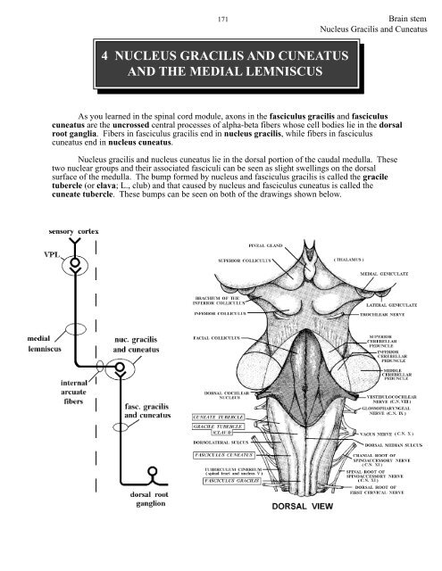
4 NUCLEUS GRACILIS AND CUNEATUS<br />
AND THE MEDIAL LEMNISCUS<br />
<strong>Brain</strong> <strong>stem</strong><br />
Nucleus Gracilis and Cuneatus<br />
As you learned in the spinal cord module, axons in the fasciculus gracilis and fasciculus<br />
cuneatus are the uncrossed central processes of alpha-beta fibers whose cell bodies lie in the dorsal<br />
root ganglia. Fibers in fasciculus gracilis end in nucleus gracilis, while fibers in fasciculus<br />
cuneatus end in nucleus cuneatus.<br />
Nucleus gracilis and nucleus cuneatus lie in the dorsal portion of the caudal medulla. These<br />
two nuclear groups and their associated fasciculi can be seen as slight swellings on the dorsal<br />
surface of the medulla. The bump formed by nucleus and fasciculus gracilis is called the gracile<br />
tubercle (or clava; L., club) and that caused by nucleus and fasciculus cuneatus is called the<br />
cuneate tubercle. These bumps can be seen on both of the drawings shown below.
<strong>Brain</strong> <strong>stem</strong> 172<br />
Nucleus Gracilis and Cuneatus
173<br />
<strong>Brain</strong> <strong>stem</strong><br />
Nucleus Gracilis and Cuneatus<br />
Axons of cells within nucleus gracilis and nucleus cuneatus cross as internal arcuate fibers<br />
and form the MEDIAL LEMNISCUS. The medial lemniscus is thus a large ascending bundle of<br />
heavily myelinated axons (fast conducting) whose cell bodies lie in the contralateral nucleus gracilis<br />
and nucleus cuneatus. The medial lemniscus passes rostrally through the medulla, pons and<br />
midbrain to terminate in the ventral posterolateral (VPL) nucleus of the thalamus. Cells in the<br />
VPL then send their axons to the postcentral gyrus (somatosensory cortex) of the cerebral cortex<br />
(areas 3, 1, 2).<br />
The dorsal column-medial lemniscal sy<strong>stem</strong> carries information from specialized touch,<br />
pressure, vibration, and joint receptors to the cerebral cortex. While lesions of the dorsal<br />
columns (fasciculi gracilis and cuneatus) in the spinal cord result in IPSILATERAL deficits, lesions<br />
of the medial lemniscus, in the brain <strong>stem</strong>, result in CONTRALATERAL deficits (since its<br />
constituent axons have crossed). This is important to understand. Also, do not confuse VPM (head,<br />
trigeminal, trigeminothalamic) with VPL (body, medial lemniscus).<br />
Lesions of the medial lemniscus results in loss of 2 pt. discrimination, vibration and<br />
conscious proprioception from the contralateral side of the body. Also remember, there is<br />
astereognosia, agraphesthesia, and a Romberg sign.
<strong>Brain</strong> <strong>stem</strong> 174<br />
Nucleus Gracilis and Cuneatus<br />
You may remember that there is a somatotopic representation of the body in the dorsal<br />
columns (fasciculus gracilis and fasciculus cuneatus). Caudal (sacral and lumbar) body parts are<br />
represented medially, while the rostral segments (upper thoracic and cervical) are represented<br />
laterally. Nucleus gracilis receives its input from about T 7 and downward, while nucleus cuneatus<br />
receives its input from spinal levels above this.<br />
There is also a somatotopic organization within the medial lemniscus. For instance, in the<br />
medullary segment of the medial lemniscus the body is represented in an upright position, so that<br />
the legs are ventral and the arms dorsal. Within the pontine portion of the medial lemniscus there is<br />
a rotation, so that the arms are represented medially and the legs laterally. Finally, in the midbrain<br />
portion of the medial lemniscus the arms are represented ventrally while the legs are represented<br />
dorsally.
PROBLEM SOLVING<br />
175<br />
RIGHT LEFT<br />
<strong>Brain</strong> <strong>stem</strong><br />
Nucleus Gracilis and Cuneatus<br />
Problem Solving<br />
Shade in the location of a single, continuous, unilateral lesion in the above drawing that will<br />
account for the following neurological problems:<br />
stabbing pain in the right eye followed by the loss of pain and temperature from the right side of the<br />
face, loss of vibratory sense from the right arm, loss of vibratory sense from the right leg
<strong>Brain</strong> <strong>stem</strong> 176<br />
Nucleus Gracilis and Cuneatus<br />
Problem Solving<br />
PROBLEM SOLVING ANSWER<br />
RIGHT LEFT
177<br />
5 ACCESSORY CUNEATE NUCLEUS<br />
(Lots of hard work for such a TINY nucleus!)<br />
<strong>Brain</strong> <strong>stem</strong><br />
Accessory Cuneate Nucleus<br />
You should recall from the spinal cord module that proprioceptive information from muscle<br />
spindles (Ia, II) and Golgi tendon organs (Ib) reaches the cerebellum via the dorsal<br />
spinocerebellar tract. The cells of origin of this tract lie in the ipsilateral Clarke’s column. This<br />
column of cells is present only at spinal cord levels C 8 -L 3 . Central processes of dorsal root neurons<br />
that enter caudal to L 3 have to ascend to reach L 3 . Consequently, Clarke’s column is quite enlarged<br />
caudally. (Clarke’s column neurons at L 3 need to serve not only entering fibers at L 3 , but all of those<br />
entering below L 3 .)<br />
Ia, Ib and type II axons of the dorsal root ganglia rostral to Clarke’s column (C 8 ) pass<br />
rostrally to reach the ipsilateral caudal medulla, where they end within the ACCESSORY<br />
CUNEATE (“wedge-shaped”) NUCLEUS. This nucleus, which is somewhat difficult to see, lies<br />
dorsal to the spinal tract and nucleus V and lateral to the most rostral pole of nucleus cuneatus. Cells<br />
in the accessory cuneate nucleus send their axons to the IPSILATERAL CEREBELLUM via a<br />
fiber bundle called the INFERIOR CEREBELLAR PEDUNCLE (together with the dorsal<br />
spinocerebellar fibers). This pathway is called the CUNEOCEREBELLAR TRACT.<br />
The accessory cuneate nucleus is concerned with relaying proprioceptive information from<br />
the arm (and neck) to the cerebellum, and the nucleus can be considered as the rostral equivalent of<br />
Clarke’s column.<br />
REMEMBER: Accessory cuneate nucleus:<br />
1). lies in the medulla.<br />
2). receives UNCROSSED fibers from dorsal root ganglia above C 8 .<br />
3). receives the same kind of information that Clarke’s column does.<br />
4). projects to the IPSILATERAL cerebellum via inferior cerebellar peduncle.<br />
5). is concerned with the arm, while Clarke’s column is concerned with the forearm, trunk<br />
and lower extremity.
<strong>Brain</strong> <strong>stem</strong> 178<br />
Accessory Cuneate Nucleus
179<br />
<strong>Brain</strong> <strong>stem</strong><br />
Accessory Cuneate Nucleus<br />
Due to its small size, lesions restricted to the accessory cuneate nucleus are rare.<br />
Understanding the laterality (which side) and specific deficits resulting from a lesion of the<br />
accessory cuneate requires knowing something about CEREBELLAR functions and connections.<br />
While this important topic is covered later in this course, we need to do some limited spade work<br />
right now to get you prepared for those lectures, and also to let you problem solve on questions<br />
regarding lesions of brain <strong>stem</strong> areas that either project to the cerebellum (like the accessory<br />
cuneate nucleus, inferior olive and pontine grey [the latter two will be discussed further up the brain<br />
<strong>stem</strong>]) or contain axons leaving the cerebellum (superior cerebellar peduncle).<br />
The cerebellum is involved in motor coordination. Unlike the cerebrum (i.e., cerebral<br />
cortex; cells of origin of the corticospinal tract), the cerebellum has no major projections to the<br />
spinal cord, but instead regulates movement indirectly by projecting to areas of the brain that do<br />
project upon the spinal cord. Lesions of the cerebellum lead to defects in the coordination of<br />
movements, but NOT paresis or paralysis. Such cerebellar defects involve errors in the rate, range<br />
or direction of voluntary movements. Disturbances following cerebellar lesions are known by a<br />
variety of terms such as nystagmus, ataxia, hypotonia, dysmetria, past pointing, rebound,<br />
dysdiadochokinesia, asynergy, intention tremor and decomposition of movement (WHEY!).<br />
RIGHT NOW, LET’S JUST CALL THESE DISTURBANCES = INCOORDINATION/ATAXIA<br />
AND REMEMBER THAT INCOORDINATION/ATAXIA RESULTS FROM LESIONS OF<br />
THE CEREBELLUM OR ITS INPUTS (ACCESSORY CUNEATE NUCLEUS) OR OUTPUTS.<br />
In addition to knowing that lesions of the cerebellum and its inputs and outputs result in<br />
incoordination, we need to know what part of the body is affected (arm, leg) and the laterality of<br />
the deficits (IPSI. or CONTRA.). The important point now is that one side of the cerebellum<br />
controls the SAME OR IPSILATERAL SIDE OF THE BODY.<br />
OPTIONAL READING<br />
This is due to TWO DECUSSATIONS of pathways involved in conveying cerebellar information<br />
to the spinal cord. To understand all of this, let’s start at the LEFT accessory cuneate nucleus,<br />
which you now know receives information from the LEFT side of the upper extremity. Cells in the<br />
LEFT accessory cuneate nucleus possess axons that comprise the LEFT cuneocerebellar tract and<br />
synapse on cells in the LEFT cerebellar cortex called granule cells. The axons of granule cells<br />
synapse on Purkinje cells. Purkinje cell axons synapse on cells in the deep white matter of the<br />
cerebellum called DEEP CEREBELLAR NUCLEI. There are four of these deep cerebellar nuclei<br />
on each side of the cerebellum. They are called fastigial, globose, emboliform and dentate. We will<br />
NOT worry about these nuclei too much at this time, but we need to know that they contain cells<br />
whose axons LEAVE the cerebellum (efferent; exit) in a large bundle called the SUPERIOR<br />
CEREBELLAR PEDUNCLE (Point #17). The superior cerebellar peduncle courses rostrally and<br />
CROSSES in the caudal midbrain (decussation #1). After crossing, axons synapse in the RED<br />
NUCLEUS (midbrain; we will discuss later in POINT #21; don’t worry about it at this time) and in<br />
the ventral lateral (VL) and ventral anterior (VA) nuclei of the thalamus. Cells in VL and VA<br />
project to the motor cortex, which of course contains the cells of origin of the CORTICOSPINAL<br />
TRACT. As you know, the corticospinal tract CROSSES in the caudal medulla (decussation #2)<br />
and innervates spinal cord neurons.
<strong>Brain</strong> <strong>stem</strong> 180<br />
Accessory Cuneate Nucleus
181<br />
This leads to two of the most important “rules” of neurology. That is:<br />
CEREBELLAR PROBLEMS = IPSILATERAL,<br />
CEREBRAL PROBLEMS = CONTRALATERAL<br />
<strong>Brain</strong> <strong>stem</strong><br />
Accessory Cuneate Nucleus<br />
The accessory cuneate nucleus projects to the IPSILATERAL CEREBELLAR<br />
HEMISPHERE (via the inferior cerebellar peduncle, along with the dorsal spinocerebellar fibers).<br />
Therefore lesions of the accessory cuneate nucleus, like the cerebellum, result in IPSILATERAL<br />
deficits. For example, the LEFT accessory cuneate nucleus receives input from Ia, Ib and type II<br />
fibers of dorsal root ganglia above C 8 on the LEFT. The LEFT accessory cuneate nucleus projects<br />
to the LEFT cerebellar hemisphere. Finally the information leaves the cerebellum to eventually<br />
influence the LEFT arm. Therefore, a lesion of the LEFT accessory cuneate nucleus would result<br />
in “bad” information reaching the LEFT cerebellar hemisphere, and in turn motor incoordination<br />
of the LEFT arm. There is NO paralysis or atrophy of these muscles. REMEMBER, for our<br />
problem solving questions involving the accessory cuneate nucleus let’s just focus on<br />
INCOORDINATION/ATAXIA OF THE IPSILATERAL ARM.<br />
To review: a lesion of the accessory cuneate nucleus results in incoordination/ataxia of the<br />
ipsilateral arm. It could not result in a Romberg because it involves a cerebellar afferent (and does<br />
not include the lower limbs anyway). A lesion of the DSCT will involve the legs but again, it is a<br />
cerebellar afferent and therefore would not give a Romberg sign. What about a lesion of the inferior<br />
cerebellar peduncle?
<strong>Brain</strong> <strong>stem</strong> 182<br />
Accessory Cuneate Nucleus<br />
Problem Solving<br />
PROBLEM SOLVING MATCHING<br />
Match the best choice in the right hand column with the pathway or cell group in the left hand<br />
column<br />
____1. right pyramid A. lesion results in a loss of vibratory sense<br />
from the left arm and leg<br />
____2. right anterolateral sy<strong>stem</strong> (ALS)<br />
and associated descending pathway B. cells project to the right VPM<br />
____3. right caudal spinal nucleus V<br />
C. cells project to the cerebellum via the right<br />
____4. right nucleus gracilis and cuneatus inferior cerebellar peduncle<br />
____5. left accessory cuneate nucleus D. axons terminate in the right VPL<br />
E. lesion results in a loss of 2 pt. discrimination<br />
from the left arm and leg<br />
F. lesion results in a loss of pain and temp from<br />
the right side of the face<br />
G. lesion results in a dilated pupil in the right<br />
eye<br />
H. cells convey 2 pt. discrimination and<br />
vibratory information to the left VPL<br />
I. cells project to the left side of the cerebellum<br />
J. lesion results in left hemiplegia
PROBLEM SOLVING<br />
183<br />
RIGHT LEFT<br />
<strong>Brain</strong> <strong>stem</strong><br />
Accessory Cuneate Nucleus<br />
Problem Solving<br />
Shade in the location of a single, continuous, unilateral lesion in the above drawing that will<br />
account for the following neurological problems:<br />
stabbing pain in the left eye followed by loss of pain and temperature from the left side of the face,<br />
incoordination of the left arm
<strong>Brain</strong> <strong>stem</strong> 184<br />
Accessory Cuneate Nucleus<br />
Problem Solving<br />
PROBLEM SOLVING ANSWER<br />
RIGHT LEFT
185<br />
<strong>Brain</strong> <strong>stem</strong>
<strong>Brain</strong> <strong>stem</strong> 186<br />
Problem Solving - ANSWERS POINTS 1-5<br />
ANSWERS TO PROBLEM SOLVING QUESTIONS RELATED TO POINTS 1-5<br />
NOTE: The answers to ALL shade-in questions are illustrated on the back side of the question.<br />
Point #1 Pyramid Point #3 Spinal Nucleus and Tract V<br />
Matching D Matching B,H,D<br />
Point #2 Anterolateral Sy<strong>stem</strong> Point #4 Nucleus Gracilis and Cuneatus<br />
Matching G,E Matching D,I,F,J<br />
Point #5 Accessory Cuneate Nucleus<br />
Matching J,D,F,H,I
187<br />
6 INFERIOR OLIVARY COMPLEX<br />
<strong>Brain</strong> <strong>stem</strong><br />
Inferior Olivary Complex<br />
This is the largest nuclear group in the brain <strong>stem</strong>. It consists of a convoluted band of cells<br />
that lie dorsal to the pyramid. This nucleus is by far the most characteristic and striking feature of<br />
the medulla. Sadly, we know little about inferior olivary function(s), but its very intimate<br />
association with the cerebellum suggests it is involved in motor coordination and most likely motor<br />
“learning”.<br />
Cells in the inferior olivary complex project to the contralateral cerebellum via the inferior<br />
cerebellar peduncle (or restiform body). Upon reaching the cerebellum, they end as “CLIMBING<br />
FIBERS” (they “climb up” the Purkinje cells; more on this later in the course). Climbing fibers<br />
arise solely from the inferior olive. Other endings seen in the cerebellar cortex are called mossy<br />
fibers. Mossy fibers do not arise from the inferior olive, but rather from places like Clarke’s column<br />
and the accessory cuneate nucleus. Thus, axons in the dorsal spinocerebellar and cuneocerebellar<br />
tracts end as “mossy fibers.” Inputs to the inferior olive will be discussed during the “Cerebellum”<br />
part of the course.
<strong>Brain</strong> <strong>stem</strong> 188<br />
Inferior Olivary Complex<br />
While many questions remain regarding the function(s) of the inferior olive, selective<br />
destruction of this nuclear complex in experimental animals has acute effects similar to those<br />
following destruction of the entire one-half of the CONTRALATERAL cerebellum. Since the<br />
inferior olive sends information to the contralateral cerebellum, and the cerebellum influences the<br />
SAME side of the body, then the loss of the LEFT olive will mean that the RIGHT half of the<br />
cerebellum is no longer receiving input from the inferior olive. This will result in incoordination/<br />
ataxia of the RIGHT side of the body. Since we will cover cerebellum later in this course, don’t<br />
worry too much about it right now. We have already discussed that cerebellar deficits involve<br />
incoordination/ataxia and are IPSILATERAL to the side of the lesion. A lesion of the inferior<br />
olive will result in incoordination/ataxia of the CONTRALATERAL ARM AND LEG (contrast<br />
this with a lesion of ACC. CUNEATE NUC. = incoordination/ataxia of IPSI ARM). What about a<br />
Romberg sign? Well, this is like a DSCT lesion. Loss of input from the olive means that you are not<br />
going to be able to stand with your feet together to begin with!! So no Romberg! Besides, it’s a<br />
cerebellar afferent!!<br />
ALSO REMEMBER:<br />
1). the sole source of climbing fibers is the inferior olive<br />
2). olivocerebellars CROSS and comprise most of the inferior cerebellar peduncle<br />
3). the inferior olive lies in the ventral medulla.
189<br />
<strong>Brain</strong> <strong>stem</strong><br />
Inferior Olivary Complex
<strong>Brain</strong> <strong>stem</strong> 190<br />
Inferior Olivary Complex<br />
PROBLEM SOLVING MATCHING<br />
Match the best choice in the right hand column with the pathway or cell group in the left hand<br />
column<br />
____1. left medial lemniscus A. lesion results in a loss of vibratory sense<br />
from the left arm and leg<br />
____2. right pyramid B. cells project to the cerebellum via the right<br />
inferior cerebellar peduncle<br />
____3. right spinal tract V C. cells project to the right VPM<br />
D. contains axons that arise from Clarke’s column<br />
____4. left inferior cerebellar peduncle on the left side of the spinal cord<br />
E. carries fibers from the right accessory cuneate<br />
____5. left inferior olive nucleus destined for the cerebellum<br />
F. “climbing fibers” from this nucleus terminate in<br />
the left side of the cerebellum<br />
G. lesion results in a Babinski sign from the left big toe<br />
H. axons convey pain and temp from the right side of<br />
the larynx<br />
I. carries information regarding 2 pt. discrimination<br />
from the right side of the body<br />
J. lesion results in a loss of pain and temp from the<br />
left side of the body
PROBLEM SOLVING<br />
191<br />
RIGHT LEFT<br />
<strong>Brain</strong> <strong>stem</strong><br />
Inferior Olivary Complex<br />
Problem Solving<br />
Shade in the location of a single, continuous, unilateral lesion in the above drawing that will<br />
account for the following neurological problems:<br />
loss of pain and temperature from the right side of the face, incoordination of the right arm and leg
<strong>Brain</strong> <strong>stem</strong> 192<br />
Inferior Olivary Complex<br />
Problem Solving<br />
PROBLEM SOLVING ANSWER<br />
RIGHT LEFT<br />
- OR -<br />
RIGHT LEFT
193<br />
7 HYPOGLOSSAL NUCLEUS (C.N. XII)<br />
<strong>Brain</strong> <strong>stem</strong><br />
Hypoglossal Nucleus<br />
This nucleus lies just off the midline beneath the floor of the fourth ventricle. Axons from<br />
cells within the hypoglossal nucleus course ventrally to exit the medulla between the pyramid and<br />
the inferior olive. The hypoglossal nerve then passes through the hypoglossal foramen to emerge<br />
from the base of the skull. Each nerve innervates the ipsilateral intrinsic and extrinsic muscles of the<br />
tongue (you referred to this nerve as somatomotor in Gross Anatomy and learned that the<br />
palatoglossus is innervated by C.N X; this is tooo detailed for neuro!). These muscles are arranged<br />
as paired groups, fused at the midline and oriented in multiple planes that allow the extremely varied<br />
and complex movement capabilities of the tongue in speaking, chewing, swallowing, and buccal<br />
cleaning processes.
<strong>Brain</strong> <strong>stem</strong> 194<br />
Hypoglossal Nucleus<br />
Following a lesion of the hypoglossal nucleus or nerve, there is ATROPHY of the muscles<br />
of the IPSILATERAL one-half of the tongue. This is a lower motor neuron lesion (the damaged<br />
neuron or axon directly innervates skeletal muscle). Upon closer examination,<br />
FASCICULATIONS (tiny, spontaneous contractions) can be seen. Both fasciculations and<br />
atrophy result from the loss of the normal innervation of the muscle by the lower motor neurons in<br />
the hypoglossal nucleus. Upon protrusion, the tongue will deviate TOWARD the side of the lesion<br />
(i.e., same side). This is due to the unopposed action of the genioglossus muscle on the normally<br />
innervated side of the tongue (the genioglossus pulls the tongue forward). Remember, the<br />
genioglossus arises laterally in the tongue and inserts on the midline of the mandible.<br />
You have already heard of corticospinal axons. By their name, they arise from cortical<br />
neurons and end in the spinal cord. Another important group of axons that arise from cortical<br />
neurons do not reach the spinal cord. Instead, they end in motor nuclei of cranial nerves. These<br />
axons are called CORTICOBULBAR (“bulb” is a term that some neuroanatomists use when<br />
referring to the medulla because of its appearance as a bulb-like expansion of the spinal cord).<br />
The corticobulbar input to the hypoglossal nucleus arises from motor cortex (you can<br />
voluntarily move your tongue) and is predominantly CROSSED. Thus, a lesion in motor cortex will<br />
result in deviation of the tongue toward the opposite side or CONTRALATERAL to the lesion. In<br />
contrast to the atrophy and fasciculations seen in lesions of the hypoglossal nucleus and nerve (lower<br />
motor neuron), NO such signs are present after lesions of the corticobulbar tract (remember, the<br />
neurons in the hypoglossal nucleus are still alive). A lesion of the corticobulbar input to the<br />
hypoglossal nucleus is called a supranuclear lesion (i.e., above or rostral to the hypoglossal<br />
nucleus). In a lesion of the motor cortex there is also involvement of corticospinal fibers. For<br />
example, a lesion in the LEFT motor cortex (which involves both corticospinal and corticobulbar<br />
axons) would result in a RIGHT hemiplegia and deviation of the tongue to the RIGHT. There<br />
would NOT be any atrophy.<br />
Weakness of the tongue manifests itself as a slurring of speech. The patient’s tongue feels<br />
“thick” and lingual sounds are slurred. This is called dysarthria (dys-articulation) and is more<br />
apparent in hypoglossal nerve lesions but can occur following supranuclear lesions.
195<br />
<strong>Brain</strong> <strong>stem</strong><br />
Hypoglossal Nucleus
<strong>Brain</strong> <strong>stem</strong> 196<br />
Hypoglossal Nucleus<br />
Problem Solving<br />
PROBLEM SOLVING MATCHING<br />
Match the best choice in the right hand column with the pathway or cell group in the left hand column<br />
____1. left spinal nucleus V A. lesion results in a loss of vibratory sense from the<br />
left arm and leg<br />
____2. right nucleus gracilis<br />
B. cells project to the cerebellum via the right<br />
____3. left accessory cuneate nucleus inferior cerebellar peduncle<br />
____4. right inferior olive C. cells project to the left VPM<br />
____5. left hypoglossal nucleus D. lesion results in incoordination of the left arm and leg<br />
E. peduncle that carries fibers from the left<br />
accessory cuneate nucleus<br />
F. lesion results in deviation of the tongue to the left<br />
upon protrusion<br />
G. lesion results in a loss of vibratory sense from<br />
the right leg<br />
H. axons convey pain and temp from the right side<br />
of the larynx<br />
I. lesion results in incoordination of only the left arm<br />
J. lesion results in a loss of pain and temp from the<br />
left side of the face
PROBLEM SOLVING<br />
197<br />
RIGHT LEFT<br />
<strong>Brain</strong> <strong>stem</strong><br />
Hypoglossal Nucleus<br />
Problem Solving<br />
Shade in the location of a single, continuous, unilateral lesion in the above drawing that will<br />
account for the following neurological problems:<br />
atrophy of the muscles of the left side of the tongue, deviation of the tongue to the left upon<br />
protrusion, loss of vibratory sense from the right arm and leg, loss of pain and temperature from the<br />
right side of the face
<strong>Brain</strong> <strong>stem</strong> 198<br />
Hypoglossal Nucleus<br />
Problem Solving<br />
PROBLEM SOLVING ANSWER<br />
RIGHT LEFT
199<br />
8 DORSAL MOTOR NUCLEUS<br />
OF THE VAGUS (C.N. X)<br />
<strong>Brain</strong> <strong>stem</strong><br />
Dorsal Motor X<br />
This nucleus lies slightly dorsal and lateral to the hypoglossal nucleus. Axons arising from<br />
cells within the dorsal motor X give rise to PREGANGLIONIC PARASYMPATHETIC fibers<br />
that course ventral and lateral from the nucleus to exit the brain <strong>stem</strong> dorsal and lateral to the inferior<br />
olive. These axons comprise the visceromotor component of the vagus nerve (C.N. X).<br />
Preganglionic parasympathetic fibers from the vagus wander all over the place, but eventually<br />
terminate in ganglia that contain postganglionic neurons. These terminal ganglia (NOT the<br />
sympathetic chain!) are located close to, or within the structures innervated by the short<br />
postganglionic fibers (do you remember Auerbach’s plexus [myenteric] and Meissner’s plexus<br />
[submucosal] within the gut?).
<strong>Brain</strong> <strong>stem</strong> 200<br />
Dorsal Motor X<br />
Preganglionic parasympathetic visceromotor fibers from the vagus activate postganglionic<br />
neurons in ganglia associated with the pharynx, larynx and esophagus. Short postganglionic<br />
parasympathetic fibers in turn innervate GLANDS and SMOOTH MUSCLE in these structures.
201<br />
<strong>Brain</strong> <strong>stem</strong><br />
Dorsal Motor X<br />
Cardiac branches of the vagus carrying visceromotor fibers synapse on ganglia within the<br />
cardiac plexuses (superficial and deep). While some investigators have shown that stimulation of<br />
the right and left vagi have different effects upon the heart, I will leave those differences for the<br />
cardiovascular section of your physiology course. For now I want you to know that vagal<br />
stimulation slows heart rate. For our PROBLEM SOLVING exercises a unilateral lesion of<br />
either dorsal motor X (right or left) will result in an INCREASE IN HEART RATE<br />
(TACHYCARDIA). This increase in heart rate is the result of losing input from the dorsal motor<br />
nucleus, which itself slows the heart (the dorsal motor X is sometimes called the “cardioinhibitory<br />
center”). The sympathetic portion of the autonomic nervous sy<strong>stem</strong> is left in control. The<br />
preganglionic sympathetic fibers arise from the lateral cell column of spinal cord segments T 1 -T 5<br />
and synapse in the three cervical sympathetic ganglia. Postganglionic sympathetic fibers arising<br />
from these ganglia pass through the cardiac plexuses and innervate sinoatrial and atrioventricular<br />
nodal tissue, conducting tissue and ventricular myocardium. Stimulation of the sympathetics<br />
increases nodal firing rate, conduction rate and ventricular force. The brain <strong>stem</strong> input to the<br />
lateral cell column will be discussed later under Point #11 (nucleus solitarius).<br />
REMEMBER:<br />
LESION OF DORSAL MOTOR X = INCREASE IN HEART RATE<br />
READ ON ONLY IF YOU ARE INTERESTED. I WILL NOT, REPEAT, WILL NOT<br />
ASK YOU ANY QUESTIONS ABOUT WHAT FOLLOWS.<br />
Preganglionic parasympathetic visceromotor fibers from the vagus also activate<br />
postganglionic neurons in ganglia associated with the lungs (pulmonary and bronchial plexuses).<br />
These postganglionic fibers innervate smooth muscle and glands of bronchioli. Stimulation of the<br />
dorsal motor nucleus results in constriction of the smooth muscle of bronchioli and increased<br />
secretion from the bronchial glands. There is mixing of the right and left vagi in the pulmonary<br />
plexuses, so a unilateral lesion of one dorsal motor X will be “covered” by the other nucleus and<br />
nerve.<br />
The visceromotor fibers from the vagus that reach the stomach and gut end in postganglionic<br />
neurons that lie near (gastric plexus) or in (myenteric and submucosal plexuses) the organs.<br />
Stimulation of the dorsal motor nucleus results in increased peristalsis and secretion of gastric and<br />
intestinal juices, and relaxation of sphincters. You might remember from Gross Anatomy that the<br />
right and left vagus nerves exchange fibers on the outer surface of the esophagus. They then enter<br />
the abdomen as anterior (or ventral) and posterior (or dorsal) vagal trunks. There is considerable<br />
mixing of the right and left vagi as they innervate the stomach and intestine.<br />
Afferent sources (inputs) to the dorsal motor nucleus include the hypothalamus, olfactory<br />
sy<strong>stem</strong>, autonomic centers in the reticular formation, and especially the nucleus solitarius. Most of<br />
these afferent sources you have never heard of. So, right now remember that the dorsal motor<br />
nucleus X plays an important role in various visceral reflexes. Thus, information about the “internal<br />
milieu” reaches the dorsal motor nucleus of X via visceral afferent pathways that we will soon talk<br />
about. The dorsal motor nucleus X receives these afferent messages and then sends information to<br />
the appropriate organ(s) (via the terminal ganglia).
<strong>Brain</strong> <strong>stem</strong> 202<br />
Dorsal Motor X<br />
PROBLEM SOLVING MATCHING<br />
Match the best choice in the right hand column with the pathway or cell group in the left hand<br />
column<br />
____1. right trigeminothalamic tract A. lesion results in left hemiplegia<br />
B. carries pain and temp information to the left VPM<br />
____2. left anterolateral sy<strong>stem</strong> C. lesion results in bilateral atrophy of the muscles<br />
and associated descending pathway of the tongue<br />
D. lesion results in deviation of the tongue to the left<br />
____3. right medial lemniscus upon protrusion<br />
E. carries pain and temp information to the right VPM<br />
____4. right hypoglossal nerve F. lesion results in a constricted pupil in the left eye<br />
and ptosis of the left eye lid<br />
____5. left dorsal motor X G. lesion results in a loss of vibratory sense from<br />
the left arm and leg<br />
H. lesion results in atrophy of the tongue muscles on<br />
the right side<br />
I. lesion results in an increased heart rate<br />
J. lesion results in a loss of pain and temp from the<br />
right side of the face
PROBLEM SOLVING<br />
203<br />
RIGHT LEFT<br />
Shade in the location of a single, continuous, unilateral lesion in the above drawing that will<br />
account for the following neurological problems:<br />
<strong>Brain</strong> <strong>stem</strong><br />
Dorsal Motor X<br />
Problem Solving<br />
increase in heart rate, atrophy of the muscles of the right side of the tongue, deviation of the tongue<br />
to the right upon protrusion and fasciculations of the muscles of the right side of the tongue
<strong>Brain</strong> <strong>stem</strong> 204<br />
Dorsal Motor X<br />
Problem Solving<br />
PROBLEM SOLVING ANSWER<br />
RIGHT LEFT
205<br />
<strong>Brain</strong> <strong>stem</strong><br />
Nucleus Ambiguus<br />
This nucleus lies dorsal and lateral to the inferior olive. Cells in nucleus ambiguus contain<br />
motor neurons associated with three cranial nerves (rostral pole =C.N. IX=glossopharyngeal; middle<br />
part =C.N. X=vagus; caudal pole =C.N. XI=spinoaccessory). Axons arising from nucleus ambiguus<br />
pass laterally and slightly ventrally to exit the medulla just dorsal to the inferior olive. These axons<br />
then course with the three cranial nerves--IX (glossopharyngeal), X (vagus) and XI<br />
(spinoaccessory)--to innervate the striated muscles of the soft palate, pharynx, larynx, and upper part<br />
of the esophagus. Since these muscles have developed embryologically from branchial arches 3, 4<br />
and 5, the cells that innervate them are called branchiomotor. Remember, NUCLEUS<br />
AMBIGUUS is “SHARED” by three cranial nerves (IX, X, XI). The general pattern of motor<br />
innervation below does not have to be memorized.<br />
IX stylopharyngeus muscle<br />
X palatal muscles; levator veli palatini (with assistance from V for the tensor veli palatini;<br />
of little clinical significance), most of the pharyngeal muscles (with assistance from IX),<br />
laryngeal muscles and striated muscles of the esophagus-palatoglossus too!<br />
XI laryngeal muscles (cranial portion)<br />
9 NUCLEUS AMBIGUUS
<strong>Brain</strong> <strong>stem</strong> 206<br />
Nucleus Ambiguus<br />
THINK SOFT PALATE, PHARYNX, LARYNX<br />
A unilateral lesion of nucleus ambiguus will result in atrophy and paralysis of all palatine<br />
muscles ipsilateral to the lesion, except the tensor veli palatini (C.N. V). Because of the palate<br />
paralysis, the patient’s speech may be nasal. This is because air is allowed to escape into the nose<br />
during speaking. Normally, the soft palate elevates in order to reduce the nasopharyngeal aperture<br />
during speaking. This elevation of the soft palate detours the air through the mouth, the path of least<br />
resistance. Due to the hemiplegic palate the patient may complain of nasal regurgitation of liquids<br />
since he/she is unable to shut off completely the nasopharynx from the buccal cavity. Moreover,<br />
during phonation (say ahhh!) the soft palate is elevated on the normal side and the UVULA<br />
DEVIATES TOWARDS THE NORMAL SIDE (contralateral to the lesion; contrast this with<br />
lesions of the hypoglossal nucleus). Remember from Gross Anatomy that the levator veli palatini<br />
raises the soft palate and, in doing so, also pulls it backward. Also, some awkwardness of<br />
swallowing, called dysphagia, may occur due to the unilateral paralysis of the constrictors of the<br />
pharynx. Due to paralysis of the laryngeal muscles, the patient exhibits dysphonia, his/her voice<br />
being husky or hoarse (speech requires phonation by the vocal cords; phono=voice, sound).<br />
Bilateral lesions of nucleus ambiguus increase the difficulties I have just described following<br />
ipsilateral lesions. Nasal regurgitation is more distressing and permanent. Dysphagia is more<br />
pronounced and speech and respiratory disorders may be profound. Respiratory disorders, induced<br />
by the paralysis of the abductor muscles (of the larynx) bilaterally may lead to suffocation unless<br />
treated by intubation.
207<br />
<strong>Brain</strong> <strong>stem</strong><br />
Nucleus Ambiguus<br />
Corticobulbar fibers (you can voluntarily swallow!) to nucleus ambiguus are BILATERAL<br />
(both crossed and uncrossed). Therefore, muscles supplied by the nucleus ambiguus are NOT<br />
noticeably weakened in the event of unilateral lesions of the corticobulbar sy<strong>stem</strong> (i.e., in the motor<br />
cortex). This means that there is NO deviation of the uvula following cortical lesions. Don’t<br />
confuse the results of lesions of the corticobulbar projection to nucleus ambiguus with lesions<br />
of nucleus ambiguus! Also don’t confuse the bilateral corticobulbar input to nucleus ambiguus<br />
with the primarily CROSSED corticobulbar input to the HYPOGLOSSAL nucleus. What it boils<br />
down to is that BILATERAL corticobulbar input is GREAT for you as students, since you don’t<br />
have to remember which way something deviates following its interruption. It is only those<br />
corticobulbar projections that are not equally bilateral (so far only that to the HYPOGLOSSAL, but<br />
more to come) that you need to worry about.
<strong>Brain</strong> <strong>stem</strong> 208<br />
Nucleus Ambiguus<br />
Motor fibers of C.N. XI that arise from the nucleus ambiguus join the vagus outside of the<br />
skull and innervate muscles of the larynx (recurrent [inferior] vagus). These fibers comprise the<br />
CRANIAL branch of C.N. XI. REMEMBER: CRANIAL XI=AMBIGUUS. In contrast, the<br />
SPINAL portion of C.N. XI consists of motor axons whose cell bodies lie in the lateral part of the<br />
ventral horn of the first five or six cervical SPINAL CORD segments. The axons of these cells pass<br />
dorsal and laterally (that is they do not exit via the ventral root), leave the spinal cord between the<br />
dorsal and ventral roots and unite to ascend in the spinal canal to enter the skull via the foramen<br />
magnum. They then exit the skull via the jugular foramen along with cranial nerves IX and X and<br />
eventually innervate the sternocleidomastoid and the upper fibers of the trapezius. REMEMBER:<br />
CAUDAL XI=SPINAL CORD.<br />
Lesions involving C.N. XI fibers to these two muscles result in atrophy of the muscles.<br />
Since the RIGHT sternocleidomastoid rotates the head to the LEFT (opposite), a lesion of the<br />
RIGHT C.N. XI will result in the chin being turned slightly to the RIGHT (paralyzed) side,<br />
especially when the head is flexed. The same RIGHT side lesion will result in paralysis of the<br />
RIGHT upper trapezius and slight sagging of the RIGHT shoulder.<br />
As for cortical input to the cells of origin of the spinal part of XI, you are lucky since it is<br />
bilateral.<br />
CORTICOBULBAR TO SPINAL PART XI=BILATERAL=GOOD=TAKE A BREAK!!!!
209<br />
<strong>Brain</strong> <strong>stem</strong><br />
Nucleus Ambiguus<br />
The normal gag reflex is a mass contraction of both sides of the posterior oral and<br />
pharyngeal musculature and an indication by the patient of an unpleasant experience. Sensory<br />
information (painful) comes into the brain <strong>stem</strong> over C.N.s IX and X (cell bodies in superior<br />
ganglia), enters the spinal tract V and terminates in caudal spinal nucleus V. Cells in spinal nucleus<br />
V then project bilaterally to nucleus ambiguus (we cannot identify these axons in our sections, but<br />
they travel over the TTT). The contractions of the pharyngeal musculature ipsilateral to the stimulus<br />
is called the DIRECT response, while the contractions of the musculature contralateral to the<br />
stimulus is called the CONSENSUAL response (consensus=agreement). Don’t forget that with a<br />
lesion of nucleus ambiguus the efferent or motor part of the GAG REFLEX is lost IPSILATERAL<br />
to the lesion. Sensory stimulation from the soft palate and pharynx can reach spinal nucleus V (via<br />
C.N.s IX and X; superior ganglia), and, via the TTT, both nuclei ambiguui. However, there is<br />
contraction of only the muscles innervated by cells in the “alive” nucleus ambiguus. Look at the<br />
diagram and contrast the effects of lesions involving (1) C.N.s IX and X, (2) caudal spinal nucleus V<br />
and (3) nucleus ambiguus. Also, do the practice questions on the next page. Good luck!
<strong>Brain</strong> <strong>stem</strong> 210<br />
Nucleus Ambiguus<br />
FYI and enjoyment<br />
The cortex gets its name from the Latin word for “bark” (of a tree).<br />
The word “hypnosis” comes from the Greek word meaning “sleep.”<br />
The term “homo sapiens” comes from the Latin words meaning “wise man.”<br />
In 1891, Wilhelm von Waldeyer coined the term “neuron.”<br />
The word “genu” comes from the Greek word gonia meaning “angle/corner.” (thanks PM)<br />
Charles Scott Sherrington coined the term “synapse” in 1897.<br />
The dura mater is the outermost covering of the brain. The term “dura mater” comes from Latin meaning<br />
“hard mother.”<br />
Written about 1,700 B.C., the Edwin Smith surgical papyrus contains the first recorded use of the word<br />
“brain.”<br />
The word “glia” comes from the Greek word meaning “glue.”<br />
The word “carotid” (carotid artery) comes from the Greek word karotis meaning “deep sleep.” This is<br />
because it has been known for a long time that pressure on the carotid arteries causes animals to become<br />
sleepy.<br />
The term “dendrite” was introduced by C. Golgi in about 1870. (From Afifi, A.K. and Bergman, R.A.,<br />
Functional Neuroanatomy, New York: McGraw-Hill, 1998.)<br />
The part of the brain called the “amygdala” gets its name from the Greek word for “almond” because of<br />
the similarities in shape.<br />
The word “axon” comes from the Greek word meaning “axle” or “axis.”<br />
The cerebral cortex makes up about 77% of the total volume of the human brain. (Statistic from Trends in<br />
Neuroscience, November 1995.)<br />
The cerebral cortex is composed of six layers of cells.<br />
The Neanderthals brain was larger than a humans<br />
Bicycle helmets reduce the risk for head injury by as much as 85% and reduce the risk for brain injury by<br />
as much as 88%. (Statistics from the Center for Disease Control.)<br />
Head injuries account for 62% of bicycle-related deaths. (Statistic from Morbidity and Mortality Weekly<br />
Report, Feb. 17, 1995.)<br />
Each year in the United States, about 200,000 people require hospitalization for head injury and 52,000<br />
people die due to head injuries. Another 1.74 million people have mild traumatic brain injury that requires<br />
them to visit a doctor or disables them for at least one day. (Statistics from Traumatic <strong>Brain</strong> Injury, edited<br />
by D.W. Marion, 1999, page 9 and 11.)<br />
Humans experience a biological urge to fantasize every 90 minutes.<br />
Scientists think that every time you have a new thought or memory, you are making a new brain<br />
connection.
PROBLEM SOLVING<br />
211<br />
RIGHT LEFT<br />
<strong>Brain</strong> <strong>stem</strong><br />
Nucleus Ambiguus<br />
Problem Solving<br />
Shade in the location of a single, continuous, unilateral lesion in the above drawing that will<br />
account for the following neurological problems:<br />
dysphagia, dysphonia, incoordination of the right arm and leg, atrophy of the pharyngeal constrictors<br />
on the left, deviation of the uvula to the right upon saying “ahhhh”, and loss of pain and temperature<br />
from the left side of the face
<strong>Brain</strong> <strong>stem</strong> 212<br />
Nucleus Ambiguus<br />
Problem Solving<br />
PROBLEM SOLVING ANSWER<br />
RIGHT LEFT
PROBLEM SOLVING MATCHING<br />
213<br />
<strong>Brain</strong> <strong>stem</strong><br />
Nucleus Ambiguus<br />
Problem Solving<br />
Match the best choice in the right hand column with the pathway or cell group in the left hand column<br />
____1. right trigeminothalamic tract (TTT) A. receives only crossed corticobulbar input<br />
____2. left anterolateral sy<strong>stem</strong> B. carries pain and temp information to the left VPM<br />
and associated descending pathway<br />
____3. left nuclei gracilis and cuneatus C. lesion results in bilateral atrophy of the muscles<br />
of the tongue<br />
____4. right dorsal motor X<br />
D. lesion results in deviation of the uvula to the left<br />
____5. left nucleus ambiguus<br />
E. arises from cells in the left caudal spinal nucleus V<br />
F. lesion results in a loss of pain and temp from the<br />
right side of the face<br />
G. lesion results in a loss of 2 pt. discrimination<br />
from the left arm and leg<br />
H. lesion results in atrophy of pharyngeal<br />
constrictors on the left side<br />
I. lesion results in an increased heart rate<br />
J. lesion results in a constricted pupil in the left eye<br />
and slight ptosis of the left eye lid
<strong>Brain</strong> <strong>stem</strong> 214<br />
Nucleus Ambiguus
215<br />
10 INFERIOR SALIVATORY NUCLEUS<br />
<strong>Brain</strong> <strong>stem</strong><br />
Inferior Salivatory Nucleus<br />
We have already discussed one PREGANGLIONIC PARASYMPATHETIC (visceromotor<br />
nuclei) nucleus, the dorsal motor nucleus X. There are three other areas in the brain <strong>stem</strong> that<br />
contain preganglionic parasympathetic cell bodies. Unlike the dorsal motor nucleus X, these nuclei<br />
cannot be seen in your brain <strong>stem</strong> sections. The most caudal of these cell groups is the<br />
INFERIOR SALIVATORY nucleus of C.N. IX. This nucleus lies in the medulla just MEDIAL to<br />
the nucleus ambiguus. Preganglionic parasympathetic axons arising from cells in the inferior<br />
salivatory nucleus end within the OTIC GANGLION. Postganglionic axons then pass to the<br />
parotid gland where they stimulate secretion.<br />
For our problem solving exercises you need to remember that the inferior salivatory<br />
nucleus lies medial to nucleus ambiguus in the medulla. Remember, the inferior salivatory<br />
nucleus is the visceromotor component of C.N. IX. Cells within this nucleus possess preganglionic<br />
parasympathetic axons that pass out of the brain <strong>stem</strong> just dorsal and lateral to the inferior olive and<br />
eventually synapse in the otic ganglion. A lesion involving the inferior salivatory nucleus will<br />
result in a loss of SALIVATION from the ipsilateral parotid gland.<br />
ANYTIME IN THE PROBLEM SETS THAT THE LESION INVOLVES NUCLEUS<br />
AMBIGUUS AND THE AREA OF THE MEDULLA MEDIAL TO THE NUCLEUS<br />
AMBIGUUS, YOU MUST ASSUME THAT BOTH NUCLEUS AMBIGUUS AND<br />
THE INFERIOR SALIVATORY NUCLEUS ARE INVOLVED.<br />
Keep in mind that a lesion of the inferior salivatory nucleus does not result in a complete<br />
loss of salivation because the opposite inf. sal. nuc. is OK and C.N. VII is OK. C.N. VII (via the<br />
Superior Salivatory Nucleus; to be discussed later) is involved in innervating the submandibular<br />
ganglion. Postganglionic neurons in this ganglion innervate the submandibular and sublingual<br />
glands (more on this later!).
<strong>Brain</strong> <strong>stem</strong> 216<br />
Inferior Salivatory Nucleus<br />
PROBLEM SOLVING MATCHING<br />
Match the best choice in the right hand column with the pathway or cell group in the left hand<br />
column<br />
____1. right inferior cerebellar peduncle A. axons project to the left side of the cerebellum<br />
B. lesion results in deviation of the uvula to the left<br />
____2. left corticobulbar pathway C. cells of origin lie in the right accessory cuneate<br />
nucleus, right Clarke’s’ column and left inferior olive<br />
____3. left nucleus ambiguus D. lesion results in bilateral atrophy of the muscles of<br />
the soft palate, pharynx and larynx<br />
____4. dorsal motor X E. cells contribute preganglionic parasympathetic<br />
input to the heart and lungs<br />
____5. right inferior salivatory nucleus F. cells project directly to the right otic ganglion<br />
G. lesion results in deviation of the uvula to the right<br />
H. lesion results in bilateral atrophy of the muscles<br />
of the tongue<br />
I. lesion results in deviation of the tongue to the<br />
right upon protrusion<br />
J. lesion results in a loss of pain and temp from the<br />
left side of the pharynx
PROBLEM SOLVING<br />
217<br />
RIGHT LEFT<br />
<strong>Brain</strong> <strong>stem</strong><br />
Inferior Salivatory Nucleus<br />
Problem Solving<br />
Shade in the location of a single, continuous, unilateral lesion in the above drawing that will<br />
account for the following neurological problems:<br />
decrease in saliva, incoordination of the right arm and leg, hoarseness and dysphagia, ptosis of<br />
the left eyelid and constriction of the left pupil, pain in the left eye followed by loss of pain and<br />
temperature from the left side of the face, and pain in the right arm and leg followed by loss of<br />
pain and temp from the right arm and leg
<strong>Brain</strong> <strong>stem</strong> 218<br />
Inferior Salivatory Nucleus<br />
Problem Solving<br />
PROBLEM SOLVING ANSWER<br />
RIGHT LEFT
219<br />
<strong>Brain</strong> <strong>stem</strong><br />
Problem Solving<br />
ANSWERS Points 6-10<br />
ANSWERS TO PROBLEM SOLVING QUESTIONS RELATED TO POINTS 6-10.<br />
NOTE: The answers to ALL shade-in questions are illustrated on the back side of the question.<br />
Point #6 Inferior Olive<br />
Matching I,G,H,D,B<br />
Point #7 Hypoglossal<br />
Matching J,G,I,D,F<br />
Point #8 Dorsal Motor X<br />
Matching E,F,G,H,I<br />
Point #9 Nucleus Ambiguus<br />
Matching E,J,G,I,H<br />
Point #10 Inferior Salivatory<br />
Matching C,I,G,E,F
<strong>Brain</strong> <strong>stem</strong> 220
221<br />
<strong>Brain</strong> <strong>stem</strong>
<strong>Brain</strong> <strong>stem</strong> 222<br />
Inferior Salivatory Nucleus<br />
Problem Solving