

Inoculum 63(3) - Mycological Society of America
Inoculum 63(3) - Mycological Society of America
Inoculum 63(3) - Mycological Society of America

Create successful ePaper yourself
Turn your PDF publications into a flip-book with our unique Google optimized e-Paper software.
Newsletter <strong>of</strong> the <strong>Mycological</strong> <strong>Society</strong> <strong>of</strong> <strong>America</strong><br />
— In This Issue —<br />
Articles<br />
At the grave <strong>of</strong> William Alphonso Murrill<br />
Yeast Diversity Studied from the Gut <strong>of</strong> Australian<br />
Passalid Beetles<br />
MSA Business<br />
Secretary’s Report<br />
<strong>Mycological</strong> News<br />
Erast Parmasto, 1928-2012<br />
Items Needed for MSA Auction<br />
MSA Student Section<br />
Invitation to a Roundtable discussion: a Roadmap<br />
for Fungal Conservation Research in North <strong>America</strong><br />
Dominican Amber with Unknown Inclusions<br />
(help needed)<br />
MSA 2012 Abstracts<br />
<strong>Mycological</strong> Bookshelf<br />
Books in Need <strong>of</strong> Reviewers!<br />
Review: Waxcap Mushrooms <strong>of</strong> Eastern North<br />
<strong>America</strong><br />
Review: Biology <strong>of</strong> Lichens – Symbiosis, Ecology,<br />
Environmental Monitoring, Systematics and Cyber<br />
Applications<br />
<strong>Mycological</strong> Classifieds<br />
Mold and fungus testing and identification services<br />
Biological control, biotechnology and regulatory<br />
services<br />
Mycology On-Line<br />
Calendar <strong>of</strong> Events<br />
Sustaining Members<br />
— Important Dates —<br />
June 30<br />
Late registration deadline<br />
MSA annual meeting<br />
July 15<br />
Deadline for submission to <strong>Inoculum</strong> <strong>63</strong>(4)<br />
July 15-19<br />
MSA Annual Meeting<br />
Yale University<br />
July 31<br />
Registration Deadline<br />
EMBO Conference on Comparative Genomics<br />
<strong>of</strong> Eukaryotic Microorganisms<br />
Editor — Donald O. Natvig<br />
Department <strong>of</strong> Biology<br />
University <strong>of</strong> New Mexico<br />
Albuquerque, NM 87131<br />
Telephone: (505) 277-5977<br />
Fax: (505) 277-0304<br />
Email: dnatvig@gmail.com<br />
Supplement to<br />
Mycologia<br />
Vol. <strong>63</strong>(3)<br />
June 2012<br />
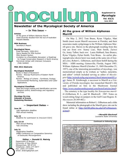
At the grave <strong>of</strong> William Alphonso<br />
Murrill<br />
On May 2, 2012 Tom Bruns, Rytas Vilgalys, Matt<br />
Smith (most recent Murrill successor at Florida), and their<br />
associates made a pilgrimage to the William Alphonso Murrill<br />
grave site. Shown in the photograph resulting from this<br />
trip are front row: Sunny Liao, Matt Smith, Zaiwei<br />
Ge, Jenny Talbot; back row: Laura Hubbard, Sara Branco,<br />
Rytas Vilgalys, Dylan Smith, Tom Bruns. An earlier photograph<br />
(also shown here) was made at the same site <strong>of</strong> Josiah<br />
Lowe, Robert L. Gilbertson, and Edson Setliff during the<br />
MSA – AIBS meeting, Gainesville, Florida, August 1985.<br />
William Alphonso Murrill (October 13, 1869-December 25,<br />
1957), one <strong>of</strong> the interesting personalities <strong>of</strong> mycology, was<br />
memorialized simply as he wished, as “author, naturalist,<br />
and editor” (which included serving as editor <strong>of</strong> Mycologia)<br />
(http://sciweb.nybg.org/science2/hcol/intern/murrill1.a<br />
sp). James W. Kimbrough, a successor to Murrill as a University<br />
<strong>of</strong> Florida mycologist, has researched Murrill’s life<br />
and published on him in the Mushroom Journal<br />
(http://www.mushroomthejournal.com/best<strong>of</strong>/articles.html)<br />
. The cemetery is the type locality for Xenosperma murrillii<br />
(Gilbertson, R. L., and M. Blackwell. 1987. Notes on<br />
wood-rotting fungi on junipers in the Gulf Coast Region. II.<br />
Mycotaxon 28:369-402.)<br />
Memorial information on Robert L. Gilbertson and a slide<br />
show including the photograph at the Murrill grave site can be<br />
found online at (http://lsb380.plbio.lsu.edu/Gil%20memorial.html).<br />
—Meredith Blackwell<br />
Louisiana State University<br />
Continued on following page
Yeast Diversity Studied from the Gut <strong>of</strong> Australian Passalid Beetles<br />
Hector Urbina, LSU graduate student from Caracas,<br />
was awarded an MSA Graduate Fellowship in<br />
2011, and he is very grateful to the <strong>Society</strong> for the<br />
award. MSA funding and a small grant from a program<br />
sponsored by the National Science Foundation<br />
and the Louisiana Board <strong>of</strong> Regents provided resources<br />
for a month in tropical forests in Queensland,<br />
Australia. The objective<br />
<strong>of</strong> the trip was to collect<br />
Australian passalid beetles<br />
(Passalidae,<br />
Coleoptera) and to isolate<br />
and purify yeasts<br />
from the gut <strong>of</strong> the<br />
wood-ingesting beetles.<br />
Strains associated with<br />
the gut <strong>of</strong> individuals in<br />
this family <strong>of</strong> the lignicolous<br />
insects are able to<br />
degrade and ferment a<br />
number <strong>of</strong> plant cell<br />
wall components.<br />
This family <strong>of</strong> insects<br />
mostly feed on decaying<br />
wood, and more<br />
than 30 species <strong>of</strong> passalids<br />
have been report-<br />
2 <strong>Inoculum</strong> <strong>63</strong>(3), June 2012<br />
ed in from Australia. Yeasts from the Australian beetles<br />
as well as from the southeastern US, Guatemala,<br />
Panama, and Thailand will give Hector a snapshot <strong>of</strong><br />
the diversity <strong>of</strong> passalid-associated yeasts in several<br />
tropical regions <strong>of</strong> the world. Hector was successful<br />
in isolating more than 1000 yeasts from 200 adult<br />
Continued on following page<br />
Fig. 1. View <strong>of</strong> the Lamington National Park, Brisbane (Australia)
eetles in at least seven species <strong>of</strong> passalids. The beetle collecting<br />
was done under the guidance <strong>of</strong> his Australian colleagues,<br />
Roger Shivas, Justin Bartlett, Alistar McTaggant, and<br />
Desley Tree <strong>of</strong> the Department <strong>of</strong> Employment, Economic Development<br />
and Innovation, Queensland, Brisbane, Australia;<br />
and Dr. Owen Seeman, expert in Australian passalids and Collection<br />
Manager for Arachnida and Myriapoda, Biodiversity<br />
Program at the Queensland Museum. The research team visited<br />
a number <strong>of</strong> national parks including, Barron Gorge,<br />
Wooroonooran, Daintree, D’Agilar, Lamington, Main Rage and<br />
others. The beetle vouchers have arrived in Baton Rouge, and<br />
the yeasts are on their way to characterization. This work will<br />
increase our understanding <strong>of</strong> the diversity, phylogeny, and biochemical<br />
abilities <strong>of</strong> yeasts associated with the passalid gut.<br />
Hector also thanks his other Australian collaborators, Tomas<br />
Marney, who has helped in preparation <strong>of</strong> media and culturing<br />
<strong>of</strong> yeasts, and Dean Beasley for his help in depositing the yeast<br />
strains into the BRIP culture collection in Brisbane.<br />
Secretary’s Report<br />
Hello everyone! By the time you read this, summer<br />
will be upon us and we will be getting ready<br />
for the 2012 Annual Meeting at Yale University<br />
in New Haven, CT. Jean Lodge and the Program<br />
Committee have put together an exciting program<br />
for us this year; it will be a meeting to remember,<br />
and my final meeting as MSA Secretary. It has<br />
been an honor to serve!<br />
Council Business: Since my last report, the<br />
Council approved 6 actions by email poll. These<br />
included: Council support for Honorary Members<br />
and MSA Fellows selected by the Honorary<br />
Awards Committee; approval <strong>of</strong> Louise Glass for<br />
the Editorial Advisory Committee; and the approval<br />
<strong>of</strong> Tim James and Ignazio Carbone as Associate<br />
Editors <strong>of</strong> Mycologia.<br />
New Members: It is my pleasure to extend a<br />
warm welcome to new or returning members.<br />
Their membership will be formally approved at the 2012 Annual<br />
Business Meeting at Yale University.<br />
Canada - Tanay Bose, Michelle Hubbard, Hai Dt Nguyen, Judith<br />
Gagnon<br />
Czech Republic - Petr Baldrian<br />
Guyana - Dillon Husbands<br />
India- Krishnendu Acharya<br />
Mexico - Fidel Jaime<br />
—Hector Urbina<br />
MSA BUSINESS<br />
Jessie Glaeser,<br />
Secretary<br />
(Photo by Tom Volk)<br />
Fig. 2. Hector Urbina with a passalid collection<br />
at Jumrum Creek Conservation Park,<br />
Kuranda, Photo by Berndt Reinhard<br />
Netherlands - Maria Dam<br />
Sri Lanka - Dimuthu Manamgoda<br />
Thailand - Saranyaphat Boonmee<br />
Turkey - Mustafa Isiloglu<br />
United States - Suzette Senerez Arcibal, Silvia<br />
Bibbo, Kathryn E. Bushley, Lauren S C<strong>of</strong>fua,<br />
Sharifa Crandall, Andrea Davis, Wade Elmer,<br />
Karen Fisher, Serita D Frey, Erica Goldberger,<br />
Christine V Hawkes, Benjamin Held, Elizabeth<br />
Heppenheimer, Jaqueline X Hess, Miriam I<br />
Hutchinson, Eric Johnson, Justin Kaffenberger,<br />
Prasanna Kandel, Chinyere Knight , Melina<br />
Kozanitas, Samantha Lee, James Lendeme,<br />
Lotus A L<strong>of</strong>gren, Sara R Lopez, Shiloh<br />
Lueschow, Jing Luo, Benjamin Morgan, Eric<br />
Morrison, Kyle Patton, Eimy Plata, Elizabeth<br />
Roberts, Sarah Robinson, Katy Ryan, Jesse J Sadowsky,<br />
Jeffrey Shaw, Xiaomei Shu, Jason Slot,<br />
Daniel J Spakowicz, Iman Sylvain, Alexander Tice, Jeffrey<br />
Townsend, Gilberto Uribe Valdez, Roo W. Vandegrift, Gloria<br />
Wada, Eric Walberg, Holly Williams, Yuka Yajima, Kolea C<br />
Zimmerman<br />
Viet Nam - Hoang Pham<br />
Emeritus Members: There were no applications for Emeritus<br />
status in the past two months. Emeritus status is granted to<br />
MSA members who are retired from active pr<strong>of</strong>essional em-<br />
Continued on following page<br />
<strong>Inoculum</strong> <strong>63</strong>(3), June 2012 3
ployment and who have been MSA members for 15 years or<br />
more. There is no membership fee for Emeritus members although<br />
there is a reduced fee for access to Mycologia. If you<br />
are interested in becoming an Emeritus member, please contact<br />
me directly at msasec01@yahoo.com.<br />
Deceased Members: We are saddened to hear <strong>of</strong> the passing<br />
<strong>of</strong> Erast Parmasto, an Honorary Member <strong>of</strong> MSA. Historian<br />
Ron Petersen has information about Dr. Parmasto’s career later<br />
in this issue. We were also sorry to learn, belatedly, <strong>of</strong> the passing<br />
<strong>of</strong> B.T. Lingappa, Dexter H. Howard, and Marjorie Anchel.<br />
Reminder: Renewing your MSA membership is easier than<br />
ever! Just log in to the MSA website at http://www.msafungi.org.<br />
There is now an email reminder system available if you<br />
have forgotten your MSA user id or password.<br />
REMINDER: MSA Directory Update: Is your information<br />
up-to-date in the MSA directory? The <strong>Society</strong> is relying more<br />
and more on email to bring you the latest MSA news, awards<br />
announcements and other timely information. To ensure that<br />
4 <strong>Inoculum</strong> <strong>63</strong>(3), June 2012<br />
you receive <strong>Society</strong> blast emails and the <strong>Inoculum</strong> as soon as it<br />
comes out, and so that your colleagues can keep in touch,<br />
please check the accuracy <strong>of</strong> your email address and contact information<br />
in the online directory. This can be accessed via our<br />
web site at www.msafungi.org. If you need assistance with<br />
updating your membership information, please contact our Association<br />
Manager at Allen Press, the always-helpful Kay Rose<br />
at krose@allenpress.com.<br />
Please do not hesitate to contact me about MSA Business or any<br />
questions that you may have about the <strong>Society</strong>. In recent years<br />
we have suffered an alarming decline in membership and it<br />
would be wonderful to reverse this trend. The first step is for<br />
everyone who is currently a member to renew for the upcoming<br />
year. And don’t forget to recommend MSA to your amateur or<br />
pr<strong>of</strong>essional colleagues who are interested in fungi – be they<br />
pathologists, geneticists, ecologists, or people who just like to<br />
wander around in the woods. There is room in MSA for all!<br />
MYCOLOGICAL NEWS<br />
Erast Parmasto, 1928-2012<br />
The MSA has lost one <strong>of</strong> its honorary members, Erast<br />
Parmasto <strong>of</strong> Tallin, Estonia.<br />
Erast was an academician <strong>of</strong> the Academy <strong>of</strong> Sciences<br />
<strong>of</strong> Estonia and expert in biosystematics, especially in the taxonomy<br />
<strong>of</strong> Hymenomycetes and statistics <strong>of</strong> spore production<br />
and measurement. An outstanding student in Estonian<br />
schools during World War II, Erast thereafter became a student<br />
<strong>of</strong> Apollinari Bondarzew in Leningrad (now St. Petersburg),<br />
thereby establishing a promising foundation on which<br />
to build a career, were it not the early days <strong>of</strong> the “Cold War”<br />
and difficult communication outside the Soviet hegemony.<br />
By 1972, Erast was elected a member <strong>of</strong> the Estonian Academy<br />
<strong>of</strong> Sciences, at the beginning <strong>of</strong> a career spent at the Institute<br />
<strong>of</strong> Biology <strong>of</strong> the Estonian Academy <strong>of</strong> Sciences in<br />
Tartu, Estonia.<br />
Like many <strong>of</strong> his age and geographic location, Erast<br />
lived through the days <strong>of</strong> Germany occupation, followed by<br />
years <strong>of</strong> Soviet Russian domination. During the latter<br />
period, it was almost impossible to gain recognition be-<br />
yond the “Iron Curtain,” but Erast persisted in publishing<br />
(always correctly in Russian) scientific papers, including<br />
several major monographs and, when possible,<br />
putting them in the hands <strong>of</strong> workers outside the Russian<br />
hegemony.<br />
A personal note: I first met Erast in Stockholm,<br />
Sweden. He had been invited to join a foray to the<br />
Swedish north, and we spent hours on the train from<br />
Stockholm to Umeå. Although we plied him with questions<br />
about conditions in Estonia, he would not answer<br />
directly, implying that “the walls have ears.” Even on<br />
the ferry he would not comment on such things. At last<br />
we reached our destination near the Norwegian border<br />
—Jessie A. Glaeser<br />
MSA Secretary<br />
msasec1@yahoo.com<br />
and the following morning we climbed a nearby hill and rested<br />
on a large rock, where Erast, as was his habit, lit a cigarette.<br />
There, surrounded for many meters by nothing by nature,<br />
he <strong>of</strong>fered, “Now I will tell you about conditions in<br />
Estonia.” And they were grim but revelatory to those <strong>of</strong> us<br />
working in freer, less ideological circumstances. Only relatively<br />
recently did life become appreciably more comfortable<br />
for science in the Baltic States.<br />
Author <strong>of</strong> over 400 scientific papers, an obituary with<br />
photos is available at http://erast.ut.ee/ErastParmasto/. He is<br />
survived by his wife and scientific collaborator, Ilmi. Erast<br />
will be remembered affectionately for his dry wit (necessary<br />
for survival in his lifetime) and careful attention to scientific<br />
detail. He mentored numerous younger colleagues who will<br />
carry on with the benefit <strong>of</strong> his experience.<br />
MSA Auction Items<br />
—Ron Petersen<br />
University <strong>of</strong> Tennessee<br />
The MSA auction (Wednesday evening, July 18) is one <strong>of</strong><br />
the major fundraising efforts <strong>of</strong> the society. All donations to the<br />
auction, silly or serious, are very welcome. If you plan to donate<br />
an item (or items) to the auction, please bring them with you<br />
and drop me an e-mail to let me know what you are bringing. If<br />
you don’t plan to come this year but still would like to donate,<br />
please send items to the address below.<br />
—Karen Hughes<br />
Ecology and Evolutionary Biology<br />
Hesler Biology Building Rm 330<br />
University <strong>of</strong> Tennessee, Knoxville 37920
MSA Student Section<br />
At the 2012 meeting this year in<br />
New Haven, the <strong>Mycological</strong> <strong>Society</strong><br />
<strong>of</strong> <strong>America</strong> will be launching a Student<br />
Section. This student-run group<br />
within the MSA will provide opportunities<br />
for students to network with<br />
other students in their own fields and<br />
beyond. It will also be valuable for<br />
student members <strong>of</strong> MSA seeking<br />
connections with those performing<br />
cutting edge research in mycology<br />
and thus has the potential to inspire<br />
future collaborative research. The<br />
Student Section is open and inclusive;<br />
we welcome the participation <strong>of</strong> all<br />
students (and faculty!) in building this<br />
group. We look forward to your participation<br />
in our new mentorship program<br />
and hope you can also join us at<br />
the following events.<br />
Sunday July 15 MSA Student Workshop:<br />
Using fungi to get a job<br />
Join us as Harvard University mycology pr<strong>of</strong>essor Anne<br />
Pringle leads our first MSA Student Section event. This pr<strong>of</strong>essional<br />
development workshop will take place after the<br />
Foray and before the Welcome Reception on Sunday.<br />
Abstract. Some ads specifically ask for mycologists,<br />
more <strong>of</strong>ten, departments are looking for conceptually oriented<br />
biologists. Fungi <strong>of</strong>fer myriad opportunities for connecting<br />
to broader themes in ecology, evolution, and other fields;<br />
making those connections can be critical to a successful job<br />
hunt. I’ll talk about my own experiences on the job market<br />
and share the research and teaching statements I used to find<br />
my current position. We’ll then practice job strategies by tak-<br />
Invitation to a Roundtable Discussion<br />
Toward a Roadmap for Fungal Conservation<br />
Research in North <strong>America</strong>: Tools,<br />
Data Needs and Collaborations<br />
MSA Annual Meeting<br />
Monday 16 July, 4-6 PM<br />
The 80 th annual meeting <strong>of</strong> the MSA will include a<br />
roundtable discussion on the tools, data, and collaborations<br />
that are needed for fungal conservation in North <strong>America</strong>.<br />
The threat <strong>of</strong> large-scale species extinctions during this<br />
century makes understanding the extent, functions, and consequences<br />
<strong>of</strong> loss <strong>of</strong> biodiversity one <strong>of</strong> the most imperative<br />
areas <strong>of</strong> biological research <strong>of</strong> our time. At the same time<br />
that environmental DNA studies highlight the depths <strong>of</strong> unknown<br />
fungal diversity, taxonomic specialists are continuing<br />
to decrease in number. And while studies <strong>of</strong> fungal community<br />
ecology are abundant, there have been very few studies<br />
ing the time to talk about our research<br />
“mission statements” and<br />
“cocktail-party tidbits”. I’ll explain<br />
why these are useful and we’ll finish<br />
by sharing our newly crafted statements<br />
with each other.<br />
Monday July 16th: First<br />
Annual Student Section<br />
Mixer<br />
Join us before the poster session<br />
as we gather in the poster area for a<br />
casual meet and greet with c<strong>of</strong>fee,<br />
dessert, and other drinks provided.<br />
The mixer will go from 6:00-6:30<br />
p.m.<br />
Mentorship Program<br />
Mentorship programs are an excellent<br />
way to introduce students to<br />
other researchers with similar interests.<br />
In addition, mentors may assist in steering student research<br />
or by providing information about previous studies or<br />
future opportunities.<br />
For these reasons, all students who expressed interest on<br />
their registration form have been assigned a senior mycologist<br />
who will serve as their mentor for the duration <strong>of</strong> the<br />
New Haven meeting. Students will meet their senior mycologist<br />
mentor at the Student Section Mixer on Monday, July<br />
16th.<br />
We appreciate your involvement in the commencement<br />
<strong>of</strong> the new MSA Student Section and look forward to seeing<br />
you all in New Haven!<br />
—Mia Maltz<br />
Department <strong>of</strong> Ecology and Evolutionary Biology<br />
University <strong>of</strong> California, Irvine<br />
in North <strong>America</strong> conducted with an explicit conservation<br />
biology focus; the large-scale Survey and Manage program<br />
<strong>of</strong> the U.S. Forest Service under the Northwest Forest Plan<br />
(Molina 2008) is a rare example. The extent <strong>of</strong> species endemism<br />
and rarity is unknown for most habitat types, and<br />
fungi have not been considered for conservation action (e.g.,<br />
RED data lists) in the U.S.A.<br />
Our goal for this roundtable is to consider ways to make<br />
progress in fungal conservation by considering the tools, data<br />
needs, and collaborations necessary to place fungi in a conservation<br />
framework comparable to that which exists for plant<br />
and animal species. In terms <strong>of</strong> tools, potential topics include<br />
harnessing molecular tools, training new taxonomic specialists,<br />
and informatics tools for information gathering and transfer<br />
for herbarium collections and ecological and distribution<br />
data. Potential types <strong>of</strong> data necessary for conservation in-<br />
Continued on following page<br />
<strong>Inoculum</strong> <strong>63</strong>(3), June 2012 5
clude threat assessments; taxonomic identifications; distributions;<br />
life cycle and fruiting patterns; habitat requirements;<br />
species interactions; diversity, endemism and the characteristics<br />
<strong>of</strong> fungal biodiversity hotspots; and the dynamics inherent<br />
to many <strong>of</strong> these data. We have to consider how to integrate<br />
existing conservation-relevant, but not explicitly<br />
conservation-oriented, data into a clearer conservation framework;<br />
for example, whether inferences about fungal distribution<br />
and occurrence can be made from studies <strong>of</strong> ecological<br />
community composition based on small plots (e.g., recent<br />
studies using high-throughput environmental DNA sequencing).<br />
Potential types <strong>of</strong> collaboration include stronger networks<br />
between ecologists, taxonomists, and population geneticists,<br />
but also pr<strong>of</strong>essional-amateur collaborations as a<br />
means to address the “dwindling taxonomists” issue.<br />
An anticipated result <strong>of</strong> the roundtable will be increased<br />
clarity on whether to – and if so, how to – bring greater attention<br />
to fungal conservation and lay out a roadmap for a<br />
collaborative, scientifically rigorous way forward – specific<br />
areas in which we need to concentrate our research efforts,<br />
how to set priorities, how to integrate knowledge from many<br />
different sources, how to include conservation issues in our<br />
Dominican Amber with Unknown Inclusions<br />
Can you identify the inclusion? Trapped in a Dominican<br />
amber specimen is a cluster <strong>of</strong> what looks<br />
like fungi, including one that is suspended in<br />
the amber. There appears to be five gilled clusters,<br />
which are partially exposed on the back <strong>of</strong> the piece.<br />
I would like assistance in classifying these inclusions.<br />
Please contact me at mail.acton@gmail.com if<br />
you can <strong>of</strong>fer guidance in identifying these mystery<br />
inclusions.<br />
6 <strong>Inoculum</strong> <strong>63</strong>(3), June 2012<br />
—Jeanette Acton<br />
other research (Molina et al. 2011), and how to educate, inform<br />
and involve the general public.<br />
We invite all attenders <strong>of</strong> the upcoming annual meeting<br />
to attend the roundtable discussion, ready to bring your<br />
thoughts and experiences to these issues. Invited facilitators<br />
will provide brief (approximately 5-minute) introductions on<br />
several topics (see the meeting website,<br />
http://msa2012.net/schedule/symposia.php#roadmap, for<br />
more details) to stimulate discussion, but for the main part it<br />
will be an ‘open mike’ format with active participation <strong>of</strong><br />
anyone in attendance. We hope to see you there.<br />
—On behalf <strong>of</strong> the MSA Conservation Committee:<br />
Todd Osmundson and Else Vellinga<br />
University <strong>of</strong> California, Berkeley<br />
REFERENCES<br />
Molina R. 2008. Protecting rare, little known, old-growth forest-associated<br />
fungi in the Pacific Northwest USA: a case<br />
study in fungal conservation. <strong>Mycological</strong> Research 112:<br />
613-<strong>63</strong>8.<br />
Molina R, Horton TR, Trappe JM, Marcot BG. 2011. Addressing<br />
uncertainty: How to conserve and manage rare or<br />
little-known fungi. Fungal Ecology 4: 134-146.
MSA 2012 ABSTRACTS<br />
Adenipekun, Clementina O 1 , Omasan E Ejoh 2 , and Adeniyi A Ogunjobi 2 .<br />
1 Department <strong>of</strong> Botany, University <strong>of</strong> Ibadan, Ibadan, Nigeria, 2 Department <strong>of</strong><br />
Microbiology, University <strong>of</strong> Ibadan, Ibadan, Nigeria. Effect <strong>of</strong> Pleurotus tuberregium<br />
Singer and microorganisms on degradation <strong>of</strong> unsterilized soil contaminated<br />
with cutting fluids<br />
The study <strong>of</strong> Pleurotus tuber-regium Singer and indigenous microorganisms<br />
isolated from unsterilized soil polluted with cutting fluids were investigated<br />
over an incubation period <strong>of</strong> 0 and 2 months. The ability <strong>of</strong> these organisms to degrade<br />
Total Petroleum hydrocarbon (TPH) present in the cutting fluids, lignin<br />
content <strong>of</strong> rice straw, their enzyme activity as well as their ability to accumulate<br />
heavy metals present in the polluted soils were monitored. In all the soil samples<br />
(unsterilized soil polluted with cutting fluids (US) and unsterilized soil polluted<br />
with cutting fluids incubated with P. tuber-regium (USP), a significant increase in<br />
the nutrient content was observed with USP recording the highest. The heavy<br />
metal content <strong>of</strong> the three soil samples decreased with increase in incubation period<br />
showing that bioaccumulation <strong>of</strong> the heavy metals had occurred. The indigenous<br />
microbes alone in US accumulated heavy metals better than the indigenous<br />
microbes in USP. TPH loss (36.23%) was recorded at 10% cutting fluids concentration<br />
in USP compared to US with 19.6%. The unsterilized contaminated<br />
soil inoculated with P. tuber-regium recorded the highest lignin degradation as the<br />
polyphenol oxidase and peroxidase activity <strong>of</strong> the organisms in all the samples<br />
showed a gradual increase. The microorganisms isolated from unsterilized soil<br />
samples included Bacillus licheniformis, B. cereus, Bacillus sp., Pseudomonas<br />
aeruginosa, Pseudomonas sp., Corynebacterium sp., Neisseria sp., Trichoderma<br />
harzanium, Aspergillus niger, A. flavus, A. glaucus, Mucor sp. and Rhizopus sp.<br />
Acharya, Krishnendu 1 , Arun K Dutta 1 , Prakash Pradhan 1 , and Anirban Roy 2 1<br />
.<br />
Molecular and Applied Mycology and Plant Pathology Laboratory, Department<br />
<strong>of</strong> Botany, University <strong>of</strong> Calcutta, Kolkata, INDIA, 2 West Bengal Biodiversity<br />
Board, Paribesh Bhawan, Kolkata, INDIA. Habitat diversity <strong>of</strong> macr<strong>of</strong>ungi in<br />
the Indian part <strong>of</strong> Sundarban<br />
The Sundarban Biosphere Reserve covers an area <strong>of</strong> approximate 25,500<br />
sq km. However, only 9,<strong>63</strong>0 sq km is in India, with the rest located in Bangladesh.<br />
This area in India is demarcated by the Hoogly River in the west, the Bay-<strong>of</strong>-Bengal<br />
in the south, and the Harinbhanga and Raimangal Rivers in the east and the<br />
Dampier-Hodges line in the north. The wide range <strong>of</strong> phyto-topographical features,<br />
presence <strong>of</strong> spatial and temporal variability in hydrological regimes and the<br />
diverse substrata has contributed to the high biodiversity <strong>of</strong> this region. Mangrove<br />
forest dominates in the core area and some margins <strong>of</strong> the deltic island, where inhabited<br />
lands show typical coastal vegetation. Several anthropogenic activities,<br />
recurring natural calamities and recent incidences <strong>of</strong> cyclones (e.g., Aila) have<br />
added to the vulnerability <strong>of</strong> this biologically rich world heritage site. The climate<br />
is characterized by relatively high temperature and humidity (>80%) throughout<br />
the year, and well-distributed rainfall during the monsoon season. Temperatures<br />
rise from a daily minima <strong>of</strong> 10ºC in the winter to a maximum <strong>of</strong> about 43ºC in<br />
March and may exceed 32ºC during the monsoon season. The average annual<br />
rainfall in the Indian part <strong>of</strong> the Sundarban region is 1,662 mm. During our survey<br />
(2010-2011), 77 quadrates were studied each measuring 20 _ 20 meters.<br />
Macr<strong>of</strong>ungi were collected, photographed, identified and were preserved with accession<br />
numbers. A total <strong>of</strong> 71 species <strong>of</strong> macr<strong>of</strong>ungi belonging to 29 families, 53<br />
genera were identified and their ecology was evaluated.<br />
Afshan, Najam-ul-Sahar, Abdul Nasir Khalid, and A R Niazi. Centre for Undergraduate<br />
Studies, University <strong>of</strong> the Punjab, Quaid-e-Azam Campus, Lahore,<br />
54950, Pakistan. Rust fungi <strong>of</strong> Himalayan Moist Temperate forests <strong>of</strong> Pakistan,<br />
their diversity and distribution: An overview<br />
Himalayan Moist Temperate forests <strong>of</strong> Pakistan have coniferous species,<br />
mixed evergreen and deciduous patches and broad leaved forests. These forests <strong>of</strong><br />
Pakistan merge downward with the tropical thorn forests and upwards with the<br />
alpine meadows. The precipitation may exceed 600 mm, thus making <strong>of</strong> these<br />
montane forests a bio-geographical crossroad between submontane and alpine<br />
meadow vegetation. Being rich in plant diversity, these forests harbor a large<br />
number <strong>of</strong> rust fungi that are obligate parasites <strong>of</strong> plants. Although the fungal flora<br />
<strong>of</strong> Pakistan has been explored by several workers in the past, the important group<br />
<strong>of</strong> rust fungi has largely been neglected resulting in a paucity <strong>of</strong> literature and very<br />
fragmentary knowledge <strong>of</strong> these fungi, particularly in Himalayan Moist Temperate<br />
forests <strong>of</strong> Pakistan. This study was undertaken to explore and assess the diversity<br />
and distribution <strong>of</strong> rust fungi along with their respective host plants in this<br />
floristically rich area. This preliminary study <strong>of</strong> the hosts and their rusts includes<br />
approximately 169 species and 19 genera <strong>of</strong> rust fungi on approximately 260<br />
species <strong>of</strong> host plants. These rust fungi include one species each <strong>of</strong> genus Cronartium,<br />
Hyalopsora, Miyagia, Monosporidium, Pucciniastrum, Pucciniostele,<br />
Thekopsora and Uredinopsis respectively; two species each <strong>of</strong> Gymnosporangium,<br />
Peridermium, Pucciniastrum and Uredo; three and four species <strong>of</strong><br />
Caeoma and Coleosporium respectively; seven <strong>of</strong> Melampsora; nine <strong>of</strong> Aecidium;<br />
eleven <strong>of</strong> Phragmidium; twenty four species <strong>of</strong> Uromyces and ninety five<br />
species <strong>of</strong> largest genus <strong>of</strong> rust fungi, Puccinia. This study enlists rust fungi <strong>of</strong> Himalayan<br />
Moist Temperate forests <strong>of</strong> Pakistan and presents their species richness<br />
and geographical distribution. This work will help to prepare a checklist <strong>of</strong> rust<br />
fungi <strong>of</strong> this area and will ultimately lead to the documentation and preparation <strong>of</strong><br />
monograph <strong>of</strong> the rust fungi <strong>of</strong> Pakistan.<br />
Ahrendt, Steven R and Jason E Stajich. Department <strong>of</strong> Plant Pathology and Microbiology,<br />
University <strong>of</strong> California, Riverside, CA 92521. Analysis <strong>of</strong> a putative<br />
sensory rhodopsin in the Chytridiomycota<br />
Rhodopsin is a seven-transmembrane G-protein coupled receptor<br />
(GPCR) that responds to light through photoisomerization <strong>of</strong> a covalently bound<br />
11-cis-retinal molecule. This action forces helical motion and subsequent signal<br />
transduction through activation <strong>of</strong> the coupled G-protein. Rhodopsin homologs<br />
have only been identified in metazoan lineages, and while Dikarya fungi are<br />
known to have opsins, phytochromes, and cryptochromes to sense light, this type<br />
<strong>of</strong> rhodopsin has only been found encoded in the genomes <strong>of</strong> the early diverging<br />
Chytridiomycota and Blastocladiomycota fungi. Previous and current work has<br />
shown that zoosporic fungi are phototaxic but the molecular mechanisms <strong>of</strong> light<br />
sensing in early diverging fungi has not been explored. Here we describe structural<br />
and functional analyses <strong>of</strong> rhodopsin proteins identified in two species <strong>of</strong><br />
Chytridiomycota, the amphibian pathogen Batracochytrium dendrobatidis and<br />
the terrestrial saprotroph Spizellomyces punctatus. Comparative genomics analyses<br />
<strong>of</strong> rhodopsin and flagellum genes in the Chytridiomycota, Zygomycetes, and<br />
Dikarya show a correlation <strong>of</strong> flagella and rhodopsin presence across the fungi.<br />
Computational modeling <strong>of</strong> the B. dendrobatidis and S. punctatus proteins indicates<br />
that they both adopt the seven transmembrane helix formation typical <strong>of</strong><br />
rhodopsin proteins. Additionally, structural features associated with rhodopsin<br />
proteins are observed as well. The B. dendrobatidis protein sequence is notably<br />
lacking the conserved lysine residue, however this residue is present in the S.<br />
punctatus sequence. Phototaxis assays were performed to determine in vivo function<br />
<strong>of</strong> the rhodopsin proteins. Ongoing work to characterize protein function<br />
through expression in Pichia pastoris may provide insight into the mechanism <strong>of</strong><br />
retinal binding and photoisomerization in these chytrid rhodopsins.<br />
Aime, M Catherine. Louisiana State University Agricultural Center, Department<br />
<strong>of</strong> Plant Pathology and Crop Physiology, Baton Rouge, LA 70803. Basidiomycete<br />
taxonomy without dual nomenclature<br />
The new International Code <strong>of</strong> Nomenclature for algae, fungi, and plants<br />
(Melbourne Code) discontinued the application <strong>of</strong> dual nomenclature in Fungi.<br />
Under earlier Codes Article 59 permitted more than one name for pleomorphic<br />
non-lichenized basidiomycetes. Effective immediately all names will compete for<br />
priority. As <strong>of</strong> January 1, 2013, publication <strong>of</strong> alternative names for species or<br />
genera will not be allowed. These changes affect basidiomycete taxonomy in several<br />
ways. To make one example, anamorphic yeast states occur in all three subphyla<br />
<strong>of</strong> Basidiomycota. Under previous versions <strong>of</strong> the Code these were treated<br />
separately from teleomorphic species and assigned to form genera principally<br />
based on carbon assimilation tests. Molecular data are now used to place these<br />
within a phylogenetic framework, highlighting the artificiality <strong>of</strong> the old system.<br />
For example, species <strong>of</strong> Sporobolomyces occur across most <strong>of</strong> the yeast-forming<br />
Pucciniomycotina classes and species <strong>of</strong> Rhodotorula can be found across both<br />
Ustilaginomycotina and Pucciniomycotina, although the type species for both<br />
genera are placed in Sporidiobolales. Thus restriction <strong>of</strong> generic names to include<br />
only those species within a monophyletic lineage will dictate the necessity <strong>of</strong><br />
many name changes within these groups as extra-Sporobolomyces, -Rhodotorula<br />
and other anamorphic basidiomycete yeast species are integrated into a phylogenetic-based<br />
taxonomy. These and many other taxonomic challenges for the community<br />
will be discussed as they apply to different groups <strong>of</strong> impacted Basidiomycota.<br />
Ainsworth, A Martyn 1 , David Parfitt 2 , Hilary J Rogers 2 , and Lynne Boddy 2 1<br />
.<br />
Royal Botanic Gardens, Kew / Natural England, Mycology Section, Jodrell<br />
Lab., Kew. TW9 3AB. United Kingdom, 2 Cardiff University, Cardiff School <strong>of</strong><br />
Biosciences, Cardiff. CF10 3AX. United Kingdom. Species concepts in European<br />
stipitate hydnoid fungi<br />
Continued on following page<br />
<strong>Inoculum</strong> <strong>63</strong>(3), June 2012 7
The standard taxonomic treatment <strong>of</strong> European stipitate hydnoid species<br />
(ectomycorrhizal tooth fungi in the genera Bankera, Hydnellum, Phellodon and<br />
Sarcodon), written by R.A. Maas Geesteranus, is almost 40 years old. It is timely<br />
therefore to re-examine the morphological species concepts therein using the<br />
results <strong>of</strong> a combined molecular and morphological approach. These fungi have<br />
received conservation-related publicity across Europe and are now generally regarded<br />
as nitrogen-sensitive tree symbionts frequently associating with roots <strong>of</strong><br />
Fagaceae and Pinaceae. A decline in European fruiting populations is mainly ascribed<br />
to increased habitat loss and aerial nitrogen deposition. Of the 18 extant<br />
stipitate hydnoid species currently included on the British and Irish checklist, 15<br />
are recognised as <strong>of</strong> conservation importance (priority BAP species) in the UK.<br />
However all conservation status assessments currently depend on morphological<br />
taxonomic concepts and accurate identification <strong>of</strong> fruit bodies. Recognising that a<br />
mushroom is a stipitate hydnoid is <strong>of</strong>ten straightforward, but naming the species<br />
on existing fruit body criteria can be fraught with difficulty. This is due to a combination<br />
<strong>of</strong> identification and taxonomic issues. In order to improve assessments<br />
<strong>of</strong> species distributions, conservation status and ecological/conservation requirements,<br />
we have adopted a combined approach using molecular (ITS1 sequencing)<br />
and traditional (fruit body and spore morphology) methods. We have discovered<br />
that the list <strong>of</strong> extant taxa is rather longer (and increasing) than the list <strong>of</strong> currently<br />
accepted names. There are undoubtedly cryptic and not-so-cryptic hydnoid taxa<br />
to be described in Britain and elsewhere in Europe. We are now comparing<br />
species names with sequence-based groupings in order to pin-point the undescribed<br />
species. This combined approach will facilitate stipitate hydnoid identification,<br />
help to locate their “best” sites for conservation and accelerate their belowground<br />
ecological study.<br />
Albu, Sebastian, Tomas A Rush, and M Catherine Aime. Louisiana State University<br />
Agricultural Center, Department <strong>of</strong> Plant Pathology and Crop Physiology,<br />
Baton Rouge, Louisiana, 70803. Description <strong>of</strong> two anamorphic yeasts in the<br />
Ustilaginales<br />
Two basidiomycete yeasts belonging to the Ustilaginales were isolated in 2011<br />
from the leaves <strong>of</strong> several fern species in Baton Rouge, Louisiana. Based on a<br />
combination <strong>of</strong> assimilation tests and phenotypic characterization, Farysizyma sp.<br />
nov. SA209 (Anthracoideaceae) and Pseudozyma sp. nov. SA575 (Ustilaginaceae)<br />
represent previously undescribed anamorphs in the Ustilaginomycetes.<br />
Colony morphologies are initially yeast-like, subsequently developing pseudohyphal<br />
tufts around the growing margin. Maximum likelihood analyses using four<br />
loci, the internal transcribed spacer (ITS) region, large subunit (LSU) and small<br />
subunit <strong>of</strong> the nuclear rDNA cistron, and translation elongation factor 1-alpha indicate<br />
that both isolates belong within Ustilaginales. The LSU and ITS regions <strong>of</strong><br />
these isolates were compared to sequences <strong>of</strong> other available Farysizyma and<br />
Pseudozyma anamorphs and to related teleomorphs. Farysizyma sp. nov. SA209<br />
is part <strong>of</strong> a Farysizyma/Farysia clade in Anthracoideaceae containing all other<br />
known species <strong>of</strong> Farysizyma. Pseudozyma sp. nov. is sister to Sporisorium<br />
hwangense within a larger clade <strong>of</strong> predominantly Sporisorium species that includes<br />
the type (S. sorghi). Comparison <strong>of</strong> our data with phenotypic descriptions<br />
and sequence data from all known species <strong>of</strong> Farysizyma and Pseudozyma indicates<br />
that neither isolate has been previously described in the anamorphic yeast<br />
state. However, without sequence information from all known members <strong>of</strong> Ustilaginales,<br />
we cannot rule out the possibility that either <strong>of</strong> these isolates may represent<br />
a previously described teleomorphic Farysia or Ustilago/Sporisorium<br />
species.<br />
Allen, Michael F. Center for Conservation Biology, University <strong>of</strong> California,<br />
Riverside, CA 92521 USA. Mycorrhizae and resource acquisition: dynamics<br />
in fluctuating environments<br />
Mycorrhizae exist from organic layers deep into soil pr<strong>of</strong>iles, even into fracturing<br />
bedrock. As soils dry, deeper water either from groundwater or from water bound<br />
in cracks and pockets is acquired and utilized or reallocated via hydraulic redistribution.<br />
N is acquired from surface soils even under drought conditions. We utilized<br />
shifts in natural abundance isotope ratios to better understand spatial and<br />
temporal patterns <strong>of</strong> resource acquisition. Subsequently, utilizing continuous sensor<br />
and observation platforms, we monitored shifts in roots, mycorrhizal fungi,<br />
and respiration in response to changing temperature and moisture. Acute perturbations<br />
consisting <strong>of</strong> either large storms or drought result in plant mortality, but<br />
also subsequently re-order root and fungal symbiosis shifting both the types and<br />
spatial structure <strong>of</strong> resource acquisition. This may be because in seasonal environments,<br />
root and fungal activity show complex and <strong>of</strong>ten rapid responses to environmental<br />
change. We can utilize these shifts to develop a better understanding<br />
<strong>of</strong> belowground response dynamics to more chronic climate change or to acute<br />
perturbations such as hurricanes or drought.<br />
Annis, Seanna L 1 , Rafael Garcia 2 , Beth Calder 2 , and Kathryn L Hopkins 3 1 2<br />
.<br />
School <strong>of</strong> Biology and Ecology, University <strong>of</strong> Maine, Orono, ME, 04469, Department<br />
<strong>of</strong> Food Science and Human Nutrition, University <strong>of</strong> Maine, Orono,<br />
8 <strong>Inoculum</strong> <strong>63</strong>(3), June 2012<br />
ME, 04469, 3 Cooperative Extension, University <strong>of</strong> Maine, Orono, ME, 04469.<br />
Identification <strong>of</strong> fungal contamination in bottled maple syrup<br />
Maple syrup processors in the northeastern USA occasionally observe fungal contamination<br />
in their bottled maple syrup containers. The concern is that the contaminants<br />
may pose a health risk. We identified fungal organisms in 32 bottles <strong>of</strong><br />
syrup from different processors in the northeastern USA. Most containers had one<br />
fungus, but some were contaminated with multiple fungi. Fungi were identified<br />
by morphology and DNA sequences <strong>of</strong> their ribosomal internal transcribed spacer<br />
regions and B tubulin genes. Multiple species <strong>of</strong> the genera Penicillium and Aspergillus,<br />
single species <strong>of</strong> Wallemia, and as yet unidentified yeasts were isolated<br />
from multiple bottles. Species <strong>of</strong> Paecolimyces and Cladosporium were isolated<br />
from single bottles <strong>of</strong> syrup. Some <strong>of</strong> the genera <strong>of</strong> Penicillium and Aspergillus<br />
identified are known to produce mycotoxins, and the production <strong>of</strong> these compounds<br />
in maple syrup is being evaluated. Syrup is typically bottled at 82 C to decrease<br />
the risk <strong>of</strong> microbial contamination. Spores from some <strong>of</strong> the fungi were<br />
able to germinate after treatment at 70 C for 3 minutes. A higher bottling temperature<br />
may be one <strong>of</strong> the changes in bottling practices required to minimize fungal<br />
contamination. This research will result in new recommendations to maple syrup<br />
processors on preventing future fungal contamination <strong>of</strong> bottled syrup.<br />
Baldrian, Petr, Tomás Vetrovsky, Jana Vorísková, Ivana Eichlerová, Jaroslav<br />
Snajdr, Lucia Zifcáková, and Martina Stursová. Laboratory <strong>of</strong> Environmental Microbiology,<br />
Institute <strong>of</strong> Microbiology <strong>of</strong> the ASCR, Prague, Czech Republic. Exploring<br />
fungal community structure and function in forest soils: challenges<br />
and limitations <strong>of</strong> current methodologies<br />
Presently, the structure and function <strong>of</strong> soil fungal communities receives considerable<br />
attention. This attention is fuelled by the recognition <strong>of</strong> the key role <strong>of</strong> fungi<br />
in the C and N cycling in soils, especially <strong>of</strong> the forest biomes. In addition, the recent<br />
establishment <strong>of</strong> high-throughput-sequencing methods, labelling with stable<br />
isotopes or metaproteomics <strong>of</strong>fers a much higher resolution <strong>of</strong> the current studies.<br />
The interpretation <strong>of</strong> experimental results is, however, still challenging due to the<br />
fact that many methods may potentially contain more or less apparent biasses.<br />
This contribution aims to point at the most important limitations <strong>of</strong> current<br />
methodologies to explore fungal abundance, community composition and function<br />
in forest soils as studied using biomass quantification techniques (PLFA, ergosterol,<br />
qPCR), shotgun or amplicon-based next-generation-sequencing, stable<br />
isotope probing and environmental metaproteomics. In the case <strong>of</strong> microbial biomass<br />
surveys, our results show that various methods (rDNA quantification, PLFA<br />
or ergosterol assays) yield widely different results <strong>of</strong> the fungal biomass content<br />
or fungal/bacterial biomass ratio and identify the differences in the composition<br />
<strong>of</strong> fungal mycelia / genomes as a source <strong>of</strong> such errors. For the next-generationsequencing<br />
data, we show that shotgun methods are not consistent with PCRbased<br />
methods and that the use <strong>of</strong> rDNA markers is highly biased due to uneven<br />
content <strong>of</strong> ITS copies per fungal genome as can be demonstrated if single-copy<br />
genes are sequenced. The approaches to explore the active fraction <strong>of</strong> the total<br />
fungal community by the analysis <strong>of</strong> RNA-derived sequencing or the use <strong>of</strong> Stable<br />
Isotope Probing and, most recently, environmental metaproteomics <strong>of</strong>fer an<br />
attractive insight into the functioning <strong>of</strong> fungal communities. However, even the<br />
use <strong>of</strong> these methods must be careful in order to avoid experimental errors.<br />
Barge, Edward G and Cathy L Cripps. Plant Sciences and Plant Pathology Department,<br />
Montana State University, Bozeman, MT 59715. Systematics <strong>of</strong> Lactarius<br />
in the Rocky Mountain alpine zone<br />
Lactarius is an important ectomycorrhizal genus in the Arctic-Alpine<br />
Biome where it associates primarily with Salix and Betula species. The Arctic-<br />
Alpine Biome covers roughly 8% <strong>of</strong> the earth’s land and in the Rocky Mountains<br />
<strong>of</strong> North <strong>America</strong> the alpine is comprised <strong>of</strong> scattered “islands” above timberline<br />
in mountainous areas. Beginning in 1999 Cripps, Horak and others surveyed arctic-alpine<br />
macromycete distributions in the Rocky Mountains <strong>of</strong> Montana, Colorado<br />
and Wyoming. Studies <strong>of</strong> the ectomycorrhizal fungi present in arctic-alpine<br />
areas are <strong>of</strong> importance as climate change impacts this biome, which includes the<br />
range expansion <strong>of</strong> Salix species, a key alpine ectomycorrhizal phytobiont. This<br />
study focuses on the systematics <strong>of</strong> Lactarius in alpine areas <strong>of</strong> the Rocky Mountains.<br />
Macromorphological descriptions made at the time <strong>of</strong> collection, microscopic<br />
examination <strong>of</strong> dried material and phylogenetic analysis (rough) have contributed<br />
to identification. Drawings <strong>of</strong> spores, pleuromacrocystidia and<br />
cheilomacrocystidia were completed for each species using a Leica drawing tube<br />
and are complemented with SEM photographs for each species. DNA from upwards<br />
<strong>of</strong> 48 collections was successfully extracted and the ITS region amplified<br />
using primers ITS1-F and ITS4. Preliminary analysis shows morphologically<br />
identified species grouping together nicely. Thus far, the study has resulted in<br />
identification <strong>of</strong> six alpine Lactarius species: L. glyciosmus, L. lanceolatus, L.<br />
nanus, L. pseudouvidus, L. repraesentaneus and L. salicis-reticulatae. Other than<br />
a preliminary report, most are first documentations for these species in alpine<br />
Continued on following page
habitats south <strong>of</strong> the Canadian border. Host patterns are possible among the<br />
species that occur with dwarf and shrub Salix and one species seems to occur only<br />
with Betula. These species are known from many arctic-alpine habitats throughout<br />
the world in places such as Greenland, Iceland, the Alps, Svalbard and Scandinavia<br />
and are now documented for the Rocky Mountains <strong>of</strong> Montana,<br />
Wyoming and Colorado.<br />
Baroni, Timothy J 1 , Ana Esperanza Franco-Molano 2 , Marcelo Betancur 2 , and<br />
Tatiana Sanjuan 2 . 1 Department <strong>of</strong> Biological Sciences, P. O. Box 2000, State<br />
University <strong>of</strong> New York - College at Cortland, Cortland, NY 13045, USA, 2 Laboratorio<br />
de Taxonomía y Ecología de Hongos, Instituto de Biología, Universidad<br />
de Antioquia, A.A. 1226, Medellín, Colombia. New species <strong>of</strong> agarics from the<br />
Páramo region in Colombia<br />
In May <strong>of</strong> 2011 a one-day field excursion was made to the Páramo<br />
ecosystem (Luteyn, 1999) in the department <strong>of</strong> Tolima, Colombia, as part <strong>of</strong> a<br />
longer research excursion to the cloud forest mountainous regions <strong>of</strong> south central<br />
Colombia. Fifteen collections <strong>of</strong> mostly agarics were made in the Páramo,<br />
several growing directly on decaying leaves <strong>of</strong> standing Espeletia. Two <strong>of</strong> these<br />
collections are clearly new species, one a Melanotus, the other a Hypsizygus.<br />
These new taxa will be illustrated and the other collections found on that excursion<br />
will also be displayed and discussed. This isolated area with its distinctive<br />
vegetation clearly deserves further exploration over longer time periods as it appears<br />
to be under explored.<br />
Bartnicki-Garcia, Salomon. Dept. Microbiology, CICESE, Ensenada Center for<br />
Research & Higher Education, Baja California, Mexico. Lysine pathways and<br />
cell wall chemistry revealed the existence <strong>of</strong> two evolutionary lines within the<br />
Kingdom Fungi<br />
Henry Vogel’s discovery (1960’s) splitting fungi into two camps based on<br />
lysine biosynthesis provided the first and strongest piece <strong>of</strong> evidence for the existence<br />
<strong>of</strong> two evolutionary lines within Fungi. By synthesizing lysine via the common<br />
route characteristic <strong>of</strong> bacteria, algae and plants, diaminopimelic acid path<br />
(DAP), Oomycetes and Hypochytridomycetes proved to be distinct from most<br />
Fungi, namely Chytridiomycetes, Zygomycetes, Ascomycetes and Basidiomycetes<br />
which synthesize lysine via aminoadipic acid pathway (AAA), a<br />
unique route found almost exclusively in fungi. Concurrent studies on cell wall<br />
chemistry <strong>of</strong> various fungi revealed drastic differences that correlated with lysine<br />
pathways, thus DAP fungi became members <strong>of</strong> the Cellulosic line, and AAA<br />
fungi <strong>of</strong> the Chitinous line. Later, molecular phylogeny convincingly confirmed<br />
the evolutionary schism among organisms traditionally called fungi but it also<br />
placed Mycology in an existential dilemma: either preserve phylogenetic purity<br />
by removing a most important group <strong>of</strong> organisms, the cellulosic fungi, from the<br />
kingdom or continue maintaining both cellulosic and chitinous fungi within the<br />
kingdom boundaries. While the first alternative has become widely adopted, it has<br />
serious negative repercussions. The second alternative would seem more practical<br />
and beneficial to the science <strong>of</strong> Mycology and to other fields where cellulosic<br />
fungi are <strong>of</strong> great importance. It benefits nobody to exclude Oomycetes from the<br />
real world <strong>of</strong> fungi namely, publications, review articles, congresses, classrooms,<br />
textbooks, and all sorts <strong>of</strong> academic and research opportunities focused on Fungi.<br />
I advocate setting aside the issue <strong>of</strong> phylogenetic purity, and declare the Kingdom<br />
Fungi as made <strong>of</strong> two separate evolutionary lines each giving rise to individuals<br />
different in biochemical details but displaying common physiologic and morphologic<br />
features... and please let’s not refer to a classic fungus <strong>of</strong> the genus Phytophthora<br />
as a “protista-like” organism.<br />
Bates, Scott T 1 , J Gregory Caporaso 2 , D Lee Taylor 3 , and Noah Fierer 1 . 1 Cooperative<br />
Institute for Research in Environmental Sciences, University <strong>of</strong> Colorado,<br />
Boulder, CO, USA, 2 Department <strong>of</strong> Computer Science, Center for Micro-<br />
3<br />
bial Genetics and Genomics, Northern Arizona University, Flagstaff, AZ, USA,<br />
Institute <strong>of</strong> Arctic Biology and Department <strong>of</strong> Biology and Wildlife, University<br />
<strong>of</strong> Alaska Fairbanks, Fairbanks, AK, USA. High-throughput sequencing to explore<br />
the biogeography <strong>of</strong> soil fungi and the use <strong>of</strong> network analyses to examine<br />
fungal-bacterial interactions<br />
High-throughput DNA sequencing technologies have paved the way for<br />
new analytical approaches that move beyond basic descriptive inventories <strong>of</strong> microbial<br />
communities. With the aim <strong>of</strong> deciphering the structure <strong>of</strong> complex microbial<br />
communities across spatial or temporal gradients, network analysis allows<br />
for the examination <strong>of</strong> potential interactions between microbial taxa in natural systems<br />
using large environmental sequence datasets. Here, we sampled soils from<br />
numerous sites across North and South <strong>America</strong>, and used fungal ITS and bacterial<br />
16S rRNA gene sequencing with Illumina technology to recover millions <strong>of</strong><br />
environmental sequences from our sample sites. In addition to examining biogeography<br />
for soil fungi, we also applied network analysis approaches to identify<br />
significant co-occurrence patterns between fungi and bacteria on the basis OTU<br />
abundance and occupancy. We outline factors that influence broad-scale biogeographical<br />
patterns for fungi in terrestrial systems, and describe the topology <strong>of</strong> the<br />
resulting fungal/bacterial co-occurrence network. Finally, we discuss the utility <strong>of</strong><br />
the network approach for exploring inter-taxon correlations to gain a more integrated<br />
understanding <strong>of</strong> microbial community structure and the ecological rules<br />
guiding community assembly.<br />
Beard, Charles E 1 and Abdullah Inci 2 . 1 Clemson University, School <strong>of</strong> Agricultural,<br />
Forest, and Environmental Sciences, 114 Long Hall, Clemson, SC,<br />
29<strong>63</strong>4, USA, 2 Ericyes University, Faculty <strong>of</strong> Veterinary Medicine, Parasitology<br />
Department, Kayseri, Turkey. Potential <strong>of</strong> the trichomycete fungus Harpella<br />
melusinsae to inhabit the midgut <strong>of</strong> mosquito hosts.<br />
Trichomycete fungi are symbiotic inhabitants <strong>of</strong> the guts <strong>of</strong> aquatic<br />
Diptera. The midguts and hindguts <strong>of</strong> Chironomidae (midges) and Simuliidae<br />
(black flies) have trichomycetes, whereas only the hindguts <strong>of</strong> Culicidae (mosquitoes)<br />
have trichomycetes. We asked why no trichomycetes use the midguts <strong>of</strong><br />
mosquitoes even though the midguts are similar among the three families. We hypothesized<br />
four broadly defined limitations that prevent trichomycetes from occupying<br />
mosquito midguts: behavioral, structural, physiological, or host habitat.<br />
The first three limitations were investigated by exposing mosquitoes to Harpella<br />
melusiane, which normally grows only in black fly larval midguts. Larval black<br />
flies from the field sites were allowed to feed in glass jars and shed frass that contained<br />
trichomycete spores. Mosquito larvae were then exposed to the water containing<br />
shed Harpella melusinae trichospores. They were allowed to feed on the<br />
spores, and then assayed for colonization. We found that Harpella melusinae<br />
grew in the mosquito midguts (prevalence up to 66%). These results suggest that<br />
mosquito behavior, gut structure, or physiological limitations do not explain the<br />
lack <strong>of</strong> trichomycete fungi in the midguts <strong>of</strong> field-collected mosquitoes. We suggest<br />
that host-habitat limitations are probably important in limiting trichomycete<br />
colonization <strong>of</strong> mosquito midguts.<br />
Beaulieu, Wesley T 1 , Wittaya Kaonongbua 1 , Daniel G Panaccione 2 , and Keith<br />
Clay 1 . 1 2<br />
Department <strong>of</strong> Biology, Indiana University, Bloomington, IN 47405,<br />
Division <strong>of</strong> Plant and Soil Sciences, West Virginia University, Morgantown,<br />
WV 26506. Molecular, chemical and morphological diversity <strong>of</strong> Periglandula,<br />
clavicipitaceous symbionts <strong>of</strong> Convolvulaceae (Morning Glories)<br />
Fungal endophytes in the Clavicipitaceae (Ascomycota: Hypocreales)<br />
have received considerable attention for production <strong>of</strong> bioactive ergot alkaloids in<br />
associations with monocotyledonous plants, primarily economically important<br />
grasses. The source <strong>of</strong> ergot alkaloids in the dicotyledonous Convolvulaceae<br />
(morning glories) was unknown until the description <strong>of</strong> two clavicipitaceous<br />
fungi, Periglandula ipomoeae and P. turbinae, which form associations with different<br />
host species in the Convolvulaceae. There are dozens <strong>of</strong> Convolvulaceae<br />
known to contain ergot alkaloids, with potentially hundreds more, suggesting<br />
each harbors a clavicipitaceous symbiont responsible for alkaloid production.<br />
Here we report on a molecular, chemical and morphological survey <strong>of</strong> six species<br />
<strong>of</strong> Ipomoea (Convolvulaceae) which contain ergot alkaloids for the presence <strong>of</strong><br />
clavicipitaceous fungal symbionts. All six host species exhibited characteristic<br />
epiphytic mycelia on adaxial surfaces <strong>of</strong> young leaves similar to those <strong>of</strong> described<br />
Periglandula (visualized with SEM) and contained unique pr<strong>of</strong>iles <strong>of</strong><br />
ergot alkaloids in seeds (measured with HPLC). There were considerable intraspecific<br />
differences in the density <strong>of</strong> epiphytic mycelia on young leaves and the<br />
presence and composition <strong>of</strong> ergot alkaloids in foliage. Using PCR and molecular<br />
cloning we sequenced two loci from fungi derived from each host: the internal<br />
transcribed spacer (ITS) region and the dimethylallyl tryptophan synthase gene<br />
(dmaW), which codes for the enzyme that catalyzes the first step in ergot alkaloid<br />
biosynthesis. In phylogenetic analyses, fungal ITS sequences from each host<br />
species were quite similar except for those from the two most divergent hosts (I.<br />
hildebrandtii and I. amnicola) and together formed a clade with described<br />
Periglandula spp. Sequences <strong>of</strong> dmaW were more variable, and sequences from<br />
each fungus formed a distinct clade, suggesting they are unique species, except for<br />
fungi from the two most closely related species (I. gracilis and I. muelleri), which<br />
grouped together. These results suggest there are many species <strong>of</strong> Periglandula<br />
beyond the two described.<br />
Benitez, Maria S 1 , Michelle H Hersh 2 , Brantlee S Richter 3 , Rytas Vilgalys 4 ,<br />
and James S Clark 1 . 1 Nicholas School <strong>of</strong> the Environment, Duke University,<br />
Durham, NC, 2 Program in Biology, Bard College and Cary Institute <strong>of</strong> Ecosystem<br />
Studies, Annandale-on-Hudson, NY, 3 Plant Pathology, University <strong>of</strong> Florida,<br />
Gainesville, FL, 4 Department <strong>of</strong> Biology, Duke University, Durham, NC. Endophyte<br />
and pathogen communities <strong>of</strong> symptomatic and asymptomatic<br />
seedlings <strong>of</strong> tree species from a temperate forest<br />
Fungal pathogens can impact plant community composition through negative<br />
density dependence regulation. Pathogen identification is required to quantify<br />
their effects on plant host species and diversity. To identify potential seedling<br />
pathogens we compared fungal communities associated with symptomatic and<br />
Continued on following page<br />
<strong>Inoculum</strong> <strong>63</strong>(3), June 2012 9
asymptomatic seedlings from seven plant species sampled at the Duke Forest<br />
(NC) on multiple years. Surface sterilized plant tissue was used for fungal pure<br />
culture isolation and total DNA extraction for community analysis. Taxonomic<br />
identification <strong>of</strong> pure cultures was performed based on sequence information <strong>of</strong><br />
the internal transcribed spacer region (ITS). A collection <strong>of</strong> over 1300 pure cultures,<br />
classified into 316 fungal taxa (97% similarity at the ITS region), was generated<br />
from 320 seedlings. The most abundant fungal groups present in symptomatic<br />
seedlings differed from those most commonly isolated from asymptomatic<br />
seedlings and <strong>of</strong>ten represent known plant pathogenic groups. In addition, the<br />
most abundant pathogen taxa were isolated from five <strong>of</strong> the seven host species<br />
studied, and were isolated at lower frequency from asymptomatic seedlings. The<br />
most abundant isolates from asymptomatic seedlings most closely matched samples<br />
from GenBank with no clear taxonomic identification. For individual<br />
seedlings studied, the fungal community appears to be simple at this stage, dominated<br />
by one or two taxa. At the isolate level, no host-specificity was observed;<br />
however, preliminary results from culture-independent analyses reveal community<br />
structure separation according to host species. The implications <strong>of</strong> host specificity<br />
<strong>of</strong> fungal pathogens in forest seedlings are discussed.<br />
Berbee, Mary L 1 , Satoshi Sekimoto 1 , Ludovic LeRenard 1 , Joseph W Spatafora<br />
2 , and AFTOL2 Working Group 1 . 1 Dept. <strong>of</strong> Botany, University <strong>of</strong> British Columbia,<br />
Vancouver BC Canada, 2 Dept. <strong>of</strong> Botany and Plant Pathology, Oregon<br />
State University, Corvallis OR USA. Becoming A Fungus: Comparative Phylogenetic<br />
Studies <strong>of</strong> Evolution <strong>of</strong> Absorptive Nutrition<br />
Most fungi gain nutrients by secreting digestive enzymes from their hyphae<br />
into the surrounding matrix, and then absorbing the nutrients that diffuse<br />
back. We are exploring data from our community sequencing proposal to the US<br />
Joint Genome Institute to better understand the metabolic capabilities <strong>of</strong> early<br />
fungi. With new genome sequences from early-diverging fungi including aquatic<br />
chytrids and terrestrial zygomycetes, it becomes possible to compare secreted enzymes<br />
across phyla and to speculate on the evolutionary origins <strong>of</strong> their underlying<br />
genes. The enzymes that fungi secrete to break down cellulose or proteins<br />
have paralogs that are not secreted but function instead in housekeeping within the<br />
cell, in modifying polysaccharides or in recycling proteins. Duplication <strong>of</strong> the<br />
housekeeping genes, modification for secretion, and addition <strong>of</strong> domains for binding<br />
to particular substrates can result in enzymes adapted for extracellular function.<br />
The Ascomycota and Basidiomycota share secreted enzymes with functions<br />
including breaking down cellulose. The chytrids and zygomycetes also have the<br />
housekeeping homologs to the secreted enzymes. However, true orthologs to the<br />
secreted enzymes <strong>of</strong> the Ascomycota and Basidiomycota seem to be rare among<br />
the early diverging clades. Assuming that it was the accumulation <strong>of</strong> plant biomass<br />
on land that selected for the maintenance <strong>of</strong> the enzymes <strong>of</strong> the Ascomycota<br />
and Basidiomycota, the absence <strong>of</strong> conserved secreted enzymes across other<br />
phyla could be explained if other fungi diverged too early to encounter land plants<br />
as a readily available source <strong>of</strong> nutrients.<br />
Birkebak, Joshua M and Brandon Matheny. Department <strong>of</strong> Ecology and Evolutionary<br />
Biology, University <strong>of</strong> Tennessee, 332 Hesler Biology Building<br />
Knoxville, TN 37996-1610. A systematic revision <strong>of</strong> Clavariaceae (Agaricales)<br />
from the Pacific Northwest<br />
The diversity <strong>of</strong> Clavariaceae has been underestimated in the Pacific<br />
Northwest <strong>of</strong> North <strong>America</strong> and thorough taxonomic revisions are much needed.<br />
Molecular methods <strong>of</strong> species recognition have not been extensively used in<br />
the Clavariaceae and are found to elucidate species level diversity previously<br />
unidentified. Several species complexes exhibiting high morphological and sequence<br />
diversity have been observed. Morphological differentiation <strong>of</strong> some molecularly<br />
distinctive species is not yet possible, and cryptic species may be present.<br />
A monographic revision <strong>of</strong> the Clavariaceae in the Pacific Northwestern North<br />
<strong>America</strong> with taxonomic keys, illustrations and descriptions is being compiled<br />
and will be published pending further investigation. At present, twenty-eight<br />
species in the family have been identified based on herbarium and recent field collections.<br />
Ten <strong>of</strong> these are tentatively considered undescribed. Overall, the Pacific<br />
Northwest is represented by two species <strong>of</strong> Camarophyllopsis (one new), eight<br />
species <strong>of</strong> Clavaria (one new), one species <strong>of</strong> Clavicorona, four species <strong>of</strong><br />
Clavulinopsis (two new), four species <strong>of</strong> Mucronella, and nine species <strong>of</strong> Ramariopsis<br />
(six new).<br />
Bittleston, Leonora S and Anne Pringle. Harvard University,16 Divinity Avenue<br />
Cambridge, MA 02138. The insect and yeast communities <strong>of</strong> carnivorous<br />
pitcher plants<br />
Carnivorous pitcher plants are models for food web dynamics. The Northern<br />
pitcher plant, Sarracenia purpurea, has modified leaves, or pitchers, which<br />
are sterile until they open. Soon after opening the pitchers are filled with rainwater,<br />
and accumulate a diverse community <strong>of</strong> organisms. The microbes in pitchers<br />
are still relatively unknown, although recent studies have shown that a keystone<br />
predator, the mosquito Wyeomyia smithii, controls bacterial diversity. Numerous<br />
10 <strong>Inoculum</strong> <strong>63</strong>(3), June 2012<br />
yeasts have been found within the pitchers, and they are different from those present<br />
in the surrounding bog water. We examined how insects affected the diversity<br />
and abundance <strong>of</strong> yeasts in S. purpurea pitcher plants. Insect exclusion with<br />
gauze coverings successfully excluded W. smithii, and the abundance <strong>of</strong> yeasts<br />
was positively correlated with insect counts. The gauze treatment did not reduce<br />
the abundance or diversity <strong>of</strong> yeasts in the pitchers, suggesting that insects introduce,<br />
attract, or promote the growth <strong>of</strong> yeasts. Additionally, we found that one<br />
commonly associated yeast, Candida globosa, is present internally in surfacesterilized<br />
adult W. smithii, indicating that the pitcher plant mosquito acts as a vector<br />
and may have a more complex association with this yeast. Convergently<br />
evolved pitcher plants in the genus Nepenthes exist in Southeast Asia, and our preliminary<br />
research show that there is also convergent community assembly <strong>of</strong> insect<br />
and arachnid associates between Nepenthes and Sarracenia. These results<br />
may extend to the communities <strong>of</strong> yeasts found within similar pitcher habitats on<br />
opposite sides <strong>of</strong> the planet.<br />
Blair, Jaime E and Nahill H Matari. Department <strong>of</strong> Biology, Franklin & Marshall<br />
College, Lancaster, PA 17603. A timescale for Oomycete evolution estimated<br />
from conserved regulators <strong>of</strong> gene expression<br />
The fungal-like oomycetes are ubiquitous in nature, occupying niches in<br />
marine, freshwater, and terrestrial ecosystems. While the diversity <strong>of</strong> saprophytic<br />
oomycetes is most certainly underestimated, this group is primarily known for the<br />
important plant and animal pathogens it contains. The oldest accepted fossil evidence<br />
<strong>of</strong> biotrophic oomycetes associated with vascular plants comes from the<br />
Early Devonian Rhynie chert (approx 400 Ma); however, the affinity <strong>of</strong> fossil<br />
oomycetes to modern lineages remains unclear. Currently, genomic resources<br />
exist for a number <strong>of</strong> oomycete species, as well as close relatives within the Stramenopiles<br />
(diatoms, brown algae). The goal <strong>of</strong> this project was to use a comparative<br />
genomics approach to identify conserved regulators <strong>of</strong> gene expression<br />
within each <strong>of</strong> these genomes, and use Bayesian techniques to co-estimate phylogeny<br />
and divergence times. We focused primarily on genes involved in key<br />
processes such as RNA silencing and chromatin modification, as well as core eukaryotic<br />
transcription factors. Oomycete genomes do contain the necessary complement<br />
<strong>of</strong> genes for RNA silencing, including orthologs <strong>of</strong> Dicer, Argonaut,<br />
dsRNA-binding proteins, and an RNA-dependent RNA polymerase; other researchers<br />
have shown that these genes are expressed during various life stages in<br />
Phytophthora infestans. Oomycetes also possess a full array <strong>of</strong> genes for histone<br />
modification, including deacetylases, acetyltransferases, methyltransferases, and<br />
chromodomain-containing proteins. Surprisingly, no homologs to canonical eukaryotic<br />
DNA methyltransferases could be identified in the genomes <strong>of</strong><br />
oomycetes or in the outgroups. Bayesian divergence times were estimated from a<br />
dataset <strong>of</strong> 53 proteins using conservative calibrations from the diatoms and the<br />
Rhynie chert oomycetes. The sensitivity <strong>of</strong> estimated divergence times to model<br />
specification on the prior parameters will also be discussed.<br />
Blair, Jaime E, Lauren C<strong>of</strong>fua, Alison Greidinger, Amy Chabitnoy, and Lauren<br />
Cook. Department <strong>of</strong> Biology, Franklin & Marshall College, Lancaster, PA<br />
17603. Measuring oomycete biodiversity in aquatic, forest, and agricultural<br />
ecosystems: culture-based and metagenomic approaches<br />
The fungal-like Oomycota have long been studied by mycologists given<br />
their osmotrophic lifestyle and habit <strong>of</strong> hyphal growth. Many species <strong>of</strong><br />
oomycetes are devastating pathogens, such as the notorious potato late blight<br />
agent, Phytophthora infestans, and the cause <strong>of</strong> rare human pythiosis, Pythium insidiosum;<br />
it is not surprising then that most research has focused on host-pathogen<br />
interactions, with relatively little study concerning the natural diversity <strong>of</strong> saprotrophic<br />
species. This knowledge gap has important ramifications on our estimates<br />
<strong>of</strong> oomycete biodiversity, as well as on our understanding <strong>of</strong> the evolutionary history<br />
<strong>of</strong> this important group. The goal <strong>of</strong> this study is to estimate and compare<br />
oomycete biodiversity from several distinct habitats, including aquatic environments,<br />
undisturbed forest soils, and highly managed agricultural settings. Our ongoing<br />
culture-based surveys have relied on various baiting techniques and have<br />
revealed a high level <strong>of</strong> diversity in aquatic and agricultural environments. However,<br />
it has been shown that baiting methods can lead to biased estimates <strong>of</strong> biodiversity<br />
as they tend to favor fast-growing organisms and those producing motile<br />
zoospores. We have therefore developed a complementary metagenomic approach<br />
using massively parallel pyrosequencing to more thoroughly sample<br />
species diversity from the different environments. We are currently verifying two<br />
markers for analysis, the mitochondrial cytochrome c oxidase subunit 1 (cox1)<br />
locus and the nuclear ribosomal RNA internal transcribed spacers (ITS) and large<br />
subunit (LSU) region; previous studies have shown that these loci are able to discriminate<br />
among closely related species, and provide phylogenetic signal at a<br />
number <strong>of</strong> taxonomic levels. We expect that this combination <strong>of</strong> culture-based<br />
and sequence-based identification methods will enhance our understanding <strong>of</strong><br />
oomycete ecology and evolution, and perhaps give us more insight into the roles<br />
<strong>of</strong> certain species in the outbreak <strong>of</strong> disease.<br />
Continued on following page
Boddy, Lynne. Cardiff School <strong>of</strong> Biosciences, Cardiff University, Cardiff CF10<br />
3AS, UK. Interactions among saprotrophic fungi and invertebrate grazers<br />
under climate change<br />
Interspecific interactions between fungi drive changes in fungal community<br />
structure. The progress and final outcome <strong>of</strong> these interactions, i.e. deadlock<br />
or replacement, are affected by the abiotic environment, changes in temperature,<br />
water potential and CO2 regime sometimes completely reversing outcomes. Similarly,<br />
invertebrate grazers can exert selective pressures on fungal decomposer<br />
communities in soil by feeding selectively on certain fungi and reversing the outcomes<br />
<strong>of</strong> competitive interactions. For example, by feeding selectively on the<br />
cord-forming fungus Resinicium bicolor, isopods prevented the competitive exclusion<br />
<strong>of</strong> Hypholoma fasciculare and Phanerochaete velutina in soil and wood.<br />
Nematode populations also reversed the outcomes <strong>of</strong> competitive interactions by<br />
stimulating growth <strong>of</strong> less competitive fungi. Composition <strong>of</strong> fungal communities<br />
affects decomposition rate, but so also can fungal-fungal and fungal-invertebrate<br />
interactions themselves stimulate or inhibit decomposition and extracellular enzyme<br />
production. Climate change has the potential to alter the activity <strong>of</strong>, and interactions<br />
between, saprotrophic fungi and soil invertebrate grazers, with implications<br />
for decomposer community composition, decomposition, ecosystem<br />
regulation and carbon feedback.<br />
Bojantchev, Dimitar. 345 Shipwatch Ln, Hercules, CA 94547. Vernal fruiting<br />
Cortinarii from the mountains <strong>of</strong> California and the Pacific Northwest<br />
The mountains <strong>of</strong> California and Western North <strong>America</strong> feature a diverse<br />
set <strong>of</strong> Cortinarius species that fruit in the spring, during, or shortly after<br />
snowmelt. This vernal-fruiting phenomenon is much more prevalent in the <strong>America</strong>n<br />
West than in Europe. Based on several years <strong>of</strong> intense collecting in California<br />
and adjacent states, utilizing morphological and molecular methods, we have<br />
become familiar with 26 Cortinarius species that fruit in the spring and early summer.<br />
Their subgeneric breakdown is 16 species in subg. Telamonia, 8 in subg.<br />
Phlegmacium (including 2 hypogeous forms), 2 in subg. Dermocybe, 1 in sect.<br />
Leprocybe and 1 in sect. Anomali. About one third <strong>of</strong> these species are very rare<br />
and so far represented by single collections only. Every season we encounter<br />
species that we had not collected before. Almost all <strong>of</strong> these are new to science<br />
and we are in the process <strong>of</strong> describing them. Some <strong>of</strong> these vernal fruiting Cortinarii<br />
have a broad distribution throughout the West, while others seem to exhibit<br />
a degree <strong>of</strong> endemism, as so far they are known only from California. Most <strong>of</strong><br />
these vernal fruiting species span the clades within genus Cortinarius, except for<br />
one group <strong>of</strong> Telamonia featuring UV active universal veil that form a well-defined<br />
clade (/colymbadinus). Only two <strong>of</strong> the vernal species are known to fruit in<br />
the fall. In this session will review the current state <strong>of</strong> the studies and present the<br />
challenges and directions <strong>of</strong> future research.<br />
Bonito, Gregory 1 , Christopher Schadt 2 , Gerald (Jerry) Tuskan 2 , Mitchel Doktycz<br />
2 , and Rytas Vilgalys 1 . 1 2<br />
Biology Department, Duke University, Durham NC,<br />
Oak Ridge National Laboratory, Department <strong>of</strong> Energy, Oak Ridge TN 37831.<br />
Populus species, genotype, and soil inoculum influences on the assemblage <strong>of</strong><br />
rhizospheric fungal and bacterial communities<br />
Populus deltoides is a common riparian tree species in southeastern North<br />
<strong>America</strong>. Populus forms root associations with both arbuscular and ectomycorrhizal<br />
fungi and also with bacterial and fungal endophytes. To address the influence<br />
<strong>of</strong> edaphic and genotypic factors on the structuring <strong>of</strong> rhizospheric assemblages,<br />
we carried out a series <strong>of</strong> trap-plant experiments using rooted cuttings <strong>of</strong><br />
various Populus genotypes. Two other tree species (Quercus phellos and Pinus<br />
taeda) were included for comparison. Plants were grown in field soils in growth<br />
chambers. We used 454 multiplex amplicon pyrosequencing to characterize fungal<br />
and bacterial root communities. Specifically, we were interested in the effects<br />
<strong>of</strong> host species, P. deltoides genotype, and soil inoculum on the structuring <strong>of</strong> rhizospheric<br />
communities. Although species richness <strong>of</strong> endophytic root fungi was<br />
higher for Populus than for oak or pine, Populus hosted fewer ectomycorrhizal<br />
taxa. Total richness <strong>of</strong> root associated bacteria taxa (283) was also significantly<br />
greater for Populus than for oak (175-f;184-b) or pine (157-f;185-b) and Populus<br />
was characterized by a higher relative abundance <strong>of</strong> Actinobacteriales and Sphingobacteriales,<br />
and a lower abundance <strong>of</strong> Rhizobiales and Burkholderiales. There<br />
were minor responses <strong>of</strong> bacterial and fungal communities to Populus genotype,<br />
but the Populus hybrid associated with a wider variety <strong>of</strong> fungi and a greater frequency<br />
and relative abundance <strong>of</strong> ectomycorrhizal taxa (e.g. Inocybe, Tomentella,<br />
Hebeloma). Arbuscular mycorrhizal fungi belonging to the Glomerales and<br />
Paraglomales were present in all Populus genotypes but sequences belonging to<br />
the Diversisporales were only detected in the hybrid Populus genotype. Soils had<br />
a large impact on microbial communities, and abundant species in some soils<br />
were absent in others. In conclusion, rhizospheric microbiota <strong>of</strong> P. deltoides is diverse<br />
and unique from other tree species, and appears to be structured both by the<br />
microbial inoculum available in soils and, to a lesser extent, by plant genotype.<br />
Bonito, Gregory1 , Matthew Smith, Michael Nowak, Rosanne Healy, Gonzalo<br />
Guevara, Efren Cázares, Akihiko Kinoshita, Eduardo Nouhra, Laura Domínguez,<br />
Leho Tedersoo, Claude Murat, Yun Wang, Baldomero Arroyo Moreno, Donald<br />
Pfister, Kazuhide Nara, Alessandra Zambonelli, James Trappe, and Rytas Vilgalys.<br />
1Deparment <strong>of</strong> Biology, 125 Science Drive, Duke University, Durham NC<br />
27708, USA. Phylogeography and diversification <strong>of</strong> Truffles in the Tuberaceae<br />
Truffles have evolved from epigeous (above-ground) ancestors in nearly<br />
every major lineage <strong>of</strong> fleshy fungi. Because accelerated rates <strong>of</strong> morphological<br />
evolution accompany the transition to the truffle form, closely related epigeous<br />
ancestors remain unknown for most truffle lineages. This is the case for the quintessential<br />
truffle genus Tuber, which includes species with socio-economic importance<br />
and esteemed culinary attributes. Ecologically, Tuber spp. form obligate<br />
mycorrhizal symbioses with a diverse species <strong>of</strong> plant hosts, including pines,<br />
oaks, poplars, orchids, and commercially important trees such as hazelnut and<br />
pecan. Unfortunately, limited geographic sampling and inconclusive phylogenetic<br />
relationships have obscured our understanding <strong>of</strong> their origin, biogeography,<br />
and diversification. To address this problem, we present a global sampling <strong>of</strong> Tuberaceae<br />
based on DNA sequence data from four loci for phylogenetic inference<br />
and molecular dating. Our well-resolved Tuberaceae phylogeny shows high levels<br />
<strong>of</strong> regional and continental endemism. We also identify a previously unknown<br />
epigeous member <strong>of</strong> the Tuberaceae - the South <strong>America</strong>n cup-fungus Nothojafnea<br />
thaxteri (Cash) Gamundí. Phylogenetic resolution was further improved<br />
through the inclusion <strong>of</strong> a previously unrecognized Southern Hemisphere sister<br />
group <strong>of</strong> the Tuberaceae. This morphologically diverse assemblage <strong>of</strong> fungi includes<br />
truffle forms endemic to Australia and South <strong>America</strong> (e.g. Gymnohydnotrya<br />
spp.) and non-truffle species. The Southern hemisphere taxa appear to<br />
have diverged more recently than the Northern Hemisphere lineages. Our analysis<br />
<strong>of</strong> the Tuberaceae suggests that Tuber evolved from an epigeous ancestor in<br />
close association with angiosperm hosts. Molecular dating estimates <strong>of</strong> ~134 million<br />
years ago for Tuberaceae are in accord with this hypothesis. Intra-continental<br />
diversification, limited long-distance dispersal, and ecological adaptations<br />
have driven evolution and Tuberaceae biodiversity.<br />
Boonmee, Saranyaphat and Amy Y Rossman. Systematic Mycology and Microbiology<br />
Laboratory, USDA-ARS, Beltsville, Maryland 20705, USA. Tropical<br />
species <strong>of</strong> Tubeufiaceae (Dothideomycetes) in northern Thailand<br />
Fifteen species <strong>of</strong> Tubeufiaceae were found on decaying wood in northern<br />
Thailand. Genera in this family are characterized by pseudothecial ascomata<br />
that are superficial, light brown, dark brown to black, <strong>of</strong>ten smooth. The bitunicate<br />
asci are cylindrical to broadly clavate and contain eight ascospores that are<br />
filiform, cylindrical to narrowly fusiform, tapering towards rounded to sub-acute<br />
ends, multiseptate, hyaline, and pale yellow or brown. Some <strong>of</strong> these fungi produce<br />
a hyphomycetous asexual state in culture such as Chlamydotubeufia khunkornensis<br />
with chlamydospores, Thaxteriella inthanonensis with helicoma-like<br />
conidia, and Thaxteriellopsis lignicola with helicomyces-like conidia. The relationship<br />
between sexual and asexual states is poorly known especially for species<br />
from Thailand. Several <strong>of</strong> these taxa appear to be new to science.<br />
Borovicka, Jan 1,2 , Martin Mihaljevic 3 , and Milan Gryndler 4 . 1 Academy <strong>of</strong> Sciences<br />
<strong>of</strong> the Czech Republic, Nuclear Physics Institute, v.v.i., Department <strong>of</strong> Nuclear<br />
2<br />
Spectroscopy, Rez 130, CZ-250 68 Rez near Prague, Czech Republic,<br />
Academy <strong>of</strong> Sciences <strong>of</strong> the Czech Republic, Institute <strong>of</strong> Geology, v.v.i., Laboratory<br />
<strong>of</strong> Environmental Geology and Geochemistry, Rozvojová 269, CZ-165 00<br />
Prague 6, Czech Republic, 3 Charles University, Faculty <strong>of</strong> Science, Institute <strong>of</strong><br />
Geochemistry, Mineralogy and Mineral Resources, Albertov 6, CZ-12843 Prague<br />
2, Czech Republic, 4 Academy <strong>of</strong> Sciences <strong>of</strong> the Czech Republic, Institute <strong>of</strong> Microbiology,<br />
v.v.i., Laboratory <strong>of</strong> Fungal Biology, Vídenská 1083, CZ-142 20<br />
Prague 4, Czech Republic. Lead isotopic composition <strong>of</strong> macr<strong>of</strong>ungi: possible<br />
applications in fungal ecology<br />
Macr<strong>of</strong>ungi are able to effectively take up various trace elements, including toxic<br />
heavy metals, translocate them through mycelia and deposit them in fruiting bodies.<br />
This ability is species-specific; certain macr<strong>of</strong>ungal species are effective accumulators<br />
<strong>of</strong> such metals as cadmium, zinc, mercury, silver, gold and vanadium.<br />
Despite growing at sites with an arsenic/silver soil concentration at “background”<br />
levels, several macr<strong>of</strong>ungal species have been reported to hyperaccumulate arsenic<br />
or silver. The mechanisms <strong>of</strong> uptake and transport <strong>of</strong> the accumulated elements<br />
are unclear. However, a lead isotopic analysis might represent an efficient<br />
tool for tracing the origin <strong>of</strong> elements in soil pr<strong>of</strong>ile. Lead is a non-essential toxic<br />
metal whose biogeochemical cycle has been affected to a great degree by man and<br />
lead isotopes have been thus introduced as “fingerprints” <strong>of</strong> environmental pollution.<br />
Each source <strong>of</strong> lead can have distinct or sometimes overlapping isotopic ratio<br />
ranges and the isotopic composition <strong>of</strong> lead in soils reflects a mixing <strong>of</strong> these<br />
sources. At sites with contrasting 206 Pb/ 207 Pb and 208 Pb/ 206 Pb ratios in partic-<br />
Continued on following page<br />
<strong>Inoculum</strong> <strong>63</strong>(3), June 2012 11
ular soil horizons, the isotopic composition <strong>of</strong> macr<strong>of</strong>ungal fruiting bodies might<br />
be a useful tool for tracing lead origin. Our preliminary data from a case study in<br />
Agaricus bernardii have supported this hypothesis. Furthermore, the isotopic<br />
composition <strong>of</strong> lead in saprotrophic macr<strong>of</strong>ungi (Agaricus campestris, A.<br />
bernardii, A. xanthodermus and Leucoagaricus leucothites) <strong>of</strong> various origins has<br />
reflected more or less the expected pollution sources.<br />
Brewer, Marin T and Ashley N Turner. Department <strong>of</strong> Plant Pathology, University<br />
<strong>of</strong> Georgia, Athens, GA 30602. Genetic diversity and reproductive isolation<br />
in Didymella bryoniae, a fungal pathogen <strong>of</strong> cucurbits<br />
We are interested in determining the center <strong>of</strong> diversity, as well as historical<br />
and ongoing migration patterns in the cucurbit pathogen Didymella bryoniae.<br />
This fungus causes gummy stem blight and black rot <strong>of</strong> cucurbits everywhere they<br />
are grown, yet relatively little is known about how it is dispersed. Previous studies<br />
with dominant markers have indicated that this species is diverse and contains<br />
at least two distinct genetic groups. We sequenced four nuclear regions, including<br />
calmodulin (CAL), beta-tubulin (TUB2), chitin synthase 1 (CHS-1), and the internal<br />
transcribed spacer <strong>of</strong> the rDNA (ITS) <strong>of</strong> thirty-four isolates from diverse cucurbit<br />
hosts across the United States (US). Isolates from both genetic groups were<br />
represented in our sample. A total <strong>of</strong> 1415 bp were sequenced, resulting in 41<br />
polymorphic sites and 13 unique haplotypes. A haplotype network constructed<br />
with TCS demonstrated that there are two distinct genetic groups, which is consistent<br />
with previous studies. However, a single isolate collected from a greenhouse<br />
in California was quite distinct from both groups. There is no association<br />
with genetic group and host species <strong>of</strong> origin, which is not surprising because earlier<br />
studies indicated that host specialization is not evident with D. bryoniae. Geographic<br />
associations were detected, however, as one <strong>of</strong> the groups was present<br />
only in the northeastern US whereas the other group was found throughout the<br />
US. There are no detectable morphological differences between the two genetic<br />
groups indicating that these groups represent cryptic species that are reproductively<br />
isolated. Studies on a worldwide collection <strong>of</strong> isolates are currently underway.<br />
Coalescent analyses on this collection will be aimed at understanding migration<br />
patterns, geographic origin, and approximate age <strong>of</strong> the divergence <strong>of</strong> the<br />
genetic groups to elucidate the evolutionary processes contributing to the observed<br />
patterns <strong>of</strong> diversity.<br />
Broders, Kirk 1 , Andre Boraks 1 , Laura Barbison 2 , John Brown 2 , and Greg<br />
Boland 2 . 1 Department <strong>of</strong> Biological Sciences, University <strong>of</strong> New Hampshire,<br />
Durham, NH 03824, 2 School <strong>of</strong> Environmental Sciences, University <strong>of</strong> Guelph,<br />
Guelph, ON N1G 2W1. Invasion biology <strong>of</strong> the butternut canker fungus<br />
Ophiognomonia clavigignenti-juglandacearum<br />
Butternut canker caused by the fungal pathogen Ophiognomonia<br />
clavigignenti-juglandacearum (Oc-j) remains the primary cause for range-wide<br />
mortality <strong>of</strong> butternut trees. The disease was first reported in Wisconsin in 1967,<br />
however the fungus may have been present for several years prior. Several questions<br />
still remain largely unanswered regarding the invasion by Oc-j. These questions<br />
include; how many times was the fungus introduced; where was it introduced;<br />
were more virulent strains introduced; and how was it able to spread so<br />
rapidly within the native butternut population. Therefore, our objective was to<br />
evaluate the invasion biology <strong>of</strong> this fungus including investigations into the ecology,<br />
epidemiology, and population structure <strong>of</strong> Oc-j in North <strong>America</strong>. To complete<br />
these objective flowers, developing seeds, and mature seeds <strong>of</strong> butternut,<br />
heartnut and black walnut were assessed as potential vectors <strong>of</strong> Oc-j; Sixteen isolates<br />
were evaluated for virulence on butternut, heartnut, and black walnut; and<br />
100 isolates <strong>of</strong> Oc-j from across North <strong>America</strong> were used to analyze the population<br />
structure <strong>of</strong> the fungus. Bayesian analyses based on 16 SNP markers revealed<br />
that the Oc-j population in North <strong>America</strong>n is composed <strong>of</strong> four genetically distinct<br />
clonal lineages, suggesting multiple introductions <strong>of</strong> Oc-j, through successive<br />
or simultaneous introductions <strong>of</strong> isolates having differentiated genetic backgrounds.<br />
Isolates <strong>of</strong> Oc-j recovered from heartnut and black walnut caused larger<br />
lesions on all three Juglans species compared to isolates originally recovered from<br />
butternut. Four <strong>of</strong> the five isolates, which caused the largest lesions on butternut<br />
belonged to a single genetic cluster. The pathogenicity data in combination with<br />
geographic and population structure data indicate this fungus was introduced into<br />
North <strong>America</strong> on multiple occasions, and a more virulent clonal lineage was recently<br />
introduced into Minnesota and Wisconsin and subsequently spread<br />
throughout the rest <strong>of</strong> the butternut canker population in North <strong>America</strong>.<br />
Buchanan, Peter K and Peter R Johnston. Landcare Research, Private Bag<br />
92170, Auckland 1142, New Zealand. Conservation <strong>of</strong> Fungi - threat status <strong>of</strong><br />
fungi in New Zealand and globally<br />
Fungi have been included in threat status assessments <strong>of</strong> New Zealand’s<br />
biota since 2002. Assessments have mainly addressed macr<strong>of</strong>ungi, as well as obligate<br />
species <strong>of</strong> fungi on threatened plants. The initial inclusion <strong>of</strong> fungi, and subsequent<br />
need for reassessment <strong>of</strong> threat status, has generated new research initiatives<br />
and raised awareness <strong>of</strong> fungal conservation. Aspects <strong>of</strong> fungal biology such<br />
12 <strong>Inoculum</strong> <strong>63</strong>(3), June 2012<br />
as ephemeral reproductive stages add difficulty to threat status assessments. Examples<br />
are presented <strong>of</strong> fungal species listed in the highest (Nationally Critical)<br />
and other threat categories. Over 1,000 species <strong>of</strong> fungi are listed as Data Deficient<br />
due to inadequate distribution data. Fungi have been included in a prioritization<br />
exercise spanning all New Zealand’s threatened taxa that are in decline, to<br />
evaluate methodology, feasibility, and cost <strong>of</strong> long-term recovery plans. Using<br />
molecular techniques, new records <strong>of</strong> ectomycorrhizal fungi listed as Data Deficient<br />
have been discovered by comparing environmental sampling <strong>of</strong> ectomycorrhizas<br />
with DNA sourced from herbarium specimens. Responding to increasing<br />
global awareness <strong>of</strong> the need for fungal conservation, the International <strong>Society</strong> for<br />
Conservation <strong>of</strong> Fungi was formed in 2010, and five Fungal Specialist Groups<br />
have been established under IUCN.<br />
Burchhardt, Kathleen M and Marc A Cubeta. North Carolina State University,<br />
Department <strong>of</strong> Plant Pathology, Raleigh, NC 27695. Population dynamics <strong>of</strong><br />
Monilinia vaccinii-corymbosi in blueberry fields throughout the United<br />
States<br />
Mummy berry disease <strong>of</strong> blueberry (Vaccinium spp.) is caused by the ascomycete<br />
Monilinia vaccinii-corymbosi (Mvc). The fungus produces asexual and<br />
sexual spores that infect blueberry shoots and fruit. While spore dispersal and sexual<br />
reproduction can contribute to the spread and evolution <strong>of</strong> fungal populations,<br />
little is known about the population dynamics <strong>of</strong> Mvc. The primary objective <strong>of</strong><br />
this study was to utilize microsatellite-based genetic markers to examine intraspecific<br />
genetic diversity, population structure, and gene flow among populations<br />
<strong>of</strong> Mvc throughout blueberry growing regions <strong>of</strong> the United States. In this<br />
study, 438 isolates <strong>of</strong> Mvc sampled from 15 blueberry fields in New York, New<br />
Jersey, Massachusetts, Georgia, Mississippi, Oregon, Washington, Michigan, and<br />
North Carolina were screened with 10 polymorphic microsatellite markers. Results<br />
based on analyzing a geographically diverse subsample <strong>of</strong> 55 isolates from<br />
six fields indicate high intraspecific genetic diversity. Forty-eight private alleles<br />
were detected across the 10 loci, providing evidence for population differentiation<br />
and restricted gene flow among fields. Results from population genetic analyses<br />
<strong>of</strong> a more comprehensive dataset <strong>of</strong> 438 isolates will be presented.<br />
Burleigh, J Gordon 1 , Keith Crandall 2 , Karen Cranston 3 , Karl Gude 4 , David S<br />
Hibbett 5 , Mark Holder 6 , Laura A Katz 7 , Richard H Ree 7 , Stephen A Smith 7 ,<br />
Douglas E Soltis 7 , and Tiffani Williams 7 . 1 Department <strong>of</strong> Biology, University <strong>of</strong><br />
Florida, Gainesville, FL 32611, 2 Department <strong>of</strong> Biology, Brigham Young University,<br />
Provo, UT 84602, 3 NESCENT, Durham, NC 27705, 4 College <strong>of</strong> Communication,<br />
5<br />
Arts and Sciences, Michigan State University, E. Lansing<br />
6<br />
MI 45622,<br />
Biology Department, Clark University, Worcester MA 01610, Department <strong>of</strong><br />
Ecology<br />
7<br />
and Evolutionary Biology, University <strong>of</strong> Kansas, Lawrence KS 66045,<br />
and elsewhere. Open Tree <strong>of</strong> Life: Community driven synthesis <strong>of</strong> the Tree<br />
<strong>of</strong> Life<br />
Reconstructing the phylogeny <strong>of</strong> all species has been a grand challenge<br />
ever since Darwin. The scope <strong>of</strong> the problem is immense: current estimates <strong>of</strong> extant<br />
biodiversity range from 1.8 million to 8.7 million species, a large fraction <strong>of</strong><br />
which are Fungi. Much progress has recently been made in resolving the tree, and<br />
systematists continue to generate new phylogenetic knowledge at all depths <strong>of</strong> ancestry.<br />
Nevertheless, despite 150 years <strong>of</strong> effort, 55 AToL projects, and numerous<br />
other funded projects, we lack a comprehensive tree <strong>of</strong> life. Synthesis is currently<br />
inhibited by limits <strong>of</strong> available data, analytical power, and informatics infrastructure.<br />
Perhaps more importantly, it is also limited by a lack <strong>of</strong> compelling<br />
means and incentives for community participation. A comprehensive synthesis<br />
would yield great benefits across the life sciences, especially if it were self-sustaining,<br />
community-driven, and continually updated. We describe a recently funded<br />
project named “Open Tree <strong>of</strong> Life” that aims to establish a community-driven,<br />
continually updated estimate <strong>of</strong> the entire tree, and develop new s<strong>of</strong>tware tools<br />
and new methods for merging and sharing data. Open Tree <strong>of</strong> Life will: 1) within<br />
one year, compile the first comprehensive draft tree <strong>of</strong> life by synthesizing existing<br />
phylogenetic and taxonomic knowledge; 2) enable the community to improve,<br />
annotate, and expand this initial tree; 3) initiate a cultural transformation in<br />
systematics towards pervasive and ingrained practices <strong>of</strong> data sharing; and 4) develop<br />
novel methods for synthetic tree reconstruction. By engaging the systematics<br />
community, including mycologists, our overarching goal is to cultivate ongoing<br />
synthesis on a large scale, in a manner that will transform current cultural<br />
norms in the field.<br />
Bushley, Kathryn E and Joseph W Spatafora. Department <strong>of</strong> Botany and Plant<br />
Pathology, Oregon State University, Corvallis, OR 97330. Convergent and divergent<br />
evolution <strong>of</strong> nonribosomal peptide synthetases<br />
Nonribosomal peptide synthetases (NRPSs) are large multimodular enzymes<br />
which produce small bioactive peptides (NRPs) without the aid <strong>of</strong> ribosomes.<br />
While the function <strong>of</strong> many NRPs remains unknown, it is becoming clear<br />
Continued on following page
that many help shape the ecologies and lifestyles <strong>of</strong> their producing fungi. Production<br />
<strong>of</strong> a host-selective toxin, for example, confers the ability to colonize a specific<br />
host while production <strong>of</strong> antibiotics may increase competitive ability in a particular<br />
habitat. As such, the ability to produce these compounds is likely under<br />
selective pressure. Drawing on examples from across the fungi and focusing on<br />
the order Hypocreales, which consists <strong>of</strong> fungi with diverse ecologies and<br />
lifestyles ranging from plant pathogens to insect/animal pathogens, to fungal<br />
pathogens, we explore correlations between the evolution <strong>of</strong> NRPSs biosynthetic<br />
pathways, their chemical products, and fungal lifestyles. We examine expansions<br />
and contractions <strong>of</strong> phylogenetic clades <strong>of</strong> NRPS adenylation domains correlated<br />
with lifestyle and address evolutionary and genetic mechanisms that may lead to<br />
diversification <strong>of</strong> chemical products. We also address the extent to which these<br />
correlations result from independent expansions versus acquisition by horizontal<br />
transfer or non-vertical evolutionary pathways.<br />
Cafaro, Matias J. Department <strong>of</strong> Biology, University <strong>of</strong> Puerto Rico, Mayaguez,<br />
PR 00681. The protist trichomycete genus Enterobryus in the Caribbean<br />
Trichomycetes are organisms that live within the digestive tracts <strong>of</strong> arthropods.<br />
Historically, they have been considered one class in the Zygomycota, but<br />
molecular studies have determined that the group is not monophyletic. The current<br />
phylogenetic position <strong>of</strong> the orders Amoebidiales and Eccrinales falls within the<br />
protist group Ichthyosporea (also known as Mesomycetozoa). Eccrinales is a morphologically<br />
diverse order with 17 genera, which inhabit a wide range <strong>of</strong> hosts:<br />
crustaceans, beetles and millipedes in varied habitats (marine, freshwater, and terrestrial).<br />
The order is characterized by unbranched, non-septate, multinucleate thalli<br />
and sporangiospores that are formed basipetally from the thallus apex. No species<br />
have been axenically cultured. Our current effort in the Eccrinales is to understand<br />
the diversity and systematics <strong>of</strong> the largest genus, Enterobryus, typically associated<br />
with millipedes. Enterobryus produces two types <strong>of</strong> sporangiospores, a primary<br />
infestation type, which is typically uninucleate, isodiametric and thin-walled, and<br />
a secondary infestation type, which is multinucleate, cylindrical and also thinwalled.<br />
Species delimitation in this genus is extremely difficult with morphology<br />
alone. Intraspecific variation is greater than interspecies variation in some species.<br />
In addition, most species descriptions are based on few collections, which do not<br />
include details <strong>of</strong> character variation. In the Caribbean, most islands have endemic<br />
millipedes and almost all hosts investigated seem to be infected with Enterobryus.<br />
We have been identifying morphological species and including molecular<br />
markers to test species hypothesis. Sequence data from multiple clones <strong>of</strong> 28S<br />
rDNA present significant sequence variation within samples indicating high heterogeneity<br />
in DNA repeats and hence possible relaxation in the concerted evolution<br />
process. New protein markers are needed to generate robust phylogenies.<br />
Capelari, Marina 1 , Fernanda Karstedt 1 , Jadson JS Oliveira 1 , and Nelson<br />
Menolli, Jr. 1,2 1 Instituto de Botânica, Núcleo de Pesquisa em Micologia, Caixa<br />
Postal 68041, 04045-972 São Paulo, SP, Brazil, 2 Instituto Federal de Educação,<br />
Ciência e Tecnologia de São Paulo, Campus São Paulo, CCT / Biologia, Rua<br />
Pedro Vicente, 625, 01109-010 São Paulo, SP, Brazil. A preliminary checklist<br />
<strong>of</strong> Agaricales from Reserva Biológica do Alto da Serra de Paranapiacaba,<br />
Santo André, SP, Brazil<br />
The Atlantic Forest is a long but discontinuous strip <strong>of</strong> tropical rainforest<br />
more than one hundred kilometers wide along the coast, from north to south<br />
Brazil. The “Reserva Biológica do Alto da Serra de Paranapiacaba” located at<br />
23°46´00´´-23°47´10´´S and 46°18´20´´-46°20´40´´W, comprises 336 ha <strong>of</strong> this<br />
forest, with an annual rainfall and temperature <strong>of</strong> 3,300 mm and 14-15°C in the<br />
winter to 21-22°C in the summer, respectively. Altitude varies from 750 m to-891<br />
m and the vegetation comprises high forests, low forests, scrub, dark scrub and<br />
grassland in different successional stages. The fungi were collected randomly<br />
across several trails in the forest, during the period <strong>of</strong> April/2006 to April/2010,<br />
with ca. <strong>of</strong> 367 collections. 140 collections represent 52 species belonging to 18<br />
genera (Calliderma, Collybia, Crepidotus, Cyptotrama, Cystoderma, Gymnopus,<br />
Hypholoma, Inocephalus, Leptonia, Marasmius, Melanotus, Mycena, Nolanea,<br />
Pholiota, Pluteus, Pyrrhoglossum, Ripartitella and Stropharia). The remainder<br />
belong to, at least, 32 genera (Agaricus, Amanita, Camarophyllus, Collybia s.l.,<br />
Conocybe, Coprinus, Crepidotus, Dictyopanus, Favolaschia, Filoboletus, Gerronema,<br />
Hygrocybe, Gloiocephala, Gymnopilus, Gymnopus s.l., Hypholoma, Lepiota,<br />
Leptonia, Leucocoprinus, Marasmiellus, Marasmius, Melanotus, Mycena,<br />
Pholiota, cf. Physocystidium, Pluteus, Psathyrella, Psilocybe, Rimbachia, Trogia,<br />
Tetrapyrgos and Tubaria). New taxa were recorded, some <strong>of</strong> them, already described<br />
(Calliderma fibulatum Karstedt & Capelari, Marasmius cystidioccultus J.<br />
S. Oliveira & Capelari, Marasmius plenicystidiosus J. S. Oliveira & Capelari),<br />
and some are in process <strong>of</strong> publication, especially <strong>of</strong> the genus Marasmius.<br />
Carlson, Alexis L, Alfredo Justo, and David S Hibbett. Clark University. Biology<br />
Department. 950 Main St, 01610, Worcester (MA). Species delimitation in<br />
Trametes (Polyporales, Basidiomycota): a comparison <strong>of</strong> ITS, TEF1, RPB1<br />
and RPB2 phylogenies<br />
Trametes is a cosmopolitan genus <strong>of</strong> white rot polypores, including the<br />
“turkey tail” fungus, T. versicolor. Although Trametes is one <strong>of</strong> the most familiar<br />
genera <strong>of</strong> polypores, its species-level taxonomy is unsettled. The ITS region is the<br />
most commonly used molecular marker for species delimitation in fungi and, despite<br />
some problems and limitations, it generally <strong>of</strong>fers a good level <strong>of</strong> resolution.<br />
During our phylogenetic study <strong>of</strong> the genus Trametes we observed a low level <strong>of</strong><br />
molecular variation in the ITS that resulted in poorly resolved phylogenies and<br />
unclear species boundaries, especially in the Trametes versicolor species group<br />
(T. versicolor, T. ochracea, T. pubescens, T. ectypa). In this study we will evaluate<br />
the performance <strong>of</strong> three protein-coding genes (TEF1, RPB1, RPB2) for<br />
species delimitation and phylogenetic reconstruction in Trametes. Preliminary results<br />
suggest that all three protein-coding genes outperform ITS for separating<br />
species in the T. versicolor complex<br />
Carris, Lori, Kalyn Thomas, Sean McCotter, and Tobin Peever. Department <strong>of</strong><br />
Plant Pathology, Washington State University, Pullman WA 99164-6430. Identification<br />
<strong>of</strong> the anamorphs <strong>of</strong> morels and false morels in Pacific Northwestern<br />
U.S. forests<br />
Morels (Morchella), false morels (Gyromitra) and allied genera are widely<br />
distributed spring macr<strong>of</strong>ungi in the Pacific Northwestern (PNW) US, and recent<br />
studies have supported a western North <strong>America</strong>n origin and diversification<br />
for morels. However, relatively little is known about the biology and ecology <strong>of</strong><br />
this iconic group <strong>of</strong> fungi, and many parts <strong>of</strong> the inland PNW remain under sampled.<br />
As part <strong>of</strong> an ongoing study on the diversity and mating systems <strong>of</strong> black<br />
morels (M. elata group) and allied genera, specimens from 15 morel and false<br />
morel populations were sampled at three locations in northern Idaho and eastern<br />
Washington in May-June 2011. Putative anamorphs were collected at these same<br />
sites in Oct-Dec. Anamorph colonies and ascocarps were photographed, morphologically<br />
characterized, and established in culture. Brown and white anamorph<br />
morphotypes were distinguished based on mycelium color and presence or absence<br />
<strong>of</strong> setae, respectively. Portions <strong>of</strong> ITS rDNA, RPB1, RPB2, and EF1-alpha<br />
were sequenced, and used to confirm anamorph-teleomorph connections for<br />
brown anamorphs with black morel lineages, and white anamorphs with Disciotis<br />
and Gyromitra spp. This is the first report <strong>of</strong> anamorphs for Disciotis and Gyromitra,<br />
and the first report <strong>of</strong> the Morchella anamorph from nature. An anamorphic<br />
state corresponding to the genus Costantinella has been reported in cultured<br />
morels, but nothing is known about the role <strong>of</strong> the anamorph in the morel life<br />
cycle in nature. Identification <strong>of</strong> the widespread occurrence <strong>of</strong> the anamorphs <strong>of</strong><br />
morels and false morels promises to fill major gaps in our understanding <strong>of</strong> the biology<br />
and ecology <strong>of</strong> these fungi.<br />
Carver, Jonathan P and Thomas J Volk. Department <strong>of</strong> Biology, University <strong>of</strong><br />
Wisconsin, La Crosse, WI. Fungal root endophytes in barley, tomato, and eggplant<br />
In recent years it has become apparent that fungal root endophytes are<br />
both ubiquitous in nature and can have significant effects on host plant fitness. In<br />
fact many fungal root endophytes have extraradical hyphae that may allow them<br />
to function in a manner similar to mycorrhizae. Many <strong>of</strong> these extraradical root<br />
endophytes (ERE) are also facultative associates and can be easily cultured in the<br />
lab. The ability <strong>of</strong> these endophytes to be easily cultured, along with their ability<br />
to improve plant growth, means that there is a great potential for using these fungi<br />
to improve agricultural productivity and sustainability. Despite this potential we<br />
still know very little about ERE ecology and diversity and even less about the<br />
EREs naturally present in agricultural systems. In this research project putative<br />
fungal endophytes were isolated from three crop plants grown in garden soil; barley<br />
(Hordeum vulgare), tomato (Solanum lycopersicum), and eggplant (Solanum<br />
melongena). From these plants a total <strong>of</strong> 119 fungal isolates were obtained. Microscopic<br />
traits and culture morphology were used to group isolates into operational<br />
taxonomic units (OTUs) likely equivalent to morphological species or<br />
strains. Twelve <strong>of</strong> the most abundant OTUs were separated for further study and<br />
identified by sequencing the ITS region. These endophytes were subsequently cocultured<br />
with each <strong>of</strong> the three crop plants in axenic growth media under indoor<br />
lights for 30 days. Effect <strong>of</strong> endophyte inoculation on shoot and root biomass was<br />
assessed and endophytes were re-isolated in order to fulfill Koch’s postulates. Additionally,<br />
roots were stained with Trypan Blue and Sudan IV to confirm ERE<br />
habit <strong>of</strong> isolates and to compare percent colonization with effects on plant growth.<br />
The results <strong>of</strong> this study may be useful in improving agriculture by expanding our<br />
current knowledge <strong>of</strong> the specific extraradical root endophytes found in crop<br />
plants and their ecology.<br />
Chen, Ko-Hsuan 1 , Gregory Bonito 1 , Hannah Reynolds 1 , Chris Schadt 2 , and<br />
Rytas Vilgalys 1 . 1 Dept. <strong>of</strong> Biology, Duke University Box 90338, Durham,<br />
27708, 2 Oak Ridge National Laboratory, Department <strong>of</strong> Energy, Oak Ridge TN<br />
37831. From trees to grasses: Arbuscular mycorrhizal fungi community is<br />
shaped by host plant preference<br />
Continued on following page<br />
<strong>Inoculum</strong> <strong>63</strong>(3), June 2012 13
Arbuscular Mycorrhizal fungi (AMF) are important to terrestrial ecosystems;<br />
however, most <strong>of</strong> AMF community studies focused on AMF associate with<br />
grasses rather than trees. We examined the AMF community on a forest tree, Populus<br />
deltoides, which is associated with ectomycorrhizal fungi as well as AMF. Soil<br />
samples from different localities (North Carolina and Tennessee) were used to inoculate<br />
P. deltoides cuttings. Mycorrhization <strong>of</strong> P. deltoides roots was characterized<br />
using 454 pyrosequencing and cloning <strong>of</strong> the AMF SSU rRNA genes. Subsequently,<br />
fungal communities from P. deltoides roots were transferred to Sorghum bicolor,<br />
a common plant host used for retaining AMF in pot cultures. To detect AMF<br />
community associated with S. bicolor, spores were extracted and identified based on<br />
PCR amplification <strong>of</strong> the SSU rRNA gene using several different primer sets. We<br />
compared data generated by these three approaches (1) P. deltoides roots/ 454 pyrosequencing;<br />
(2) P. deltoides roots/cloning and Sanger sequencing; (3) S. bicolor<br />
roots and rhizosphere /PCR. Each method recovered 35, 10 and 10 OTUs, respectively,<br />
but only 2 common OTUs were detected by all three methods. Combined results<br />
reveal the dominance <strong>of</strong> Glomerales and Paraglomerales in the P. deltoides<br />
AMF community, with few (less than 0.01%) <strong>of</strong> the fungal OTUs detected from the<br />
Diversisporales and none from Archaeosporales. P. deltoides appears to be preferably<br />
mycorrhized by Glomerales and Paraglomerales. Several Rhizophagus OTUs<br />
were only detected by S. bicolor/PCR method, suggesting Rhizophagus fungi might<br />
exist in Populus roots in very low numbers, but are favored by S. bicolor once this<br />
host is available. These results suggest that Sorghum may not always be an optimal<br />
alternate host for culturing AMF fungi, particularly those that preferentially associate<br />
with Populus. Currently we are investigating the transferability <strong>of</strong> AMF communities<br />
from S. sorghum back to P. deltoides.<br />
Choi, Jong-In 1 , Jeon Dae-Hoon 1 , Tai- Moon Ha 1 , Young-Cheul Ju 1 and Geon-<br />
Sik Seo 2 . 1 Mushroom Research Institute, Gyeonggido Agiricultural Research &<br />
Extension Services, 430-8 Sam-Ri Goniliam-Eup Gwangju-Si, Gyeonggi-Do<br />
464-873, Republic <strong>of</strong> Korea. 2 Korea National College <strong>of</strong> Agriculture and Fisheries,<br />
11-1 Donghwa-Ri Bongdam-Eup Hwaseong-Si Kyeonggi-Do 445-760,<br />
Republic <strong>of</strong> Korea. Characteristics <strong>of</strong> a new mid-high temperature adaptable<br />
oyster mushroom variety ‘Gonji-5ho’ for bag culture<br />
‘Gonji-5ho,’ a new variety <strong>of</strong> oyster mushroom that is suitable for production<br />
in bag culture, was bred by matings between monokaryons isolated from<br />
chiak-3ho and Suhan-1ho. The major characteristics <strong>of</strong> the fruit body included a<br />
thick and gray pileus and thick, long and s<strong>of</strong>t stipes. Tissue elasticity and cohesivness<br />
was superior to Suhan-1ho. The optimum temperature for mycelial<br />
growth was around 26 to 29C and that for pinheading and growth <strong>of</strong> fruitbodies<br />
was around 18 to 20C. In the bag culture, it 18 days <strong>of</strong> incubation was required<br />
and 3 days for the formation <strong>of</strong> primordia. The fruit bodies grew to maturity and<br />
were uniform in size, yielding 221.4g/1kg bag.<br />
Cifuentes, Joaquín B and Rosalva A Vázquez-Estup. Biología, Facultad de<br />
Ciencias, UNAM, México DF. Cystolepiota and Echinoderma: tropical spp. in<br />
México<br />
Cystolepiota as currently defined (Vellinga, 2007) contains between 10<br />
and 55 taxa (Kirk et al., 2008; Vellinga, 2010). Echinoderma is considered either<br />
a synomyn <strong>of</strong> Lepiota (Kirk et al., op. cit., Vellinga, 2010) or a separate genus<br />
with up to 14 taxa (Bon, 1992; Knudsen et Versterholt, 2008). Phylogenetic<br />
analysis suggested a close relationship between these genera (Vellinga, 2003).<br />
Only the following species have been reported from Mexico (under different genera):<br />
C. acutesquamosa, C. cygnea and L. hemisclera (Herrera and Guzmán,<br />
1972; Bandala et al., 1988; Cifuentes, 2008). Our ongoing revision <strong>of</strong> lepiotaceous<br />
fungi from Mexico has revealed some interesting tropical taxa in these genera.<br />
Here we describe and illustrate two taxa in Cystolepiota (one C. aff. petasiformis<br />
and a putative new taxon) and two putative new taxa in Echinoderma.<br />
Corradi, Nicolas and Stefan Amyotte. University <strong>of</strong> Ottawa, Ottawa, Ontario,<br />
Canada. Comparative transcriptomics and the arbuscular mycorrhizal fungi<br />
Arbuscular mycorrhizal fungi (AMF) are notorious for benefiting most<br />
land plants by establishing a widespread association with their roots; the mycorrhizal<br />
symbiosis. Despite their tremendous importance for terrestrial plants worldwide,<br />
however, the content and nature <strong>of</strong> their genomes has long remained elusive.<br />
Here, we report the acquisition <strong>of</strong> large-scale sequence data from the<br />
transcriptome <strong>of</strong> two different strains <strong>of</strong> AMF in the genus Rhizophagus (previously<br />
known as Glomus) using Illumina sequencing. A total <strong>of</strong> 39,000 contigs<br />
from these strains were assembled, annotated and compared against sequence data<br />
(over 40,000 contigs) recently acquired by others from the model AMF Rhizophagus<br />
(Glomus) intraradices. Overall, these high-throughput approaches resulted<br />
in the first large-scale comparison <strong>of</strong> biochemical processes occurring among<br />
closely related AMF strains; providing first-hand and long-awaited information<br />
about the diversity, evolution, origin and plasticity <strong>of</strong> the AMF proteome. In particular,<br />
our study revealed a number <strong>of</strong> evolutionary novelties and molecular<br />
mechanisms that were previously unknown to occur in the genome <strong>of</strong> these ecologically<br />
critical organisms.<br />
14 <strong>Inoculum</strong> <strong>63</strong>(3), June 2012<br />
Crandall, Sharifa G and Gregory S Gilbert. University <strong>of</strong> California, Santa Cruz,<br />
1156 High Street, Santa Cruz, CA 95064. Airborne fungal spore dispersal and<br />
trait diversity in coastal mixed-evergreen forests in California<br />
Although some studies characterize fungal spore dispersal dynamics in<br />
tropical forests, spore dynamics in temperate forests is still largely unknown. Airborne<br />
spore abundance in tropical forests is stratified: there is a higher density <strong>of</strong><br />
spores in the still, dark, and moist understory compared to the windy, light, and<br />
dry conditions <strong>of</strong> the forest canopy. This spatial stratification suggests that spore<br />
dispersal is influenced by abiotic factors and that species may be dispersal limited.<br />
Moreover, once spores are discharged, specific spore traits such as size or ornamentation<br />
may restrict or facilitate spore movement. This study examines the<br />
spatial dynamics <strong>of</strong> airborne spores in a temperate coastal mixed-evergreen forest<br />
in California. We ask: 1) is there is a difference in spore density under different<br />
site conditions (open vs. closed canopy)? 2) do fungal spore traits differ among<br />
sites and vertically from the forest floor into the canopy? 3) which spore traits are<br />
phylogenetically conserved across taxa? Spores from different fungal species<br />
were trapped in closed versus open canopy forest sites at the University <strong>of</strong> California,<br />
Santa Cruz Upper Campus Reserve during the wet season in 2012. Spore<br />
density and traits are characterized by imaging <strong>of</strong> spores under a light microscope<br />
and analyzed using the s<strong>of</strong>tware analysis tool imageJ®. Traits measured include<br />
spore diameter, size, outer ornamentation, cell wall thickness, and presence/absence<br />
<strong>of</strong> melanin. Spore samples will be identified using next generation sequencing<br />
(NGS) methods. We report on the spatial distribution <strong>of</strong> airborne spores<br />
and suggest phylogenetically conserved traits across fungal communities.<br />
Cripps, Cathy L 1 , Erin Lonergan 1 , and Cyndi M Smith 2 . 1 Plant Sciences and<br />
Plant Pathology Department, Montana State University Bozeman MT 59717,<br />
USA, 2 Parks Canada, Waterton Lakes National Park, P.0. 200, Waterton Park,<br />
AB TOK 2MO, Canada. Linking fungal biology to restoration: Use <strong>of</strong> hostspecific<br />
ectomycorrhizal fungi to restore whitebark pine, a ‘threatened’<br />
species in western North <strong>America</strong><br />
North <strong>America</strong>’s only stone pine (Pinus albicaulis) is under consideration<br />
for ‘endangered species’ status in the USA and is assessed as ‘endangered’ in<br />
Canada awaiting legal listing. This five-needle pine forms extensive forests <strong>of</strong><br />
magnificent old growth or gnarled krummholz at treeline in western North <strong>America</strong>.<br />
It has a unique ecology that requires bird dispersal <strong>of</strong> seeds; squirrels compete<br />
for seeds and grizzly bears raid squirrel caches in a complicated food web that includes<br />
ectomycorrhizal fungi (EMF). The invasive white pine blister rust and<br />
mountain pine beetle have reduced forests 12-70% in parts <strong>of</strong> their original range.<br />
Large restoration efforts are underway as summarized in the 2012 ‘Range-Wide<br />
Restoration Strategy for Whitebark Pine’ that promotes plantings <strong>of</strong> rust-resistant<br />
nursery seedlings. However survival rates <strong>of</strong> previously planted seedlings are low.<br />
We are investigating the use <strong>of</strong> native EMF as a tool to enhance survival for various<br />
projects including “Working together to Restore Terrestrial Ecosystems” for<br />
Parks Canada. Our surveys <strong>of</strong> EMF with this pine have revealed limited EMF diversity<br />
and an important set <strong>of</strong> suilloids. Some <strong>of</strong> the suilloids also occur with<br />
stone pines in Europe and Asia; others appear limited to whitebark pine. Host-specific<br />
fungi could confer an advantage not available to competitive trees. However<br />
host-specific fungi are also at risk and could decline with the tree species making<br />
restoration more difficult. Screening <strong>of</strong> 26 native EMF showed Suillus species<br />
related to those used to inoculate cembrean pine in Europe to be effective colonizers<br />
in the nursery; Rhizopogon lagged in colonization. We are currently developing<br />
inoculation methods for effective colonization <strong>of</strong> this tree species. Large<br />
out-plantings <strong>of</strong> inoculated seedlings are being monitored in Waterton Lakes National<br />
Park to determine if survival is enhanced. Restoration recommendations<br />
specific to whitebark pine will be presented with preservation strategies for hostspecific<br />
EMF.<br />
Cui, Yunluan and Nicholas P Money. Miami University 316 Pearson Hall Oxford<br />
Ohio 45056. Actin dynamics in constricting ring traps formed by nematophagous<br />
fungi<br />
The micr<strong>of</strong>ilament network <strong>of</strong> F-actin plays an essential role in fungal<br />
morphogenesis by coordinating cell wall synthesis, cytoplasmic migration, and<br />
organelle positioning. We are investigating the connection between the behavior<br />
<strong>of</strong> the actin cytoskeleton and trap development in nematophagous fungi that form<br />
constricting ring traps. We have constructed a fluorescence-tagged actin-binding<br />
protein (Lifeact) driven by the ToxA promoter through recombinant PCR. The<br />
vector pBChygro harboring an ampicillin resistant gene and hygromycin B phosphotransferase<br />
gene was used as a backbone to accept the chimeric gene into multiple<br />
clone sites through enzyme digestion and linkage. Restriction enzyme mediated<br />
integration (REMI) and the CaCl2-polyethylene glycol methods were used<br />
to transform fungal protoplasts with the Lifeact-expressing plasmid. This efficient<br />
transformation system facilitated the random insertion <strong>of</strong> the HindIII linearized<br />
plasmid into the fungal genome. Transformed protoplasts were imaged using flu-<br />
Continued on following page
orescence microscopy. Treatment with restriction enzymes had no effect on protoplast<br />
survival and hyphae generated from protoplasts expressed the fluorescent<br />
protein tag. This indicates that heterogeneous Lifeact protein is expressed under<br />
the ToxA promoter, and that integration <strong>of</strong> the plasmid into the fungal genome was<br />
successful. This is the first report <strong>of</strong> the successful transformation <strong>of</strong> a fungus that<br />
produces ring traps using heterogeneous Lifeact by REMI. Data obtained from<br />
these experiments will be helpful in the future investigation <strong>of</strong> functional genes<br />
contributing to trap development.<br />
Davey, Marie L 1,2 , Håvard Kauserud 2 , and Mikael Ohlson 1 . 1 Department <strong>of</strong><br />
Ecology and Natural Resource Management, Norwegian University <strong>of</strong> Life Sciences,<br />
N-1432 Ås, Norway, 2 Molecular Evolution Research Group, Department<br />
<strong>of</strong> Biology, University <strong>of</strong> Oslo, N-0316 Oslo, Norway. Amplicon pyrosequencing<br />
reveals shifts in the fungal communities associated with boreal<br />
bryophytes along an elevation and vegetation-type gradient<br />
Bryophytes are a dominant vegetation component <strong>of</strong> the boreal and alpine<br />
ecosystems and are thought to host a high diversity <strong>of</strong> fungi that play important<br />
ecological roles in nutrient cycling. However, relatively little is known about fungal<br />
community size and composition dynamics in these systems. Four bryophyte<br />
hosts (Dicranum scoparium, Hylocomium splendens, Pleurozium schreberi, and<br />
Polytrichum formosum) were collected along elevational transects spanning three<br />
distinct vegetation zones: low alpine meadow, sub-alpine birch forest, and montane<br />
conifer forest. 454 amplicon pyrosequencing <strong>of</strong> the ITS2 region <strong>of</strong> rDNA<br />
was used to characterize the fungal communities associated with these mosses and<br />
to investigate community dynamics in composition and diversity along the gradient.<br />
The biochemical marker compound ergosterol was quantified and used as a<br />
proxy for fungal biomass to allow for estimations <strong>of</strong> the size <strong>of</strong> these fungal communities.<br />
The overall fungal biomass associated with the moss communities varied<br />
between both the host species and the photosynthetic and senescent tissues <strong>of</strong><br />
the moss but remained constant across the different vegetation types. Although<br />
community size (fungal biomass) remained constant across the gradient, distinct<br />
shifts in community composition occurred between the three vegetation types,<br />
with a transition occurring between the community associated with the hosts in<br />
the subalpine birch vegetation type and those in the montane conifer forest type.<br />
The four moss hosts displayed varying degrees <strong>of</strong> patchiness and spatial auto-correlation<br />
in their associated fungal communities.<br />
Davidson, Bill E and Marcelo D Serpe. Department <strong>of</strong> Biological Sciences, 1910<br />
University Drive, Boise State University, Boise, ID 83725-1515. Improvement<br />
in colonization and seedling survival <strong>of</strong> Wyoming big sagebrush following inoculation<br />
with native arbuscular mycorrhizal fungi<br />
Inoculation <strong>of</strong> seedlings with arbuscular mycorrhizal fungi (AMF) is a<br />
common practice aimed at improving seedling establishment. The success <strong>of</strong> this<br />
practice largely depends on the ability <strong>of</strong> the inoculum to colonize the growing<br />
root system after transplanting. These events were investigated in Wyoming big<br />
sagebrush (Artemisia tridentata ssp. wyomingensis) seedlings inoculated with native<br />
AMF. Seedlings were first grown in a greenhouse in 50 mL containers containing<br />
sterilized pot cultures (control seedlings) or pot cultures having a mixture<br />
<strong>of</strong> seven mycorrhizal phylotypes (inoculated seedlings). In early spring, threemonth<br />
old seedlings were transplanted to 24 L pots filled with soil collected from<br />
a sagebrush habitat and grown under natural climatic conditions. At the time <strong>of</strong><br />
transplanting the percent colonization was 0.3 and 57% for control and inoculated<br />
seedlings, respectively. Two and a half and five months after transplanting, the<br />
percent colonization remained higher in inoculated than in control seedlings with<br />
values <strong>of</strong> about 48 and 20%, respectively. These differences in colonization were<br />
associated with differences in seedling survival, which was 24% higher in inoculated<br />
than control seedlings. In contrast, biomass per plant and the shoot over root<br />
ratio were similar for both treatments. Overall, our results indicate that the inoculum<br />
contributed to the colonization <strong>of</strong> the roots that developed after transplanting<br />
resulting in higher levels <strong>of</strong> colonization than those naturally occurring in the soil.<br />
The increased colonization by mycorrhizal fungi may have led to an increase in<br />
seedling survival.<br />
Davis, William J, Martha J Powell, Peter M Letcher, and Jonathan Antonetti. Department<br />
<strong>of</strong> Biological Sciences, The University <strong>of</strong> Alabama, Tuscaloosa, AL,<br />
35487. Exploring sporangial surface features to characterize members <strong>of</strong> the<br />
Chytriomycetaceae (Chytridiales)<br />
Understanding species boundaries is critical to understanding biodiversity.<br />
In the past plasticity <strong>of</strong> the chytrid thallus led to the consolidation <strong>of</strong> morphologically<br />
variable isolates into a single species. Recent molecular phylogenetic<br />
analyses have <strong>of</strong>ten revealed great genetic diversity among isolates and validated<br />
the separation <strong>of</strong> these isolates into distinct species. It has also been revealed that<br />
morphologically similar isolates can show great genetic diversity. This is true <strong>of</strong><br />
Chytriomyces hyalinus, one <strong>of</strong> the most commonly collected chytrids. Isolates<br />
identified as C. hyalinus based on thallus morphology and habitat form four wellsupported<br />
clades in molecular analyses. These results suggest the presence <strong>of</strong><br />
cryptic species. Characters that distinguish these potential cryptic species need to<br />
be identified in order to delineate and describe them as new species. Examination<br />
<strong>of</strong> sporangial cell surface features with scanning electron microscopy (SEM) has<br />
revealed that many <strong>of</strong> these isolates <strong>of</strong> C. hyalinus have ridge-like surfaces, which<br />
are not readily detected with light microcopy and were not observed in the original<br />
description <strong>of</strong> C. hyalinus. The purpose <strong>of</strong> this study is to explore whether sporangial<br />
surface features can be used to distinguish members in different sub-clades<br />
<strong>of</strong> the C. hyalinus complex. Thus, representatives <strong>of</strong> each <strong>of</strong> the sub-clades <strong>of</strong> the<br />
C. hyalinus complex, as well as sister clades, were examined with SEM.<br />
Davoodian, Naveed. New York Botanical Garden, Institute <strong>of</strong> Systematic<br />
Botany, 2900 Southern Blvd, Bronx, NY 10458. Toward a monograph <strong>of</strong> Gyroporus<br />
The genus Gyroporus is a group <strong>of</strong> poroid ectomycorrhizal mushrooms in<br />
the Boletales with representatives documented from every major continent except<br />
Antarctica. They are strongly implicated as symbionts with Pinaceae, Fagaceae,<br />
Myrtaceae, and Casuarinaceae and there is evidence for associations with Betulaceae,<br />
Salicaceae, Euphorbiaceae, and Fabaceae as well. Though the genus is<br />
fairly easily distinguished (by a yellow spore print, broadly circumferentially<br />
arranged stipe hyphae, and clamp connections), and some members have been<br />
known since before Fries, the group has never been monographed. Research is<br />
currently underway on a contribution toward a monograph <strong>of</strong> Gyroporus. A preliminary<br />
overview <strong>of</strong> the research is presented, with emphasis on problems in the<br />
study <strong>of</strong> this group. Several major nomenclatural problems exist within the group.<br />
Also, preliminary phylogenetic studies by other investigators have yielded equivocal<br />
results; this is further compounded by the problem <strong>of</strong> morphologically similar<br />
taxa exhibiting disjunct, multi-continental distributions. As such, some <strong>of</strong> the<br />
well-accepted species in the genus are suspected to actually be species complexes.<br />
A global approach with broader sampling has been adopted to address these<br />
issues, and recent fieldwork in Australia has generated important collections and<br />
likely new species.<br />
de Beer, Z Wilhelm 1 , Keith A Seifert 2 , and Michael J Wingfield 1 . 1 Department<br />
<strong>of</strong> Microbiology and Plant Pathology, FABI, University <strong>of</strong> Pretoria, Pretoria,<br />
South Africa, 2 Biodiversity (Mycology and Botany), Agriculture and Agri-Food<br />
Canada, Ottawa, Ontario, K1A 0C6, Canada. The impacts <strong>of</strong> 1F:1N on the<br />
Ophiostomatales (Sordariomycetes, Ascomycota)<br />
The Ophiostomatales includes mainly bark beetle associated fungi, the<br />
majority <strong>of</strong> which are responsible for blue stain <strong>of</strong> freshly exposed timber, while<br />
a few species are tree-killing pathogens. Under the dual nomenclature system,<br />
four teleomorph genera were recognized in the Ophiostomatales: Ophiostoma,<br />
Ceratocystiopsis, Grosmannia, and Fragosphaeria. Anamorphs in the order were<br />
treated in recent years primarily in Sporothrix, Leptographium, Pesotum, Raffaelea<br />
and Hyalorhinocladiella, while seven additional anamorph genera were treated<br />
as synonyms <strong>of</strong> these five genera. The adoption <strong>of</strong> the one fungus one name<br />
(1F:1N) principles in the ICN has ‘released’ the unused genus names for application<br />
to newly emerging lineages in the Ophiostomatales, which include species<br />
with known teleomorphs. The aims <strong>of</strong> this study were to consider the impact <strong>of</strong><br />
the application <strong>of</strong> the 1F:1N principles on the Ophiostomatales, to redefine genera,<br />
to reconsider synonymies and to resurrect generic names where applicable.<br />
To achieve this, we re-assessed all published rDNA sequences for the Ophiostomatales,<br />
selected the most reliable sequences representing 269 taxa, and conducted<br />
phylogenetic analyses using these data. The resulting phylogenies confirmed<br />
the monophyly <strong>of</strong> Ceratocystiopsis and Fragosphaeria, but also showed<br />
that Ophiostoma, Grosmannia, and Raffaelea are polyphyletic. The S. schenckii-<br />
O. stenoceras- and P. fragrans species complexes, previously treated in Ophiostoma,<br />
emerged as distinct genera, namely Sporothrix and Graphilbum. We thus<br />
redefined both these genera to include species with teleomorphs previously assigned<br />
to Ophiostoma. However, presently available data are insufficient to clarify<br />
the generic status <strong>of</strong> several other lineages currently treated in Ophiostoma s.l.,<br />
including the status <strong>of</strong> Leptographium versus Grosmannia, or that <strong>of</strong> the R. lauricola<br />
complex. Because it is presently not possible to propose definitive generic<br />
names for some clades, the relevant species are left in their current classification<br />
in order to minimize nomenclatural changes, pending the selection <strong>of</strong> acceptable<br />
generic names.<br />
Dentinger, Bryn TM 1,2 , D Jean Lodge 3 , A Martyn Ainsworth 1 , Paul F Cannon<br />
1 , and Gareth W Griffith 2 . 1 Jodrell Laboratory, Royal Botanic Gardens, Kew,<br />
Richmond, Surrey TW9 3DS, UK, 2 Institute <strong>of</strong> Biological, Environmental and<br />
Rural Sciences, Prifysgol Aberystwyth, Aberystwyth, Ceredigion Wales SY23<br />
3DD, UK, 3 Center for Forest Mycology Research, US Department <strong>of</strong> Agriculture,<br />
Forest Service, Northern Research Station, P.O. Box 1377, Luquillo, Puerto<br />
Rico 00773. Systematics and DNA barcoding <strong>of</strong> waxcap fungi<br />
Continued on following page<br />
<strong>Inoculum</strong> <strong>63</strong>(3), June 2012 15
Fungi are crucial components <strong>of</strong> all ecosystems and their conservation requires<br />
strategies specifically tailored to them. However, a major stumbling block<br />
in fungal conservation is our lack <strong>of</strong> knowledge <strong>of</strong> their true diversity. In the UK,<br />
waxcap fungi (Hygrocybe s.l. and Cuphophyllus) have been the subject <strong>of</strong> longterm<br />
monitoring, they have excited the attention <strong>of</strong> non-specialists, their presence<br />
has led to designation <strong>of</strong> a few Special Sites <strong>of</strong> Scientific Interest in the UK, and<br />
they are included in the sites analyzed in the Important Fungus Areas survey.<br />
Moreover, all four national statutory conservation bodies have funded survey and<br />
monitoring <strong>of</strong> these fungi. However, over the last decades, application <strong>of</strong> molecular<br />
markers to species diagnosis in fungi has called into question the reliability<br />
<strong>of</strong> a strictly morphology-based approach. A key requirement for many fungi <strong>of</strong><br />
conservation concern in the UK is the need for molecular diagnostic tools to assist<br />
in species definition (including recognition <strong>of</strong> cryptic taxa). For this project,<br />
we focused on generating DNA barcodes from waxcap fungi to assist in identification<br />
<strong>of</strong> cryptic species that may need to be considered for conservation management.<br />
New citizen-led survey work has contributed over 500 new collections<br />
across Great Britain. In addition, sequencing information was harvested from over<br />
650 fungarium specimens representing most <strong>of</strong> the known species in the UK.<br />
Over 200 new ITS barcode sequences were analyzed in combination with all <strong>of</strong><br />
the Hygrocybe ITS sequences available on GenBank plus unpublished reference<br />
sequences, revealing a number <strong>of</strong> cryptic species and species with identification<br />
problems in the UK and USA. A reevaluation <strong>of</strong> morphological characters correlated<br />
with distinct ITS clades will inform future rapid, field-based surveys and<br />
monitoring efforts. In addition, these sequences have helped in revising infrageneric<br />
names in Hygrophoraceae - the first full revision since Hesler and Smith<br />
(19<strong>63</strong>).<br />
Desai, Nikhilesh S. Chicago Botanic Garden, Plant Science Center, 1000 Lake<br />
Cook Rd, Glencoe, IL 60022. Mycorrhizal community composition <strong>of</strong> Quercus<br />
oleoides as a function <strong>of</strong> stand maturity in a regenerating dry tropical<br />
forest<br />
Species rich dry tropical forests (DTFs) are the most endangered ecosystems<br />
in the tropical biome, threatened by many anthropogenic activities. In Costa<br />
Rica, deforestation <strong>of</strong> the DTF has occurred for centuries; however, conservation<br />
efforts over the past 30 years have restored these forests to 47.9% <strong>of</strong> their original<br />
extent. To better understand the role <strong>of</strong> mycorrhizal fungi in the regeneration<br />
<strong>of</strong> DTFs, this project studies the mycorrhizal communities associated with a<br />
younger (10 years old) and older (25 years old) stand <strong>of</strong> Quercus oleoides (tropical<br />
live oak). The study site is Sector Santa Rosa <strong>of</strong> the Área de Conservación<br />
Guanacaste (ACG) in northwestern Costa Rica, where Q. oleoides were once regionally<br />
dominant. This project is intended to evaluate: 1) the ectomycorrhizal<br />
(EM) diversity in older and younger stands <strong>of</strong> oak and the overlap in community<br />
composition between stands; 2) whether older stands contain more rare EM<br />
species than younger stands; and 3) how the balance between arbuscular mycorrhizal<br />
(AM) and EM abundance reflects stand age. Data collection occurred over<br />
two weeks in July 2011. Using two 20-m x 50-m plots from young and old stands,<br />
soil cores were collected and the associated EM root tips were pooled for analysis<br />
<strong>of</strong> species composition using molecular methods. Sporocarps were collected<br />
for DNA analysis <strong>of</strong> fresh fungal tissue. DNA sequences were used to identify<br />
EM fungi using BLAST searches against GenBank and UNITE databases. Oak<br />
roots without mantles were analyzed for AM presence by staining with Trypan<br />
blue. More EM root tip colonization was observed in soil cores from older stands.<br />
Preliminary RFLP results suggest that this reflects a greater EM richness in these<br />
communities. Further results and discussion will be presented in the poster.<br />
Li, De-Wei 1 and Guihua Zhao 2 . 1 The Connecticut Agricultural Experiment Station,<br />
Valley Laboratory, 153 Cook Hill Road, Windsor, CT 06095, 2 Center <strong>of</strong><br />
Biotechnology R&D, Jiangsu Polytechnic College <strong>of</strong> Agriculture and Forestry, 3<br />
Jushu Road, Jurong, Jiangsu 212400, China. Two new hyphomycetes, Strelitziana<br />
windsorensis sp. nov. and Zeloasperisporium toonae sp. nov., from<br />
Toona sinensis in Connecticut<br />
Two new hyphomycetes, Strelitziana windsorensis and Zeloasperisporium<br />
toonae, that were collected from Toona sinensis in Windsor, Connecticut were<br />
found to be new to science according to the morphological characters and analysis<br />
<strong>of</strong> ITS sequences. Strelitziana windsorensis is described and illustrated with<br />
filiform conidia, smooth, <strong>63</strong>-103 _ 2.0-2.5 _m and conidiophores reduced to conidiogenous<br />
cells and Zeloasperisporium toonae with conidia, smooth, 12.3-17 µ<br />
3.5-4.4 µm, 0-2 (1) septate. Keys to the species <strong>of</strong> Strelitziana and Zeloasperisporium<br />
are provided.<br />
van Diepen, Linda TA 1 , Serita D Frey 1 , and W Kelley Thomas 2 . 1 Department<br />
<strong>of</strong> Natural Resources and the Environment, University <strong>of</strong> New Hampshire,<br />
Durham, NH 03824, 2 Hubbard Center for Genome Studies, University <strong>of</strong> New<br />
Hampshire, Durham, NH 03824. Soil metatranscriptomics reveals changes in<br />
expression <strong>of</strong> transcripts encoding lignocellulolytic enzymes in the forest<br />
floor <strong>of</strong> a temperate forest under increased nitrogen deposition<br />
16 <strong>Inoculum</strong> <strong>63</strong>(3), June 2012<br />
Fungi are ubiquitous in terrestrial ecosystems and play an important role<br />
in biogeochemical cycling because <strong>of</strong> their function as litter decomposers. It has<br />
been demonstrated that increased nitrogen (N) deposition decreases fungal biomass<br />
and changes the relative abundance <strong>of</strong> particular groups and species. In addition<br />
increased N can slow litter decomposition and reduce lignolytic enzyme activity.<br />
To understand the functioning <strong>of</strong> fungi under increased N addition in more<br />
detail, we performed a soil metatranscriptomic analysis on forest floor samples<br />
from a chronic N-addition experiment at Harvard Forest (MA, USA). We extracted<br />
total RNA, followed by reverse transcription <strong>of</strong> poly-A tailed mRNA and<br />
Illumina paired-end sequencing <strong>of</strong> the cDNA. In the forest floor <strong>of</strong> control and N<br />
amended plots, a total <strong>of</strong> ~460,000 genes were expressed at the time <strong>of</strong> sampling,<br />
<strong>of</strong> which 172,000 and 95,000 were uniquely expressed in the N addition and control<br />
plots, respectively. More than 20,000 genes (~5% <strong>of</strong> all expressed genes)<br />
were either significantly up-regulated or down-regulated in the control versus the<br />
N-amended plots (P5, Reads Per Kilobase <strong>of</strong> exon per Million<br />
mapped sequence reads) in either the control or N-amended plots. Most <strong>of</strong> the differences<br />
in expression were found within primary metabolic and cellular processes.<br />
More specifically, several genes closely related to fungal glycoside hydrolases<br />
and laccases, involved in lignocellulose degradation, were found to be down-regulated<br />
in the N-amended plots compared to the control plots. Within our study,<br />
metatranscriptomics proved to be a useful technique to study the activity <strong>of</strong> specific<br />
genes <strong>of</strong> interest, while at the same time revealing shifts in the activity <strong>of</strong><br />
genes that would be overlooked in gene-specific qPCR analysis.<br />
Dillon, Husbands R 1 and Henkel W Terry 2 . 1 Department <strong>of</strong> Agriculture, Faculty<br />
<strong>of</strong> Agriculture and Forestry, University <strong>of</strong> Guyana, Georgetown, Guyana, 2 Department<br />
<strong>of</strong> Biological Sciences, Humboldt State University, Arcata, CA 95521.<br />
Synopsis <strong>of</strong> Boletaceae from the central Guiana Shield<br />
<strong>Mycological</strong> exploration in the remote tropical forests <strong>of</strong> Guyana, in the<br />
central Guiana Shield phytogeographic region <strong>of</strong> northeastern South <strong>America</strong>, has<br />
uncovered upwards <strong>of</strong> 30 species <strong>of</strong> the pore mushroom family Boletaceae sensu<br />
lato. Boletes have heret<strong>of</strong>ore been poorly documented from the South <strong>America</strong>n<br />
tropics. These species are distributed across eight sensu lato genera including Austroboletus,<br />
Boletellus, Chalciporus, Fistulinella, Phylloporus, Pulveroboletus,<br />
Tylopilus, and Xerocomus. Eighteen <strong>of</strong> these species, most <strong>of</strong> which were new to<br />
science, have been formally described. Molecular analysis <strong>of</strong> roots has confirmed<br />
that at least sixteen <strong>of</strong> these bolete species are ECM associates <strong>of</strong> the Fabaceae<br />
hosts Dicymbe spp., Aldina insignis, the endemic dipterocarp Pakaraimaea dipterocarpacea,<br />
or Coccoloba spp. (Polygonaceae). We will discuss current work on<br />
new species <strong>of</strong> Xerocomus from this bolete-rich region.<br />
Duong, Tuan A 1 , Z Wilhelm de Beer 2 , Brenda D Wingfield 1 , and Michael J<br />
Wingfield 1 . 1 Department <strong>of</strong> Genetics, Forestry and Agricultural Biotechnology<br />
Institute, University <strong>of</strong> Pretoria, Pretoria 0002, South Africa, 2 Department <strong>of</strong> Microbiology<br />
and Plant Pathology, Forestry and Agricultural Biotechnology Institute,<br />
University <strong>of</strong> Pretoria, Pretoria 0002, South Africa. Cloning, characterization<br />
and population analysis <strong>of</strong> the mating-type genes from Leptographium<br />
procerum and L. pr<strong>of</strong>anum<br />
Leptographium procerum and its sibling species, L. pr<strong>of</strong>anum, are ascomycetes<br />
associated with root-infesting beetles <strong>of</strong> respectively pines and hardwood<br />
trees. Both species are native to North <strong>America</strong>, although L. procerum has<br />
been introduced into Europe, New Zealand, and South Africa. The latest introduction<br />
<strong>of</strong> L. procerum was into China in association with the red turpentine beetle<br />
(Dendroctonus valens), where the fungus apparently contributes to killing <strong>of</strong><br />
millions <strong>of</strong> native Chinese pine trees. As for many other Leptographium species,<br />
sexual states have never been observed in L. procerum and L. pr<strong>of</strong>anum. The objectives<br />
<strong>of</strong> this study were to clone and characterize the mating type loci <strong>of</strong> these<br />
fungi, and to develop markers that can be used to characterize the mating-type <strong>of</strong><br />
individual isolates. To achieve this, we amplified a partial sequence <strong>of</strong> MAT1-2-<br />
1 using degenerate primers targeting the high mobility group (HMG) box. A complete<br />
MAT1-2 idiomorph <strong>of</strong> L. procerum was subsequently obtained by screening<br />
a genomic library using the HMG sequence as a probe. Long range PCRs were<br />
used to amplify the complete MAT1-1 idiomorph <strong>of</strong> L. procerum and both the<br />
MAT1-1 and MAT1-2 idiomorphs <strong>of</strong> L. pr<strong>of</strong>anum. Characterization <strong>of</strong> the MAT<br />
idiomorphs suggested that the MAT genes are fully functional and that individuals<br />
<strong>of</strong> both these species are self-sterile in nature with a heterothallic mating system.<br />
Mating type markers were developed and tested on a population <strong>of</strong> L. procerum<br />
isolates from the USA, the assumed center <strong>of</strong> origin for this species. The<br />
results suggested that cryptic sex is occurring or has recently taken place within<br />
this population. The information regarding the MAT genes and mating type markers<br />
developed in this study will now be used in studies considering the population<br />
genetics and origin <strong>of</strong> these fungi.<br />
Continued on following page
Duong, Tuan A 1 , Z Wilhelm de Beer 2 , Brenda D Wingfield 1 , and Michael J<br />
Wingfield 1 . 1 Department <strong>of</strong> Genetics, Forestry and Agricultural Biotechnology<br />
Institute, University <strong>of</strong> Pretoria, Pretoria 0002, South Africa., 2 Department <strong>of</strong> Microbiology<br />
and Plant Pathology, Forestry and Agricultural Biotechnology Institute,<br />
University <strong>of</strong> Pretoria, Pretoria 0002, South Africa. Characterization <strong>of</strong><br />
MAT genes reveals unexpected patterns <strong>of</strong> sexual compatibility in Leptographium<br />
and Grosmannia<br />
Species <strong>of</strong> Leptographium sensu lato (Ophiostomatales, Ascomycetes)<br />
are sap-stain fungi vectored by bark beetles (Coleoptera, Scolytinae) and some<br />
cause or are associated with root diseases in conifers. Sexual states have been reported<br />
for more than 30 species, and following the dual nomenclature system,<br />
these have been treated in the teleomorph genus Grosmannia. No sexual state is<br />
known for at least 47 additional species and these reside in the anamorph genus<br />
Leptographium. The discovery <strong>of</strong> sexual states for species <strong>of</strong> Leptographium relies<br />
mainly on the presence <strong>of</strong> fruiting bodies on host tissue at the time <strong>of</strong> isolation<br />
and/or intensive laboratory mating studies, which are <strong>of</strong>ten undirected and<br />
labor intensive with a low success rate. Very little is known regarding the mating<br />
systems in Leptographium and Grosmannia and the aim <strong>of</strong> this study was to investigate<br />
the sexual compatibility <strong>of</strong> species in these two genera by characterization<br />
<strong>of</strong> the mating type loci. To achieve this objective, we cloned and identified<br />
mating type genes in six selected species by screening genomic libraries in combination<br />
with long range PCR. Based on these sequences, primers were designed<br />
to amplify MAT genes in other Grosmannia and Leptographium species. The<br />
primers successfully amplified portions <strong>of</strong> the mating type genes <strong>of</strong> more than 30<br />
species and confirmed their thallism, in many species for the first time. Populations<br />
<strong>of</strong> several species were also investigated to obtain evidence <strong>of</strong> sexual reproduction<br />
by comparing the mating type ratio in available isolates. The results suggested<br />
that sexual reproduction probably occurs in nature for several <strong>of</strong> these<br />
species, even though their sexual states have never been seen. The mating type<br />
genes and primers developed in this study represent important tools to further<br />
study the biology and population diversity in the Ophiostomatales.<br />
Eberlein, Chris, Angela M Schäfer, and Dominik Begerow. Ruhr-University<br />
Bochum, Geobotanik, ND03/174, 44780 Bochum, Germany. Alaska - Europe:<br />
Biogeography <strong>of</strong> arctic populations <strong>of</strong> Microbotryum<br />
Climatic changes over millions <strong>of</strong> years have shaped the vegetation <strong>of</strong> our<br />
earth. Especially plants <strong>of</strong> extreme habitats were heavily influenced and many<br />
species are extinct due to various ice ages in Europe. While there have been several<br />
studies on the population structure <strong>of</strong> higher plants <strong>of</strong> artic and alpine distribution,<br />
very little is known about the associated, host specific parasites. The aim<br />
<strong>of</strong> this study is the establishment <strong>of</strong> microsatellites for Microbotryum silenesacaulis<br />
based on low coverage genome sequencing and the analysis <strong>of</strong> populations<br />
from the Alps, Scandinavia and Alaska. Although the coverage is low,<br />
genome sequence data revealed hundreds <strong>of</strong> microsatellites <strong>of</strong> various sizes.<br />
While the number <strong>of</strong> bi- and trinucleotid repeats seems somehow limited, there<br />
are especially many longer motives <strong>of</strong> up to nine basepairs repeated many times.<br />
To differentiate neighbouring populations as well, we used highly variable satellites<br />
to analyse the structure <strong>of</strong> alpine and arctic populations. The link <strong>of</strong> M.<br />
silenes-acaulis collected one year ago during the MSA meeting in Fairbanks to<br />
European alpine or arctic populations will be discussed.<br />
Eyre, Catherine A, Melina Kozanitas, and Matteo Garbelotto. Forest Mycology<br />
and Pathology Laboratory, Department <strong>of</strong> Environmental Science, Policy and<br />
Management, University <strong>of</strong> California, Berkeley, CA 94720, USA. Population<br />
dynamics <strong>of</strong> aerial and terrestrial populations <strong>of</strong> Phytophthora ramorum in a<br />
California watershed under different climatic conditions<br />
In the attempt to better understand the epidemiology <strong>of</strong> Sudden Oak<br />
Death in California, we present the first combined genetic analysis <strong>of</strong> soil and leaf<br />
genotypes performed for a plant pathogen. Isolates <strong>of</strong> Phytophthora ramorum<br />
were collected from leaves and soil in six plots during a drought and an averagerainfall<br />
year, and analyzed using polymorphic SSRs. Leaf populations were resampled<br />
in different seasons during both years to provide information on how seasonal<br />
weather patterns may affect population dynamics. Confirmed SOD<br />
symptoms on leaves <strong>of</strong> the transmissive host Bay Laurel increased from 15 to<br />
39% between dry and wet conditions. Symptoms caused by other foliar pathogens<br />
were highest (69%) in dry conditions, suggesting that, while P. ramorum and<br />
other pathogens occupy the same niche, they are favored by different climatic<br />
conditions. Some foliar genotypes <strong>of</strong> P. ramorum were more abundant in wet than<br />
in dry conditions and persistent through time. Conversely, persistence <strong>of</strong> soil<br />
genotypes was limited. Soil and foliar populations were genetically distinct,<br />
(FST>0.15), but not segregated into different portions <strong>of</strong> a minimum spanning<br />
network, suggesting intermixing <strong>of</strong> the two. We surmise that the genetic structure<br />
between substrates results from genotype-specific differences in substrate adaptation<br />
rather than lack <strong>of</strong> gene flow; this hypothesis is supported by different genotype<br />
rank order abundances between soil and leaf populations. In climatic condi-<br />
tions unfavorable to the pathogen, genetic diversity increases both within and between<br />
sites, while in favorable conditions diversity decreases due to greater migration<br />
levels <strong>of</strong> some genotypes. Foliar genotypes can spread farther in wet<br />
years, and soil appears to be re-inoculated on a yearly basis. Cumulatively, our results<br />
indicate that leaves act as a relatively persistent source <strong>of</strong> inoculum, while<br />
soil is a sink that is genetically very diverse, but potentially inconsequential for the<br />
natural spread <strong>of</strong> the disease.<br />
Fisher, Karen E, Ricardo Reyes, David Lowry, and Robert W Roberson. School<br />
<strong>of</strong> Life Sciences, Arizona State University, Tempe, AZ 85287. Changes in hyphal<br />
growth and ultrastructure in two chitin synthase mutants <strong>of</strong> Neurospora<br />
crassa<br />
The primary fibrillar component <strong>of</strong> the fungal cell wall is chitin; a linear<br />
polymer <strong>of</strong> ß-1,4-N-acetylglucosamine. Chitin synthesis occurs at the cell surface,<br />
primarily at sites <strong>of</strong> growth, and is catalyzed by a trans-membrane enzyme known<br />
as chitin synthase (CHS). Seven CHSs have been identified in Neurospora crassa.<br />
Previous research utilizing GFP fusions <strong>of</strong> CHS-1, CHS-3 and CHS-6 has<br />
documented their accumulation in the core <strong>of</strong> the Spitzenkörper (Spk) in growing<br />
hyphae <strong>of</strong> N. crassa. The translocation <strong>of</strong> these enzymes, presumably by vesicle<br />
motility, towards the Spk from subapical locations has also been reported. Given<br />
the importance <strong>of</strong> chitin synthesis in fungal cell growth and morphogenesis, we<br />
have conducted a study <strong>of</strong> two chitin synthase deletion mutants in N. crassa:<br />
∆CHS-1 and ∆CHS-6. In this presentation, hyphal growth behavior will be described<br />
in these mutants relative to wild-type hyphae. In addition, ultrastructural<br />
analysis <strong>of</strong> cytoplasmic organization will be documented.<br />
Floudas, Dimitrios and David S Hibbett. Biology Department, Clark University,<br />
Worcester, MA, 01610. Addressing the polyphyletic origin <strong>of</strong> Phanerochaete<br />
(Polyporales, Basidiomycota) based on a multi-gene dataset: how many lineages<br />
exist?<br />
Phanerochaete Karst. is a saprotrophic basidiomycete genus <strong>of</strong> the Polyporales,<br />
which harbors white rot species that remove both lignin and carbohydrates<br />
from wood. One species in the genus, Phanerochaete chrysosporium, is a<br />
model system for the study <strong>of</strong> white rot biochemistry and its genome has been sequenced,<br />
revealing a wide range <strong>of</strong> genes related to lignocellulose degradation.<br />
Despite progress on deciphering the white rot mechanism <strong>of</strong> P. chrysosporium,<br />
we lack a deeper understanding <strong>of</strong> the phylogenetic relationships <strong>of</strong> the genus as<br />
a whole. The lack or simplicity <strong>of</strong> macro and micro-morphological characters <strong>of</strong><br />
the basidiocarps has rendered the identification <strong>of</strong> Phanerochaete species and the<br />
delimitation <strong>of</strong> the genus perplexing. Molecular studies, based mainly on ribosomal<br />
genes, have shown that the genus as traditionally delimited is <strong>of</strong> polyphyletic<br />
origin nested in Polyporales, but only a few Phanerochaete species have been<br />
incorporated in multi-locus phylogenetic studies. The fragmented phylogenetic<br />
picture for Phanerochaete hinders understanding <strong>of</strong> the morphological, biochemical<br />
and ecological evolution in the genus. We are sampling here multiple loci<br />
across Phanerochaete and related genera, as a part <strong>of</strong> the PolyPEET project, in an<br />
attempt to provide the first higher-level phylogeny <strong>of</strong> the genus, to assess its relationships<br />
with other genera in the Polyporales, and reassess the phylogenetic distribution<br />
<strong>of</strong> morphological characters. The preliminary results indicate that<br />
Phanerochaete s.l. species are found in at least 6 lineages in the phlebioid clade,<br />
highlighting the necessity for nomenclatural rearrangements in the genus.<br />
Gajdeczka, Michael T, Wenjun Li, Catharine B Kappauf, and Joseph Heitman.<br />
Department <strong>of</strong> Molecular Genetics and Microbiology, Duke University Medical<br />
Center, Durham, North Carolina, United States <strong>of</strong> <strong>America</strong>. Like a rock?: Lack<br />
<strong>of</strong> sequence variation in clinical isolates <strong>of</strong> the dermatophyte Trichophyton<br />
rubrum<br />
Trichophyton rubrum (Ascomycota) - the cause <strong>of</strong> athlete’s foot, ringworm<br />
and nail infections - is the most common dermatophyte affecting humans.<br />
Despite availability <strong>of</strong> an annotated genome, this microbe’s pathogenicity, epidemiology<br />
and genetics remain poorly understood due to an inability to conduct<br />
genetic crosses, lack <strong>of</strong> adequate sampling and apparent clonality. The only evidence<br />
<strong>of</strong> sexual mating in T. rubrum is indirect - a single genetic hybrid was<br />
formed between T. rubrum and Arthroderma simii, the teleomorph <strong>of</strong> a related<br />
species (T. mentagrophytes). T.rubrum lacks a known teleomorph, and although<br />
its genome contains mating type genes, only a single MAT allele (MAT1-1) is<br />
known. This study has three aims: (1) survey sequence variation in our 194 clinical<br />
samples using sequence-based and previously-developed variable number<br />
tandem repeat (VNTR) markers; (2) use paired-end Illumina sequencing to identify<br />
additional polymorphism within and among strains belonging to different<br />
VNTR types, and (3) search for evidence <strong>of</strong> (same-sex) mating using genetic and<br />
culture-based methods. First, we sequenced 7.5 kb <strong>of</strong> mainly non-coding regions<br />
located within 20 kb <strong>of</strong> the ABC1 and CAP59 genes in ten T. rubrum isolates collected<br />
over a span <strong>of</strong> ten years. These sequences revealed six singleton polymor-<br />
Continued on following page<br />
<strong>Inoculum</strong> <strong>63</strong>(3), June 2012 17
phisms, which distinguished only half <strong>of</strong> the strains. We are now transitioning to<br />
VNTR genotyping and whole genome Illumina sequencing to characterize the diversity<br />
more completely. Lastly, in order to test for formation <strong>of</strong> ascomata (sexual<br />
structures), we will co-culture ten <strong>of</strong> our T. rubrum isolates and a T. tonsurans<br />
isolate (MAT1-2) in single, pair-wise and triple combinations on dermatophyte<br />
mating medium. Cultures with extensive hyphal coiling (indicative <strong>of</strong> early sexual<br />
development) will be dissected and examined for presence <strong>of</strong> ascomata or ascospores.<br />
Thus far, our results provide no evidence <strong>of</strong> sexual reproduction and reconfirm<br />
low sequence variation in T. rubrum clinical isolates.<br />
Garbelotto, Matteo M and Todd W Osmundson. Department <strong>of</strong> Environmental<br />
Science, Policy and Management, University <strong>of</strong> California, Berkeley, CA 94720,<br />
USA. Is there a “best” unit <strong>of</strong> conservation for valuable mushroom species?<br />
A scientific approach based on studies on matsutakes, morels and truffles<br />
In the presence <strong>of</strong> limited economic resources, an approach is needed to<br />
maximize the results <strong>of</strong> conservation efforts focused on valuable mushroom<br />
species. Although the genomic nature <strong>of</strong> desirable mushroom phenotypes is still<br />
mostly unknown, we propose that a biogeographical approach combined with<br />
population genetics studies provide a concrete way to enact mushroom conservation<br />
plans. In the absence <strong>of</strong> precise knowledge <strong>of</strong> why a specific population<br />
should be protected, we suggest that the unit <strong>of</strong> conservation should be that <strong>of</strong> the<br />
“metapopulation” within each broad geographic region. It was first suggested in<br />
the late 1990s that habitat fragmentation was a major driver <strong>of</strong> speciation in Matsutakes.<br />
That intuition was recently confirmed by formal analysis on the effects<br />
<strong>of</strong> forest fragmentation on fungal community structure. The logistical advantage<br />
<strong>of</strong> focusing on habitat conservation, rather than on individual fungal species, is<br />
obvious. We show here that for recently evolved species, distribution <strong>of</strong> species<br />
tracks the presence <strong>of</strong> continuous forest cover, and an “isolation-by-distance” approach<br />
can be used to identify valuable conservation units. However, we present<br />
four examples <strong>of</strong> exceptions that need to be taken into account: a) The presence<br />
<strong>of</strong> significant geographic barriers, such as high mountain ranges, may accelerate<br />
drift; b) Significant bottlenecks caused by geologically recent events like glaciations<br />
may have led to genetic isolation between populations surviving in different<br />
refugia; c) Natural ecological events such as fires may be triggers <strong>of</strong> speciation<br />
and <strong>of</strong> hybridization among mushrooms; and d) Forest management may accelerate<br />
genetic drift by favoring short rotations where gene flow among populations<br />
is hindered.<br />
Gause, Justin W and Merlin M White. Boise State University, Dept. <strong>of</strong> Biological<br />
Sciences, Boise, ID 83725. Entangled and nearly deceived in a web <strong>of</strong> gut<br />
fungi: two intermingled, dimorphic Smittium species from Idaho<br />
Since 2007 we have been prospecting for gut fungi near Boise, Idaho. The<br />
Boise River, which runs parallel to Boise State’s campus, sporadically has been a<br />
convenient and favorable system for aquatic insect collection, despite that it runs<br />
through this metropolitan area. Collections have included midges (Chironomidae),<br />
which are hosts <strong>of</strong> several genera <strong>of</strong> Harpellales, including Smittium. In<br />
2008, several collections <strong>of</strong> larval midges revealed an astonishing density <strong>of</strong><br />
hindgut dwelling gut fungi. Upon morphometric analysis <strong>of</strong> vouchered slides, two<br />
species <strong>of</strong> Smittium, both with dimorphic trichospores, were found to be coexisting<br />
inhabitants in the midge population. In addition to the dimorphic trichospores,<br />
zygospores were also recovered. Zygospores are <strong>of</strong>ten the missing “piece <strong>of</strong> the<br />
puzzle” for taxonomic considerations, and we were fortunate enough to have<br />
them within a few dissections. However, these specimens were remarkably well<br />
developed and presented intermingled thalli, demanding careful scrutiny to tease<br />
apart the key morphological aspects belonging to each. We present our data on the<br />
two species <strong>of</strong> Smittium and compare them with records <strong>of</strong> putatively closely related<br />
Smittium species, with suggestions for future efforts with these and other<br />
host populations in pursuit <strong>of</strong> gut fungi worldwide, perhaps in an area near you?<br />
Geiser, David M 1 , Alejandro P Rooney 2 , Todd J Ward 2 , Takayuki Aoki 3 ,<br />
Seogchan Kang 1 , Stephen A Rehner 4 , and Kerry O’Donnell 2 . 1 Department <strong>of</strong><br />
Plant Pathology, Penn State University, University Park, PA 16802, 2 USDA-<br />
ARS-NCAUR, 1815 N. University St., Peoria, IL 61604, 3 MAFF, NIAS, 2–1–2,<br />
Kannondai, Tsukuba, Ibaraki 305–8602, Japan, 4 Systematic Mycology and Microbiology<br />
Laboratory, USDA, ARS, Beltsville, MD 20705. Phylogenetically<br />
marking the limits <strong>of</strong> the genus Fusarium for post-Article 59 usage<br />
Fusarium (Hypocreales, Nectriaceae) is one <strong>of</strong> the most important and<br />
systematically challenging groups <strong>of</strong> mycotoxigenic, plant pathogenic and human<br />
pathogenic fungi. We conducted maximum likelihood (ML), maximum parsimony<br />
(MP) and Bayesian (B) analyses on partial nucleotide sequences <strong>of</strong> genes encoding<br />
the largest (RPB1) and second largest (RPB2) RNA polymerase II subunits<br />
on 94 fusaria to infer phylogenetic relationships within the genus and 20 <strong>of</strong><br />
its near relatives. Our analysis revealed that Cylindrocarpon spp. formed a basal<br />
monophyletic sister to a ‘terminal Fusarium clade’ (TFC sensu Gräfenhan et al.<br />
2011), which comprised 20 strongly informally named clades. Inclusion <strong>of</strong> the<br />
basal most divergences within the TFC received strong support only from the<br />
18 <strong>Inoculum</strong> <strong>63</strong>(3), June 2012<br />
Bayesian analysis (posterior probability (B-PP) = 0.99-1), with clades including<br />
F. ventricosum and F. dimerum forming the two earliest diverging lineages. An<br />
internode supporting the remaining TFC, however, was strongly supported by MP<br />
and ML boostrapping (ML-BS 100%, MP-BS 87%) and a B-PP <strong>of</strong> 1. This contrasts<br />
with the published analysis <strong>of</strong> a phylogeny based on RPB2 and acyl citrate<br />
lyase gene sequences, which did not yield significant support for this clade. Diversification<br />
time estimates were made to infer the origins <strong>of</strong> the genus and its<br />
clades. Because 1) the TFC includes all economically important fusaria, 2) virtually<br />
all <strong>of</strong> its members produce a recognizable Fusarium anamorph, 3) the Fusarium<br />
anamorph names are widely used compared to the associated teleomorphs;<br />
applied mycologists rarely if ever see the latter, and 4) it corresponds most closely<br />
to a fairly stable concept <strong>of</strong> the genus that has existed for nearly a century, herein<br />
we formally combine several teleomorph genera within the TFC and their types<br />
into Fusarium. The present study provides a robust framework to guide future<br />
comparative phylogenetic and phylogenomic analyses, including work to test and<br />
extend the hypotheses developed herein.<br />
Geml, Jozsef 1 , Eduardo R Nouhra 2 , Christian Y Wicaksono 1 , Nicolas Pastor 2 ,<br />
Lisandro Fernandez 2 , and Alejandra G Becerra 2 . 1 Nationaal Herbarium Nederland,<br />
Nederlands Centrum voor Biodiversiteit Naturalis, Universiteit Leiden, P.O.<br />
Box 9514, 2300RA Leiden, The Netherlands, 2 Instituto Multidisciplinario de Biología<br />
Vegetal (CONICET), Universidad Nacional de Córdoba, cc 495, cp 5000<br />
Córdoba, República Argentina. Mycota <strong>of</strong> the Andean Yungas forests: assessments<br />
<strong>of</strong> fungal biodiversity and habitat partitioning in a threatened ecosystem<br />
The Yungas, a system <strong>of</strong> subtropical montane forests on the eastern slopes<br />
<strong>of</strong> the Andes, reach their southern limit in northwestern Argentina. These forests<br />
are extremely diverse and, despite covering only 2% <strong>of</strong> the country’s area, they<br />
harbour about 50% <strong>of</strong> Argentina’s biodiversity. Unfortunately, the Yungas are<br />
among the ecosystems most threatened by anthropogenic pressure and climatic<br />
changes. Previous mycological works in the Yungas focused on wood-decay<br />
fungi (e.g., polypores) and mycorrhizae in Alnus acuminata cloud forests, while<br />
diverse Yungas communities still remain virtually unexplored. We carried out<br />
massively parallel pyrosequencing <strong>of</strong> ITS rDNA from soil samples to provide the<br />
first kingdom-wide fungal biodiversity assessment for the Yungas. Samples were<br />
taken in the three major forest types along an altitudinal gradient: the piedmont<br />
forest (400-700 m asl), the montane forest (700-1500 m asl), and the montane<br />
cloud forest (1500-3000 m asl). Using a 97% similarity cut-<strong>of</strong>f value, we delimited<br />
1839 fungal operational taxonomic units (OTUs) in total. The majority belonged<br />
to the phylum Ascomycota (52.49%), followed by Basidiomycota<br />
(18.85%), Glomeromycota (0.012%), and various zygomycete lineages (0.02%),<br />
while 24.91% showed highest similarity to other unidentified environmental sequences<br />
and could not be assigned to phylum. The distribution <strong>of</strong> the total 1839<br />
OTUs among the major altitudinal vegetation types were as follows: 909 OTUs<br />
in the piedmont forest, 826 in the montane forest and 929 in the montane cloud<br />
forest. Fungal communities were significantly different among all three forest<br />
types, with many OTUs showing strong habitat preference for a certain altitudinal<br />
zone. Our data <strong>of</strong>fers an unprecedented insight into the fungal biodiversity <strong>of</strong><br />
the Yungas and into the zonal changes in fungal community structure, with potential<br />
applications in conservation strategies to preserve the unique biodiversity<br />
<strong>of</strong> the Andean forests.<br />
Gonzalez, Maria C 1 , Anthony E Glenn 2 , Edmundo Rosique-Gil 3 , Kevin D<br />
Hyde 4 , and Richard T Hanlin 5 . 1 Departamento de Botánica, Instituto de Biología,<br />
Universidad Nacional Autónoma de México, Mexico City, DF 04510,<br />
Mexico, 2 Russell Research Center, Toxicology & Mycotoxin Research Unit,<br />
USDA, ARS, Athens, GA 30605, USA, 3 División Académica de Ciencias Biológicas,<br />
Universidad Autónoma Juárez de Tabasco, Villahermosa Tab 86039,<br />
Mexico, 4 5<br />
School <strong>of</strong> Science, Mae Fah Luang University, Chiang Rai, Thailand,<br />
Museum <strong>of</strong> Natural History Annex, University <strong>of</strong> Georgia, Bogart, GA 30622,<br />
USA. A new species <strong>of</strong> Aliquandostipite from an urban lagoon <strong>of</strong> Tabasco,<br />
Mexico<br />
During a continued survey <strong>of</strong> Ascomycota diversity from different lentic<br />
freshwater habitats, we recorded several interesting fungi from a lagoon located<br />
in the urbanized area <strong>of</strong> the Villahermosa City, capital <strong>of</strong> the State <strong>of</strong> Tabasco. An<br />
uncommon specimen was obtained using a submerged wood-baiting technique.<br />
This Mexican microorganism shows some <strong>of</strong> the typical characteristics <strong>of</strong> Aliquandostipite<br />
type species, A. khaoyaiensis i.e. subglobose, ostiolate ascomata; 8spored,<br />
bitunicate, asci; bi-celled, multiguttulate, sheated ascospores and pigmented<br />
hyphae up to 40 µm wide. However, in addition, it has other relevant<br />
features such as the presence <strong>of</strong> very long, subcylindrical, contorted, branched, ascomatal<br />
neck that when young is covered with hyaline, straight, attenuated, 20-50<br />
µm x 2-4 µm hairs, a characteristic is not described in Aliquandostipite species.<br />
Other characteristics that the new fungus exhibits include different average size <strong>of</strong><br />
mature ascomata, asci and ascospores, the distinctly bitunicate, thin-walled, deli-<br />
Continued on following page
quescent asci and ascospores deeply constricted at the septum that separate into<br />
two part spores. The analysis <strong>of</strong> morphological set <strong>of</strong> characters and ITS rDNA<br />
sequence data confirmed this Mexican lignicolous fungus belongs to Aliquandostipite,<br />
but it is genetically different from all the other described species. Therefore,<br />
this fungus is proposed as a new species and is described and illustrated. The<br />
high frequency <strong>of</strong> occurrence <strong>of</strong> this ascomycete at heavily polluted tropical urban<br />
lagoon possibly indicates a successful adaptation to that habitat.<br />
Goodwin, Stephen B. USDA-ARS, Department <strong>of</strong> Botany and Plant Pathology,<br />
915 West State Street, Purdue University, West Lafayette, IN 47907-2054. Evolution<br />
<strong>of</strong> plant pathogenesis within Dothideomycetes<br />
The Dothideomycetes is the largest class <strong>of</strong> fungi, occurring on every continent<br />
and in very diverse habitats. In addition to many plant pathogens in the orders<br />
Capnodiales and Pleosporales, which collectively cause economically important<br />
diseases on almost every major crop, Dothideomycetes can endure<br />
extremes <strong>of</strong> heat or cold, high solar radiation, desiccation, live in fresh- or saltwater<br />
habitats, persist as lichens or can form mycorrhizal associations with plants.<br />
To better understand plant pathogenicity and the genetic basis for this huge ecological<br />
diversity, the genomes <strong>of</strong> more than 20 Dothideomycetes have been sequenced<br />
or are in progress, ranging in size from 22 Mb for the extremophile Baudoinia<br />
compniacensis to 74 Mb for the banana pathogen Mycosphaerella fijiensis.<br />
Genome expansion seems to have occurred primarily by proliferation <strong>of</strong> retrotransposons,<br />
but other mechanisms were noted including the first evidence for<br />
transduplication by DNA transposons in fungi. An unusual component <strong>of</strong> the M.<br />
graminicola genome was a “dispensome” <strong>of</strong> eight chromosomes that can be lost<br />
with no obvious effect on fitness, appears to have originated by horizontal transfer<br />
more than 10,000 years ago, and is highly variable in field populations. Some<br />
other Dothideomycetes genomes have possible dispensable chromosomes that are<br />
unrelated to those in M. graminicola. Functional characterization <strong>of</strong> the genomes<br />
revealed that many species <strong>of</strong> Mycosphaerella have reduced sets <strong>of</strong> genes for cell<br />
wall-degrading enzymes and other proteins, possibly as an adaptation to avoid detection<br />
by their hosts to facilitate stealth pathogenicity. Effectors identified in the<br />
genome <strong>of</strong> M. fijiensis by similarity to those in the tomato pathogen Cladosporium<br />
fulvum could be recognized by the heterologous resistance gene to elicit a defense<br />
response. The extensive genomic resources for the Dothideomycetes provide<br />
tools for a comprehensive understanding <strong>of</strong> the genetics and evolution <strong>of</strong><br />
adaptations to very diverse environments and modes <strong>of</strong> nutrition.<br />
Gossage, Zachary T 1 , Sagar Yeraballi 1 , Katherine Suding 2 , Robert Sinsabaugh<br />
3 , and Andrea Porras-Alfaro 1 . 1 Biological Sciences, Western Illinois University,<br />
Macomb, IL, 2 Department <strong>of</strong> Environmental Science, Policy & Management,<br />
University <strong>of</strong> California Berkeley, CA, 3 Department <strong>of</strong> Biology, University<br />
<strong>of</strong> New Mexico, Albuquerque, NM. Root-associated fungi <strong>of</strong> Geum rossii and<br />
Deschampsia cespitosa at the Niwot LTER site<br />
Plant-associated fungi are known to play an important role in plant productivity<br />
and ecosystem sustainability. Endophytic fungal diversity in the alpine<br />
tundra is unknown for the majority <strong>of</strong> plant species. The main goal <strong>of</strong> this study<br />
was to identify and describe fungal symbionts associated with two alpine tundra<br />
co-dominant plants: Geum rossii and Deschampsia cespitosa. Plants were collected<br />
at Niwot Ridge long-term nitrogen fertilization experiments in Colorado.<br />
Forty-two plants were collected from four different treatments: control, nitrogen<br />
fertilized, D. cespitosa removal with nitrogen fertilization, and D. cespitosa removal<br />
with no nitrogen addition in 2008 and additional plants were collected from<br />
control plots in 2010. Roots were harvested from each treatment and stained to determine<br />
fungal colonization. Roots were surface sterilized and plated on malt extract<br />
agar with antibiotics. One hundred and ten pure cultures <strong>of</strong> endophytic fungi<br />
were isolated and identified by sequencing the internal transcribed spacer rDNA.<br />
Analysis <strong>of</strong> the sequences at 97% similarity yielded 25 contigs and 24 unique sequences.<br />
The majority <strong>of</strong> the isolates, 91%, belong to the phylum Ascomycota<br />
and the remaining to Basidiomycota and other undetermined phyla. Dominant orders<br />
included Helotiales (23%), Hypocreales (23%) and Eurotiales (21%). Within<br />
Helotiales, dominant taxa were closely related to Phialocephala fortinii and<br />
Cryptosporiopsis ericae. Within Hypocreales some common taxa included<br />
Tolypocladium and Beauveria. Dominant taxa within Eurotiales included isolates<br />
related to Penicillium sp. and Aspergillus sp. Endophytic communities showed<br />
very little plant specificity suggesting that most taxa are adapted to colonize both<br />
<strong>of</strong> these dominant species.<br />
Grigoriev, Igor. US DOE Joint Genome Institute, 2800 Mitchell Dr, Walnut<br />
Creek, CA 94598. Fueling the future with fungal genomics<br />
Until recently, genomic diversity <strong>of</strong> Fungi was biased towards ascomycetes<br />
<strong>of</strong> medical importance. To correct this bias the Fungal Genomics Program<br />
(jgi.doe.gov/fungi) <strong>of</strong> the US Department <strong>of</strong> Energy (DOE) Joint Genome<br />
Institute (JGI) has partnered with international scientific community to explore<br />
fungal diversity in several large scale genomics initiatives aligned with the 2010<br />
Grand Challenges for Biological and Environmental Research: a long term vision.<br />
The first initiative, the Genomic Encyclopedia <strong>of</strong> Fungi, is focused on diversity<br />
among DOE relevant fungi in the areas <strong>of</strong> plant health, to explore the interactions<br />
<strong>of</strong> bioenergy crop species with symbionts and pathogens, and <strong>of</strong> biorefinery, to<br />
catalog industrially relevant genes, pathways, and hosts for bioenergy applications.<br />
The second initiative, the 1000 Fungal Genomes project, is aimed to explore<br />
fungal diversity across the Fungal Tree <strong>of</strong> Life in order to provide references<br />
for research on plant-microbe interactions and environmental metagenomics. The<br />
third initiative is to support functional studies <strong>of</strong> fungal systems <strong>of</strong> varying complexity:<br />
from new model organisms to metagenomes <strong>of</strong> complex communities.<br />
Open to all scientists around the world, these initiatives result in massive amounts<br />
<strong>of</strong> genomic information integrated with analytical tools and community-driven<br />
experiments. Several examples produced by these initiatives will illustrate how<br />
genomic data and different sampling strategies can improve our understanding <strong>of</strong><br />
fungal diversity, interactions, and evolution.<br />
Grube, Martin 1 , Cene Gostincar 2 , and Lucia Muggia 1 . 1 Institute <strong>of</strong> Plant Sciences,<br />
Karl-Franzens-University Graz, Graz, Austria, 2 Department <strong>of</strong> Biology,<br />
Biotechnical Faculty, University <strong>of</strong> Ljubljana, Ljubljana, Slovenia. Evolutionary<br />
aspects <strong>of</strong> oligotrophism and extremotolerance in Ascomycota<br />
As widely distributed colonizers <strong>of</strong> rocks, black meristematic fungi endure<br />
temperature extremes, desiccation and usually a low supply <strong>of</strong> nutrients. Certain<br />
traits, such as phenotypic plasticity and melanized cell walls have been suggested<br />
as important factors <strong>of</strong> their polyextremotolerance. Some <strong>of</strong> their adaptive<br />
abilities, e.g. growth at elevated temperatures, may also be considered as health<br />
concerns, especially since black fungi can <strong>of</strong>ten establish themselves in manmade<br />
habitats. Facultative lichen-like associations <strong>of</strong> black fungi with algae could<br />
contribute to the persistence in poor natural habitats on rocks. We detected several<br />
cases <strong>of</strong> such associations. Phylogenetic analyses already suggested an ancestral<br />
proximity <strong>of</strong> oligotrophic black fungi and certain lineages <strong>of</strong> lichen-forming<br />
fungi. We included further lichenized and lichenicolous species in phylogenetic<br />
analyses that seem to link certain groups <strong>of</strong> lichens with black fungi. We believe<br />
that the upcoming information on genomes from black fungi and comparative genomics<br />
will provide us with new insights into the pre-adaptations for extremotolerance<br />
and oligotrophism <strong>of</strong> black fungal lineages. This knowledge may also be<br />
used to predict the future evolutionary potentials <strong>of</strong> certain fungal lineages.<br />
Gryganskyi, Andrii P 1,5 , Richard A Humber 2 , Matthew E Smith 3 , Jolanta Miadlikovska<br />
1 , Steven Wu 1 , Kerstin Voigt 4 , Iryna M Anishchenko 5 , Oleksandr V<br />
Savytskyi 6 , and Rytas Vilgalys 1 . 1 Duke University, Department <strong>of</strong> Biology,<br />
Durham, NC 27708-90338, USA, 2 USDA-ARS BioIPM Research, RW Holley<br />
Center for Agriculture & Health 538 Tower Road, Ithaca, NY 14853, USA, 3 Department<br />
4<br />
<strong>of</strong> Plant Pathology, University <strong>of</strong> Florida, Gainesville, FL 32611, USA,<br />
Friedrich-Schiller University <strong>of</strong> Jena, Institute <strong>of</strong> Microbiology, 25 Neugasse,<br />
Jena 07743, Germany, 5 M. Kholodny Institute <strong>of</strong> Botany, National Academy <strong>of</strong><br />
Sciences <strong>of</strong> Ukraine, 2 Tereshchenkivska St., Kyiv 01601, Ukraine, 6 Institute <strong>of</strong><br />
Molecular Biology and Genetics, National Academy <strong>of</strong> Sciences <strong>of</strong> Ukraine, 150<br />
Acad. Zabolotnogo St., Kyiv 03680, Ukraine. Molecular phylogeny <strong>of</strong> the Entomophthoromycota.<br />
The Entomophthoromycota, a recently recognized new phylum, is a ubiquitous<br />
group <strong>of</strong> fungal pathogens <strong>of</strong> a wide variety <strong>of</strong> economically important insect<br />
pests, and other soil invertebrates. This group also includes a small number<br />
<strong>of</strong> parasites <strong>of</strong> reptiles, vertebrates (including humans), macromycetes, fern gametophytes,<br />
and desmid algae, as well as some saprobes. Here we report recent<br />
studies to resolve the phylogenetic relationships within the Entomophthoromycota<br />
and to place this group reliably among other basal fungal lineages. Bayesian Interference<br />
and Maximum Likelihood analyses <strong>of</strong> four loci (nuclear 18S and 28S<br />
rDNA, mitochondrial 16S, and the protein-coding RPB2) as well as non-molecular<br />
data consistently and unambiguously identify 31 taxa <strong>of</strong> Entomophthoromycota<br />
as a monophyletic group distinct from other Zygomycota and flagellate fungi.<br />
Using the constraints <strong>of</strong> our multi-gene dataset we constructed the most comprehensive<br />
rDNA phylogeny yet available for Entomophthoromycota. The taxa studied<br />
here belong to five distinct, well-supported lineages. The Basidiobolus clade<br />
is the earliest diverging lineage. The Conidiobolus lineage is a paraphyletic grade<br />
<strong>of</strong> trophically diverse species that include saprobes, insect pathogens, and facultative<br />
human pathogens. Three well supported lineages in the exclusively entomopathogenic<br />
Entomophthoraceae center around the genera Batkoa, Entomophthora<br />
and Zoophthora, although several genera within this latter crown clade are<br />
resolved as non-monophyletic. Ancestral state reconstruction suggests that the ancestor<br />
<strong>of</strong> all Entomophthoromycota was morphologically similar to species <strong>of</strong><br />
Conidiobolus. Analyses using strict, relaxed, and local molecular clock models<br />
documented highly variable DNA substitution rates among lineages <strong>of</strong> Entomophthoromycota.<br />
Despite the complications caused by different rates <strong>of</strong> molecular<br />
evolution among lineages, our dating analysis indicates that the Entomophthoromycota<br />
originated 405±90 million years ago. We suggest that<br />
entomopathogenic lineages in Entomophthoraceae probably evolved from sapro-<br />
Continued on following page<br />
<strong>Inoculum</strong> <strong>63</strong>(3), June 2012 19
ic or facultatively pathogenic ancestors during (or shortly after) the evolutionary<br />
radiation <strong>of</strong> the arthropods.<br />
Hameed, Khalid 1 , Gregory Bonito 1 , Michael Gajdeczka 1 , Christopher Schadt 2 ,<br />
and Rytas Vilgalys 1 . 1 Biology Department, Duke University, Durham NC<br />
27708, 2 Oak Ridge National Laboratory, Department <strong>of</strong> Energy, Oak Ridge TN<br />
37831. Fungal communities <strong>of</strong> Populus - comparisons between culture-based<br />
and culture independent approaches<br />
Populus is an economically important genus <strong>of</strong> woodland and riparian<br />
trees that forms both arbuscular and ectomycorrhizas. Populus also harbors a<br />
wide-diversity <strong>of</strong> root endophytes but this diversity is still largely uncharacterized.<br />
To address this field and greenhouse studies were conducted on different Populus<br />
species and genotypes. Fungal community diversity in roots was determined with<br />
culture-independent 454 pyrosequencing <strong>of</strong> phylogenetically informative fragments<br />
<strong>of</strong> genomic rDNA (ITS, LSU, SSU). Companion culture-based studies <strong>of</strong><br />
Populus root associates were carried out to: 1) describe the culturable diversity <strong>of</strong><br />
Populus root associates; 2) determine discrepancies between culture-based and<br />
culture-independent methods; 3) obtain a culture library <strong>of</strong> fungal root associates<br />
<strong>of</strong> Populus for future experiments and genome sequencing. Using sterile technique<br />
and selective isolation medias, we isolated >300 cultures from individual<br />
surface sterilized root tips <strong>of</strong> Populus deltoides collected from our field site and<br />
greenhouse experiments. We also isolated fungi from roots <strong>of</strong> Pinus taeda (loblolly<br />
pine) and Quercus alba (white oak) for comparison. Pure cultures were sequenced<br />
at the ITS and LSU. DNA sequences were identified by comparing them<br />
to accessions in the NCBI nucleotide database and to sequences assembled from<br />
pyrosequencing data. We estimate our cultures represent ~65 OTUs based on<br />
97% similarity <strong>of</strong> the ITS. Despite using benomyl in our media to select for basidiomycetes,<br />
the majority <strong>of</strong> OTUs from sequenced isolates were ascomycetes.<br />
We obtained cultures from a number <strong>of</strong> the fungi that were among the most abundant<br />
sequence type in our 454 datasets (e.g., Atractiellales spp., Ilionectria spp.,<br />
Mortieriella spp, and Thelephoraceae sp.). Further, fruitbodies <strong>of</strong> Laccaria were<br />
found under a number <strong>of</strong> trap-plants and this species has been obtained in pure<br />
culture. These isolates will be valuable for further studies on Populus-microbial<br />
relations.<br />
Hansen, Karen and Ibai Olariaga. Swedish Museum <strong>of</strong> Natural History, Department<br />
<strong>of</strong> Cryptogamic Botany, P.O. Box 50007, SE-104 05 Stockholm, Sweden.<br />
A multigene phylogenetic assessment <strong>of</strong> species boundaries in Otidea<br />
Species <strong>of</strong> Otidea are among the more conspicuous Pyronemataceae<br />
(Pezizomycetes), producing large, typically ear-shaped apothecia. Otidea is restricted<br />
to the Northern Hemisphere, with the highest diversity in boreal-temperate<br />
forests, and is ectomycorrhizal. The genus is monophyletic and easy to recognize,<br />
but species delimitation is highly controversial. To resolve species<br />
boundaries and relationships within Otidea, we provide a multigene (LSU rDNA,<br />
RPB1, RPB2 and EF1; 4908 bp), worldwide phylogeny <strong>of</strong> Otidea and comparative<br />
morphological data. Two main lineages are supported within Otidea: I) O.<br />
alutacea, O. apophysata, O. daliensis, O. platyspora, and O. subterranea, and II)<br />
the rest <strong>of</strong> Otidea, which encompasses several sub-lineages. Otidea papillata and<br />
a clade <strong>of</strong> O. leporina and Otidea sp., are identified as successive sister lineages<br />
to the rest <strong>of</strong> lineage II. No unique morphological characters distinguish the two<br />
main lineages. The sub-lineages are supported by a combination <strong>of</strong> morphological<br />
characters, such as the apothecia shape and color, spore shape and ornamentation,<br />
paraphyses shape and content, and outer excipulum features. Our results,<br />
including sequences <strong>of</strong> type collections, show clear species limits, except within<br />
two species complexes: the O. alutacea-cochleata and O. phlebophora-rainierensis<br />
complexes. A number <strong>of</strong> Otidea names currently in use are shown to be synonyms.<br />
Several names have been interpreted differently in Europe, North <strong>America</strong><br />
and Asia. At the same time some species seem to show continental endemism.<br />
Five new species have been discovered. The ITS region is too variable to align<br />
across Otidea, but appears to be useful for delimiting species; the ITS region is<br />
highly variable between species, but conserved within. To delimit species within<br />
the two species complexes we will perform separate analyses <strong>of</strong> these, including<br />
also the ITS region, and explore various analytical approaches for independent<br />
corroboration <strong>of</strong> the species identified. We estimate that Otidea comprises at least<br />
35 species.<br />
Hart, Andrew M and Thomas J Volk. Department <strong>of</strong> Biology, University <strong>of</strong><br />
Wisconsin, La Crosse, WI 54601, USA. Trametes versicolor: A tremendously<br />
versatile fungus<br />
Since microbes are generally seen in a negative public light, it is refreshing<br />
to take a look at one that is not only beneficial, but has the potential to serve<br />
multiple human interests. Trametes versicolor is a white rot polypore commonly<br />
referred to as the “turkey tail” mushroom. It is named for and characterized by the<br />
many concentric zones <strong>of</strong> color on the fruiting body. It is important to note that<br />
due to its prevalence in the northern forests, the turkey tail is an important saprophyte,<br />
but this might be the least useful quality <strong>of</strong> T. versicolor. The same versa-<br />
20 <strong>Inoculum</strong> <strong>63</strong>(3), June 2012<br />
tile enzyme (laccase) that degrades lignin is key to several <strong>of</strong> T. versicolor’s applications.<br />
Laccases are used in biopulping and the bioremediation <strong>of</strong> a variety <strong>of</strong><br />
environmental contaminants. T. versicolor produces this valuable enzyme, and<br />
also gives us polysaccharide-K (PSK). Polysaccharide-K is anecdotally proven to<br />
function as an adjuvant (immunostimulatory). Polysaccharide-K is thought to enhance<br />
immune response to cancer cells. In addition to medical and environmental<br />
uses, T. versicolor is also used for its pigments. The pigments increase the<br />
wood’s monetary value and in turn are prized by woodworkers and artists. The inoculated<br />
wood forms streaks <strong>of</strong> color, called spalting. T. versicolor pigments are<br />
also used to dye fabrics such as wool. The multi-colored velvety fruiting body is<br />
used to make art <strong>of</strong> all kinds. These are the uses we’ve found for the turkey tail so<br />
far. As more research is done we will see the common fungus even more. This<br />
poster is applicable for education in courses related to Mycology, Medicine,<br />
Bioremediation and Biodegradation, Environmental Toxicology, Ecology, Biochemistry,<br />
and Biology.<br />
Hayward, Jeremy and Thomas R Horton. Department <strong>of</strong> Environmental and<br />
Forest Biology, Syracuse University, Syracuse, NY 13210. Thelephoroid symbionts<br />
<strong>of</strong> Pisoniae are limited to a small group <strong>of</strong> closely related taxa<br />
throughout the range <strong>of</strong> the plant tribe<br />
While several studies have demonstrated greater ectomycorrhizal (ECM)<br />
specificity in the tropics than in the temperate zone, little is known about the patterns<br />
underlying this specificity. Here, we present preliminary results from a study<br />
investigating the structure <strong>of</strong> specificity between members <strong>of</strong> the Nyctaginaceae<br />
and the ECM /tomentella-thelephora lineage. Within the Nyctaginaceae ectomycorrhizal<br />
plants are restricted to the genera Neea, Pisonia and Guapira, all members<br />
<strong>of</strong> the tribe Pisoniae. Following a previous observation that two Pisoniae<br />
species in opposite hemispheres associated with closely related thelephoroids, we<br />
hypothesized that members <strong>of</strong> the Pisoniae, whatever their geographic context,<br />
would also associate with this clade. We sampled ectomycorrhizas from members<br />
<strong>of</strong> the Pisoniae growing in the Pacific and on the Caribbean islands <strong>of</strong> Dominica,<br />
Vieques and Puerto Rico. We used molecular barcoding to identify ECM fungi<br />
and analyzed their phylogenetic placement using Bayesian and Maximum-Likelihood<br />
techniques. We also undertook a literature review to identify fungi previously<br />
detected associating with members <strong>of</strong> the Pisoniae. We found that at the familial<br />
level, the ectomycorrhizal specificity <strong>of</strong> the Pisoniae is low: species<br />
associate with members <strong>of</strong> at least five ECM fungal families. In contrast to this<br />
pattern <strong>of</strong> family-level generalism, those thelephoroid ECM fungi associating<br />
with the Pisoniae cluster strongly within a single lineage, implying narrow specificity<br />
between the Pisoniae and their thelephoroid symbionts at a subgeneric level.<br />
This clustering pattern is well supported in nonparametric tests <strong>of</strong> phylogenetic<br />
signal. This pattern <strong>of</strong> specificity holds true in the Pacific, in the Caribbean and in<br />
South <strong>America</strong>; it also holds true for representatives <strong>of</strong> all ectomycorrhizal plant<br />
genera within the Pisoniae. Based on these results, we hypothesize that phylogenetic<br />
placement may matter more than location in determining associations<br />
among tropical trees displaying ECM specificity. This hypothesis should be tested<br />
in associations between other tropical ECM plants and fungi.<br />
Healy, Rosanne A 1 , Gregory M Bonito 2 , Matthew E Smith 3 , Gonzalo G Guevara<br />
4 , Jonathon Frank 5 , Darlene Southworth 5 , James M Trappe 6 , and David J<br />
Mclaughlin 1 . 1 Department <strong>of</strong> Plant Biology, University <strong>of</strong> Minnesota, St. Paul,<br />
MN 55108, USA, 2 Department <strong>of</strong> Biology, Duke University, Durham, NC<br />
27708, USA, 3 Department <strong>of</strong> Plant Pathology, University <strong>of</strong> Florida, Gainesville<br />
FL, USA, 4 Instituto Tecnológico de Ciudad Victoria, Biology, Ciudad Victoria,<br />
Mexico, 5 Department <strong>of</strong> Biology, Southern Oregon University, Ashland, OR<br />
97520, 6 Department <strong>of</strong> Forest Ecosystems and <strong>Society</strong>, Oregon State University,<br />
Corvalis, OR, USA. What is a cup fungus doing fruiting from a truffle tree?<br />
The case <strong>of</strong> the truffle-cup fungus lineage /pachyphloeus-scabropezia<br />
The ectomycorrhizal genera Pachyphloeus, a truffle, and Scabropezia, a<br />
cup fungus, are found primarily in broad-leaved forests <strong>of</strong> the Northern Hemisphere.<br />
Respectively they have twelve (plus two varieties) and three accepted described<br />
species. Molecular phylogenetic analyses have placed Scabropezia within<br />
Pachyphloeus. Previous inferences <strong>of</strong> truffle evolution in the Pezizales suggest<br />
that fruitbody evolution proceeds from cup fungus form to truffle form with no<br />
known reversals. Current data is unclear on whether Scabropezia branches from<br />
the base <strong>of</strong> the Pachyphloeus lineage, or is nested within Pachyphloeus, making<br />
it paraphyletic. The latter scenario may imply a reversal <strong>of</strong> form from truffle to<br />
cup fungus. Taxonomic confusion in Pachyphloeus from poorly understood type<br />
descriptions has resulted in expansion beyond original species limits, which in<br />
turn has led to confusion regarding the relationship <strong>of</strong> Scabropezia to Pachyphloeus.<br />
The goals <strong>of</strong> this study were to revise the /pachyphloeus-scabropezia lineage<br />
using multigene analyses from sporocarp and anamorph DNA sequences,<br />
and to infer the relationship <strong>of</strong> Scabropezia to Pachyphloeus. The ITS rDNA was<br />
sequenced from representative specimens from across the Northern Hemisphere,<br />
and then sorted into phylotypes based on 96% similarity. The LSU, RPB1 and<br />
Continued on following page
RPB2 loci were sequenced from selected specimens to better resolve relationships<br />
within the lineage. Morphological data was mapped onto one <strong>of</strong> the best-supported<br />
maximum likelihood phylograms to identify phylogenetically informative features.<br />
Preliminary results suggest that 1) the Scabropezia clade branches from the<br />
base <strong>of</strong> the /pachyphloeus lineage, but includes P. austro-oregonensis, 2) there are<br />
at least 25 species <strong>of</strong> Pachyphloeus and four species <strong>of</strong> Scabropezia, 3) P. lateritius<br />
and P. macrosporus are not part <strong>of</strong> the lineage, and 4) anamorph color, sporocarp<br />
color, ascus shape, and spore wall ornamentation are congruent with molecular<br />
data.<br />
Heidmarsson, Starri 1 , Sergio Perez-Ortega 2 , Holger Thüs 3 , Cecile Gueidan 3 ,<br />
Asuncíon de los Ríos 2 , and Francois Lutzoni 4 . 1 Icelandic Institute <strong>of</strong> Natural History,<br />
Borgir Nordurslod, IS-600 Akureyri, Iceland, 2 Departamento de Biología<br />
Ambiental, Museo Nacional de Ciencias Naturales, Madrid, Spain, 3 Botany, Natural<br />
History Museum, London, United Kingdom, 4 Biology, Duke University,<br />
Durham, United States. Diversity and phylogeny <strong>of</strong> marine and freshwater<br />
Verrucariaceae<br />
Verrucariaceae consists mainly <strong>of</strong> lichenized members, although the<br />
lichenicolous lifestyle can also be found among members <strong>of</strong> this family. The family<br />
consists <strong>of</strong> crustose, squamulose, foliose and even subfruticose taxa. Within<br />
Verrucaria s. lat., delimitations <strong>of</strong> taxa has been problematic because only few<br />
phenotypic characters have been useful to infer relationships. Recent DNA-based<br />
phylogenetic analyses across this family have already resulted in the description<br />
<strong>of</strong> a genus comprising only marine members <strong>of</strong> Verrucaria s. lat. (Wahlenbergiella)<br />
and another genus which contains taxa that either grow by freshwater<br />
or in marine habitat (Hydropunctaria). Recently it was shown that Mastodia,<br />
which forms symbiosis with a leafy green alga, is part <strong>of</strong> the Wahlenbergiella<br />
clade. We will present the diversity <strong>of</strong> aquatic Verrucaria s. Lat. in Iceland, from<br />
both marine and freshwater habitats. Phylogenetic analyses <strong>of</strong> the genera Hydropunctaria<br />
and Wahlenbergiella were conducted, mainly based on Icelandic<br />
material, within a broader phylogenetic framework. These analyses revealed that<br />
the strictly marine clade Wahlenbergiella consists <strong>of</strong> at least two, fairly distinct<br />
clades. One <strong>of</strong> these clades comprises members <strong>of</strong> Wahlenbergiella, such as W.<br />
mucosa, W. striatula, W. Tavaresiae, and Verrucaria ceuthocarpa. The other<br />
clade consists <strong>of</strong> Mastodia tesselata and several taxa, which have been only collected<br />
in the southern hemisphere, so far.<br />
Heimdal, Rune 1 , Marie L Davey 1,2 , and Håvard Kauserud 1 . 1 Molecular Evolution<br />
Research Group, Department <strong>of</strong> Biology, University <strong>of</strong> Oslo, N-0316 Oslo,<br />
Norway, 2 Department <strong>of</strong> Ecology and Natural Resource Management, Norwegian<br />
University <strong>of</strong> Life Sciences, N-1432 Ås, Norway. Host and tissue specificities<br />
in Galerina and Mycena species associated with boreal forest mosses<br />
revealed by data-mining <strong>of</strong> amplicon pyrosequencing fungal inventories<br />
The agaricoid basidiomycete genera Galerina and Mycena are both common<br />
saprophytes in the boreal forest that are frequently reported in ‘mossy habitats’<br />
or in the presence <strong>of</strong> mosses. ITS2 Sequences with BLAST matches to Galerina<br />
and Mycena were extracted from three large scale, amplicon<br />
pyrosequencing-based inventories <strong>of</strong> moss-associated fungi in southern Norway,<br />
and used for investigations into the genetic diversity <strong>of</strong> both groups and their ecological<br />
preferences. In both genera, genetic diversity was high relative to reported<br />
alpha diversity in those habitats. A relatively small number <strong>of</strong> OTUs occurred<br />
with high frequency and abundance, and in addition, a larger number <strong>of</strong> taxa were<br />
rare and occurred at very low frequencies and abundances. Among those common<br />
taxa, most OTUs showed a distinct preference for senescent moss tissues, supporting<br />
the idea that these fungi are decomposers <strong>of</strong> bryophytes. However, a number<br />
<strong>of</strong> OTUs also occurred with high frequency and abundance in photosynthetic<br />
tissues, suggesting some species have a facility for colonization and persistence in<br />
living moss tissues. In addition, some OTU’s showed a distinct predilection for<br />
particular moss hosts, suggesting that identifying the moss associated with fruitbodies<br />
<strong>of</strong> these fungi may prove useful in the identification <strong>of</strong> the fungus.<br />
Henkel, Terry W 1 , Michael A Castellano 2 , Hannah T Reynolds 3 , M Catherine<br />
Aime 4 , Steven L Miller 5 , and Matthew E Smith 6 . 1 Department <strong>of</strong> Biological Sciences,<br />
Humboldt State University, Arcata, CA 95521, 2 USDA, Forest Service,<br />
Northern Research Station, 3200 Jefferson Way, Corvallis, OR 97331, 3 Department<br />
<strong>of</strong> Biology, University <strong>of</strong> Akron, Akron OH 44023, 4 Department <strong>of</strong> Plant<br />
Pathology and Crop Physiology, Louisiana State University Agricultural Center,<br />
Baton Rouge, LA 70803, USA, 5 Department <strong>of</strong> Botany, University <strong>of</strong> Wyoming,<br />
Laramie, WY 82071, 6 Department <strong>of</strong> Plant Pathology, University <strong>of</strong> Florida,<br />
Gainesville FL 32611. Ectomycorrhizal ascomycetes from the Guiana Shield:<br />
Elaphomyces and Pseudotulostoma<br />
The hypogeous false truffle genus Elaphomyces Fries (Elaphomycetaceae,<br />
Eurotiales, Ascomycota) contains ectomycorrhizal (ECM) fungi with a<br />
gleba <strong>of</strong> powdery, dark ascospores, a thick peridium, and large ascomata relative<br />
to other members <strong>of</strong> the Eurotiales. Of the ~67 species <strong>of</strong> Elaphomyces currently<br />
described worldwide, many are known from north temperate forests with ECM<br />
host plants in the Pinaceae, Fagaceae, and Betulaceae. Nearly a century ago<br />
Cooke and Rodway provided the first reports <strong>of</strong> Elaphomyces from the Southern<br />
Hemisphere (Australia), but erroneously assigned European names to their specimens.<br />
Recent efforts by Castellano et al. have uncovered at least 16 new<br />
Elaphomyces species from Australia and New Zealand. While Corner and Hawker<br />
in 1955 were the first to publish new Elaphomyces species from the lowland<br />
tropics in association with Dipterocarpaceae in Singapore, few other tropical<br />
species have been described. The recently discovered Elaphomyces digitatus ined.<br />
and Elaphomyces compleximurus ined. from Guyana constitute the first records<br />
for the genus for the lowland South <strong>America</strong>n tropics. These fungi occur in rainforests<br />
dominated by ECM trees <strong>of</strong> the genus Dicymbe (Fabaceae subfam. Caesalpinioideae)<br />
and constitute the first records <strong>of</strong> Elaphomyces in association with<br />
leguminous host plants. Pseudotulostoma volvata, a stipitate, volvate member <strong>of</strong><br />
the Elaphomycetaceae, is common in the same forests. So far, these fungi are the<br />
only ECM ascomycetes known from this lowland tropical ecosystem, despite the<br />
fact that more than 200 species <strong>of</strong> ECM basidiomycetes are present. We will discuss<br />
the morphology and ecology <strong>of</strong> these taxa, the evolution <strong>of</strong> ascoma form in<br />
the Elaphomycetaceae, and “missing” Elaphomyces diversity uncovered by rootbased<br />
sequencing studies.<br />
Herr, Joshua R. 321 Forest Resources Building, School <strong>of</strong> Ecosystem Sciences,<br />
The Pennsylvania State University, University Park, PA 16801. Fungal diversity<br />
associated with differing monoculture forest types measured by next-generation<br />
metabarcoding.<br />
It has long been understood that fungi contribute to many key ecosystem<br />
processes. This is particularly important in forest soils, where saprotrophic fungi<br />
are the main drivers <strong>of</strong> plant organic matter decomposition and ectomycorrhizal<br />
fungi facilitate nutrient uptake for the host plants, thereby affecting plant growth<br />
and fitness. Additionally, the presence <strong>of</strong> soil fungi shape seedling establishment<br />
and evidence suggests that ectomycorrhizal fungi may contribute to the distribution<br />
<strong>of</strong> photosynthate from one tree to another, directly regulating the survival <strong>of</strong><br />
nurse seedlings. Despite their important ecological roles, there is a paucity <strong>of</strong> information<br />
regarding taxonomic diversity and abundance associated with plant<br />
host. Largely this is due to the fact that many fungi are unculturable, lack known<br />
sexual structures, and are recorded only by environmental sequencing. In order to<br />
understand the diversity <strong>of</strong> fungi associated with differing forest types, we have<br />
utilized next-generation sequencing techniques (454/Roche and Illumina/Solexa)<br />
to probe soil fungal diversity. Seven monoculture forest types (Fagus, Quercus,<br />
Pinus, Pseudotsuga, Picea, an “old-growth” native forest dominated by Fagus,<br />
and an early successional Populus bio-energy plantation) were sampled. Nextgeneration<br />
sequencing showed extreme diversity <strong>of</strong> fungal taxa. Only 24% <strong>of</strong> taxonomic<br />
sequence reads were able to be identified with existing fungal databases.<br />
This finding agrees with recent studies employing new sequencing techniques to<br />
probe fungal diversity. Our sequencing shows an average <strong>of</strong> more than 2000<br />
unique fungal taxa in as little as four grams <strong>of</strong> forest soil. This presentation will<br />
discuss diversity across forest type surveyed and across seasonal sampling.<br />
Herrera, Cesar S 1 , Amy Y Rossman 2 , Gary J Samuels 2 , and Priscila Chaverri<br />
1 . 1 University <strong>of</strong> Maryland, Department <strong>of</strong> Plant Sciences and Landscape Architecture,<br />
2102 Plant Sciences Building, College Park, MD 20742, 2 United<br />
States Department <strong>of</strong> Agriculture, Systematic Mycology and Microbiology Lab,<br />
Rm. 246, Bldg. 010A, BARC-West, 10300 Baltimore Ave., Beltsville, MD<br />
20705. A new monotypic genus to accommodate Cosmospora vilior and related<br />
species<br />
Cosmospora sensu Rossman (Ascomycota, Hypocreales, Nectriaceae)<br />
has included nectroid fungi with small, reddish, KOH+, smooth, thin-walled, laterally<br />
collapsing when dry, non- or weakly stromatic perithecia. Recently, the<br />
group was found to be polyphyletic based on molecular data, and has been segregated<br />
into multiple genera. However, not all Cosmospora-like fungi have been<br />
treated systematically. Some <strong>of</strong> these species form a clade that includes C. vilior<br />
(= Nectria vilior) and many species <strong>of</strong>ten labeled as “Cosmospora sp.” The objectives<br />
<strong>of</strong> this research were to designate an epitype for N. vilior, which has been<br />
misapplied based on examination <strong>of</strong> the type specimen, and determine its phylogenetic<br />
position within Cosmospora sensu lato and the Nectriaceae. A multilocus<br />
phylogeny was constructed based on six loci (ITS, LSU, RPB1, MCM7, TEF1,<br />
and TUB) to estimate the species tree. Results from the phylogenetic analyses indicated<br />
that C. vilior forms a monophyletic group with other Cosmopora-like<br />
fungi that have an Acremonium-like anamorph and that parasitize Eutypa and Eutypella<br />
(Diatrypaceae). The group is phylogenetically distinct from other previously<br />
segregated genera, and for that reason, a new genus is described to accommodate<br />
these species.<br />
Herrera, Jose and Ravin Poudel. Department <strong>of</strong> Biology, 100 E. Normal, Truman<br />
State University, Kirksville, MO <strong>63</strong>501. The effect <strong>of</strong> soil on the fungal<br />
root endophyte community <strong>of</strong> Zea mays<br />
Continued on following page<br />
<strong>Inoculum</strong> <strong>63</strong>(3), June 2012 21
Zea mays is, arguably, the most economically important grass domesticated<br />
by man. Yet, few studies have examined and fully characterized the fungal<br />
root endophyte community inhabiting the belowground portion <strong>of</strong> the plant. Taxonomic<br />
molecular identification <strong>of</strong> fungal species based upon the internal transcribed<br />
spacer (ITS) region <strong>of</strong> rDNA obtained from roots reveal soil as a prominent<br />
factor determining the composition <strong>of</strong> micr<strong>of</strong>ungal communities within the<br />
roots <strong>of</strong> maize (Zea mays subsp. mays, previously fully sequenced variety B73)<br />
and its progenitor, teosinte (Zea mays subsp. parviglumis), grown in Missouri clay<br />
soil or Missouri clay soil mixed with desert soil from New Mexico. Fungal communities<br />
in maize and teosinte grown in mixed soils were dominated by different<br />
proportions <strong>of</strong> Hypocreales spp. and dark septate endophytes (Pleosporales spp.,<br />
and Sordariales spp., primarily) while those grown only in Missouri clay soils<br />
were dominated by Glomerales spp. Moreover, fungal communities colonizing<br />
roots <strong>of</strong> Z. mays differed significantly from those colonizing roots <strong>of</strong> native (and<br />
co-habiting) fescue grass (Festuca arundinacea). Interestingly, Pleosporean sequences<br />
also include Paraphaeosphaeria spp., one <strong>of</strong> the dominant and common<br />
fungal species described in various grass species across North <strong>America</strong>. The presence<br />
<strong>of</strong> such cosmopolitan fungal root endophytes suggests that some fungal associations<br />
are general in nature and could function as evolutionary hinges that<br />
help plants develop adaptations to a variety <strong>of</strong> environmental challenges.<br />
Hess, Jaqueline 1 , Inger Skrede 2 , and Anne Pringle 1 . 1 Department <strong>of</strong> Organismic<br />
and Evolutionary Biology, Harvard University, 16 Divinity Ave., Cambridge,<br />
MA 02130, 2 Microbial Evolution Research Group (MERG), Department <strong>of</strong> Biology,<br />
University <strong>of</strong> Oslo, P.O. Box 1066 Blindern, N-0316 Oslo, Norway.<br />
Transposable element dynamics in ectomycorrhizal genomes: a close-up<br />
perspective<br />
Ectomycorrhizal (ECM) symbiosis between plants and fungi has evolved<br />
several times in the Fungal Kingdom. Ongoing genome sequencing <strong>of</strong> ectomycorrhizal<br />
(ECM) fungi is increasing the understanding <strong>of</strong> the genetic mechanisms<br />
central to the evolution <strong>of</strong> the symbiosis. Besides changes in gene content and<br />
gene family sizes, differences in genome architecture are also apparent, likely<br />
caused by changes in the ecology and population dynamics <strong>of</strong> ECM fungi. A<br />
common feature <strong>of</strong> ECM genomes sequenced to date is greatly elevated transposable<br />
element (TE) content: approximately 20% <strong>of</strong> the genome <strong>of</strong> Laccaria bicolor<br />
and 60% <strong>of</strong> the genome <strong>of</strong> Tuber melamsporum is made up <strong>of</strong> TEs. The<br />
genus Amanita encompasses an evolutionary transition from a free-living, saprotrophic<br />
to ECM niche and provides a powerful model for interrogating genome<br />
evolution across closely related, ecologically distinct species. To investigate<br />
changes in TE dynamics after evolution <strong>of</strong> the ECM niche we used next generation<br />
sequencing technologies to shotgun sequence the genomes <strong>of</strong> five species,<br />
covering the major clades <strong>of</strong> the genus Amanita (three ECMs and two saprotrophs)<br />
and the saprotrophic outgroup species Volvariella volvacea. We found the<br />
ECM genome sizes to be expanded, ranging from one and a half to nearly double<br />
the size compared to the saprotrophic genomes. Here, we will present our informatics<br />
approach for TE detection from short read data and fragmented assemblies<br />
and discuss the patterns <strong>of</strong> TE invasion in the different ECM clades <strong>of</strong> Amanita.<br />
Hicks, Sarah L 1 , Emily Farrer 2 , Robert Sinsabaugh 1 , Andrea Porras-Alfaro 1,3 ,<br />
and Katherine N Suding 2 . 1 Biology Dept, University <strong>of</strong> New Mexico, MSC03<br />
2020 1 University <strong>of</strong> New Mexico Albuquerque, NM 87105, 2 Environmental<br />
Science, Policy & Management University <strong>of</strong> California at Berkeley Berkeley,<br />
CA 94720, 3 Department <strong>of</strong> Biological Sciences Western Illinois University 1<br />
University Circle Waggoner Hall 372 Macomb, IL 61455. Effect <strong>of</strong> nitrogen<br />
pollution on root fungal endophyte communities from two co-dominant<br />
alpine tundra plants at Niwot, LTER<br />
Due to pollution from Boulder and Denver, CO, Nitrogen (N) deposition<br />
is increasing at Niwot Ridge, LTER. Over the last decade, one <strong>of</strong> the co-dominant<br />
alpine tundra plants, Geum rossii, has experienced substantial dieback, while the<br />
other co-dominant, Deschampsia caespitosa, gains dominance. G. rossii dieback<br />
has been attributed to elevated soil N rather than inter-species competition. The<br />
mechanism by which N kills G. rossii is still unknown. We hypothesize elevated<br />
soil N causes shifts in endophytic fungal communities, and that these shifts are responsible<br />
for G. rossii death. We expect G. rossii communities to become more<br />
pathogenic and parasitic in response to N. G. rossii and D. caespitosa roots were<br />
sampled from N-addition and control plots. Samples were surface sterilized prior<br />
to DNA extraction and 454 titanium pyrosequencing <strong>of</strong> fungal ITS ribosomal<br />
DNA. Sequences were clustered into OTUs by 97% similarity in Qiime. OTU<br />
correlation with N was assessed with ANOVA and community composition correlation<br />
with N was assessed with RDA, using the nlme and vegan packages in R<br />
respectively. G. rossii endophyte communities only clustered by N treatment at<br />
species level. Community shifts occurred among closely related species, and most<br />
sensitive taxa responded to N positively, including taxa that belong to pathogenic<br />
groups. Deschampsia caespitosa communities clustered by N at all taxonomic<br />
levels, and community shifts occurred between distantly related species, so that<br />
whole fungal Classes responded to N. Most affected taxa responded negatively.<br />
22 <strong>Inoculum</strong> <strong>63</strong>(3), June 2012<br />
These data indicate that endophyte communities <strong>of</strong> D. caespitosa and G. rossii respond<br />
substantially differently to N addition. Deschampsia caespitosa endophyte<br />
communities appear to change more fundamentally. These changes may be important<br />
to D. caespitosa’s ability to adapt to changing nutrients. If so, these data<br />
may provide insight into the mechanisms by which abiotic soil factors alter above<br />
ground vegetation dynamics.<br />
Hobbie, Erik A. Earth Systems Research Center, University <strong>of</strong> New Hampshire,<br />
Durham, New Hampshire, USA. Assessing fungivory in rodents, marsupials,<br />
and kiwi from stable isotope evidence<br />
Fungivory is both a common dispersal mechanism for many fungi and an<br />
important source <strong>of</strong> nutrients and energy for many small animals, particularly rodents<br />
and marsupials. Studies <strong>of</strong> fungivory generally rely on scat or gut content<br />
analyses. An alternate technique, stable isotope measurements on animal protein,<br />
relies on sporocarps <strong>of</strong> ectomycorrhizal fungi having distinctive carbon and nitrogen<br />
isotope ratios ( 13 C/ 12 C and 15 N/ 14 N, expressed as d 13 C and d 15 N) compared<br />
to other ecosystem components. This approach allows retrospective studies<br />
by using archived museum specimens; here, I present three case studies <strong>of</strong> this approach.<br />
In studies <strong>of</strong> western Oregon rodents, mean isotopic values for the five<br />
sampled taxa separate into three main groups, with creeping vole and red tree vole<br />
quite similar (low in d 13 C and d 15 N, herbivorous), red-backed vole and flying<br />
squirrel similar (high in d13C and d15N, indicating high levels <strong>of</strong> fungivory), and<br />
Douglas squirrel different from the other two groupings (high in d 13 C, low in<br />
d 15 N, probably primarily a seed-eater). In ten genera <strong>of</strong> Australian marsupials,<br />
potoroos and bettongs appeared highly fungivorous, as expected from prior dietary<br />
studies. Kiwi in New Zealand may perform the same dispersal function for<br />
truffles as small mammals do elsewhere. Kiwis were isotopically distinct from<br />
frugivorous and insectivorous birds, but truffle consumption probably cannot be<br />
distringuished isotopically from consumption <strong>of</strong> soil invertebrates. Isotopic analysis<br />
appears to be a potentially useful tool to determine levels <strong>of</strong> fungivory in historic<br />
and current animal populations.<br />
Hodgins-Davis, Andrea 1 , Aleksandra Adomas 2 , Daniel Rice 3 , and Jeffrey<br />
Townsend 1 . 1 Department <strong>of</strong> Ecology and Evolutionary Biology, Yale University,<br />
New Haven, CT 06520, 2 National Institute <strong>of</strong> Environmental Health Sciences,<br />
Research Triangle Park, NC 27709, 3 Department <strong>of</strong> Organismic and Evolutionary<br />
Biology, Harvard University, Cambridge, MA 02138. Modelling transcriptional<br />
reaction norm evolution in the model yeast Saccharomyces cerevisiae<br />
Genetic variation for plastic phenotypes has the potential to contribute a<br />
large proportion <strong>of</strong> the phenotypic variation available to selection for novel adaptation.<br />
Models for linking population variation with plasticity make predictions<br />
about how selection may constrain the evolution <strong>of</strong> gene expression variation<br />
across transcriptional reaction norms. To empirically test these models, we have<br />
characterized population variation for transcriptional reaction norms in Saccharomyces<br />
cerevisiae for an ecologically relevant gradient <strong>of</strong> copper concentrations<br />
from starvation to toxicity. We find that although the vast majority <strong>of</strong> transcriptional<br />
variation is small in magnitude, not just some, but most genes demonstrate<br />
variable expression across environments or genetic backgrounds. Functionally,<br />
the most highly expressed genes defined three distinct cellular states across the<br />
copper reaction norm consistent with previous results characterizing eukaryotic<br />
responses to copper starvation, copper-replete fermentation, and copper overdose.<br />
These cellular states included direct transcriptional responses to intracellular levels<br />
<strong>of</strong> copper ions and diverse indirect metabolic adjustments to the consequences<br />
<strong>of</strong> changed copper levels. Indirect homeostatic changes <strong>of</strong> expression were more<br />
variable among genotypes in their direction <strong>of</strong> response than were the reaction<br />
norms <strong>of</strong> genes directly regulated by copper-binding transcription factors. To interpret<br />
this variability in transcriptional reaction norms in the context <strong>of</strong> the<br />
processes <strong>of</strong> natural selection and neutral drift, we account for variation in mutation<br />
and regulatory degree across the genome by parameterizing classic models <strong>of</strong><br />
phenotypic evolution. Empirically estimating population genetic variance, mutational<br />
variance, and regulatory degree, we infer the strength <strong>of</strong> stabilizing selection<br />
operating on gene expression levels. We present evidence consistent with either<br />
weak or infrequent stabilizing selection on most gene expression phenotypes,<br />
and discuss the implications <strong>of</strong> the lack <strong>of</strong> constraint for the evolvability <strong>of</strong> transcriptional<br />
reaction norms.<br />
Hodkinson, Brendan P 1 and James C Lendemer 2 . 1 International Plant Science<br />
Center, The New York Botanical Garden, 2900 Southern Blvd., Bronx, NY<br />
10458-5126, 2 Institute <strong>of</strong> Systematic Botany, The New York Botanical Garden,<br />
2900 Southern Blvd., Bronx, NY 10458-5126. A modern, high-throughput<br />
workflow for biodiversity research integrating floristics, taxonomy, phylogeny,<br />
ecology and conservation<br />
Continued on following page
The field <strong>of</strong> Systematic Biology has increasingly become fragmented in<br />
recent years, with many scientists focusing in on a small number <strong>of</strong> methods or<br />
approaches for studying biodiversity and its origins. Here we present an integrated<br />
workflow for studying the many facets <strong>of</strong> biodiversity and systematics. This<br />
workflow is currently being used to conduct a large-scale inventory <strong>of</strong> lichens in<br />
the Mid-Atlantic Coastal Plain <strong>of</strong> the United States that includes the collection and<br />
analysis <strong>of</strong> ~35,000 new specimens. Using floristic habitat sampling (FHS), specimens<br />
representing the organismal group <strong>of</strong> interest are collected from individual<br />
sites throughout the study region, and are subsequently analyzed anatomically and<br />
chemically. Specimens that cannot be identified taxonomically with anatomical/chemical<br />
analyses alone are sequenced using a multi-locus 454-based approach.<br />
Sequences from the genes <strong>of</strong> interest (in our case, mtSSU, nucLSU and<br />
nucITS) are compared with publicly available reference sequences to determine<br />
the phylogenetic affinities <strong>of</strong> species that remain either unnamed or ‘incertae<br />
sedis.’ All collections are databased rapidly using modular databasing s<strong>of</strong>tware<br />
(KE EMu). Large-scale ecological analyses are performed on database-generated<br />
taxon lists by hijacking data management s<strong>of</strong>tware originally designed for organizing<br />
and analyzing large molecular sequence data sets. Overall results are compiled<br />
and made available on the web in the form <strong>of</strong>, e.g., checklists (for regions,<br />
sub-regions and sites), detailed taxonomic treatments, molecular sequence alignments,<br />
phylogenetic trees, and specimen-based ecological data sets. Both through<br />
these online resources and workshops/forays for the public, information is disseminated<br />
for integration into conservation and management plans to preserve<br />
biodiversity for the future.<br />
Hoppe, Björn 1 , Tiemo Kahl 2 , François Buscot 1 , and Dirk Krüger 1 . 1 UFZ -<br />
Helmholtz Centre for Environmental Research, Department <strong>of</strong> Soil Ecology, Th.-<br />
Lieser-Str. 4, D-06120 Halle/Saale, Germany, 2 University <strong>of</strong> Freiburg, Institute<br />
<strong>of</strong> Silviculture, Tennenbacherstr. 4, D-79085 Freiburg i. Brsg., Germany. A study<br />
<strong>of</strong> correlations between fungal and bacterial communities in dead wood<br />
Dead wood (a.k.a. coarse woody debris, CWD) is a major habitat in forest<br />
ecosystems that is home and food for various types <strong>of</strong> organisms. Usually, researchers<br />
study the fungi and xylobiontic insects present and active in the wood<br />
decay process. The role <strong>of</strong> bacteria in the decomposition <strong>of</strong> this complex substrate<br />
is yet underexplored. One aspect <strong>of</strong> the project FunWood <strong>of</strong> the DFG (German<br />
Science Foundation) Biodiversity Exploratories is to investigate interrelations between<br />
fungal and bacterial diversity on CWD <strong>of</strong> European beech, Norway spruce<br />
and Scots pine along a gradient <strong>of</strong> forest sites that were under different silvicultural<br />
management practices. The same DNA extractions from drilled CWD<br />
served as templates for PCR subsequently processed for functional and diversity<br />
fingerprints (nitrogenase nifH T-RFLP, ARISA) as well as Sanger sequencing.<br />
Preliminary results entail 16S rDNA sequences from highly diverse eubacterial<br />
communities in CWD comprising 10 different classes. The detected families Beijerinckiaceae,<br />
Rhizobiaceae and Bradyrhizobiaceae are known to include potent<br />
nitrogen fixing bacteria. Conceivably, the extreme C/N ratios <strong>of</strong> CWD reaching<br />
350-800:1 make it increasingly harder for wood decay fungi to meet their nitrogen<br />
demands as the wood decay process goes on. Thus, we aim to test further for<br />
the presence <strong>of</strong> actually nitrogen fixing bacteria and their potential role in filling<br />
the fungal nitrogen needs in this habitat. We hitherto found 26 different nifH sequences<br />
from 5 logs. None was closer than 97% similarity to any on GenBank,<br />
indicating the presence <strong>of</strong> uncharacterized, potentially unknown bacteria. By the<br />
time <strong>of</strong> presentation, statistically analyzed nifH sequence data <strong>of</strong> 48 CWD logs are<br />
ready to be discussed, along with fungal diversity data based on sporocarps and<br />
DNA.<br />
Horn, Bruce W 1 , Ronald B Sorensen 1 , Marshall C Lamb 1 , Victor S Sobolev 1 ,<br />
and Ignazio Carbone 2 . 1 National Peanut Research Laboratory, USDA, ARS,<br />
Dawson, GA, 39842, 2 Department <strong>of</strong> Plant Pathology, North Carolina State University,<br />
Raleigh, NC, 27606. Formation <strong>of</strong> Aspergillus flavus sclerotia on corn<br />
grown under different drought stress conditions<br />
Aspergillus flavus is a major producer <strong>of</strong> carcinogenic aflatoxins worldwide<br />
in corn, peanuts, tree nuts, cottonseed, spices and other crops. Many countries<br />
have strict limits on the amount <strong>of</strong> aflatoxins permitted in human commodities<br />
and animal feed. Sclerotia produced by A. flavus serve several functions in the<br />
life cycle <strong>of</strong> the fungus: (1) resistance to adverse environmental conditions; (2)<br />
colonization <strong>of</strong> substrates through myceliogenic and sporogenic germination; and<br />
(3) sexual reproduction. Drought stress in corn results in an increase in aflatoxin<br />
contamination, but little is known about the effects <strong>of</strong> drought on sclerotium production.<br />
Corn was grown at Shellman, Georgia, in 2010 under different drought<br />
stress conditions consisting <strong>of</strong>: rainfall only (0% irrigation) and 33, 66 and 100%<br />
<strong>of</strong> the recommended irrigation amounts. Decreasing water availability showed a<br />
progressive reduction in grain yield (r = 0.96, p < 0.001) and an increase in aflatoxin<br />
concentration (r = –0.62, p < 0.05), but had no significant effect on percentage<br />
<strong>of</strong> ears showing Aspergillus section Flavi sporulation or percentage <strong>of</strong> ears<br />
with sclerotia. Of the 8706 ears examined, sclerotia (n = 6022) were recovered<br />
from 84 ears (1%). Sclerotia <strong>of</strong> A. flavus L (large sclerotial) strain were dominant<br />
(78 ears); less common were sclerotia <strong>of</strong> A. flavus S (small sclerotial) strain (8<br />
ears), A. parasiticus (3 ears) and A. alliaceus (2 ears). The sexual stage was not<br />
detected in any sclerotia, including those <strong>of</strong> homothallic A. alliaceus, suggesting<br />
that sclerotia may require additional incubation after dispersal onto soil.<br />
Horton, Thomas R 1 , Martin A Nuñez 2 , Yazmin Rivera 1 , G Amico 2 , R Dimarco<br />
2 , N Barrios 2 , Jeremy Hayward 1 , and Dan Simberl<strong>of</strong>f 2 . 1 Department <strong>of</strong> Environmental<br />
and Foresty Biology, SUNY-ESF, Syracuse, NY 13210, 2 Department<br />
<strong>of</strong> Ecology and Evolutionary Biology, University <strong>of</strong> Tennessee, Knoxville, TN<br />
37919. On the role <strong>of</strong> ectomycorrhizal fungi in Pinaceae invasions<br />
Establishment <strong>of</strong> Pinaceae in locations with no history <strong>of</strong> the trees requires<br />
the co-introduction <strong>of</strong> compatible ectomycorrhizal (EM) fungi, which are<br />
also required to facilitate invasions. Compatible fungi disperse from plantations<br />
through mycelial networks that grow from plantation edges (short distance), and<br />
spores dispersed by wind, water and mammals (potentially long distance). Invasion<br />
<strong>of</strong> Pinaceae from plantations into native forests has been limited on Puerto<br />
Rico (USA) and Isla Victoria (Argentina). The trees do not associate with native<br />
EM fungi to any great extent, if at all. On Puerto Rico most established pines are<br />
within decimeters <strong>of</strong> plantations, but some trees have established at 1000m. Root<br />
tips collected from established pines at 1000m from plantations yielded Rhizopogon<br />
spp., as well as Pisolithus tinctorius, Wilcoxina mikolae and a member <strong>of</strong><br />
the Atheliaceae. This indicates mammalian (Rhizopogon) as well as other dispersal<br />
vectors (Pisolithus, Wilcoxina, Atheliaceae). Bioassays with dried soils collected<br />
in areas 1000m from plantations and without pines only yielded Rhizopogon.<br />
Dispersal <strong>of</strong> fungi may be facilitated by rats and mice introduced since the<br />
1500s. In addition, tropical storms and anthropogenic activity may vector the<br />
fungi over long distances. On Isla Victoria establishment is largely restricted<br />
around plantations. We have not observed the rare native deer or any rodents in<br />
or near plantations. However, nonnative deer were first introduced with conifers<br />
about 100 years ago and pigs have become numerous since 2004. A bioassay with<br />
ponderosa pine and Douglas fir using fecal pellets from the nonnative mammals<br />
collected away from plantations yielded Rhizopogon, Suillus, and Hebeloma mycorrhizae.<br />
Severe weather during the peak fruiting period is probably less important<br />
for spore dispersal on Isla Victoria than it is on Puerto Rico. We hypothesize<br />
the degree long-distance dispersal and accumulation <strong>of</strong> compatible propagules<br />
can impact the rate at which these invasions proceed.<br />
Hughes, Karen W and Ronald H Petersen. Ecology and Evolutionary Biology,<br />
University <strong>of</strong> Tennessee, Knoxville, TN 37996. Current fungal data from the<br />
Great Smoky Mountains ATBI are inadequate to determine conservation<br />
status: what is needed?<br />
Rapid urbanization and deforestation in the Southeastern US has heavily<br />
impacted native plants, and many remain only in small reserves. While there is no<br />
similar assessment for fungi, it is likely that fungal species follow similar patterns<br />
<strong>of</strong> fragmentation and localized extirpation. Determining endemism and rarity,<br />
however, is significantly hampered by a lack <strong>of</strong> focused species-level research<br />
and data concerning fungal distribution patterns. The Great Smoky Mountains<br />
National Park (GSMNP) is a protected reserve that exhibits high biological diversity<br />
for fungi, salamanders and plants. The GSMNP and adjacent regions <strong>of</strong><br />
the southern Appalachian Mountains provide an opportunity to identify remnants<br />
<strong>of</strong> native southeastern mycota. We report studies at the species level that identify<br />
putative endemics for further study, examine distributions in so far as is possible<br />
and note high levels <strong>of</strong> cryptic speciation in fungi that may influence estimates <strong>of</strong><br />
richness and endemism.<br />
Hung, Richard. Plant Biology, Rutgers University, 59 Dudley Rd., New<br />
Brunswick, NJ 08901. Trichoderma viride volatile organic compound pr<strong>of</strong>ile:<br />
The effect <strong>of</strong> individual components on Arabidopsis thaliana<br />
Several Trichoderma species are successful biocontrol agents. Their effects<br />
include plant growth promotion, stimulation <strong>of</strong> systemic defense responses,<br />
and protection from pathogens. Our research has shown that the volatile organic<br />
compounds (VOCs) <strong>of</strong> Trichoderma viride have plant growth promoting effects<br />
on Arabidopsis thaliana without physical contact. GC-MS analysis <strong>of</strong> the VOC<br />
pr<strong>of</strong>ile <strong>of</strong> T. viride has resulted in 51 unique detectable compounds <strong>of</strong> which<br />
isobutyl alcohol, isopentyl alcohol, and 3-methylbutanal were the most abundant.<br />
Experiments have been performed to determine which components <strong>of</strong> the T. viride<br />
pr<strong>of</strong>ile are responsible for the observed growth in A. thaliana. These compounds,<br />
in addition to mushroom alcohol (1-octen-3-ol) and geosmin, have been<br />
tested on model systems with A. thaliana. Exposure to 1-octen-3-ol has resulted<br />
in increased growth both above and below ground. The average root mass <strong>of</strong> control<br />
plants was 0.36g and the average mass <strong>of</strong> VOC exposed plants was 0.77g<br />
showing a 113% increase in plant mass. In addition there was a 60% increase in<br />
chlorophyll concentration (5.5mg/g control, 8.8mg/g test). Conductivity readings<br />
<strong>of</strong> the tested plants were the same as controls at 12% ± 2% indicating no cell wall<br />
Continued on following page<br />
<strong>Inoculum</strong> <strong>63</strong>(3), June 2012 23
instability. Trypan blue staining showed no necrotic lesions and hydrogen peroxide,<br />
a reactive oxygen species (ROS) that is precursor to necrotic lesions was not<br />
detected with 3,3’-diaminobenzidine (DAB) staining. Exposure to ethanol, another<br />
component <strong>of</strong> the VOC pr<strong>of</strong>ile, resulted in reduced seedling formation<br />
(73%) as compared to control (92%). More seeds were unable to throw <strong>of</strong>f their<br />
seed coat and complete seedling formation (15%) than control (2%) at the end <strong>of</strong><br />
three days.<br />
Hustad, Vincent P 1,2 and Andrew N Miller 2 . 1 Dept. <strong>of</strong> Plant Biology, University<br />
<strong>of</strong> Illinois, 505 South Goodwin Ave., Urbana, IL 61801, 2 Illinois Natural History<br />
Survey, University <strong>of</strong> Illinois, 1816 South Oak St., Champaign, IL 61820.<br />
Studies in Geoglossomycetes systematics<br />
Geoglossomycetes are a widespread and diverse class <strong>of</strong> fungi known<br />
from every continent except Antarctica. Commonly referred to as earth-tongues<br />
due to their morphology and terrestrial habitat, these fungi have long been a subject<br />
<strong>of</strong> mycological interest, although few molecular systematic studies have been<br />
conducted within the group. This research presents the latest results <strong>of</strong> our attempt<br />
to generate the first modern, multi-gene, species-level phylogeny within Geoglossomycetes.<br />
Sequences <strong>of</strong> the partial nuclear ribosomal 28S large subunit and<br />
internal transcribed spacer genes, as well as the protein-coding genes, RPB1 and<br />
MCM7, were generated for numerous species previously thought to belong in Geoglossomycetes.<br />
Results <strong>of</strong> maximum parsimony, maximum likelihood, and<br />
Bayesian analyses confirmed previous findings that Geoglossomycetes is a wellsupported<br />
monophyletic class that includes the genera Geoglossum, Nothomitra,<br />
Sarcoleotia, and Trichoglossum. Polyphyletic genera within Geoglossomycetes<br />
were also observed and two new genera, Glutinoglossum and Sabuloglossum,<br />
were established.<br />
Hutchinson, Miriam I 1 , Amy J Powell 2 , Kylea J Parchert 2 , Joanna L Redfern 1 ,<br />
Andrea M Martinez 1 , Randy M Berka 3 , Eric Ackerman 2 , Blake Simmons 4,5 ,<br />
Igor V Grigoriev 6 , and Donald O Natvig 1 . 1 Department <strong>of</strong> Biology, University<br />
<strong>of</strong> New Mexico, Albuquerque, NM 87131, 2 Sandia National Laboratories, Albuquerque,<br />
NM 87123, 3 Novozymes, Inc., Davis, CA 95618, 4 Sandia National<br />
Laboratories, Livermore, CA 94551, 5 Joint BioEnergy Institute, Emeryville, CA<br />
94608, 6 DOE Joint Genome Institute, Walnut Creek, CA 94598. Reproductive<br />
genetics and development in the fungus Myceliophthora heterothallica, a<br />
thermophilic model for the Chaetomiaceae<br />
Members <strong>of</strong> the Chaetomiaceae are among the most reported fungi in<br />
studies <strong>of</strong> biomass degradation. They are <strong>of</strong> interest for their abilities to produce<br />
thermostable carbohydrate-active enzymes, which led to the sequencing <strong>of</strong><br />
genomes from two thermophilic species, Myceliophthora thermophila and<br />
Thielavia terrestris. Until now, there has been no genetically tractable model either<br />
for this family, or more generally, for thermophilic fungi. We have characterized<br />
reproduction in the thermophile Myceliophthora heterothallica towards<br />
the goals <strong>of</strong> establishing this organism as a model for the group and developing it<br />
as an expression platform. M. heterothallica was reported to be heterothallic<br />
based on the fact that matings between two strains resulted in the production <strong>of</strong><br />
fruiting bodies and ascospores. Prior to our work, however, heterothallism had not<br />
been confirmed with independent assortment <strong>of</strong> mating loci and autosomal genes.<br />
We speculate that this lack <strong>of</strong> confirmation <strong>of</strong> true heterothallism resulted from a<br />
failure to obtain ascospore germination. We found that ascospores are resistant to<br />
germination at temperatures below 47-50 ° C. This discovery allowed us to confirm<br />
heterothallism and analyze the segregation <strong>of</strong> markers in crosses. Sequences<br />
from opposite mating types show that mating regions are conserved relative to<br />
other Sordariales. Interestingly, different stages <strong>of</strong> development have different<br />
temperature optima: ascospore germination occurs at 47 ° C and above, ascocarp<br />
formation is optimal at 30 ° C, and growth is optimal at 45 ° C. We have successfully<br />
crossed M. heterothallica strains from Indiana, New Mexico and Germany,<br />
and we are expanding the number <strong>of</strong> known strains by surveying across latitudinal<br />
and elevation gradients. In addition, we are developing methods for transformation<br />
and gene replacement. Our goal is to develop M. heterothallica as a model<br />
organism to study fundamental aspects <strong>of</strong> thermophily and the biology <strong>of</strong> Chaetomiaceae.<br />
Hwang, Jonathan 1 , Qi Zhao 2 , Zhuliang Yang 2 , Zheng Wang 1 , and Jeffrey P<br />
Townsend 1 . 1 Ecology and Evolutionary Biology Department, Yale University,<br />
165 Prospect Street, New Haven, CT 06520, USA, 2 Key Laboratory <strong>of</strong> Biodiversity<br />
and Biogeography, Kunming Institute <strong>of</strong> Botany, Chinese Academy <strong>of</strong><br />
Sciences, Kunming, Yunnan, China. Ecological diversification <strong>of</strong> saddle fungi:<br />
Helvella indicated by phylogeny and secondary structure <strong>of</strong> ITS sequences<br />
from annotated collections and environmental samples<br />
Saddle fungi Helvella (Helvellaceae, Pezizomycotina) species are morphologically<br />
well-defined and have been intensively studied for their taxonomy<br />
and classification. Ecological roles as being saprotrophic or mycorrhizal have<br />
been suggested with indirect evidence, including molecular data from environmental<br />
samples, for some species in the genus. Recent molecular phylogenies<br />
24 <strong>Inoculum</strong> <strong>63</strong>(3), June 2012<br />
based on internal transcribed spacer <strong>of</strong> rDNA (ITS) did not support morphological<br />
groupings; however, a robust phylogeny <strong>of</strong> ITS was not achieved due the large<br />
divergence observed among Helvella ITS sequences, which makes a dependable<br />
alignment <strong>of</strong> representative ITS sequences in the genus out <strong>of</strong> reach <strong>of</strong> traditional<br />
alignment programs. In this study, we applied SATé, a s<strong>of</strong>tware that combines<br />
progressive alignment and tree building to build so far the most inclusive ITS<br />
alignment for annotated Helvella species and their related environmental ITS sequences.<br />
Estimated secondary structures for selected ITS2 sequences verified the<br />
robustness <strong>of</strong> the phylogeny based on the best SATé alignment. Our results suggested<br />
a high homoplasious similarity in key morphologies in Helvella, and morphological<br />
groupings <strong>of</strong> the genus do not reflect the true evolutionary histories<br />
within the group. ITS phylogeny including environmental samples also provides<br />
strong evidence that ectomycorrhizal association is maintained in a clade <strong>of</strong><br />
Helvella and diversified to adapt for different hosts and geographic conditions.<br />
However, saprotrophic life styles in other Helvella species requires expanded<br />
metagenomic investigation on plant materials and soil samples, as no single ITS<br />
sequence was recovered from studies examining various soil samples for those<br />
common fungi.<br />
Isiloglu, Mustafa, Hakan Alli, and Hayrunisa Bas Sermenli. Mugla University<br />
Dept. <strong>of</strong> Biology, Campus, Mugla, Turkey. The Ecology <strong>of</strong> Morchella anatolica<br />
Morchella anatolica Isiloglu, Spooner, Alli & Solak was described in<br />
2010 from a very interesting area in Mugla, Turkey. In this paper, its remarkable<br />
and unique ecology is discussed.<br />
James, Timothy Y 1* , Adrian Pelin 2 , Stefan Amyotte 2 , Courtney S Frye 1 , Nicolas<br />
Corradi 2 , and Jason E Stajich 3 . 1 Dept. <strong>of</strong> Ecology and Evol. Biol., University<br />
<strong>of</strong> Michigan, Ann Arbor, MI 48109 USA, 2 Dept. <strong>of</strong> Biology, University <strong>of</strong> Ottawa,<br />
Ottawa, ON, Canada K1N 6N5, 3 Dept. <strong>of</strong> Plant Pathology and<br />
Microbiology, University <strong>of</strong> California, Riverside, CA 92521 USA. The biology<br />
and phylogeny <strong>of</strong> Rozella (phylum Cryptomycota) revealed by genome sequencing<br />
The enigmatic genus Rozella is a chytrid-like endoparasite <strong>of</strong> water molds<br />
that is related to a diverse array <strong>of</strong> organisms (Cryptomycota) known only from<br />
environmental DNA sequences. Rozella and other Cryptomycota are suggested to<br />
lack a chitinous cell wall during at least part <strong>of</strong> their trophic phase. We produced<br />
a draft genome sequence for Rozella using a combination <strong>of</strong> next generation sequencing<br />
technologies. Rozella possesses at least 5 chitin synthase genes that it<br />
uses during both resting sporangium formation and host invasion. The mitochondrial<br />
genome <strong>of</strong> Rozella is reduced in size and gene content, displaying a genome<br />
remarkably convergent with that <strong>of</strong> Plasmodium. A phylogenomic analysis confirms<br />
the placement <strong>of</strong> Rozella on the primary (earliest diverging) branch <strong>of</strong> the<br />
fungal kingdom along with other Cryptomycota such as microsporidia. The nuclear<br />
genome, however, has not undergone the major reduction in coding potential<br />
as observed in microsporidia.<br />
Jarvis, Susan G 1,2 , Andy F S Taylor 1,2 , Ian J Alexander 1 , and Steve Woodward<br />
1 . 1 School <strong>of</strong> Biological Sciences, University <strong>of</strong> Aberdeen, Aberdeen, AB24<br />
3UU, UK, 2 The James Hutton Institute, Craigiebuckler, Aberdeen, AB15 8QH,<br />
UK. Community composition <strong>of</strong> ectomycorrhizal fungi along a climatic gradient<br />
in Scotland<br />
Ectomycorrhizal (ECM) fungi play fundamental roles in forest ecosystems<br />
through tree nutrition and nutrient cycling. Understanding the responses <strong>of</strong><br />
ECM fungi to environmental change will be important to maintain forest productivity<br />
for the future. Assessing the distribution <strong>of</strong> ECM communities along existing<br />
large-scale environmental gradients provides an insight into the environmental<br />
factors driving community composition. Previous studies have suggested<br />
nitrogen deposition to be the main environmental driver <strong>of</strong> ECM community variation<br />
over large-scale gradients in Europe. However, these studies have included<br />
areas with very high nitrogen deposition, which can dramatically alter ECM communities,<br />
and this may have masked any climatic effect on these communities. In<br />
Scotland nitrogen deposition varies with latitude but is less severe than in continental<br />
Europe, with total nitrogen deposition below 20 kg N ha -1 yr -1 in most<br />
areas. Utilising the latitudinal gradient in nitrogen deposition and a longitudinal<br />
climate gradient a survey <strong>of</strong> the ECM fungi present on the roots <strong>of</strong> native Scots<br />
pine (Pinus sylvestris) across Scotland was conducted to assess the relative impacts<br />
<strong>of</strong> climate and moderate nitrogen deposition on ECM community composition.<br />
Ectomycorrhizal pine roots were collected from 15 native pinewoods and<br />
fungal species were identified from root tips by sequencing <strong>of</strong> the ITS rDNA region.<br />
Non-metric multidimensional scaling <strong>of</strong> community composition and vector<br />
fitting <strong>of</strong> 17 climatic, nitrogen and soil chemistry variables identified temperature<br />
and rainfall variables to be correlated with the primary axis <strong>of</strong> community<br />
variation. Nitrogen deposition was shown to have little influence on community<br />
Continued on following page
composition, although there was some effect <strong>of</strong> total soil nitrogen. This work<br />
highlights the potential for climate to influence ECM fungal communities and the<br />
need to understand the impacts, and possible interactions, <strong>of</strong> multiple drivers on<br />
community composition.<br />
Je, Hee Jeong, Chak Han Im, Na Hee Kwon, Mi Jin Kim, Min Keun Kim, Ki<br />
Kwan Park, and Jae San Ryu. Eco-friendliness Research Department,<br />
Gyeongsangnam-do Agricultural Research and Extension Services, Jinju 660-<br />
360, Republic <strong>of</strong> Korea. Colorimetric method for discriminating abnormal<br />
strains <strong>of</strong> Flammulina velutipes and SSR markers linked to the defective<br />
symptoms<br />
Winter mushroom, F. velutipes is one <strong>of</strong> the most popular mushrooms in<br />
Asian countries, including Korea, Japan, and China. The strain is preserved on<br />
slants or under mineral oil in mushroom farms, but occasionally it degenerates by<br />
unknown reasons, resulting in loss in yield and malformation. Therefore, a<br />
method is needed that is easy enough to be managed by farmers to detect abnormal<br />
strains using a pH indicator and carbon source. Media including MCM, PDA,<br />
YP supplemented with 0.5% <strong>of</strong> carbon sources (sawdust, lactose Avicel, CMC,<br />
etc) and pH indicators (bromothymol blue, congo red, methyl orange, bromocresol<br />
green, methyl red, bromocresol purple, phenol red, and bromophenol blue)<br />
were tested for addressing difference between normality and abnormality <strong>of</strong><br />
strains. YP supplemented popular sawdust and BTB was useful to detect 2 highly<br />
abnormal and 6 slightly abnormal strains that were undetectable by the previous<br />
method. 275 SSRs were identified to design primers and to develop markers<br />
linked to the defective symptoms. 2 SSR markers showing a polymorphism between<br />
normal and abnormal strains were screened. The difference in length <strong>of</strong><br />
bands was just 4 - 8 bases, so an automatic electrophoresis system will be required<br />
to detect such a small difference.<br />
Jenkinson, Thomas S 1,2 , Brian A Perry 3 , Rainier E Schaefer 1 , and Dennis E<br />
Desjardin 1 . 1 Department <strong>of</strong> Biology, San Francisco State University, 1600 Holloway<br />
Ave., San Francisco, CA 94132, 2 Department <strong>of</strong> Ecology and Evolutionary<br />
Biology, University <strong>of</strong> Michigan, 830 North Universty Ave., Ann Arbor, MI<br />
48109, 3 Department <strong>of</strong> Biology, University <strong>of</strong> Hawaii, Hilo, 200 West Kawili St.,<br />
Hilo, HI 96720. Cryptomarasmius gen. nov. is proposed in the Physalacriaceae<br />
to accommodate members <strong>of</strong> Marasmius section Hygrometrici<br />
Phylogenetic placement <strong>of</strong> the infrageneric section Hygrometrici (genus<br />
Marasmius sensu stricto) in prior molecular phylogenetic studies have been unresolved<br />
and problematical. Molecular analyses based on newly generated ribosomal<br />
nuc-LSU and 5.8S sequences resolve members <strong>of</strong> section Hygrometrici to<br />
the family Physalacriaceae, within the Agaricales. The new genus Cryptomarasmius<br />
is proposed in the Physalacriaceae to accommodate members <strong>of</strong> Marasmius<br />
section Hygrometrici. Taxonomic transfers <strong>of</strong> fourteen species within section Hygrometrici<br />
are recommended to reflect phylogenetic affinity.<br />
Johnson, Eric M, Eric D Tretter, Prasanna Kandel, and Merlin M White. Boise<br />
State University, Dept. <strong>of</strong> Biological Sciences, 1910 University Drive, Boise, ID<br />
83725. Molecular-based phylogenetic placement <strong>of</strong> the Asellariales, an enigmatic<br />
order <strong>of</strong> arthropod gut endosymbionts<br />
The order Asellariales is comprised <strong>of</strong> three genera, Asellaria, Orcheselleria,<br />
and Baltomyces, which are endosymbionts in the guts <strong>of</strong> isopods and springtails.<br />
Ultrastructural studies suggest placement <strong>of</strong> Asellariales in the subphylum<br />
Kickxellomycotina, and life history further suggests a relationship to the Harpellales,<br />
an order <strong>of</strong> insect gut endosymbionts. However, placement <strong>of</strong> the Asellariales<br />
at any level has eluded verification due to a lack <strong>of</strong> molecular data. Attempts<br />
to obtain gene sequences for any member <strong>of</strong> the Asellariales have been seriously<br />
hindered for a variety reasons, including sparse DNA samples due to the microscopic<br />
size <strong>of</strong> the specimens; lack <strong>of</strong> culturability; and contaminating DNA from<br />
the host animal as well as other fungi, bacteria, and even host food material in the<br />
gut. Here we report the successful amplification and sequencing <strong>of</strong> partial sequences<br />
<strong>of</strong> 5 genes (SSU and LSU rDNA, RPB1, RPB2, B-tubulin) for Asellaria<br />
ligiae, as well as a single gene (LSU rDNA) for Orchesellaria mauguoi. We<br />
also describe the development and use <strong>of</strong> custom primers to obtain the sequences,<br />
as well as the unusual nature <strong>of</strong> A. ligiae rDNA and challenges associated with it.<br />
Finally, we can place representatives <strong>of</strong> the Asellariales using Bayesian inference<br />
on the basis <strong>of</strong> molecular data from Asellaria and Orchesellaria.<br />
Johnson, Lynnaun 1 , Shiloh Lueschow 1 , Jason Stonewall 1 , Robert McCleery 2 ,<br />
Rod McClanahan 3 , Joseph Kath 4 , and Andrea Porras-Alfaro 1 . 1 Biological Sciences,<br />
Western Illinois University, Macomb, IL 61455, 2 Wildlife Ecology and<br />
Conservation PO Box 110430, University <strong>of</strong> Florida, Gainesville, FL 32611-<br />
0430, 3 4<br />
USDA Forest Service, Shawnee National Forest, Harrisburg, IL 62946,<br />
Illinois Department <strong>of</strong> Natural Resources, Springfield, IL 62702-1271. Description<br />
<strong>of</strong> mycobiomes associated with three species <strong>of</strong> bats in Illinois<br />
Geomyces destructans is the causative agent <strong>of</strong> Geomycosis better known<br />
as White Nose Syndrome (WNS). This fungus was documented in 2009 as a<br />
novel fungal species. Since 2006 it has killed more than a million bats to date with<br />
losses in bat populations <strong>of</strong> 75% or more in the northeastern United States.<br />
Though hypotheses suggest that G. destructans originated in Europe, the fungus<br />
currently shows no serious threat to European bats. With the high mortality<br />
caused by WNS in North <strong>America</strong>, much focus has been placed on the fungus G.<br />
destructans and current literature is lacking detailed analysis about the composition<br />
and structure <strong>of</strong> fungal communities (mycobiomes) in healthy bats. The main<br />
objectives <strong>of</strong> this project are 1) to document fungal flora <strong>of</strong> hibernating bats in<br />
northern IL caves, 2) to determine the presence <strong>of</strong> other potential Geomyces<br />
species and pathogens amongst bats. We sampled 30 bats including Myotis<br />
septentrionalis, Myotis sodalis, Perimyotis subflavus from four hibernacula within<br />
Illinois (before WNS arrival) and we isolated and sequenced 141 fungi using<br />
ITS primers. We found approximately 53 OTUs at 97% similarity. Fungi associated<br />
with bats were primarily dominated by Ascomycota (72% <strong>of</strong> the OTUs).<br />
Dominant orders include Eurotiales, Hypocreales, Capnodiales, Pleosporales and<br />
Sordariales. Common species isolated from bats included Cladosporium, Geomyces,<br />
Fusarium, Mortierella, Penicillium and Trichosporon. Thirteen Geomyces<br />
isolates were obtained from different bat species and phylogenetic analysis<br />
indicated at least three potential novel Geomyces clades. Temperature tests<br />
showed all Geomyces isolates were psychrotolerant, different than G. destructans,<br />
a psychrophilic fungus. The widespread distribution <strong>of</strong> Geomyces among bat mycobiomes<br />
can lead to false positives when detecting WNS. The role <strong>of</strong> these diverse<br />
bat mycobiome and their potential pathogenicity and interactions with G.<br />
destructans will require further analysis.<br />
Jumper, Katherine E 1 , Nishanta Rajakaruna 2 , and David Porter 3 . 1 122 Cottage<br />
Street Bar Harbor, ME 04609, 2 College <strong>of</strong> the Atlantic 105 Eden Street Bar Harbor,<br />
ME 04609, 3 Department <strong>of</strong> Plant Biology University <strong>of</strong> Georgia Athens, GA<br />
30602. Diversity and heavy metal content <strong>of</strong> macr<strong>of</strong>ungi found on serpentine<br />
andgranite outcrops, Deer Isles, Maine, USA<br />
Fungal diversity in relation to edaphic conditions has rarely been studied.<br />
We are conducting a year-long comparative survey <strong>of</strong> macr<strong>of</strong>ungi on serpentine<br />
and granite outcrops to determine whether there are differences in fungal biodiversity<br />
based on edaphic factors, and whether fungi tissue differ in heavy metal<br />
content based on the substrate upon which they are found. Previous studies we<br />
have conducted document differences in diversity <strong>of</strong> lichens, bryophytes, and vascular<br />
plants at these two sites, including significant differences in heavy metal accumulation<br />
in vascular plant species found at both sites. Serpentine soils contain<br />
high amounts <strong>of</strong> heavy metals and low amounts <strong>of</strong> essential nutrients and are a<br />
challenging environment for plant growth. Serpentine outcrops are known to harbor<br />
many rare and endemic plants, but very little is known about the ecology <strong>of</strong><br />
macr<strong>of</strong>ungi in serpentine habitats. We predict that we will find differences in<br />
species diversity between the two outcrops, reflecting differing abilities in the fungal<br />
species to adapt to the contrasting soil conditions. The poster presents our experimental<br />
design and findings from preliminary collections made during fall <strong>of</strong><br />
2011 and spring <strong>of</strong> 2012.<br />
Jumpponen, Ari. Division <strong>of</strong> Biology, Kansas State University, Manhattan,<br />
KS66506. Tallgrass prairie soil microbial communities are resilient to climate<br />
change<br />
Climate models for central United States predict increasing temperatures<br />
and greater variability in precipitation. Combined, these shifts in environmental<br />
conditions are likely to impact many ecosystem properties and services. The existing<br />
Rainfall Manipulation Plots (RaMPs) experiment permits addressing address<br />
soil microbial community responses to simultaneous manipulation <strong>of</strong> temperature<br />
and temporal variability in precipitation. The RaMPs experiment is<br />
located in the tallgrass prairie at Konza Prairie Biological Station and has been operational<br />
since 1998 providing therefore a potential to address soil community responses<br />
to environmental manipulations in the long term. To test whether community<br />
composition, richness, or diversity respond to environmental change,<br />
more than 100,000 bacterial and fungal amplicons across 48 experimental units<br />
were 454-sequenced. The acquired data suggest that the soil communities are<br />
compositionally resilient to predicted environmental changes. This is the case<br />
both for the communities overall as inferred from ordination analyses as well as<br />
analyses <strong>of</strong> variance for each <strong>of</strong> the most common Operational Taxonomic Units<br />
(OTUs). However, while seemingly compositionally resilient, other data sources<br />
as well as on-going functional assays strongly indicate functional shifts in these<br />
communities.<br />
Justo, Alfredo. Clark University. Biology Department. 950 Main St, 01610,<br />
Worcester (MA). Lepiotaceous fungi (Agaricaceae, Basidiomycota) in Northeastern<br />
North <strong>America</strong><br />
Collections <strong>of</strong> Lepiotaceous fungi (Lepiota and allied genera in the Agaricaceae)<br />
made in the Northeastern U.S.A between 2009-2011 were studied using<br />
Continued on following page<br />
<strong>Inoculum</strong> <strong>63</strong>(3), June 2012 25
morphological and molecular (nrITS) data. Two taxa are currently under revision<br />
and probably represent undescribed species: (i) Lepiota novae-angliae, nom.<br />
prov., recorded from Massachusetts and New Hampshire closely resembles L.<br />
clypeolaria in morphology but it’s ITS sequences do not match those <strong>of</strong> L. clypeolaria<br />
or any <strong>of</strong> the species <strong>of</strong> sect. Lepiota currently available in GenBank; (ii)<br />
Leucoagaricus allochthonus, nom. prov., was recorded in an indoors (greenhouse-like)<br />
garden in central Massachusetts and is probably a tropical alien<br />
species. Microscopically, it is characterized by the presence <strong>of</strong> pleurocystidia, a<br />
feature only shared by 3-4 other species in the very diverse Leucoagaricus/Leucocoprinus<br />
lineage (> 100 described species). Molecular data confirmed the presence<br />
<strong>of</strong> widespread cosmopolitan species in our area (e.g. Lepiota cristata, Leucoagaricus<br />
leucothites) but also revealed molecular differences between isolates<br />
<strong>of</strong> northeastern lepiotaceous fungi (e.g. Leucoagaricus rubrotinctus, Lepiota felina)<br />
and GenBank sequences deposited under the same name but with a different<br />
geographic origin.<br />
Kaffenberger, Justin T and Jonathan S Schilling. Department <strong>of</strong> Bioproducts<br />
and Biosystems Engineering, University <strong>of</strong> Minnesota, 2004 Folwell Avenue, St.<br />
Paul, MN 55108. Insights into the potential diversity <strong>of</strong> the brown rot decay<br />
mechanism provided by substrate compositional analysis<br />
Wood-degrading fungi are typically designated by the type <strong>of</strong> decay that<br />
they produce: the two main types being brown rot and white rot. While much effort<br />
has been put into understanding the biochemical mechanism by which brownrot<br />
occurs, the process remains poorly understood. Evidence suggests that brownrot<br />
fungi incorporate a Fenton reaction-based oxidation to rapidly de-polymerize<br />
substrate components and several mechanisms incorporating Fenton chemistry<br />
have been proposed. These proposed mechanisms have been based largely on<br />
studies performed on only a few brown-rot species. The polyphyletic nature <strong>of</strong><br />
brown-rot, however, suggests that there may be several routes to brown rot decay.<br />
While these fungi all ultimately produce brown decay residues, details regarding<br />
the chemical composition <strong>of</strong> these residues over the course <strong>of</strong> degradation have<br />
not been explored. Details regarding this chemistry may elucidate similarities and<br />
differences in the nature <strong>of</strong> underlying decay mechanisms. To this end, one representative<br />
from each <strong>of</strong> the known seven clades <strong>of</strong> brown rot fungi (Gloeophyllum<br />
trabeum, Fomitopsis pinicola, Ossicaulis lignatilis, Serpula lacrymans, Fistulina<br />
hepatica, Wolfiporia cocos, and Dacryopinax sp.) were used to decay<br />
representative hardwood (Populus tremuloides), s<strong>of</strong>twood (Pinus radiata), and<br />
grass (Zea mays) species. Compositional analyses <strong>of</strong> the residues were performed<br />
at six time points over the course <strong>of</strong> 4 months <strong>of</strong> exposure. Details <strong>of</strong> these compositions<br />
and their implications are discussed.<br />
Kauserud, Håvard 1 , Einar Heegaard 2 , Ulf Büntgen 3 , Simon Egli 3 , Rune<br />
Halvorsen 4 , and Lynne Boddy 5 . 1 Microbial Evolution Research Group (MERG),<br />
Department <strong>of</strong> Biology, University <strong>of</strong> Oslo, PO Box 1066 Blindern, NO-0316<br />
Oslo, Norway, 2 Norwegian Forest and Landscape Institute, Fanaflaten 4, N-5244<br />
Fana, Norway, 3 Swiss Federal Research Institute for Forest Snow and Landscape<br />
(WSL), Birmensdorf, Switzerland, 4 Department <strong>of</strong> Research and Collections,<br />
Natural History Museum, University <strong>of</strong> Oslo, PO Box 1172 Blindern, NO-0318<br />
Oslo, Norway, 5 Cardiff School <strong>of</strong> Biosciences, Biomedical Building, Museum<br />
Avenue, Cardiff CF10 3AX, UK. Temporal patterns <strong>of</strong> fungal fruiting reveal<br />
ongoing climate change effects<br />
Effects <strong>of</strong> climate change on fungi may manifest in various ways. Time<br />
<strong>of</strong> fruiting may change, yields <strong>of</strong> fruit bodies can be altered and distributional<br />
ranges <strong>of</strong> species may shift. We are studying these three aspects using statistical<br />
analyses <strong>of</strong> time series data and digitized field and herbarium records. We show<br />
that the annual fruiting period across Europe has changed during the last 50 years.<br />
In Austria, Norway, Switzerland and the UK, the average annual fruiting time <strong>of</strong><br />
autumn fruiters has been delayed. Moreover, the start and end <strong>of</strong> the annual fruiting<br />
period, using the 5 and 95 percentiles as proxies, have changed towards earlier<br />
and later, respectively. Changes in fruiting time have been especially dramatic<br />
in the UK, which deviates from the other regions by a more oceanic climate. Time<br />
series data from a Swiss forest plot indicates that the annual yields <strong>of</strong> ectomycorrhizal<br />
fruit bodies has increased dramatically during the last 40 years. We hypothesize<br />
that changes in the regional climate, including higher temperatures and<br />
more precipitation, can be linked to the increased fruit body yields. In another ongoing<br />
study we are exploring the extent to which fungi in the UK have shifted distributional<br />
ranges during the last 50 years and whether this can be linked to climate<br />
change.<br />
Kepler, Ryan M 1 , Jonathan Shao 2 , and Stephen A Rehner 1 . 1 Systematic Mycology<br />
2<br />
and Microbiology Laboratory, USDA, ARS, Beltsville, MD, 20705,<br />
Molecular Plant Pathology Laboratory, USDA, ARS, Beltsville, MD, 20705.<br />
Genome-enabled marker development for evolutionary studies <strong>of</strong> the insect<br />
pathogen Metarhizium<br />
Genome sequence resources for fungi enable development <strong>of</strong> custom genetic<br />
markers scaled to specific evolutionary inferences that improve greatly on<br />
26 <strong>Inoculum</strong> <strong>63</strong>(3), June 2012<br />
information derived from the more limited set <strong>of</strong> generic legacy genetic markers.<br />
Using recently available genome sequences for M. anisopliae and M. acridum, we<br />
have developed a suite <strong>of</strong> novel genetic markers for investigating cryptic speciation,<br />
phylogeography and population genetics within Metarhizium s.l. To exploit<br />
the inherent variability <strong>of</strong> nuclear intergenic (nucIGS) regions we have created a<br />
simple graphical approach to visually scan for variable nucIGS regions flanked by<br />
conserved gene sequences that can serve as anchors for robust primer development.<br />
Using pairwise alignments <strong>of</strong> selected genomic scaffolds from the two<br />
species created in the Mummer 3.0 genome alignment program, a simple script<br />
was developed that tabulates the number <strong>of</strong> SNPs via a scalable sliding window<br />
function, facilitating visualization <strong>of</strong> trends in nucleotide substitutions and similarity<br />
between syntenic genomic regions among pairs <strong>of</strong> sequences. We describe<br />
the development <strong>of</strong> novel markers developed using this approach and demonstrate<br />
their utility to infer cryptic phylogenetic partitions previously missed or poorly resolved<br />
by pre-existing phylogenetic markers. We demonstrate the superior performance<br />
<strong>of</strong> these new versus preexisting markers by comparison <strong>of</strong> their phylogenetic<br />
informativeness using PhyDesign and Genealogical Sorting Indices. In<br />
addition we: 1) summarize the numbers, types and genomic distribution <strong>of</strong> microsatellite<br />
markers and compare these to those developed by enrichment methods,<br />
and 2) discuss the mating locus and development <strong>of</strong> improved mating type<br />
assignment PCR assays. Through selected examples we illustrate the improvements<br />
in evolutionary inference achieved with these newly developed genetic<br />
markers.<br />
Kerekes, Jennifer F 1 , Michael Kaspari 2 , Bradley S Stevenson 3 , and Thomas D<br />
Bruns 1 . 1 111 Koshland Hall, Dept. <strong>of</strong> Plant and Microbial Biology, UC Berkeley,<br />
Berkeley CA 94720, USA, 2 Graduate Program in EEB, Dept. <strong>of</strong> Zoology,<br />
University <strong>of</strong> Oklahoma, Norman, OK 73019, USA, 3 Graduate Program in EEB,<br />
Dept. <strong>of</strong> Botany and Microbiology, University <strong>of</strong> Oklahoma, Norman, OK 73019,<br />
USA. Nutrient enrichment increased species richness <strong>of</strong> leaf litter fungal<br />
communities in a diverse lowland tropical forest<br />
We explored leaf litter fungal diversity in a diverse lowland tropical forest<br />
in which a replicated factorial N, P, K, micronutrient fertilization experiment<br />
<strong>of</strong> 40 x 40 m plots had been on going for nine years. Fertilized plots were compared<br />
with control plots that did not receive fertilization to evaluate possible nutrient<br />
effects on leaf litter fungal communities. We extracted DNA from leaf litter<br />
samples collected on the Gigante peninsula, Barro Colorado Nature Monument<br />
(BCNM), Republic <strong>of</strong> Panama and used fungal-specific amplification and a 454<br />
pyrosequencing approach to sequence two loci, the nuclear ribosomal internal<br />
transcribed spacer (ITS) region and the nuclear ribosomal large subunit (LSU) D1<br />
region. Overall, both the ITS and LSU gene regions had similar results. The use<br />
<strong>of</strong> two separate gene regions for the analysis provided a complimentary look at<br />
the data. Ascomycota (including approximately 29 orders, 44 families, and 88<br />
genera) are the dominant phylum among the leaf litter fungi, followed by Basidiomycota<br />
(including approximately 31 orders, 38 families and 52 genera). The<br />
long-term addition <strong>of</strong> nutrients increased species richness relative to the control<br />
plots, and had an effect on the taxonomic composition <strong>of</strong> the leaf litter fungal<br />
communities at lower taxonomic levels (i.e. family, genus and species), but not at<br />
higher taxonomic levels (i.e. phylum, class and order). Nitrogen has the largest effect,<br />
followed by phosphorus and potassium.<br />
Kerrigan, Julia and Virginia Waldrop. School <strong>of</strong> Agricultural, Forest, and Environmental<br />
Sciences, 114 Long Hall, Clemson University, Clemson, SC 29<strong>63</strong>4-<br />
0310. Bi<strong>of</strong>ilm formation by Aspergillus niger, Aureobasidium pullulans, and<br />
Cladosporium cladosporioides<br />
A bi<strong>of</strong>ilm is an aggregate <strong>of</strong> microorganisms adhered together, to each<br />
other and to a substrate, surrounded by an extracellular matrix. Bi<strong>of</strong>ilms are ubiquitous,<br />
occurring in natural and artificial water systems, and are <strong>of</strong> great concern<br />
because <strong>of</strong> their persistence in industrial, medical, and household environments.<br />
Although bacterial bi<strong>of</strong>ilm research is well established, fungal bi<strong>of</strong>ilm research is<br />
comparatively new and the vast majority <strong>of</strong> the research has been conducted on<br />
yeasts. This research was undertaken to develop a repeatable system for studying<br />
bi<strong>of</strong>ilm formation by different types <strong>of</strong> filamentous fungi and to document their<br />
phenotypic changes throughout bi<strong>of</strong>ilm development. Aspergillus niger, Aureobasidium<br />
pullulans, and Cladosporium cladosporioides were studied because<br />
they are cosmopolitan, commonly found in bi<strong>of</strong>ilms, and have characteristic culture<br />
morphology allowing for easy detection <strong>of</strong> contaminants. We established a<br />
method for engineering bi<strong>of</strong>ilms in a controlled reactor that models those growing<br />
under low sheer. This method can be modified for different types <strong>of</strong> filamentous<br />
fungi and other microbes. The first stage <strong>of</strong> filamentous fungal bi<strong>of</strong>ilm formation<br />
involves spore attachment and germination. Hyphal tip growth produces<br />
an extracellular polysaccharide that helps the fungus grow and allows for attachment<br />
within the liquid environment. Hyphae form an interconnected network that<br />
attaches to itself and other surfaces. As hyphal proliferation continues, spores are<br />
produced, germinate, and give rise to additional bi<strong>of</strong>ilm mass. A mature bi<strong>of</strong>ilm<br />
Continued on following page
is composed <strong>of</strong> a confluent mucilaginous hyphal matrix covered with conidia;<br />
however, the morphological changes during bi<strong>of</strong>ilm formation vary substantially<br />
among the species. The establishment <strong>of</strong> a repeatable filamentous fungus bi<strong>of</strong>ilm<br />
formation system and understanding the morphological steps involved in bi<strong>of</strong>ilm<br />
formation allow for future studies on how to prevent and remove them.<br />
Kim, Seong Hwan 1 , Hyuk Woo Kwon 1 , Min Woo Hyun 1 , Gi-Ho Sung 2 , and<br />
Hyung-Kyoon Choi 3 . 1 Department <strong>of</strong> Microbiology, Dankook University, Cheonan,<br />
Chungnam 330-714, Korea, 2 2Mushroom Research Division, National Institute<br />
<strong>of</strong> Horticultural and Herbal Science, Rural Development Administration,<br />
Suwon 441-707, Korea, 3 College <strong>of</strong> Pharmacy, Chung-Ang University, Seoul<br />
156-756, Korea. Microscopic analysis <strong>of</strong> Cordyceps bassiana (anamorph<br />
stage: Beauveria bassiana) stromata during artificial cultivation for commercial<br />
Use<br />
Cordyceps bassianais is an insect-borne medicinal fungus that has recently<br />
been cultivated artificially. To determine the proper time for harvesting<br />
fruit bodies <strong>of</strong> C. bassiana with commercial value, stereo and scanning electron<br />
microscopic images <strong>of</strong> perithecium formation on stomata were analyzed during<br />
13 weeks <strong>of</strong> artificial cultivation. Perithecia formation initiated on the stromata<br />
after 5 weeks in culture. After 6 weeks they were nearly mature on the stromata<br />
but ascospores had not been released. Fully matured perithecia producing ascospores<br />
were found on the stromata after 7 weeks. The best looking morphological<br />
features <strong>of</strong> fruit bodies with good commercial value were found after 6 to 7<br />
weeks culture. The highest level <strong>of</strong> total phenolics content was obtained from fruit<br />
bodies after 6 weeks culture. Our work suggests the formation and degree <strong>of</strong><br />
perithecia maturity could be determined according to fruit body development and<br />
applied as a criterion for harvest time determination in the production <strong>of</strong> secondary<br />
metabolites from artificially grown C. bassiana stromata.<br />
Kluting, Kerri L 1 , Sarah E Bergemann 1 , and Timothy J Baroni 2 . 1 Middle Tennessee<br />
State University, Biology Department, PO Box 60, Murfreesboro TN<br />
37132, 2 State University <strong>of</strong> New York, College at Cortland, Department <strong>of</strong> Biological<br />
Sciences, PO Box 2000, Cortland NY 13045. Resolving the generic<br />
boundaries within the Rhodocybe/Clitopilus clade (Agaricomycetes, Basidiomycota)<br />
The Entolomataceae (Agaricomycetes, Basidiomycota) is a highly diverse<br />
assemblage <strong>of</strong> euagarics with well over 1000 taxa worldwide. These macr<strong>of</strong>ungi<br />
are recognized as a cohesive unit by their pink or flesh brown basidiospores<br />
that are angular in polar view, have some form <strong>of</strong> a ridged or pustulate ornamentation<br />
and the walls <strong>of</strong> the spores are evenly cyanophilic. Recent published phylogenetic<br />
analyses support the separation <strong>of</strong> these taxa into two clades, typically<br />
referred to as the ‘Entoloma’ and ‘Rhodocybe/Clitopilus’ clades. The<br />
Rhodocybe/Clitopilus clade is the focus <strong>of</strong> our investigation, as a recent publication<br />
suggests that the genus Rhodocybe (pustulate spores) should be subsumed<br />
under the genus Clitopilus (longitudinally ridged spores) in order to form a monophyletic<br />
clade. Our goal is to conduct a systematic revision <strong>of</strong> the Rhodocybe/Clitopilus<br />
clade, focusing on delimitation <strong>of</strong> generic boundaries using a phylogenetic<br />
framework. Initial efforts have focused on obtaining sequences <strong>of</strong><br />
mitochondrial and low-copy nuclear loci [mitochondrial small subunit ribosomal<br />
RNA (mtSSU) and ATPase subunit 6 (ATP6), the nuclear RNA polymerase second<br />
largest subunit (RPB2) and the translation elongation factor alpha gene<br />
(TEF)] to define the generic boundaries. To date, we have sequenced and performed<br />
a phylogenetic analysis utilizing 86 collections <strong>of</strong> Clitopilus and<br />
Rhodocybe. The results <strong>of</strong> our combined analysis <strong>of</strong> the four genes provide significant<br />
support for the delineation <strong>of</strong> the following clades within Rhodocybe: i) a<br />
clade containing Clitopilopsis hirneola (formerly as Rhodocybe hirneola); ii) a<br />
fallax/mundula/popinalis clade nested between Clitopilus and Clitopilopsis; iii) a<br />
Rhodocybe s.s. clade with several Rhodocybe taxa, including the type for the<br />
genus, R. caelata and; iv) a clade containing R. nitellina. A discussion <strong>of</strong> possible<br />
scenarios to explain evolution in the Rhodocybe/Clitopilus clade will be presented.<br />
Koch, Rachel A 1 , Terry W Henkel 2 , and M Catherine Aime 1 . 1 Department <strong>of</strong><br />
Plant Pathology and Crop Physiology, Louisiana State University Agricultural<br />
Center, Baton Rouge, Louisiana 70803 USA, 2 Department <strong>of</strong> Biological Sciences,<br />
Humboldt State University, Arcata, California 95521 USA. Resolving the<br />
phylogenetic relationship between Armillaria and the newly discovered<br />
genus Guyanagaster<br />
Recent mycological work in the remote Pakaraima Mountains <strong>of</strong> Guyana<br />
uncovered Guyanagaster necrorhiza, a new gasteromycete genus and species in<br />
the Physalacriaceae (Agaricales). The original study showed that Guyanagaster is<br />
most closely related to the agaricoid genus Armillaria and has a similar root parasitism/wood<br />
decay habit. While Armillaria species exhibit typical lamellate<br />
mushroom morphologies Guyanagaster basidiomata are entirely sequestrate with<br />
a thick, hard peridium, tough, decay-resistant gleba with highly modified hymenium<br />
and heavily ornamented basidiospores, and a short stipe attached directly to<br />
root substrata. Molecular phylogenetic analyses using different gene regions and<br />
varying numbers <strong>of</strong> taxa were, however, not in complete agreement as to the exact<br />
relationship between Guyanagaster and Armillaria; some analyses suggested that<br />
Guyanagaster is sister to Armillaria, whereas others indicated that Guyanagaster<br />
is nested within Armillaria. To better understand the phylogenetic relationships <strong>of</strong><br />
Guyanagaster and Armillaria within the Physalacriaceae as well as implications<br />
for evolution <strong>of</strong> basidioma forms, we built an expanded three-gene supermatrix<br />
(ITS, LSU and EF1a) from an increased number <strong>of</strong> Armillaria species, Guyanagaster,<br />
and other exemplar Physalacriaceae taxa. A maximum likelihood analysis<br />
<strong>of</strong> these data suggested that Guyanagaster may be derived from within Armillaria,<br />
a result consistent with demonstrated origins <strong>of</strong> sequestrate taxa within other<br />
lamellate lineages. In light <strong>of</strong> these results we feel that retaining generic status for<br />
Guyanagaster is warranted due to its extreme morphological divergence from<br />
Armillaria; such an approach will, however, render Armillaria sensu lato paraphyletic.<br />
We also present a new species <strong>of</strong> Armillaria that occurs sympatrically<br />
with Guyanagaster and is the first record <strong>of</strong> an Armillaria species in Guyana.<br />
Kozanitas, Melina, Todd W Osmundson, and Matteo Garbelotto. Forest Mycology<br />
and Pathology Laboratory, Department <strong>of</strong> Environmental Science, Policy and<br />
Management, University <strong>of</strong> California, Berkeley, CA 94720, USA. Temporal<br />
fluctuations and the role <strong>of</strong> disturbance in disease progression <strong>of</strong> the Sudden<br />
Oak Death epidemic<br />
With its high host mortality and ability to cause landscape-scale alterations<br />
in forest cover and composition, Sudden Oak Death (etiological agent<br />
Phytophthora ramorum, Stramenopila, Oomycota) mirrors past forest disease<br />
epidemics such as Chestnut Blight and Dutch Elm Disease. In contrast with these<br />
past epidemics, however, the appearance <strong>of</strong> Sudden Oak Death converges with a<br />
time <strong>of</strong> significant advancement in the development <strong>of</strong> molecular genetic tools<br />
that allow the movement <strong>of</strong> individuals (or individual genotypes) to be tracked<br />
and the role <strong>of</strong> evolutionary processes in disease progression to be assessed. Such<br />
methods have been instrumental in reconstructing the likely origins and geographic<br />
pathways <strong>of</strong> spread <strong>of</strong> the pathogen; however, little is yet known about<br />
the local-scale processes contributing to disease maintenance and progression.<br />
Since 2008, we have surveyed a network <strong>of</strong> sampling plots in the San Francisco<br />
Bay Area multiple times per year in order to examine seasonal patterns in the incidence<br />
and population genetic structure <strong>of</strong> infection foci. A period <strong>of</strong> severe<br />
drought in 2007-2009 followed by a period <strong>of</strong> normal to above-normal precipitation<br />
in 2010-2011 provide a significant opportunity to examine the effects <strong>of</strong> abiotic<br />
disturbance on disease progression. Here, we present recent and ongoing research<br />
focused on assessing seasonal patterns in genotypic diversity <strong>of</strong> viable<br />
infections that can act as reservoirs <strong>of</strong> new infectious propagules, assessing the effect<br />
<strong>of</strong> drought as a potential agent <strong>of</strong> selection on pathogen genotypes, examining<br />
the relationship between infections on dead-end (Oak) and amplifying (California<br />
Bay Laurel) hosts, and assessing the possible role <strong>of</strong> competitive<br />
interactions with sympatric Phytophthora species (P. pseurosyringae, P.<br />
nemorosa) using culture-based surveys, culture-independent (qPCR) assays, and<br />
analyses <strong>of</strong> population genetic structure (based on variable microsatellite loci) to<br />
infer the underlying processes <strong>of</strong> pathogen demographic expansion, contraction<br />
and spread.<br />
Krüger, Dirk 1 , Björn Hoppe 1 , Witoon Purahong 1 , Danuta Kapturska 1 , Kristin<br />
Baber 2 , Marek Pecyna 3 , Tobias Arnstadt 3 , Renate Rudl<strong>of</strong>f 1 , Harald Kellner 3 ,<br />
Peter Otto 2 , Tiemo Kahl 4 , François Buscot 1 , Jürgen Bauhus 4 , and Martin<br />
H<strong>of</strong>richter 3 . 1 UFZ - Helmholtz Centre for Environmental Research, Department<br />
<strong>of</strong> Soil Ecology, Th.-Lieser-Str. 4, D-06120 Halle/Saale, Germany, 2 University<br />
<strong>of</strong> Leipzig, Institute <strong>of</strong> Biology I, Herbarium Universitatis Lipsiensis (LZ), Johannisallee<br />
21-23, D-04103 Leipzig, Germany, 3 International Graduate School<br />
Zittau – IHI, Markt 23, D-027<strong>63</strong> Zittau, Germany, 4 University <strong>of</strong> Freiburg, Institute<br />
<strong>of</strong> Silviculture, Tennenbacherstr. 4, D-79085 Freiburg i. Brsg., Germany.<br />
Overview <strong>of</strong> research activities <strong>of</strong> the FunWood and FuPerS project consortia<br />
<strong>of</strong> the German Biodiversity Exploratories: fungal and bacterial diversity<br />
and wood and litter decay<br />
In forests ecosystems, coarse woody debris (CWD, a.k.a. dead wood) and<br />
litter are dominant habitats rich in lignin and cellulose. Their degradation, mainly<br />
by Fungi, is an important part <strong>of</strong> nutrient cycling. Intensive use <strong>of</strong> forests for timber<br />
production increases loss <strong>of</strong> CWD, thus may affect the diversity <strong>of</strong> organisms<br />
sharing this habitat. The FunWood project <strong>of</strong> the DFG (German Science Foundation)<br />
Biodiversity Exploratories investigates the postulated effect <strong>of</strong> forest management<br />
intensity on the diversity <strong>of</strong> wood-decaying fungi and dead wood decomposition.<br />
In the first project phase the fungal diversity was approached both<br />
by molecular biological methods (fingerprinting and sequencing <strong>of</strong> clone libraries)<br />
and surveying fruit bodies. In addition, preliminary investigations on bacterial<br />
diversity, including nitrogen fixation genes (nifH), were performed. Potentially<br />
new bacteria were identified by nifH. The follow-up, second phase <strong>of</strong> this<br />
project (started 2011) scales up the study on wood decay fungi to encompass 150<br />
Continued on following page<br />
<strong>Inoculum</strong> <strong>63</strong>(3), June 2012 27
plots in all three exploratory locations (Schwäbische Alb, Hainich-Dün,<br />
Schorfheide-Chorin). Thirteen species <strong>of</strong> trees whose logs were placed there as<br />
the BeLongDead experiment are now included. A two-year experiment <strong>of</strong> adding<br />
nitrogen to dead wood was also initiated. We discuss some results <strong>of</strong> phase 1 and<br />
some planned activities in phase 2. In addition, some current aspects <strong>of</strong> a related<br />
project, FuPerS, studying leaf litter degradation, are discussed.<br />
LeBlanc, Nicholas, Linda Kinkel, and H Corby Kistler. Department <strong>of</strong> Plant<br />
Pathology, University <strong>of</strong> Minnesota, 495 Borlaug Hall 1991 Upper Buford Circle,<br />
St. Paul, MN 55108. Soil fungal lineages respond to host plant identity and<br />
community diversity in a model grassland system<br />
Grassland soils are known to harbor diverse fungal communities but the<br />
effects <strong>of</strong> host plant identity and the diversity <strong>of</strong> the surrounding plant community<br />
on these fungi is unknown. We have focused on rhizosphere fungal communities<br />
in a model grassland system using targeted metagenomics and well-annotated<br />
reference material for taxonomic assignment. A total <strong>of</strong> twenty-four samples<br />
were analyzed from hosts grown in monoculture or polyculture. Hosts included<br />
Big Bluestem, Little Bluestem, Round-Headed Bush Clover, and Sundial Lupine.<br />
We hypothesized that host family (grasses and legumes) and plant community diversity<br />
(monoculture and polyculture) would influence fungal community compositions<br />
and diversity. Amplicon libraries, derived from rhizosphere DNA, were<br />
sequenced using 454 technology. Sequences were aligned to a custom database <strong>of</strong><br />
4030 unique accessions published in phylogenetic studies, derived from genome<br />
databases, and obtained from the AFTOL consortium. Initial NCBI taxon IDs<br />
were used to assign reads to higher taxonomies using Perl scripts. Approximately<br />
800 fungal genera were detected from the prairie soils. More diverse fungal<br />
communities were associated with hosts grown in polyculture. Many <strong>of</strong> the assigned<br />
taxonomies represent slow-growing, poorly- studied fungi, such as<br />
Chaetothyriales and Verrucariales, which were enriched in the grass rhizosphere.<br />
Other taxa, like Helotiales, were preferentially enriched in more diverse plant<br />
communities but also significantly correlated with soil potassium. This work<br />
highlights the first steps towards linking plant host and community diversity with<br />
the abundance <strong>of</strong> root-associated fungi that likely play important, but poorly understood<br />
roles in plant growth and development.<br />
Lehr, Nina A 1 , Zheng Wang 1 , Francesc Lopez-Giraldez 1 , Ning Li 1 , Frances<br />
Trail 2 , and Jeffrey Townsend 1 . 1 2<br />
Yale University, New Haven, CT, USA,<br />
Michigan State University, East Lansing, MI, USA. The importance <strong>of</strong><br />
carotenoids for sexual development <strong>of</strong> fungi<br />
Carotenoids are terpenes which belong to the most widespread group <strong>of</strong><br />
compounds in nature and which are produced by plants, bacteria and fungi. Terpenes<br />
fulfill diverse functions such as protection against reactive oxygen species<br />
(ROS) and are beneficial for the nutrition <strong>of</strong> humans and animals. The orange pigmentation<br />
<strong>of</strong> the fungus Neurospora crassa derives from the accumulation <strong>of</strong> the<br />
xanthophyll neurosporaxanthine and other precursor carotenoids in response to<br />
asexual spore development upon exposure to light. Carotenoid biosynthesis in N.<br />
crassa has been studied intensively and it is known that blue light controls induction<br />
<strong>of</strong> carotenoid production in the mycelium, formation <strong>of</strong> protoperithecia as<br />
well as phototropism <strong>of</strong> perithecial beaks and perithecial polarity. Carotenoid<br />
biosynthesis involves five different enzymes, amongst which are three “albino”<br />
genes al-1, al-2 and al-3, encoding enzymes essential for carotenogenesis but not<br />
for growth. In its life cycle, Neurospora undergoes either an asexual or a sexual<br />
phase depending on environmental conditions. The asexual phase results in hyphal<br />
growth leading to the formation <strong>of</strong> macro- and microconidia while in the sexual<br />
phase complex, three-dimensional fruiting bodies, perithecia, are formed. So<br />
far there is no information on the linkage between carotenoid biosynthesis and the<br />
sexual life cycle. We have performed Illumina next generation sequencing <strong>of</strong><br />
three Neurospora species over the time course <strong>of</strong> sexual development, followed<br />
by a comparative gene expression analysis with a pipeline we have generated to<br />
measure the levels <strong>of</strong> gene expression. We have quantified the amount <strong>of</strong> total<br />
carotenoids present in the mycelium at each individual time point. A screen <strong>of</strong><br />
knockout mutants has revealed a linkage <strong>of</strong> carotenoid biosynthesis to the formation<br />
<strong>of</strong> perithecia.<br />
Lehr, Nina 1 , Zheng Wang 1 , Usha Sikhakolli 2 , Francesc Lopez-Giraldez 1 , Ning<br />
Li 1 , Frances Trail 2 , and Jeffrey P Townsend 1 . 1 Department <strong>of</strong> Ecology and<br />
Evolutionary Biology and Program in Computational Biology and Bioinformatics,<br />
Yale University, New Haven, Connecticut 06520, USA., 2 Department <strong>of</strong><br />
Plant Biology, Michigan State University, East Lansing, MI 48824. Comparative<br />
transcriptomics reveals new Neurospora crassa genes important to<br />
perithecial development<br />
In recent years, a plethora <strong>of</strong> genomic sequences have been released for<br />
fungal species, accompanied by functional predictions for genes based on protein<br />
sequence comparisons. However, identification <strong>of</strong> genes involved in particular<br />
processes has been extremely slow, and new methodologies for identifying genes<br />
involved in a particular process have not kept pace with the exponential increase<br />
28 <strong>Inoculum</strong> <strong>63</strong>(3), June 2012<br />
in genome sequence availability. We have performed transcriptional pr<strong>of</strong>iling <strong>of</strong><br />
five species <strong>of</strong> Neurospora and Fusarium during six stages <strong>of</strong> perithecium development.<br />
Because we maintained a strictly common medium across experiments,<br />
our transcriptomic data revealed solely evolved differences in the transcriptional<br />
basis <strong>of</strong> morphological changes. We estimated ancestral gene expression pr<strong>of</strong>iles<br />
and transcriptional shifts across this developmental process, facilitating identification<br />
<strong>of</strong> genes whose transcription had substantially and significantly shifted during<br />
the evolutionary process. We examined one hundred genes whose expression<br />
greatly increased in Neurospora crassa perithecial development compared to<br />
Neurospora tetrasperma, compared to Neurospora discreta, or compared to<br />
Fusarium spp. Phenotypes <strong>of</strong> knockouts <strong>of</strong> these genes included substantial<br />
changes in the timing and environmental sensitivity <strong>of</strong> perithecial development.<br />
These genes were not previously identified as candidates for function in perithecium<br />
development, illustrating the utility <strong>of</strong> this method for identification <strong>of</strong> genes<br />
associated with specific functional processes.<br />
Lendemer, James C 1 and Brendan P Hodkinson 2 . 1 Institute <strong>of</strong> Systematic<br />
Botany, The New York Botanical Garden, Bronx, NY 10458-5126, U.S.A., 2 International<br />
Plant Science Center, The New York Botanical Garden, Bronx, NY<br />
10458-5126, USA. Sterile asexually reproducing crustose lichens: improving<br />
classification, conservation, and communication in the 21st century<br />
Although many lichens reproduce through the dispersal <strong>of</strong> sexual diaspores,<br />
asexual reproduction is not uncommon and occurs in nearly all <strong>of</strong> the diverse<br />
lineages that comprise lichen-forming fungi. This type <strong>of</strong> reproduction can<br />
occur through either purely fungal diaspores or lichenized diaspores. The latter<br />
comprise a morphologically diverse array <strong>of</strong> structures that have evolved to facilitate<br />
the co-dispersal <strong>of</strong> the components <strong>of</strong> the lichen microbiome. The majority<br />
<strong>of</strong> crustose lichens that reproduce exclusively via lichenized diaspores are poorly<br />
understood, under collected, or undescribed and they represent a large component<br />
<strong>of</strong> lichen biodiversity that is currently overlooked and underestimated. We present<br />
a conceptual summary <strong>of</strong> the results <strong>of</strong> five years <strong>of</strong> research on this group in<br />
the context <strong>of</strong> a revision <strong>of</strong> the sterile crustose genus Lepraria s.l. In addition to<br />
extensive morphological studies <strong>of</strong> thallus ultrastructure, and integrated studies <strong>of</strong><br />
molecular and non-molecular datasets to resolve species boundaries, we show that<br />
Lepraria s.l. is polyphyletic at the ordinal level and contains disparate elements<br />
belonging to four families in three orders, one <strong>of</strong> which was previously unrecognized.<br />
Drawing upon this body <strong>of</strong> work we present a standard protocol to improve<br />
our understanding <strong>of</strong> sterile crustose lichens, communicate this understanding to<br />
a diverse array <strong>of</strong> interdisciplinary researchers, and translate this understanding<br />
into meaningful applications to promote real-world conservation.<br />
Letcher, Peter M, Satoshi Sekimoto, and Martha J Powell. Department <strong>of</strong> Biological<br />
Sciences, The University <strong>of</strong> Alabama, Tuscaloosa, AL 35487. Characterizing<br />
the paracrystalline inclusion in zoospores <strong>of</strong> members <strong>of</strong> the<br />
Chytridiales (Chytridiomycota)<br />
Orders within Chytridiomycota (=chytrids) have now been circumscribed<br />
as monophyletic lineages, each with zoospores exhibiting unique suites <strong>of</strong> character<br />
states. Molecular phylogenetic analyses allow us to hypothesize zoospore<br />
character evolution through tracking modifications, losses, or appearances <strong>of</strong><br />
characters along these lineages. The presence <strong>of</strong> a paracrystalline inclusion is one<br />
zoosporic character within Chytridiomycota that has been reported only among<br />
members <strong>of</strong> the Chytridiales. We are investigating the structure and function <strong>of</strong><br />
the paracrystalline inclusion in zoospores <strong>of</strong> the Chytridiales. Although the<br />
Chytridiales includes a variety <strong>of</strong> zoospore sub-types defining the families<br />
Chytridiaceae and Chytriomycetaceae, zoospores <strong>of</strong> all members studied with<br />
electron microscopy contain a paracrystalline inclusion. Consequently, the presence<br />
<strong>of</strong> a paracrystalline inclusion unites the Chytridiales and serves as a diagnostic<br />
character for this order within Chytridiomycota. Because paracrystalline inclusions<br />
found in organisms outside the Chytridiomycota are typically associated<br />
with the accumulation <strong>of</strong> proteins, our hypothesis is that the paracrystalline inclusion<br />
in zoospores <strong>of</strong> Chytridiales is composed <strong>of</strong> protein. We have found that<br />
number, size, and complexity <strong>of</strong> paracrystalline inclusions vary with species. The<br />
long-term aim <strong>of</strong> this study is to isolate the paracrystalline inclusion and characterize<br />
it biochemically.<br />
Levesque, C Andre, Gregg Robideau, Chen Wen, and Lewis Christopher. Agriculture<br />
& Agri-Food Canada, Ottawa, Canada. Oomycete barcoding and applications<br />
The ITS region is the de facto DNA barcode in oomycetes as in true<br />
Fungi. However, the cytochrome oxidase 1 (COI) region is the NCBI accepted<br />
barcode region and it must be ruled out to pave the way for an alternative marker<br />
such as ITS. COI does not work for the true Fungi, primarily because <strong>of</strong> numerous<br />
introns, but in oomycetes COI works at least as well as ITS. The prevalence<br />
<strong>of</strong> ITS in mycology has led to the proposal that it be included as barcode in addi-<br />
Continued on following page
tion to COI for oomycetes. Recently, the oomycete COI database was expanded<br />
to have comparable coverage <strong>of</strong> oomycetes species to the existing ITS data. COI<br />
has the advantage <strong>of</strong> being easy to align across all oomycetes because there are no<br />
introns and indels, whereas it is impossible to align ITS between genera. However,<br />
ITS was found to be a more valuable target in a DNA array system developed<br />
for most Phytophthora species. Specificity <strong>of</strong> hybridization probes is increased<br />
when they fall within an insertion or span deletions in closely related species. On<br />
the other hand, changes at the synonymous third codon positions are not as effective<br />
as they <strong>of</strong>ten yield very few base pair differences between closely related<br />
species over short oligonucleotides. Next generation sequencing provides additional<br />
applications <strong>of</strong> barcode sequence databases. The most common universal<br />
primers for ITS do amplify oomycete DNA and generate oomycete sequences in<br />
the large pool <strong>of</strong> PCR products processed by pyrosequencing. However, when<br />
studying ecology <strong>of</strong> oomycetes, the use <strong>of</strong> oomycete specific primers provides a<br />
better pr<strong>of</strong>ile <strong>of</strong> species that are present at low propagule concentration.<br />
Li, Ning and Jeffrey P Townsend. Department <strong>of</strong> Ecology and Evolutionary Biology<br />
and Program in Computational Biology and Bioinformatics, Yale University,<br />
New Haven, Connecticut 06520, USA. Estimating selective pressure<br />
across the coding sequences <strong>of</strong> the model yeast Saccharomyces cerevisiae<br />
The impact <strong>of</strong> natural selection on the genome has been well studied in<br />
diverse model species. However, genome-wide scans <strong>of</strong> polymorphism and divergence<br />
in synonymous and replacement sites within genes have yielded surprisingly<br />
few genes whose tallies reject a null hypothesis, especially in organisms<br />
with very high effective population sizes such as the model yeast Saccharomyces<br />
cerevisiae. The surprising dearth may arise because targets <strong>of</strong> selection are <strong>of</strong>ten<br />
likely to be restricted to a single functional domain or a few local, interacting sites<br />
within a protein. Unfortunately, most data sets lack sample size under previous<br />
methodologies to effectively detect selection at these levels, leading to a tyranny<br />
<strong>of</strong> the null hypothesis. Additionally, local variation in mutation rate <strong>of</strong> synonymous<br />
sites presents a challenge for fine-scale detection <strong>of</strong> a history <strong>of</strong> natural selection.<br />
To address these challenges, we have developed an approach that performs<br />
model-averaged clustering <strong>of</strong> intragenic polymorphism and divergence.<br />
Applying Poisson Random Field theory to the model-averaged polymorphism<br />
and divergence over all potential clusters, we graphically pr<strong>of</strong>ile the model-averaged<br />
level <strong>of</strong> selection and its 95% confidence intervals for each site. Our genomic<br />
analysis <strong>of</strong> genes across the S. cerevisiae genome demonstrates that although<br />
much <strong>of</strong> the coding sequence is under purifying rather than adaptive<br />
natural selection, nearly 50% <strong>of</strong> coding sequence overall has been evolving under<br />
positive selection. Moreover, adaptively evolving sites are distributed across<br />
about 80% <strong>of</strong> the genes in the whole genome. The presence <strong>of</strong> extensive regions<br />
<strong>of</strong> purifying selection explains the finding that few genes are significant under traditional<br />
tests <strong>of</strong> polymorphism and divergence, even though many critical sites or<br />
regions within individual genes are playing important adaptive roles in an evolutionary<br />
history <strong>of</strong> adaptive natural selection.<br />
Lim, Young Woon 1 , Hyun Lee 1 , Changmu Kim 2 , and Jin Sung Lee 2 . 1 School<br />
<strong>of</strong> Biological Science, Seoul National University, Seoul, 151-747, Korea, 2 National<br />
Institute <strong>of</strong> Biological Resources (NIBR), Incheon, 404-708, Korea. Fungal<br />
diversity associated with oaks in Korea<br />
Eleven species <strong>of</strong> oak trees are common in Korean forests in association<br />
with evergreen pines. Although they have great economic value, oak trees have<br />
received little attention. With the goal to manage oak forests effectively, we investigated<br />
the diversity <strong>of</strong> decay-causing fungi and ectomycorrhizal (EM) fungi<br />
from the eight species <strong>of</strong> oak trees. Through examination <strong>of</strong> the specimens deposited<br />
in the Seoul National University Fungus Collection (SFC) and the National<br />
Institute <strong>of</strong> Biological Resources (NIBR) <strong>of</strong> the Ministry <strong>of</strong> Environment,<br />
a total <strong>of</strong> 523 decay fungi (104 species) associated with oak trees were identified<br />
and listed. No host specificity <strong>of</strong> decay fungi was evident. However, many decay<br />
fungi preferentially attacked specific sites within oak trees. To investigate the diversity<br />
<strong>of</strong> the ectomycorrhizal fungi, cloning and pyrosequencing were carried<br />
out. Through the sequence analysis <strong>of</strong> 243 clones, 67 sequences were identified<br />
as the plant and 176 sequences (62 phylotypes) were the fungi (Basidiomycota-<br />
142 (43), Ascomycota-34 (19)). Major phylotypes were ecotomycorrhizal fungi<br />
such as Russula and Lactarius, and the others were litter/decay rotters and dark<br />
septate endophytes. A total <strong>of</strong> 17,694 sequence reads were obtained from pyrosequencing,<br />
where 9,951 were fungi and 5,597 were plant. Compared with RFLP<br />
and cloning, the pyrosequencing approach yielded more diverse results.<br />
Little, Damon P. Cullman Program for Molecular Systematics, The New York<br />
Botanical Garden, Bronx, NY 10458. The use <strong>of</strong> DNA barcode techniques to<br />
identify the constituents <strong>of</strong> teas and herbal dietary supplements<br />
Dried fragmentary plant materials, such as those found in teas and herbal<br />
dietary supplements, are difficult to identify to species using only morphological<br />
characteristics. Fortunately, PCR amplifiable DNA can be extracted from most<br />
dried fragments and the resulting DNA barcodes can be used to reliably make<br />
identifications. For example, black cohosh (Actaea racemosa) herbal dietary supplements<br />
are commonly consumed to treat menopausal symptoms. Accidental<br />
misidentification and/or deliberate adulteration results in harvesting other, related,<br />
species that are then marketed as black cohosh. Some <strong>of</strong> these species are known<br />
to be toxic to humans. Two nucleotides in the plant barcode region consistently<br />
distinguish black cohosh from related species. Of 36 dietary supplements sequenced,<br />
27 (75%) have a sequence that exactly matches black cohosh. The remaining<br />
9 samples (25%) have a sequence identical to that <strong>of</strong> three Asian Actaea<br />
species (A. cimicifuga, A. dahurica, and A. simplex). Another example comes<br />
from commercial herbal tea products that are <strong>of</strong>ten composed <strong>of</strong> a number <strong>of</strong> plant<br />
species. Although barcode sequences could be generated for 60 <strong>of</strong> 71 (84%)<br />
herbal tea products examined, matching DNA identifications to listed ingredients<br />
was limited by incomplete databases, shared or nearly identical barcodes among<br />
some species, and a lack <strong>of</strong> standardized common names for all plant species.<br />
Analysis <strong>of</strong> the barcodes generated indicates that 25 (35%) <strong>of</strong> the herbal teas examined<br />
contained ingredients not mentioned on their labels.<br />
Lonergan, Erin R and Cathy L Cripps. Plant Sciences and Plant Pathology Department,<br />
Montana State University, Bozeman, MT 59715. The use <strong>of</strong> native ectomycorrhizal<br />
fungi in the restoration <strong>of</strong> whitebark pine<br />
Whitebark pine is a keystone species in high elevation ecosystems <strong>of</strong><br />
Western North <strong>America</strong>; it is rapidly declining throughout much <strong>of</strong> its range due<br />
to blister rust, a mountain pine beetle epidemic and fire suppression. The U.S. Fish<br />
and Wildlife Service found it warranted but currently precluded for protection<br />
under the Endangered Species Act. Huge restoration efforts are on going with<br />
over 200,000 nursery-grown seedlings planted in the Western U.S.; however,<br />
seedling survival rates are low. Ectomycorrhizal fungi can enhance conifer survival<br />
after out-planting, but previous to this research, whitebark pine seedlings<br />
were not inoculated before out-planting. Rhizopogon is <strong>of</strong>ten used for inoculation,<br />
but we found Suillus species specific to five needle pines to be important ectomycorrhizal<br />
partners for young and mature whitebark pine trees. Suillus species<br />
are also used for inoculation <strong>of</strong> cembran pines in Europe. Here the effects <strong>of</strong> inoculation<br />
<strong>of</strong> whitebark pine seedlings with native strains <strong>of</strong> Suillus sibiricus were<br />
investigated in greenhouse and field studies. In the greenhouse, lower levels <strong>of</strong><br />
fertilization resulted in a greater abundance <strong>of</strong> ectomycorrhizae, although all treatments<br />
with low N fertilizer allowed for colonization. Long containers promoted<br />
colonization, while short containers appeared to promote root disease. The field<br />
study in Waterton Lakes National Park, Alberta, Canada, is examining the survival<br />
<strong>of</strong> out-planted whitebark seedlings inoculated with native ectomycorrhizal<br />
fungi (Suillus sibiricus). A thousand seedlings were planted in “clusters” to mimic<br />
the planting strategy <strong>of</strong> Clark’s nutcracker. Each cluster included one, two, or<br />
three inoculated seedlings (in addition to non-inoculated controls). Clusters were<br />
planted in burned and un-burned areas, with and without beargrass. First year survival<br />
<strong>of</strong> seedlings was high with 95% <strong>of</strong> seedlings surviving regardless <strong>of</strong> treatment<br />
or habitat type. Monitoring in 2012 will examine survival rates in the second<br />
year when higher mortality rates are likely to occur.<br />
Lopez, Sara R 1 , Robert L Sinsabaugh 1 , David T Hanson 1 , and Andrea Porras-<br />
Alfaro 2 . 1 Department <strong>of</strong> Biology, University <strong>of</strong> New Mexico, Albuquerque, NM,<br />
87131, 2 Department <strong>of</strong> Biological Sciences, Western Illinois University, Macomb,<br />
IL, 61455. Estimating fungal contributions to nitrous oxide (N2O) emissions from nitrogen fertilized grasslands using tunable diode laser absorption<br />
spectroscopy.<br />
The use <strong>of</strong> nitrogen (N) fertilizers has created international concern as<br />
denitrification <strong>of</strong> fertilizer-derived nitrate could result in higher rates <strong>of</strong> N2O release<br />
to the atmosphere. The discovery that many eukaryotic fungi can transform<br />
N through nitrification and denitrification pathways is significant from both biochemical<br />
and ecological perspectives, because fungi dominate microbial metabolism<br />
in many terrestrial ecosystems. It is known that roots from semi-arid grasslands<br />
are colonized by a diverse fungal community, and suppression <strong>of</strong> such<br />
fungal biomass can cause a substantial reduction <strong>of</strong> N2O production. N2O production<br />
by six soil fungal species was estimated by analyzing gas samples from<br />
pure cultures isolated from blue-grama rhizosphere soils at the Sevilleta Long-<br />
Term Ecological Research site in Central New Mexico. Cultures were maintained<br />
in liquid medium containing nitrate as N source and incubated at 25°C for 10 days<br />
prior to sealing the flasks to limit aeration. Gas samples obtained after 24, 48 and<br />
72 hours <strong>of</strong> incubation were injected into a Tunable Diode Laser (Campbell Scientific).<br />
CO2 production was recorded to normalize N2O generation by fungal<br />
respiration. Preliminary results indicate that there is a high variability in N2O production<br />
among species but, overall, N2O production tends to increase with time<br />
<strong>of</strong> incubation. After 72 hours <strong>of</strong> incubation, the highest levels <strong>of</strong> N2O and CO2 were produced by Gibberella avenacea (4.<strong>63</strong>x10 -6 µmol 100 ml -1 mg dry mass -<br />
1 -1 -1<br />
and 0.3 µmol 100 ml mg dry mass respectively) and an unidentified group<br />
(Genbank AJ875391; 4.07x10 -4 µmol 100 ml -1 mg dry mass -1 and 4.95 µmol<br />
100 ml -1 mg dry mass -1 respectively.<br />
Continued on following page<br />
<strong>Inoculum</strong> <strong>63</strong>(3), June 2012 29
Lueschow, Shiloh 1 , Lynnaun Johnson 1 , Robert McCleery 2 , Rod McClanahan 3 ,<br />
and Andrea Porras-Alfaro 1 . 1 Department <strong>of</strong> Biological Sciences, Western Illinois<br />
University, Macomb, IL, 2 Wildlife Ecology and Conservation, University <strong>of</strong><br />
Florida, Gainesville, FL, 3 USDA Forest Service, Shawnee National Forest, Harrisburg,<br />
IL. Morphological, physiological and molecular characterization <strong>of</strong><br />
different strains <strong>of</strong> Geomyces<br />
Geomyces destructans is a fungus responsible for the mycoses in bats<br />
known as White Nose Syndrome (WNS), which has already killed millions <strong>of</strong><br />
bats and may be driving some species towards extinction. Different Geomyces<br />
species are commonly found in the natural environment such as soils and caves,<br />
but it is not clear why G. destructans shows greater pathogenicity. G. destructans<br />
is considered a psychrophilic fungus (optimal grow is at low temperatures), but<br />
little is known about the growth requirements <strong>of</strong> other closely related Geomyces<br />
strains and species. The main goal <strong>of</strong> this study was to characterize the growth<br />
rates <strong>of</strong> various Geomyces isolates from Illinois and compare them to the<br />
pathogen G. destructans. Fourteen Geomyces isolates including G. destructans<br />
were plated on sabouraud dextrose agar. Isolates were identified using ITS rDNA.<br />
The isolates were obtained from bats collected in Illinois before the arrival <strong>of</strong><br />
WNS. Isolates were incubated at 25ºC and 6 ºC. Growth rates were measured<br />
weekly to determine differences between the various isolates and G. destructans.<br />
Geomyces destructans showed no growth after three weeks at 25º C. The other<br />
Geomyces isolates obtained from bats in Illinois showed growth after the first<br />
week at 25º C. In all cases except for G. destructans, the isolates grew faster at 25º<br />
C than at 6º C. Excluding G. destructans, the growth rate range for 25º C was 5<br />
mm/week to 9 mm/week. The growth rate range for 6º C was 3mm/week to<br />
5mm/week. Our study suggests that the psychrophilic (cold loving) nature <strong>of</strong> G.<br />
destructans may be an important factor influencing its pathogenecity.<br />
Luo, Jing and Ning Zhang. Department <strong>of</strong> Plant Biology and Pathology, Rutgers<br />
University, New Brunswick, New Jersey 08901. A phylogenetic study on the<br />
Magnaporthaceae (Ascomycota)<br />
The phylogenetic relationships among taxa in the Magnaporthaceae were<br />
investigated based on DNA sequences <strong>of</strong> multiple genes including ITS, SSU,<br />
LSU, RPB1, TEF and MCM7. The genera Magnaporthe and Gaeumannomyces<br />
were shown to be polyphyletic and their members were divided into four major<br />
groups based on the phylogenetic analyses. With the combination <strong>of</strong> morphological,<br />
biological and molecular data, we propose to establish and typify a new<br />
genus for the well-supported clade including M. poae, M. rhizophila, and G. incrustans.<br />
The current generic concepts <strong>of</strong> Gaeumannomyces, Magnaporthe and<br />
Pyricularia are revised. The constituent species <strong>of</strong> these genera are listed.<br />
Maltz, Mia R 1 , Kathleen Treseder 1 , and Jutta Burger 2 . 1 Department <strong>of</strong> Ecology<br />
and Evolutionary Biology, University <strong>of</strong> California, Irvine, CA, 2 Irvine Ranch<br />
Conservancy. Mycorrhizal associations in restored and invaded grasslands<br />
Mycorrhizal fungi perform a variety <strong>of</strong> ecosystem services, including facilitating<br />
plant establishment and improving nutrient uptake by plants. However,<br />
little is known about which Southern Californian mycorrhizal fungi facilitate native<br />
plant biodiversity in grasslands invaded by Brassica nigra. To compare<br />
among restoration sites, soil samples were taken from intact native grasslands<br />
(>50% native), manually managed B. nigra plots (30-50% native) highly invaded<br />
B. nigra cover (
microsatellite markers were developed and used to genotype ascospore progeny<br />
from 44 perithecia isolated from two nearly adjacent sites at West Rock Ridge<br />
State Park in New Haven, CT. The 13 markers ranged in allelic diversity from 2-<br />
15 alleles/locus, with an average <strong>of</strong> 4.3 alleles per locus in this population. Pooled<br />
sibling ascospore haplotypes for each <strong>of</strong> the 44 perithecia were used to infer 27<br />
unique multilocus diplotypes, from which parameters <strong>of</strong> population structure<br />
were estimated. Minimally significant (p≤0.05) linkage disequilibrium (LD) was<br />
observed between several pairs <strong>of</strong> loci, but a standardized estimate <strong>of</strong> multilocus<br />
LD was not significant. Genetic differentiation between the two subpopulations<br />
was minimally significant at three loci (Dest = 0.11, 0.17, 0.19), while the harmonic<br />
mean across all loci was not (Dest = 0.01); therefore, all subsequent analyses<br />
were performed on the entire population. Absence <strong>of</strong> segregation among sibling<br />
haplotypes was observed in 23 <strong>of</strong> the 44 perithecia, yielding a selfing rate, s,<br />
<strong>of</strong> 0.52. Heterozygote deficiencies were observed at all loci; Fis , the inbreeding<br />
coefficient, ranged from 0.39 to 1.0, and averaged 0.71 over all loci. Fis estimated<br />
from only the outcrossed portion <strong>of</strong> the population (n=21) was 0.42, supporting<br />
the hypothesis that biparental inbreeding contributed significantly to overall<br />
inbreeding in this population. These observations support theoretical models that<br />
posit the importance <strong>of</strong> biparental inbreeding to the evolutionary stability <strong>of</strong><br />
mixed mating.<br />
McCluskey, Kevin 1 and Jessie Glaeser 2 . 1 University <strong>of</strong> Missouri-KC, 5100<br />
Rockhill Road, Kansas City, MO 64110, 2 Center for Forest Mycology Research,<br />
USDA-Forest Service, NRS, Forest Products Laboratory, One Gifford Pinchot<br />
Drive, Madison, WI 53726. Research Coordination Network for US culture<br />
collections <strong>of</strong> microorganisms associated with plants<br />
A network bringing together scientists working with laboratory-based collections<br />
<strong>of</strong> microbes is being established. The network will hold numerous workshops<br />
teaching best practices for managing, preserving, and distributing bacteria, fungi,<br />
and other microscopic organisms in the context <strong>of</strong> formal culture collections.<br />
Biosecurity and regulatory issues will also be emphasized at workshops. Additional<br />
goals include re-establishing a pr<strong>of</strong>essional society <strong>of</strong> culture collection researchers<br />
in the US, developing internet based collection management tools, and<br />
fostering communication between US collections, foreign collections, and international<br />
collection networks. Collections <strong>of</strong> living microbes assure that current<br />
and past research and innovation are available to future generations <strong>of</strong> scientists.<br />
Biological materials that are made available via well-managed collections represent<br />
the foundation <strong>of</strong> the modern biotechnology industry. Materials in culture<br />
collections impact fields as diverse as human health, agricultural productivity,<br />
biotechnology, and biodiversity research. While there are several pr<strong>of</strong>essional collections<br />
in the US, most collections in the US are small and do not have long-term<br />
strategies for survival. The network supported by this grant will ensure that smaller<br />
collections benefit from expertise available at larger living microbe collections.<br />
NSF has recommended our RCN proposal for funding, but funding has not yet<br />
been approved. All interested parties are invited to participate.<br />
McCormick, Meghan A, Marc A Cubeta, and Larry F Grand. North Carolina<br />
State University, Department <strong>of</strong> Plant Pathology, Box 7567, Raleigh, NC 27695.<br />
Geographic distribution <strong>of</strong> Fomes fasciatus and F. fomentarius in the United<br />
States<br />
Fomes fasciatus and F. fomentarius can cause a white heart rot in multiple<br />
species <strong>of</strong> trees and play important roles in forest ecology and management.<br />
F. fasciatus is distributed across the Southeastern U.S., throughout Central <strong>America</strong><br />
and into South <strong>America</strong>., while F. fomentarius is distributed throughout the<br />
boreal forests <strong>of</strong> the Northern Hemisphere. North Carolina is a unique geographic<br />
transition area for these species as it represents the northern limit for F. fasciatus<br />
and the southern limit for F. fomentarius. In this study, U.S. distribution maps<br />
(by county) <strong>of</strong> F. fasciatus and F. fomentarius were developed based on records<br />
from 26 mycological herbaria, publications, and collections made for this study.<br />
The geographic distribution for both species was expanded to include six counties<br />
in four states not included in previous publications. Both species are associated<br />
with a diverse range <strong>of</strong> trees, which includes hardwood species from multiple genera.<br />
In the US, F. fasciatus has been reported predominantly on species <strong>of</strong> oak<br />
(Quercus) and hickory (Carya), while F. fomentarius typically decays species <strong>of</strong><br />
birch (Betula), beech (Fagus), and maple (Acer). The distribution <strong>of</strong> host species<br />
in many cases extends beyond the known limits <strong>of</strong> the distribution <strong>of</strong> F. fomentarius<br />
and F. fasciatus indicating that other delimiting factors contribute to the distribution<br />
and occurrence <strong>of</strong> these fungi.<br />
McCormick, Meghan A, Larry F Grand, and Marc A Cubeta. North Carolina<br />
State University, Department <strong>of</strong> Plant Pathology, Box 7567, Raleigh, NC 27695.<br />
Characterization <strong>of</strong> the wood decay fungi Fomes fasciatus and F. fomentarius<br />
using sequence analysis, morphology, and growth response to temperature<br />
in vitro<br />
The wood decay fungi Fomes fasciatus and F. fomentarius are endemic<br />
to the United States. Both fungi cause a white heart rot <strong>of</strong> multiple species <strong>of</strong> trees<br />
and have essential roles in forest ecology and management. Little is known about<br />
the genotypic and phenotypic diversity <strong>of</strong> these species. For this study, basidiocarps<br />
<strong>of</strong> F. fasciatus and F. fomentarius were collected from 13 states within the<br />
United States. Pure cultures <strong>of</strong> each species were isolated from basidiocarp context<br />
tissue and/or single basidiospores. Ten morphological characteristics were<br />
recorded for basidiocarps and pure cultures. F. fasciatus and F. fomentarius were<br />
morphologically similar, but could be differentiated based on the smaller average<br />
basidiospore size <strong>of</strong> F. fasciatus. The average colony diameter (mm) was measured<br />
every 3 d for 9 d using five isolates each <strong>of</strong> F. fasciatus and F. fomentarius<br />
on malt extract agar at temperatures ranging from 12 to 36 °C in 4 °C increments.<br />
The temperature optima <strong>of</strong> F. fasciatus and F. fomentarius were 32 °C and 28 °C,<br />
respectively. In a separate study, we found that F. fasciatus grew at 39 °C while<br />
F. fomentarius did not. The genetic relatedness <strong>of</strong> the two species was examined<br />
using maximum parsimony analysis <strong>of</strong> the internal transcribed spacer region<br />
(ITS) <strong>of</strong> the ribosomal DNA (rDNA) and the RNA polymerase II gene (RPB2).<br />
Preliminary results based on 100 replications indicate that the two species represent<br />
distinct lineages that form two distinct clades with 100 percent bootstrap support.<br />
Further analysis <strong>of</strong> ITS rDNA and RPB2 sequences will be discussed.<br />
McLaughlin, David J, TK Arun Kuman, and Rosanne Healy. Department <strong>of</strong><br />
Plant Biology, University <strong>of</strong> Minnesota, St. Paul, MN 55108. What can nuclear<br />
division, spindle pole body, and septal pore characters reveal about fungal<br />
phylogeny?<br />
A major goal <strong>of</strong> the AFTOL2 project has been to integrate molecular and<br />
structural data for key taxa at critical branches in the fungal tree <strong>of</strong> life to better<br />
understand phylogenetic relationships and character evolution. Nuclear division,<br />
spindle pole body (SPB) and septal pore characters have supported molecular<br />
phylogenies, but structural data is <strong>of</strong>ten absent for major clades or for taxa at key<br />
branch points within the fungal tree <strong>of</strong> life. We will consider recent data related to<br />
a number <strong>of</strong> hypotheses addressed by AFTOL2 and how the results <strong>of</strong> structural<br />
studies bear on them. These hypotheses include multiple losses <strong>of</strong> the flagellum<br />
and the transition to SPB’s in Fungi. In the Kickxellomycotina a ring-shaped SPB<br />
occurs in Coemansia reversa, which differs from SPB’s reported for other subphyla<br />
<strong>of</strong> zygomycetous fungi. The hypothesis that Orbiliomycetes is the earliest<br />
diverging lineage <strong>of</strong> Pezizomycotina gains support from septal pore analyses<br />
within the fruiting body <strong>of</strong> an Orbilia sp. and from ascus characters. The link between<br />
Ascomycota and Basidiomycota based on similarities in septal pore structure<br />
remains unresolved despite new insights into septal pore structure in Neolecta<br />
irregularis (Taphrinomycotina) and Helicogloea spp. (Pucciniomycotina),<br />
while a new septal type has emerged in Agaricomycotina through serial section<br />
reconstruction <strong>of</strong> the septal pore apparatus <strong>of</strong> Wallemia sebi. The latter supports<br />
the early divergence <strong>of</strong> the Wallemiomycetes. These data are retrievable for character<br />
analysis from the AFTOL Structural and Biochemical Database, but the<br />
database is a work in progress with a need for greater community involvement to<br />
make it more complete. Major gaps remain in the analysis <strong>of</strong> nuclear division,<br />
SPB and septal pore characters with some phyla and subphyla unstudied and a<br />
major phylum greatly understudied.<br />
McTaggart, Alistair R and M Catherine Aime. Department <strong>of</strong> Plant Pathology<br />
and Crop Physiology Louisiana State University Agricultural Center, 302 Life<br />
Sciences Bldg. Baton Rouge, LA 70803. ITS, problematic to amplify a rust<br />
barcode<br />
DNA barcoding <strong>of</strong>fers a rapid and accurate means to identify fungi and<br />
other organisms in comparison to traditional, morphological methods. Rusts (Pucciniales,<br />
Pucciniomycotina), the largest group <strong>of</strong> phytopathogenic fungi, are challenging<br />
for both morphological and molecular identification. They have a dynamic<br />
life cycle with five spore stages that infect different, <strong>of</strong>ten unrelated hosts.<br />
They are obligate parasites and cannot be maintained in pure culture, thus pure<br />
DNA is difficult, if not impossible to obtain. A molecular barcode that could differentiate<br />
between rust species and identify cryptic taxa is a necessity for diagnosticians<br />
and plant pathologists who rely mainly on morphology and host associations<br />
for species identification. One genus <strong>of</strong> rusts, Coleosporium, is<br />
taxonomically challenging and infects several agricultural and ornamental plants,<br />
such as pine and frangipani. Within northern <strong>America</strong> and Asia, C. asterum is believed<br />
to form a species complex. This study used Coleosporium as a model organism<br />
to investigate: 1. The systematic relationships <strong>of</strong> eight species <strong>of</strong><br />
Coleosporium in northern <strong>America</strong>, including the C. asterum species complex. 2.<br />
The utility <strong>of</strong> the ITS region as a molecular barcode for rust fungi. 3. The potential<br />
<strong>of</strong> other loci, such as the LSU region, as rust barcodes.<br />
Medina Rivera, Mariely and Matias J Cafaro. Department <strong>of</strong> Biology, University<br />
<strong>of</strong> Puerto Rico, Mayaguez, PR 00681. Micr<strong>of</strong>ungi community associated<br />
with yeast agriculture ant Cyphomyrmex minutus<br />
Continued on following page<br />
<strong>Inoculum</strong> <strong>63</strong>(3), June 2012 31
Fungus-growing ants belong to the tribe Attini. They cultivate a single<br />
fungus (Basidiomycota) colony as food. To protect their cultivar from specific<br />
parasites in the genus Escovopsis (Ascomycota: Hypocreales) the ants engage in<br />
another association with antibiotic-producing Actinobacteria. In addition, the ants<br />
have specific hygienic behaviors that include farming and grooming <strong>of</strong> the cultivar.<br />
The ants also rearrange and transport the refused organic material inside and<br />
out <strong>of</strong> the nest. Because the cultivar pathogen is more abundant at the refuse material<br />
<strong>of</strong> the nest <strong>of</strong> other Attini ants in comparison with the cultivar garden is very<br />
important for the health <strong>of</strong> the cultivar to maintain this behavior. Cyphomyrmex<br />
minutus is the only Lower Attini species reported from Puerto Rico that practices<br />
yeast agriculture. We have investigated the micr<strong>of</strong>ungi community from both, the<br />
yeast cultivar and the refuse material piles. Our main objective was to detect and<br />
identify the cultivar pathogen Escovopsis and other micr<strong>of</strong>ungi associated with C.<br />
minutus. We obtained 152 cultures from 26 nests collected in Cambalache Forest<br />
in Puerto Rico. We also extracted total DNA from refuse samples and amplified<br />
the ITS region and cloned the products. The micr<strong>of</strong>ungi community associated to<br />
C. minutus is diverse. Three major classes <strong>of</strong> fungi are represented in our isolates<br />
(Ascomycota, Basidiomycota and Zygomycota). Aspergillus, Bionectria, Fusarium,<br />
Microdochium, Penicillium and Pestalotiopsis were shared between the cultivar<br />
and refuse material among 48 different genera detected. So far C. minutus<br />
yeast cultivar does not show signs <strong>of</strong> infection with Escovopsis or any other<br />
pathogens. We hypothesize that Escovopsis may not be present in Puerto Rico or<br />
that yeast agriculture is an effective adaptation to prevent pathogen infection.<br />
Mena-Ali, Jorge, Erica Goldberger, Elizabeth Heppenheimer, and Maggie Serpi.<br />
Department <strong>of</strong> Biology, Franklin & Marshall College, Lancaster PA 17603. An<br />
Evolutionary examination <strong>of</strong> ecological and physiological resistance within<br />
host-pathogen interactions: a Montiaceae and Microbotryum model<br />
Historically, coevolution is considered the primary process to justify disease<br />
evolution between hosts and their pathogens. However recent research has<br />
highlighted the relative importance <strong>of</strong> host shift events in the evolutionary divergence<br />
<strong>of</strong> pathogen species. The purpose <strong>of</strong> this project is to examine the evolutionary<br />
history <strong>of</strong> the association between smut fungi (Microbotryum spp.) that infect<br />
plants within the Montiaceae family. In this system, when fungal spores infect<br />
a susceptible host, a severe form <strong>of</strong> anther-smut disease develops, leading to complete<br />
sterilization <strong>of</strong> both male and female fertility in the plant. Through largescale<br />
herbarium surveys we were able to analyze the prevalence and global distribution<br />
<strong>of</strong> the disease. To date, we have surveyed 11,864 sheets from 21 herbaria<br />
representing 206 species. Of these, 51 sheets showed disease giving a 0.43% disease<br />
rate. However, if we exclude those species without signs <strong>of</strong> disease, the disease<br />
rate increases to 9.9%. Additionally, we collected spore samples from infected<br />
specimens for molecular analysis. These samples were analyzed with<br />
fungal-specific markers (NADH, ITS). Initial phylogenetic analysis suggests a<br />
pattern <strong>of</strong> divergence among Microbotryum isolates that reflects host taxonomic<br />
classification. However, geographic distribution also seems to have contributed to<br />
isolation and speciation. These results will be used to analyze the evolutionary history<br />
<strong>of</strong> Microbotryum species associated with Montiaceae in the context <strong>of</strong> varying<br />
pathogenicity and host specificity.<br />
Menolli, Nelson, Jr 1,2 and Marina Capelari 1 . 1 Instituto de Botânica, Núcleo de<br />
Pesquisa<br />
2<br />
em Micologia, Caixa Postal 68041, 04045-972 São Paulo, SP, Brazil,<br />
Instituto Federal de Educação, Ciência e Tecnologia de São Paulo, Campus São<br />
Paulo, CCT / Biologia, Rua Pedro Vicente 625, 01109-010 São Paulo, SP, Brazil.<br />
One hundred and twelve years <strong>of</strong> Pluteus knowledge in Brazil: revision <strong>of</strong> the<br />
first collections studied by J. Rick and P. Hennings<br />
The genus Pluteus comprises ca. 300 species worldwide <strong>of</strong> which 70 have<br />
been mentioned from Brazil; however, it is believed that about 23 are certainly<br />
known. To solve some <strong>of</strong> the taxonomic problems, techniques <strong>of</strong> molecular biology<br />
can be used, but there is some limitations e.g. when studying old collections,<br />
and an accurate morphological study may be an alternative. Complementing the<br />
Brazilin knowledge <strong>of</strong> Pluteus, the first collections studied in the early 20th century<br />
were re-examined. At first, Hennings described P. scruposus, P. cervinus var.<br />
griseoviridis and P. termitum. Later, between 1907 and 1961, Rick recorded 21<br />
more Pluteus taxa. The holotypes <strong>of</strong> P. scruposus and P. termitum deposited in<br />
Berlin were probably destroyed. Collections <strong>of</strong> nine taxa recorded by Rick (P.<br />
cervinus var. patricius, P. cristatulus Rick, P. eximius, P. fibrillosus Rick, P.<br />
melanodon, P. nanus var. podospileus, P. umbrosus) also were not found. All<br />
other collections are deposited at PACA and SP and despite the bad conservation,<br />
some micromorphological structures were recovered and the following considerations<br />
could be made correcting the European names attributed in the past by Rick<br />
and Hennings. P. brunneopictus: is probably P. tucumanus Singer; P. cervinus<br />
and P. cervinus var. griseoviridis: represent P. xylophillus (Speg.) Singer; P. exiguus:<br />
is probably P. jamaicensis Murrill; P. granulatus: is possibly P. glaucotinctus<br />
Horak; P. hispidulus: is probably P. yungensis Singer; P. leptonia Rick: is<br />
a species <strong>of</strong> Entoloma s.s.; P. nanus: is possibly P. pulverulentus Murrill; P. pellitus:<br />
is probably P. petasatus; P. phlebophorus: represents two collections which<br />
32 <strong>Inoculum</strong> <strong>63</strong>(3), June 2012<br />
probably are P. pulverulentus and P. tucumanus; P. sensitivus Rick: is probably<br />
P. albostipitatus; P. straminellus Rick: is a nomen dubium; P. velatus Rick: is a<br />
nomen dubium; P. wehlianus: is a species <strong>of</strong> Bolbitiaceae or Strophariaceae.<br />
Methven, Andrew S 1 and Andrew N Miller 2 . 1 Department <strong>of</strong> Biological Sciences,<br />
Eastern Illinois University, Charleston, IL 61920, 2 Illinois Natural History<br />
Survey, University <strong>of</strong> Illinois, Champaign, IL 61820. Evolutionary relationships<br />
<strong>of</strong> the gomphoid genus Clavariadelphus: One genus or two?<br />
The genus Clavariadelphus includes a group <strong>of</strong> club-shaped basidiomes<br />
most commonly collected in late summer and fall in northern, boreal forests<br />
throughout North <strong>America</strong>. A monograph <strong>of</strong> Clavariadelphus in North <strong>America</strong><br />
divided the genus into two subgenera: subgenus Clavariadelphus which includes<br />
ectomycorrhizal species that are associated with coniferous or deciduous trees,<br />
broadly ellipsoid basidiospores (length:width ratio < 2.5), and little or no hyphae<br />
at the base <strong>of</strong> the basidiomes; and, subgenus Ligulus with saprotrophic species<br />
that function ecologically as litter decomposers in coniferous forests, narrowly ellipsoid<br />
basidiospores (length:width ratio > 2.5), and copious amounts <strong>of</strong> hyphae<br />
which bind the substrate to the base <strong>of</strong> the basidiomes. While some agaricologists<br />
have argued that these two subgenera are distinct enough to be recognized as separate<br />
genera, questions about the range <strong>of</strong> variation in morphological characters,<br />
chemical spot tests and cultural characters have precluded recognition <strong>of</strong> the two<br />
groups as segregate genera. We hypothesize that the genus Clavariadelphus is<br />
polyphyletic and, in order to adhere to a natural system <strong>of</strong> classification, needs to<br />
be subdivided into two monophyletic groups or genera. Two nuclear ribosomal<br />
genes, ITS and LSU were amplified, sequenced and analyzed in a phylogenetic<br />
context to determine if Clavariadelphus should be segregated into two genera.<br />
Based on ITS and LSU sequences, Subgenus Ligulus is well supported as a monophyletic<br />
group that is distinct from and basal to Subgenus Clavariadelphus. Since<br />
the type species <strong>of</strong> the genus, C. pistillaris, belongs to Subgenus Clavariadelphus,<br />
a new genus will be proposed for the taxa included in Subgenus Ligulus.<br />
Miadlikowska, Jolanta 1 , Bernie Ball 1 , Francesc López-Giráldez 2 , Jeffrey P<br />
Townsend 2 , Ester Gaya 1 , Tami McDonald 1 , Suzanne Joneson 1 , Andrii Gryganskyi<br />
1 , Teresita M Porter 1 , Brandon Matheny 3 , Kassian Kobert 4 , Alexandros Stamatakis<br />
4 , Barbara Robbertse 5 , Joseph Spatafora 5 , David Hibbett 6 , Rytas Vilgalys<br />
1 , and François Lutzoni 1 . 1 Department <strong>of</strong> Biology, Duke University,<br />
Durham, NC 27708, 2 Department <strong>of</strong> Ecology and Evolutionary Biology, Yale<br />
University, New Haven, CT 06520, 3 Department <strong>of</strong> Ecology and Evolutionary<br />
Biology, University <strong>of</strong> Tennessee, Knoxville, TN 37996, 4 Heidelberg Institute for<br />
Theoretical Studies, Schloss-Wolfsbrunnenweg 35, D-69118 Heidelberg, Germany,<br />
5 Department <strong>of</strong> Botany and Plant Pathology, Oregon State University,<br />
Corvallis, OR 97331, 6 Department <strong>of</strong> Biology, Clark University, Worcester, MA<br />
01610. Novel molecular markers and their utility in molecular systematics <strong>of</strong><br />
Fungi<br />
Although next generation sequencing methods have proven to be very<br />
successful in accelerating data acquisition, selecting the optimal set <strong>of</strong> molecular<br />
markers for phylogenetic studies has remained a complex endeavor. Existing fungal<br />
phylogenies demonstrate the urgent need for novel single-copy protein-coding<br />
genes to resolve phylogenetic relationships among fungi at all taxonomic levels<br />
with high confidence. As part <strong>of</strong> the Assembling the Fungal Tree <strong>of</strong> Life project<br />
(AFToL 2), a comparative genomic approach was adopted to select all singlecopy<br />
orthologous genes with the greatest potential to resolve the most challenging<br />
supraordinal nodes <strong>of</strong> the fungal tree <strong>of</strong> life. Based on the comparison <strong>of</strong> 39<br />
fungal genomes, 71 potentially single-copy orthologous genes were selected, and<br />
a total <strong>of</strong> 243 universal primer pairs were designed and tested on six exemplar<br />
species representing Ascomycota, Basidiomycota and early-diverging fungi. The<br />
successful amplification and sequencing <strong>of</strong> 19 new gene regions adds a total <strong>of</strong><br />
ca. 13,000 bp per taxon, in addition to the commonly sequenced nucLSU, nuc-<br />
SSU, mitSSU, MCM7, RPB1 and RPB2. The final datasets used to evaluate the<br />
performance <strong>of</strong> these new genes include 37 non-lichenized fungi, for which genomic<br />
sequence data are available, and up to 24 lichen-forming members <strong>of</strong> the<br />
Arthoniomycetes, Dothideomycetes, Eurotiomycetes, Lecanoromycetes, and<br />
Lichinomycetes, for which the sequences were obtained mostly from cultures <strong>of</strong><br />
the mycobionts using single-gene Sanger sequencing, but also through next generation<br />
genome sequencing. Maximum likelihood analyses were completed on<br />
each <strong>of</strong> the 19 novel and 8 commonly used gene regions independently and on<br />
various multi-locus combinations on the same set <strong>of</strong> taxa when possible. Phylogenetic<br />
efficiency (i.e., the level <strong>of</strong> resolution and internode robustness) and phylogenetic<br />
informativeness (sensu Townsend) among all loci was compared. The<br />
performance <strong>of</strong> novel versus older genes used to infer phylogenetic relationships<br />
among fungi is also compared.<br />
Miller, Stephen J, Jr 1 , Hayato Masuya 2 , and Ning Zhang 1 . 1 Dept Plant Biology<br />
and Pathology, Rutgers University, New Brunswick, NJ 08901, USA, 2 Dept<br />
Forest Microbiology, Forestry & Forest Products Research Institute, Matsunosato<br />
Continued on following page
1, Tsukuba, Ibaraki 305-8687, JAPAN. Assessing the effects <strong>of</strong> radiation after<br />
the Fukushima Daiichi meltdown on dogwood endophyte communities<br />
Radionuclide contamination in the environment is one <strong>of</strong> the predominate<br />
concerns to human and environmental health. Radionuclide contamination enters<br />
the environment in the form <strong>of</strong> fallout from nuclear weapons, nuclear waste generated<br />
from industries, medical use <strong>of</strong> radioisotopes, and accidents from nuclear<br />
meltdowns. Previously, the role <strong>of</strong> fungi played in mediating radionuclide movement<br />
after the Chernobyl accident was examined. These studies were conducted<br />
a few years after the initial accident. Recently, the Fukushima Daiichi nuclear<br />
meltdown has released radionuclides in the environment, with radiation levels detectable<br />
in North <strong>America</strong>. In this study, we analyzed the fungal endophytic communities<br />
associated with Cornus kousa (Kousa dogwood) leaf samples collected<br />
in Japan in June 2010 and June 2011, which was three months after the initial incident.<br />
For the June 2010 samples, 320 <strong>of</strong> 600 leaf segments from 6 samples<br />
yielded fungal cultures. For the June 2011 samples, fungi were recovered in 116<br />
<strong>of</strong> 400 leaf segments from 4 samples located 60-100 km from the Fukushima Daiichi<br />
site. In the June 2010 samples, Colletotrichum and Penicillium were the dominant<br />
genera. Cosmospora, Phomopsis, and Diaporthe were the dominant genera<br />
in the samples recovered after the nuclear meltdown. More melanized fungi were<br />
isolated from the 2011 dogwood samples than those collected in 2010. The results<br />
indicate that the sampled fungal endophyte communities changed significantly<br />
from 2010 to 2011, likely affected by radiation pollution.<br />
Minnis, Andrew M. Center for Forest Mycology Research, Northern Research<br />
Station, USDA-Forest Service, Madison, WI, 53726. Ascomycete taxonomy<br />
and the outset <strong>of</strong> the one name era for fungi<br />
Multiple correct names, including one for each stage <strong>of</strong> non-lichen forming<br />
ascomycetes with pleomorphic life cycles, have been allowed under formalized<br />
Codes used to name fungi for a significant period <strong>of</strong> the history <strong>of</strong> mycology.<br />
Due to philosophical preferences <strong>of</strong> a large group <strong>of</strong> mycologists, the most<br />
important article that allowed for multiple names (Art. 59) was essentially deleted<br />
at the 2011 Melbourne International Botanical Congress. The replacement text<br />
(Art. 59) in the International Code <strong>of</strong> Nomenclature for algae, fungi, and plants allows<br />
only one correct name for non-lichen forming ascomycetes with pleomorphic<br />
life cycles. Although already enacted, these changes, which will formally and<br />
retroactively take effect in 2013 and have substantial impact on how mycologists<br />
and other users <strong>of</strong> fungal names will communicate about fungi, will be summarized.<br />
Pragmatic considerations will be noted and illustrated by examples <strong>of</strong> the<br />
impact on ascomycete taxonomy at the various ranks, especially in regards to priority,<br />
legitimacy, and validity <strong>of</strong> names as well as author citations and types. Some<br />
observations on what in general will be “the rise <strong>of</strong> the anamorphs” will be made.<br />
The means by which the mycological community can avoid disadvantageous<br />
changes to names during the transitional period to one name while maintaining a<br />
connection to important historical works will be outlined and a call for much<br />
needed participation and help in the process will be shared. The importance <strong>of</strong><br />
studying the whole fungus and integrating the taxonomy <strong>of</strong> anamorphs and teleomorphs<br />
in future works will be emphasized.<br />
Mohan, Jacqueline E 1 , Jerry M Melillo 2 , James S Clark 3 , Fahkri A Bazzaz 4 ,<br />
Katherine J Bridges 1 , Charles Cowden 1 , Paul Frankson 1 , Richard Shefferson 1 ,<br />
Richard Sicher 5 , Timothy Sipe 6 , and Lewis Ziska 5 . 1 Odum School <strong>of</strong> Ecology,<br />
University <strong>of</strong> Georgia Athens, GA 30602, 2 The Ecosystems Center, Marine Biological<br />
Laboratory Woods Hole, MA 02543, 3 Nicholas School <strong>of</strong> the Environment<br />
Duke University Durham, NS 27278, 4 Harvard University Department <strong>of</strong><br />
Organismic and Evolutionary Biology Cambridge, MA 02138, 5 USDA, Crop<br />
Systems and Global Change Beltsville, MD 20705-2350, 6 Franklin and Marshall<br />
College Lancaster, PA 17604-3003. The arbuscular advantage: Soil warming<br />
stimulates the growth <strong>of</strong> arbuscular mycorrhizal (AM)-associated tree<br />
species and poison ivy over 8 years at Harvard Forest<br />
Impacts <strong>of</strong> warmer soils for nutrient biogeochemical cycling are fairly<br />
well known, particularly from soil warming experiments situated on fertile soils<br />
<strong>of</strong> mid-high latitudes; namely, warmer soils cycle nitrogen (N), carbon (C), and<br />
in one previous study, phosphorus (P) faster. Potential impacts <strong>of</strong> warmer soils<br />
and increased availabilities <strong>of</strong> inorganic soil nutrients on plant-mycorrhizal interactions<br />
are less clear, as is the scaling up <strong>of</strong> plant-mycorrhizal relations with<br />
warming for ecosystem composition and functioning. Here we present results<br />
from the Harvard Forest large-plot (900-m 2 ) soil warming experiment demonstrating<br />
that warmer soils with higher availabilities <strong>of</strong> inorganic N enhance the<br />
growth <strong>of</strong> arbuscular mycorrhizal (AM)-associated woody species with important<br />
implications for future carbon sequestration by forests. In the pre-treatment growing<br />
seasons <strong>of</strong> 2001 and 2002, juvenile trees had similar growth rates in the to-be-<br />
Heated and the Control plots. Following warming, commenced May 2003, juvenile<br />
trees associating with AM fungi displayed significant and consistent growth<br />
increases in the Heated plot. This enhanced growth was sustained over time<br />
(2003-2010) and by the following taxa: sugar maple (Acer saccharum), red maple<br />
(A. rubrum), black cherry (Prunus serotina), white ash (Fraxinus americana), and<br />
poison ivy (Toxicodendron radicans). Co-dominant oaks, pines, and birches did<br />
not grow faster with warming. Further, mycorrhizal root colonization was significantly<br />
less in the maple species growing under warming while foliar %N and %P<br />
values were maintained, suggesting faster growth <strong>of</strong> AM-species may be due to<br />
decreased C costs <strong>of</strong> mycorrhizal associates. These results suggest a future shift<br />
in eastern temperate composition towards less productive tree species with important<br />
implications for future carbon dynamics. These finding may partially explain<br />
the increasing abundance <strong>of</strong> red maple and woody vines observed over the<br />
last several decades in eastern forests and globally, respectively.<br />
Moll, Julia 1,2 , Dörte Dibbern 3 , Susanne Kramer 4 , François Buscot 1,2 , Dirk<br />
Krüger 1 , Tillmann Lueders 3 , Sven Marhan 4 , and Ellen Kandeler 4 . 1 UFZ -<br />
Helmholtz Centre for Environmental Research, Department <strong>of</strong> Soil Ecology, Th.-<br />
Lieser-Str. 4, D-06120 Halle/Saale, Germany, 2 University <strong>of</strong> Leipzig, Institute <strong>of</strong><br />
Biology I, Johannisallee 21-23, D-04103 Leipzig, Germany, 3 Helmholtz Zentrum<br />
München, German Research Center for Environmental Health, Institute <strong>of</strong><br />
Groundwater<br />
4<br />
Ecology, Ingolstädter Landstr. 1, D-85764 Neuherberg, Germany,<br />
University <strong>of</strong> Hohenheim, Institute <strong>of</strong> Soil Science and Land Evaluation, Soil<br />
Biology Group, D-70593 Stuttgart, Germany. Fungal and bacterial diversity in<br />
connection with soil decomposition processes resolved using 13 C rRNA-SIP<br />
Soil fungi and bacteria play an important role in global nutrient cycles because<br />
<strong>of</strong> their decomposition <strong>of</strong> organic materials. The use <strong>of</strong> rRNA-based stable<br />
isotope probing (SIP) allows us to follow the carbon flux into soil microbial communities.<br />
Using this novel approach, we aimed at quantifying the carbon flow<br />
from plant-derived substrates into the bacterial and fungal channel at different<br />
stages <strong>of</strong> decomposition and to link these microorganisms to specific degradation<br />
processes. Highly 13 C-labeled labile (glucose) and recalcitrant (cellulose, maize<br />
roots, maize leaves) substrates were mixed into soil microcosms and incubated for<br />
32 days. Following cesium chloride density gradient ultracentrifugation <strong>of</strong> RNA<br />
extracts<br />
13<br />
(separating heavy labeled from light unlabeled RNA) from controls and<br />
C-microcosms sampled at 2, 8, 16 and 32 days post-incubation, rRNA was converted<br />
to cDNA and amplified. T-RFLP on the SSU and 16S amplicons was applied<br />
to detect the fungi and bacteria utilizing these substrates. It was possible to<br />
identify carbon-assimilating microbes for all substrates and time points representing<br />
substrate degradation stages. For both fungi and bacteria, the community (<strong>of</strong><br />
operational taxonomic units, OTUs) that consumed carbon from labile substrate<br />
clearly differed from that consuming it from recalcitrant material. The bacterial<br />
communities that appeared to be actively assimilating carbon in an early stage <strong>of</strong><br />
decomposition differed from that active in later stages. The fungal utilizers, however,<br />
rather remained the same over time. To validate our results and to better<br />
identify active taxa, clone library or pyrosequencing approaches are currently<br />
added onto the analysis <strong>of</strong> our experiment. By using SIP, this study will thus gain<br />
first insights into the relationship between carbon quality and availability and it<br />
will link soil microbial taxonomic with functional diversity.<br />
Monacell, James T and Ignazio Carbone * . Department <strong>of</strong> Plant Pathology,<br />
North Carolina State University, Raleigh, NC 27606. Mobyle SNAP Workbench:<br />
a phylogenetic and genomic analysis web portal<br />
The SNAP Workbench toolkit is a stand-alone s<strong>of</strong>tware application that integrates<br />
a wide array <strong>of</strong> bioinformatics tools for phylogenetic and population genetic<br />
analyses. We have developed a web-portal frontend, using the Mobyle portal<br />
framework, which runs all <strong>of</strong> the programs available in the stand-alone SNAP<br />
Workbench on a high-performance Linux cluster. Additionally, we have expanded<br />
the selection <strong>of</strong> programs to over fifty tools, including 1) a suite <strong>of</strong> genome assembly<br />
and analysis tools such as Fastx-Toolkit, Quake, Burrows-Wheeler Alignment<br />
Tool, Velvet, SOAPdenovo, GeneMark-ES, and Augustus, 2) large-scale<br />
phylogenetic methods as implemented in RAxML, and 3) metagenomic analysis<br />
tools such as Mothur and Esprit. The Mobyle SNAP Workbench web-portal allows<br />
researchers to seamlessly execute and manage otherwise complex commandline<br />
programs with multiple input files and parameters, as exemplified in the coalescent<br />
approaches implemented in Genetree, MDIV, IM, IMa, MIGRATE, and<br />
LAMARC; these tools are parameter-rich and therefore computationally intensive,<br />
<strong>of</strong>ten requiring several days or weeks to run. Our optimization includes 1) selecting<br />
appropriate number <strong>of</strong> compute nodes and machine architecture for MPI programs<br />
and 2) parallelization <strong>of</strong> computational tasks to execute on multiple machines<br />
when possible (e.g. Genetree). A unique feature <strong>of</strong> the portal is the<br />
implementation <strong>of</strong> workflows that link together several programs sequentially.<br />
This greatly facilitates exploratory analyses for the beginner but also allows for efficient<br />
use <strong>of</strong> cluster resources. The possibility <strong>of</strong> making the portal accessible to<br />
the public, as a web service on Amazon EC2 or at NC State, will be discussed.<br />
Moore, Geromy G and Kenneth C Ehrlich. Southern Regional Research Center,<br />
USDA, ARS, New Orleans, LA, USA. Competition experiments to test the efficacy<br />
<strong>of</strong> Aspergillus flavus biocontrol strains<br />
Continued on following page<br />
<strong>Inoculum</strong> <strong>63</strong>(3), June 2012 33
Several non-aflatoxigenic strains <strong>of</strong> Aspergillus flavus have been in use<br />
for nearly a decade to inhibit colonization <strong>of</strong> important food commodities by toxigenic<br />
Aspergilli such as A. flavus and A. parasiticus. Annual application <strong>of</strong> these<br />
biocontrol strains is necessary because they do not appear to persist in the field.<br />
The reason(s) for this are unknown. One possible mechanism to explain the loss<br />
<strong>of</strong> an atoxigenic biocontrol competitor strain is that it could be recombining with<br />
the native toxigenic population and reacquiring toxigenic properties. A second<br />
possibility is that aflatoxin production increases the likelihood <strong>of</strong> survival for toxigenic<br />
strains in the soil, during overwintering, compared to that <strong>of</strong> an atoxigenic<br />
strain. To test the relative efficacy <strong>of</strong> these non-aflatoxigenic strains as bio-competitors,<br />
and to assess the reason for the loss <strong>of</strong> the field inoculum with time (posttreatment),<br />
we transformed six different biocontrol strains with a plasmid containing<br />
DNA for expressing an ‘enhanced green fluorescent protein’ (eGFP) tag.<br />
Competition experiments in petri plates have been set up to test the efficacy <strong>of</strong> the<br />
transformed biocontrol strains to colonize cotton seed. Tests included: GFP-transformed<br />
biocontrol strains vs non-transformed (homologous and heterologous)<br />
biocontrol strains, and GFP-transformed biocontrol strains vs toxigenic strains <strong>of</strong><br />
A. flavus and A. parasiticus. These in vitro studies will test if the GFP tag diminishes<br />
the aggressiveness <strong>of</strong> the biocontrol strain, as well as the relative abilities <strong>of</strong><br />
the individual strains to compete against toxigenic strains. If these tests show<br />
promise in using GFP-tagged strains to track the introduced fungi, subsequent<br />
studies will be done under conditions imitating the growing environment <strong>of</strong> the<br />
cotton plant in order to track the longevity and recombining potential <strong>of</strong> these biocontrol<br />
strains.<br />
Morgado, Luis N 1 , Manon Neilen 1 , Machiel E Noordeloos 1 , D Lee Taylor 2 , Ina<br />
Timling 2 , and József Geml 1 . 1 Netherlands Centre for Biodiversity Naturalis, Leiden<br />
University, P.O. Box 9514, Einsteinweg 2, 2300 RA Leiden, The Netherlands,<br />
2 Institute <strong>of</strong> Arctic Biology, University <strong>of</strong> Alaska Fairbanks, Fairbanks,<br />
AK 99775-7000. Phylogenetic diversity <strong>of</strong> the ectomycorrhizal genus Cortinarius<br />
(Agaricales, Basidiomycota) in the Arctic<br />
Mycorrhizal associations are abundant and widespread in almost all<br />
ecosystems, and c. 80% <strong>of</strong> land plant species form associations with mycorrhizal<br />
fungi. They play a particularly important role in the functioning <strong>of</strong> terrestrial arctic<br />
ecosystems, where arctic plants are highly dependent on mutualistic relationships<br />
with mycorrhizal fungi for survival in these nutrient-poor environments. Ectomycorrhiza<br />
(ECM) is the predominant mycorrhiza type in arctic and alpine<br />
environments, and ECM fungi are crucial for the survival <strong>of</strong> arctic shrubs (e.g. Betula,<br />
Dryas, Salix). Although recent molecular studies have revealed high diversity<br />
in arctic ECM communities, the systematic treatment <strong>of</strong> several arctic ECM<br />
genera is still not adequate. Cortinarius is one <strong>of</strong> the most abundant genera in the<br />
Arctic. Here we present preliminary results on the phylogenetic diversity <strong>of</strong> the<br />
genus Cortinarius in the North <strong>America</strong>n and European Arctic. We analyzed internal<br />
transcribed spacer (ITS) rDNA sequences from basidiomata and soil samples<br />
collected in northern Alaska and Svalbard using likelihood-based phylogenetic<br />
methods. We detected at least 28 phylogroups, from which 18 matched<br />
sequences from sporocarps deposited in public databases. These included C. umbilicatus,<br />
C. biformis, C. delibutus, C. favrei, C. cinnamomeus, C. aureomarginatus,<br />
C. urbicus, C. fulvescens, among others. The other 8 represent previously<br />
unsequenced taxa that may or may not be newly discovered species. Future<br />
investigations will include multi-gene phylogenetics and morphological analyses.<br />
Morgado, Luis N 1 , Machiel E Noordeloos 1 , Delia Co-David 1 , Yves Lamourex<br />
2 , and József Geml 1 . 1 National Herbarium <strong>of</strong> the Netherlands, Netherlands<br />
Centre for Biodiversity Naturalis, Leiden University, P.O. Box 9514, Einsteinweg<br />
2, 2300 RA Leiden, The Netherlands, 2 505, Rue Saint-Alexandre, app. 401,<br />
Longueuil (Québec), Canada, J4H3G3. Biogeographic and phylogenetic relationships<br />
<strong>of</strong> four easily recognizable morphospecies <strong>of</strong> Entoloma Section Entoloma<br />
(Basidiomycota), inferred from molecular and morphological data<br />
Species from Entoloma section Entoloma are commonly recorded from<br />
both the Northern and Southern Hemispheres and, according to literature, most <strong>of</strong><br />
them have, at least, Nearctic-Palearctic distribution. However, all records are<br />
based on morphological analysis, and studies relating morphology, molecular data<br />
and species distribution are lacking. In this study, we selected four morphospecies<br />
from Section Entoloma, to answer specific questions considering species concept<br />
and geographic distribution: E. sinuatum (E. lividum auct.), E. prunuloides (typespecies<br />
<strong>of</strong> section Entoloma), E. nitidum and the European red-listed E. bloxamii,<br />
with collections from Europe, North <strong>America</strong>, Australia and New Zealand. We<br />
combined molecular phylogenetics (based on nuclear LSU, ITS and rpb2 and mitochondrial<br />
SSU sequences) with morphological analysis to infer interspecific relationships<br />
and distribution patterns. Our results indicate that most species appear<br />
to have more restricted distribution than previously assumed. None <strong>of</strong> the collections<br />
studied from North Hemisphere and South Hemisphere proved to be conspecific.<br />
Entoloma sinuatum and E. nitidum contain phylogeographical groups<br />
that are partly recognizable using morphological characters. Entoloma bloxamii is<br />
paraphyletic, falling apart into 3 distinct lineages, one represented in Europe, a<br />
34 <strong>Inoculum</strong> <strong>63</strong>(3), June 2012<br />
second restricted to North <strong>America</strong>, and a third represented in both these continents.<br />
The European and North <strong>America</strong>n E. prunuloides appear to be conspecific.<br />
Furthermore the accepted taxa appear to belong to two distinct clades, (1) Entoloma<br />
clade, with E. sinuatum, E. subsinuatum, E. whiteae, E. flavifolium, and<br />
(2) Prunuloides clade, with E. prunuloides, E. bloxamii and E. nitidum.<br />
Morgenstern, Ingo 1,2 , Justin Powlowski 1,3 , and Adrian Tsang 1,2 . 1 Centre for<br />
Structural and Functional Genomics, Concordia University, 7141 Sherbrooke<br />
Street West, Montreal, Quebec, Canada, 2 Department <strong>of</strong> Biology, Concordia<br />
University, 3 Department <strong>of</strong> Chemistry and Biochemistry, Concordia University.<br />
Transcriptional activity <strong>of</strong> “GH61”-encoding genes from various fungal<br />
species grown on straws<br />
The efficient degradation <strong>of</strong> lignocellulosic biomass using microbial enzymes<br />
is considered a key step for the production <strong>of</strong> second generation bi<strong>of</strong>uel<br />
and value-added by-products. Efforts to improve commercial cellulase mixtures<br />
include the spiking with auxiliary enzymes. Recently, members <strong>of</strong> the so-called<br />
glycosyl hydrolase family 61 (GH61) have attracted much interest due to their cellulase-enhancing<br />
properties in enzyme mixtures. Copies for GH61 encoding<br />
genes are present in the majority <strong>of</strong> fungal genomes; however, the copy number<br />
<strong>of</strong> GH61s differs tremendously among the investigated genomes, exceeding thirty<br />
copies in some species. Currently, it is not known whether this is merely an example<br />
<strong>of</strong> high redundancy or how far functional differences are present among<br />
GH61 paralogs. We are examining the transcriptional pr<strong>of</strong>ile <strong>of</strong> “GH61s” both<br />
from ascomycete and basidiomycete species grown on alfalfa and/or barley straw<br />
as substrate and are interpreting the results in a phylogenetic context.<br />
Morrison, Eric W 1 , Serita D Frey 2 , W Kelley Thomas 3 , and Anne Pringle 4 1<br />
.<br />
Department <strong>of</strong> Molecular, Cellular and Biomedical Sciences, University <strong>of</strong> New<br />
Hampshire, Durham, NH, 2 Department <strong>of</strong> Natural Resources and the Environment,<br />
University <strong>of</strong> New Hampshire, Durham, NH, 3 Hubbard Center for Genome<br />
Studies, University <strong>of</strong> New Hampshire, Durham, NH, 4 Organismic and Evolutionary<br />
Biology, Harvard University, Cambridge, MA. Diversity and composition<br />
<strong>of</strong> soil fungal communities under long-term nitrogen enrichment<br />
Nitrogen (N) deposition from fossil fuel burning has the potential to affect<br />
ecosystem processes such as the decomposition and storage <strong>of</strong> soil organic matter.<br />
The Harvard Forest Chronic Nitrogen Addition experiment (HFCN) was established<br />
in 1989 to test the effects <strong>of</strong> long-term N fertilization on ecosystem<br />
processes in a northeastern mixed-hardwood forest. Three plots receive one <strong>of</strong><br />
three treatments: ambient N deposition (control), 50 kg N ha -1 yr -1 (low N), or<br />
150 kg N ha -1 yr -1 (high N). Researchers at this site have observed an accumulation<br />
<strong>of</strong> soil C in the N fertilized plots and a decrease in fungal biomass, ligninolytic<br />
enzyme activity, and rates <strong>of</strong> litter decay. Soil fungi are the primary decomposers<br />
<strong>of</strong> lignin in these communities. We hypothesized that decreased<br />
decomposition rates in N fertilized plots may be due to decreased diversity and<br />
changes in the composition <strong>of</strong> the fungal community. We performed a marker<br />
gene study <strong>of</strong> the fungal community in the organic soil horizon using 454 sequencing<br />
<strong>of</strong> three loci: ITS1, ITS2, and rDNA large subunit D2-D3 region. The<br />
dominant fungal family in soils under ambient N deposition was the Russulaceae.<br />
This family underwent a significant decrease in relative abundance in N treated<br />
soils. Unknown fungi dominated N treated soils. Control soils had significantly<br />
fewer unknown OTUs. High N soils had higher numbers <strong>of</strong> OTUs and singleton<br />
OTUs then control soils and low N soils, and had higher predicted richness. Fungal<br />
communities in high N soils had different community structure than control<br />
and low N soils as predicted with OTU based and phylogenetic beta-diversity<br />
metrics. Differences in community composition and higher numbers <strong>of</strong> unknown<br />
OTUs in high N soil fungal communities may suggest that a previously uncatalogued<br />
portion <strong>of</strong> the community is released by the decline <strong>of</strong> the dominant Russulaceae.<br />
Mueller, Olaf 1 , Scott Baker 3 , Guillaume Blanc 2 , Frank Collart 3 , Fred Dietrich 1 ,<br />
Peter Larsen 3 , Jon Magnuson 3 , Francis Martin 4 , Emmanuelle Morin 4 , François<br />
Lutzoni 1 , and Daniele Armaleo 1 . 1 Department <strong>of</strong> Biology, Duke University,<br />
Durham, NC 27708 USA, 2 Information Génomique et Structurale (IGS), CNRS-<br />
UPR2589, IFR-88, Marseille, 3 Argonne National Laboratory, 9700 S. Cass Avenue,<br />
Argonne, IL 60439, 4 IUMR 1136, INRA-Nancy University, Interactions<br />
Arbres/Microorganismes, INRA-Nancy, 54280 Champenoux, France. Insights<br />
from comparative genomics in lichen symbiosis<br />
Mutual recognition and response among plants and fungi are central to either<br />
pathogenic or symbiotic associations. Genomes <strong>of</strong> the lichen-forming ascomycete<br />
Cladonia grayi (mycobiont) and its photoautotrophic symbiont, the single-celled<br />
green alga Asterochloris sp. (photobiont), were sequenced and<br />
analyzed to identify genes specific for the establishment and maintenance <strong>of</strong><br />
lichen symbiosis. Annotated gene models <strong>of</strong> C. grayi and Asterochloris were<br />
compared to reference genomes representing related Ascomycota taxa (Euro-<br />
Continued on following page
tiomycetes, Leotiomycetes, Sordariomycetes and Dothideomycetes) and Chlorophyta<br />
taxa (Chlorophyceae, Trebouxiophyceae and Prasinophyceae), respectively.<br />
A mutual best-hit blast approach was performed to identify similarities and differences<br />
in gene inventories and to provide a scaffold for detailed phylogenetic<br />
analyses. Markov cluster (MCL) studies compiled expanded and contracted gene<br />
families in the Cladonia symbionts. Gene families were investigated phylogenetically.<br />
Reduced rates <strong>of</strong> evolution driven by purifying selection are expected for<br />
genes essential to the lichen symbiosis, in order to maintain the lichen-symbiosis<br />
for a period <strong>of</strong> more than 400 million years. Candidate genes identified in this<br />
manner will be discussed in light <strong>of</strong> results from transcriptomic analyses derived<br />
from RNA isolated from the mycobiont and photobiont grown separately and together<br />
in experiments aimed at reconstituting the initial developmental interactions<br />
<strong>of</strong> the lichen symbiosis.<br />
Mujic, Alija B and Joseph W Spatafora. Department <strong>of</strong> Botany and Plant Pathology,<br />
Oregon State University, Corvallis, OR, 97331. A draft genome <strong>of</strong> Rhizopogon<br />
vesiculosus using the next generation Illumina sequencing platform<br />
The ectomycorrhizal (EM) fungi Rhizopogon vinicolor and R. vesiculosus<br />
(Boletales, Basidiomycota) are cryptic sister species <strong>of</strong> false truffles that form<br />
obligate EM relationships with Pseudotsuga menziesii (Douglas Fir). They both<br />
form unique tuberculate mycorrhizae and possess a sympatric distribution<br />
throughout their range where sampled. Previous and ongoing studies have revealed<br />
that while the sporocarp and EM morphology <strong>of</strong> these fungi may be highly<br />
similar, they possess striking life history differences. Rhizopogon vesiculosus<br />
produces larger vegetative genets and shows patterns <strong>of</strong> inbreeding within a patch<br />
size <strong>of</strong> approximately 120 meters. These trends are also reflected at the landscape<br />
scale, where R. vesiculosus has been found to display moderate population differentiation.<br />
In contrast, R. vinicolor produces smaller genets and shows no patterns<br />
<strong>of</strong> restricted gene flow within sites and only marginal population differentiation at<br />
the landscape scale. The frequency and distribution <strong>of</strong> both EM root tips and<br />
sporocarps <strong>of</strong> these species are relatively equivalent at all sites sampled and surveys<br />
<strong>of</strong> the nuclear count <strong>of</strong> basidiospores have revealed equal potential for secondary<br />
homothallism. These findings afford some explanation <strong>of</strong> the forces driving<br />
divergence between these species, but basic elements <strong>of</strong> these species’ biology<br />
such as the mating type system and biogeography remain unknown and limit our<br />
ability to test evolutionary hypotheses <strong>of</strong> sympatric and allopatric speciation. Recent<br />
advancements in high throughput sequencing technologies allow for greater<br />
applicability <strong>of</strong> genomic data for population and species-boundaries studies <strong>of</strong><br />
non-model organisms. In this study we present the partial draft genome <strong>of</strong> R.<br />
vesiculosus as sequenced on the Illumina HiSeq platform. This genome will allow<br />
for comparative analysis <strong>of</strong> gene content and mating type system with other Basidiomycota<br />
and ultimately for population/species-level genomic studies between<br />
R. vesiculosus and R. vinicolor.<br />
Nagy, Laszlo G 1 , Dimitrios Floudas 1 , Manfred Binder 1 , Igor Grigoriev 2 , Francis<br />
Martin 3 , and David S Hibbett 1 . 1 2<br />
Biology Department, Clark<br />
3<br />
University,<br />
Fungal Genomics Program Lead, DOE Joint Genome Institute, Plant Biology<br />
Department <strong>of</strong> Nancy University. Does ECM induce a clean sweep <strong>of</strong> decay related<br />
genes in Agaricomycetes?<br />
Ectomycorrhizae (ECM) are symbiotic interactions between fungi and<br />
green plants with an enormous impact on many ecosystems. The ECM association<br />
can be found in several lineages <strong>of</strong> both the Agaricomycetes and the<br />
Viridiplantae, with most <strong>of</strong> the species found in the Agaricales and Boletales in<br />
mushrooms and the Pinales the Rosidae in plants. Macroevolutionary aspects <strong>of</strong><br />
the origin <strong>of</strong> ECM formation have been studied extensively by phylogenetic comparative<br />
methods, resulting in highly discordant estimates regarding the number<br />
<strong>of</strong> gains and losses throughout the Agaricomycetes, however, its genetic basis is<br />
very poorly known. Recent and preliminary results suggest that the evolution <strong>of</strong><br />
decay-related gene families show spectacular patterns in switches between different<br />
nutritional modes <strong>of</strong> fungi. Therefore, in this study, we set out to record gene<br />
family expansions and contractions in selected ECM and saprotrophic taxa and<br />
correlate these changes with switches in nutritional mode. Gene phylogenies and<br />
comparisons <strong>of</strong> gene copy numbers reinforce predictions based on single<br />
genomes. Of the 9 gene families studied, GH6, GH7, GH61, GH12 and fPOX<br />
show considerable loss <strong>of</strong> gene copies in ECM lineages, whereas gene copy numbers<br />
<strong>of</strong> the Multicopper peroxidase, Dye decolorizing peroxidase, GH5 and GH28<br />
gene families are comparable to those in white-rot and brown rot lineages. This<br />
suggests that while some <strong>of</strong> the decay-related gene families tend to be released<br />
from selection in mycorrhizal lineages, eventually leading to extensive gene loss<br />
over longer timescales, others retain a function, presumably because these gene<br />
families confer more general functions and/or ECM lineages maintain some capability<br />
to attack wood.<br />
Neilen, Manon, Machiel E Noordeloos, Luis N Morgado, and Jozsef Geml. Nationaal<br />
Herbarium Nederland, Nederlands Centrum voor Biodiversiteit Naturalis,<br />
Universiteit Leiden, PO Box 9514, 2300RA Leiden, The Netherlands. Willows -<br />
the arctic connection: biodiversity <strong>of</strong> arctic-alpine ectomycorrhizal fungi in<br />
Salix repens communities in Dutch North Sea sand dunes<br />
Dwarf willow (Salix) species are ubiquitous in the Arctic and are the primary<br />
hosts for the majority <strong>of</strong> arctic ectomycorrhizal (ECM) fungi. In Western<br />
Europe, the sand dune communities <strong>of</strong> creeping willow (S. repens) are known to<br />
harbour a rich myc<strong>of</strong>lora and are highly important for nature conservation, water<br />
resource management, and recreational purposes. In this project, we assess the<br />
biodiversity <strong>of</strong> ECM fungi in the Arctic (Alaska and Svalbard) and in Dutch<br />
coastal dune communities. We generated DNA sequence data from both soil and<br />
fruitbody samples and conducted phylogenetic analyses for the major Salix-symbiont<br />
ECM genera, such as Cortinarius, Russula, Hebeloma, Inocybe and Tomentella.<br />
Our results show that the majority <strong>of</strong> the ECM basidiomycete species<br />
found in soil samples taken from S. repens beds in the Dutch maritime sand dunes<br />
also occur in various arctic tundra communities. When comparing our results with<br />
long-term records <strong>of</strong> aboveground diversity (i.e., sporocarp mapping data generated<br />
by the Werkgroep Paddenstoelenkartering Nederland, WPN), we found that<br />
many <strong>of</strong> these ‘arctic’ species are considered rare and/or threatened, and therefore,<br />
red-listed in the Netherlands. In addition, we could confidently identify several<br />
OTUs to arctic species that had never been reported from the Netherlands before,<br />
e.g., Cortinarius favrei, Cortinarius laetissimus, Inocybe straminipes, Russula<br />
nana. Based on the many similarities in the ECM communities in arctic tundra<br />
and maritime sand dunes, it appears that, in the temperate zone, S. repens may<br />
serve as an ecological analog <strong>of</strong> arctic dwarf willows. In coastal dunes, S. repens<br />
is among the very few ECM hosts and its presence strongly enhances fungal biodiversity<br />
in these areas. Knowledge on ECM biodiversity can provide insight into<br />
the functioning and sustainability <strong>of</strong> these ecosystems.<br />
Nguyen, Nhu H 1 , Karen Hansen 2 , Rosanne Healy 3 , and Else C Vellinga 1 . 1<br />
Dept. <strong>of</strong> Plant and Microbial Biology, 111 Koshland Hall, UC Berkeley, Berkeley<br />
CA 94720, USA, 2 Swedish Museum <strong>of</strong> Natural History, Department <strong>of</strong> Cryptogamic<br />
Botany, PO Box 50007, SE-104 05 Stockholm, Sweden, 3 Department <strong>of</strong><br />
Plant Biology, University <strong>of</strong> Minnesota, St. Paul, MN 55108, USA. Helvella vespertina<br />
and Helvella dryophila, two new Helvella lacunosa look-alikes from<br />
western North <strong>America</strong><br />
Fungal taxonomists have long suspected that the macr<strong>of</strong>ungi <strong>of</strong> western<br />
North <strong>America</strong> are distinct from eastern North <strong>America</strong> and Europe. Yet, we continue<br />
to apply European species name to western North <strong>America</strong>n species. One<br />
such case is Helvella “lacunosa”, a very common ectomycorrhizal species. We<br />
collected and sequenced ITS and LSU rRNA genes <strong>of</strong> Helvella “lacunosa” from<br />
California and compared them to European and eastern North <strong>America</strong>n H. lacunosa.<br />
Specimens from western North <strong>America</strong> are distinct from European specimens<br />
and from <strong>America</strong>n specimens east <strong>of</strong> the Rocky Mountains. Furthermore,<br />
the western North <strong>America</strong>n “lacunosa” resolved into two species, one occurring<br />
with conifers (pines and Douglas fir) and one occurring with hardwood, mostly<br />
oak. These two species will be described as Helvella vespertina and Helvella<br />
dryophila. Intensive sampling in eastern North <strong>America</strong> and Mexico is desperately<br />
needed to resolve the H. “lacunosa” complex occurring in North <strong>America</strong>.<br />
This effort is a contribution to the North <strong>America</strong>n Myc<strong>of</strong>lora Project.<br />
Nguyen, Hai, Nancy Nickerson, and Keith Seifert. Agriculture and Agri-Food<br />
Canada | 960 Carling Avenue | Ottawa, Ontario, Canada | K1A 0C6. Basidioascus:<br />
a new lineage <strong>of</strong> heat resistant and xerotolerant basidiomycetes<br />
Basidioascus was originally described from soil in Australia by Matsushima,<br />
who considered the type species, B. undulatus, to be an ascomycete with<br />
some basidiomycete characters. We isolated this fungus commonly from soil in<br />
temperate areas or Canada and the United States, using methods designed to isolate<br />
heat resistant and xerotolerant fungi. Molecular phylogenetic analysis <strong>of</strong><br />
rDNA loci reveals that Basidioascus is a member <strong>of</strong> the Basidiomycota, with a<br />
sister-group relationships with Wallemia, a xerotolerant basidiomycetous mould<br />
that contaminates food and house dust. Microscopy and molecular phylogenetic<br />
analysis <strong>of</strong> the internal transcribed spacer (ITS) barcode region support the recognition<br />
<strong>of</strong> B. undulatus and two undescribed species. Our interpretation <strong>of</strong> microscopic<br />
observations is that ontogenesis starts with the development <strong>of</strong> lateral or<br />
terminal clavate, probasidia on hyphae, which become obovate when mature.<br />
After self-fertilization, the basidium ruptures, producing 1-3 dark, thick-walled<br />
globose to subglobose basidispores near the surface <strong>of</strong> the agar, leaving behind a<br />
collapsed basidium.<br />
Njambere, Evans N 1 , Frank Wong 2 , and Ning Zhang 1 . 1 Dept. <strong>of</strong> Plant Biology<br />
and Pathology, Rutgers University, New Brunswick, NJ 08901, 2 Bayer Environmental<br />
Science, Alexandria, VA. Microsatellite markers reveal isolation by<br />
distance and recent population expansion <strong>of</strong> Waitea circinata var. circinata<br />
Waitea circinata var. circinata (Wcc) is an emerging pathogen <strong>of</strong> turf<br />
grass in North <strong>America</strong>. It causes brown ring patch <strong>of</strong> turf grass in golf courses<br />
Continued on following page<br />
<strong>Inoculum</strong> <strong>63</strong>(3), June 2012 35
and amenity areas. The population dynamics and dispersal pathway <strong>of</strong> this<br />
pathogen in the US have not been well studied. In attempt to elucidate this, we<br />
isolated eight promising microsatellite markers from an enriched genomic library<br />
<strong>of</strong> Wcc. Seven <strong>of</strong> these markers were used to study the population diversity <strong>of</strong><br />
eastern and western US populations <strong>of</strong> Wcc and the association with phenotypic<br />
characteristics. In general, the microsatellite markers were highly polymorphic,<br />
with an observed heterozygosity <strong>of</strong> > 0.5. The eastern population was differentiated<br />
from the western population but both populations showed the presence <strong>of</strong> immigrant<br />
individuals across boundaries. Analysis <strong>of</strong> population structure with geography<br />
revealed a significant clinal expansion, r 2 = 0.11, P = 0.01, <strong>of</strong> Wcc within<br />
a radius <strong>of</strong> about 150 km. However, it became more stochastic with increase in<br />
distance. No host specific clusters were observed based on UPGMA clustering,<br />
suggesting a lack <strong>of</strong> association between host and pathogen genotypes.<br />
Norvell, Lorelei L 1 and Scott A Redhead 2 . 1 Mycotaxon, PNW Mycology<br />
Service, Portland OR 97229-1309 USA, 2 National <strong>Mycological</strong> Herbarium, Eastern<br />
Cereal & Oilseed Research Centre C.E.F., Agriculture & Agri-Food Canada,<br />
Ottawa, ON K1A 0C6 Canada. A mycologist’s guide to the Melbourne Code.<br />
The International Code <strong>of</strong> Nomenclature for algae, fungi, and plants<br />
(ICN) replaced the International Code <strong>of</strong> Botanical Nomenclature (ICBN) during<br />
the 2011 International Botanical Congress (IBC) in Melbourne. The new ICN permits<br />
diagnoses/descriptions in English (no longer requiring Latin) and electronic<br />
publication <strong>of</strong> fungal names (via PDFs in ISSN/ISBN numbered publications).<br />
With registration <strong>of</strong> fungal names required for valid publication in 2013, the IBC<br />
Nomenclature Committee for Fungi (NCF) is now moving to approve one or<br />
more nomenclatural registries. The ICN also excludes names <strong>of</strong> microsporidians<br />
and organisms treated in other Codes, refines typification procedures and spelling<br />
<strong>of</strong> sanctioned names, and recommends how to designate type cultures. The Melbourne<br />
Congress also referred mechanical typification issues and governance <strong>of</strong><br />
fungal nomenclature to special committees and approved NCF recommendations<br />
on the conservation and rejection <strong>of</strong> notable fungal names. The new ICN does not<br />
permit multiple names for fungi with pleomorphic life cycles, which the ICBN<br />
(under Art. 59) previously allowed for non-lichenized ascomycetes and basidiomycetes.<br />
After 2013, alternative new names will be either illegitimate or not<br />
valid, although alternatives published through 2012 may be valid or legitimate.<br />
Although teleomorph- and anamorph-typified names will compete equally for priority,<br />
the ICN dictates delay in adopting anamorph-typified names with priority<br />
over commonly used teleomorph-typified names. To buffer the effects <strong>of</strong> this<br />
major change regarding alternative names, NCF-sanctioned subcommittees will<br />
compile and adjudicate lists <strong>of</strong> conserved/rejected names <strong>of</strong> non-lichenized fungi<br />
(not restricted to the Ascomycota and Basidiomycota) for approval by the IBC.<br />
The NCF and International Commission on the Taxonomy <strong>of</strong> Fungi (ICTF) - collaborating<br />
to issue joint guidelines, updates, and advice - recognize that mycological<br />
opinion over the Art. 59 change is still deeply divided and must be considered.<br />
Nuhn, Mitchell, Roy E Halling, Manfred Binder, David S Hibbett, and Todd W<br />
Osmundson. Department <strong>of</strong> Biology, Clark University, 950 Main St, Worcester,<br />
MA 01610. The Boletineae <strong>of</strong> Queensland, Australia<br />
The Boletineae is a group <strong>of</strong> mushrooms with many species highly sought<br />
after by both animals and humans as food. The majority <strong>of</strong> Boletineae species are<br />
ectomycorrhizal with woody plants, making them important members <strong>of</strong> forest<br />
ecosystems. This study will provide the first biodiversity inventory <strong>of</strong> the Boletineae<br />
in Queensland, Australia, including three <strong>of</strong> Australia’s Biodiversity<br />
Hotspots and World Heritage listed sites. To accomplish the inventory, modern<br />
molecular and phylogenetic techniques will be used, in combination with traditional<br />
taxonomic methods. A sequence dataset <strong>of</strong> nuclear ribosomal large subunit<br />
(nuclsu) and translation elongation factor 1-alpha (tef1) sequences has been generated<br />
for the systematic overview <strong>of</strong> the Boletineae <strong>of</strong> Queensland. The study has<br />
already resulted in one publication, designating a new cosmopolitan genus and at<br />
least two species for a previously described species, Sutorius eximius and S. australianses<br />
(previously Tylopilus eximius). Further study is ongoing, providing an<br />
overview <strong>of</strong> amorphologically distinct, cosmopolitan taxon, Tylopilus chromapes,<br />
that appears to be polyphyletic based on preliminary results. An overview <strong>of</strong> additional<br />
Boletineae species occurring in Queensland, Australia and their relationships<br />
in the context <strong>of</strong> a worldwide phylogeny <strong>of</strong> Boletineae is in progress.<br />
Olarte, Rodrigo A 1 , Bruce W Horn 2 , Carolyn J Worthington 1 , and Ignazio Carbone<br />
1 . 1 Department <strong>of</strong> Plant Pathology, North Carolina State University,<br />
Raleigh, NC 27695, 2 National Peanut Research Laboratory, Agricultural Research<br />
Service, U.S. Department <strong>of</strong> Agriculture, Dawson, GA 39842. Matingtype<br />
heterokaryosis and population shifts in Aspergillus flavus<br />
Aspergillus flavus is a fungal pathogen <strong>of</strong> many agronomically important<br />
crops worldwide. We sampled A. flavus strains from a cornfield in Rocky Mount,<br />
NC. This field was planted in 2010 and plots were inoculated at tasseling with either<br />
AF36 or NRRL 21882 (=Afla-Guard) biocontrol strains, both <strong>of</strong> which are<br />
36 <strong>Inoculum</strong> <strong>63</strong>(3), June 2012<br />
mating type MAT1-2. Subsequently, toxigenic strain NRRL 3357 (MAT1-1) was<br />
applied to all plots, including control plots not inoculated with biocontrol strains.<br />
Sclerotia were harvested from infected corn ears approximately 4.5 months after<br />
planting (2.5 months after biocontrol treatment); ninety single-ascospore isolates<br />
were isolated from ascocarps originating from plots treated with AF36 and NRRL<br />
21882. In addition, eighty A. flavus isolates were collected from soil one month<br />
after planting (before biocontrol application) and one year after biocontrol application,<br />
for a grand-total <strong>of</strong> 250 isolates. Aflatoxin (AF) and cyclopiazonic acid<br />
(CPA) production were determined using standard thin-layer chromatography<br />
and HPLC. Three distinct toxin classes were identified: AF-/CPA-, AF+/CPA+<br />
and AF-/CPA+. PCR amplification revealed grouping <strong>of</strong> isolates into three distinct<br />
mating-type classes: MAT1-1, MAT1-2 and MAT1-1/MAT1-2. A significant<br />
proportion (54%) <strong>of</strong> isolates sampled prior to biocontrol treatments were heterokaryotic<br />
for mating type (MAT1-1/MAT1-2), and 39% <strong>of</strong> isolates obtained<br />
from ascospores were heterokaryotic as well as 9% <strong>of</strong> isolates from soil after biocontrol<br />
treatments. The vertical transmission <strong>of</strong> MAT1-1/MAT1-2 to progeny ascospore<br />
isolates suggests that heterokaryosis can be maintained in subsequent<br />
generations. The population genetic structure before and after the application <strong>of</strong><br />
biocontrol treatments will be discussed. Further characterization <strong>of</strong> heterokaryons<br />
and their frequency in A. flavus populations may be important in understanding<br />
the adaptation <strong>of</strong> these fungi to changing environmental conditions.<br />
Oliver, Jason P and Jonathan S Schilling. University <strong>of</strong> Minnesota, 320 Kaufert<br />
Laboratory, 2004 Folwell Avenue, St. Paul, MN 55108-6130. Manure gas eating<br />
microbes: Effect <strong>of</strong> fungal and bacterial biomass on bi<strong>of</strong>iltration <strong>of</strong> livestock<br />
production emissions<br />
Though specific data is only just being analyzed, scientists and regulators<br />
are beginning to recognize the significant emission <strong>of</strong> particulate matter, odor,<br />
hazardous and greenhouse gases from livestock production and manure management.<br />
Bi<strong>of</strong>iltration - use <strong>of</strong> a biologically active porous media to capture and degrade<br />
gaseous pollutants - is a low-cost and adaptable mitigation technology suitable<br />
for manure storage emissions. Improving the design and management <strong>of</strong><br />
bi<strong>of</strong>ilters (and ultimately adoption by producers) rests on our ability to understand<br />
the microbial ecology <strong>of</strong> the filter media, the underpinnings <strong>of</strong> bi<strong>of</strong>ilter effectiveness<br />
and efficiency. Studying bi<strong>of</strong>ilters at multiple scales, we are beginning to resolve<br />
the effect <strong>of</strong> fungal and bacterial biomass on reductions <strong>of</strong> gases generated<br />
by long-term storage <strong>of</strong> livestock manure.<br />
Orwin, Kate H 1 , Miko U F Kirschbaum 2 , Julie R Deslippe 2 , and Ian A Dickie<br />
3 . 1 Lancaster Environment Centre, Lancaster University, Lancaster, LA1 4YQ,<br />
United Kingdom, 2 Landcare Research, Private Bag 11052, Palmerston North<br />
4442, New Zealand, 3 Landcare Research, Box 40, Lincoln 7640, New Zealand.<br />
The impact <strong>of</strong> mycorrhizal fungal traits on ecosystem responses to global<br />
change: a modeling approach<br />
Ecosystems are currently being subjected to multiple global change factors,<br />
from nitrogen deposition to increasing CO2 concentrations and changes in<br />
temperature. Understanding the consequences <strong>of</strong> these effects on ecosystem<br />
processes such as carbon cycling is <strong>of</strong> prime importance, as this will determine<br />
whether ecosystems are likely to cause positive or negative feedbacks to climate<br />
change. Current evidence suggests that carbon cycling can be strongly affected by<br />
the traits <strong>of</strong> the plant community, with most studies focusing on litter quality and<br />
plant growth rates. However, recent modelling work has demonstrated that the<br />
traits <strong>of</strong> plant symbionts can also play a significant role in determining ecosystem<br />
carbon storage. In particular, the ability <strong>of</strong> mycorrhizal fungi to directly access organic<br />
nutrients can be as important as a major shift in litter quality for carbon storage.<br />
The two main mechanisms behind this effect were direct access to organic<br />
nutrients causing i) an increase in plant nutrition resulting in enhanced photosynthetic<br />
rates and subsequent carbon inputs to the soil and ii) a reduction in microbial<br />
access to nutrients, which reduced the rate <strong>of</strong> soil organic matter decomposition.<br />
We have little understanding <strong>of</strong> the way in which such interactions among<br />
mycorrhizal symbionts, their hosts and other soil decomposers may modify the effect<br />
<strong>of</strong> global change factors on ecosystem carbon dynamics. Here, we use a<br />
newly developed model (MySCaN: Mycorrhizal Status, Carbon and Nutrient cycling)<br />
to examine the impact <strong>of</strong> global change factors on carbon storage, and how<br />
these trends can be affected by mycorrhizal traits. We show that global change effects<br />
on carbon storage are not universal, but are modified by the interaction between<br />
plants and their mycorrhizal symbionts.<br />
Osmundson, Todd W and Matteo Garbelotto. Forest Mycology and Pathology<br />
Laboratory, Department <strong>of</strong> Environmental Science, Policy and Management,<br />
University <strong>of</strong> California, Berkeley, CA 94720, USA. Fire drives habitat specialization,<br />
speciation, and hybridization <strong>of</strong> Morel mushrooms in western<br />
North <strong>America</strong><br />
Continued on following page
Morels (Morchella spp.) are commercially valuable edible mushrooms<br />
and non-timber forest products. Several species reproduce prolifically the year<br />
after wildfires, suggesting a response to - and perhaps important role in - post-fire<br />
forest regeneration. Although post-fire seed regeneration may accelerate evolutionary<br />
diversification compared to adult persistence in plants, similar comparisons<br />
for fungi are lacking. Using multilocus phylogenies and diversification<br />
analyses, we demonstrate the evolutionary importance <strong>of</strong> fire-associated reproduction<br />
in morels through (i) high ecological specificity <strong>of</strong> fire association; (ii)<br />
phylogenetic conservatism <strong>of</strong> fire-association, with diversification concurrent<br />
with shifts in fire-association status; and (iii) gene introgression between phylogenetically<br />
distinct but ecologically similar fire-associated species. Rates <strong>of</strong> phylogenetic<br />
diversification and molecular evolution do not differ between fire- and<br />
non-fire-associated lineages, suggesting that rate-enhancing factors (e.g., population<br />
turnover, large population sizes) are counteracted by rate-limiting ones (e.g.,<br />
long fire-return intervals). Genetic introgression was observed only between fireassociates,<br />
suggesting that fire can counteract diversification by synchronizing<br />
sexual reproduction between species, a process potentially relevant to other<br />
species that reproduce in response to disturbance.<br />
Owensby, Alisha C, Kathryn E Bushley, and Joseph W Spatafora. Department<br />
<strong>of</strong> Botany and Plant Pathology, Oregon State University, Corvallis, OR. Comparative<br />
genomics reveals complex patterns <strong>of</strong> secondary metabolite evolution<br />
in the genus Elaphocordyceps<br />
Hypocreales is an order characterized by a dynamic evolutionary history<br />
<strong>of</strong> interkingdom host jumping, with members that parasitize animals, plants, and<br />
other fungi. The monophyly <strong>of</strong> taxa attacking members <strong>of</strong> the same kingdom has<br />
not been supported by molecular phylogenetics, however. For example, Trichoderma<br />
spp. and Elaphocordyceps spp. are both mycoparasitic, but are members<br />
<strong>of</strong> two different families within the Hypocreales, the Hypocreaceae and Ophiocordycipitaceae,<br />
respectively. In fact, both species are more closely related to insect<br />
pathogens, than they are to each other. Three species <strong>of</strong> Trichoderma have<br />
sequenced genomes, and more recently the genomes <strong>of</strong> several insect pathogens<br />
in the Hypocreales have been published (e.g. Cordyceps militaris, Metarhizium<br />
anisopliae, M. acridum) and others are in progress (e.g. Tolypocladium inflatum).<br />
Elaphocordyceps is a genus within this clade that primarily parasitizes ectomycorrhizal<br />
truffles in the genus Elaphomyces, representing a yet unsampled ecology<br />
within Hypocreales. To compare gene space <strong>of</strong> a truffle pathogen with closely related<br />
insect pathogens and distantly related mycoparasites, we sequenced the<br />
genome <strong>of</strong> Elaphocordyceps ophioglossoides. Our draft assembly <strong>of</strong> the E.<br />
ophioglossoides genome has a size <strong>of</strong> approximately 32 MB and 10,779 predicted<br />
genes. Here, we present a survey <strong>of</strong> E. ophioglossoides genes involved in secondary<br />
metabolism. Notably, E. ophioglossoides is missing the cyclosporin producing<br />
nonribosomal peptide synthetase (NRPS) found in its congener, T.<br />
inflatum, but possesses the destruxin NRPS, recently identified in Metarhizium<br />
anisopliae. The three largest genes in E. ophioglossoides are related to peptaibol<br />
NRPS genes, which are only described from Trichoderma spp., and are indicated<br />
as being important to mycoparasitism. Our work represents the first genome <strong>of</strong> an<br />
Elaphocordyceps species parasitic on other fungi and will provide insights into<br />
mechanisms underlying mycoparasitism and interkingdom host jumping.<br />
Padamsee, Mahajabeen 1 , Stanley E Bellgard 2 , Ian Dickie 3 , Scott Fraser 4 ,<br />
Duckchul Park 1 , Bryan Stevenson 4 , and Bevan Weir 1 . 1 Biosystematics Team,<br />
Landcare Research, Auckland, New Zealand, 2 Biodiversity and Conservation<br />
Team, Landcare Research, Auckland, New Zealand, 3 Ecosystem Processes,<br />
Landcare Research, Lincoln, New Zealand, 4 Soils and Landscape Responses,<br />
Landcare Research, Hamilton, New Zealand. Stuff in Duff: a metagenomic<br />
study <strong>of</strong> fungi in kauri forests<br />
Kauri (Agathis australis, Araucariaceae) is restricted in distribution to the<br />
northern tip <strong>of</strong> the North Island <strong>of</strong> New Zealand. Living to 2,500 years or more<br />
and having trunks to 7 m diam., kauri is a keystone species. Kauri was extensively<br />
logged and now less than 1% remains <strong>of</strong> undisturbed old-growth forests. Since<br />
the 1970s these trees have also been under threat from the exotic invasive, Phytophthora<br />
taxon Agathis (PTA) that causes kauri dieback. We are interested in understanding<br />
how the fungal community associated with the large amount <strong>of</strong> kauri<br />
duff (i.e., leaf litter) will be affected after infection <strong>of</strong> the tree by this exotic root<br />
pathogen. However, there has been no thorough study <strong>of</strong> the fungi associated with<br />
kauri and as a result we have no baseline data <strong>of</strong> the composition <strong>of</strong> a “healthy”<br />
fungal community in kauri leaf litter. We used pyrosequencing to study fungal<br />
biodiversity and assess changes in fungal community composition after invasion<br />
by PTA. The sampling scheme was optimized and several methods <strong>of</strong> analyzing<br />
the data were tested. A sequence database <strong>of</strong> known fungi was created with which<br />
to compare the metagenomic data.<br />
Padamsee, Mahajabeen and Eric McKenzie. Biosystematics Team, Landcare<br />
Research, Auckland, New Zealand. Are there New Zealand-only clades <strong>of</strong> rust<br />
fungi?<br />
Of the 250 species <strong>of</strong> rust fungi (Pucciniales) recorded in New Zealand,<br />
approximately 90 are believed to be endemic since they occur on native plants.<br />
Over the last 150 years, the number <strong>of</strong> non-native rusts has risen sharply from 33<br />
to over a hundred. Although most <strong>of</strong> these rusts are recorded on non-native plants,<br />
several have been also recorded on native hosts. This begs the question, if host<br />
jumping has been observed in such a short time, what implication does this have<br />
for the origin <strong>of</strong> endemic rusts, i.e., are they really endemic? If not, then when and<br />
from where were the rusts introduced? Recent evidence suggests that many <strong>of</strong> the<br />
non-native rusts have been introduced from Australia by trans-Tasman airflows,<br />
which suggests that historically dispersal from Australia may have influenced the<br />
current distribution <strong>of</strong> rust fungi. A multi-gene phylogeny <strong>of</strong> New Zealand rust<br />
fungi was constructed and when combined with data from non-native rusts suggested<br />
the strong likelihood that there are New Zealand-only clades <strong>of</strong> rust fungi.<br />
Preliminary results indicate that several rust fungi may have evolved on closely<br />
related hosts in New Zealand.<br />
Pagliaccia, Deborah P, Elinor P Pong, Brandon M McKee, and Greg W Douhan.<br />
Department <strong>of</strong> Plant Pathology and Microbiology, University <strong>of</strong> California,<br />
Riverside, CA 92521. Population genetic structure <strong>of</strong> Phytophthora cinnamomi<br />
Rands associated with Phytophthora root rot <strong>of</strong> avocado within California<br />
and the potential introduction <strong>of</strong> a new clonal lineage<br />
Phytophthora root rot (PRR) <strong>of</strong> avocado (Persea americana Mill), caused<br />
by Phytophthora cinnamomi, is the most serious disease <strong>of</strong> avocado worldwide.<br />
Previous studies have determined that this pathogen exhibits a primarily clonal reproductive<br />
mode but no population level studies have been conducted in the avocado<br />
growing regions <strong>of</strong> California (Ca). Therefore, we used AFLP and matingtype<br />
to investigate pathogen diversity from 138 isolates collected in 2009-2010<br />
from 15 groves from the Northern and Southern avocado growing regions. Additional<br />
isolates collected from avocado from 1970 to 2007 as well as isolates from<br />
other countries and hosts were also used for comparative purposes. Three distinct<br />
clades were found based on UPGMA analysis <strong>of</strong> 22 polymorphic loci. One clade<br />
contained only A1 isolates collected from various hosts and locations. Two A2<br />
clades were found; one clade contained both newer and older collections from<br />
Northern and Southern Ca whereas the other clade only contained isolates collected<br />
in 2009-10. A total <strong>of</strong> 16 genotypes were found with only 1 to 4 genotypes<br />
identified from any one location. The results indicate significant population structure<br />
in the Ca avocado P. cinnamomi population, low genotypic diversity consistent<br />
with asexual reproduction, potential evidence for the movement <strong>of</strong> clonal<br />
genotypes between the two growing regions, and a potential introduction <strong>of</strong> a new<br />
clonal lineage into Southern CA. Current studies are underway to investigate the<br />
phenotypic variation (in vitro and in vivo) within our collection <strong>of</strong> P. cinnamomi<br />
isolates to determine if these new findings will affect our efforts towards breeding<br />
PRR tolerant rootstocks <strong>of</strong> avocado to control this important disease.<br />
Perry, Brian A, Erin Datl<strong>of</strong>, and Mali‘o Kodis. Biology Department, University<br />
<strong>of</strong> Hawai‘i at Hilo, 200 W. Kawili St., Hilo, HI 96720. Foliar endophytic community<br />
structure in wild and cultivated stands <strong>of</strong> Hawaiian ‘ohi’a lehua<br />
(Metrosideros polymorpha)<br />
Although associated with all plants that have been investigated, the taxonomic,<br />
genetic and functional diversity <strong>of</strong> endophytic fungi remains undocumented<br />
for many regions, including the Hawaiian Islands. Fungal endophytes<br />
have been shown to confer such benefits as increased draught tolerance, resistance<br />
to pathogens, and anti-herbivory properties. Additionally, fungal endophyte composition<br />
has been shown to play a large role in altering vegetation dynamics and<br />
plant community composition. Given the potential roles <strong>of</strong> fungal endophytes, it<br />
is clear that a detailed understanding the taxonomic and genetic diversity <strong>of</strong> these<br />
symbionts should be addressed as conservation and management plans for host<br />
species are developed. To assess such diversity in a native Hawaiian taxon, we examined<br />
foliar fungal endophytic community structure in elevational phenotypes<br />
<strong>of</strong> wild and cultivated Metrosideros polymorpha (‘ohi’a lehua) using environmental<br />
PCR methods. Preliminary results based on ITS sequence data indicate<br />
that low and high elevation populations on Hawai’i Island harbor disparate endophytic<br />
communities. Additionally, seeds collected from high and low elevation<br />
populations and reared together in a mid-elevation garden harbor endophytic<br />
communities similar to those <strong>of</strong> their parent populations, suggesting strong host<br />
selection and/or vertical symbiont transmission. Seeds isolated from cultivated M.<br />
polymorpha individuals are currently being examined as potential vectors <strong>of</strong> endophytic<br />
communities similar to those recovered from foliar samples.<br />
Petersen, Ronald H and Karen W Hughes. Ecology & Evolutionary Biology,<br />
University <strong>of</strong> Tennessee, Knoxville, Tn 37996-1100. The “Big Ditch” project:<br />
TransAtlantic mushroom disjunction tested using multiple taxonomic tools<br />
Written descriptions and illustrations by historical Euro-Scandinavian<br />
mycologists crossed the Atlantic to <strong>America</strong>, where they were matched to fungi<br />
Continued on following page<br />
<strong>Inoculum</strong> <strong>63</strong>(3), June 2012 37
<strong>of</strong> the New World. Doubts about the accuracy <strong>of</strong> name applications across the<br />
ocean have persisted, but molecular phylogenies now provide a new level <strong>of</strong> resolution.<br />
Our research targets fleshy fungi whose names originated in Europe, but<br />
which bear the same name and superficial form in eastern North <strong>America</strong>. Several<br />
examples can be identified in which DNA sequences reveal differences across<br />
the ocean, including saprophytic (Marasmius rotula; Baeospora myosura;<br />
Sparassis crispa) and ectomycorrhizal (Strobilomyces strobilaceus; Tricholoma<br />
populinum) fungi. In addition, it appears that genes for sexual compatibility and<br />
recognition are more highly conserved than the internally transcribed spacer<br />
(ITS), but morphological differences vary with the individual taxon. Implications<br />
<strong>of</strong> this research include the necessity for new names for heret<strong>of</strong>ore cryptic taxa on<br />
both continents and resultant adjustment <strong>of</strong> biodiversity inventory reports.<br />
Peterson, Stephen W. USDA-ARS NCAUR, 1815 North University Street, Peoria,<br />
IL 61604. Aspergillus section Versicolores: nine new species and multilocus<br />
DNA sequence based phylogeny<br />
ß-tubulin, calmodulin, internal transcribed spacer and partial lsu-rDNA,<br />
RNA polymerase, DNA replication licensing factor Mcm7, and pre-rRNA processing<br />
protein Tsr1 were amplified and sequenced from 62 A. versicolor clade<br />
isolates and analyzed phylogenetically using the concordance model to establish<br />
species boundaries. We used phylogeny to define species and phenotype from 10d<br />
CYA cultures to describe the species. Aspergillus austroafricanus, A. creber, A.<br />
cvjetkovicii, A. fructus, A. jensenii, A. puulaauensis, A. subversicolor, A. tennesseensis<br />
and A. venenatus are described as new species and A. amoenus, A. protuberus,<br />
A. tabacinus and A. versicolor are accepted as distinct species on the<br />
basis <strong>of</strong> molecular and phenotypic differences. PCR primer pairs used to detect A.<br />
versicolor in sick building syndrome studies have a positive reaction for all <strong>of</strong> the<br />
newly described species.<br />
Phookamsak, Rungtiwa 1 , Amy Y Rossman 2 , Ekachai Chukeatirote 1 , and<br />
Kevin D Hyde 1 . 1 School <strong>of</strong> science, Mae Fah Luang University, 57100, Chiang<br />
Rai, Thailand., 2 Mycology & Microbiology Laboratory, USDA-ARS, Beltsville,<br />
MD 20705, USA. Taxonomic and phylogenetic studies <strong>of</strong> Dothideomycetes<br />
on bamboo in Chiang Rai Province, Northern <strong>of</strong> Thailand<br />
Bamboos are fascinating and useful plants that have a wide range <strong>of</strong> uses.<br />
The traditional people in Southeast Asia, China, Japan, India and South <strong>America</strong><br />
have used bamboos in their cultures and survival since ancient times. There are<br />
several studies on endophytic, pathogenic and saprobic fungi on bamboo. However,<br />
phylogenetic information based on molecular data is poorly known for many<br />
bambusicolous fungi. We studied the taxonomy and phylogeny <strong>of</strong> Dothideomycetes<br />
on bamboo in Chiang Rai Province, Northern Thailand. Specimens<br />
were collected from six locations in Chiang Rai. Pure cultures were obtained<br />
using single spore isolation. We identified bitunicate fungi on dead stems or<br />
branches <strong>of</strong> bamboo that belong to the families <strong>of</strong> Aigialaceae, Didymosphaeriaceae,<br />
Lophiostomataceae, Massarinaceae and Melanommataceae. Some fungi<br />
are illustrated and a checklist <strong>of</strong> fungi examined on bamboo is presented. Among<br />
the 33 saprobic fungi on dead materials two isolates <strong>of</strong> Aigialaceae, four <strong>of</strong> Didymosphaeriaceae,<br />
three <strong>of</strong> Lophiostomataceae and Massarinaceae and 11 <strong>of</strong><br />
Melanommataceae were identified. The molecular phylogeny <strong>of</strong> Dothideomycetes<br />
on bamboo is inferred based on combined sequences <strong>of</strong> rDNA, the<br />
internal transcribed spacers (ITS rDNA), small subunit nuclear rDNA (18S,<br />
SSU), large subunit nuclear rDNA (28S, LSU) and partial RNA polymerase second<br />
largest subunit (RPB2).<br />
Picard, Kathryn T 1 , Rowena F Stern 2 , and François Lutzoni 1 . 1 Duke University,<br />
Durham, NC 27708, 2 Sir Alister Hardy Foundation for Ocean Science, Plymouth,<br />
UK. Investigating early-diverging fungi from marine and estuarine<br />
habitats in North <strong>America</strong> and Europe<br />
Despite increasing efforts to characterize and catalog early-diverging<br />
fungi, marine and estuarine habitats remain largely unexplored. Further, traditional<br />
culturing methods commonly employed in taxonomic surveys provide only<br />
limited insight into the breadth <strong>of</strong> zoosporic fungal diversity. However, with the<br />
application <strong>of</strong> culture-independent molecular techniques, including environmental<br />
cloning and, increasingly, high-throughput sequencing, the presence, distribution,<br />
and variety <strong>of</strong> these previously unculturable microbes is being elucidated.<br />
For this study, marine and estuarine habitats in North <strong>America</strong> and Europe were<br />
surveyed for novel fungal phylotypes using next-generation sequencing methods.<br />
North <strong>America</strong>n samples originated from a yearlong sampling effort in coastal<br />
North Carolina. Monthly sediment and plankton samples were collected from<br />
three small estuarine islands located within the Beaufort Inlet in Pamlico Sound.<br />
Quarter-annual collections <strong>of</strong> marine sediments from the shallow waters <strong>of</strong> Cape<br />
Lookout Bight were also made. Using the Ion Torrent platform, the 5’ end <strong>of</strong> the<br />
nuclear large subunit (28S) was sequenced from these samples, resulting in ~200<br />
bp reads. European samples originated from open water collections made across<br />
the English Channel over the course <strong>of</strong> three months. Partial nuclear small subunit<br />
(18S) rDNA sequences (~200-400 bp) were generated from English Channel<br />
38 <strong>Inoculum</strong> <strong>63</strong>(3), June 2012<br />
samples using 454 pyrosequencing. The conserved 18S and 28S markers were<br />
chosen over more variable regions, such as ITS1 and ITS2, based on greater availability<br />
<strong>of</strong> publicly accessible reference sequences. Preliminary analysis <strong>of</strong> both<br />
North <strong>America</strong>n and European sequence data reveals novel fungal phylotypes<br />
from across the fungal tree.<br />
Porras-Alfaro, Andrea 1,4 , Liu Kuan-Liang 3 , Zachary Gossage 1 , Lynnaun<br />
Johnson 1 , Tabitha Williams 1 , Gary Xie 2 , and Cheryl R Kuske 2 . 1 Department <strong>of</strong><br />
Biological Sciences, Western Illinois University, Illinois, USA, 2 Los Alamos National<br />
Laboratory, Bioscience Division, Los Alamos, New Mexico, USA, 3 Institute<br />
<strong>of</strong> Information Management, National Cheng Kung University, Tainan City,<br />
Taiwan, Republic <strong>of</strong> China, 4 Department <strong>of</strong> Biology, University <strong>of</strong> New Mexico,<br />
Albuquerque, USA. ITS and LSU automated classification: the fungal RDP<br />
naïve Bayesian classifier<br />
The introduction <strong>of</strong> next generation sequencing (NGS) in ecological studies<br />
has created a major revolution in fungal ecology. Analyses <strong>of</strong> large fungal<br />
datasets are common but currently limited by the lack <strong>of</strong> databases and reliable<br />
tools that will allow the classification <strong>of</strong> thousands <strong>of</strong> fungal sequences from environmental<br />
samples. The main objective <strong>of</strong> this project was to create curated<br />
databases for the Internal Transcribed Spacer (ITS) region and the large-subunit<br />
rRNA (LSU) gene and to evaluate their performance in the Ribosomal Database<br />
Project (RDP) naïve Bayesian classifier. We created and compared hand-curated<br />
LSU and ITS databases (more than 8 000 fungal sequences per database). Taxonomic<br />
gaps in fungal databases and challenges in the creation and maintenance <strong>of</strong><br />
these databases will be discussed. When compared with the traditional BLASTN<br />
approach, the RDP fungal classifier was more rapid (>460-fold with our system)<br />
with similar or superior accuracy. Performance <strong>of</strong> fungal classifier at different taxonomic<br />
levels, sequence lengths and regions was also evaluated. Classification<br />
was more accurate with 400-bp sequence reads than with 100-bp reads and location<br />
<strong>of</strong> hyper variable regions played an important role in accuracy <strong>of</strong> sequence<br />
classification. The fungal classifier shows to be a highly effective tool to analyze<br />
large NGS datasets from environmental surveys, and to determine target regions<br />
in LSU and ITS sequences that will improve accuracy <strong>of</strong> taxonomic classification<br />
when short sequences are obtained. The LSU training set and tool are publicly<br />
available through the Ribosomal Database Project (http://rdp.cme.msu.edu/classifier/classifier.jsp)<br />
Porter, Teresita M and G Brian Golding. McMaster University, Biology Department,<br />
Life Sciences Building, 1280 Main Street West, Hamilton, ON L8S<br />
4K1 Canada. Assigning ITS and LSU rDNA ‘barcodes’ with confidence<br />
Whereas PCR, cloning and Sanger-based sequencing can generate libraries<br />
comprised <strong>of</strong> thousands <strong>of</strong> sequences up to about 600-1000 bp in length;<br />
current highly parallel sequencers are now generating amplicon libraries comprised<br />
<strong>of</strong> millions <strong>of</strong> relatively short sequences <strong>of</strong> about 200-400 bp in length. We<br />
are interested in how these shorter sequences affect the quality <strong>of</strong> automated taxonomic<br />
assignments. Here we present the results from two simulation studies that<br />
specifically address the accuracy and error rate <strong>of</strong> ITS and LSU rDNA markers<br />
using several tools suitable for automating assignments. We compare the performance<br />
<strong>of</strong> programs using similarity-, phylogeny-, and composition-based<br />
methods. Generally, we find that database completeness is perhaps the biggest<br />
factor determining assignment accuracy, followed by query sequence length, assignment<br />
method, primer choice, and sequence error. Specifically, we find that<br />
MEGAN lowest common ancestor (LCA) parsing produces the lowest error rate<br />
when assigning unknown ITS and LSU rDNA sequences; and, the fungal LSU<br />
naïve Bayesian classifier available through the Ribosomal Database Project website<br />
performs significantly faster than BLAST-based methods.<br />
Powell, Martha J and Peter M Letcher. Department <strong>of</strong> Biological Sciences, The<br />
University <strong>of</strong> Alabama, Tuscaloosa, AL 35487. Using cellular structure and<br />
biochemistry for insights into fungal cell biology and phylogeny<br />
Chytridiomycota (chytrids) are now recognized as highly adaptable fungi,<br />
not limited in geography or habitat by the constraints <strong>of</strong> reproduction with a flagellated,<br />
unwalled-spore (=zoospore). As our examination <strong>of</strong> chytrid zoospore ultrastructure<br />
has expanded, we have discovered tremendous variety in organellar<br />
structures and organization. Molecular-based reconstructions <strong>of</strong> phylogeny have<br />
also revealed great genetic diversity among chytrids. The intersection <strong>of</strong> molecular<br />
phylogenies with analyses <strong>of</strong> organellar function is helping us understand how<br />
zoospore structures have evolved within lineages. Zoospore architectural differences<br />
are good predictors <strong>of</strong> molecular-based phylogenies. Selective pressures associated<br />
with habitat differences may be drivers in the evolution <strong>of</strong> zoospore ultrastructure.<br />
Evolution <strong>of</strong> zoospore architecture in the Chytridiales is discussed as<br />
an example <strong>of</strong> how structures have been modified and lost within evolutionary lineages.<br />
Our challenge now is discovering more about the functional roles <strong>of</strong> architectural<br />
design differences in chytrid zoospores.<br />
Continued on following page
Presley, Gerry N and Jonathan Schilling. Department <strong>of</strong> Bioproducts and<br />
Biosystems Engineering, University <strong>of</strong> Minnesota, St. Paul, MN 55108. Spatial<br />
mapping <strong>of</strong> gene expression in the brown rot fungus Postia placenta during<br />
wood decay<br />
Wood is an abundant, renewable feedstock for the production bi<strong>of</strong>uels<br />
and biomaterials, however it is very recalcitrant and obtaining fermentable sugars<br />
from it and other lignocellulosic material is energetically costly. Brown rot fungi<br />
are able to obtain carbohydrates from wood at low temperatures by using a unique<br />
mechanism that is thought to involve the production <strong>of</strong> hydroxyl radicals that<br />
modify lignin and allow endoglucanases to access to cellulose in wood cell walls<br />
to aid in the release <strong>of</strong> glucose. Analysis <strong>of</strong> spruce wafers degraded by Postia placenta<br />
suggest that the fungus pretreats its substrate ahead <strong>of</strong> the advancing hyphal<br />
front with oxidative, lignin-modifying chemical reactions followed by saccharification<br />
<strong>of</strong> the substrate, indicated by an increase in endoglucanase activity behind<br />
the hyphal front. This project will focus on determining spatial gene expression<br />
patterns along a spatial gradient in wood degraded by Postia placenta. Wood sections<br />
taken along the length <strong>of</strong> degrading spruce wafers will be analyzed using<br />
Fluorescence in situ hybridization to determine where mRNA from genes thought<br />
to be involved in oxidative pretreatment such as quinone reductases and saccharification<br />
related genes such as endoglucanases are localized in the degrading<br />
wafer. Further analysis <strong>of</strong> gene expression will be done using qRT-PCR to determine<br />
the levels <strong>of</strong> gene expression for pretreatment and saccharification related<br />
genes along the length <strong>of</strong> degrading spruce wafers. This work will spatially map<br />
the expression <strong>of</strong> genes involved brown rot pretreatment and saccharification and<br />
help determine which genes are important in mediating those processes.<br />
Pringle, Anne. Organismic and Evolutionary Biology, Harvard University, Cambridge,<br />
MA, 02138. MSA Student Workshop: Using fungi to get a job<br />
Some ads specifically ask for mycologists, more <strong>of</strong>ten, departments are<br />
looking for conceptually oriented biologists. Fungi <strong>of</strong>fer myriad opportunities for<br />
connecting to broader themes in ecology, evolution, and other fields; making<br />
those connections can be critical to a successful job hunt. I’ll talk about my own<br />
experiences on the job market and share the research and teaching statements I<br />
used to find my current position. We’ll then practice job strategies by taking the<br />
time to talk about our research “mission statements” and “cocktail-party tidbits”.<br />
I’ll explain why these are useful and we’ll finish by sharing our newly crafted mission<br />
statements and tidbits with each other.<br />
Pringle, Anne 1 , Shugeng Cao 2 , and Jon Clardy 2 . 1 Organismic and Evolutionary<br />
Biology, Harvard University, Cambridge, MA, 02138, 2 Harvard Medical School,<br />
Harvard University, Boston, MA, 02115. Chemical diversity across individual<br />
lichens<br />
Fungi <strong>of</strong>ten grow as modular networks, and fungi in the genus Xanthoparmelia<br />
form foliose lichens. Foliose lichens grow radially from a center.<br />
Edges are younger than the middle <strong>of</strong> a thallus, and to test whether modules <strong>of</strong><br />
different ages are chemically equivalent, we took samples from the edges and centers<br />
<strong>of</strong> transects laid across lichens <strong>of</strong> different sizes and used HPLC to characterize<br />
chemical diversity. The modules <strong>of</strong> a single thallus are not equivalent. The<br />
same suite <strong>of</strong> compounds is found across an individual, but dramatic differences<br />
in amounts <strong>of</strong> the different compounds mean the diversity <strong>of</strong> chemicals is greatest<br />
at lichen edges, regardless <strong>of</strong> the size <strong>of</strong> the thallus. The centers <strong>of</strong> very large,<br />
old lichens are chemically depauperate, as compared to edges, but even in small<br />
individuals centers possess fewer amounts <strong>of</strong> compounds as compared to edges,<br />
suggesting the pattern is not driven by age, and is caused instead by the need for<br />
edges to maintain abundant chemical arsenals to colonize new habitats. These<br />
data provide a context for our previous work on bacterial diversity across the thalli<br />
<strong>of</strong> lichens, and are also contextualized by ongoing research on demographies<br />
and life histories <strong>of</strong> filamentous fungi.<br />
Quintanilla, Laura A and Brian D Shaw. Department <strong>of</strong> Plant Pathology and<br />
Microbiology, Program for the Biology <strong>of</strong> Filamentous Fungi, 2132 TAMU,<br />
Texas A&M University, College Station, TX 77843, USA. Actin dynamics in<br />
Aspergillus nidulans<br />
Actin is a major cytoskeletal protein required for the polarized growth <strong>of</strong><br />
filamentous fungi. To study actin dynamics in living cells, Aspergillus nidulans<br />
was transformed with the Lifeact reporter construct that has been used to document<br />
actin patch, cable, and ring dynamics in other systems. Lifeact is a 17 amino<br />
acid peptide derived from the Saccharomyces cerevisiae actin binding protein<br />
Abp140p. Localization patterns for strains expressing Lifeact from three different<br />
promoters displayed similar actin localization patterns and all strains that were<br />
tested grew and developed as wild-type. Lifeact labeled actin localized to a subapical<br />
collar <strong>of</strong> endocytic actin patches that was located approximately 2 µm from<br />
the apex. Cortical actin patches were also present along the periphery <strong>of</strong> the hyphae.<br />
Actin patches displayed anterograde and retrograde movement throughout<br />
the hypha. The presence <strong>of</strong> a dense, complex, and highly dynamic network <strong>of</strong><br />
actin cables (SAW - Sub-apical Actin Web) located at varying distances from the<br />
apex was also observed. This network <strong>of</strong> actin cables was hypothesized to be associated<br />
with branch site or septation site selection. An alternative hypothesis was<br />
that the SAW acted as a diffusion barrier. Actin cables also spanned the length <strong>of</strong><br />
growing hyphae and were associated with the Spitzenkörper. When a hyphal<br />
branch was newly formed, an actin cable network formed inside the parent hypha<br />
and then entered the newly formed branch. A complex <strong>of</strong> actin cables predicted<br />
septation sites and then condensed to form a double acto-myosin ring, which<br />
eventually depolymerized as septal wall material was deposited. Further experiments<br />
designed to test our hypotheses will be discussed.<br />
Ratekin, Angela K 1 , Bernadette C Taylor 1 , and Thomas J Volk 2 . 1 Department<br />
<strong>of</strong> Microbiology, University <strong>of</strong> Wisconsin La Crosse, La Crosse, WI 54601, 2 Department<br />
<strong>of</strong> Biology, University <strong>of</strong> Wisconsin La Crosse, La Crosse, WI 54601.<br />
How bad is BAD1? Looking at the relationship between Blastomyces dermatitidis<br />
Virulence Factor BAD1 and Human Neutrophils, the “Infantry” <strong>of</strong><br />
our Initial Immune Response<br />
Blastomycosis, a fungal infection, presents with a variety <strong>of</strong> disease states.<br />
The causative agent, Blastomyces dermatitidis, a thermally dimorphic fungal<br />
pathogen, is capable <strong>of</strong> infecting immunocompetent hosts. The mechanisms <strong>of</strong> invasion<br />
and the evasion <strong>of</strong> the immune response mounted by the human host are not<br />
clearly understood. The broad based budding yeast form is large, making it difficult<br />
for the phagocytic cells <strong>of</strong> the immune system to engulf. Blastomyces ADhesin<br />
Factor (BAD1) is a virulence factor associated with the yeast form <strong>of</strong> B. dermatitidis.<br />
Neutrophils are the predominant immune cell involved in phagocytosis and<br />
the predominant cell populating the initial defense <strong>of</strong> the immune system. BAD1<br />
attaches to these phagocytic cells resulting in immunomodulation, a change in<br />
chemical messaging. This change may result in an increased activation <strong>of</strong> neutrophils,<br />
resulting in an increase in Neutrophil Extracellur Trap (NET) production.<br />
Since the yeast cells are too large to be readily engulfed by the neutrophils, a net<br />
with a DNA backbone embedded with antimicrobial peptides and enzymes may<br />
aid in the containment and elimination <strong>of</strong> pathogens. However, with some strains<br />
the activation <strong>of</strong> neutrophils may become overzealous resulting in excess lung tissue<br />
damage, presenting as Acute Respiratory Distress Syndrome (ARDS). Looking<br />
at isolates with known clinical presentation, BAD1 expression is measured by<br />
flow cytometry. The production <strong>of</strong> NETs is observed by fluorescence microscopy<br />
and quantified by measuring elastase, a component <strong>of</strong> NETs. A percent killed assay<br />
is being used to determine the survival <strong>of</strong> B. dermatitidis post NET production. Our<br />
hypothesis is that the level <strong>of</strong> BAD1 expression correlates with neutrophil activation<br />
and NET production, which in turn plays a part in determining the type <strong>of</strong> clinical<br />
manifestation. A high level <strong>of</strong> BAD1 expression results in tissue damage,<br />
ARDS, whereas a lower level results in dissemination <strong>of</strong> the organism.<br />
Reynolds, Hannah T 1 , M Kevin Keel 2 , and Hazel A Barton 1 . 1 Department <strong>of</strong><br />
Biology, University <strong>of</strong> Akron, Akron, OH 44325, 2 Department <strong>of</strong> Population<br />
Health, University <strong>of</strong> Georgia, Athens, GA 30602. Assessment <strong>of</strong> the saprotrophic<br />
ability <strong>of</strong> the White Nose Syndrome agent, Geomyces destructans<br />
White Nose Syndrome, an emerging disease <strong>of</strong> hibernating bats, has<br />
killed over 5.5 million bats in northeastern North <strong>America</strong>. Its rapid spread across<br />
the country, beginning in New York in 2006 and currently reaching as far south<br />
as Alabama, has led to numerous cave closures across the United States. The fungus<br />
Geomyces destructans was isolated and described from infected bats and was<br />
recently shown to be the causal agent <strong>of</strong> the disease. Geomyces, the anamorph <strong>of</strong><br />
Pseudogymnoascus, consists <strong>of</strong> 11 species known predominately from cold soils.<br />
Understanding the natural history <strong>of</strong> Geomyces destructans, particularly its ecology<br />
in caves, is crucial for disease management. To investigate the saprotrophic<br />
potential <strong>of</strong> Geomyces destructans, we used a combination <strong>of</strong> enzyme bioassays<br />
and cave community ecology. First, we tested G. destructans and other Geomyces<br />
species for the ability to decompose several common substrates, including chitin,<br />
keratin, and cellulose. We also tested for the production <strong>of</strong> siderophores, ironchelating<br />
molecules that allow survival in low iron conditions and are important<br />
virulence factors for several pathogens. For positive tests, we compared the enzyme<br />
activity <strong>of</strong> G. destructans with that <strong>of</strong> closely related species. Second, we<br />
tested the growth ability <strong>of</strong> G. destructans in sterilized and unsterilized soils and<br />
sediments collected from multiple locations in caves in Cumberland Gap National<br />
Historical Park. For each sample, the microbial communities and soil composition<br />
were analyzed to determine the abiotic and biotic factors that permit the survival<br />
and growth <strong>of</strong> Geomyces destructans in caves.<br />
Reynolds, Nicole K 1 , Emma R Wilson 1 , Prasanna Kandel 1 , Matthew Laramie 1 ,<br />
David Pilliod 2 , and Merlin M White 1 . 1 Boise State University, Department <strong>of</strong> Biological<br />
Sciences, 1910 University Dr. Boise, ID 83725, 2 U.S. Geological Survey,<br />
Snake River Field Station, 970 Lusk St. Boise, ID, 83706. Extending<br />
knowledge <strong>of</strong> Trichomycetes by fixing on preserved immature aquatic insects<br />
in Idaho<br />
Continued on following page<br />
<strong>Inoculum</strong> <strong>63</strong>(3), June 2012 39
Trichomycetes (gut fungi) are obligate symbionts <strong>of</strong> various arthropods<br />
and have been found in marine, freshwater and terrestrial habitats on every continent<br />
except Antarctica. Minimally, gut fungi associate commensally with their immature<br />
aquatic hosts (including black flies, mayflies, stoneflies, isopods, and others)<br />
attaching to the chitinous lining <strong>of</strong> the mid- or hindgut, although relationships<br />
may shift depending on the situation. Both the geographic distribution and the biodiversity<br />
<strong>of</strong> gut fungi are vastly underestimated. Idaho is no exception, as it presents<br />
many opportunities for discovery in unique habitats, including the sagebrush<br />
steppe. Fourteen locations in remote forest streams <strong>of</strong> Idaho were surveyed for<br />
macroinvertebrates, which were immediately preserved in 95% ethanol upon collection.<br />
Inspection <strong>of</strong> fixed specimens revealed gut fungi were occasionally protruding<br />
beyond the anus <strong>of</strong> the host. Specimens were rehydrated, dissected, slide<br />
mounted and those vouchers <strong>of</strong> gut fungi were used for identification. Hosts included<br />
black flies, mayflies and a few stoneflies. Gut fungi are more typically dissected<br />
from living hosts. These specimens, fixed in the field, presented both challenges<br />
and rewards. Whereas some fungi naturally extend beyond the anus at<br />
maturity, such extension in black fly larvae is not well known. Interestingly, a new<br />
species <strong>of</strong> Genistellospora, which has unusually large tricho- and zygospores, and<br />
is being described from a separate, less remote sampling location, was also observed<br />
in many <strong>of</strong> the black fly samples. We suggest that examination <strong>of</strong> fixed<br />
specimens may present similar opportunities for discovery. Additionally, with<br />
these data collected from fixed specimens, it is clear that these and similar other<br />
habitats in Idaho provide a healthy repository and diversity <strong>of</strong> trichomycetes.<br />
Richards, Thomas A. Dept. <strong>of</strong> Zoology. DC1 406C, The Natural History Museum,<br />
Cromwell Road, London, SW7 5BD, United Kingdom. Horizontal gene<br />
transfer and public goods games in fungi and fungi-like organisms<br />
Horizontal gene transfer (HGT) is the transmission <strong>of</strong> genetic material between<br />
organisms, specifically across species boundaries. A growing body <strong>of</strong> data<br />
suggests that fungi and fungal-like protists have gained genes by HGT. This is an<br />
exciting result because fungi at first glance represent the most recalcitrant <strong>of</strong> all<br />
organisms to gene transfer, possessing robust cell walls and having lost<br />
phagotrophic capacities because they feed exclusively by osmotrophy. Using phylogenetic<br />
methods we investigate the role HGT has played in the evolution <strong>of</strong><br />
fungi and fungal-like protists, including HGT between plants and fungi. This<br />
work demonstrates that HGT has actually played a role in shaping osmotrophic<br />
phenotypes and furthermore has been important for the evolution <strong>of</strong> plant parasitic<br />
mechanisms in the oomycetes. I then use this data to argue that HGT has also<br />
shaped public goods games, an important factor in the evolution <strong>of</strong> osmotrophic<br />
eukaryotes. Together this data suggests HGT in osmotrophic eukaryotes, although<br />
relatively rare compared to prokaryotes, seems to be an important factor for shaping<br />
the evolution <strong>of</strong> biological functions in these groups.<br />
Riley, Rohan 1 , Alexander Idnurm 2 , Philippe Charron 1 , Yolande Dalpé 3 , and<br />
Nicolas Corradi 1 . 1 Canadian Institute for Advanced Research, Department <strong>of</strong> Biology,<br />
2<br />
University <strong>of</strong> Ottawa, 30 Marie Curie Priv. Ottawa ON Canada K1N 6N5,<br />
School <strong>of</strong> Biological Sciences 5007 Rockhill Road University <strong>of</strong> Missouri-<br />
Kansas City Kansas City, MO 64110, USA, 3 Eastern Cereal and Oilseed Research<br />
Center, 960 Carling Ave. Ottawa, Ontario K1A 0C6. Searching for clues<br />
<strong>of</strong> sexual reporoduction in the genomes <strong>of</strong> arbuscular mycorrhizal fungi<br />
Arbuscular Mycorrhizal Fungi (AMF) represent an ecologically important<br />
and evolutionary intriguing group <strong>of</strong> land plant symbionts that have been long<br />
regarded as an ancient asexual lineage. However, the recent acquisition <strong>of</strong> large<br />
sequence datasets has revealed the presence <strong>of</strong> an alternative scenario for their<br />
supposed long evolutionary clonal history; including the presence <strong>of</strong> cryptic sexuality.<br />
Here we report the identification <strong>of</strong> many AMF genes that are commonly<br />
linked with the presence <strong>of</strong> sex in many fungi across the genome <strong>of</strong> different<br />
strains <strong>of</strong> Glomus. These include homologues <strong>of</strong> MATA_HMG proteins, which to<br />
our surprise were found to be extremely diverse in both number and sequence<br />
variant. This elevated diversity is unmatched by any other known fungal species,<br />
and preliminary evidence suggests that some <strong>of</strong> these loci might be involved in<br />
partner recognition in these ecologically important organisms.<br />
Rivas Plata, Eimy, François Lutzoni, Orvo Vitikainen, Trevor Goward, Emmanuel<br />
Sérusiaux, Nicolas Magain, and Jolanta Miadlikowska. Department <strong>of</strong> Biology,<br />
Duke University. 130 Science Drive, Durham, NC 27708-0338. Cophylogenetic<br />
study <strong>of</strong> the lichen-forming fungus Peltigera and its cyanobiont<br />
Nostoc at an intercontinental spatial scale<br />
The Peltigera aphthosa and P. leucophlebia species complexes are classified<br />
within two sections, Peltidea and Chloropeltigera, respectively. These sections<br />
include mostly tripartite lichens associated with a green alga (Coccomyxa) as the<br />
primary photobiont (in the thallus) and a cyanobacterium (Nostoc) as the secondary<br />
photobiont (in the external cephalodia). Only two species, P. malacea and P.<br />
frippii, both from section Peltidea, are bipartite with Nostoc as the only photobiont.<br />
Members <strong>of</strong> both sections are restricted to the coldest regions <strong>of</strong> the northern<br />
hemisphere with a peak <strong>of</strong> species richness in boreal forests. Although P. aph-<br />
40 <strong>Inoculum</strong> <strong>63</strong>(3), June 2012<br />
thosa and P. leucophlebia are morphologically similar, to the point <strong>of</strong> being difficult<br />
to distinguish phenotypically, genetically they have diverged extensively<br />
with both clades recovered as paraphyletic but never well supported by bootstrap<br />
analyses. The phenotypical variation in both clades has triggered the description<br />
<strong>of</strong> several species, such as P. britannica, P. chionophila, P. latiloba, and P. nigripunctata.<br />
Using sequence data <strong>of</strong> the internal transcribed spacer (ITS) and two<br />
protein-coding genes (beta-tubulin and RPB1), we inferred phylogenetic relationships<br />
for the fungal partner <strong>of</strong> lichen thalli sampled from North <strong>America</strong>, Europe,<br />
and Asia. Our phylogenies partially support existing species but also suggest a<br />
more complex taxonomic structure that requires a reassessment <strong>of</strong> current species<br />
delimitations in these sections. In parallel, we generated a phylogeny for Nostoc<br />
from the same thalli using the rbcLx locus, and found a pattern <strong>of</strong> associations between<br />
clades <strong>of</strong> lichen mycobionts and Nostoc photobionts. In particular, samples<br />
<strong>of</strong> P. malacea, regardless <strong>of</strong> their geographic origin, appear to be associated with<br />
a unique clade <strong>of</strong> Nostoc, absent in other species, indicating a mycobiont-photobiont<br />
cospeciation event during their coevolution. This rare case <strong>of</strong> cospeciation<br />
could result from a secondary acquisition <strong>of</strong> the bipartite state, following a transition<br />
to a tri-partite state in that section.<br />
Roberson, Robert W. School <strong>of</strong> Life Sciences, Arizona State University, Tempe,<br />
AZ 85287. The structure <strong>of</strong> the hyphal apex: Spitzenkörper or not?<br />
The defining feature <strong>of</strong> filamentous fungi is the hypha. Hyphal growth has<br />
aided the Fungi to successfully utilize a wide range <strong>of</strong> ecological habitats and develop<br />
multiple lifestyles. Cellular and molecular studies <strong>of</strong> hyphal growth have<br />
placed great emphasis on the Spitzenkörper (Spk). The Spk is a dense, roughly<br />
spheroidal cluster <strong>of</strong> vesicles, cytoskeletal components and signaling proteins<br />
found at the tips <strong>of</strong> most growing hyphae. It plays crucial roles in optimizing hyphal<br />
extension rates and in determining patterns <strong>of</strong> growth and morphogenesis by<br />
acting as a dynamic organizing center in the reception <strong>of</strong> secretory vesicles and<br />
orchestrating their delivery to the apical membrane. The Spk appears to have<br />
evolved only in filamentous fungi where it is present in all members <strong>of</strong> the Basidiomycota<br />
and Ascomycota studied thus far. Interestingly, a structural equivalent<br />
<strong>of</strong> the Spk is not common or has not been observed in hyphae <strong>of</strong> the zygomycete<br />
fungi (e.g., Mucoromycotina), or many the other relatively early<br />
diverging lineages. As notable exceptions to this, Spk have been identified in hyphae<br />
<strong>of</strong> Basidiobolus sp. (a zygomycete <strong>of</strong> uncertain phylogeny) and Allomyces<br />
macrogynus (Blastocladiomycota). In fungi that lack a recognizable Spk, vesicle<br />
clusters are <strong>of</strong>ten present in the hyphal apex. Although the arrangement <strong>of</strong> these<br />
vesicles is less organized and complex, there are characteristic patterns that are<br />
recognized. For example, in Rhizopus species, and other members <strong>of</strong> the Mucoromycotina,<br />
a thin crescent-shaped band <strong>of</strong> closely packed vesicles is present<br />
just beneath the apical plasma membrane. Although there have been great advances<br />
in understanding the biology and distribution <strong>of</strong> the Spk, pr<strong>of</strong>ound questions<br />
remain. In this presentation, light and electron microscopy data will used to<br />
review cytoplasmic features <strong>of</strong> the hyphal apex from diverse fungal groups in<br />
hopes to better understand hyphal cell biology and fungal phylogeny.<br />
Robert, Vincent, Joost Stalpers, and Pedro W Crous. CBS-KNAW, Fungal Biodiversity<br />
Center, Uppsalalaan 8, 3534CT Utrecht, The Netherlands. Nomenclatural<br />
databases as working tools for taxonomists: opportunities and challenges<br />
During the 18th IBC held in Melbourne, important decisions were made<br />
to adapt the CODE to accommodate novel developments. One <strong>of</strong> them was to enforce<br />
registration in online repositories. This will guarantee easy access to newly<br />
published taxa and associated data. A number <strong>of</strong> issues remain to be addressed: 1.<br />
possibility <strong>of</strong> multiple repositories that could act as <strong>of</strong>ficial registrars has major<br />
implications. Presently Mycobank (IMA) and Index Fungorum (CABI) are the<br />
only repositories, but more might emerge in the future; 2. coexistence <strong>of</strong> several<br />
repositories necessitates heavy synchronization to reach the desired aims. However,<br />
no collaboration rules are imposed; 3. decisions <strong>of</strong> the NCF are needed with<br />
regards to the data that have to be deposited (minimum and extended datasets); 4.<br />
future scientific developments will require regular adaptations <strong>of</strong> the system (e.g.<br />
environmental sampling, genomic data), with cost implications for repositories; 5.<br />
access to type information <strong>of</strong> taxa should be available and unequivocal to allow<br />
proper taxonomic revisions, especially in the light <strong>of</strong> one fungus one name; 6. registration<br />
<strong>of</strong> species data should be regulated, especially concerning types (epitype,<br />
neotype, lectotype) and barcodes; 7. updates from and relations with journals will<br />
have to be clearly established in order to prevent differences between deposits and<br />
publications; 8. taxonomic data are not always up to date and finding experienced<br />
curators willing to spend time on maintaining taxonomic databases is a challenge<br />
that should not be underestimated; 9. high availability <strong>of</strong> the system has to be ensured<br />
and no serious down-time is acceptable; 10. continuous and pr<strong>of</strong>essional<br />
services will have cost implications with regard to both hard- and s<strong>of</strong>tware. Such<br />
questions will have to be addressed and solved by the mycological community before<br />
January 1, 2013.<br />
Continued on following page
Robinson, Sarah L and Daniel G Panaccione. West Virginia University, Division<br />
<strong>of</strong> Plant and Soil Sciences, Morgantown, WV 26505. Chemotypic and<br />
genotypic diversity in the ergot alkaloid pathway <strong>of</strong> Aspergillus fumigatus<br />
Aspergillus fumigatus is an opportunistic human pathogen that synthesizes<br />
a group <strong>of</strong> mycotoxins via a branch <strong>of</strong> the ergot alkaloid pathway. This fungus<br />
is globally distributed, and genetic data indicate that isolates recombine freely<br />
over that range; however, previous work on ergot alkaloids has focused on a limited<br />
number <strong>of</strong> isolates. We hypothesized that A. fumigatus harbors variation in<br />
the chemotype <strong>of</strong> ergot alkaloids and genotype <strong>of</strong> the ergot alkaloid gene cluster.<br />
Analysis <strong>of</strong> thirteen isolates by high performance liquid chromatography revealed<br />
four distinct ergot alkaloid pr<strong>of</strong>iles or chemotypes. Five isolates completed the A.<br />
fumigatus branch <strong>of</strong> the ergot alkaloid pathway to fumigaclavine C. Six independent<br />
isolates accumulated fumigaclavine A, the pathway intermediate immediately<br />
prior to fumigaclavine C. One isolate accumulated only the early pathway<br />
intermediates chanoclavine-I and chanoclavine-I aldehyde, and one isolate lacked<br />
ergot alkaloids altogether. A genetic basis for each <strong>of</strong> the observed chemotypes<br />
was obtained either by PCR analysis <strong>of</strong> the ergot alkaloid gene cluster or through<br />
sequencing <strong>of</strong> easL, the gene encoding the prenyl transferase that reverse prenylates<br />
fumigaclavine A to fumigaclavine C. Isolates also exhibited differences in<br />
pigmentation and sporulation. The ergot alkaloid chemotypes were widely distributed<br />
geographically and among substrate <strong>of</strong> origin.<br />
Rodriguez Estrada, Alma E 1 , Michael L Draney 1 , and Vicki Medland 2 . 1 University<br />
<strong>of</strong> Wisconsin Green-Bay. Department <strong>of</strong> Natural and Applied Sciences<br />
ES-317. 2420 Nicolet Dr. Green Bay, WI 54311, 2 University <strong>of</strong> Wisconsin<br />
Green-Bay. C<strong>of</strong>rin Center for Biodiversity. Mary Ann C<strong>of</strong>rin Hall 218E. 2420<br />
Nicolet Dr. Green Bay, WI 54311. <strong>Mycological</strong> Research in Panama: A highimpact<br />
field experience for students with variable levels <strong>of</strong> training<br />
Research experiences for undergraduate students have been recently recognized<br />
as a high-impact educational practice to accomplish excellence in any academic<br />
field. The University <strong>of</strong> Wisconsin-Green Bay encourages research experiences<br />
and international courses where undergraduate students have the chance<br />
to fully participate in formal scientific work while experiencing life in a different<br />
culture. The upper level course “Research Experience in Panama” is <strong>of</strong>fered every<br />
year in January. During this course, faculty members and students (biology and<br />
environmental science majors) travel to Panama to perform research for two<br />
weeks. For four years, research themes related to mammals, birds, fish, spiders,<br />
and coral reef and seagrass benthic invertebrates. In 2012, mycological research<br />
was implemented for the first time. This work utilizes a protocol that allows students<br />
with different levels <strong>of</strong> mycological training (undergraduate and masterlevel<br />
students, inexperienced and students who took an upper level mycology<br />
course) to help with ecological macromyetes data collection. This protocol also<br />
allows to rapidly assess the diversity <strong>of</strong> ground-accessible macromycetes using<br />
standardized, repeatable and randomized samples. Data were collected to study<br />
the ecology <strong>of</strong> ectomycorrhizal and saprotrophic macr<strong>of</strong>ungi at a variety <strong>of</strong> sites<br />
near Bocas del Toro and Gamboa field stations (Smithsonian Tropical Research<br />
Institute). Specifically, this research seeks to study the effects <strong>of</strong> altitude and vegetation<br />
on macromycetes richness and abundance across a range <strong>of</strong> Panamanian<br />
rainforest (lowland, mid and high elevation). Parallel transects <strong>of</strong> 250 m 2 (50 x 5<br />
m) were delimited within 0.25 hectare sites where macromycetes were collected,<br />
described and counted. The sites sampled in 2012 will be sampled in subsequent<br />
years, and different students will participate. Ecological research on<br />
macromycetes is a high-impact practice that strengths students’ understanding <strong>of</strong><br />
science and elevates students’ awareness <strong>of</strong> the importance <strong>of</strong> fungi in the ecosystems.<br />
Rosales, Antonio 1 , Terri Tobias 1 , Katherine Suding 2 , Robert Sinsabaugh 3 , and<br />
Andrea Porras-Alfaro 1 . 1 Department <strong>of</strong> Biological Sciences, Western Illinois<br />
University, 2 Department <strong>of</strong> Environmental Sciences, University <strong>of</strong> California,<br />
Berkeley, 3 Department <strong>of</strong> Biology, University <strong>of</strong> New Mexico. Vertically transmitted<br />
endophytes from plants in the alpine tundra<br />
Alpine tundra plants are able to survive harsh conditions with the help <strong>of</strong><br />
endophytes and mycorrhizal fungi. Endophytes are fungi or bacteria that live<br />
within plants without causing apparent damage. These symbiotic relationships between<br />
plants and endophytic fungi are known to have a direct impact on plant<br />
community structure, fitness, and diversity. Endophytes can colonize plants by either<br />
horizontal or vertical transmission and little is known about their abundance,<br />
taxonomy and function. The objective <strong>of</strong> this study was to isolate and quantify<br />
vertically transmitted endophytes from plant seeds that were collected at the<br />
Niwot LTER Site in Colorado. Six plant species (Geum rossii, Erigeron simplex,<br />
Artemisia scopulorum, Deschampsia cespitosa, Bistorta bistortoides, and Trisetum<br />
spicatum) were studied. Thirty seeds <strong>of</strong> each plant were surface sterilized and<br />
plated on malt extract agar with antibiotics. Fungi were sequenced using the ITS<br />
rDNA region. Thirty-three endophytes were isolated from the seeds. Ten different<br />
morphotypes were present. E. simplex and D. cespitosa show the highest ger-<br />
mination rates. Germination rates varied between 67% to 0% and colonization<br />
rates varied between 6% to 33%. B. bistortoides seeds did not germinate and<br />
showed the highest fungal colonization rates. Preliminary identification <strong>of</strong> some<br />
isolates using ITS rDNA shows that G. rossii and D. cespitosa seeds are colonized<br />
by a fungus closely related to Cladosporium uredinicola. Additional tests need to<br />
be conducted to determine the potential role that these seed endophytes may have<br />
in plant germination, seed establishment and the structure <strong>of</strong> plant communities in<br />
the alpine tundra.<br />
Runa, Farhana 1 , Ignazio Carbone 1 , Deepak Bhatnager 2 , and Gary A Payne 1 1<br />
.<br />
2<br />
Department <strong>of</strong> Plant pathology, North Carolina State University, Raleigh, NC,<br />
Southern Regional Research Center, USDA , New Orleans, LA. Nuclear condition<br />
<strong>of</strong> Aspergillus flavus by heterokaryosis and ploidy analysis<br />
Conidia <strong>of</strong> Aspergillus flavus are multinucleate but it is unknown whether<br />
they are predominantly homo- or heterokaryotic. A heterokaryon could serve as a<br />
source <strong>of</strong> genetic diversity within strains for pathogenicity and aflatoxin production<br />
if homokaryotic as well as heterokaryotic nuclei are packaged selectively into<br />
conidia during conidiation. To study nuclear condition in A. flavus we transformed<br />
strain AFC (arg7, pyrG) with either plasmid HH2B-ECFP to create AFC-<br />
ECFP (pyr), a strain expressing nuclei with cyan fluoresce, or with plasmid<br />
HH2A-EYFP to create AFC-EYFP (arg), a strain expressing yellow fluorescing<br />
nuclei. Protoplasts from the two strains were subjected to polyethylene glycol mediated<br />
cell fusion. Putative fusants were selected for their ability to grow on minimal<br />
medium, which should not allow the growth <strong>of</strong> neither nutritional mutant<br />
AFC-ECFP (pyr) nor AFC-EYFP (arg). The putative fusants produced mycelia<br />
with some nuclei expressing ECFP and other nuclei expressing EYFP. We also<br />
observed that conidia from these fusants have three types <strong>of</strong> nuclei: only EYFP<br />
expressing, only ECFP expressing, or both EYFP and ECFP expressing. Ploidy<br />
analysis by flow cytometry indicated that the putative 11-2 fusant is heterokaryotic.<br />
Moreover, flow cytometry and merged fluorescence microscopy image data<br />
showed that the majority <strong>of</strong> conidia within 11-2 are homokaryotic, expressing either<br />
EYFP or ECFP in subsequent generations; however, a very small percentage<br />
<strong>of</strong> conidia maintain nuclei expressing both. Conidia having nuclei with merged<br />
fluorescence have been separated and sorted by FACS. We will determine<br />
whether the merged fluorescence microscopy images are indicative <strong>of</strong> nuclei expressing<br />
EYFP+ECFP, maintained predominantly as heterokaryons or as<br />
diploids. Confocal microscopy will be applied to track the migration <strong>of</strong><br />
EYFP+ECFP nuclei during germination and conidiation. This work will allow us<br />
to understand the mechanism whereby heterokaryons or diploids are maintained<br />
in multinucleated conidia <strong>of</strong> A. flavus.<br />
Ryan, Katy L and Daniel G Panaccione. West Virginia University, Division <strong>of</strong><br />
Plant and Soil Sciences, Morgantown, WV 26505. Partial reconstruction <strong>of</strong> the<br />
ergot alkaloid pathway in Aspergillus nidulans<br />
Ergot alkaloids are pharmaceutically and agriculturally important secondary<br />
metabolites produced by several species <strong>of</strong> fungi. A better understanding <strong>of</strong><br />
the biosynthetic pathway <strong>of</strong> ergot alkaloids may allow for modification <strong>of</strong> these<br />
compounds to produce effective pharmaceuticals or control their accumulation in<br />
agriculture. Ergot alkaloid pathways vary among fungi, but the pathway intermediate<br />
chanoclavine appears to be evolutionarily conserved among ergot alkaloid<br />
producers. Chanoclavine synthesis occurs in at least five steps, and knockout and<br />
heterologous expression studies have demonstrated that four genes, dmaW, easF,<br />
easE, and easC, are required for pathway steps prior to chanoclavine. However,<br />
it is currently unknown whether these genes are sufficient to synthesize chanoclavine.<br />
DmaW prenylates tryptophan to produce dimethylallyltryptophan<br />
(DMAT), and easF encodes a methyl transferase that produces the second intermediate,<br />
N-Me-DMAT. The roles <strong>of</strong> EasE and EasC have not been established.<br />
Four genes, dmaW, easF, easE, and easC were amplified from the human<br />
pathogen Aspergillus fumigatus and transformed into Aspergillus nidulans, which<br />
does not contain any <strong>of</strong> the ergot alkaloid synthesis genes and produces no ergot<br />
alkaloids. We determined by HPLC and LC/MS that the A. nidulans strains containing<br />
the four A. fumigatus genes produced chanoclavine and earlier pathway intermediates.<br />
The specific roles <strong>of</strong> EasE and EasC are being investigated through<br />
the generation <strong>of</strong> transformants containing DmaW, EasF, and EasC, or DmaW,<br />
EasF, and EasE, compared to appropriate controls. HPLC and LC/MS data<br />
demonstrated that EasC catalyzes the formation <strong>of</strong> a new intermediate; the role <strong>of</strong><br />
EasE is still being investigated. We conclude that the four genes listed above are<br />
sufficient for the synthesis <strong>of</strong> chanoclavine in A. nidulans and that our approach<br />
<strong>of</strong> expressing ergot pathway genes in A. nidulans will provide a better understanding<br />
<strong>of</strong> the early steps in ergot alkaloid synthesis.<br />
Ryu, Jae San, Kyung Hee Kim, Min Keun Kim, Su Myung Chae, Hee Jeong Je,<br />
and Won Du Song. Eco-friendliness Research Department, Gyeongsangnam-do<br />
Agricultural Research and Extension Services, Jinju 660-360, Republic <strong>of</strong> Korea.<br />
The genetic structure <strong>of</strong> mating type locus B3 in Pleurotus eryngii<br />
Continued on following page<br />
<strong>Inoculum</strong> <strong>63</strong>(3), June 2012 41
King oyster mushroom Pleurotus eryngii is a heterothallic homobasidiomycete<br />
with a tetrapolar incompatibility system controlled by the two mating<br />
type loci, A and B. RAPD and BSA were performed to screen primers linked to<br />
the specific mating types. Out <strong>of</strong> 100 random primers, OPS-03 showed the specific<br />
band, OPS-032100 co-segregated with AxB3. The mating type B3 locus was<br />
identified based on a SCAR marker derived from OPS-032100 from a genomic<br />
library <strong>of</strong> the dikaryotic strain KNR2312. Sequence analysis suggested the genetic<br />
basis for B3 locus in P. eryngii was similar to those in model mushrooms. Interestingly,<br />
4 putative pheromone receptor genes, instead <strong>of</strong> 3 that are typical, and<br />
6 putative pheromone genes were predicted. Every pheromone receptor was<br />
packed with two pheromone precursors in three receptors while the extra one receptor;<br />
pepr3 does not have any cognate pheromone beside its sequences. B3<br />
locus specific RNAs were isolated and reverse transcribed further into cDNA to<br />
characterize their sequence and role as signal factors. qRT-PCR showed transcription<br />
levels <strong>of</strong> the pheromone and the pheromone receptor in monokaryons<br />
were much higher than in a dikaryon. Such a study could be useful for developing<br />
self-fertilizing monokaryotic strains using RNAi (knock-down) and for deciphering<br />
comparative gene expression.<br />
Ryu, Jae San 1 , Song Hee Lee 2 , Kyung Hee Kim 1 , Hee Jeong Je 1 , Min Keun<br />
Kim 1 , Hyun sook Lee 2 , and Su Myung Chae 1 . 1 Eco-friendliness Research Department,<br />
Gyeongsangnam-do Agricultural Research and Extension Services,<br />
Jinju 660-360, Republic <strong>of</strong> Korea, 2 Departments <strong>of</strong> Microbiology, Gyeongsang<br />
National University, Jinju, 660-900, Republic <strong>of</strong> Korea. Complete genome sequence<br />
<strong>of</strong> Pleurotus eryngii using the next generation sequencing<br />
Pleurotus eryngii (King oyster mushroom) is an edible mushroom with potential<br />
health benefits that thrives on a parasitic host in the steppe area. P. eryngii<br />
is a basidiomycete with a tetrapolar incompatibility system comprising two mating<br />
type loci, A and B. Two monokaryons P5 and P6 from P. eryngii KNR 2312 strain<br />
by a de-dikaryotization were analyzed by CHEF, revealing 10 chromosomal bands<br />
with a size range from 3.0 to 7.1 Mbp and an estimated total genome size <strong>of</strong> 43.8<br />
Mbp. GS-FLX and HiSeq TM Sequencing analysis <strong>of</strong> P5 as a Next Generation Sequencing<br />
(NGS) strategy was performed to clarify its genetic composition. Shotgun<br />
and mate paired libraries were constructed for NGS. The read count was<br />
650,812 and 433,267,606 total bases for the shotgun library. Average length <strong>of</strong><br />
read was over 665 bases. For the mate paired library, read count was 122,368, total<br />
bases was 47,889,870, and average <strong>of</strong> length was 391.36. Scaffolding was performed<br />
to assemble contigs and mate-paired data. The number <strong>of</strong> scaffolds and<br />
bases were 1,506 and 43,991,976, respectively. The scaffold accounted for 100%<br />
<strong>of</strong> the genome size estimated by karyotyping. To estimate function, sequences<br />
were compared with those in the NCBI blastx database. Over 1,000 genes were<br />
aligned with the sequences within the contigs. Functional genomics will be required<br />
to elucidate unknown genes. Results <strong>of</strong> this genome project will soon allow<br />
sequence-based breeding like MAS (marker assisted selection).<br />
Sadowsky, Jesse J, Linda TA van Diepen, and Serita D Frey. University <strong>of</strong> New<br />
Hampshire, 114 James Hall, Durham, NH 03824. Contributions <strong>of</strong> ectomycorrhizal<br />
fungi to organic matter formation and degradation in response to<br />
chronic nitrogen deposition<br />
Nitrogen deposition promotes carbon storage in temperate forests by altering<br />
organic matter decomposition processes and decomposer communities. Ectomycorrhizal<br />
(ECM) fungi participate in soil organic matter dynamics by producing<br />
abundant mycelium and actively degrading labile and recalcitrant organic<br />
matter, but have not been considered explicitly for their role in organic matter contribution<br />
and decomposition in simulated long-term nitrogen enrichment studies.<br />
Plots within a temperate mixed-hardwood stand at the Harvard Forest Long-Term<br />
Ecological Research site in Petersham, MA, USA have been amended with 50 or<br />
150 kg N ha -1 yr -1 since 1988 to simulate projected future N deposition levels or<br />
N saturation, respectively. We determined how chronic N deposition affects annual<br />
organic matter input by ECM fungi by collecting hyphae in root-excluded<br />
sand bags. We assessed potential organic matter decomposition by ECM fungal<br />
communities by measuring activity <strong>of</strong> five C, N, or P hydrolases and two oxidases<br />
on the mantle <strong>of</strong> ECM root tips and <strong>of</strong> foraging extraradical mycelium collected<br />
in sand bags. Fungal symbionts were identified by DNA sequencing. ECM hyphal<br />
production was reduced at the highest nitrogen deposition rate. Nitrogen<br />
deposition increased cellulolytic and chitinolytic activity <strong>of</strong> ECM root tips in the<br />
organic soil horizon and decreased aminopeptidase activity in the mineral soil<br />
horizon. This generally agrees with earlier observations in bulk soil at this and<br />
other sites receiving multiple years <strong>of</strong> nitrogen enrichment. In root-excluded sand,<br />
chitinase activity increased by three-fold at both rates <strong>of</strong> added nitrogen, while<br />
laccase activity declined by one-third and three-fourths at low and high nitrogen<br />
rates. ECM fungal species richness was least at the highest nitrogen deposition<br />
rate. Further efforts towards determining the fate <strong>of</strong> organic matter pools contributed<br />
and decomposed by ECM fungi communities will help to clarify their<br />
role in soil organic matter dynamics under elevated nitrogen deposition and other<br />
anthropogenic disturbances.<br />
42 <strong>Inoculum</strong> <strong>63</strong>(3), June 2012<br />
Salamone, Amy and Allison Walker. Department <strong>of</strong> Coastal Sciences, University<br />
<strong>of</strong> Southern Mississippi, 703 East Beach Dr., Ocean Springs, MS 39564. On<br />
the rocks: the first look at fungal biodiversity in marine bi<strong>of</strong>ilms <strong>of</strong> submerged<br />
artificial reefs<br />
This study was the first characterization <strong>of</strong> natural mixed-species fungal<br />
communities in bi<strong>of</strong>ilm on artificial reefs in the marine environment. Previous<br />
fungal bi<strong>of</strong>ilm studies have failed to observe the naturally occurring biodiversity<br />
in coastal areas, which are the most utilized zones <strong>of</strong> the marine environment.<br />
Fungi have evolved to form bi<strong>of</strong>ilms for protection, to communicate via quorum<br />
sensing, to gain access to nutrients, and to exchange genetic information. These<br />
communities fuel many primary consumers that support artificial reef habitats.<br />
Fungi, along with bacteria, archaea, protists, algae, and diatoms, quickly form a<br />
complex bi<strong>of</strong>ilm on immersed surfaces in seawater. This settlement and succession<br />
calls for further examination, given the major trophic implications therein, as<br />
properties <strong>of</strong> the bi<strong>of</strong>ilm can influence future establishment <strong>of</strong> other marine organisms.<br />
For this study, an optimized extraction technique for fungal DNA from<br />
bi<strong>of</strong>ilm was developed. Fungal bi<strong>of</strong>ilm communities were characterized by culturing,<br />
morphological identification, ITS T-RFLP analysis, and ITS gene sequencing.<br />
The role <strong>of</strong> seasonality and other abiotic factors in structuring these<br />
communities was also examined. Fungal biodiversity in these bi<strong>of</strong>ilms is high,<br />
spanning over 25 genera, including species <strong>of</strong> Ascomycetes, Basidiomycetes, Zygomycetes,<br />
and potential novel yeast species. This assessment <strong>of</strong> fungal community<br />
biodiversity in marine bi<strong>of</strong>ilm yields insight into artificial reef fungal succession<br />
and functionality in the North-Central Gulf <strong>of</strong> Mexico.<br />
Salgado-Salazar, Catalina 1 , Amy Y Rossman 2 , and Priscila Chaverri 1 . 1 2102<br />
Plant Sciences Bld, University <strong>of</strong> Maryland, College Park, MD 20742, 2 USDA-<br />
ARS, Systematic Mycology and Microbiology Laboratory, Beltsville, Maryland<br />
20705. Molecular systematics <strong>of</strong> saprophytic and plant pathogenic isolates <strong>of</strong><br />
the cosmopolitan fungus Thelonectria discophora (Nectriaceae, Hypocreales,<br />
Ascomycota)<br />
Thelonectria discophora, previously placed in Neonectria, (anamorph<br />
“Cylindrocarpon” ianthothele var. majus) is a cosmopolitan species found on a<br />
wide range <strong>of</strong> living or recently dead woody plants, including gymnosperms and<br />
dicotyledonous plants and less frequently on palm trunks and herbaceous materials.<br />
A variety <strong>of</strong> this species, “Neonectria” discophora var. rubi, has been associated<br />
with a distinctive basal canker <strong>of</strong> cultivated Rubus sp. Molecular techniques<br />
have been useful to define species limits and have revealed that many widespread<br />
and cosmopolitan organisms can be formed by multiple genetically distinct cryptic<br />
species or sub-species that may be geographically or ecologically correlated.<br />
In order to test if T. discophora is a group <strong>of</strong> cryptic species with geographic or<br />
ecological restrictions, multi-gene phylogenetic analyses were conducted using<br />
Maximum Likelihood and Bayesian Inference approaches on a worldwide collection<br />
<strong>of</strong> isolates. Fifteen cryptic groups could be distinguished within T. discophora<br />
distributed in three major clades. These clades were supported by high<br />
bootstrap values and posterior probabilities. Some cryptic groups were composed<br />
by isolates coming from close geographic locations; however geographic clustering<br />
is not the rule as isolates from distant regions also grouped together. Interestingly,<br />
saprophytic isolates on plants, soil and pathogens were found to form separate<br />
groups seemingly isolated by ecology. Genetic divergence and fixation<br />
index values indicate that the cryptic groups are highly divergent, not forming a<br />
single panmictic population.<br />
Sanchez-Garcia, Marisol and P Brandon Matheny. Department <strong>of</strong> Ecology and<br />
Evolutionary Biology, University <strong>of</strong> Tennessee, Knoxville, TN, 37996-1610,<br />
USA. A phylogenetic evaluation <strong>of</strong> the tribe Leucopaxilleae: polyphyly, the<br />
LPD grade, and novel taxa from the Southern Appalachians<br />
The tribe Leucopaxilleae Singer comprises eight genera that have been<br />
placed at various taxonomic ranks. Phylogenetic analysis <strong>of</strong> a four-gene region<br />
supermatrix (nuclear rRNA regions, rpb2) shows the tribe to be highly polyphyletic,<br />
with genera distributed in the Tricholomatoid, Marasmioid, Hygrophoroid<br />
and Pluteoid clades. Leucopaxillus, Porpoloma and Dennisiomyces,<br />
occur in the Tricholomatoid clade. Dennisiomyces, primarily a neotropical genus,<br />
has not been previously considered part <strong>of</strong> the Leucopaxilleae. We present preliminary<br />
data that suggest Dennisiomyces is closely related to Leucopaxillus and<br />
Porpoloma forming the ‘LPD’ grade from which the ectomycorrhizal (ECM)<br />
genus Tricholoma appears to be derived. These results also suggest that Leucopaxillus<br />
and Porpoloma are polyphyletic. Additionally, three unknown species<br />
that form a clade nested in the ‘LPD’ grade have been collected recently from the<br />
Southern Appalachians <strong>of</strong> the southeast United States. They are distinguished<br />
from other species in the LPD grade by possession <strong>of</strong> distinct cheilocystidia and<br />
pleurocystidia, presence <strong>of</strong> clamp connections and inamyloid, smooth spores. At<br />
present, we are unable to ascribe any known genus to this clade.<br />
Continued on following page
Sandberg, Dustin C and A Elizabeth Arnold. School <strong>of</strong> Plant Sciences, The University<br />
<strong>of</strong> Arizona, Tucson, AZ 85721 USA. Host- and geographic structure <strong>of</strong><br />
endophyte communities in aquatic plants <strong>of</strong> northern Arizona<br />
Many <strong>of</strong> the general themes in endophyte biology have been developed<br />
from studies <strong>of</strong> terrestrial plants. However, aquatic plants represent a suite <strong>of</strong> morphological<br />
and physical traits that are distinct from those living on land: thin cuticles,<br />
frequently open stomata, and specialized roots that are <strong>of</strong>ten adapted for<br />
oxygen uptake. The goal <strong>of</strong> this study was to evaluate the diversity, composition,<br />
host- and tissue affiliations, and geographic structure <strong>of</strong> fungal endophytes associated<br />
with diverse aquatic plants. In Fall 2011, the four most abundant species <strong>of</strong><br />
submergent and emergent macrophytes were collected from three sites in each <strong>of</strong><br />
six reservoirs and lakes in north-central Arizona. Endophytes were isolated in culture<br />
from roots, stems, and photosynthetic tissues <strong>of</strong> each species, and were evaluated<br />
using a culture-free approach by cloning directly from plant genomic DNA<br />
isolated from each tissue type. All isolates and clones were sequenced for a<br />
1000bp fragment comprising the internal transcribed spacers, 5.8S gene, and partial<br />
nuclear ribosomal large subunit. The resulting data were used to evaluate five<br />
predictions: (1) endophytes <strong>of</strong> aquatic plants will be dominated by cosmopolitan<br />
genera <strong>of</strong> fungi that can take advantage <strong>of</strong> the limited structural defenses <strong>of</strong> aquatic<br />
plants to colonize them more readily than terrestrial plants; (2) endophytes <strong>of</strong><br />
aquatic plants will be diverse, with many different lineages capable <strong>of</strong> persisting<br />
in tissues <strong>of</strong> macrophytes; (3) endophyte communities will differ as a function <strong>of</strong><br />
tissue type and depth beneath the water surface; (4) endophyte communities will<br />
be similar among hosts within lakes but will differ between lakes; (5) culture-free<br />
approach will reveal more specialized endophytes, including aquatic specialists,<br />
than culture-based surveys. These analyses provide a first quantitative estimation<br />
<strong>of</strong> endophytic associations in diverse, ecologically important, and economically<br />
relevant aquatic plants, and provide a basis for evaluating general trends based on<br />
plants in terrestrial ecosystems.<br />
Sandona, Katrina 1 , Terri Tobias 1 , Antonio Rosales 1 , Katherine Suding 2 ,<br />
Robert Sinsabaugh 3 , and Andrea Porras-Alfaro 1 . 1 Western Illinois University, 1<br />
University Circle, Macomb IL. 61455, 2 University <strong>of</strong> California Berkeley, 130<br />
Mulford Hall 3144, Berkeley CA. 94720, 3 University <strong>of</strong> New Mexico, 167A<br />
Castetter Hall, Albuquerque NM. 87131. Effect <strong>of</strong> a dominant endophytic fungus,<br />
Phialocephala fortinii, on plant growth<br />
Dark septate endophytes have been detected in over 600 plant species<br />
worldwide. Phialocephala fortinii have been reported multiple times as a common<br />
dark septate endophyte in the alpine tundra, heathlands, and forests. The objective<br />
<strong>of</strong> this study was to determine the effect on seed germination and plant<br />
growth <strong>of</strong> Phialocephala fortinii isolates on different commercial plants. Fungi<br />
were isolated from roots that were collected at the Niwot Long Term Ecological<br />
Research Site in Colorado. Root isolates were identified using ITS rDNA and<br />
Phialocephala fortinii was selected for germination experiments. The fungus<br />
SS37 was plated in five culture jars containing malt extract agar with antibiotics.<br />
Five jars with no fungus were used as controls. After a week six surfaced sterilized<br />
seeds <strong>of</strong> Zea mays (corn) and Glycine max (soybean) where planted in each<br />
jar. Seeds were allowed to grow for one week. Plants were harvested from the<br />
jars; the roots and stems were measured. The fungus Phialocephala significantly<br />
stimulated the growth <strong>of</strong> corn seeds with an average number <strong>of</strong> roots <strong>of</strong> 8.3±0.3<br />
(SE) with respect to 2.4±0.3 SE for the control (P
merization <strong>of</strong> wood with little weight loss, using a reduction-oxidation pathway<br />
that yields highly reactive hydroxyl radicals through a chelator-mediated Fenton<br />
reaction (CMFR). While hydroxyl radicals would readily damage cellulases, the<br />
fungus is believed to conduct both reactions, enzymatic and oxidative, in the same<br />
wood sample. We are examining the spatial and temporal relationship <strong>of</strong> these<br />
two reaction systems, complemented by pro<strong>of</strong>-<strong>of</strong>-concept biomimick experiments.<br />
The results have shown clear reaction partitioning at coarse scale in wood<br />
wafers, which will be presented. The outcomes from such fundamental inquiry in<br />
a novel application context are useful, given the traditional pest-oriented focus on<br />
these fungi and the targeted focus on wood strength loss. Our systems-based research<br />
should help better understand these fungi, not only for potential in application<br />
but as key players in the global carbon cycle.<br />
Seifert, Keith A 1 and Andrew N Miller 2 . 1 Eastern Cereal & Oilseed Research<br />
Centre,<br />
2<br />
Agriculture & Agri-Food Canada, Ottawa, Ontario, Canada K1A 0C6,<br />
University <strong>of</strong> Illinois, Illinois Natural History Survey, Champaign, IL 61820-<br />
6970 USA. Subcommissions, working groups, and protected lists<br />
The International Commission on the Taxonomy <strong>of</strong> Fungi (ICTF) is a 20member<br />
group affiliated with both the International <strong>Mycological</strong> Association and<br />
the International Union <strong>of</strong> Microbiological Societies. Its activities over the past 30<br />
years have been focused on the taxonomy and nomenclature <strong>of</strong> economically important<br />
fungal groups, with subcommissions or affiliated commissions working<br />
on Penicillium and Aspergillus, Trichoderma and Hypocrea, and Fusarium. Informal<br />
working groups on Ceratocystis and Ophiostoma, and on the Mycosphaerella<br />
complex, have been periodically active. In the wake <strong>of</strong> the nomenclature<br />
changes discussed in this symposium, additional groups on Colletotrichum<br />
and the Hypocreales are in the process <strong>of</strong> forming. The activities <strong>of</strong> these groups<br />
will briefly be reviewed in this talk. Although over the long term the ICTF will<br />
continue its focus on economically important fungi, in the short term we plan to<br />
assist with the formation <strong>of</strong> working groups to address the broadest possible taxonomic<br />
scope. This is an exciting time for increased cooperation and collaboration<br />
among fungal taxonomists, as we work towards developing consensus on the<br />
selection <strong>of</strong> genus and species names, and the development <strong>of</strong> lists <strong>of</strong> protected<br />
and/or rejected names.<br />
Shaw, Brian D, Da-Woon Chung, Srijana Upadhyay, Charles Greenwald,<br />
Sheng-Li Deng, Heather H Wilkinson, and Daniel J Ebbole. Program for the Biology<br />
<strong>of</strong> Filamentous Fungi, Department <strong>of</strong> Plant Pathology and Microbiology,<br />
2132 TAMU, Texas A&M University, College Station TX 77843, USA. Regulation<br />
<strong>of</strong> conidiation in Aspergillus nidulans and Neurospora crassa<br />
Did conidiation arise more than once in ascomycete lineages or did extant<br />
condiation strategies diverge from an ancestral form? To begin to address these<br />
questions, transcriptional regulation <strong>of</strong> conidiation from the two best-studied conidiating<br />
ascomycetes, Aspergillus nidulans and Neurospora crassa, was examined.<br />
We hypothesized that a conidiation regulatory pathway was present in the<br />
ancestral species, and became specialized in the extant species by gaining and/or<br />
losing specific regulators. N. crassa orthologs <strong>of</strong> seven regulatory genes in A.<br />
nidulans (fluG, flbC, flbD, abaA, wetA, medA, and stuA) were examined. Expression<br />
<strong>of</strong> the N. crassa orthologs complemented defective conidiation in the A. nidulans<br />
fluG, flbD, wetA, medA, and stuA mutants. We detected four patterns for the<br />
conidiation regulators: (i) Non-homologous genes with analogous roles in conidiation<br />
(brlA and fl), (ii) Orthologs with retained biochemical function that lack an<br />
analogous role in conidiation (fluG, flbD, and wetA), (iii) Orthologs with retained<br />
biochemical function with analogous roles in conidiation (medA and stuA), and<br />
(iv) Orthologs with biochemical function not conserved with analogous roles in<br />
conidiation (abaA). It is reasonable to expect that if the extant conidiation strategies<br />
for these two species evolved from the same ancestral pathway the sets <strong>of</strong><br />
genes co-regulated with known targets <strong>of</strong> conidiation regulators would exhibit<br />
considerable overlap. However, transcriptional pr<strong>of</strong>iling <strong>of</strong> both species across a<br />
time course <strong>of</strong> conidiation using RNASeq revealed no overlap in co-regulaiton <strong>of</strong><br />
these gene sets, thus providing no evidence for shared mechanisms below the regulators.<br />
Taken together, our data for the roles <strong>of</strong> conidiation regulator orthologs<br />
and the behavior <strong>of</strong> known conidiation associated genes that are likely targets <strong>of</strong><br />
regulators across these distantly related species provides little support for a common<br />
pathway.<br />
Shaw, Jeffrey, Daniel Spakowicz, Cambria Alpha, Brian Dunican, Kaury<br />
Kucera, and Scott Strobel. Department <strong>of</strong> Molecular Biophysics and Biochemistry,<br />
Yale University, 260 Whitney Ave., JW Gibbs Room 321, New Haven, CT<br />
06520. Biological and volatile organic compound diversity in fungal endophytes<br />
from Malaysian and Ecuadorian rainforests<br />
Fungal endophytes comprise a ubiquitous yet understudied source <strong>of</strong> biological<br />
diversity. We have targeted this endophytic diversity for study as part <strong>of</strong><br />
an effort to identify new secondary metabolite chemistry in microorganisms. The<br />
concentration <strong>of</strong> endophyte diversity has been shown be greatest in tropical rainforests<br />
so, through collaborations with the governments <strong>of</strong> Ecuador and the<br />
44 <strong>Inoculum</strong> <strong>63</strong>(3), June 2012<br />
Malaysian state <strong>of</strong> Sarawak, we have undertaken collection expeditions to investigate<br />
the biological and chemical diversity <strong>of</strong> endophytes in these regions. The<br />
Ecuador expedition is done in conjunction with the Yale “Rainforest Expedition<br />
and Laboratory” course which includes 15 undergraduate students. Based on sequencing<br />
<strong>of</strong> the internal transcribed spacer (ITS) region <strong>of</strong> the ribosomal RNA in<br />
our isolates and comparison to GenBank, the endophytes isolated from the rainforests<br />
<strong>of</strong> Ecuador and Malaysia are among the most divergent fungi currently<br />
known. Of the isolates collected, 23% <strong>of</strong> those from Sarawak (36 total) and 18%<br />
<strong>of</strong> those from Ecuador (121 total) have less than 95% identity to any cultured organism.<br />
This metric can be used to roughly estimate the number <strong>of</strong> new species<br />
in our collection, although ITS divergence among species <strong>of</strong> fungi can vary widely.<br />
Of the total sample, 72 isolates (approximately 9%) have less than 85% identity<br />
to the cultured organisms in GenBank. Isolates from these expeditions were<br />
assayed for volatile organic compound production (VOC) by GC/MS. We have<br />
identified several unique metabolites produced by these organisms that have not<br />
been previously observed. Among these are a series <strong>of</strong> molecules, produced by<br />
several different fungi, identified as nonatetraenes. Other hydrocarbons were produced<br />
with a range <strong>of</strong> carbon chain lengths (C8 - C20 ) and varying degrees <strong>of</strong> unsaturation,<br />
branching and cyclization. Several <strong>of</strong> the compounds identified have<br />
interesting bioactivity or properties that may make them attractive bi<strong>of</strong>uels.<br />
Shu, Xiaomei. Department <strong>of</strong> Plant Pathology, Box 7567, 267 Partners III, North<br />
Carolina State University, Raleigh, NC 27695-7567. Pathogenesis and host response<br />
during infection <strong>of</strong> seeds by Aspergillus flavus and Fusarium verticillioides<br />
Both Aspergillus flavus, a saprophyte, and Fusarium verticillioides, an endophyte,<br />
can colonize developing maize seeds and contaminate them with mycotoxins.<br />
Susceptibility is conditioned by plant stress, but little is known about the<br />
infection processes by the two fungi, and whether their different trophic lifestyles<br />
affect tissue colonization and host response. To unravel the biology and pathogenesis<br />
<strong>of</strong> these two fungi, we followed fungal infection and host defense response<br />
<strong>of</strong> developing maize seed for three days after introducing the conidial suspension<br />
through the seed pericarp. Histological examination <strong>of</strong> thin sections <strong>of</strong><br />
maize seeds showed that the fungi were restricted to the site <strong>of</strong> inoculation 48<br />
hours post inoculation. Within the next 24 hours mycelium was detected in the<br />
aleurone, the endosperm and the embryo-endosperm interface. Mycelium <strong>of</strong> A.<br />
flavus was observed on the surface <strong>of</strong> either enlarged or shrunken cells <strong>of</strong> the aleurone,<br />
but never inside intact cells. In contrast, F. verticillioides was sometimes observed<br />
inside intact aleurone cells. RNA in situ hybridization showed the transcriptional<br />
induction <strong>of</strong> two maize defense genes, PRms (Pathogenesis related<br />
protein, maize seeds) and UGT (UDP-glucosyltransferases), in specific seed tissues<br />
in advance <strong>of</strong> visible fungal colonization, suggesting that the fungal virulent<br />
factors or plant defense signaling transduction molecules may be involved. Induction<br />
<strong>of</strong> gene transcription <strong>of</strong>ten appeared first in the tip <strong>of</strong> scutellum, which is<br />
the cotyledon <strong>of</strong> the seed. This observation suggests that the scutellum may be<br />
very important in plant-microbe communication. Even though these two fungi are<br />
characterized as having different trophic lifestyles, they follow a similar pattern <strong>of</strong><br />
colonization, and induce two known defense genes in maize seeds. Perhaps structural<br />
or chemical characteristics <strong>of</strong> seeds favor a common path <strong>of</strong> colonization, or<br />
the two fungi share a common toolbox for the infection <strong>of</strong> maize seeds.<br />
Sikhakolli, Usha 1 , Nicholas Harrison 1 , Nina Lehr 2 , Zheng Wang 2 , Ning Li 2 ,<br />
Frances Trail 1* , and Jeffrey P Townsend 2 . 1 Department <strong>of</strong> Plant Biology,<br />
Michigan State University, East Lansing, MI, 2 Department <strong>of</strong> Ecology and Evolutionary<br />
Biology, Yale Univeristy, New Haven CT. Comparative transcriptomics<br />
identifies new genes important to perithecium development and function<br />
in Fusarium<br />
In recent years, a plethora <strong>of</strong> genomic sequences have been released for<br />
fungal species, accompanied by functional predictions for genes based on protein<br />
sequence comparisons. However, identification <strong>of</strong> genes involved in particular<br />
processes has been extremely slow, and new methodologies for identifying genes<br />
involved in a particular process have not kept pace with the exponential increase<br />
in genome sequence availability. We have performed transcriptional pr<strong>of</strong>iling <strong>of</strong><br />
five species <strong>of</strong> Neurospora and Fusarium during six stages <strong>of</strong> perithecium development.<br />
We estimated the ancestral transcriptional shifts during the developmental<br />
process among the species and identified genes whose transcription had substantially<br />
and significantly shifted during the evolutionary process. We examined<br />
genes whose expression greatly increased in Fusarium graminearum perithecium<br />
development compared to Fusarium verticilloides or compared to Neurospora<br />
spp. Functional studies through gene disruption resulted in substantial changes in<br />
Fusarium graminearum perithecium morphology in the mutants <strong>of</strong> many <strong>of</strong> these<br />
genes. These genes were not previously identified as candidates for function in<br />
perithecium development, illustrating the utility <strong>of</strong> this method for identification<br />
<strong>of</strong> genes associated with specific functional processes.<br />
Continued on following page
Slot, Jason C, Matthew A Campbell, Han Zhang, John G Gibbons, and Antonis<br />
Rokas. Department <strong>of</strong> Biological Sciences, Vanderbilt University, Nashville, TN.<br />
Fungal gene clusters, transfer, tinkering and death<br />
The association <strong>of</strong> metabolically linked genes in chromosomal gene clusters<br />
has been shown to facilitate accelerated evolution <strong>of</strong> fungal genomes through<br />
whole pathway acquisition by horizontal gene transfer (HGT) and through accelerated<br />
pathway loss. How gene clusters originate and integrate into novel fungal<br />
genomes following HGT is not well understood, however. In order to address<br />
these questions, we investigated the evolution <strong>of</strong> several secondary metabolite<br />
gene clusters with highly restricted distributions among closely related fungal<br />
taxa. Here we present evidence that novel gene clusters can persist along with alternative<br />
assemblages <strong>of</strong> genes in polymorphic loci. These variable loci could explain<br />
the dramatic differences in secondary metabolic diversity between closely<br />
related fungi. Recently acquired clusters may alternatively be scrapped, with some<br />
components being repurposed and the rest left to decay. These results provide<br />
more insight into the importance <strong>of</strong> gene clusters in fungal metabolic diversity and<br />
suggest that the rate <strong>of</strong> gene cluster HGT is likely underestimated.<br />
Smith, Matthew E 1 , Terry W Henkel 2 , H David Clarke 3 , Alex K Fremier 4 , and<br />
Rytas Vilgalys 5 . 1 Department <strong>of</strong> Plant Pathology, University <strong>of</strong> Florida,<br />
Gainesville FL 32611, 2 Department <strong>of</strong> Biological Sciences, Humboldt State University,<br />
Arcata CA 95521, 3 Department <strong>of</strong> Biology, University <strong>of</strong> North Carolina-Asheville,<br />
Asheville, NC 28804, 4 Department <strong>of</strong> Fish and Wildlife Resources,<br />
University <strong>of</strong> Idaho, Moscow ID 83844, 5 Department <strong>of</strong> Biology, Duke University,<br />
Durham NC 27708. Ectomycorrhizal fungi associated with Pakaraimaea<br />
dipterocarpacea, an endemic neotropical dipterocarp<br />
Ectomycorrhizal (ECM) plants and fungi can be well represented in certain<br />
tropical regions. For example, the predominantly paleotropical ECM Dipterocarpaceae<br />
is one <strong>of</strong> the most widespread and speciose tree families in Southeast<br />
Asia. Pakaraimaea dipterocarpacea is one <strong>of</strong> only two species <strong>of</strong> dipterocarp<br />
known from the western hemisphere, and is the only member <strong>of</strong> the monotypic<br />
Dipterocarpaceae subfam. Pakaraimoideae. The species was discovered in<br />
Guyana by Bassett Maguire <strong>of</strong> the New York Botanical Garden in the 1950’s, and<br />
is endemic to the sandstone highlands <strong>of</strong> Guyana and Venezuela in dense stands<br />
<strong>of</strong> multi-stemmed trees. In 2006 Bernard Moyersoen showed that Pakaraimaea<br />
forms ectomycorrhizas and provided evidence for a vicariant distribution relative<br />
to its Old World relatives. Despite the unique phylogenetic position <strong>of</strong> P. dipterocarpacea<br />
within the Dipterocarpaceae and its unique geographical distribution,<br />
little is known about the ECM fungi associated with the species. In 2010 we sampled<br />
ECM fungi on roots <strong>of</strong> P. dipterocarpacea and the leguminous ECM tree Dicymbe<br />
jenmanii where these species co-occur in the Upper Mazaruni River Basin<br />
<strong>of</strong> Guyana. Using molecular methods we documented 52 species <strong>of</strong> ECM fungi<br />
across 11 independent lineages. While many <strong>of</strong> ECM fungi found on<br />
Pakaraimaea roots were conspecific with taxa known from the region as sporocarps,<br />
a number <strong>of</strong> “unknowns” were found across several basidiomycete families<br />
and genera. Due to the phylogenetic distance between host taxa we hypothesized<br />
that P. dipterocarpacea would host ECM fungi not found on D. jenmanii,<br />
and vice versa. While statistical tests suggested that several ECM fungal species<br />
exhibited preference for either P. dipterocarpacea or D. jenmanii, most were generalists.<br />
In addition to these findings, unique ECM fungi collected as sporocarps<br />
in the Pakaraimaea forests will be discussed.<br />
Soares, William RO, Eliane AS Armando, and José C Dianese * . Depto. de Fitopatologia,<br />
Universidade de Brasília, 70910900 Brasília, DF, Brazil. A new<br />
species, and first record <strong>of</strong> Meliola weigeltii var. fraxinifoliae, on anacardiaceous<br />
hosts from the Brazilian Pantanal<br />
A new Meliola species was found on leaves <strong>of</strong> Spondias lutea (Brazilian<br />
plum), a cultivated fruit crop also occurring in wild environments through the<br />
Neotropics. Our specimen was collected in the Pantanal Matogrossense (UB<br />
Mycol. Col. 21456) showing characteristic features related to: mycelial setae up<br />
to 267 _ 5.5-9.5 µm, mostly erect, sometimes curved, with a broad apex when single<br />
containing 2-4 denticles up to 13.5 µm tall or 2-3 furcate with denticles up to<br />
22 µm tall; ascomata 90-162 µm diam., superficial, glabrous, dark brown, globose<br />
to subglobose, spread on the superficial mycelium; ascospores 31-41 _ 9.5-<br />
13 µm, oblong to elliptical with obtuse terminal cells, dark brown when mature,<br />
4-septate, with constrictions at each septum. This specimen morphologically differs<br />
from all known Meliola species, strongly indicating that it belongs in a new<br />
taxon. This is also the fourth record <strong>of</strong> a Meliola species on plants <strong>of</strong> the genus<br />
Spondias. Meliola weigeltii var. fraxinifoliae was found on Astronium fraxinifolium,<br />
which is the first record for the entire Pantanal Region, although it is already<br />
known from the Cerrado and from the Northeastern region <strong>of</strong> Brazil.<br />
Soares, William RO, Maria DM Santos, Érica SC Souza, and José C Dianese * .<br />
Depto. de Fitopatologia, Universidade de Brasília, 70910900 Brasília, DF, Brazil.<br />
A new synnematous hyphomycete parasitic on a Phyllachora species<br />
A new Phyllachora species on leaves <strong>of</strong> an unknown angiosperm was recently<br />
found. However, very seldom the stromata <strong>of</strong> the fungus and its immerse<br />
ascomata were free <strong>of</strong> an absolutely inedited hyphomycete. This new hyperparasite<br />
was studied both using light and scanning electron microscopy, and shown to<br />
invade the Phyllachora ascomata and the surrounding pseudostromata that resulted<br />
from the incorporation <strong>of</strong> the neighboring plant tissue by the host fungus. The<br />
hyphomycete develops into a widespread internal compact hyaline mycelium that<br />
later will grow erumpently through the plant epidermis as well as through the<br />
melanin-impregnated clypeus <strong>of</strong> the fungus. Thus, cylindrical compact synnemata<br />
showing textura intrincata grow to 285-500 µm long 57-70 µm diam., completely<br />
covered, from base to apex, by a multitude <strong>of</strong> closely packed verrucose<br />
polyphialidic conidiogenous cells (1-2 µm diam.). The conidia [(4)-13-18 x 1-3<br />
µm)] formed are hyaline, mostly 1-septate, elliptical to fusiform. Given the absolutely<br />
inedited morphology <strong>of</strong> the hyphomycete, it becomes clear that it has to<br />
be assigned to a new anamorphic genus.<br />
Song, Zewei and Jonathan Schilling. Department <strong>of</strong> Bioproducts and Biosystems<br />
Engineering, University <strong>of</strong> Minnesota, St. Paul, MN 55108. Initial population<br />
abundance shaped the competition between two wood-degrading fungi<br />
Competition <strong>of</strong> wood-degrading fungi determines the structure <strong>of</strong> their<br />
community and the resulting ecological processes. During wood decomposition,<br />
brown and white rot fungi utilize different forms <strong>of</strong> carbon while competing for<br />
the same space. In this research, we studied the effect <strong>of</strong> initial population abundance<br />
on the competition outcome and residue properties <strong>of</strong> two wood-degrading<br />
fungi on four wood types. A microcosm system was used to allow brown and<br />
white rot fungi invade and compete on wood substrates (oak, birch, pine and<br />
spruce). The initial population size was controlled by the area <strong>of</strong> agar inoculated<br />
into the microcosm. A brown rot fungus (Gloeophyllum trabeum) was inoculated<br />
in larger amounts (646mm 2 ) than a white rot fungus (Irpex lacteus, 77mm 2 ), simulating<br />
a stronger brown rot population due to earlier colonization. Decay residues<br />
were harvested after 3 and 8 weeks. A previous experiment showed that when<br />
both fungi were inoculated at the same size, the white rot fungus always dominated<br />
the system. The wood residue after 3 weeks showed distinctive white rot<br />
properties, with low solubility (~20%) and high pH (~4). However, after 8 weeks,<br />
a proportion <strong>of</strong> wood residues showed distinctive brown rot properties, which<br />
were high solubility (~60%) and low pH (~2). Quantitative PCR also showed that<br />
at 3 weeks the white rot fungus was dominant in wood. But after 8 weeks, some<br />
<strong>of</strong> the residues showed much higher percentage <strong>of</strong> the brown rot fungus. It is possible<br />
that the brown rot fungus was unsuccessful in invading wood at an early<br />
stage <strong>of</strong> competition, but was able to survive in feeder strips and outcompeted the<br />
white rot fungus after 8 weeks. Our study indicated that initial population abundance<br />
has a pr<strong>of</strong>ound effect on the competition outcome.<br />
Spakowicz, Daniel J, Jeffrey J Shaw, Rahul S Dalal, Gang Fang, Brian F Dunican,<br />
and Scott A Strobel. Dept <strong>of</strong> Molecular Biophysics and Biochemistry, Yale<br />
University, New Haven, CT 06511. Genomic and transcriptomic analysis <strong>of</strong><br />
the novel endophytic isolate E5202H for the identification <strong>of</strong> natural product<br />
pathways<br />
Endophytes are a diverse niche <strong>of</strong> fungi that produce potentially useful<br />
secondary metabolites. Isolate E5202H is an endophyte that was isolated from the<br />
plant Guazuma ulmifolia (Sterculiaceae) in the Cerro Blanco dry forest <strong>of</strong><br />
Ecuador in 2009. This isolate is likely a new species in the genus Massarina, with<br />
86% similarity in its internal transcribed spacer rDNA sequence to the closest<br />
named organism in GenBank by Smith-Waterman alignment. In addition,<br />
E5202H produces a novel molecule with anti-oomycete activity, shown by mass<br />
spectrometry and NMR to be (2E,4E,6E)-6-methylocta-2,4,6-trienoic acid. Also,<br />
it produces a volatile hydrocarbon with potential bi<strong>of</strong>uel properties, shown by<br />
mass spectrometry and NMR to be (3E,5E,7E)-nona-1,3,5,7-tetraene. Metabolic<br />
labeling studies suggest that (3E,5E,7E)-nona-1,3,5,7-tetraene is produced by a<br />
polyketide synthase-like mechanism. Genomic and transcriptomic analyses <strong>of</strong><br />
E5202H are being pursued to identify the genes involved in the production <strong>of</strong><br />
these molecules. The E5202H genome has been assembled to 20 scaffolds with<br />
an estimated size <strong>of</strong> 52 MB. We pursued transcriptome sequencing from five different<br />
conditions where different volatile metabolites were produced, as determined<br />
by GCMS. Correlating the transcription with metabolite production and<br />
isotopic labeling data allows us to generate hypotheses for the genes responsible<br />
for the production <strong>of</strong> these products. The functions <strong>of</strong> candidate genes will be determined<br />
by heterologous expression studies.<br />
Spiegel, Frederick W. Department <strong>of</strong> Biological Sciences, University <strong>of</strong><br />
Arkansas, Fayetteville, AR 72701, USA. Nucletmycea, Holozoa, Opisthokonta,<br />
and the problem with the “Entagled Bank”<br />
Continued on following page<br />
<strong>Inoculum</strong> <strong>63</strong>(3), June 2012 45
In the famous last paragraph <strong>of</strong> the Origin, Darwin states, “There is<br />
grandeur in this view <strong>of</strong> life...that...from so simple a beginning endless forms most<br />
beautiful and most wonderful have been, and are being, evolved.” From this, in<br />
part, comes the hard to shake idea that the progenitors <strong>of</strong> lineages were both primitive<br />
and simple. By definition, the characters <strong>of</strong> the ancestor <strong>of</strong> a lineage are primitive.<br />
However, many aspects <strong>of</strong> an ancestor, even a deep ancestor, were very<br />
complex, not simple at all. The last common ancestor <strong>of</strong> living eukaryotes was a<br />
sexual organism that had to have had a flagellate stage in its life cycle. These characters<br />
had to have been true <strong>of</strong> the first opisthokont, and it was certainly more<br />
complex than many members <strong>of</strong> the extant opisthokonts. Rather than worry about<br />
whole organisms, perhaps it is better to compare sets <strong>of</strong> characters that we can hypothesize<br />
result from share ancestry. For instance, multicellularity has arisen numerous<br />
times in eukaryotes. In opisthokonts, multicellularity that includes cell migration<br />
has arisen at least three times, at least once in Holozoa (Metazoa), and at<br />
least twice in Nucletmycea (the sorocarpic Fonticula and the dikaryomycetes).<br />
Do the seeds for using cell migration in multicellular development come from<br />
characters <strong>of</strong> the last common ancestor <strong>of</strong> the extant opisthokonts? We do not yet<br />
know, but if we learn to “think outside the box” as comparative biologists, it will<br />
become easier to recognize those characters that need to be compared and to develop<br />
a set <strong>of</strong> model organisms and that allow hypotheses to be tested.<br />
Stajich, Jason E 1 , David A Hewitt 2 , and Gregory Jedd 3 . 1 Plant Pathology and<br />
Microbiology, University <strong>of</strong> California, Riverside, CA USA, 2 Department <strong>of</strong><br />
Botany,<br />
3<br />
Academy <strong>of</strong> Natural Sciences <strong>of</strong> Philadelphia, Philadelphia, PA,<br />
Temasek Life Sciences Laboratory and Department <strong>of</strong> Biological Sciences, The<br />
National University <strong>of</strong> Singapore, Singapore. Insights into independent origins<br />
<strong>of</strong> multicellularity in Neolecta from comparative genomics<br />
The evolution <strong>of</strong> multicellular tissues in fungi, from the most fundamental<br />
aspects <strong>of</strong> hyphal growth to development <strong>of</strong> complex fruiting bodies, required<br />
a host <strong>of</strong> changes in cellular biology <strong>of</strong> fungi. Genome sequence comparisons provide<br />
a means to study the molecular evolutionary changes across species through<br />
the inventories <strong>of</strong> gene content and genome organization. Phylogenomic comparison<br />
<strong>of</strong> genes in the filamentous Pezizomycotina fungi reveals a collection <strong>of</strong> lineage<br />
specific genes including some known to be important for the filamentous<br />
lifestyle such as the Hex proteins <strong>of</strong> Woronin body organelles, which are necessary<br />
for septal pore plugging after hyphal wounding. Neolecta, a Taphrinamycotina<br />
fungus, is phylogenetically distinct from the Pezizomycotina and may represent<br />
an independent lineage where hyphal growth and complex fruiting bodies<br />
evolved. By sequencing and comparing the genome <strong>of</strong> Neolecta irregularis to the<br />
rest <strong>of</strong> the Ascomycete fungi we have found further evidence that filamentousspecific<br />
genes in Pezizomycotina remain specific to the clade. Microscopy reveals<br />
Wornin-body like structures in the hyphae <strong>of</strong> Neolecta, yet Woronin body genes<br />
hex, wsc, leashin and spa9 are not found, suggesting independent origins for this<br />
adaptation. The NADPH oxidases appear to be expressed in all multicellular fruiting<br />
bodies, and one <strong>of</strong> these is encoded in the Neolecta genome, suggesting shared<br />
traits that might have been present in a complex Dikarya ancestor. These and ongoing<br />
analyses will reveal further patterns <strong>of</strong> genome evolution that link cell biology<br />
changes to evolutionary transitions between unicellular and multicellular<br />
forms in fungal history.<br />
Sthultz, Christopher M 1 , Linda TA Van Diepen 2 , Serita D Frey 2 , and Anne<br />
Pringle 1 . 1 Organismic & Evolutionary Biology Harvard University 16 Divinity<br />
Avenue Cambridge, MA 02138, 2 Department <strong>of</strong> Natural Resources and the Environment<br />
University <strong>of</strong> New Hampshire 56 College Road James Hall Durham,<br />
NH 03801. Influences <strong>of</strong> nitrogen deposition and soil warming on saprophytic<br />
fungal community structure, fungal growth, and litter decomposition<br />
Impacts <strong>of</strong> global change on microbial communities remain poorly understood.<br />
However, because microbes are important drivers <strong>of</strong> biogeochemical<br />
cycles, anthropogenically mediated changes may have strong influences on<br />
ecosystem function. Saprophytic fungi are primarily responsible for decomposition<br />
in temperate forest systems, yet relatively little is known about whether global<br />
changes, including nitrogen deposition and soil warming, will alter community<br />
assembly processes. Here we focus on results from the first year <strong>of</strong> a multi-year<br />
litterbag/decomposition experiment using long-term experimental N addition<br />
(control, low and high N treatments) and soil warming plots (control and elevated<br />
5 o C treatments) in a northeastern hardwood forest. We examined saprophytic<br />
fungal community structure, fungal growth, and decomposition from 95 litter<br />
bags harvested after 1 year in the field. Using metagenomics, culturing, and laboratory<br />
microcosms we tested the influence <strong>of</strong> nitrogen deposition and soil warming<br />
on fungal communities and ecosystem function. We document four major patterns:<br />
1) Fungal communities in elevated nitrogen deposition and soil warming<br />
plots were different from control plots. 2) Decomposition varied between treatment<br />
and control plots in both experiments. 3) In the nitrogen experiment fungal<br />
community composition and decomposition were influenced not only by treatment<br />
but also by the source <strong>of</strong> the litter in the bags (from control, low, or high N<br />
plots). There was a significant interaction effect on the community composition.<br />
46 <strong>Inoculum</strong> <strong>63</strong>(3), June 2012<br />
4) There were differences in growth rates <strong>of</strong> the same fungal species cultured from<br />
each <strong>of</strong> the three nitrogen treatments. Our results increase understanding <strong>of</strong> the<br />
ecology and evolution <strong>of</strong> saprophytic fungi in a global change context, and add to<br />
what is known about the biodiversity <strong>of</strong> decomposer fungi. In addition, our results<br />
suggest global change may influence the fungi found in the environment, but also<br />
the evolution <strong>of</strong> some fungal species.<br />
Sweeney, Katarina, Michael Freitag, and Jeffrey Stone. Oregon State University,<br />
Department <strong>of</strong> Botany and Plant Pathology, 2082 Cordley Hall, Corvallis, OR<br />
97331-2902. Estimating genetic diversity <strong>of</strong> Cronartium ribicola populations<br />
in Oregon by RAD marker genotyping<br />
The pathogen, Cronartium ribicola, was introduced to North <strong>America</strong> in<br />
1910 and found in SW British Columbia and NW Washington by 1922. Evidence<br />
<strong>of</strong> evolutionary change in North <strong>America</strong>n C. ribicola since its introduction is apparent<br />
and spread <strong>of</strong> two virulent races pathogenic to WPBR-resistant genotypes<br />
<strong>of</strong> Pinus monticola and P. lambertiana have been observed. The C. ribicola vcr1<br />
race is virulent to P. lambertiana carrying a major gene (Cr1) that confers resistance<br />
to “wild-type” C. ribicola and was first found in California in 1978. The vcr2<br />
race is virulent to P. monticola carrying Cr2 that also confers resistance to “wildtype”<br />
C. ribicola, and was found in Oregon in the early 1970s. Neither vcr1 or<br />
vcr2 races are virulent to the resistant genotypes <strong>of</strong> the other species. Few studies<br />
have been completed to estimate the genetic diversity <strong>of</strong> natural populations <strong>of</strong> C.<br />
ribicola in western North <strong>America</strong>, but accurate characterization <strong>of</strong> host phenotypes<br />
in resistance breeding programs requires more detailed understanding <strong>of</strong> the<br />
diversity <strong>of</strong> C. ribicola populations. Resistance screening is carried out with wild<br />
populations known to be predominantly vcr1, vcr2, or “wild-type” C. ribicola. In<br />
order to gain a more precise understanding <strong>of</strong> the molecular basis <strong>of</strong> natural variation<br />
in wild-type strains <strong>of</strong> C. ribicola in Oregon, we are employing restriction<br />
site associated DNA (RAD) marker genotyping <strong>of</strong> C. ribicola aeciospores. RAD<br />
sequencing generates short sequence reads adjacent to restriction enzyme sites<br />
that serve to identify restriction polymorphisms between samples or can be used<br />
to identify the presence <strong>of</strong> SNPs. We are using aeciospore samples from multiple<br />
locations in Oregon. RAD mapping will allow us to relate the variation seen in<br />
pine host reactions to infection by C. ribicola with informative SNPs in the<br />
pathogen.<br />
Sylvain, Iman A and Timothy Y James. University <strong>of</strong> Michigan, Department <strong>of</strong><br />
Ecology and Evolutionary Biology. 830 North University Ann Arbor, MI 48109.<br />
Global population genetic structure <strong>of</strong> Aspergillus niger and Eurotium<br />
rubrum in green c<strong>of</strong>fee beans<br />
Whether small species are ubiquitous and cosmopolitan remains a significant<br />
question in mycological research. Whether “everything is everywhere,” and<br />
how habitat properties or historical contingency influence the distribution <strong>of</strong> fungi<br />
has yet to be fully understood. Determining species ranges for fungi can be challenged<br />
by reliance on morphological characters for the identification <strong>of</strong> species,<br />
and enlisting molecular techniques has shown that fungi may have varied ranges<br />
that scale from largely global to narrowly endemic. Fungi associated with highly<br />
mobile agricultural products may have global distributions that mirror the trade <strong>of</strong><br />
their plant hosts, or show patterns <strong>of</strong> biogeographic structure influenced by the<br />
ecology <strong>of</strong> distinct agroecosystems. Trans-global gene flow in fungi may be facilitated<br />
by the movement <strong>of</strong> agricultural commodities, resulting in panmictic<br />
populations, and the observation that fungi show no correspondence between genetic<br />
and geographic ranges. We are developing global c<strong>of</strong>fee agriculture as a<br />
model system to understand how the movement <strong>of</strong> food products influences the<br />
dispersal <strong>of</strong> fungi. We have used a multi-locus sequencing technique to analyze<br />
the population structure <strong>of</strong> two mycotoxigenic Aspergillus species, A. niger and<br />
Eurotium rubrum, cultured from green c<strong>of</strong>fee beans. C<strong>of</strong>fee beans were sourced<br />
from eight countries: Kenya, Ethiopia, Mexico, Costa Rica, Guatemala, Papa<br />
New Guinea, Bali, and Sumatra. The c<strong>of</strong>fee was either produced as USDA-certified<br />
organic, or by conventional agricultural methods, and either wet or dryprocessed.<br />
Phylogenetic trees constructed from rRNA ITS sequences suggest that<br />
multiple distinct, possibly cryptic species are present within the sampled A. niger<br />
and E. rubrum clades. Beta-tubulin and calmodulin sequences show no geographic<br />
structure in these species, suggesting that Aspergillus species associated<br />
with c<strong>of</strong>fee production may truly be everywhere.<br />
Taerum, Stephen 1 , Z Wilhelm de Beer 2 , Tuan A Duong 1 , Min Lu 3 , Nancy<br />
Gilette 4 , Jianghua Sun 3 , and Michael J Wingfield 1 . 1 Department <strong>of</strong> Genetics,<br />
Forestry and Agricultural Biotechnology Institute (FABI), University <strong>of</strong> Pretoria,<br />
Pretoria 002, South Africa, 2 Department <strong>of</strong> Microbiology and Plant Pathology,<br />
Forestry and Agricultural Biotechnology Institute (FABI), University <strong>of</strong> Pretoria,<br />
Pretoria 002, South Africa, 3 State Key Laboratory <strong>of</strong> Integrated Management <strong>of</strong><br />
Pest Insects and Rodents, Institute <strong>of</strong> Zoology, Chinese Academy <strong>of</strong> Sciences,<br />
Beijing 100101, P. R. China, 4 Ecosystem Function and Health, PSW Research<br />
Station, 800 Buchanan Street, Albany, CA 94710. Fungal symbionts suggest an<br />
Continued on following page
alternative origin for the red turpentine beetle (Dendroctonus valens) invasion<br />
in China<br />
Species invasions are among the greatest threats to biodiversity, ecosystem<br />
stability, and the global economy. Invasive species frequently co-invade with<br />
numerous symbionts, some <strong>of</strong> which are themselves problematic in their new environments.<br />
Interestingly, the symbionts associated with an invading species can<br />
be useful in discerning the possible origins <strong>of</strong> invasive pests. We used phylogenetic<br />
analyses to determine and compare the composition <strong>of</strong> fungal communities<br />
associated with a forest pest, the red turpentine beetle (Dendroctonus valens), in<br />
its native [eastern (ENA) and western (WNA) North <strong>America</strong>] and invaded<br />
(China) environments. In total, 28 different species <strong>of</strong> Ophiostomatalean fungi<br />
were isolated in the three regions: 14 spp. in China, 13 in ENA and 11 in WNA.<br />
Five fungal species were shared between RTB populations in ENA and WNA.<br />
Two species (O. ips and O. floccosum) occurred in both China and WNA, but<br />
both were present in very low numbers and have a global distribution. Three<br />
species were present in ENA and China, including O. abietinum, also with a global<br />
distribution, L. koreanum, thus far only reported from East Asia, and L. procerum.<br />
The first two species respectively constituted 1% and 6% <strong>of</strong> the ophiostomatalean<br />
isolates obtained in both regions, while L. procerum was the dominant<br />
fungus in both regions, constituting 50% <strong>of</strong> the isolates in ENA and 56% in China.<br />
This fungus is pathogenic to pine trees in China, but not in the USA, and was conspicuously<br />
absent from RTB in WNA. These findings suggest that the RTB may<br />
have been introduced into China from ENA, contrary to previous reports based on<br />
population studies <strong>of</strong> the beetle, suggesting that RTB was introduced into China<br />
from WNA.<br />
Talbot, Jenny, Lotus L<strong>of</strong>gren, and Kabir Peay. University <strong>of</strong> Minnesota Plant<br />
Pathology 1991 Upper Buford Circle 495 Borlaug Hall St. Paul, MN 55108-<br />
6030A. Degradative enzyme production: a function <strong>of</strong> phylogenetic or niche<br />
relatedness?<br />
Fungi produce an array <strong>of</strong> extracellular enzymes that assist in the degradation<br />
<strong>of</strong> organic materials. Fungi occupying different ecological niches are expected<br />
to have different enzymatic capabilities but the extent to which these capabilities<br />
are restricted by genetic lineage is unclear. We are testing the hypothesis<br />
that fungi occupying similar ecological niches have similar enzyme pr<strong>of</strong>iles but<br />
that this capability is restricted by genetic lineage. To test this hypothesis, we are<br />
using 18 sets <strong>of</strong> closely related fungi across the Basidiomycota in a laboratorybased<br />
microcosm study to directly quantify the production <strong>of</strong> degradative enzymes<br />
among functionally and phylogenetically distinct taxa. Each set consists <strong>of</strong><br />
‘three pairs’ <strong>of</strong> fungi occupying distinct ecological niches: a mycorrhizal fungus,<br />
a white rot fungus, and a brown rot fungus, for a total <strong>of</strong> 54 taxa. We are measuring<br />
the production <strong>of</strong> cellulases, hemicellulases, proteases, and oxidases by each<br />
taxa as they colonize litter and soil in axenic culture. We will then correlate genetic<br />
distance to functional distance among taxa and test the effect <strong>of</strong> ecological<br />
niche on enzyme production using ANOVA. ECM fungi form associations with<br />
host plants under which fungal-sequestered nutrients are exchanged for plantfixed<br />
carbon. By contrast, white rot fungi and brown rot fungi acquire carbon<br />
from detritus, with white rot fungi having stronger lignin degrading capabilities<br />
than brown rot. Viewing enzyme production as an adaptive functional trait, we expect<br />
that the enzymatic pr<strong>of</strong>iles <strong>of</strong> different fungal species will vary most strongly<br />
with ecological niche, with a lesser effect <strong>of</strong> phylogenetic distance between lineages.<br />
However, we expect that closely related species will produce a similar suite<br />
<strong>of</strong> enzymes.<br />
Tanney, Joey B and Keith A Seifert. Eastern Cereal and Oilseed Research Centre,<br />
960 Carling Avenue, Ottawa, Ontario, K1A 0C6. The occurrence <strong>of</strong><br />
Phialosimplex in the built environment<br />
House dust samples were collected from 14 countries as part <strong>of</strong> an ongoing<br />
project comparing the indoor mycota detected by dilution-to-extinction culturing<br />
and pyrosequencing. A modified isolation method using microtubes and<br />
low water activity agar media was used to culture slow-growing and xerophilic<br />
fungi that would potentially be missed by classical methods, which favor fastgrowing<br />
mesophilic molds. Strains identified as Phialosimplex comprised approximately<br />
15% <strong>of</strong> identified cultures from house dust samples from Micronesia,<br />
0.8% from Indonesia, and an as yet unquantified percentage <strong>of</strong> strains from<br />
Thailand. Phialosimplex is a recently described anamorphic genus within the Trichocomaceae.<br />
Morphologically, it is characterized by narrow phialides borne on<br />
vegetative hyphae or on short, unbranched conidiophores, with aseptate subglobose<br />
to ovoid conidia in long chains. There are three known species, P. caninus,<br />
P. chlamydosporus, both associated with disseminated mycoses in dogs, and P.<br />
sclerotialis, originally isolated from hay. In a recently published study, the latter<br />
species comprised 37% <strong>of</strong> fungi identified in a poultry facility using pyrosequencing.<br />
Preliminary ITS data suggest the presence <strong>of</strong> at least one novel<br />
Phialosimplex species among the house dust isolates. Our ITS sequences <strong>of</strong><br />
Phialosimplex will be used to identify OTUs in the 454 pyrosequencing data generated<br />
from the same dust samples, allowing us to compare the sensitivity <strong>of</strong> the<br />
microbiological and molecular methods for detecting species <strong>of</strong> this genus. The<br />
frequent occurrence in house dust <strong>of</strong> Phialosimplex, a genus previously considered<br />
as an obscure agent <strong>of</strong> canine disease, should encourage further investigation<br />
into its significance in the built environment.<br />
Taylor, Andy F S 1,2 , Greg W Douhan 3 , Allan E Hills 4 , Giampaolo Simonini 5 ,<br />
Manfred Binder 6 , and Ursula Eberhardt 7 . 1 The James Hutton Institute,<br />
Craigiebuckler, Aberdeen, AB15 8QH, Scotland, UK, 2 Institute <strong>of</strong> Biological and<br />
Environmental Sciences, University <strong>of</strong> Aberdeen, Cruickshank Building, St<br />
Machar Drive, Aberdeen, AB24 3UU, UK, 3 Department <strong>of</strong> Plant Pathology and<br />
Microbiology, University <strong>of</strong> California, Riverside, CA 92521, USA, 4 ‘Megera’,<br />
Acremead Road, Wheatley, Oxon, OX33 1NZ, England, UK, 5 Via Bellaria 8,<br />
42100 Reggio Emilia, Italy, 6 Clark University, Biology Department, Lasry Biosciences<br />
7<br />
Center, 950 Main Street Worcester, Massachusetts 01610-1477, U.S.A.,<br />
CBS-KNAW Fungal Biodiversity Centre, Centraalbureau voor Schimmelcultures,<br />
P. O. Box 85167, NL-3508 AD UTRECHT, The Netherlands. A molecular<br />
analysis <strong>of</strong> European and North <strong>America</strong>n taxa within the Xerocomus<br />
chrysenteron complex and the description <strong>of</strong> X. redeuilhii Taylor, Eberhardt<br />
& Simonini sp. nov.<br />
The taxonomy within the genus Xerocomus has not been fully resolved<br />
and remains controversial. For example, in North <strong>America</strong>, Smith and Thiers collapsed<br />
all species within the genus Xerocomus into the genus Boletus in the<br />
1970’s. Nevertheless, Xerocomus is widely recognized as a distinct genus in Europe<br />
and phylogenetic analysis based on LSU-rDNA supports that Xerocomus is<br />
a distinct genus. Phylogenetic analyses have also revealed that the type species for<br />
the genus, Xerocomus subtomentosus, is distinct from other Xerocomus species<br />
that have been studied at the molecular level, which suggests that additional genera<br />
could be split <strong>of</strong>f. In this paper we examine the taxa in the Xerocomus chrysenteron<br />
complex in Europe and North <strong>America</strong> using rDNA-ITS sequence data.<br />
We describe Xerocomus redeuilhii A.F.S. Taylor, Eberhardt & Simonini sp. nov.,<br />
a widespread species from southern Europe that was formerly considered to be the<br />
North <strong>America</strong>n species X. dryophilus (Theirs) Singer. Xerocomus fennicus (Harmaja)<br />
Ladurner & Simonini described from Finland is shown to be synonymous<br />
with the North <strong>America</strong>n taxon Xerocomus intermedius (Smith & Thiers) Heinemann,<br />
Rammeloo &. Rullier and X. marekii Šutara & Skála is shown to be a red<br />
form <strong>of</strong> X. porosporus Imler. Phylogenetic analysis <strong>of</strong> the complex demonstrated<br />
that few taxa are shared between North <strong>America</strong> and Europe, with a number <strong>of</strong><br />
lineages within the complex requiring taxonomic re-evaluation in North <strong>America</strong>.<br />
Taylor, Lee 1 , Jack W McFarland 2 , Teresa N Hollingsworth 3 , and Roger W<br />
Ruess 1 . 1 Institute <strong>of</strong> Arctic Biology, 311 Irving I Building, University <strong>of</strong> Alaska<br />
Fairbanks, Fairbanks, AK 99775, 2 U.S. Geological Survey, 345 Middlefield<br />
Road, M.S. 962, Menlo Park, CA 94025, 3 Boreal Ecology Cooperative Research<br />
Unit, PNW Research Station, USDA Forest Service, Fairbanks, AK 99775. Saturated<br />
molecular census <strong>of</strong> soil fungi in boreal Alaska suggests 6 million<br />
species <strong>of</strong> fungi on earth<br />
There are 98,000 described fungal species and the true number <strong>of</strong> fungal<br />
species is estimated to range from 0.6M based on a comparison <strong>of</strong> animal species<br />
to 1.5 M based on the number <strong>of</strong> pathogenic fungi associated with plants. Because<br />
systematics in fungi and other microbes is vastly different from that in animals,<br />
because most fungi are not plant pathogens, and because molecular methods were<br />
not used, existing estimates are likely to be very wrong. We used an environmental,<br />
molecular approach to census plant and fungal diversity in a simple<br />
ecosystem, with particular concern for adequacy <strong>of</strong> sampling and accuracy <strong>of</strong><br />
species assessment. Our estimate, based on fungi found in soil associated with<br />
plants at a ratio <strong>of</strong> 17 fungi to 1 plant, is 6M fungal species, itself a conservative<br />
estimate. Our result indicates fungal diversity has been greatly underestimated.<br />
Our data <strong>of</strong>fer an explanation for this underestimate; niche partitioning on a very<br />
fine scale. Furthermore, we find that species in the same genus and broad functional<br />
guild <strong>of</strong>ten occupy different niches. With the ongoing reorganization <strong>of</strong> biodiversity<br />
on Earth, it is critical to accelerate our documentation <strong>of</strong> the distribution<br />
and function <strong>of</strong> the likely 98% <strong>of</strong> fungal species that are currently undescribed.<br />
Taylor, Steven J 1 , Andrew N Miller 1* , Anthony C Yannarell 2 , Joseph F Merritt<br />
1 , Nohra Mateus-Pinilla 1 , Edward J Heske 1 , Vincent P Hustad 3 , H M Lin 2 ,<br />
Joseph A Kath 4 , and Rod D McClanahan 5 . 1 Illinois Natural History Survey, University<br />
<strong>of</strong> Illinois, 1816 South Oak St., Champaign, IL 61820, 2 Dept. <strong>of</strong> Natural<br />
Resources and Environmental Science, University <strong>of</strong> Illinois, 1102 South Goodwin<br />
Ave., Urbana, IL 61801, 3 Dept. <strong>of</strong> Plant Biology, University <strong>of</strong> Illinois, 505<br />
South Goodwin Ave., Urbana, IL 61801, 4 Illinois Department <strong>of</strong> Natural Resources,<br />
One Natural Resources Way, Springfield, IL 62702, 5 US Forest Service,<br />
Shawnee National Forest - Hidden Springs RD, 602 North First St., Vienna, IL<br />
62995. White-nose syndrome and Illinois bat hibernacula<br />
Continued on following page<br />
<strong>Inoculum</strong> <strong>63</strong>(3), June 2012 47
We are monitoring the invasion <strong>of</strong> Geomyces destructans in bat hibernacula<br />
in Illinois. Our team includes three mammalogists, a mycologist, a microbial<br />
ecologist, a wildlife veterinary epidemiologist, and a cave biologist, and is assisted<br />
by several resource managers. We use molecular and culture-based<br />
approaches to evaluate dead and live-caught bats and cave and mine substrates for<br />
the presence <strong>of</strong> G. destructans, and describe the microbial and fungal communities<br />
<strong>of</strong> sampled animals and caves. Beginning in winter 2012, we will visit about<br />
eight hibernacula per year for three years, and sample active bats during summer.<br />
We are collecting swab and wing-punch samples from asymptomatic and symptomatic<br />
bats; soil, air, and various other substrate samples from hibernacula; and<br />
temperature, humidity, and light data to characterize cave environments. Our<br />
study will provide data on the occurrence and distribution <strong>of</strong> G. destructans in hibernacula<br />
on the leading edge <strong>of</strong> the spread <strong>of</strong> white-nose syndrome, and the microbial<br />
ecosystems in which it becomes established. Circumstances permitting<br />
(i.e., timing and extent <strong>of</strong> the invasion), we hope to better understand potential<br />
competing or synergistic interactions between G. destructans and microbial communities<br />
on bats and in caves that influence the establishment <strong>of</strong> G. destructans.<br />
Thiers, Barbara M, Deb Paul, Cathy Bester, and Jason Grabon. iDigBio,<br />
FLMNH, Dickinson Hall, Gainesville, FL 32611. Integrated Digitized Biocollections<br />
(iDigBio): A national resource for digitization<br />
iDigBio, Integrated Digitized Biocollections, is the National Resource<br />
funded by the National Science Foundation for Advancing Digitization <strong>of</strong> Biological<br />
Collections (ADBC). Through iDigBio, data and images for millions <strong>of</strong> biological<br />
specimens are being curated, connected and made available in electronic<br />
format for the biological research community, government agencies, students,<br />
educators, and the general public. The mission <strong>of</strong> iDigBio is to develop a national<br />
infrastructure that supports the vision <strong>of</strong> ADBC by overseeing implementation<br />
<strong>of</strong> standards and best practices for digitization; building and deploying a customized<br />
cloud computing environment for collections; recruiting and training personnel,<br />
including underserved groups; engaging the research community, collections<br />
community, citizen scientists, and the public through education and outreach<br />
activities; and planning for long-term sustainability <strong>of</strong> the national digitization effort.<br />
In addition to the iDigBio central digitization HUB, there are partner institutions<br />
referred to as “Thematic Collection Networks” (TCNs) that consists <strong>of</strong> networks<br />
<strong>of</strong> institutions with a strategy for digitizing information that addresses a<br />
particular research theme. The current TCNs include InvertNet - An Integrative<br />
Platform for Research on Environmental Change, Species Discovery and Identification;<br />
Plants, Herbivores and Parasitoids: A Model System for the Study <strong>of</strong> Tri-<br />
Trophic Associations; and North <strong>America</strong>n Lichens and Bryophytes: Sensitive Indicators<br />
<strong>of</strong> Environmental Quality and Change. Additional TCNs will be added<br />
yearly, with four new TCNs to be announced in April 2012. Through the iDigBio<br />
HUB cyberinfrastructure, compilation and the inter-linking <strong>of</strong> data from the<br />
TCNs and existing collaborative databases, there will be opportunities to address<br />
research questions and education interests regarding biodiversity, climate change,<br />
species invasions, natural disasters, and the spread <strong>of</strong> pests and diseases. New<br />
TCNs will be funded in succeeding years based on solicitations from NSF. The<br />
iDigBio HUB is based at the University <strong>of</strong> Florida (UF), in partnership with Florida<br />
State University (FSU).<br />
Thomas, Elizabeth, Nick Taylor, Pio Moises Figueroa-Contreras, Zhi Zhang,<br />
Kelly L Ivors, and Marc A Cubeta. Department <strong>of</strong> Plant Pathology, North Carolina<br />
State University, Raleigh, NC 27695. Investigation <strong>of</strong> sclerotial morphogenesis<br />
in the soil fungus Rhizoctonia solani<br />
The soil fungus Rhizoctonia solani (teleomorph=Thanatephorus cucumeris)<br />
is an important pathogen <strong>of</strong> cultivated and native species <strong>of</strong> plants. The<br />
fungus is a competitive saprobe that promotes the decomposition <strong>of</strong> organic matter<br />
and survives in soil in the absence <strong>of</strong> a host plant by forming sclerotia. Although<br />
there has been an accumulating body <strong>of</strong> knowledge generated on sclerotial<br />
forming fungi, little is known about the molecular mechanisms involved in the<br />
formation <strong>of</strong> sclerotia by R. solani. To examine the biochemical and physiological<br />
changes <strong>of</strong> the fungus during sclerotial morphogenesis, an in vitro assay was<br />
developed to induce sclerotia formation in a wild type strain <strong>of</strong> R. solani anastomosis<br />
group 3 (Rhs 1AP) and two protoplast-derived strains with a reduced nuclear<br />
genome (Rhs 1AP-115 and Rhs 1AP-123E) that had lost their ability to produce<br />
monilioid cells and sclerotia on nutrient media and potato tubers. The<br />
addition <strong>of</strong> xylose, arabinose, galactose, mannose, maltose, trehalose, raffinose,<br />
starch, cellulose, xylan oat-spelt, and casein to minimal medium stimulated radial<br />
mycelial growth <strong>of</strong> Rhs1AP but not Rhs 1AP-115 and Rhs 1AP-123E. The supplementation<br />
<strong>of</strong> minimal media with arabinose, mannitol, sorbitol, xylitol, glycerol,<br />
and myo-inositol induced formation <strong>of</strong> sclerotia in strain Rhs 1AP but not in<br />
strains Rhs 1AP-115 and Rhs 1AP-123E. Growth <strong>of</strong> the latter strains in the presence<br />
<strong>of</strong> cAMP or under nutrient deprived conditions also did not result in the formation<br />
<strong>of</strong> sclerotia. Results from the analysis <strong>of</strong> the transcriptome <strong>of</strong> strains Rhs<br />
1AP (sclerotia plus) and Rhs 1AP-123E (sclerotia minus) will be discussed.<br />
48 <strong>Inoculum</strong> <strong>63</strong>(3), June 2012<br />
Tobias, Terri L 1 , Katrina P Sandona 1 , Antonio D Rosales 1 , Andrea Porras-Alfaro<br />
1 , Robert L Sinsabaugh 2 , and Katharine N Suding 3 . 1 Western Illinois University,<br />
1 University Circle, Macomb IL. 61455, 2 University <strong>of</strong> New Mexico,<br />
167A Castetter Hall, Albuquerque NM. 87131, 3 University <strong>of</strong> California Berkeley,<br />
130 Mulford Hall 3144, Berkeley CA. 94720. In search for a function: Endophytic<br />
fungi in the alpine tundra and their effect on cultivated plants.<br />
Plants in the alpine tundra are anatomically adapted to survive harsh environmental<br />
conditions. They also rely on endophytic and mycorrhizal fungi for<br />
acquisition <strong>of</strong> nutrients and potential protection against pathogens. Microbial interactions<br />
with plants are known to play an important role in maintaining the biodiversity<br />
in the alpine tundra. Besides their importance, very little is known about<br />
fungal endophytic abundance, taxonomic and functional relationships with dominant<br />
plants. The main objective <strong>of</strong> this research is to examine the mechanism <strong>of</strong><br />
transmission, diversity and function <strong>of</strong> seed and root associated fungi in an alpine<br />
tundra site located at the Niwot Long Term Ecological Research Site in Colorado.<br />
Seed endophytes were compared with root and soil communities using sequencing<br />
and culturing techniques. Dominant fungi were also tested in germination experiments.<br />
Dominant plants in alpine tundra harbor a community <strong>of</strong> endophytic<br />
fungi dominated by Ascomycota. A total <strong>of</strong> 33 endophytes were isolated from<br />
surface sterilized seeds. Greenhouse experiments with a dominant root fungal isolate<br />
identified as Philocephala fortinii showed significant positive effects on both<br />
germination and plant growth in two commercially cultivated plants (Zea mays<br />
and Glycine max). The average number <strong>of</strong> roots for soybeans after eleven days<br />
was 8.6 roots per plant as compared to the control <strong>of</strong> only 1.9 roots per plant. Likewise<br />
corn showed similar results with an average <strong>of</strong> 6.8 roots compared to the<br />
controls with 4.8 roots after seven days. Modern agriculture faces the serious issue<br />
<strong>of</strong> feeding a growing population while preserving the environment. This research<br />
will not only help understand the role <strong>of</strong> these fungi in natural ecosystems but has<br />
the potential to provide new insights in plant health and management.<br />
Toome, Merje and M Catherine Aime. Department <strong>of</strong> Plant Pathology and Crop<br />
Physiology, Louisiana State University Agricultural Center, Baton Rouge,<br />
Louisiana 70803, USA. Microbotryomycetes: Higher-level classification and<br />
description <strong>of</strong> a new genus<br />
Microbotryomycetes, i.e., the “anther smuts” are a class <strong>of</strong> diverse Pucciniomycotina<br />
for which higher-level (e.g., order and family) classification has<br />
been largely unresolved. Recently, phylogenetic reevaluation <strong>of</strong> Microbotryomycetes<br />
was prompted by the isolation <strong>of</strong> a previously undescribed ballistosporic<br />
yeast from a neotropical forest in South <strong>America</strong>. The isolate, recovered<br />
from the surface <strong>of</strong> a fern leaf, forms butyrous cream-colored colonies with polar<br />
budding cells on solid media and rosette-like formations in liquid media. Based<br />
on phylogenetic, morphological and physiological analyses a new genus and<br />
species, Blackwellia eburnea nom. prov., are proposed. Phylogenetic analyses<br />
show that B. eburnea is most closely related to Kriegeria eriophori, a sedge parasite,<br />
the aquatic fungus Camptobasidium hydrophilum, and several recently described<br />
anamorphic yeasts that have been isolated from various plant or psychrophilic<br />
environments. Blackwellia eburnea makes an interesting addition to<br />
this clade since it represents the only species known thus far from the neotropics;<br />
all other species are either from temperate or cold environments. This indicates<br />
that additional studies <strong>of</strong> fungal biodiversity from poorly examined habitats like<br />
ice, water and tropical plant surfaces could greatly contribute to the species discovery<br />
and provide valuable information for improving the taxonomy <strong>of</strong> predominantly<br />
anamorphic clades in Microbotryomycetes. A higher-level classification<br />
is proposed to accommodate this and other species that until now have been<br />
incertae sedis within the Microbotryomycetes.<br />
Tournas, V H 1 , J Rivera Calo 2 , and C Sapp 3 . 1 Center for Food Safety and Applied<br />
2<br />
Nutrition/FDA, 5100 Paint Branch Parkway,<br />
3<br />
College Park, MD, USA,<br />
University <strong>of</strong> Arkansas, Fayetteville, AR, USA, Johns Hopkins School <strong>of</strong> Medicine,<br />
Baltimore, MD, USA. Fungal pr<strong>of</strong>iles in various milk thistle botanicals<br />
from the US market<br />
Milk thistle (MT) dietary supplements are widely consumed due to their<br />
possible beneficial effect on liver health. As botanicals, they can be contaminated<br />
with a variety <strong>of</strong> fungi and their secondary metabolites, mycotoxins. This study<br />
was conducted in an effort to determine the mycological levels and pr<strong>of</strong>iles in various<br />
MT botanical supplements (seeds, herb, tea bags, liquid seed extracts, capsules<br />
and s<strong>of</strong>t gels) obtained from U.S. retail. Conventional plating methods were<br />
used for the isolation and enumeration <strong>of</strong> fungi, while conventional microscopy<br />
as well as molecular methods were employed for species-level identification <strong>of</strong><br />
the isolated fungal strains. Results showed that a high percentage (62%) <strong>of</strong> the MT<br />
samples tested were contaminated with fungi. Total yeast and mould (Y&M)<br />
counts ranged between
tentially toxigenic moulds from the Aspergillus flavus and A. niger groups as well<br />
as Eurotium, Penicillium, Fusarium and Alternaria species were isolated from<br />
MT supplements. The predominant moulds were A. flavus, Eurotium spp. (E.<br />
repens, E. amstelodami and E. rubrum), A. tubingensis, A. niger and A. candidus.<br />
To our knowledge, this is the first study reporting on fungal contamination pr<strong>of</strong>iles<br />
<strong>of</strong> MT botanicals.<br />
Tournas, V H 1 , Naomi Santillan 2 , and Nicholas S Niazi 3 . 1 CFSAN/FDA, 5100<br />
Paint Branch Parkway, College Park, MD, USA, 2 HACU/University <strong>of</strong> Texas,<br />
Rio Grande Valley, TX, USA, 3 JIFSAN/University <strong>of</strong> Maryland, College Park,<br />
MD, USA. Viability and efficacy <strong>of</strong> probiotic yeast supplements from U.S. retail<br />
Yeast (Saccharomyces boulardii) probiotic preparations from three U.S.<br />
companies (A, B and C) were tested for viability and efficacy. A total <strong>of</strong> 33 samples<br />
were analyzed for levels <strong>of</strong> live organisms per gram and per capsule using the<br />
dilution plating method. All samples were tested before their expiration dates. Results<br />
showed that overall S. boulardii levels ranged between 2.6 x 10 9 to 4.0 x<br />
10 10 colony forming units per gram (cfu/g) or 1.4 x 10 9 -8.0 x 10 9 cfu/capsule.<br />
Probiotic supplements from company A had S. boulardii levels similar to/or higher<br />
than those on the label, while all samples from company B had significantly<br />
higher counts than the ones claimed on the label. Most samples from company C<br />
contained significantly lower yeast counts than the ones claimed on their respective<br />
labels. Variation in viable yeast levels existed among companies, among lots<br />
from the same company as well as within lots.<br />
Treseder, Kathleen K and Krista L McGuire. Dept <strong>of</strong> Ecology and Evolutionary<br />
Biology, University <strong>of</strong> California Irvine, Irvine CA 92697. The role <strong>of</strong> fungi<br />
in mediating ecosystem responses to global change<br />
We address whether trait-based mechanisms determine which fungal<br />
species respond to global warming, and whether the species that proliferate are<br />
those that will target recalcitrant C compounds, thereby reduce long-term C sequestration.<br />
The phylogenetic breadth <strong>of</strong> each trait should determine the degree to<br />
which the two traits may overlap within species. We hypothesized that larger,<br />
more complex (i.e., more recalcitrant) organic C compounds will be decomposed<br />
by a narrower phylogenetic range <strong>of</strong> taxa than are more labile C compounds, because<br />
the enzymatic machinery required to break down more recalcitrant compounds<br />
may be more complex and should evolve less frequently. Conversely, we<br />
hypothesized that a relatively broad phylogenetic range <strong>of</strong> taxa would respond to<br />
warming, given that multiple physiological changes can confer adaptation. We<br />
tested these predictions in an Alaskan boreal ecosystem. We used DNA sequencing<br />
to identify fungal taxa that were positively or negatively affected by warming.<br />
In addition, we assessed the relative abundance <strong>of</strong> recalcitrant- and labile-C users<br />
in situ by using nucleotide analog labeling <strong>of</strong> DNA. We found that taxa that fungi<br />
that used lignocelluloses and tannin-proteins, which are relatively recalcitrant<br />
compounds, were more phylogenetically narrow than expected by random chance<br />
(net relatedness index or NRI = 0.6 and 1, respectively). Fungal taxa that occupied<br />
warmed plots were neutrally-related to one another, as were those occupying<br />
control plots. The two traits were linked within fungal taxa, with recalcitrant C<br />
users responding positively to warming (P = 0.026). Ultimately, warming may<br />
lead to decreases in long-term C sequestration in the boreal forest.<br />
Tretter, Eric D, Eric M Johnson, Yan Wang, Prasanna Kandel, and Merlin<br />
White. Boise State University, Dept. <strong>of</strong> Biological Sciences, 1910 University<br />
Drive, Boise, ID 83725. Utilizing new genes (MCM7 and TSR1) for the phylogenetic<br />
analysis <strong>of</strong> early-diverging fungi<br />
The Kickxellomycotina is a group <strong>of</strong> early-diverging fungi with highly diverse<br />
morphologies and life histories that were previously part <strong>of</strong> the non-monophyletic<br />
Zygomycota phylum. Phylogenetic studies <strong>of</strong> the Kickxellomycotina<br />
have been limited by our current inability to culture many members <strong>of</strong> the Harpellales<br />
and all members <strong>of</strong> the Asellariales, and the tendency <strong>of</strong> some clades to suffer<br />
from severe long-branch attraction (particularly the Dimargaritales). This reduces<br />
our ability to sample taxa within each order, and makes it difficult to find<br />
congruent gene trees to produce concatenated multi-gene analyses. Traditional<br />
gene loci may also have severe problems with alignment that limit phylogenetic<br />
power (ITS), have problems with paralogs (beta-tubulin), or may prove very difficult<br />
to amplify for samples not taken from culture (RPB1, RPB2). In an attempt<br />
to address these problems and find additional genes to improve our resolution<br />
within this subphylum, we have investigated the use <strong>of</strong> the genes MCM7 and<br />
TSR1, which have been proven to be powerful for phylogenetic inference within<br />
the Dikarya and congruent with trees produced by phylogenomic analysis. We developed<br />
primers suitable for the amplification <strong>of</strong> MCM7 and TSR1, within three<br />
<strong>of</strong> the current orders <strong>of</strong> Kickxellomycotina (Harpellales, Dimargaritales, and<br />
Kickxellales) and four genera that may represent new orders. To determine if<br />
these genes reveal phylogenies congruent with the evolutionary history <strong>of</strong> the organisms,<br />
we compare the TSR1 and MCM7 phylogenies to an analysis based on<br />
ribosomal RNA genes, the most commonly used and accepted gene locus for<br />
these taxa. Finally, we report on the success <strong>of</strong> attempts to amplify species within<br />
other groups <strong>of</strong> early-diverging fungi, and provide suggestions as to which<br />
primers could potentially work with other clades.<br />
Udayanga, Dhanushka 1,2 , Xingzhong Liu 2 , Amy Y Rossman 3 , Ekachai<br />
Chukeatirote 1 , and Kevin D Hyde 1 . 1 Institute <strong>of</strong> Excellence in Fungal Research,<br />
School<br />
2<br />
<strong>of</strong> Science, Mae Fah Luang University, Chiang Rai 57100, Thailand,<br />
State Key Laboratory <strong>of</strong> Mycology, Institute <strong>of</strong> Microbiology, Chinese Academy<br />
<strong>of</strong> Sciences, No 3 1st West Beichen Road, Chaoyang District, Beijing 100101,<br />
P.R. China, 3 Systematic Mycology & Microbiology Laboratory, USDA-ARS,<br />
Beltsville, MD 20705, USA. Taxonomic and molecular phylogenetic studies<br />
<strong>of</strong> the phytopathogenic genus Diaporthe (Phomopsis)<br />
The genus Diaporthe (anamorph Phomopsis) comprises phytopathologically<br />
important micr<strong>of</strong>ungi with a diverse host association and worldwide distribution.<br />
Species recognition <strong>of</strong> Diaporthe has historically been based on morphology,<br />
cultural characteristics and host affiliation. Difficulties in accurate species<br />
identification using morphology have led to the application <strong>of</strong> alternative approaches<br />
to differentiate species; these include virulence and pathogenicity, biochemistry,<br />
metabolites, physiology, antagonism, molecular phylogenetics and<br />
mating. We investigate and evaluate the taxonomy and phylogeny based on specimens<br />
collected from northern Thailand, specimens and cultures obtained from<br />
BPI and various other collections. The complexes <strong>of</strong> species that occur on each <strong>of</strong><br />
the selected hosts and families are also considered. Fresh specimens are identified<br />
by means <strong>of</strong> molecular phylogenetic data based on the ITS rDNA, partial elongation<br />
factor 1-a (EF), B-tubulin (TUB), calmodulin (CAL) coupled with morphology.<br />
Backbone phylogenetic trees are inferred based on type derived sequences<br />
and used to identify the species and define novel taxa and phylogenetic species<br />
recognition <strong>of</strong> Diaporthe is re-evaluated. The older generic name Diaporthe is put<br />
forward and several species combinations and new taxa are proposed. The need<br />
for epitypification <strong>of</strong> prevalent phytopathogens and adoption to a single generic<br />
name is emphasized.<br />
Uehling, Jessie K 1 , Terry W Henkel 1 , Rytas Vilgalys 2 , and Matthew E<br />
Smith 2,3 . 1 Department <strong>of</strong> Biological Sciences, Humboldt State University, Arcata<br />
CA 95521, USA, 2 Department <strong>of</strong> Biology, Duke University, Durham NC<br />
27708, USA, 3 Department <strong>of</strong> Plant Pathology, University <strong>of</strong> Florida, Gainesville<br />
FL 32611, USA. Membranomyces species are common ectomycorrhizal symbionts<br />
across the northern hemisphere<br />
Membranomyces Jülich is an ectomycorrhizal (ECM) resupinate basidiomycete<br />
genus that currently contains two species, M. delectabilis and M.<br />
spurius, known from Europe and Canada. Studies have suggested that Membranomyces<br />
falls within the Clavulinaceae (Cantharellales), which otherwise consists<br />
<strong>of</strong> the largely coralloid genus Clavulina. Some phylogenetic analyses have<br />
resolved Membranomyces as sister to Clavulina, while others suggested that<br />
Membranomyces is nested within Clavulina. With the recent discovery <strong>of</strong> two<br />
tiny, resupinate Clavulina species in Guyana, we hypothesized that some <strong>of</strong> the<br />
“missing” Clavulina species diversity indicated by belowground molecular studies<br />
in Guyana may be composed other similarly cryptic taxa. Given that a number<br />
<strong>of</strong> temperate belowground ECM studies have found Clavulinaceae root tip sequences<br />
that were unidentified at the species level, we suspected that a missing<br />
diversity situation might also apply to Membranomyces. We sequenced ITS<br />
rDNA from herbarium specimens <strong>of</strong> M. delectabilis and M. spurius to provide a<br />
database from which to determine if conspecific sequences were already present<br />
from root or soil samples deposited in GenBank. We found numerous Membranomyces<br />
environmental sequences on GenBank; subsequent phylogenetic analyses<br />
including these and specimen sequences indicated that 1) Membranomyces<br />
species are more widely distributed in the northern hemisphere than previously<br />
thought, forming ectomycorrhizas with a diverse array <strong>of</strong> gymnosperm and angiosperm<br />
host plants, and 2) unidentified Membranomyces species exist. Host<br />
plant genera with Membranomyces associates included Arctostaphylos, Betula,<br />
Carpinus, Fagus, Populus, Quercus, Pinus and Tsuga, with an apparent new distribution<br />
including Europe, the Mediterranean Arc, the Middle East, Japan, New<br />
Zealand, and United States. We discuss the taxonomic, ecological, and biogeographical<br />
implications <strong>of</strong> these findings.<br />
Urbina, Hector 1 , Janet J Luangsa-ard 2 , Sung O Suh 3 , and Meredith Blackwell 1 1<br />
.<br />
Department <strong>of</strong> Biological Sciences, Louisiana State University, Baton Rouge,<br />
Louisiana 70803, USA, 2 Phylogenetics Laboratory, BIOTEC, Thailand Science<br />
Park, 113 Paholyothin Rd., Khlong 1, Khlong Luang, Pathum Thani 12120, Thailand,<br />
3 Mycology and Botany Program, <strong>America</strong>n Type Culture Collection<br />
(ATCC), 10801 University Blvd., Manassas, Virginia 20110, USA. Xylose-fermenting<br />
yeasts from the gut <strong>of</strong> passalid beetles: geographical distribution<br />
Passalid beetles (Passalidae, Polyphaga, Coleoptera) have a worldwide<br />
distribution with approximately 960 species classified in five subfamilies. Our<br />
Continued on following page<br />
<strong>Inoculum</strong> <strong>63</strong>(3), June 2012 49
previous studies <strong>of</strong> gut symbionts <strong>of</strong> Odontotaenius disjunctus, a common passalid<br />
beetle in the eastern USA, showed that several xylose-fermenting (X-F)<br />
yeasts are almost always associated with the beetle species. However, similar information<br />
on yeast associates <strong>of</strong> other passalids remains limited. In order to understand<br />
more about the associations between these beetles and X-F yeasts, a variety<br />
<strong>of</strong> passalids were collected from Guatemala and Thailand. Over 2500 yeast<br />
strains were isolated from about 200 adult beetles belonging to 32 passalid species<br />
collected in the two countries. Multi-locus phylogenetic analyses and biochemical<br />
characterizations indicated that the X-F yeasts were present in all <strong>of</strong> the passalid<br />
beetles studied. The most notable geographical difference was that the X-F<br />
members <strong>of</strong> the Scheffersomyces clade, especially S. shehatae and S. stipitis, were<br />
the most common ascomycete yeasts in the gut <strong>of</strong> passalids collected in<br />
Guatemala. By contrast, Scheffersomyces species, except for Scheffersomyces lignicola,<br />
were rare in the gut <strong>of</strong> passalids from Thailand. The most common Thai<br />
yeasts were X-F species in the Spathaspora clade. Based on molecular and other<br />
taxonomic characters, several X-F yeasts were determined to be new species <strong>of</strong><br />
Scheffersomyces and Spathaspora. These results support the conclusion that the<br />
X-F yeasts are closely associated with passalid beetles across continents, although<br />
further studies with more extensive sampling from additional distant regions will<br />
be necessary to understand the nature <strong>of</strong> the yeast associations with passalid beetles.<br />
Urbina, Hector 1 , Sung-Oui Suh 2 , and Meredith Blackwell 1 . 1 Department <strong>of</strong> Biological<br />
2<br />
Sciences, Louisiana State University, Baton Rouge, LA 70803, USA,<br />
Mycology and Botany Program, <strong>America</strong>n Type Culture Collection (ATCC),<br />
Manassas, Virginia 20110, USA. The gut <strong>of</strong> arthropods: a source for isolation<br />
<strong>of</strong> new Trichomonascus species<br />
The gut <strong>of</strong> insects has been a source for the isolation <strong>of</strong> several unrelated<br />
groups <strong>of</strong> yeasts, including members <strong>of</strong> the Candida tanzawaensis and Candida<br />
kruisii clades and species related to Candida albicans (Lodderomyces clade). Recently,<br />
these clades have been significantly enlarged as a result <strong>of</strong> intensive exploration<br />
<strong>of</strong> gut yeast community. The genus Trichomonascus (Trichomonascaceae:<br />
Saccharomycotina) is currently composed <strong>of</strong> 25 yeast species<br />
found primarily on decayed and fresh fungal sporophores or in association with<br />
insects. During our survey <strong>of</strong> gut yeasts, we isolated 93 Trichomonascus strains<br />
from the gut <strong>of</strong> a number <strong>of</strong> beetle families, including Anthribidae, Erotylidae,<br />
Endomychidae, Mycetophagidae, Nitidulidae, Scarabaeidae, Staphylinidae, and<br />
Tenebrionidae collected from Panama and the southeastern USA. Our molecular<br />
and morphological characterization identified the insect-associated Trichomonascus<br />
taxa as Trichomonascus petasosporus and as many as 15 undescribed<br />
species. The new Trichomonascus species are related to Trichomonascus<br />
indianensis and Trichomonascus farinosus previously isolated from fungal<br />
sporophores, as well as Trichomonascus capitulata and Trichomonascus nivea<br />
isolated from plant tissues. The gut <strong>of</strong> fungus-feeding beetles has proven to be a<br />
rich source <strong>of</strong> a number <strong>of</strong> yeasts, including undescribed Trichomonascus species.<br />
U’Ren, Jana M and A Elizabeth Arnold. School <strong>of</strong> Plant Sciences, University <strong>of</strong><br />
Arizona, Tucson, AZ, 85721. Multilocus phylogenetic analysis <strong>of</strong> the Xylariaceae:<br />
what are the roles <strong>of</strong> previously unknown endophytic and endolichenic<br />
fungi?<br />
The Xylariaceae (Sordariomycetes) comprise one <strong>of</strong> the largest and most<br />
diverse families <strong>of</strong> filamentous Ascomycota, with at least 73 accepted genera and<br />
ca. 800 species. Currently recognized species include many facultative saprotrophs<br />
<strong>of</strong> wood, litter, soil and dung, and a few plant pathogens that cause canker<br />
diseases, root rots, and needle blight in agricultural and natural systems. Despite<br />
a strong tradition <strong>of</strong> taxonomic work on Xylariaceae, recent studies illustrate that<br />
an enormous amount <strong>of</strong> undescribed diversity within the family occurs as previously<br />
unstudied endophytic and endolichenic fungi. Previous studies have examined<br />
the evolutionary relationships between described Xylariaceae species and<br />
unknown fungi for a limited number <strong>of</strong> genera using SSU rDNA and ITS rDNA,<br />
but ribosomal genes either lack resolution (i.e., SSU) or are unalignable across diverse<br />
taxa (i.e, ITS), precluding robust phylogenetic analyses at the family level.<br />
We isolated fungi from the interior <strong>of</strong> living leaves, senescent leaves, decaying<br />
leaves, and lichen thalli in five biotically and geographically diverse locations<br />
across North <strong>America</strong>, and sequenced ITS-partial LSU rDNA for 2062 xylariaceous<br />
isolates. Cluster analyses suggested the presence <strong>of</strong> 94 putative species<br />
(based on 95% sequence similarity) and 249 unique genotypes. For one representative<br />
<strong>of</strong> each putative species, we sequenced two protein-coding genes (RPB2<br />
and beta-tubulin), which were aligned with sequence data from 300 described<br />
species <strong>of</strong> Xylariaceae and analyzed using maximum likelihood and Bayesian inference<br />
methods. Preliminary analyses indicate that endophytic and endolichenic<br />
fungi frequently form novel well-supported clades relative to described species.<br />
Some endophytic/endolichenic fungi were found in clades not previously known<br />
to contain symbiotrophic taxa. On-going work will pair phylogenetic analyses<br />
with ancestral state reconstructions to examine the evolution <strong>of</strong> major trophic<br />
modes and the role <strong>of</strong> symbiosis in diversification <strong>of</strong> this important group <strong>of</strong> fungi.<br />
50 <strong>Inoculum</strong> <strong>63</strong>(3), June 2012<br />
U’Ren, Jana M and A Elizabeth Arnold. University <strong>of</strong> Arizona, School <strong>of</strong> Plant<br />
Sciences, Tucson, AZ 85719. Surveys at five sites across North <strong>America</strong> reveal<br />
taxonomic and functional differences among fungi from living, senescent,<br />
and fallen leaves<br />
Some fungi recovered from the interior <strong>of</strong> living, asymptomatic leaves<br />
(endophytes) have been isolated from leaf litter, suggesting that many <strong>of</strong> these<br />
symbiotrophs are decomposer fungi with only a transiently endophytic life stage.<br />
However, communities rarely are sampled to statistical completion and surveys<br />
rarely encompass multiple host species and sites. We examined the specificity,<br />
composition, and geographic turnover <strong>of</strong> fungi inhabiting the interior <strong>of</strong> leaves <strong>of</strong><br />
the same host individuals and in the same seasonal cohorts but that represented<br />
three different stages (living, senescent/dead attached to branches, fallen/decomposing).<br />
Analysis <strong>of</strong> 2064 isolates from 3-4 abundant species <strong>of</strong> woody plants in<br />
five temperate and boreal sites consistently revealed that fungal communities<br />
change in response to leaf senescence and incorporation into leaf litter. Although<br />
boundaries among leaf life-stages were blurred by some fungi that occurred in<br />
both living and dead leaves, genotypes that were highly abundant in leaf litter<br />
were seldom found as endophytes. Communities in each leaf stage differed<br />
among hosts and sites, but fungi in dead leaves were less host- and geographically<br />
specific than those in living leaves. Fungi from each leaf class differed in their<br />
relationships with environmental factors, with fungi in dead leaves demonstrating<br />
less strict association with precipitation and growing season duration than endophytes.<br />
On-going work pairing these data with functional traits will inform the<br />
breadth <strong>of</strong> ecological roles <strong>of</strong> endophytes and provide the basis for hypothesis<br />
testing with culture-free methods.<br />
Uribe Valdez, Gilberto and Greg W Douhan. Department <strong>of</strong> Plant Pathology<br />
and Microbiology, University <strong>of</strong> California, Riverside, CA 92521, USA. Hostparasite<br />
relationships between California boletes and bolete-infecting Hypomyces<br />
species.<br />
Fungi in the genus Hypomyces are mycoparasites <strong>of</strong> various fungal<br />
groups, including boletes. Until recently, most studies within this genus primarily<br />
dealt with taxonomic descriptions without much focus on their biology in natural<br />
ecosystems. Currently, there are eight formally described bolete-infecting<br />
Sepedonium anamorphs linked to the Hypomyces teleomorph. Our current objective<br />
is to determine the level <strong>of</strong> host specificity between these parasites and their<br />
hosts using a phylogenetic approach, since previous reports regarding specificity<br />
have been based mostly on morphological observations and a small molecular<br />
dataset. Our long-term goal is to then determine whether we can infer any patterns<br />
<strong>of</strong> co-evolution within this system. As determined in a previous study in California,<br />
two cryptic species were found within H. microspermus and H. chrysospermus<br />
sensu lato, though based on a very limited sample size. We have set out to<br />
corroborate the presence <strong>of</strong> these multiple, phylogenetic species with a multi-gene<br />
phylogeny approach. Currently, sequencing <strong>of</strong> the ITS regions, actin, and elongation<br />
factor genes support the multispecies complex hypothesis, though more sequencing<br />
will be done to further strengthen our findings. Also, two additional<br />
species previously reported to occur in California, H. ampullosporum and H. laevigatum,<br />
will be further analyzed and included in our new host-parasite analyses.<br />
Currently, we have collected over 400 parasitized boletes within California, cultured<br />
the parasites, and isolated DNA from both the hosts and pathogens. Putative<br />
basidiomycete-specific primers are being used to identify and establish host relationships<br />
based on sequencing <strong>of</strong> the variable ITS regions and a partial region <strong>of</strong><br />
the more conserved LSU-rDNA. These host sequences are being used to create<br />
species level phylogenies that will be compared to phylogenies <strong>of</strong> the parasite. We<br />
are finding that many bolete clades are being parasitized by closely related Hypomyces<br />
isolates, and results for all samples in our collection will be presented.<br />
Valent, Barbara. Department <strong>of</strong> Plant Pathology, Kansas State University, Manhattan<br />
KS 66506-5502 USA. Stealth strategies <strong>of</strong> a cereal killer<br />
Magnaporthe oryzae includes host-adapted populations that collectively<br />
threaten rice, wheat and other cereal crops worldwide. Past studies have focused<br />
on understanding development and function <strong>of</strong> the pathogen’s appressorium, the<br />
dome-shaped cell that builds up and focuses enormous pressures to force a tiny<br />
penetration peg through the tough outer plant surface. Now, new understanding<br />
<strong>of</strong> host tissue colonization after penetration has emerged from live cell imaging <strong>of</strong><br />
the fungus expressing fluorescently-labelled components during invasion <strong>of</strong> optically-clear<br />
rice sheath cells. The blast fungus colonizes its host using specialized<br />
intracellular hyphae that successively invade living plant cells. Filamentous hyphae<br />
that enter each rice cell invaginate the host plasma membrane, and then differentiate<br />
into enlarged, bulbous invasive hyphae that are cloaked in a tight coat<br />
<strong>of</strong> plant membrane. Fungal cells directly involved in the morphological switch are<br />
associated with a highly-localized biotrophic interfacial complex (BIC) that appears<br />
to function as the staging center for translocation <strong>of</strong> effectors, pathogen proteins<br />
that hijack host processes, into the cytoplasm <strong>of</strong> the invaded host cell. In-<br />
Continued on following page
deed, some <strong>of</strong> the translocated effectors move ahead into neighboring rice cells<br />
before the fungus enters them, possibly preparing rice cells before invasion. The<br />
thickened invasive hypha again undergoes extreme constriction, forming a tiny<br />
penetration peg-like structure for crossing the plant cell wall into the next cell. The<br />
fungus searches for locations to cross the wall, suggesting that it recognizes and<br />
manipulates the plant’s plasmodesmata, the tiny communication channels between<br />
plant cells, for its cell-to-cell movement. Knowledge important for controlling<br />
a major disease threat to global food security comes back down to understanding<br />
fundamental mechanisms for hyphal polymorphism, for fungal protein<br />
secretion and targeting, and for sensing and adapting to harsh environments.<br />
Vujanovic, Vladimir. University <strong>of</strong> Saskatchewan, Saskatoon, SK, S7N 5A8<br />
Canada. Monitoring Fusarium complex mycelia replacement by mycopathogenic<br />
Sphaerodes using ATPtest, qPCR and HPLC<br />
The biotrophic mycoparasite-host interface and replacement remain poorly<br />
described, yet their understanding is important for effective biocontrol <strong>of</strong><br />
Fusarium plant pathogens. Alcohol percentage test (APT) and real-time PCR<br />
quantification <strong>of</strong> DNA were employed to evaluate the efficiency <strong>of</strong> mycopathogenic<br />
Sphaerodes mycoparasitica mycelia to replace an array <strong>of</strong> pathogenic/toxigenic<br />
Fusaria in co-cultures. Shifts in Fusarium mycelial growth and hydrophobicity<br />
measured (APT) at the interaction zone with S. mycoparasitica indicated<br />
the degree <strong>of</strong> Fusarium mycelia replacement by the mycoparasite (P
associated with Panicum. In 2011, we sampled cultivated P. virgatum in New Jersey<br />
(NJ) and naturally occurring Panicum from New Jersey and Hawaii. Root and<br />
shoot tissue was surface sterilized and plated on acidified malt extract agar. Fungi<br />
were recovered from 79 <strong>of</strong> 1600 root and shoot segments and were identified<br />
based on morphology and DNA sequences <strong>of</strong> the internal transcribed spacer (ITS)<br />
region <strong>of</strong> ribosomal RNA genes. Isolation <strong>of</strong> fungi was twice as high in NJ natural<br />
locations than cultivated. The dominant fungal species isolated from the natural<br />
NJ locations were Fusarium, Bionectria, and Stagnospora. For cultivated NJ<br />
samples, Fusarium, Bionectria, and Cladosporium were the most frequently isolated<br />
fungi. The most prevalent species in all locations was Fusarium oxysporum.<br />
Impact <strong>of</strong> the F. oxysporum strain on the switchgrass host is under investigation.<br />
Wang, Yan 1 , Eric D Tretter 1 , Eric M Johnson 1 , Prasanna Kandel 1 , Robert W<br />
Lichtwardt 2 , and Merlin M White 1 . 1 Department <strong>of</strong> Biological Sciences, Boise<br />
State University, 1910 University Drive, Boise, ID 83725, 2 Department <strong>of</strong> Ecology<br />
& Evolutionary Biology, University <strong>of</strong> Kansas, 1200 Sunnyside Avenue,<br />
Lawrence, KS 66045. Ribosomal RNA gene-based and multi-gene phylogenies<br />
<strong>of</strong> Smittium (Harpellales) and allies-toward unraveling relationships<br />
among early-diverging fungi<br />
Smittium is one <strong>of</strong> the oldest members <strong>of</strong> the Harpellales, a group commonly<br />
referred to as the gut fungi. Gut fungi are endosymbiotic microorganisms<br />
that live in the digestive tracts <strong>of</strong> various Arthropods, worldwide. During the 75<br />
years since the first species, Smittium arvernense, was described, Smittium has<br />
grown to include 81 species. This genus has also helped to advance our understanding<br />
<strong>of</strong> the gut fungi, by serving as a “model” for laboratory studies <strong>of</strong> the fungal<br />
trichomycetes. Many isolates <strong>of</strong> Smittium have been used for ultrastructural,<br />
physiological, host feeding, serological, as well as isozyme, and now ongoing molecular<br />
systematic studies. Previous and current molecular studies have shown<br />
that Smittium is polyphyletic but with consistent separation <strong>of</strong> Smittium culisetae,<br />
one <strong>of</strong> the most common and widespread species, from the remainder <strong>of</strong> Smittium<br />
species. Morphological (zygospore and trichospore shape), molecular (18S and<br />
28S rDNA), immunological, and isozyme evidence were used to establish a new<br />
genus Zancudomyces, and to accommodate Smittium culisetae. A multi-gene<br />
dataset, consisting <strong>of</strong> 18S and 28S rDNA, RPB1, RPB2, and MCM7 translated<br />
protein sequences for Smittium and related Harpellales (Austrosmittium,<br />
Coleopteromyces, Furculomyces, Pseudoharpella, Stachylina and Trichozygospora),<br />
was used for phylogenetic analyses and provided strong support at multiple<br />
levels in the trees generated. The clades and branches <strong>of</strong> the consensus tree<br />
were assessed relative to morphological traits for the taxa <strong>of</strong> interest, including<br />
thallus branching type, holdfast shape, trichospore or zygospore characters as an<br />
aid to inform the taxonomy and eventual systematic revisions and reclassification.<br />
Wang, Zheng 1 , Nina Lehr 1 , Francesc Lopez-Giraldez 1 , Marta Farré 2 , Frances<br />
Trail 3 , and Jeffrey P Townsend 1 . 1 Department <strong>of</strong> Ecology and Evolutionary Biology,<br />
Yale University, New Haven, CT06520, USA, 2 2Departament de Biologia<br />
Cellular, Fisiologia i Immunologia. Universitat Autònoma deBarcelona. Campus<br />
UAB, 08193, Cerdanyola del Vallès, Barcelona, Spain, 3 Department <strong>of</strong> Plant<br />
Biology, Department <strong>of</strong> Plant Pathology, Michigan State University, East Lansing,<br />
MI 48824, USA. Multidimensional regulation in transcriptome during<br />
sexual development in Neurospora crassa revealed with RNA sequencing<br />
Fungi exhibit a huge diversity in morphology and ecology. The genetic<br />
basis <strong>of</strong> morphological development in sexual reproduction <strong>of</strong> multi-cellular<br />
fungi, including the model organism Neurospora crassa, has not been fully investigated<br />
with genome-wide approaches. RNA were sampled for eight time<br />
points cross perithecial development, including two points right before and after<br />
crossing, for which mat A protoperithecia were fertilized with mat a conidia.<br />
Transcription pr<strong>of</strong>iles for 9717 genes were obtained for all eight time-points using<br />
multi-targeting priming and for five time-points using random hexamer priming.<br />
The majority <strong>of</strong> the genome showed continually up- or down- regulated expression<br />
patterns during the later perithecial development stages after 72 and 96 h.<br />
Functionally unclassified proteins were counted for most up-regulated genes,<br />
while genes were enriched with different functions for various down-regulated<br />
patterns. We observed a significant increase in transcription for mat a-1 and<br />
pheromone precursor ccg-4 during perithecial development. In addition, expression<br />
<strong>of</strong> genetic markers for morphological traits in perithecium, ascus, and ascospore,<br />
and for development traits in meiosis <strong>of</strong>ten showed multiple peaks during<br />
the experiment. Expression patterns for genes involved in Meiotic Silencing<br />
and Repeat Induced Point Mutation, and for a gene similar to stc-1, were correlated<br />
and correspondent to morphological development. Interesting transcription<br />
pr<strong>of</strong>iles were also observed for genes involved in heterokaryotic and vegetative<br />
incompatibility, in the heterotrimeric G protein signaling systems, and in the MAP<br />
kinase signaling pathways. Knockouts <strong>of</strong> these regulatory genes are being studied<br />
for their phenotypes in sexual development <strong>of</strong> N. crassa.<br />
Weete, John D 1 , Maritza Abril 2 , and Meredith Blackwell 2 . 1 Auburn Research<br />
& Technology Foundation, Auburn University, Auburn, Alabama 36832 USA,<br />
52 <strong>Inoculum</strong> <strong>63</strong>(3), June 2012<br />
2<br />
Department <strong>of</strong> Biological Sciences, Louisiana State University, Baton Rouge,<br />
Louisiana 70803 USA. Biochemical characters support fungal lineages<br />
In the mid 20 th Century, prominent mycologists began to express the<br />
view that fungal biologists had accepted the “strong and growing opinion ... that<br />
the fungi may have originated from animal-like forms.” Before there were DNA<br />
sequences, morphological and biochemical characters (e.g., cell wall components,<br />
flagellation, site <strong>of</strong> meiosis, carbohydrate storage, lysine synthesis, membrane<br />
sterols) were important to separate fungi from other organisms and define monophyletic<br />
groups. The characters have been useful in establishing the degree <strong>of</strong> relatedness<br />
among organisms, but they have supported lineages that later were validated<br />
by DNA analyses. On closer inspection some <strong>of</strong> the characters are more<br />
variable than were appreciated previously. For example the type <strong>of</strong> sterol, previously<br />
thought to be ergosterol for all fungi, actually varies qualitatively within the<br />
fungi. Although ergosterol is the major sterol in the vast majority <strong>of</strong> Dikarya and<br />
certain zygosporic fungi (Mucorales, Zoopagales, Dimargaritales), ergosterol is<br />
not known in all fungi. In fact, end products <strong>of</strong> other sterol pathways (cholesterol,<br />
24-methyl cholesterol, 24-ethyl cholesterol, and brassicasterol) occur as major<br />
sterols among the lineages. For example members <strong>of</strong> Pucciniomycota, the earliest<br />
diverging Basidiomycota, contain 24-ethyl cholesterol; the few zoosporic fungi<br />
tested (Chytridiomycota, Blastocladiomycota) contain a variety <strong>of</strong> sterols with<br />
double bonds only in the C-5 position such as cholesterol and 24-ethyl cholesterol,<br />
and Zygosporic groups outside <strong>of</strong> Mucorales, Zoopagales, and Dimargaritales,<br />
also lack ergosterol and contain a variety <strong>of</strong> C28 sterols. Several studies have used<br />
ergosterol as a measure <strong>of</strong> fungal biomass in the soil, but the method omits arbuscular<br />
mycorrhizal fungi (Glomeromycota), which contain 24-ethyl cholesterol<br />
as the major membrane sterol. Taxon sampling is poor in certain groups, but biochemical<br />
characters hold great promise for looking at lineages as genomics information<br />
becomes readily available.<br />
Williams, Gwendolyn C 1 , Matthew E Smith 2 , Terry W Henkel 3 , and Rytas J<br />
Vilgalys 1 . 1 Department <strong>of</strong> Biology, Duke University, Durham, NC 27708, 2 Department<br />
<strong>of</strong> Plant Pathology, University <strong>of</strong> Florida, Gainesville, FL 32611, 3 Department<br />
<strong>of</strong> Biological Sciences, Humboldt State University, Arcata, CA 95521.<br />
Ectomycorrhizal fungi associated with seedlings <strong>of</strong> a monodominant tree in<br />
the Neotropics<br />
The ectomycorrhizal (EM) leguminous canopy tree Dicymbe corymbosa<br />
forms monodominant stands in the tropical forests <strong>of</strong> the central Guiana Shield region.<br />
Within these stands, D. corymbosa maintains a large bank <strong>of</strong> shade-tolerant<br />
seedlings that result from mast fruiting events. It has been proposed that common<br />
mycorrhizal networks support Dicymbe seedling recruitment and facilitate persistent<br />
monodominance, and that an absence <strong>of</strong> appropriate symbionts may inhibit<br />
these seedlings from successfully establishing in nearby mixed forests <strong>of</strong> AM-associated<br />
trees. Little is known, however, about the actual fungi forming mycorrhizae<br />
with these seedlings. Using molecular methods we examined EM fungi associated<br />
with roots <strong>of</strong> D. corymbosa seedlings within monodominant stands and<br />
along transition zones into AM-dominated mixed forest. Seedlings within monodominant<br />
stands hosted significantly higher EM fungal species diversity than<br />
those in transition zones, but no effect <strong>of</strong> distance from parent tree was detected<br />
in either location. EM fungi commonly recovered on seedlings included members<br />
<strong>of</strong> the Clavulinaceae, Inocybaceae, Russulaceae, and Thelephoraceae, all <strong>of</strong><br />
which have been consistently found on adult D. corymbosa roots. The Boletaceae,<br />
however, were notably under-represented on seedlings. These preliminary results<br />
will be discussed in relation to high-throughput environmental sequencing <strong>of</strong> soils<br />
from the monodominant stands and transition zones.<br />
Williams, Tabitha F 1 , Robert McCleery 2 , Rod McClanahan 3 , and Andrea Porras-Alfaro<br />
1 . 1 Biology Department, Western Illinois University, Macomb, IL<br />
61455, 2 Wildlife Ecology and Conservation PO Box 110430, University <strong>of</strong> Florida,<br />
Gainesville, FL 32611, 3 United States Forest Service, Shawnee National Forest,<br />
Harrisburg, IL 62946. Comparison <strong>of</strong> fungal communities associated with<br />
different bat species in Southern Illinois<br />
Bats are indispensable to ecosystem stability; they play an important role<br />
in the control <strong>of</strong> insect populations and plant pollination. White Nose Syndrome<br />
(WNS), caused by the fungus Geomyces destructans, is a new disease infecting<br />
bats in the USA and Canada. WNS was first reported in 2006 in New York and<br />
has since then spread across the eastern and central United States where it had<br />
made its way to the Illinois borders. The major objective <strong>of</strong> this research was to<br />
identify, characterize and compare psychrophilic (“cold loving”) fungal communities<br />
associated with different bat species in southern Illinois. Bats were trapped<br />
using a harp trapping technique following USGS National Wildlife Center protocols.<br />
Swabs were taken from the bat wings and were inoculated in situ on petri<br />
plates in MEA (Malt Extract Agar) with antibiotics. Samples were incubated at<br />
6°C to select for psychrophilic fungi. We evaluated seven different bat species;<br />
approximately 20 to 30 colonies were obtained in each petri dish, with an average<br />
Continued on following page
<strong>of</strong> four unique morphospecies per swabbed- area (2-3cm 2 ). From the swab plates<br />
about 300 psychrotolerant pure cultures were obtained. DNA extractions, PCR<br />
and sequencing reactions were used to identify fungal cultures. Fungal communities<br />
were dominated by Ascomycota followed by Zygomycota and Basidiomycota.<br />
Common fungi in bat wings include Penicillium corylophilum, Cladosporium<br />
spp, Epicoccum, and Alternaria. Most abundant species were isolated multiple<br />
times from different bat species and caves. This research will increase our knowledge<br />
about all fungal communities associated with bats that are endangered and<br />
currently facing local extinctions.<br />
Wilson, Andrew W 1 , Norman Wickett 1 , Paul Grabowski 2 , Jeremie Fant 1 ,<br />
Justin Borevitz 2,3 , and Gregory M Mueller 1 . 1 Chicago Botanic Garden, Plant<br />
Conservation Science, 1000 Lake Cook Rd, Glencoe, IL 60022, 2 University <strong>of</strong><br />
Chicago, Ecology and Evolution, 1101 E 57th Street, Chicago, IL 60<strong>63</strong>7, 3 Australian<br />
National University, Research School <strong>of</strong> Biology, Canberra, Australia.<br />
Using new techniques in genotyping by sequencing to rejuvenate population<br />
genetic studies in fungi.<br />
Challenges in obtaining sufficient numbers <strong>of</strong> informative population<br />
level markers has been an impediment to understanding the population genetics<br />
<strong>of</strong> fungi. Recent developments in high-throughput technologies have led to the innovation<br />
<strong>of</strong> new methods in genotype by sequencing (GBS) analysis. Restriction<br />
site associated DNA (RAD) sequencing is a relatively new technology that allows<br />
for identification <strong>of</strong> single-nucleotide polymorphisms (SNPs) by sequencing<br />
many small fragments in highly variable regions <strong>of</strong> the genome resulting from a<br />
restriction digest. Restriction sites are fairly conserved within a species, and RAD<br />
has been demonstrated as an effective way to generate loci from which SNPs can<br />
be identified for population genetic analysis. We tested the effectiveness <strong>of</strong> a<br />
novel GBS approach on 18 samples <strong>of</strong> the ectomycorrhizal species Laccaria bicolor.<br />
Initial data collection (one multiplexed lane <strong>of</strong> paired-end HiSeq 2000) produced<br />
over 64 million 101 bp reads. Preliminary analysis <strong>of</strong> all samples identified<br />
13,528 SNPs consisting <strong>of</strong> 1919 multi-allelic loci. A comparison <strong>of</strong> Illinois (3<br />
samples) and Wisconsin (2 samples) populations found 268 heterozygous loci,<br />
which produced an FST <strong>of</strong> 0.3229. Despite the small sample size, these preliminary<br />
results demonstrate the ability <strong>of</strong> the GBS approach to produce the data necessary<br />
for studying Agaricomycete populations. The results <strong>of</strong> a comprehensive<br />
analysis <strong>of</strong> the data generated in this pilot study as well as further discussion on<br />
the potential <strong>of</strong> these methods for fungal population genetic research will be presented.<br />
Wilson, Nathan J 1 , Kathryn M Dunn 2 , Han Wang 2 , and Deborah L McGuinness<br />
2 . 1 Marine Biological Laboratory, Center for Library and Informatics, 7<br />
MBL St., Woods Hole, MA 02543, 2 Rensselaer Polytechnic Institute, 110 Eighth<br />
Street, Troy, NY USA 12180. Application <strong>of</strong> semantic technology to define<br />
names for fungi<br />
The need for well-defined, persistent descriptions <strong>of</strong> taxa that can be accurately<br />
interpreted by computers is becoming increasingly clear. The goal <strong>of</strong> this<br />
work is to develop named descriptions <strong>of</strong> Fungi that enable automated reasoning<br />
by computers. We encode these descriptions using the Web Ontology Language<br />
(OWL). The initial target audience is field mycologists using the Mushroom Observer<br />
website, who range from pr<strong>of</strong>essional scientists to beginning mushroom<br />
enthusiasts. We describe our mycology ontology along with a transparent, community-based<br />
ontology evolution process. The ontology was designed to focus on<br />
properties that can be observed in the field, but the framework is proving to be<br />
suitable for microscopic, chemical, and genomic properties as well. Concrete examples<br />
are provided where field mycologists need names for groups <strong>of</strong> similarlooking<br />
Fungi that are known to belong to different species, and where our approach<br />
can significantly increase the precision <strong>of</strong> information recorded by the<br />
observer. Such a system is important for enabling the field mycologist to make<br />
more meaningful contributions to the modern scientific literature. In addition, the<br />
resulting ontology and descriptions provides a foundation for consistent, unambiguous,<br />
computational representations <strong>of</strong> Fungi. Finally, we expect that such a<br />
system will enable more people to become active field mycologists by providing<br />
a more robust way to document field observations and connect those observations<br />
with information about similar fungi.<br />
Wong, Valerie l, Christopher Schwebach, and Georgiana May. Department <strong>of</strong><br />
Ecology, Evolution, and Behavior, 100 Ecology Building, 1987 Upper Buford<br />
Circle, University <strong>of</strong> Minnesota, Saint Paul, MN 55108. Effects <strong>of</strong> heating on the<br />
frequency and diversity <strong>of</strong> endophytic fungi in boreal Picea and Populus<br />
Fungal endophytes, which live in plant tissue without causing obvious<br />
signs <strong>of</strong> disease, constitute an understudied, hyperdiverse group <strong>of</strong> organisms that<br />
have been implicated in mediating interactions between plants and herbivores,<br />
other fungi, and abiotic factors. This study examines the effect <strong>of</strong> heating on endophytes.<br />
We sampled leaves from Picea glauca and Populus tremuloides in the<br />
B4Warmed experiment, located at the Cloquet Forestry Center, which examines<br />
how Minnesota trees respond to climate warming. Plots <strong>of</strong> seedlings are at ambi-<br />
ent temperature or heated to 1.8°C or 3.8°C above ambient, and these heat treatments<br />
are replicated in closed and open canopy. Over 1,500 leaf samples were<br />
collected from each tree species and plated for culturing. We find a relatively low<br />
isolation frequency in both P. glauca (2%) and P. tremuloides (1.8%). We also<br />
found a comparable frequency <strong>of</strong> non-filamentous isolates (2.4% in P. glauca and<br />
1.3% in P. tremuloides). Slightly more endophytes were found in heat treatment<br />
plots. To our knowledge, our efforts represent the first characterization <strong>of</strong> the endophyte<br />
community in Minnesota’s boreal forest and the repercussions <strong>of</strong> climate<br />
change on that community.<br />
Wu, Tiehang 1 and Dan Chellemi 2 . 1 Department <strong>of</strong> Biology, Georgia Southern<br />
University, Statesboro, GA 30460, 2 USDA-ARS-USHRL, Fort Pierce, FL<br />
34945. Molecular approaches pr<strong>of</strong>iling <strong>of</strong> soil fungal diversity and composition<br />
under different land and crop management systems<br />
Diversity and composition <strong>of</strong> soil and root colonizing fungi were examined<br />
in tomato (Lycopersicum esculentum) plots subjected to diverse land and<br />
crop management systems. Culture-dependent colony counting was used to identify<br />
communities <strong>of</strong> fungi colonizing roots, and length heterogeneity polymerase<br />
chain reaction (LH-PCR) analysis <strong>of</strong> internal transcribed spacer (ITS) pr<strong>of</strong>iles was<br />
used to characterize soil fungal communities. Diversity <strong>of</strong> soil fungi in bahiagrass<br />
(Paspalum notatum var. notatum ‘Argentine’) or undisturbed weed fallow plots<br />
was significantly lower when compared to diversity in organically managed plots.<br />
However, the diversity <strong>of</strong> root colonizing fungi was significantly lower in organic<br />
plots when compared to the bahiagrass or weed fallow plots. Multivariate<br />
analysis <strong>of</strong> root colonizing fungi and genetic ITS-1 pr<strong>of</strong>iles <strong>of</strong> soil fungi both indicated<br />
a higher degree <strong>of</strong> similarity among fungal communities in weed fallow<br />
and bahiagrass plots. Soil fungal communities in organically managed plots displayed<br />
a high degree <strong>of</strong> similarity to each other and were unique when compared<br />
to communities in other land management systems. A dominant 341-bp amplicon<br />
was identified in all soil fungal communities except those from organic plots. The<br />
amplicon was identified as Fusarium oxysporum by cloning and sequence analysis<br />
and confirmed by the LH-PCR amplicon size and sequences for a known<br />
Fusarium oxysporum isolate. Fusarium oxysporum dominated all communities <strong>of</strong><br />
root colonizing fungi except those subjected to organic management practices.<br />
This study addresses the benefits <strong>of</strong> integrating molecular approaches to pr<strong>of</strong>ile<br />
whole soil fungal community with a correlation to target the functional subset <strong>of</strong><br />
root colonizing fungi.<br />
Wu, Yuan. Chang Bai Road 155, Chang Ping District, Beijing, China. Yeast infections<br />
in an intensive care unit <strong>of</strong> a general hospital in Beijing<br />
Hospital infections caused by pathogenic yeasts has increased in recent<br />
years. In the past year, we collected 100 yeasts strains from different medical departments<br />
in a general hospital in Beijing. Thirty-one yeast strains were isolated<br />
from the intensive care unit (ICU) from blood, urine, feces and drainage. Identification<br />
<strong>of</strong> the 31 strains revealed that 13 were Candida tropicalis, 7 were C. albicans,<br />
4 were C. glabrata and C. parasilosis, 2 were Pichia sp. and 1 was Saccharomyces<br />
cerevisiae. In this study, C. tropicalis was the predominant pathogen<br />
isolated from ICU patients, which further confirms that non-albicans Candida infections<br />
in hospitals are increasing.<br />
Young, Darcy 1 , Rachael Martin 1 , James Rice 2 , Dimitrios Floudas 1 , Igor V<br />
Grigoriev 3 , and David S Hibbett 1 . 1 Biology Department, Clark University,<br />
Worcester, MA, 01610, 2 School <strong>of</strong> Engineering, Brown University, Providence,<br />
RI, 02912, 3 US Department <strong>of</strong> Energy Joint Genome Institute, Walnut Creek,<br />
CA, 94598. Analyzing the role <strong>of</strong> white-rot fungi in bioremediation using<br />
transcriptome and degradation assays following growth on Number 6 fuel oil<br />
The extracellular enzymes that white-rot fungi secrete during lignin decay<br />
have been proposed as promising agents for oxidizing persistent pollutants like<br />
polycyclic aromatic hydrocarbons, synthetic dyes, explosives, and pesticides. We<br />
investigated the abilities <strong>of</strong> the white-rot fungi Punctularia strigoso-zonata, Irpex<br />
lacteus, Trichaptum biforme, Phlebia brevispora, Trametes versicolor, and Pleurotus<br />
ostreatus to degrade Number 6 fuel oil in wood sawdust cultures. Our goals<br />
are to advise the bioremediation efforts underway at a brownfield redevelopment<br />
site on the Blackstone River in Grafton, Massachusetts and to contribute to a<br />
broader understanding <strong>of</strong> the mechanisms <strong>of</strong> decay in white-rot fungi. We have<br />
preliminary results from gas chromatography-mass spectrometry analyses suggesting<br />
that Irpex lacteus degrades a significant portion <strong>of</strong> the compounds in No.<br />
6 oil. Punctularia strigoso-zonata is also the subject <strong>of</strong> transcriptome pr<strong>of</strong>iling to<br />
determine if there are genes that are differentially regulated during growth in the<br />
presence <strong>of</strong> No. 6 oil on two different wood substrates. We plan to characterize<br />
the genes in the four sequenced transcriptomes using the unpublished annotated<br />
genome <strong>of</strong> P. strigoso-zonata. A clearer understanding <strong>of</strong> the decay mechanisms<br />
<strong>of</strong> P. strigoso-zonata could streamline the use <strong>of</strong> other white-rot fungi in bioremediation.<br />
Continued on following page<br />
<strong>Inoculum</strong> <strong>63</strong>(3), June 2012 53
Young-Cheol, Ju, Lee Yun-Hae, and Jang Myoung-Jun. Mushroom Research<br />
Institute, Gyeonggido Agricultural Research & Extension Services 430-8 Sam-Ri<br />
Silchon-Eup Gwangju-Si Gyeonggi-Do, Postal code : 464-873. Utility <strong>of</strong> Albasia<br />
sawdust for cultivating Pleurotus ostreatus in bottles<br />
In this study, we attempted to find substitute materials for cottonwood<br />
sawdust used in the cultivation <strong>of</strong> Pleurotus ostreatus in bottles. Chemical characteristics<br />
<strong>of</strong> mixed substrates substituting albasia sawdust for cottonwood sawdust<br />
were not different significantly. Tests for 184 pesticides (Acrinathrin, etc.)<br />
showed that none were detected in the sawdust. The incubation period was shorter<br />
using 50% albasia sawdust compared to 100% albasia sawdust. The yield <strong>of</strong><br />
fruit-bodies was similar to the control and those obtained with 50% albasia sawdust.<br />
However, the 50% albasia sawdust treatment was higher in bio-efficiency<br />
than the control. Therefore, it is suggested that the 50% albasia sawdust treatment<br />
be substituted for cottonwood sawdust for cultivation <strong>of</strong> P. ostreatus.<br />
Zelski, Steven E 1 , Andrew N Miller 2 , and Carol A Shearer 1 . 1 Department <strong>of</strong><br />
Plant Biology, University <strong>of</strong> Illinois, 505 South Goodwin Ave., Urbana, IL<br />
61801, 2 Illinois Natural History Survey, University <strong>of</strong> Illinois, 1816 South Oak<br />
St., Champaign, IL 61820. Environmental sampling as a means <strong>of</strong> rapid identification<br />
<strong>of</strong> freshwater fungal assemblages on submerged woody debris<br />
Quickly identifying the members <strong>of</strong> a fungal community colonizing submerged<br />
woody debris is a goal for those interested in studying aquatic fungi. Traditional<br />
methods are both time consuming and require expert knowledge <strong>of</strong> freshwater<br />
fungal taxonomy, thus a molecular identification method is desirable to<br />
address these issues. While environmental sampling has been done on various<br />
other substrates, no previous work has been performed on submerged woody debris.<br />
To fill this void a sampling protocol for submerged woody debris was developed<br />
to follow changes in the fungal community on submerged balsa wood<br />
baits over a seven month time period. Balsa wood baits were attached to bricks<br />
and set out in two transects in an Illinois stream. One sample from each transect<br />
was retrieved each month <strong>of</strong> the study and water temperature and pH were recorded.<br />
Water temperature ranged from 2.2-19.5 C and pH ranged from 8.0-10.0.<br />
Whole community fungal DNA was extracted from the wood samples, the Internal<br />
Transcribed Spacer (ITS) <strong>of</strong> the nuclear rDNA was amplified, clone libraries<br />
were constructed, and individual clones were sequenced. Sequences were then<br />
sorted into operational taxonomic units and BLAST searches performed in Gen-<br />
Bank. In total, 25 taxa were recovered over the course <strong>of</strong> the study. Species diversity<br />
was found to be lower on balsa wood baits than on naturally occurring substrates,<br />
which may reflect differences in substrate quality. This study illustrates<br />
that not only is it possible to rapidly assess the species present on submerged<br />
woody debris, but that there is also the potential to recover a greater diversity <strong>of</strong><br />
species using this method.<br />
Zelski, Steven E 1 , Carol A Shearer 1 , Huzefa A Raja 3 , and Andrew N Miller 2 1<br />
.<br />
Department <strong>of</strong> Plant Biology, University <strong>of</strong> Illinois, 505 S. Goodwin Ave., Urbana,<br />
IL 61801, 2 Illinois Natural History Survey, University <strong>of</strong> Illinois, 1816<br />
South Oak St., Champaign, IL 61820, 3 Department <strong>of</strong> Chemistry and Biochemistry,<br />
University <strong>of</strong> North Carolina Greensboro, 457 Sullivan Science Building,<br />
Greensboro, NC 27402-6170. Species richness and distribution patterns <strong>of</strong><br />
freshwater ascomycetes along an altitudinal gradient in the Peruvian Andes<br />
We sampled freshwater ascomycetes along an altitudinal gradient in the<br />
Peruvian Andes to determine their spatial distribution patterns along a climatic<br />
gradient. Previous, but as yet unpublished work has shown a near complete<br />
turnover <strong>of</strong> species along a latitudinal gradient reaching from the sub-Arctic to the<br />
tropics and one might expect a similar pattern to emerge with elevation, as has<br />
been shown for terrestrial plants and aquatic invertebrates. Submerged woody debris<br />
was collected from a variety <strong>of</strong> aquatic habitats including palm swamps,<br />
ponds, streams and rivers along with environmental data such as water temperature,<br />
pH, dissolved oxygen content and GPS location. For data analyses, sites<br />
were categorized as low, medium, or high elevation along an altitudinal gradient<br />
ranging from 218-3870 m. Approximately 30 pieces <strong>of</strong> decomposing plant debris<br />
were collected at each site and examined in the laboratory for the presence <strong>of</strong> fun-<br />
54 <strong>Inoculum</strong> <strong>63</strong>(3), June 2012<br />
gal fruiting structures. Data for this abstract is based on two collecting trips, one<br />
in May and the other in September and October <strong>of</strong> 2010. Over 1250 individual<br />
fungal specimens have been documented, representing nearly 300 species. Preliminary<br />
analysis <strong>of</strong> the distribution data suggests that the three elevational levels<br />
differ in their community assemblages and that species turnover occurred along<br />
the altitudinal gradient. Species richness was highest at mid elevation (153<br />
species), followed by low elevation (95 species) and high elevation (39 species).<br />
Low and mid elevation showed the highest degree <strong>of</strong> similarity (.481 Sørensen<br />
similarity index), while both low and mid elevations showed low similarity to<br />
high elevation (.285 and .2<strong>63</strong> Sørensen similarity index, respectively). Comparison<br />
<strong>of</strong> the Peru species with a summary list <strong>of</strong> species from several inventories<br />
from another tropical region, namely Thailand, reveals a Sørensen similarity<br />
index <strong>of</strong> .152 suggesting there are pantropical species, but differences in longitudinal<br />
assemblages do exist.<br />
Zhang, Ning. Dept. <strong>of</strong> Plant Biology and Pathology, Rutgers University, New<br />
Brunswick, NJ 08901. Barcodes and a MycoCHIP - a case study on turfgrass<br />
pathogen barcoding and diagnosis<br />
The internal transcribed spacer (ITS) region <strong>of</strong> ribosomal RNA genes is<br />
one <strong>of</strong> the most commonly sequenced loci in fungi and has been declared as the<br />
<strong>of</strong>ficial fungal barcode. Usually ITS sequences allow differentiation <strong>of</strong> fungi at<br />
the species level, which makes them ideal for designing species specific probes in<br />
molecular detection methods such as DNA diagnostic array. The diagnostic array<br />
tool <strong>of</strong>fers a fast, culture-independent alternative for the detection <strong>of</strong> microbes<br />
from field samples. It supports simultaneous detection and differential identification<br />
<strong>of</strong> multiple target species in a single assay. Our current objective is to develop<br />
a highly sensitive Turf PathoCHIP for rapid detection <strong>of</strong> known and emerging<br />
turfgrass pathogens, based on ITS sequences <strong>of</strong> fungi. The development <strong>of</strong> this<br />
product will allow for efficient pathogen identification resulting in more effective<br />
disease management, improved plant health and potential reductions in pesticide<br />
usage. As the bacterial PhyloChip, this technology can be extended to form a<br />
“MycoCHIP”, which may cover fungi <strong>of</strong> all known diversity, in order to detect<br />
and identify fungi in other habitats on earth.<br />
Zimmerman, Kolea C 1 , Daniel Levitis 2 , and Anne Pringle 1 . 1 Department <strong>of</strong><br />
Organismic<br />
2<br />
and Evolutionary Biology, Harvard University, Cambridge MA,<br />
Max Planck Institute for Demographic Research, Rostock, Germany. High<br />
throughput analyses <strong>of</strong> early life history traits <strong>of</strong> Neurospora crassa<br />
Many organisms experience high mortality during initial growth. This is<br />
especially relevant for most fungi because <strong>of</strong> the large ratio between propagules<br />
produced and the number <strong>of</strong> those propagules that germinate and grow into a mature<br />
mycelium. In this study, we developed flow cytometry methods that can be<br />
used to determine the fine scale germination dynamics <strong>of</strong> Neurospora crassa<br />
conidia and ascospores. Specifically, we determined germination dynamics <strong>of</strong><br />
conidia harvested from eight genetically distinct WT strains and we also optimized<br />
conditions for the cytometry <strong>of</strong> ascospores. Conidia and ascospores were<br />
germinated in liquid culture and sampled at intervals. Aliquots were fixed and nucleic<br />
acids <strong>of</strong> the germinating spores were fluorescently labeled. Processed samples<br />
were measured with a BD Fortessa flow cytometer using a 488 nm laser for<br />
all parameters. Spearman rank correlations showed that side-scatter-width and<br />
fluorescence-width were the most informative parameters for germination over<br />
time. Increased nucleic acid content preceded physical growth and nucleic acid<br />
content was the most variable growth trait. This indicates that nucleic acid content<br />
and rate <strong>of</strong> change are informative parameters for further quantitative genetic and<br />
heritability analyses. Germination rates and percent germination <strong>of</strong> conidia differed<br />
significantly amongst the wild isolates tested. Finally, we developed a<br />
framework to test the effects <strong>of</strong> parent relatedness on the viability <strong>of</strong> meiotically<br />
produced <strong>of</strong>fspring. We calculated precise genetic distances between environmental<br />
isolates <strong>of</strong> N. crassa using publicly available RNAseq data and devised<br />
crosses to represent the range <strong>of</strong> calculated distances. Our research will facilitate<br />
further experimental work on the evolutionary demography <strong>of</strong> early life history<br />
traits in the Fungi.
MYCOLOGIST’S BOOKSHELF<br />
We have two reviews for this issue, and I’ve added a few new titles to the list <strong>of</strong> available books below.<br />
Just as a reminder, I’ve adopted a new process for getting books to you, whereby books will be “drop-shipped”<br />
directly from publisher to reviewer; this will streamline the process at my end and will save the <strong>Society</strong> the<br />
shipping expense. Also <strong>of</strong> note, and perhaps a trend: one <strong>of</strong> the publishers whose books we frequently review<br />
is adopting a new policy that currently only applies to some <strong>of</strong> their books: the review will have to be done<br />
using an online version (though not a pdf or something for a Kindle or e-reader); once they receive a copy <strong>of</strong><br />
the published review, they’ll ship the reviewer the hard copy. This will be the case with the Springer “Laboratory<br />
Protocols in Fungal Biology” by Gupta et al.<br />
If you would like to review a book or CD, please contact me (robert.marra@ct.gov). A book goes to the<br />
first person requesting it, and I ask that you get your reviews to me in a reasonably timely manner. Also, if you<br />
know <strong>of</strong> a newly published book that might be <strong>of</strong> interest to mycologists, please let me know so I can request<br />
it from the publisher.<br />
—Bob Marra<br />
Books in Need <strong>of</strong> Reviewers<br />
• Biodiversity in Dead Wood. 2012. Juha Siitonen, Bengt Gunnar<br />
Jonsson. Cambridge University Press, Cambridge, UK.<br />
www.cambridge.org. ISBN: 9780521717038. 524 pp, 92 b/w<br />
illus. 21 tables. Price: £38.00 (paperback). New this Issue.<br />
• Fungi <strong>of</strong> Serbia and Western Balkans [Gljive Srbije I Zapadnog<br />
Balkana]. 2009. Branislav Uzelac (photographs by<br />
Goran Milosevic). BGV Logik, Belgrade, Serbia. Branislav<br />
Uzelac. 2009. BGV Logik, Serbia (english@glijvari.org.rs) or<br />
(goran.milosevic@poducavanje.co.rs. ISBN 978-86-912677-0-<br />
4. 464 pp., ca. 1200 color photographs. Price 120 €. New this<br />
Issue.<br />
• Cytology and Plectology <strong>of</strong> the Hymenomycetes. 2012 (2 nd revised<br />
edition). Heinz Clémençon. Schweizerbart Science Publishers.<br />
Stuttgart. www.schweizerbart.de. ISBN 978-3-443-<br />
50037-5. 520 pp, <strong>63</strong>6 figures, 12 tables. Price: €98.00<br />
(paperback). New this Issue.<br />
• The Mycota, Vol. 10: Industrial Applications, 2 nd ed. 2011.<br />
Martin H<strong>of</strong>richter (Ed.). Karl Esser (Series Ed.). Springer-Verlag,<br />
Berlin. www.springer.com. ISBN: 978-3-642-11457-1. 485<br />
pp, 152 illus. Price: $269.00 (hardcover).<br />
• Biomonitoring, Ecology, and Systematics <strong>of</strong> Lichens. Bibliotheca<br />
Lichenologica vol 106. 2011. Scott T Bates, Frank Bungartz,<br />
Robert Lucking, Maria A Herrera-Campos, Angel Zambrano<br />
(Eds.). Borntraeger/Schweizerbart Science Publishers,<br />
Stuttgart. www.schweizerbart.de. ISBN: 978-3-443-58085-8 442<br />
pp, 102 figures, 33 tables, 16 color plates. Price: €109.00 (s<strong>of</strong>tcover).<br />
• Plant Fungal Pathogens: Methods and Protocols. 2012.<br />
Melvin D Bolton, Bart PHJ Thomma (Eds). Part <strong>of</strong> the “Methods<br />
in Molecular Biology” series, v.835. Springer-Verlag, Berlin.<br />
www.springer.com. ISBN: 978-1-61779-5008. 769 pp, 138<br />
illus., 74 in color. Price: $159.00 (hardcover).<br />
• Key For Identification <strong>of</strong> Common Phytophthora Species.<br />
2011. Jean Beagle Ristaino. APS Press, St. Paul, MN. www.apsnet.org.<br />
ISBN: 978-0-89054-397-9. CD-ROM. Price: $269.00<br />
(single user).<br />
• Pollination Biology. 2012. D. P. Abrol. Biodiversity, Conservation,<br />
and Agricultural Production. Springer-Verlag, Berlin.<br />
www.springer.com. ISBN: 978-94-007-1941-5. 792pp, 23 illus.,<br />
18 in color. Price: $209.00 (hardcover).<br />
• Fundamentals <strong>of</strong> Mold Growth in Indoor Environments and<br />
Strategies for Healthy Living. 2011. Olaf CG Adan & Robert<br />
A Samson (Eds). Wageningen Academic Press, Wageningen.<br />
www.wageningenacademic.com. 978-90-8686-135-4. 524 pp.<br />
(hardback). Price: €97.<br />
• Elements <strong>of</strong> Evolutionary Genetics. 2010. Brian Charlesworth<br />
and Deborah Charlesworth. Roberts & Company Publishers,<br />
Greenwood Village, CO. www.roberts-publishers.com/. ISBN:<br />
978-0-9815-1942-5. 768 pp, b&w. Price: $64 (s<strong>of</strong>tcover).<br />
• The Analysis <strong>of</strong> Biological Data. 2009. Michael Whitlock and<br />
Dolph Schluter. Roberts & Company Publishers, Greenwood<br />
Village, CO. www.roberts-publishers.com/. ISBN: 978-0-9815-<br />
1940-1. 704 pp, full color. Price: $80 (hardback).<br />
• Evolution: Making Sense <strong>of</strong> Life. Available August 2012<br />
(copyright year 2013). Carl Zimmer and Douglas J. Emlen.<br />
Roberts & Company Publishers, Greenwood Village, CO.<br />
www.roberts-publishers.com/. ISBN: 978-1-9362-2117-2. 800<br />
pp, printed in four colors. Price: $92 (hardback).<br />
• Laboratory Protocols in Fungal Biology: Current Methods<br />
in Fungal Biology. Avail June 2012. Vijai Kumar Gupta, Maria<br />
Tuohy, Eds. Springer-Verlag, Berlin. www.springer.com. ISBN:<br />
978-1-4614-2355-3. 802 pp, 105 illus., 40 in color. Price:<br />
$279.00 (hardcover). Review Copy online only; hardcover copy<br />
provided following receipt <strong>of</strong> published review.<br />
• Fungal Plant Pathogens (Principles and Protocols Series).<br />
March 2012. CR Lane, P Beales, KJK Hughes (Eds). CABI, Oxfordshire,<br />
UK. www.cabi.org. 978-1-8459-3668-6. 324 pp. Price:<br />
$75.<br />
<strong>Inoculum</strong> <strong>63</strong>(3), June 2012 55
Books with Reviewers Assigned<br />
• Waxcap Mushrooms <strong>of</strong> Eastern North <strong>America</strong>. 2012. Alan E.<br />
Bessette, William C. Roody, Walter E. Sturgeon, and Arleen R.<br />
Bessette. Syracuse University Press, Syracuse, NY. www.syracuseuniversitypress.syr.edu.<br />
ISBN: 978-0-8156-3268-9. 192 pp,<br />
167 color, 3 b&w illustrations. Price: $95.00 (hardcover). Reviewed<br />
this issue.<br />
• Biology <strong>of</strong> Lichens – Symbiosis, Ecology, Environmenta Monitoring,<br />
Systematics and Cyber Applications. Bibliotheca<br />
Lichenologica vol 105. 2010. Thomas Nash III, Linda Geiser,<br />
Bruce McCune, Dagmar Triebel, Alexandru M Tomescu, William<br />
Sanders (Eds.). Borntraeger/Schweizerbart Publishers, Stuttgart.<br />
www.schweizerbart.de. ISBN: 978-3-443-58084-1 256 pp, 81 figures,<br />
19 tables. Price: €79.00 (s<strong>of</strong>tcover). Reviewed this issue.<br />
• Practical Guide to Turfgrass Fungicides. 2011. Richard Latin.<br />
APS Press, St. Paul, MN. www.apsnet.org. ISBN: 978-0-89054-<br />
392-4. 280 pp, 115 images, 29 chemical structures. Price: $139.95<br />
(hardcover).<br />
• Coalescent Theory: An Introduction. 2009. John Wakely.<br />
Roberts & Company Publishers, Greenwood Village, CO.<br />
www.roberts-publishers.com/. ISBN: 978-0-9747-0775-4. 352 pp,<br />
b&w. Price: $48 (s<strong>of</strong>tcover).<br />
• The Tangled Bank: An Introduction to Evolution. 2010. Carl<br />
Zimmer. Roberts & Company Publishers, Greenwood Village,<br />
CO. www.roberts-publishers.com/. ISBN: 978-0-9815-1947-0.<br />
394 pp, full color. Price: $48 (hardback).<br />
• Biology <strong>of</strong> Marine Fungi. 2012. Chandralata Raghukumar (Ed).<br />
Part <strong>of</strong> the “Progress in Molecular and Subcellular Biology” series,<br />
v.53. Springer-Verlag, Berlin. www.springer.com. ISBN: 978-3-<br />
642-23341-8. 354 pp, 83 illus., 27 in color. Price: $209.00 (hardcover).<br />
• Tree Thinking: An Introduction to Phylogenetic Biology.<br />
Available July 2012. David Baum and Stacey Smith. Roberts &<br />
Waxcap Mushrooms <strong>of</strong> Eastern North <strong>America</strong><br />
Waxcap Mushrooms<br />
<strong>of</strong> Eastern North <strong>America</strong>.<br />
2012. By Alan E. Bessette,<br />
William C. Roody,<br />
Walter E. Sturgeon, and<br />
Arleen R. Bessette. Syracuse<br />
University Press.<br />
ISBN: 978-0-8156-3268-<br />
9. 179 pages. Cloth<br />
$95.00.<br />
Mushrooms belonging<br />
to the Hygrophoraceae<br />
are some <strong>of</strong><br />
the most visually striking<br />
among the agaric fungi.<br />
Many are brightly colored,<br />
<strong>of</strong>ten easily identified on macroscopic appearance<br />
alone, and consequently some <strong>of</strong> these are routinely included<br />
in general mushroom field guides. Commonly referred to<br />
as “waxcaps,” they are <strong>of</strong>ten encountered in field and forest<br />
56 <strong>Inoculum</strong> <strong>63</strong>(3), June 2012<br />
Company Publishers, Greenwood Village, CO. www.roberts-publishers.com/.<br />
ISBN: 978-1-9362-2116-5. 400 pp, b&w. Price: $60<br />
(hardback).<br />
• Parasites in Ecological Communities: from Interactions to<br />
Ecosystems. 2011. Melanie J. Hatcher and Alison M. Dunn. Part<br />
<strong>of</strong> the Ecology, Biodiversity and Conservation series. Cambridge<br />
University Press, Cambridge, UK. www.cambridge.org. ISBN:<br />
9780521718226. 464 pp, 113 b/w illus., 7 tables. Price: $60.00 (paperback).<br />
• Forest Health: An Integrated Perspective. 2011. John D. Castello,<br />
Stephen A. Teale (eds.). Cambridge University Press, Cambridge,<br />
UK. www.cambridge.org. ISBN: 9780521766692. 404 pp,<br />
80 b/w illus. 7 tables. Price: £65.00 (hardcover).<br />
• Yeast Research: An Historical Overview. 2011. James A. Barnett,<br />
Linda Barnett. ASM Press, Washington, DC. www.asmpress.org.<br />
ISBN: 978-1-55581-516-5. 392 pp, illus. Price: $159.95<br />
(hardcover).<br />
• Medically Important Fungi: A Guide to Identification, 5 th ed.<br />
2011. Davise H. Larone. ASM Press, Washington, DC. www.asmpress.org.<br />
ISBN: 978-1-55581-660-5. 508 pp, illus, color plates.<br />
Price $109.95 (hardcover).<br />
• The Mycota, Vol. 14: Evolution <strong>of</strong> Fungi and Fungal-Like Organisms.<br />
2011. Stefanie Pöggeler, Johannes Wöstemeyer (Eds.).<br />
Karl Esser (Series Ed.). Springer-Verlag, Berlin.<br />
www.springer.com. ISBN: 978-3-642-19973-8. 345 pp, 60 illus.,<br />
10 in color. Price: $269.00 (hardcover).<br />
• Microbial Bi<strong>of</strong>ilms: Current Research and Applications. 2012.<br />
Gavin Lear, Gillian D. Lewis (Eds.). Caister Academic Press, Norfolk,<br />
UK. www.caister.com. 228 pp. Price: GB £159, US $310<br />
(hardcover).<br />
as saprobes or mycorrhizal symbionts in both tropical and<br />
temperate zones.<br />
This guide is touted as comprehensive coverage to the<br />
waxcaps as they occur in eastern North <strong>America</strong>. That region<br />
is defined as portions <strong>of</strong> Canada and the United States east <strong>of</strong><br />
the Rocky Mountains and is illustrated on a map. The authors<br />
have divided the book into logical portions: a preface, acknowledgements,<br />
and map precede an introductory section<br />
outlining the group covered, attribution to those who have<br />
contributed expertise to waxcap diversity, enumeration <strong>of</strong><br />
macroscopic features, edibility, and possible lookalikes from<br />
other genera. While several genera are almost universally<br />
recognized in the Hygrophoraceae, the authors justify their<br />
acceptance <strong>of</strong> two in this guide, Hygrocybe and Hygrophorus.<br />
In defining the group, the authors give some attention to<br />
the waxy nature <strong>of</strong> the basidiomes as well as microscopic details<br />
(e.g., the orientation <strong>of</strong> the lamellar trama) used to characterize<br />
genera. Descriptions and color images follow and<br />
the guide finishes up with a glossary, bibliography, separate<br />
Continued on following page
indices to common and scientific names, photo credits, and<br />
short bio-sketches <strong>of</strong> the authors.<br />
The main part <strong>of</strong> the book is taken up with 70 pages <strong>of</strong><br />
descriptions followed by slightly more than 160 high quality<br />
color images. The descriptive text is alphabetical by species<br />
(Hygrocybe then Hygrophorus) and for a particular taxon<br />
gives the accepted name, synonymy, if any, page number <strong>of</strong><br />
illustration, common name, concise macroscopic description,<br />
spores size and morphology, followed by observations<br />
on distribution and ecology, a note on edibility, and a commentary<br />
usually giving comparisons to and distinctions from<br />
other species. The color images are mostly confined to an approximately<br />
80 page color plate section where 2, 8.5 × 13 cm<br />
pictures reside on a page; but there are 8 exceptions (pp. 89,<br />
91, 98, 109, 116, 118, 124, 153) where some images are<br />
slightly smaller. Image quality is outstanding, and the majority<br />
<strong>of</strong> the images were captured in natural settings, although<br />
there are few studio images as well. These color plates are<br />
grouped by genus, Hygrocybe then Hygrophorus, and within<br />
those, the arrangement follows a color scheme from white or<br />
pale colored species to those that are more highly pigmented.<br />
There is one subgroup <strong>of</strong> Hygrocybe, called “Chameleons”<br />
for those usually reddish hygrocybes with changeable colors<br />
(e.g., H. conica).<br />
There are no technical identification aids (i.e., dichotomous<br />
or synoptic keys) <strong>of</strong> any kind. If the user is attempting<br />
an identification <strong>of</strong> a waxcap, then the only way to do this is<br />
to compare a collection with an image and/or read a description.<br />
This method may well work for the average to advanced<br />
user, and I suspect that this modus is one employed more<br />
Biology <strong>of</strong> Lichens<br />
Biology <strong>of</strong> Lichens –<br />
Symbiosis, Ecology, EnvironmentalMonitoring,<br />
Systematics and<br />
Cyber Applications.<br />
2010. By Thomas Nash<br />
III, Linda Geiser, Bruce<br />
McCune, Dagmar<br />
Triebel, Alexandru M<br />
Tomescu, William<br />
Sanders (Eds.). Bibliotheca<br />
Lichenologica vol 105.<br />
Borntraeger/Schweizerbart<br />
Publishers, Stuttgart.<br />
256 pp, 81 figures, 19 tables.<br />
ISBN: 978-3-443-<br />
58084-1. Price: €79.00<br />
(s<strong>of</strong>tcover).<br />
Bibliotheca Lichenologica is a series including several<br />
monographs <strong>of</strong> lichens within various taxonomic groups or<br />
from cryptogamically unexplored geographic regions. To<br />
date, the series comprises 107 volumes, the earliest <strong>of</strong> which<br />
date back to the mid-1980s. Biology <strong>of</strong> Lichens – Symbiosis,<br />
Ecology, Environmental Monitoring, Systematics and Cyber<br />
<strong>of</strong>ten than not with general field guides. The authors clearly<br />
do not intend this guide as an authoritative scientific treatment,<br />
but rather as a comprehensive illustrative guide to hygrophori<br />
which they believe fills a gap between general field<br />
guides and monographic treatments. This lack may seem as<br />
a fault to some, but many <strong>of</strong> these taxa are readily identifiable<br />
from macroscopic features.<br />
There are two idiosyncrasies <strong>of</strong> note. In more than one<br />
instance, nomenclatural novelties are provided. For example,<br />
Hygrocybe basidiosa (Peck) on p 17 is <strong>of</strong>fered as a new combination,<br />
but the authors do not provide the original bibliographic<br />
reference even though the basionym is given. Further,<br />
in the observations portion <strong>of</strong> the description, another<br />
comb. nov., Hygrocybe albipes (Peck) is presented without<br />
basionym and without original bibliographic citation. Both<br />
<strong>of</strong> these instances are invalid and that is a pity because proper<br />
nomenclatural compliance would have been easily accomplished.<br />
Secondly, publishers and authors <strong>of</strong> mushroom field<br />
guides always publish a disclaimer about using the book for<br />
determining if an entity is edible. I would then question why<br />
there is a two page section on edibility, and in the descriptive<br />
section, why there is a note on edibility <strong>of</strong> a species. Those<br />
quirks aside, there are quite satisfactory descriptions coupled<br />
with some <strong>of</strong> the best photographs <strong>of</strong> mushrooms that I have<br />
ever seen. The goal <strong>of</strong> providing non-technical, well-illustrated<br />
documentation for waxcaps <strong>of</strong> eastern North <strong>America</strong><br />
is well-done.<br />
—Roy E. Halling<br />
The New York Botanical Garden<br />
rhalling@nybg.org<br />
Applications (Volume 105) includes selected papers related<br />
to the International Association for Lichenology (IAL) 6<br />
symposium held in Asilomar, California in 2008. The first<br />
IAL symposium, held at the University <strong>of</strong> Münster in 1986,<br />
was published in Volume 25 <strong>of</strong> Bibliotheca Lichenologica,<br />
and IAL 3 and IAL 4 were published in Volumes 68 and 82,<br />
respectively. Proceedings for IAL 2 were published in Cryptogamic<br />
Botany, while Folia Cryptogamica Estonia contained<br />
several contributions from IAL 5.<br />
Biology <strong>of</strong> Lichens – Symbiosis, Ecology, Environmental<br />
Monitoring, Systematics and Cyber Applications includes<br />
26 papers compiled into 9 sections. As the title implies, the<br />
papers cover a broad spectrum <strong>of</strong> lichen topics. This volume<br />
includes: holes in the symbiosis literature; current advances<br />
in data networking as related to lichenology; recent studies<br />
using lichens to quantify air pollution; and reports <strong>of</strong> new<br />
species. The remaining third <strong>of</strong> the papers fit roughly into the<br />
Ecology placeholder specified in the book subtitle, yet have<br />
little true topical overlap.<br />
As paper and poster presentations from an international<br />
symposium, each <strong>of</strong> the studies <strong>of</strong>fers novel research, which<br />
contributes to the field <strong>of</strong> lichenology as a whole. The greatest<br />
original strength <strong>of</strong> this compilation, however, is the section<br />
Continued on following page<br />
<strong>Inoculum</strong> <strong>63</strong>(3), June 2012 57
on data networking. Significant progress has been made in the<br />
past two decades to query the volumes <strong>of</strong> information amassed<br />
in the literature and in herbaria on lichens. These search tools<br />
(e.g., LIAS, CNALH, RLL and KeyToNature) are freely<br />
available to novices and experts alike, from every corner <strong>of</strong> the<br />
earth where a lichen grows and piques someone’s curiosity.<br />
This section <strong>of</strong> four informative papers provides a tidy description<br />
<strong>of</strong> this work largely lacking from the lichen literature<br />
(as evidenced from RLL and Mattick’s index).<br />
My favorites among this melting pot <strong>of</strong> lichen bioinformation,<br />
however, were the two papers by Tomescu et al.<br />
(2010) and Schwartzman (2010). Tomescu and others’ inventive<br />
methodology to simulate fossilization (i.e., 4 days <strong>of</strong> wet<br />
compression followed by 4 hours <strong>of</strong> heating at 130°C), represents<br />
a novel way to understand the prevalence <strong>of</strong> problematic<br />
cryptogamic fossils or paucity <strong>of</strong> non-vascular plant and<br />
fungal fossils all together. Moreover, their study compliments<br />
Schwartzman’s insights on the environmental triggers for the<br />
lichen symbiosis. I found his paper a refreshing read <strong>of</strong> big<br />
ideas and little data. His hypothesis transported me millions<br />
<strong>of</strong> years into the past, and let me watch a gasping photobiont<br />
clutch its way around its surroundings until finally collapsing<br />
into the hyphae <strong>of</strong> its partner and taking in a deep breath.<br />
58 <strong>Inoculum</strong> <strong>63</strong>(3), June 2012<br />
The primary weakness <strong>of</strong> the book is its lack <strong>of</strong> coherence.<br />
Within 256 pages, there were 9 sections, which made<br />
the text read like a journal rather than a book. Only one line<br />
on the inside cover clarifies that this volume was intended to<br />
reflect the variety <strong>of</strong> lichen information presented at IAL 6;<br />
nevertheless, as a single work it is a bit disjointed. Upon<br />
reading the title and subtitle, I had anticipated a general introduction<br />
on lichen biology providing a primer on a variety<br />
<strong>of</strong> topics for new lichenologists. In retrospect, the title aptly<br />
describes the contents <strong>of</strong> this text, yet its generality led me to<br />
anticipate a book suitable for novices. Prolific esoteric lichen<br />
terminology, the journal-like format, and studies narrowly<br />
focusing on one geographic problem or taxonomic group<br />
limit its utility to this audience. Ironically, this text may be<br />
more appropriate for pr<strong>of</strong>essional lichenologists who, in all<br />
likelihood, attended the IAL symposium themselves. So if<br />
you prepare yourself for a journal-like read, you will be rewarded<br />
with gems <strong>of</strong> lichen inquiry and documentation <strong>of</strong><br />
the technological transformation <strong>of</strong> our field.<br />
REMINDER: MSA Directory Update<br />
—Emily A Holt<br />
Department <strong>of</strong> Biology<br />
Utah Valley University<br />
eholt@uvu.edu<br />
MYCOLOGICAL CLASSIFIEDS<br />
Mold and Fungus Testing and Identification Services<br />
Biochallenge tests for ink, micr<strong>of</strong>luidic materials; testing for resistance<br />
<strong>of</strong> materials to fungal invasion. Identification <strong>of</strong> fungal contaminants<br />
in manufactured products. Epifluorescent microbial detection<br />
in deionized water systems, micr<strong>of</strong>luidic devices, medical<br />
fluids, manufactured goods. Identification <strong>of</strong> fungi from buildings,<br />
animal and plant diseases. 10% discount for regular and sustaining<br />
MSA members. Email info@pacificanalytical.com. For more information<br />
see www.pacificanalytical.com<br />
Biological Control, Biotechnology and Regulatory Services<br />
Center for Regulatory Research, LLC specializes in regulatory<br />
permit application services for biological control and biotechnology<br />
organisms/products. Let us evaluate your research discoveries for<br />
commercial potential and environmental impacts. We also <strong>of</strong>fer assistance<br />
with writing proposals for SBIR grant programs (Small<br />
Business Innovation Research) that fund new commercial ventures.<br />
Contact Dr. Sue Cohen by email (sdcohen@regresearch.com) or by<br />
phone (612-246-3838). For more information about our company,<br />
visit our website at www.regresearch.com.<br />
Is your information up-to-date in the MSA directory? The <strong>Society</strong> is relying more and more on email to<br />
bring you the latest MSA news, awards announcements and other timely information, and our newsletter. To<br />
ensure that you receive <strong>Society</strong> blast emails and the <strong>Inoculum</strong> as soon as it comes out, and so that your colleagues<br />
can keep in touch, please check the accuracy <strong>of</strong> your email address and contact information in the online<br />
directory. This can be accessed via our web site at www.msafungi.org. If you need assistance with updating<br />
your membership information, or help with your membership log-in ID and password, please contact<br />
Kay Rose, Association Manager at Allen Press, at krose@allenpress.com.
MYCOLOGY ON-LINE<br />
Below is an alphabetical list <strong>of</strong> websites featured in <strong>Inoculum</strong>. Those wishing to add sites to this directory or to edit addresses should email<br />
dnatvig@gmail.com. Unless otherwise notified, listings will be automatically deleted after one year (at the editors discretion).<br />
A New Web Page About Tropical Fungi, Hongos Del Parque “El Haya” (58-5)<br />
hongosdelhaya.blogspot.com/<br />
ASCOFrance.com, a very useful site for illustrations <strong>of</strong> ascomycetes including<br />
anamorphs (accessible in both French and English)<br />
asc<strong>of</strong>rance.com/?lang=us<br />
Ascomycota <strong>of</strong> Sweden<br />
www.umu.se/myconet/asco/indexASCO.html<br />
Basidiomycete Research Group (University <strong>of</strong> Helsinki, Finland) studies<br />
systematics, ecology and evolution <strong>of</strong> fungi in forest environment.<br />
www.basidio.fi<br />
Bibliography <strong>of</strong> Systematic Mycology<br />
www.speciesfungorum.org/BSM/bsm.htm<br />
Cold Spring Harbor Laboratory; Meetings & Courses Programs (58-2)<br />
meetings.cshl.edu<br />
Collection <strong>of</strong> 800 Pictures <strong>of</strong> Macro- and Micro-fungi<br />
www.mycolog.com<br />
Cordyceps Website<br />
www.mushtech.org<br />
Cornell Mushroom Blog (58-1)<br />
http://blog.mycology.cornell.edu/<br />
Cortbase (58-2)<br />
andromeda.botany.gu.se/cortbase.html<br />
Corticoid Nomenclatural Database (56-2)<br />
www.phyloinformatics.org/<br />
The Cybertruffle internet server for mycology seeks to provide information<br />
about fungi from a global standpoint (59-3).<br />
www.cybertruffle.org.uk<br />
Cyberliber, a digital library for mycology (59-3).<br />
www.cybertruffle.org.uk/cyberliber<br />
Cybernome provides nomenclatural and taxonomic information about<br />
fungi and their associated organisms, with access to over 548,000 records<br />
<strong>of</strong> scientific names (59-3).<br />
www.cybertruffle.org.uk/cybernome<br />
Dictionary <strong>of</strong> The Fungi Classification<br />
www.indexfungorum.org/names/fundic.asp<br />
Distribution Maps <strong>of</strong> Caribbean Fungi (56-2)<br />
www.biodiversity.ac.psiweb.com/carimaps/index.htm<br />
Entomopathogenic Fungal Culture Collection (EFCC)<br />
www.mushtech.org<br />
Fungal Environmental Sampling and Informatics Network (58-2)<br />
www.bio.utk.edu/fesin/<br />
Fungi <strong>of</strong> Ecuador<br />
www.mycokey.com/Ecuador.html<br />
German <strong>Mycological</strong> <strong>Society</strong> DGfM<br />
www.dgfm-ev.de<br />
Glomeromycota PHYLOGENY<br />
amf-phylogeny.com<br />
MYCO-LICH facilitates mycology and lichenology studies in Iran.<br />
www.myco-lich.com<br />
Mycologia<br />
mycologia.org<br />
Humboldt Institute — Located on the eastern coast <strong>of</strong> Maine, the institute<br />
is known for the extensive series <strong>of</strong> advanced and pr<strong>of</strong>essional-level natural<br />
history seminars it has <strong>of</strong>fered in Maine since 1987, along with ecological<br />
restoration seminars and expeditions to the neotropics. It publishes<br />
the Northeastern Naturalist and Southeastern Naturalist, two scholarly,<br />
peer-reviewed, natural history science journals.<br />
www.eaglehill.us<br />
Website relating to the taxonomy <strong>of</strong> the Hysteriaceae & Mytilinidiaceae<br />
(Pleosporomycetidae, Dothideomycetes, Ascomycota) to facilitate species<br />
identification using a set <strong>of</strong> updated and revised keys based on those first<br />
published by Hans Zogg in 1962. 59(4)<br />
www.eboehm.com/<br />
Index <strong>of</strong> Fungi<br />
www.indexfungorum.org/names/names.asp<br />
Interactive Key to Hypocreales <strong>of</strong> Southeastern United States (57-2)<br />
nt.ars-grin.gov/sbmlweb/fungi/keydata.cfm<br />
ISHAM: the International <strong>Society</strong> for Human and Animal Mycology<br />
www.isham.org<br />
JSTOR (58-3)<br />
jstor.org<br />
Libri Fungorum <strong>Mycological</strong> Publications (58-3)<br />
194.203.77.76/LibriFungorum/<br />
Mold Testing and Identification Services (58-2)<br />
www.pioneer.net/~microbe/abbeylab.html<br />
McCrone Research Institute is an internationally recognized not-for-pr<strong>of</strong>it<br />
institute specializing primarily in teaching applied microscopy. 59(4)<br />
www.mcri.org<br />
Mountain Justice Summer (58-3)<br />
www.MountainJusticeSummer.org<br />
Mycology Education Mart where all relevant mycology courses can be<br />
posted. www2.bio.ku.dk/mycology/courses/<br />
MycoKey<br />
www.mycokey.com<br />
The Myconet Classification <strong>of</strong> the Ascomycota<br />
www.fieldmuseum.org/myconet<br />
New Electronic Journal about mushrooms from Southeast Mexico (61-4)<br />
http://fungavera.blogspot.com<br />
Northeast <strong>Mycological</strong> Federation (NEMF) foray database (58-2)<br />
www.nemfdata.org<br />
Pacific Northwest Fungi — A peer-reviewed online journal for information<br />
on fungal natural history in Alaska, British Columbia, Idaho, Montana,<br />
Oregon and Washington, including taxonomy, nomenclature, ecology,<br />
and biogeography.<br />
www.pnwfungi.org/<br />
Pleurotus spp.<br />
www.oystermushrooms.net<br />
Rare, Endangered or Under-recorded Fungi in Ukraine (56-2)<br />
www.cybertruffle.org.uk/redlists/index.htm<br />
Registry <strong>of</strong> Mushrooms in Art<br />
members.cox.net/mushroomsinart/<br />
Robigalia provides information about field observations, published records<br />
and reference collection specimens <strong>of</strong> fungi and their associated organisms,<br />
with access to over 685,000 records (59-3).<br />
www.cybertruffle.org.uk/robigalia<br />
Searchable database <strong>of</strong> culture collection <strong>of</strong> wood decay fungi (56-6)<br />
www.fpl.fs.fed.us/rwu4501/index.html<br />
Small Things Considered — A microbe blog on microbes in general, but<br />
carries occasional pieces specifically on fungi.<br />
schaechter.asmblog.org/schaechter/<br />
Tree canopy biodiversity project University <strong>of</strong> Central Missouri (58-4)<br />
faculty.cmsu.edu/myxo/<br />
Trichomycete site includes monograph, interactive keys, a complete<br />
database, world literature, etc. (61-4)<br />
www.nhm.ku.edu/~fungi<br />
The TRTC Fungarium (58-1)<br />
bbc.botany.utoronto.ca/ROM/TRTCFungarium/home.php<br />
U.S. National Fungus Collections (BPI)<br />
Complete Mushroom Specimen Database (57-1)<br />
www.ars.usda.gov/ba/psi/sbml<br />
Valhalla provides information about past mycologists, with names, dates <strong>of</strong><br />
birth and death and, in some cases, biographies and/or portraits (59-3).<br />
www.cybertruffle.org.uk/valhalla<br />
Website for the mycological journal Mycena (56-2)<br />
www.mycena.org/index.htm<br />
Wild Mushrooms From Tokyo<br />
www.ne.jp/asahi/mushroom/tokyo/<br />
<strong>Inoculum</strong> <strong>63</strong>(3), June 2012 59
CALENDAR OF EVENTS<br />
NOTE TO MEMBERS:<br />
Those wishing to list upcoming mycological courses, workshops, conventions, symposia, and forays<br />
in the Calendar <strong>of</strong> Events should include complete postal/electronic addresses and submit to<br />
<strong>Inoculum</strong> editor Don Natvig at dnatvig@gmail.com.<br />
July 9-13, 2012<br />
The XVII th Biennial Workshop on the Smuts<br />
and Bunts<br />
Shenzhen, Guangdong, China<br />
Contact Dr. Wu Pinshan, smut2012@1<strong>63</strong>.com<br />
July 15-19, 2012<br />
2012 MSA Meeting<br />
Yale University, New Haven, CT<br />
July 31 - Aug 5, 2012<br />
Yeast Genetics and Molecular Biology Meeting<br />
Princeton University, Princeton, NJ<br />
http://www.yeast-meet.org/2012/<br />
August 2-5, 2012<br />
36th Annual Samuel Ristich Foray, NEMF<br />
East Stroudsburg, PA<br />
http://www.nemf.org/files/menu.htm<br />
August 4-8, 2012<br />
<strong>America</strong>n Phytopathological <strong>Society</strong><br />
Providence, RI<br />
http://www.apsnet.org/Pages/default.aspx<br />
60 <strong>Inoculum</strong> <strong>63</strong>(3), June 2012<br />
Aug 5-10, 2012<br />
Ecological <strong>Society</strong> <strong>of</strong> <strong>America</strong><br />
Portland, OR<br />
http://www.esa.org/portland/<br />
Sept 3-6, 2012<br />
BMS Fungal Interactions<br />
Alicante, Spain<br />
http://www.britmycolsoc.org.uk/science/scientific-meetings/2012-alicante/<br />
Dec 13-16, 2012<br />
NAMA Foray<br />
Scotts Valley, CA<br />
http://www.namyco.org/<br />
Mar 12-17, 2013<br />
The 27th Fungal Genetics Conference<br />
Asilomar Conference Center<br />
Pacific Grove, CA<br />
http://www.fgsc.net/announc.html<br />
<strong>Mycological</strong> <strong>Society</strong> <strong>of</strong> <strong>America</strong> — Gift Membership Form<br />
Sponsoring a gift membership in MSA <strong>of</strong>fers tangible support both for the re cip i ent <strong>of</strong> the membership as well as for<br />
my col o gy in general. Pro vid ing both Mycologia and <strong>Inoculum</strong>, a gift membership is an excellent way to further the efforts<br />
<strong>of</strong> our mycological col leagues, es pe cial ly those who cannot afford an MSA membership. In addition to a feeling<br />
<strong>of</strong> great sat is fac tion, you also will receive a convenient reminder for renewal <strong>of</strong> the gift membership the following year.<br />
I want to provide an MSA Gift Membership to the following individual:<br />
Name ________________________________________________________________________________________<br />
Institution ______________________________________________________________________________________<br />
Complete Address ______________________________________________________________________________<br />
Phone _____________________ FAX _________________________ Email _______________________<br />
Please send renewal notices to:<br />
(YOUR name) __________________________________________________________________________________<br />
(YOUR address) ________________________________________________________________________________<br />
Phone _______________________ FAX _______________________ Email _______________________<br />
I agree to pay $98* for this membership by check (payable to MSA, drawn on US bank) ___ VISA ___ Mastercard ___<br />
Acct. # _________________ Name (as it appears on card) _____________________________ Exp. date __________<br />
Send this form to: MSA Business Office, PO Box 1897, Lawrence KS 66044<br />
or FAX to (785) 843-1274, Attn: Processing Department<br />
*If this membership is given after June 1, please add $10 to cover postage for past issues.
The <strong>Mycological</strong> <strong>Society</strong> <strong>of</strong> <strong>America</strong><br />
Sustaining Members 2012<br />
The <strong>Society</strong> is extremely grateful for the continuing support <strong>of</strong> its Sus tain ing Members.<br />
Please patronize them and, whenever possible, let their representatives know <strong>of</strong> our appreciation.<br />
Fungi Perfecti<br />
Attn: Paul Stamets<br />
PO Box 7<strong>63</strong>4<br />
Olympia, WA, 98507<br />
(360)426-9292<br />
info@fungi.com<br />
Mycotaxon, Ltd.<br />
Attn: Richard P. Korf<br />
PO Box 264<br />
Ithaca, NY, 14851-0264<br />
(607) 273-0508<br />
info@mycotaxon.com<br />
Triarch, Inc.<br />
Attn: P.L. Conant - President<br />
PO Box 98<br />
Ripon, WI, 54971<br />
(920)748-5125<br />
Sylvan, Inc.<br />
Attn: Mark Wach<br />
Research Dept Library<br />
198 Nolte Drive<br />
Kittanning, PA, 16201<br />
(724)543-3948<br />
mwach@sylvaninc.com<br />
Syngenta Seeds, Inc.<br />
Attn: Rita Kuznia<br />
Dept Head, Plant Pathology<br />
317 330th Street<br />
Stanton, MN, 55018-4308<br />
(507) 6<strong>63</strong>-7<strong>63</strong>1<br />
rita.kuznia@syngenta.com<br />
Genencor Internation, Inc.<br />
Attn: Michael Ward<br />
925 Page Mill Rd<br />
Palo Alto, CA, 94304<br />
(650)846-5850<br />
mward@genencor.com<br />
Novozymes, Inc.<br />
Attn: Wendy Yoder<br />
1445 Drew Ave<br />
Davis, CA, 95618<br />
(530) 757-8110<br />
wty@novozymes.com<br />
BCN Research Laboratories, Inc.<br />
Attn: Emilia Rico<br />
2491 Stock Creek Blvd<br />
Rockford, TN, 37853<br />
(865)558-6819<br />
emirico@msn.com<br />
You are encouraged to inform the Membership Committee (D. Jean Lodge, Chair,<br />
djlodge@caribe.net) <strong>of</strong> firms or foundations that might be approached about Sustaining<br />
Membership in the MSA. Sustaining members have all the rights and privileges<br />
<strong>of</strong> individual members in the MSA and are listed as Sustaining Members in all<br />
issues <strong>of</strong> Mycologia and <strong>Inoculum</strong>.<br />
<strong>Inoculum</strong> <strong>63</strong>(3), June 2012 61
inoculum<br />
The Newsletter<br />
<strong>of</strong> the<br />
<strong>Mycological</strong><br />
<strong>Society</strong> <strong>of</strong> <strong>America</strong><br />
Supplement to Mycologia<br />
Volume <strong>63</strong>, No. 3<br />
June 2012<br />
<strong>Inoculum</strong> is published six times a year in<br />
even numbered months (February, April,<br />
June, August, October, December). Submit<br />
copy to the Editor by email as attachments,<br />
preferably in MS Word. If you submit pictures,<br />
these need to be sent as separate<br />
JPGs or GIFFs, not embedded in the word<br />
document. The Editor reserves the right to<br />
edit copy submitted in accordance with the<br />
policies <strong>of</strong> <strong>Inoculum</strong> and the Council <strong>of</strong> the<br />
<strong>Mycological</strong> <strong>Society</strong> <strong>of</strong> <strong>America</strong>.<br />
Donald O. Natvig, Editor<br />
Department <strong>of</strong> Biology<br />
University <strong>of</strong> New Mexico<br />
Albuquerque, NM 87131<br />
Telephone: (505) 277-5977<br />
Fax: (505) 277-0304<br />
dnatvig@gmail.com<br />
MSA Officers<br />
President, David Hibbett<br />
Department <strong>of</strong> Biology<br />
Clark University<br />
950 Main St.<br />
Worcester, MA 01610<br />
Phone: 508-793-7332<br />
Fax: 508-793-8861<br />
dhibbett@clarku.edu<br />
President Elect, Mary Berbee<br />
Department <strong>of</strong> Botany<br />
University <strong>of</strong> British Columbia<br />
6270 University Blvd.<br />
Vancouver, BC V6T 1Z4, Canada<br />
Phone: 604-822-3780<br />
Fax: 604-822-6089<br />
berbee@interchange.ubc.ca<br />
Vice president, Joey Spatafora<br />
Dept <strong>of</strong> Botany & Plant Pathology<br />
Oregon State University<br />
Corvallis, OR 97331<br />
Phone: 541-737-5304<br />
Fax: 541-737-3573<br />
spatafoj@science.oregonstate.edu<br />
Secretary, Jessie A. Glaeser<br />
USDA-Forest Service<br />
Forest Products Lab<br />
One Gifford Pinchot Dr.<br />
Madison, WI 53726<br />
Phone: 608-231-9215<br />
Fax: 608-231-9592<br />
msasec1@yahoo.com<br />
Treasurer, Marc Cubetta<br />
Department <strong>of</strong> Plant Pathology<br />
Center for Integrated Fungal Research<br />
North Carolina State University<br />
Box 7567 Partners III Room 225<br />
Raleigh, NC 27695<br />
Phone: 919-513-1227<br />
Fax: 919-513-0024<br />
marc_cubeta@ncsu.edu<br />
Past President: Thomas D. Bruns<br />
pogon@berkeley.edu<br />
MSA Homepage: msafungi.org<br />
62 <strong>Inoculum</strong> <strong>63</strong>(3), June 2012<br />
MSA Endowment Funds<br />
Contributions<br />
I wish to contribute $________ to the following named fund(s):<br />
____ Alexopoulos ____ Emerson-Fuller-Whisler ____ Miller<br />
____ Barksdale-Raper ____ Fitzpatrick ____ Thiers<br />
____ Barr ____ Gilbertson ____ Trappe<br />
____ Bigelow ____ Korf ____ Uecker<br />
____ Butler ____ Luttrell ____ Wells<br />
____ Denison<br />
Research Funds<br />
____ Alexander H. and Helen V. Smith Award<br />
____ Myron P. Backus Graduate Award<br />
____ Clark T. Rogerson Award<br />
____ George W. Martin/Gladys E. Baker Award<br />
____ John Rippon Graduate Research Award<br />
____ Undergraduate Research Award<br />
____ Salomón Bartnicki-García Research Award<br />
Other Funds<br />
____ Constantine J. Alexopoulos Prize<br />
____ John S. Karling Lecture Fund<br />
____ Uncommitted Endowment<br />
____ Other (specify)<br />
I wish to pledge $_____________ a year for ____________ years<br />
_____ to the following fund (s) ____________________________<br />
_____ to some other specified purpose ______________________<br />
_____ to the uncommitted endowment<br />
Name: ________________________________________________<br />
Address: _________________________________________________<br />
_________________________________________________<br />
___ Check ____ Credit Card (Visa, MC, etc): ________________<br />
Credit Card No. ____________________ Exp. Date: _________<br />
Signature: __________________________________________<br />
Please send this completed form and your contribution to:<br />
Christy Classi<br />
Association Manager<br />
Allen Press Inc.<br />
810 East 10th St<br />
Lawrence Kansas 66044<br />
Please make checks payable to the<br />
<strong>Mycological</strong> <strong>Society</strong> <strong>of</strong> <strong>America</strong>
An Invitation to Join MSA<br />
THE MYCOLOGICAL SOCIETY OF AMER I CA<br />
(Please print clearly)<br />
2012 MEMBERSHIP FORM<br />
(You may apply for membership on-line at msafungi.org)<br />
Last name ______________________________ First name _________________________________ M.I. ______<br />
Dept./Street _______________________________________________________________________________________<br />
Univ./Organization __________________________________________________________________________________<br />
City __________________________ State/Prov. __________ Country ____________________ ZIP_________________<br />
Telephone: (____)________ ______________ Email _______________________ Fax (____)______________________<br />
TYPE OF MEMBERSHIP<br />
Cyber Memberships<br />
____ Regular $98 (Includes on-line access to Mycologia and <strong>Inoculum</strong>)<br />
____ Student $50 (Includes on-line access to Mycologia and <strong>Inoculum</strong>)<br />
Hardcopy Memberships<br />
____ Regular $98 (Includes print Mycologia, and on-line access<br />
to Mycologia and <strong>Inoculum</strong>)<br />
____ Student $50 (Includes print Mycologia, and on-line access<br />
to Mycologia and <strong>Inoculum</strong>)<br />
____ Sustaining $278 (Includes print Mycologia, and on-line access to Mycologia<br />
and <strong>Inoculum</strong>, plus listing in Mycologia and <strong>Inoculum</strong>)<br />
____ Life $1,500 + $20 for each family member (One-time payment, Includes print<br />
Mycologia, and on-line access to Mycologia and <strong>Inoculum</strong>)<br />
____ Family $98 (Includes one print copy <strong>of</strong> Mycologia, and on-line<br />
access to Mycologia and <strong>Inoculum</strong>)<br />
____ Emeritus $50 (Includes print Mycologia, and on-line access<br />
to Mycologia and <strong>Inoculum</strong>)<br />
Other Memberships<br />
____ Associate $50 (Includes on-line access to <strong>Inoculum</strong>)<br />
____ Emeritus $0 (Includes on-line access to <strong>Inoculum</strong>)<br />
AREAS OF INTEREST<br />
Mark most appropriate area(s)<br />
____ Cell Biology – Physiology (including cytological, ultrastructural, metabolic regulatory and developmental<br />
aspects <strong>of</strong> cells)<br />
____ Ecology – Pathology (including phytopathology, medical mycology, symbiotic associations, saprobic<br />
relationships and community structure/dynamics)<br />
____ Genetics – Molecular Biology (including transmission, population and molecular genetics and molecular<br />
mechanisms <strong>of</strong> gene expression)<br />
____ Systematics – Evolution (including taxonomy, comparative morphology molecular systematics,<br />
phylogenetic inference, and population biology)<br />
PAYMENT<br />
_____ CHECK [Payable to <strong>Mycological</strong> <strong>Society</strong> <strong>of</strong> <strong>America</strong> and<br />
drawn in US dollars on a US bank]<br />
_____ CREDIT CARD: _____ VISA _____ MASTERCARD<br />
Expiration Date: ____________________________________________<br />
Account No: _______________________________________________<br />
Name as it appears on the card: _______________________________<br />
Mail membership form and payment to:<br />
<strong>Mycological</strong> <strong>Society</strong> <strong>of</strong> <strong>America</strong><br />
Attn: Kay Rose<br />
P.O. Box 1897, Lawrence, KS 66044-8897<br />
Phone: (800) 627-0629 or (785) 843-1221<br />
Fax: (800) 627-0326 or (785) 843-1234<br />
Email: krose@allenpress.com


