T2 Humeral Nailing System Operative Technique - Stryker
T2 Humeral Nailing System Operative Technique - Stryker
T2 Humeral Nailing System Operative Technique - Stryker

Create successful ePaper yourself
Turn your PDF publications into a flip-book with our unique Google optimized e-Paper software.
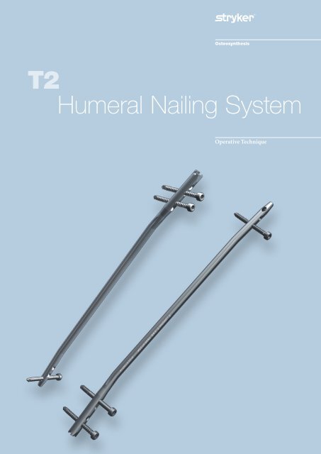
<strong>T2</strong><br />
<strong>Humeral</strong> <strong>Nailing</strong> <strong>System</strong><br />
<strong>Operative</strong> <strong>Technique</strong>
<strong>T2</strong> <strong>Humeral</strong> <strong>Nailing</strong> <strong>System</strong><br />
Contributing Surgeons<br />
Rupert Beickert, M. D.<br />
Senior Trauma Surgeon<br />
Murnau Trauma Center<br />
Murnau<br />
Germany<br />
Rosemary Buckle, M. D.<br />
Orthopaedic Associates, L. L. P.<br />
Clinical Instructor<br />
University of Texas Medical School<br />
Houston, Texas<br />
USA<br />
Prof. Dr. med. Volker Bühren<br />
Chief of Surgical Services<br />
Medical Director of Murnau Trauma Center<br />
Murnau<br />
Germany<br />
Michael D. Mason, D. O.<br />
Assistant Professor of Orthopaedic Surgery<br />
Tufts University School of Medicine<br />
New England Baptist Bone & Joint Institute<br />
Boston, Massachusetts<br />
USA<br />
2<br />
This publication sets forth detailed<br />
recommended procedures for using<br />
<strong>Stryker</strong> Osteosynthesis devices and<br />
instruments.<br />
It offers guidance that you should<br />
heed, but, as with any such technical<br />
guide, each surgeon must consider<br />
the particular needs of each patient<br />
and make appropriate adjustments<br />
when and as required.<br />
A workshop training is required prior<br />
to first surgery.<br />
All non-sterile devices must be<br />
cleaned and sterilized before use.<br />
Follow the instructions provided in<br />
our reprocessing guide (L24002000).<br />
Multi-component instruments must<br />
be disassembled for cleaning. Please<br />
refer to the corresponding assembly/<br />
disassembly instructions<br />
See package insert (L22000007) for<br />
a complete list of potential adverse<br />
effects, contraindications, warnings<br />
and precautions. The surgeon must<br />
discuss all relevant risks, including the<br />
finite lifetime of the device, with the<br />
patient, when necessary.<br />
Warning:<br />
All bone screws referenced in<br />
this document here are not<br />
approved for screw attachment or<br />
fixation to the posterior elements<br />
(pedicles) of the cervical, thoracic<br />
or lumbar spine.
Contents<br />
Page<br />
1. Introduction 4<br />
Implant Features 4<br />
Technical Details 5<br />
Instrument Features 6<br />
2. References 7<br />
3. Indications, Precautions and Contraindications 8<br />
Indications 8<br />
Precautions 8<br />
Relative Contraindications 8<br />
4. Pre-operative Planning 9<br />
5. Locking Options 10<br />
6. <strong>Operative</strong> <strong>Technique</strong> – Antegrade <strong>Technique</strong> 12<br />
Patient Positioning and Fracture Reduction 12<br />
Incision 12<br />
Entry Point 13<br />
Unreamed <strong>Technique</strong> 14<br />
Reamed <strong>Technique</strong> 14<br />
Nail Selection 16<br />
Nail Insertion 17<br />
Guided Locking Mode (via Target Device) 18<br />
Static Locking Mode 19<br />
Freehand Distal Locking 22<br />
End Cap Insertion 24<br />
Dynamic Locking Mode 24<br />
Apposition /Compression Locking Mode 25<br />
Advanced Locking Mode 27<br />
Nail Removal 28<br />
7. <strong>Operative</strong> <strong>Technique</strong> – Antegrade <strong>Technique</strong> 29<br />
Patient Positioning 29<br />
Incision 29<br />
Entry Point 30<br />
Unreamed <strong>Technique</strong> 30<br />
Reamed <strong>Technique</strong> 31<br />
Nail Selection 32<br />
Nail Insertion 32<br />
Guided Locking Mode (via Target Device) 35<br />
Static Locking Mode 36<br />
Freehand Proximal Locking 39<br />
End Cap Insertion 40<br />
Dynamic Locking Mode 40<br />
Apposition /Compression Locking Mode 41<br />
Advanced Locking Mode 43<br />
Nail Removal 44<br />
Ordering Information – Implants 45<br />
Ordering Information – Instruments 47<br />
3
Introduction<br />
Over the past several decades antegrade<br />
humeral nailing has become<br />
the treatment of choice for most<br />
hu meral shaft fractures. Retrograde<br />
humeral nailing has expanded the<br />
use of in tramedullary nails.<br />
Studies have shown the following<br />
benefits to be associated with<br />
<strong>Humeral</strong> <strong>Nailing</strong> :<br />
Brief operative time (1)<br />
Minimal morbidity (1)<br />
Early return to function of the<br />
extremity (2)<br />
In 90% of the cases, no external support<br />
is needed (1, 2)<br />
Closed technique (4)<br />
Low infection rate (2, 5, 6)<br />
Very good pain relief in stabilization of<br />
pathological fractures (2, 4)<br />
Implant Features<br />
The <strong>T2</strong> <strong>Humeral</strong> <strong>Nailing</strong> <strong>System</strong> is the<br />
realization of superior biomechanical<br />
intramedullary stabilization.<br />
The system offers the option of<br />
different locking modes :<br />
• Static, transverse/oblique<br />
• Dynamic<br />
• Apposition/compression<br />
• Advanced locking<br />
In some indications, a controlled<br />
apposition/compression of bone fragments<br />
can be applied by intro ducing<br />
a compression screw from the top of<br />
nail. To further increase rotational<br />
stability, the nail can be locked after<br />
utilizing the apposition/compression<br />
feature.<br />
The beneficial effect of apposition/<br />
compression in treating long-bone<br />
fractures in cases involving transverse<br />
and short oblique fractures that are<br />
axially stable is well documented<br />
(15, 16, 19).<br />
Compared to Plate and Screw<br />
Osteosynthesis :<br />
Minimal damage to muscle, connective<br />
tissue and vasculature (1, 3, 7)<br />
Reduced periosteal stripping and concomitant<br />
soft tissue damage(1)<br />
Fewer radial nerve palsies (3, 4)<br />
Designed for load sharing instead of<br />
load bearing (2)<br />
Cosmetically smaller incision<br />
The <strong>T2</strong> <strong>Humeral</strong> <strong>Nailing</strong> <strong>System</strong> is<br />
one of the first humeral nailing<br />
systems to offer an option for either an<br />
antegrade or a retrograde approach to<br />
repair fractures of the humerus.<br />
One Implant, Two Approaches<br />
<strong>Stryker</strong> Osteosynthesis has created a<br />
locking nail system, bring-ing together<br />
all the capabilities and benefits of separate<br />
antegrade and ret rograde nailing<br />
systems to create a single, integrated<br />
surgical resource for fixation of long-<br />
The compression screw is pushed<br />
against the proximal Partially<br />
Threaded Locking Screw (Shaft<br />
Screw) that has been placed in the<br />
oblong hole, drawing either the distal<br />
or the proximal segment towards the<br />
fracture site. In stable fractures, this<br />
has the biomechanical advantage of<br />
creating active circumferential compression<br />
to the fracture site, transferring<br />
axial load to the bone, and<br />
reducing the function of the nail as a<br />
load bearing device (17).<br />
This ability to transfer load back to<br />
the bone can reduce the incidence of<br />
implant failure secondary to fatigue.<br />
Typical statically locked nails functioning<br />
as load bearing devices have<br />
report ed failure rates in excess of 20%<br />
(18).<br />
4<br />
bone fractures.<br />
Furthermore, the development of<br />
the <strong>T2</strong> <strong>Humeral</strong> <strong>Nailing</strong> <strong>System</strong> offers<br />
the competitive advantages of :<br />
• Dual nailing approach : Antegrade<br />
and Retrograde<br />
• Accommodating reamed or<br />
unreamed procedures<br />
• Static, controlled dynamic and<br />
apposition/compression locking<br />
options<br />
• Advanced Locking Mode for<br />
increased rotational stability.<br />
Through the development of a common,<br />
streamlined and intuitive surgical<br />
approach, both in principle and in<br />
detail, the <strong>T2</strong> <strong>Humeral</strong> <strong>Nailing</strong> <strong>System</strong><br />
offers significantly increased speed<br />
and functionality for the treatment<br />
of fractures as well as simplifying the<br />
training requirements for all person nel<br />
involved.<br />
Common 4mm cortical screws s i m -<br />
plify the surgical procedure. Fully<br />
Threaded Locking Screws are available<br />
for regular locking procedures.<br />
Partially Threaded Locking Screws<br />
(Shaft Screws) are designed for application<br />
of apposition/compression.<br />
One common <strong>Humeral</strong> Compression<br />
Screw to close the fracture site, and<br />
End Caps in six sizes are available to<br />
provide an improved fit for every indication<br />
to allow nail length adaptation<br />
after insertion and to prevent bone<br />
ingrowth.<br />
All implants of the <strong>T2</strong> <strong>Humeral</strong><br />
<strong>Nailing</strong> <strong>System</strong> are cannulated<br />
and made of Type II anodized<br />
titanium alloy (Ti6AL4V) for<br />
enhanced biomechanical and biomedical<br />
performance.<br />
See the detailed chart on the next page<br />
for the design specifications and size<br />
offerings.
Introduction<br />
Technical Details<br />
Nails<br />
Diameter 7−9mm<br />
Sizes 140−320mm<br />
Humerus Advanced<br />
Compression Screw<br />
(Diameter = 6mm)<br />
4.0mm Fully Threaded<br />
Locking Screws<br />
L = 20−60mm<br />
4.0mm Partially Threaded<br />
Locking Screws<br />
(Shaft Screws)<br />
L = 20−60mm<br />
End Caps<br />
Standard +5mm +10mm +15mm +20mm +25mm<br />
5<br />
Compression<br />
Range*<br />
Bend, 4 °<br />
* Compression Range<br />
30<br />
36<br />
Bend, 6 °<br />
0mm<br />
12<br />
23<br />
28<br />
38<br />
62<br />
48mm<br />
28<br />
18<br />
10<br />
0mm<br />
Total Length of Slot 10mm<br />
Less Screw Diameter (–) 4mm<br />
Maximum Movement of Screw 6mm<br />
S<br />
L<br />
O T
Introduction<br />
Instrument Features<br />
The major advantage of the instru- Symbol coding on the instruments<br />
ment system is a breakthrough in the indicates the type of procedure, and<br />
integration of the instrument plat -<br />
form which can be used not only for<br />
must not be mixed.<br />
the complete <strong>T2</strong> <strong>Nailing</strong> <strong>System</strong>, but<br />
will be the platform for all future<br />
Symbol<br />
<strong>Stryker</strong> nailing systems, thereby reducing<br />
complexity and inventory.<br />
Square = Long instruments<br />
The instrument platform offers<br />
ad vanced precision and usability, and<br />
features ergonomically styled<br />
target ing devices.<br />
Drills<br />
Triangular = Short instruments<br />
Drills feature color coded rings :<br />
3.5mm = Orange<br />
For 4.0mm Fully Threaded Locking<br />
Screws and for the second cortex<br />
when using 4.0mm Partially Threaded<br />
Locking Screws (Shaft Screws).<br />
4.0mm = Grey<br />
For the first cortex when using 4.0mm<br />
Partially Threaded Locking Screws<br />
(Shaft Screws).<br />
6
References<br />
1. J. Blum et al., Unreamed <strong>Humeral</strong><br />
Nails Prove Reliable, Safe in<br />
Multicentre Study, International<br />
Edition Orthopaedics Today,<br />
Volume 1 Number 2, March/April<br />
1998.<br />
2. B. Redmond et al., Interlocking<br />
Intramedullary <strong>Nailing</strong> of<br />
Pathological Fractures of the Shaft<br />
of the Humerus, The Journal of<br />
Bone and Joint Surgery,<br />
Vol. 78-A, No. 6, June 1996.<br />
3. P. Rommens et al., Die<br />
Verriegelungsnagelung der<br />
Humerusschaftfraktur. Eine<br />
kritische Bewertung von 100<br />
Fällen, Swiss Surg. Suppl. 2/1996, p. 7<br />
4. P. Rommens et al., Retrograde<br />
Verriegelungs nagelung der<br />
Humerusschaftfraktur, Eine<br />
klinische Studie, Unfallchirurg,<br />
1995, 98 pp. 133−138.<br />
5. The Unreamed Nail – A Biological<br />
Osteosynthesis of the upper arm<br />
Acta Chir. Belg. 1997 August ; 97(4) :<br />
184−189.<br />
6. J. Stapert et al., Behandlung von<br />
Humerus-frakturen mit dem TLN<br />
(Telescopic Locking Nail),<br />
Swiss Surg. Suppl. 2/1996, p.<br />
7. D. Wheeler, M. Colville,<br />
Biomechanical Comparison of<br />
Intramedullary and Percutaneous<br />
Pin Fixation for Proximal <strong>Humeral</strong><br />
Fracture Fixation, Journal of<br />
Orthopaedic Trauma, Vol. 11, No. 5,<br />
pp. 363−367.<br />
8. ASTM Designation : F136−92,<br />
Standard Specification for Wrought<br />
Titanium 6A I−4V ELI Alloy for<br />
Surgical Implant Applications.<br />
9. ASTM Designation : F138−92,<br />
Standard Specification for Stainless<br />
Steel Bar and Wire for Surgical<br />
Implants (Special Quality).<br />
10. Leventhal G., Journal of Bone and<br />
Joint Surgery, Vol 33−A, No 2,<br />
April 1951, p475.<br />
11. Bothe RT, Beaton KE, Davenport<br />
HA (1940) Reaction of Bone<br />
to Multiple Metallic Implants,<br />
Surg. Gynaecol. Obstet. 71, 598.<br />
12. Bardos DI, “Titanium and<br />
Titanium Alloys”, in D. Williams<br />
(ed.) Concise encyclopedia of<br />
medical and dental materials,<br />
Pergamon Press, Oxford, 1990,<br />
360−365.<br />
13. Solar R. : Corrosion Resistance of<br />
Titanium Surgical Implant Alloys :<br />
A Review, in Corrosion and<br />
Degradation of Implant Materials,<br />
ASTM STP 684, American Society<br />
for Testing and Materials, 1979,<br />
pp 259−273.<br />
14. AO/ASIF Titanium − 6%<br />
Aluminum − 7% Niobium<br />
Implant Material, AO/ASIF<br />
Materials Technical Commission,<br />
First Edition June 1993.<br />
15. Müller ME, et al Manual for<br />
Internal Fixation. Springer-Verlag<br />
Berlin, 1991.<br />
7<br />
16. O. Gonschorek, G. O. Hofmann,<br />
V. Bühren, Interlocking<br />
Compression <strong>Nailing</strong> : A report<br />
on 402 Applications, Arch Orthop<br />
Trauma Surg (1998)<br />
117 : 430−437.<br />
17. T. E. Richardson, M. Voor,<br />
D. Seligson, Fracture Site<br />
Compression and Motion with<br />
Three Types of Intramedullary<br />
Fixation of the Femur,<br />
In : Osteosynthese International<br />
(1998), 6 : 261−264.<br />
18. Hutson et al., Mechanical Failures<br />
of Intramedullary Tibial Nails<br />
Applied without Reaming, In :<br />
Clin. Orthop. (1995), 315 : 129−137.<br />
19. Bühren V., Kompressionsnagelung<br />
langer Röhrenknochen,<br />
Unfallchirurg 103,2000, 708−720.<br />
20. Mehdi Mousavi, et al., Pressure<br />
Changes During Reaming with<br />
Different Parameters and Reamer<br />
Designs, Clinical Orthopaedics<br />
and Related Research, Number 373,<br />
pp. 295−303, 2000.
Indications, Precautions and Contraindications<br />
Indications Precautions<br />
•<br />
•<br />
•<br />
•<br />
The <strong>T2</strong> <strong>Humeral</strong> Nail is intended<br />
to provide temporary stabilization<br />
of various types of fractures,<br />
malunions and nonunions of the<br />
humerus.<br />
The nails are inserted using<br />
an opened or closed technique<br />
and can be static, dynamic and<br />
compression locked.<br />
The subject and predicate devices<br />
are indicated for use in the<br />
humerus.<br />
Types of fractures include, but<br />
not limited to fractures of the<br />
humeral shaft, non-unions,<br />
malalignments, pathological<br />
humeral fractures, and impending<br />
pathological fractures.<br />
Relative Contraindications<br />
The physician’s education, training<br />
and professional judgement must<br />
be relied upon to choose the most<br />
appropriate device and treatment.<br />
Conditions presenting an increased<br />
risk of failure include:<br />
•<br />
•<br />
•<br />
•<br />
•<br />
•<br />
Any active or suspected latent<br />
infection or marked local<br />
inflammation in or about the<br />
affected area.<br />
Compromised vascularity that<br />
would inhibit adequate blood<br />
supply to the fracture or the<br />
operative site.<br />
Bone stock compromised by<br />
disease, infection or prior<br />
implantation that can not provide<br />
adequate support and/or fixation<br />
of the devices.<br />
Material sensitivity, documented<br />
or suspected.<br />
Obesity. An overweight or obese<br />
patient can produce loads on the<br />
implant that can lead to failure<br />
of the fixation of the device or to<br />
failure of the device itself.<br />
Patients having inadequate tissue<br />
coverage over the operative site.<br />
The <strong>T2</strong> <strong>System</strong> has not been evaluated<br />
for safety and compatibility in the MR<br />
environment.<br />
The <strong>T2</strong> <strong>System</strong> has not been tested<br />
for heating or migration in the MR<br />
environment.<br />
•<br />
•<br />
•<br />
8<br />
Implant utilization that would<br />
interfere with anatomical<br />
structures or physiological<br />
performance.<br />
Any mental or neuromuscular<br />
disorder which would create<br />
an unacceptable risk of fixation<br />
failure or complications in<br />
postoperative care.<br />
Other medical or surgical<br />
conditions which would preclude<br />
the potential benefit of surgery.
Pre-operative Planning<br />
An X-Ray Template (1806-0003) is<br />
available for pre-operative planning.<br />
Thorough evaluation of pre-operative<br />
radiographs of the affected extremity<br />
is critical. Careful radiographic examination<br />
can help prevent intra-operative<br />
complications.<br />
If X-Rays show a very narrow<br />
intramedullary canal in the distal part<br />
of the humerus, retrograde humeral<br />
nailing is not possible.<br />
The proper nail length when in serted<br />
antegrade should extend from subchondral<br />
bone proximally, to 1cm<br />
above the olecranon fossa distally.<br />
The retrograde nail length is determined<br />
by measuring the distance from<br />
1cm above the olecranon fossa to the<br />
center of the humeral head.<br />
In either approach, the surgeon should<br />
consider the apposition/compression<br />
feature of the <strong>T2</strong> <strong>Humeral</strong> Nail,<br />
know ing that 6mm of active apposition/compression<br />
is possible, prior to<br />
determining the final length of the<br />
implant.<br />
Note:<br />
Check with local representative<br />
regarding availability of nail sizes.<br />
9
Locking Options<br />
Antegrade<br />
Retrograde<br />
10<br />
Static Mode transverse<br />
Static Mode oblique
Locking Options<br />
Dynamic Mode Apposition/Compression Mode<br />
11<br />
Advanced Locking Mode
<strong>Operative</strong> <strong>Technique</strong> – Antegrade <strong>Technique</strong><br />
Patient Positioning and Fracture Reduction<br />
The patient is placed in a semireclined<br />
“beach chair position” or supine on<br />
a radiolucent table. Patient positioning<br />
should be checked to ensure<br />
that imag ing and access to the entry<br />
site are possible without excessive<br />
manipulation of the affected extremity<br />
(Fig. 1). The image intensifier is placed<br />
at the legside of the patient ; the surgeon<br />
is positioned at the headside.<br />
Incision<br />
A small incision is made in line with<br />
the fibers of the deltoid muscle anterolateral<br />
to the acromion. The deltoid<br />
is split to expose the subdeltoid bursa.<br />
Palpate to identify the anterior and<br />
posterior margins of the greater<br />
tuberosity and supraspinatus tendon.<br />
The supraspinatus tendon is then<br />
incised in line with its fibers<br />
(Fig. 2).<br />
The real rotation of the proximal fragment<br />
is checked (inversion or reversion),<br />
considering that the entry point<br />
is at the tip of the greater tubercle. If<br />
the proximal fragment is inverted,<br />
the entry point is more anterior. If the<br />
proximal fragment is in external rotation,<br />
the entry point is more lateral. It<br />
is recommended to localize the entry<br />
point under image intensifier control,<br />
also palpating the bicipital groove,<br />
the portal is about 10mm posterior to<br />
the biceps tendon. This will make the<br />
entry portal concentric to the medullary<br />
canal.<br />
12<br />
Fig. 1<br />
Fig. 2
<strong>Operative</strong> <strong>Technique</strong> – Antegrade <strong>Technique</strong><br />
Entry Point<br />
The entry point is made with the<br />
Curved, cannulated Awl (1806-0040)<br />
(Fig. 3). The 2.5 × 800mm Ball Tip<br />
Guide Wire (1806-0083S) is then<br />
introduced through the awl under<br />
image intensification into the metaphysis,<br />
central to the long axis of the<br />
humerus.<br />
Alternatively, the optional Crown Drill<br />
(1806-2020) may be used over the<br />
K-Wire with Washer (1806-0051S) for<br />
entry point preparation. The K-Wire<br />
will help to guide the Crown Drill<br />
centrally (Fig. 3a).<br />
Then, the 3 × 285mm K-Wire (1806-<br />
0050S) is introduced under image<br />
intensification into the meta-physis,<br />
central to the long axis of the humerus.<br />
The cannulated Ø10mm Rigid Reamer<br />
(1806-2010) may be used over the<br />
K-Wire and the proximal metaphysis<br />
should be drilled to a depth of at least<br />
6cm.<br />
Note:<br />
During opening the entry portal<br />
with the Awl, dense cortex may<br />
block the tip of the Awl. An Awl<br />
Plug (1806-0032) can be inserted<br />
through the Awl to avoid penetration<br />
of bone debris into the cannulation<br />
of the Awl shaft.<br />
13<br />
Fig. 3<br />
Fig. 3a
<strong>Operative</strong> <strong>Technique</strong> – Antegrade <strong>Technique</strong><br />
Unreamed <strong>Technique</strong><br />
If an unreamed technique is pre -<br />
ferred, the nail may be inserted over<br />
the 2.2 × 800mm Smooth Tip Guide<br />
Wire (1806-0093S) (Fig. 4).<br />
Reamed <strong>Technique</strong><br />
For reamed techniques, the<br />
2.5 × 800mm Ball Tip Guide Wire<br />
(1806-0083S) is inserted across the<br />
fracture site.<br />
The Reduction Rod (1806-0363) may<br />
be used as a fracture reduction tool to<br />
facilitate Guide Wire insertion across<br />
the fracture site (Fig. 5).<br />
Reaming is commenced in 0.5mm<br />
increments until cortical contact is<br />
appreciated. Final reaming should<br />
be 1mm−1.5mm larger than the diameter<br />
of the nail to be used.<br />
The Guide Wire Pusher can be used to<br />
help keep the Guide Wire in position<br />
during reamer shaft extraction. The<br />
metal cavity at the end of the handle<br />
pushed on the end of the power tool<br />
facilitates to hold the Guide Wire in<br />
place when starting to pull the power<br />
tool. When close to the Guide Wire<br />
end place the Guide Wire Pusher with<br />
its funnel tip to the end of the power<br />
tool cannulation. While removing the<br />
power tool the Guide Wire Pusher will<br />
keep the Guide Wire in place<br />
(Fig. 6 & 7).<br />
14<br />
Fig. 4<br />
Fig. 5<br />
Fig. 6
<strong>Operative</strong> <strong>Technique</strong> – Antegrade <strong>Technique</strong><br />
Note:<br />
• 8mm <strong>Humeral</strong> Nails cannot be<br />
inserted over the 3 × 1000mm Ball<br />
Tip Guide Wire (1806-0085S).<br />
The Ball Tip Guide Wire must<br />
be exchanged for the 3 × 800mm<br />
Smooth Tip Guide Wire (1806-<br />
0090S) prior to nail insertion.<br />
• Use the Teflon Tube (1806-0073S)<br />
for the Guide Wire exchange.<br />
When reaming is completed, the<br />
Teflon Tube (1806-0073S) should be<br />
used to exchange the Ball Tip Guide<br />
Wire (1806-0083S) with the Smooth<br />
Tip Guide Wire (1806-0093S) for nail<br />
insertion (Fig. 8 and 9).<br />
An unreamed technique can be<br />
considered in cases, where the<br />
medullary canal has the appropriate<br />
diameter. In these cases, the nail can<br />
be introduced over the 2.2×800mm<br />
Smooth Tip Guide Wire (1806-0093S).<br />
Note:<br />
• X-Ray Templates should be used<br />
pre-operatively to determine the<br />
canal size radiographically.<br />
• The driving end of 7mm nails is<br />
8mm.<br />
15<br />
Fig. 7<br />
Fig. 8<br />
Fig. 9
<strong>Operative</strong> <strong>Technique</strong> – Antegrade <strong>Technique</strong><br />
Transverse Oblique Compression Slot<br />
(Oblique or Dynamic)<br />
End of Guide Wire Ruler<br />
is the measurement reference<br />
Hole<br />
Positions<br />
240mm<br />
Diameter Length<br />
Length<br />
The Guide Wire Ruler can be easily<br />
folded and unfolded.<br />
16<br />
Fig. 11<br />
Fig. 12<br />
Nail Selection<br />
The X-Ray Template should be used<br />
pre-operatively to determine the canal<br />
size radiographically. This information<br />
may be utilized in conjunction with<br />
the clinical assessment of canal size<br />
as determined by the size of the last<br />
reamer used.<br />
Fig. 10<br />
Diameter<br />
The diameter of the selected nail<br />
should be 1mm−1.5mm smaller than<br />
the last reamer used.<br />
Length<br />
Nail length may be determined with<br />
the X-Ray Ruler (Fig. 10). The Guide<br />
Wire Ruler (1806-0022) may be<br />
used by placing it on the Guide Wire<br />
reading the correct nail length at the<br />
end of the Guide Wire on the Guide<br />
Wire Ruler (Fig. 11 and 12). Confirm<br />
the position of the tip of the Guide<br />
Wire prior to measurement.<br />
Note:<br />
If the fracture is suitable for<br />
ap position/compression, the<br />
implant selected should be<br />
6−10mm shorter than measured<br />
to help avoid migra tion of the nail<br />
beyond the insertion site.
<strong>Operative</strong> <strong>Technique</strong> – Antegrade <strong>Technique</strong><br />
Nail Insertion<br />
The selected nail is assembled onto<br />
the Target Device (1806-0143) with<br />
the Nail Holding Screw (1806-0163).<br />
Tighten the Nail Holding Screw<br />
securely with the Insertion Wrench<br />
(1806-0135) so that it does not loosen<br />
during nail insertion (Fig. 13).<br />
Note:<br />
Prior to nail insertion please<br />
check correct alignment by<br />
inserting a drill bit through the<br />
assembled Tissue Protection and<br />
Drill Sleeve placed in the required<br />
holes of the targeting device.<br />
Upon completion of reaming and<br />
Guide Wire exchange, the appropriate<br />
size nail is ready for insertion.<br />
Advance the nail through the entry<br />
point past the fracture site to the<br />
appropriate level.<br />
Gentle rotation of the nail may be<br />
necessary to start the nail insertion.<br />
The nail should be advanced with<br />
manual pressure. Aggressive use of<br />
the slotted hammer can result in<br />
additional fractures. If the nail does<br />
not advance easily, a check with image<br />
intensification should be made to see<br />
if the nail angle is too steep resulting<br />
in the nail impinging on the medial<br />
cortex.<br />
The Slotted Hammer (1806-0170)<br />
can be used to insert the nail over the<br />
Guide Wire.<br />
DO NOT hit the Target Device.<br />
Note:<br />
A chamfer is located on the<br />
working end of the nail to<br />
denote the end under X-Ray.<br />
Three circumferential grooves<br />
are located on the insertion<br />
post at 2mm, 6mm, and 10mm<br />
from the driving end of the nail<br />
(Fig. 14-16). Depth of insertion<br />
may be visualized with the aid of<br />
fluoroscopy.<br />
The 3 × 285mm K-Wire may be<br />
inserted through the Target Device<br />
which identifies the junction of the<br />
nail and insertion post (Fig. 17).<br />
2mm<br />
6mm<br />
10mm<br />
Static<br />
Dynamic<br />
Fig. 13<br />
Fig. 14 Fig. 15<br />
Apposition/Compression<br />
17<br />
Fig. 16<br />
Fig. 17
<strong>Operative</strong> <strong>Technique</strong> – Antegrade <strong>Technique</strong><br />
Guided Locking Mode<br />
(via Target Device)<br />
Prior to guided locking via the Target<br />
Device, the Nail Holding Screw must<br />
be firmly tightened using the Insertion<br />
Wrench, to ensure that the nail is in<br />
correct alignment with the Target<br />
Device.<br />
The Target Device is designed to provide<br />
four options for guided locking<br />
(Fig. 18.1−18.4).<br />
In the Static Oblique Locking Mode,<br />
the two static holes closest to the<br />
end of the nail may be used for static<br />
oblique (30°) locking (Fig. 18.1).<br />
1. S t at ic<br />
2 . S t at ic<br />
In the Static Transverse Locking Mode,<br />
the next static hole and the dynamic<br />
hole are used for static transverse<br />
locking (Fig. 18.2).<br />
3. Static<br />
4. Dynamic<br />
In the Controlled Dynamic Mode,<br />
and/or Controlled Apposition/Compression<br />
Mode, the dynamic hole is<br />
required (Fig. 18.3).<br />
4. Dynamic<br />
In the Advanced Locking Mode, the<br />
dynamic hole is required. After<br />
utilizing compression with the<br />
Advanced Compression Screw, the<br />
static hole is used (Fig. 18.4).<br />
4. Dynamic<br />
3. Static<br />
The Short Tissue Protection Sleeve,<br />
(1806-0180), together with the Short<br />
Drill Sleeve (1806-0210) and the Short<br />
Trocar (1806-0310), are inserted into<br />
the Target Device by pressing the safety<br />
clip (Fig. 19).<br />
The friction lock mechanism is<br />
designed to keep the sleeve in place<br />
and prevent it from falling out. It is<br />
designed to also keep the sleeve from<br />
sliding during screw measurement.<br />
To release the Tissue Protection Sleeve,<br />
the safety clip must be pressed again.<br />
18<br />
1 2<br />
3<br />
3<br />
4<br />
4<br />
4<br />
Fig. 18.1<br />
Fig. 18.2<br />
Fig. 18.3<br />
Fig. 18.4<br />
Fig. 19
<strong>Operative</strong> <strong>Technique</strong> – Antegrade <strong>Technique</strong><br />
Static Locking Mode<br />
Static Transverse<br />
Locking Mode<br />
In unstable or comminuted fractures,<br />
the nail should be used as a standard<br />
interlocking nail. Static locking of the<br />
distal holes will help maintain the<br />
length of the bone and the rotational<br />
stability of the fracture.<br />
The Short Tissue Protection Sleeve,<br />
together with the Short Drill Sleeve<br />
and the Short Trocar, are positioned<br />
through the static locking hole on the<br />
Target Device. A small skin incision<br />
is made, and the assembly is pushed<br />
through until it is in contact with the<br />
lateral cortex of the humerus (Fig. 20).<br />
Note:<br />
Especially in the proximal<br />
humerus, use image intensification<br />
to help ensure the Tissue<br />
Protection Sleeve is flush with the<br />
cortex or you could lose 1–2mm of<br />
screw measurement accuracy.<br />
The Trocar is removed while the<br />
Tissue Protection Sleeve and the Drill<br />
Sleeve remain in position.<br />
For accurate drilling and easy determination<br />
of screw length, use the centertipped,<br />
Ø3.5 × 230mm calibrated Drill<br />
(1806-3540S). The centered Drill is<br />
forwarded through the Drill Sleeve<br />
and pushed onto the cortex.<br />
After the first cortex is drilled to the<br />
appropriate level the screw length may<br />
be read directly off of the Drill at the<br />
end of the Drill Sleeve (Fig. 21).<br />
Caution:<br />
Make sure the Tissue Protection<br />
Sleeve/Drill Sleeve Assembly is<br />
seated on bone prior to selecting<br />
final screw length.<br />
50mm<br />
19<br />
Fig. 20<br />
Fig. 21<br />
50mm
<strong>Operative</strong> <strong>Technique</strong> – Antegrade <strong>Technique</strong><br />
Warning:<br />
Do not drill through the far cortex<br />
as this will penetrate the joint.<br />
Note:<br />
• The position of the end of the<br />
Drill as it relates to the far cortex<br />
is equal to where the end of the<br />
screw will be. Therefore, if the end<br />
of the Drill is 3mm beyond the far<br />
cortex, the end of the screw will<br />
also be 3mm beyond.<br />
• The Screw Gauge, Short, is calibrated<br />
so that with the bend at the<br />
end pulled back flush with the far<br />
cortex, the screw tip will end 3mm<br />
beyond the far cortex (Fig. 21).<br />
When the Drill Sleeve is removed,the<br />
correct 4.0mm Locking Screw is<br />
inserted through the Tissue Protection<br />
Sleeve using the Screwdriver Shaft,<br />
Short (1806-0222) with the Teardrop<br />
Handle (702429, Fig. 22). The screw is<br />
near its’ proper seating position when<br />
the groove around the shaft of the<br />
screwdriver is approaching the end of<br />
the Tissue Protection Sleeve.<br />
Use image intensification to confirm<br />
screw position through the nail as well<br />
as screw length.<br />
Repeat the locking procedure for the<br />
other statically positioned Locking<br />
Screw (Fig. 23).<br />
Caution:<br />
The coupling of Elastosil handles<br />
contains a mechanism with one<br />
or multiple ball bearings. In case<br />
of applied axial stress on the<br />
Elastosil handle, those components<br />
are pressed into the surrounding<br />
cylinder resulting in a<br />
complete blockage of the device<br />
and possible bending.<br />
To avoid intra-operative complications<br />
and secure long-term<br />
functionality, we mandate that<br />
Elastosil handles be used only for<br />
their intended use.<br />
DO NOT HIT any Elastosil handles.<br />
20<br />
Fig. 22<br />
Fig. 23
<strong>Operative</strong> <strong>Technique</strong> – Antegrade <strong>Technique</strong><br />
Static Oblique<br />
Locking Mode<br />
In cases that may be locked in the<br />
Static Oblique Locking Mode, place<br />
the assembly of the Tissue Protection<br />
Sleeve together with the Drill Sleeve<br />
and the Trocar through the Oblique<br />
static hole closest to the driving end<br />
of the nail (Fig. 24). Refer to the procedure<br />
for Locking Screw insertion.<br />
The second Fully Threaded Locking<br />
Screw is inserted through the static<br />
hole (Fig. 25) next to the first hole, and<br />
placed in an oblique manner through<br />
the oblong hole of the nail (Fig. 26).<br />
Confirm screw position and screw<br />
length with image intensification.<br />
W a s h e r<br />
The Washer, either Rectangular or<br />
Round, may be used in cases of osteoporotic<br />
bone to bridge the bone gap<br />
and allow for enhanced purchase of<br />
the Locking Screw (Fig. 27).<br />
Fig. 27<br />
21<br />
Fig. 24<br />
Fig. 25<br />
Fig. 26
<strong>Operative</strong> <strong>Technique</strong> – Antegrade <strong>Technique</strong><br />
Freehand Distal Locking<br />
The freehand technique is used to<br />
insert Locking Screws into both the<br />
A/P and M/L holes in the nail.<br />
Rotational alignment must be checked<br />
prior to distal locking.<br />
Multiple locking techniques and radiolucent<br />
drill devices are available for<br />
freehand locking. The critical step<br />
with any freehand locking technique,<br />
proximal or distal, is to visualize a<br />
perfectly round locking hole with the<br />
C-Arm.<br />
Caution:<br />
In order to avoid damage to<br />
the neurovascular structure, a<br />
limited open approach should be<br />
considered.<br />
The center-tipped Ø3.5 × 230mm<br />
Drill (1806-3540S), or the optional<br />
Ø3.5 × 130mm Drill (1806-3550S), is<br />
held at an oblique angle to the center<br />
of the locking hole (Fig. 28 and 29).<br />
Upon X-Ray verification, the Drill is<br />
placed perpendicular to the nail and<br />
drilled through the anterior cortex.<br />
Confirm these views in both the A/P<br />
and M/L planes by X-Ray.<br />
22<br />
Fig. 28<br />
Fig. 29
<strong>Operative</strong> <strong>Technique</strong> – Antegrade <strong>Technique</strong><br />
After drilling both cortices, the screw<br />
length may be read directly off of<br />
the Screw Scale, Short (1806-0360)<br />
at the orange color coded ring on the<br />
center-tipped Drill (Fig. 30). As with<br />
proximal locking (Fig. 21, p. 19), the<br />
position of the end of the drill is equal<br />
to the end of the screw as they relate to<br />
the far cortex.<br />
Routine Locking Screw insertion is<br />
employed with the assembled Short<br />
Screwdriver Shaft and the Teardrop<br />
Handle.<br />
If possible, the distal humerus should<br />
be locked with two Fully Threaded<br />
Locking Screws. Additional locking of<br />
the M/L hole(s) is possible if the image<br />
intensifier can be adjusted (Fig. 31).<br />
Note:<br />
Use image intensification to<br />
confirm screw position through<br />
the nail as well as screw length.<br />
Alternatively, the Screw Gauge can be<br />
used to measure the screw length.<br />
35mm<br />
23<br />
orange ring<br />
Fig. 30<br />
Fig. 30a<br />
Fig. 31
<strong>Operative</strong> <strong>Technique</strong> – Antegrade <strong>Technique</strong><br />
End Cap Insertion<br />
After removal of the Target Device, an<br />
End Cap is used to reduce the potential<br />
for bony ingrowth into the proximal<br />
threads of the nail.<br />
End Caps are available in six sizes<br />
(Fig. 32).<br />
The End Cap is inserted with the Short<br />
Screwdriver Shaft assembled on the<br />
Teardrop Handle after intra-operative<br />
radiographs show satisfactory reduction<br />
and hardware implantation<br />
(Fig. 33). Fully seat the End Cap to<br />
minimize the potential for loosening.<br />
Caution:<br />
To avoid impingement, carefully<br />
select the length of the End Cap.<br />
Close the wound using standard technique.<br />
Fig. 34<br />
Standard +5mm +10mm +15mm +20mm +25mm<br />
Dynamic Locking Mode<br />
When the fracture profile permits,<br />
controlled dynamic locking may be<br />
utilized for transverse or axially stable<br />
fractures.<br />
Antegrade dynamization is performed<br />
by statically locking the nail distally.<br />
The guided Partially Threaded<br />
Locking Screw (Shaft Screw) is then<br />
placed in the dynamic position of<br />
the oblong hole. This allows the nail<br />
to move, and the fracture to settle<br />
while torsional stability is maintained<br />
(Fig. 34).<br />
24<br />
Fig. 32<br />
Fig. 33
<strong>Operative</strong> <strong>Technique</strong> – Antegrade <strong>Technique</strong><br />
Apposition/Compression Locking Mode<br />
In transverse or axially stable fracture<br />
patterns, active apposition/compression<br />
increases fracture stability and<br />
enhances fracture healing. The antegrade<br />
<strong>T2</strong> <strong>Humeral</strong> Nail provides the<br />
option to treat a humerus fracture<br />
with active mechanical apposition/<br />
compression prior to leaving the<br />
operating room.<br />
Note:<br />
Distal freehand static locking<br />
must be performed prior to<br />
applying active, controlled apposition/compression<br />
to the fracture<br />
site.<br />
If active apposition/compression is<br />
required, a Partially Threaded Locking<br />
Screw (Shaft Screw) is inserted via the<br />
Target Device in the dynamic position<br />
of the oblong hole. This will allow for<br />
a maximum of 6mm of active, controlled<br />
apposition/compression. In<br />
order to insert the Partially Threaded<br />
Locking Screw (Shaft Screw), drill<br />
both cortices with the Ø3.5 × 230mm<br />
Drill (1806-3540S). Next, the near<br />
cortex ONLY is overdrilled with the<br />
Ø4.0 × 180mm Drill (1806-4000S).<br />
Note:<br />
After the opposite cortex is drilled<br />
with the Ø3.5 × 230mm drill, the<br />
correct screw length can be read<br />
directly off of the calibrated Drill<br />
at the end of the Drill Sleeve.<br />
After the Partially Threaded Locking<br />
Screw (Shaft Screw) is inserted, the<br />
Nail Holding Screw is removed,<br />
leaving the insertion post intact with<br />
the nail (Fig. 35). This will act as a<br />
guide for the Compression Screw. The<br />
Compression Screw with the Compression<br />
Screwdriver Shaft (1806-0263)<br />
assembled on the Teardrop Handle is<br />
inserted through the insertion post<br />
(Fig. 36).<br />
25<br />
Fig. 35<br />
Fig. 36
<strong>Operative</strong> <strong>Technique</strong> – Antegrade <strong>Technique</strong><br />
Note:<br />
It may be easier to “insert” the<br />
Compres sion Screw prior to fully<br />
seating the nail. Once the nail<br />
tip has cleared the fracture site,<br />
the Guide Wire (if used) is withdrawn.<br />
With the proximal portion<br />
of the nail not fully seated and<br />
extending out of the bone, the<br />
Advanced Compression Screw is<br />
inserted. Care should be taken<br />
that the shaft of the Compression<br />
Screw does not extend into the<br />
area of the oblong hole.<br />
The Short Tissue Protection Sleeve<br />
is removed and the Compression<br />
Screw is gently tightened utilizing<br />
the two-finger technique (Fig. 37). As<br />
the Compression Screw is advanced<br />
against the 4.0mm Partially Threaded<br />
Locking Screw (Shaft Screw), it draws<br />
the distal fracture segment towards the<br />
fracture site, employing active apposition/compression<br />
(Fig. 38). Image<br />
intensification will enable the surgeon<br />
to visualize active apposition/compression.<br />
Some bending of the transverse<br />
Partially Threaded Locking Screw<br />
(Shaft Screw) may be seen.<br />
Note:<br />
• Apposition/compression must be<br />
carried out under X-Ray control.<br />
Over-compression may cause the<br />
nail or the Partially Threaded<br />
Locking Screw (Shaft Screw) to<br />
fail.<br />
• When compressing the nail,<br />
the im plant must be inserted a<br />
safe distance from the entry point<br />
to accommodate for the 6mm of<br />
active compression. The three<br />
grooves on the insertion post are<br />
designed to help attain accurate<br />
insertion depth of the implant.<br />
26<br />
Fig. 37<br />
Fig. 38
<strong>Operative</strong> <strong>Technique</strong> – Antegrade <strong>Technique</strong><br />
Advanced Locking Mode<br />
In order to achieve additional fixation<br />
and to reduce the load on the Partially<br />
Threaded Locking Screw (Shaft Screw),<br />
the design of the <strong>T2</strong> <strong>Humeral</strong> Nail<br />
provides the opportunity to insert a<br />
Fully Threaded Locking Screw in the<br />
other transverse hole at the driving<br />
end of the nail after apposition/compression<br />
is utilized.<br />
Prior to guided locking via the Target<br />
Device, the Nail Holding Screw must<br />
be tightened using the Insertion<br />
Wrench.<br />
Fix the Advanced Compression Screw<br />
on the self-retaining Compression<br />
Screwdriver Shaft. Remove the Nail<br />
Holding Screw leaving the Target<br />
Device in place (Fig. 39). Advance the<br />
Compression Screw through the Target<br />
Device until the desired amount of<br />
compression is achieved. Visualize<br />
depth of insertion with the aid of<br />
flouroscopy (Fig. 40).<br />
Note:<br />
As previously described, it may be<br />
easier to insert the Compression<br />
Screw prior to fully seating the<br />
nail.<br />
To reattach the Target Device to the<br />
nail, detach the Teardrop Handle from<br />
the Compression Screwdriver Shaft<br />
and screw the Nail Holding Screw over<br />
the Compression Screwdriver Shaft<br />
into its required position.<br />
To insert the second transverse Fully<br />
Threaded Locking Screw, follow the<br />
locking procedure for static locking<br />
(Fig. 41).<br />
Finally, an End Cap should be inserted,<br />
as shown on page 24.<br />
27<br />
Fig. 39<br />
Fig. 40<br />
Fig. 41
<strong>Operative</strong> <strong>Technique</strong> – Antegrade <strong>Technique</strong><br />
Nail Removal<br />
Nail removal is an elective procedure.<br />
If used, first remove the End Cap with<br />
the Short Screwdriver Shaft and the<br />
Teardrop Handle (Fig. 42).<br />
If Advanced Locking Mode was utilized,<br />
the most proximal screw is<br />
extracted first, allowing access to the<br />
compression screw. Next, disengage<br />
the Advanced Compres sion Screw<br />
from the Fully Threaded Locking<br />
Screw (Shaft Screw) by turning the<br />
Compression Screwdriver one full turn<br />
in a counter-clockwise direction<br />
(Fig. 43).<br />
Note:<br />
There is no need to attempt to<br />
remove the Advanced Compression<br />
Screw from the nail,<br />
which with the nail implanted,<br />
may be difficult.<br />
The Universal Rod, Short is inserted<br />
into the driving end of the nail before<br />
all Locking Screws are removed with<br />
the Short Screwdriver Shaft and the<br />
Teardrop Handle (Fig. 43).<br />
Note:<br />
Attaching of the Universal Rod<br />
to the nail first will reduce the<br />
potential for nail migration,<br />
then the locking screws may be<br />
removed safely.<br />
The Slotted Hammer is used to extract<br />
the nail in a controlled manner<br />
(Fig. 44).<br />
28<br />
Advanced Compression<br />
Screw − Disengaged<br />
Fig. 42<br />
Fig. 43<br />
Fig. 44
<strong>Operative</strong> <strong>Technique</strong> – Retrograde <strong>Technique</strong><br />
Patient Positioning<br />
The patient is placed on a radiolu -<br />
cent table in the prone position or<br />
lateral decubitus position. The affected<br />
arm is supported on an arm board or<br />
hand table. The shoulder is in 90°<br />
abduction, the elbow joint flexed also<br />
in a 90° position. In this position, fractures<br />
can be reduced in correct rotation.<br />
Patient positioning should be checked<br />
to ensure that imaging of the entry site<br />
at the proximal humerus is possible.<br />
This allows the elbow to be hyper<br />
flexed to accommodate insertion of the<br />
implant parallel to the humerus.<br />
Incision<br />
A posterior approach is used to<br />
access the distal humerus. Starting at<br />
the tip of the olecranon, a 6cm incision<br />
is made in a proximal direction. The<br />
triceps tendon is split and muscle tissue<br />
is bluntly dissected and retracted<br />
until the upper edge of the olecranon<br />
fossa is displayed.<br />
The distal insertion point for the<br />
nail is one centimeter above the olecranon<br />
fossa. The Insertion Site<br />
Template (703117) may be used to help<br />
determine the appropriate insertion<br />
site (Fig. 45). The medullary canal is<br />
opened using the Drill Ø3.5 × 130mm<br />
(1806-3550S) by drilling a set of<br />
linear holes (Fig. 46). The holes are<br />
then joined with the Self-guiding Rigid<br />
Reamer (703125) (Fig. 47).<br />
Note:<br />
The drill guide slots of the<br />
retrograde Insertion Site Template<br />
(703117), must be centered and<br />
parallel to the medullary canal<br />
(long axis of the humerus).<br />
29<br />
Fig. 45<br />
Fig. 46<br />
Fig. 47
<strong>Operative</strong> <strong>Technique</strong> – Retrograde <strong>Technique</strong><br />
Entry Point<br />
Final insertion site preparation is performed<br />
with the Conical Rigid Reamer<br />
(703126) to create a longitu-dinal oval<br />
cortical hole at least 3cm in length and<br />
1cm in width (Fig. 49).<br />
The cortical bone is removed distally<br />
to the level of the olecranon fossa with<br />
the rigid reamers or small rongeur.<br />
Caution :<br />
Although the tip of the nail has a 4<br />
degree bend that facilitates distal<br />
nail insertion, high compressive<br />
forces during nail insertion can<br />
result in fractures of the distal<br />
humerus if the insertion opening<br />
is too short or too steep.<br />
Unreamed <strong>Technique</strong><br />
The <strong>T2</strong> <strong>Humeral</strong> Nail is cannulated,<br />
and may be introduced in an un-<br />
reamed fashion over a Smooth Tip<br />
Guide Wire. This simplifies frac ture<br />
reduction and reduces the risk of iatrogenic<br />
distal fractures caused by trying<br />
to reduce the fracture with the nail.<br />
The 2.2 × 800mm Smooth Tip Guide<br />
Wire (1806-0093S) is inserted under<br />
image control through the distal fragment<br />
and into the desired position<br />
within the proximal humerus using<br />
the Guide Wire Handle and Chuck<br />
(1806-1095 and 1806-1096) (Fig. 48<br />
and 50).<br />
The Reduction Rod (1806-0363) may<br />
be used as a fracture reduction tool<br />
to facilitate Guide Wire insertion<br />
(Fig. 51). The Guide Wire is advanced<br />
until the tip rests at the center of the<br />
humeral head. The Guide Wire should<br />
lie in the center of the metaphysis in<br />
both the A/P and Lateral views to help<br />
avoid offset positioning of the nail.<br />
The Guide Wire Handle is removed<br />
leaving the Guide Wire in place.<br />
30<br />
Fig. 48<br />
Fig. 49<br />
Fig. 50<br />
Fig. 51
<strong>Operative</strong> <strong>Technique</strong> – Retrograde <strong>Technique</strong><br />
Reamed <strong>Technique</strong><br />
For reamed techniques, the<br />
2.5 × 800mm Ball Tip Guide Wire<br />
(1806-0083S) is inserted through the<br />
fracture site. The Reduction Rod or<br />
the Universal Rod, Short with the<br />
“optional” Reduction Spoon may be<br />
used as a fracture reduction tool to<br />
facilitate Guide Wire insertion across<br />
the fracture site (see Fig. 51).<br />
Reaming is commenced in 0.5mm<br />
increments until cortical contact is<br />
appreciated. (Fig. 50). The final reamer<br />
should be 1mm−1.5mm larger than<br />
the diameter of the nail to be used.<br />
The Guide Wire Pusher can be used to<br />
help keep the Guide Wire in position<br />
during reamer shaft extraction. The<br />
metal cavity at the end of the handle<br />
pushed on the end of the power tool<br />
facilitates to hold the Guide Wire in<br />
place when starting to pull the power<br />
tool. When close to the Guide Wire<br />
end place the Guide Wire Pusher with<br />
its funnel tip to the end of the power<br />
tool cannulation. While removing the<br />
power tool the Guide Wire Pusher will<br />
keep the Guide Wire in place (Fig. 53<br />
and 54).<br />
Note:<br />
The driving end of the 7mm nail<br />
is always 8mm.<br />
When reaming is complete, the Teflon<br />
Tube (1806-0073S) should be used to<br />
exchange the Ball Tip Guide Wire with<br />
the Smooth Tip Guide Wire for nail<br />
insertion.<br />
Note:<br />
Do not insert any <strong>T2</strong> <strong>Humeral</strong><br />
Nail over any Ball Tip Guide Wire.<br />
An unreamed technique can be<br />
considered in cases, where the medullary<br />
canal has the appropriate diameter.<br />
In these cases, the nail can<br />
be introduced over the 2.2×800mm<br />
Smooth Tip Guide Wire (1806-0093S).<br />
Note:<br />
X-Ray Templates should be used<br />
pre-operatively to determine the<br />
canal size radiographically.<br />
31<br />
Fig. 52<br />
Fig. 53<br />
Fig. 54
<strong>Operative</strong> <strong>Technique</strong> – Retrograde <strong>Technique</strong><br />
Nail Selection<br />
The X-Ray Template (1806-003) should<br />
be used preoperatively to determine<br />
canal size radiographically. This information<br />
may be utilized in conjunction<br />
with the clinical assessment of canal<br />
size as determined by the size of the<br />
last reamer used.<br />
Diameter<br />
The diameter of the selected nail<br />
should be 1mm smaller than the last<br />
reamer used.<br />
Length<br />
Nail length may be determined with<br />
the X-Ray Ruler (1806-0013) (Fig. 55).<br />
The Guide Wire Ruler (1806-0022)<br />
may be used by placing it on the Guide<br />
Wire and then reading the correct nail<br />
length at the end of the Guide Wire<br />
on the Guide Wire Ruler (Fig. 56).<br />
Confirm the position of the tip of the<br />
Guide Wire prior to measurement.<br />
Note:<br />
If the fracture is suitable for apposition/compression,<br />
the implant<br />
selected should be 6−10mm<br />
shorter than measured to help<br />
avoid migration of the nail beyond<br />
the insertion site.<br />
Nail Insertion<br />
The selected nail is assembled onto<br />
the Target Device (1806-0143) with<br />
the Nail Holding Screw (1806-0163).<br />
Tighten the Nail Holding Screw with<br />
the Insertion Wrench (1806-0135)<br />
securely so that it does not loosen<br />
during nail insertion (Fig. 57).<br />
32<br />
The Guide Wire Ruler can be easily<br />
folded and unfolded.<br />
Fig. 55<br />
Fig. 56<br />
Fig. 57
<strong>Operative</strong> <strong>Technique</strong> – Retrograde <strong>Technique</strong><br />
Note:<br />
Prior to nail insertion please<br />
check correct alignment by<br />
inserting a drill bit through the<br />
assembled Tissue Protection and<br />
Drill Sleeve placed in the required<br />
holes of the targeting device.<br />
Upon completion of reaming and<br />
Guide Wire ex change, the appropriate<br />
size nail is ready for insertion and is<br />
advanced through the entry point past<br />
the frac ture site to the appropriate<br />
level.<br />
Gentle rotation of the nail may be<br />
necessary to start nail insertion. The<br />
nail should be advanced with manual<br />
pressure (Fig. 58). Aggressive use<br />
of the slot ted hammer can result in<br />
additional fractures. If the nail does<br />
not advance easily, a check with image<br />
intensification should be made to see if<br />
the nail angle is too steep and the nail<br />
is impinging on the anterior cortex. In<br />
this case, it may be necessary to further<br />
widen the insertion opening.<br />
Note:<br />
A chamfer is located on the driv -<br />
ing end of the nail to denote the<br />
end under X-Ray. Three circumferential<br />
grooves are located on<br />
the insertion post at 2mm, 6mm,<br />
and 10mm from the driving end<br />
of the nail (Fig. 59). Depth of<br />
insertion may be visualized with<br />
the aid of fluoroscopy.<br />
The 3 × 285mm K-Wire may be<br />
inserted through the Target Device<br />
which identifies the junction of the<br />
nail and insertion post.<br />
Insertion of the nail into the fracture<br />
zone should be monitored under<br />
image intensification.<br />
The nail can be inserted over the<br />
Smooth Tip Guide Wire by gentle<br />
impaction on the Nail Holding Screw/<br />
Insertion Wrench assembly (Fig. 60)<br />
or the Nail Holding Screw/Strike Plate<br />
assembly (Fig. 61).<br />
33<br />
Fig. 58<br />
Fig. 59<br />
Fig. 60
<strong>Operative</strong> <strong>Technique</strong> – Retrograde <strong>Technique</strong><br />
Repositioning may be carried out<br />
either by hand or by attaching the<br />
Universal Rod, Short to the Nail<br />
Holding Screw. The slotted hammer<br />
may be used to reposition the nail<br />
smoothly (Fig. 62). DO NOT hit on<br />
the Target Device.<br />
When locking the retrograde nail in<br />
the Static Mode, the nail is countersunk<br />
a minimum of 6mm below the<br />
surface. When the implant is inserted<br />
in the Dynamic Mode, with active<br />
apposition/compression, or in the<br />
Advanced Locking Mode, the recommended<br />
insertion depth is 10mm.<br />
Note :<br />
Remove the Guide Wire prior to<br />
drilling and inserting the Locking<br />
Screws.<br />
34<br />
Fig. 61<br />
Fig. 62
<strong>Operative</strong> <strong>Technique</strong> – Retrograde <strong>Technique</strong><br />
Guided Locking Mode<br />
(via Target Device)<br />
Prior to guided locking via the Target<br />
Device, the Nail Holding Screw must<br />
be firmly tightened using the Insertion<br />
Wrench, to ensure that the nail<br />
is in cor rect alignment with the Target<br />
Device.<br />
The Target Device is designed to provide<br />
four options for guided locking<br />
(Fig. 63.1−63.4).<br />
In Static Oblique Locking Mode, the<br />
two static holes closest to the end of<br />
the nail may be used for static oblique<br />
(30°) locking (Fig. 63.1).<br />
1. S t at ic<br />
2 . S t at ic<br />
In Static Transverse Locking Mode,<br />
the next static hole and the dynamic<br />
hole are used for static transverse<br />
locking (Fig. 63.2).<br />
3. Static<br />
4. Dynamic<br />
In controlled Dynamic Mode, and/<br />
or controlled Apposition/Compression<br />
Mode, the dynamic hole is required<br />
(Fig. 63.3).<br />
4. Dynamic<br />
In Advanced Locking Mode, the<br />
dynamic hole is required. After<br />
utilizing compression with the<br />
Ad vanced Compression Screw, the<br />
static hole is used (Fig. 63.4).<br />
4. Dynamic<br />
3. Static<br />
The Tissue Protection Sleeve, Short<br />
(1806-0180) together with the Drill<br />
Sleeve, Short (1806-0210) and the<br />
Trocar, Short (1806-0310) are inserted<br />
into the Target Device by pressing<br />
the safety clip (Fig. 64). The friction<br />
lock mechanism is designed to keep<br />
the sleeve in place and prevent it from<br />
falling out. It is designed to also keep<br />
the sleeve from sliding during screw<br />
measurement. To release the Tissue<br />
Protection Sleeve, the safety clip must<br />
be pressed again (Fig. 65).<br />
1 2<br />
3<br />
4<br />
4<br />
3<br />
4<br />
35<br />
Fig. 63.1<br />
Fig. 63.2<br />
Fig. 63.3<br />
Fig. 63.4<br />
Fig. 64 & 65
<strong>Operative</strong> <strong>Technique</strong> – Retrograde <strong>Technique</strong><br />
Static Locking Mode<br />
Static Transverse Locking Mode<br />
In unstable or comminuted fractures,<br />
the nail should be used as a standard<br />
interlocking nail. Static locking will<br />
help maintain the length of the nail<br />
and the rotational stability of the fracture.<br />
The Short Tissue Protection Sleeve<br />
together with the Short Drill Sleeve<br />
and the Short Trocar are positioned<br />
through the static locking hole on the<br />
Target Device. A small skin incision<br />
is made and the assembly is pushed<br />
through, until it is in contact with the<br />
posterior cortex of the humerus.<br />
Note:<br />
Especially in the proximal<br />
humerus, use image intensification<br />
to help ensure the Tissue<br />
Protection sleeve is flush with the<br />
cortex or you could lose 1−2mm of<br />
screw measurement accuracy.<br />
The Trocar is removed, while the<br />
Tissue Protection Sleeve and the Drill<br />
Sleeve remain in position.<br />
For accurate drilling and easy determination<br />
of screw length, use the<br />
center-tipped Ø3.5 × 230mm calibrated<br />
Drill (1806-3540S). The Drill is forwarded<br />
through the Drill Sleeve and<br />
pushed onto the cortex.<br />
Caution:<br />
Make sure the Tissue Protection<br />
Sleeve/Drill Sleeve Assembly is<br />
seated on bone prior to selecting<br />
final screw length.<br />
After drilling both cortices, the screw<br />
length may be read directly off of the<br />
calibrated Drill at the end of the Drill<br />
Sleeve (Fig. 66 and 67).<br />
Note :<br />
The position of the end of the<br />
Drill as it relates to the far cortex<br />
is equal to where the end of the<br />
screw will be. Therefore, if the end<br />
of the Drill is 3mm beyond the far<br />
cortex, the end of the screw will<br />
also be 3mm beyond.<br />
36<br />
50mm<br />
Fig. 66<br />
Fig. 67<br />
50mm
<strong>Operative</strong> <strong>Technique</strong> – Retrograde <strong>Technique</strong><br />
Note:<br />
The Screw Gauge, Short, is calibrated<br />
so that with the bend at the<br />
end pulled back flush with the far<br />
cortex, the screw tip will end 3mm<br />
beyond the far cortex (Fig. 67)<br />
When the Drill Sleeve is removed, the<br />
correct 4mm Locking Screw is inserted<br />
through the Tissue Protection Sleeve<br />
using the Short Screwdriver Shaft<br />
(1806-0222) with the Teardrop Handle<br />
(702429). The screw is driven through<br />
both cortices and is near its’ proper<br />
seating position when the center of the<br />
groove around the shaft of the screwdriver<br />
is ap proach ing the end of the<br />
Tissue Protec tion Sleeve (Fig. 68).<br />
Use image intensification to confirm<br />
screw position through the nail as well<br />
as screw length. Repeat the locking<br />
procedure for the other statically positioned<br />
Locking Screw (Fig. 69).<br />
Note:<br />
Only the Static Transverse<br />
Locking Option allows the nail to<br />
be easily compressed in a secondary<br />
procedure. The static Locking<br />
Screw closest to the driving end<br />
of the nail may be removed and<br />
the Compression Screw can be<br />
inserted into the nail.<br />
Caution:<br />
The coupling of Elastosil handles<br />
contains a mechanism with one<br />
or multiple ball bearings. In case<br />
of applied axial stress on the<br />
Elastosil handle, those components<br />
are pressed into the surrounding<br />
cylinder resulting in a<br />
complete blockage of the device<br />
and possible bending.<br />
To avoid intra-operative complications<br />
and secure long-term<br />
functionality, we mandate that<br />
Elastosil handles be used only for<br />
their intended use. DO NOT HIT<br />
any Elastosil handles.<br />
37<br />
Fig. 68<br />
Fig. 69
<strong>Operative</strong> <strong>Technique</strong> – Retrograde <strong>Technique</strong><br />
Static Oblique Locking Mode<br />
For the Static Oblique Locking Mode,<br />
place the assembly of Tissue Protection<br />
Sleeve together with the Drill Sleeve<br />
and the Trocar through the Static hole<br />
closest to the driving end of the nail.<br />
Refer to the procedure described for<br />
Locking Screw insertion<br />
(Fig. 70).<br />
The second Fully Threaded Locking<br />
Screw is inserted through the Static<br />
hole next to the first hole and placed in<br />
an oblique manner through the oblong<br />
hole of the nail (Fig.71).<br />
Confirm screw position and length<br />
with image intensification.<br />
Washer<br />
If the nail insertion opening is too<br />
long, or if osteoporotic bone is encoun -<br />
tered, it may not be possible to achieve<br />
bicortical purchase for the most distal<br />
Fully Threaded Locking Screw. A<br />
Wash er, either Rectangular or Round,<br />
may be used to bridge the bone gap<br />
and allow for enhanced purchase of<br />
the Locking Screw (Fig. 72).<br />
38<br />
Fig. 70<br />
Fig. 71<br />
Fig. 72
<strong>Operative</strong> <strong>Technique</strong> – Retrograde <strong>Technique</strong><br />
Freehand Proximal Locking<br />
The freehand technique is used to<br />
insert Locking Screws into both the<br />
A/P and M/L holes in the nail.<br />
Rotational alignment must be checked<br />
prior to locking the nail statically.<br />
Multiple locking techniques and<br />
radiolucent drill devices are available<br />
for freehand locking. The critical step<br />
with any freehand locking technique,<br />
proximal or distal, is to visualize a<br />
perfectly round locking hole with the<br />
C-Arm.<br />
The center-tipped Ø3.5 × 230mm<br />
Drill (1806-3540S), or the optional<br />
Ø3.5 × 130mm Drill (1806-3550S), is<br />
held at an oblique angle to the center<br />
of the locking hole (Fig. 73). Upon<br />
X-Ray verification, the Drill is placed<br />
perpendicular to the nail and drilled<br />
through the anterior cortex. Confirm<br />
these views in both the A/P and M/L<br />
planes by X-Ray.<br />
After drilling both cortices, the screw<br />
length may be read directly off of the<br />
Screw Scale, Short (1806-0360) at the<br />
orange color coded ring on the centertipped<br />
Drill (Fig. 74). As with distal<br />
locking (Fig. 67, p. 27), the position of<br />
the end of the drill is equal to the end<br />
of the screw as they relate to the far<br />
cortex.<br />
Routine Locking Screw insertion is<br />
employed with the assembled Short<br />
Screwdriver Shaft and the Teardrop<br />
Handle.<br />
If possible, the proximal humerus<br />
should be locked with two Fully<br />
Threaded Locking Screws (Fig. 75).<br />
Note :<br />
Use image intensification to confirm<br />
screw position within the<br />
nail as well as screw length.<br />
Alternatively, the Screw Gauge can be<br />
used to measure the screw length.<br />
35mm<br />
Orange ring<br />
39<br />
Fig. 73<br />
Fig. 74<br />
Fig. 75
<strong>Operative</strong> <strong>Technique</strong> – Retrograde <strong>Technique</strong><br />
End Cap Insertion<br />
After removal of the Target Device, an<br />
End Cap is used to reduce the potential<br />
for bony ingrowth into the proximal<br />
threads of the nail.<br />
End Caps are available in six sizes<br />
(Fig. 76).<br />
The End Cap is inserted with the Short<br />
Screwdriver Shaft assembled on the<br />
Teardrop Handle after intra-operative<br />
radiographs show satisfactory reduction<br />
and hardware implantation<br />
(Fig. 77). Fully seat the End Cap to<br />
minimize the potential for loosening.<br />
Caution:<br />
To avoid impingement, carefully<br />
select the length of the End Cap.<br />
Close the wound using standard technique.<br />
Dynamic Locking Mode<br />
Controlled dynamic locking may be<br />
utilized for transverse or axially stable<br />
fractures.<br />
Retrograde dynamization is performed<br />
by statically locking the nail proximally.<br />
The guided Locking Screw is then<br />
placed in the dynamic position of the<br />
oblong hole. This allows the nail to<br />
move, and the fracture to settle while<br />
torsional stability is maintained<br />
(Fig. 78).<br />
40<br />
Standard +5mm +10mm +15mm +20mm +25mm<br />
Fig. 76<br />
Fig. 77<br />
Fig. 78
<strong>Operative</strong> <strong>Technique</strong> – Retrograde <strong>Technique</strong><br />
Apposition/Compression Locking Mode<br />
In transverse or axially stable fracture<br />
patterns, active apposition/compression<br />
increases fracture stability and<br />
enhances fracture healing. The<br />
retrograde <strong>T2</strong> <strong>Humeral</strong> Nail provides<br />
the option to treat a humerus fracture<br />
with active mechanical apposition/<br />
compression prior to leaving the<br />
operating room.<br />
Note :<br />
Proximal freehand static locking<br />
must be performed prior to applying<br />
active, controlled apposition/<br />
compression to the fracture site.<br />
If active apposition/compression is<br />
required, a Partially Threaded Locking<br />
Screw (Shaft Screw) is inserted via the<br />
Target Device in the dynamic position<br />
of the oblong hole. This will allow for<br />
a maximum of 6mm of active, controlled<br />
apposition/compression. In<br />
order to insert the Partially Threaded<br />
Locking Screw (Shaft Screw), drill<br />
both cortices with the Ø3.5 × 230mm<br />
Drill (1806-3540S). Next, the near<br />
cortex ONLY is overdrilled with the<br />
Ø4.0 × 180mm Drill (1806-4000S).<br />
Note :<br />
• After the opposite cortex is drilled<br />
with the Ø3.5 × 230mm Drill, the<br />
correct screw length can be read<br />
directly off of the calibrated Drill<br />
at the end of the Drill Sleeve.<br />
• It may be easier to “insert” the<br />
•<br />
Compression Screw prior to fully<br />
seating the nail. Once the nail<br />
tip has cleared the fracture site,<br />
the Guide Wire (if used) is withdrawn.<br />
With the proximal portion<br />
of the nail not fully seated and<br />
extending out of the bone, the<br />
Advanced Compression Screw is<br />
inserted.<br />
Care should be taken that the<br />
shaft of the Compression Screw<br />
does not extend into the area of<br />
the oblong hole.<br />
41<br />
Fig. 79
<strong>Operative</strong> <strong>Technique</strong> – Retrograde <strong>Technique</strong><br />
After the Partially Threaded Locking<br />
Screw (Shaft Screw) is inserted, the<br />
Nail Holding Screw is removed, leaving<br />
the insertion post intact with<br />
the nail (Fig. 79). This will act as a<br />
guide for the Compression Screw.<br />
The Compression Screw is inserted<br />
with the Compression Screwdriver<br />
Shaft (1806-0263) assembled on the<br />
Teardrop Handle through the insertion<br />
post (Fig. 80).<br />
The Short Tissue Protection Sleeve<br />
is removed and the Compression<br />
Screw is gently tightened utilizing<br />
the two-finger technique (Fig. 81). As<br />
the Compression Screw is advanced<br />
against the 4.0mm Partially Threaded<br />
Locking Screw (Shaft Screw), it draws<br />
the proximal fracture segment towards<br />
the fracture site, employing active<br />
apposition/compression. Image intensification<br />
will enable the surgeon to<br />
visualize active apposition/compression.<br />
Some bending of the transverse<br />
Partially Threaded Locking Screw<br />
(Shaft Screw) may be seen.<br />
Note :<br />
• Apposition/compression must be<br />
carried out under X-Ray control.<br />
Over-compression may cause the<br />
nail or the Partially Threaded<br />
Locking Screw (Shaft Screw) to<br />
fail.<br />
• When compressing the nail, the<br />
implant must be inserted a safe<br />
distance from the entry point<br />
to accommodate for the 6mm of<br />
active compression. The three<br />
grooves on the insertion post are<br />
designed to help attain accurate<br />
insertion depth of the implant.<br />
42<br />
Fig. 80<br />
Fig. 81
<strong>Operative</strong> <strong>Technique</strong> – Retrograde <strong>Technique</strong><br />
Advanced Locking Mode<br />
In order to achieve additional fixation<br />
and to reduce the load on the Partially<br />
Threaded Locking Screw (Shaft<br />
Screw), the design of the <strong>T2</strong> <strong>Humeral</strong><br />
Nail provides the opportunity to insert<br />
an additional Fully Threaded Locking<br />
Screw in the other transverse hole at<br />
the driving end of the nail after<br />
apposition/compression is utilized.<br />
Prior to guided locking via the Target<br />
Device, the Nail Holding Screw must<br />
be tightened using the Insertion<br />
Wrench.<br />
The Compression Screw is inserted<br />
with the Compression Screwdriver<br />
Shaft. Fix the Compression Screw on<br />
the Compression Screwdriver Shaft.<br />
Remove the Nail Holding Screw<br />
leaving the Target Device in place<br />
(Fig. 82). Advance the Compression<br />
Screw through the Target Device until<br />
the desired amount of compression is<br />
achieved. Visualize depth of insertion<br />
with the aid of flouroscopy (Fig. 83).<br />
Note:<br />
As previously described, it may be<br />
easier to insert the Compression<br />
Screw prior to fully seating the<br />
nail.<br />
To reattach the Target Device to the<br />
nail, detach the Compression Screw<br />
Driver and screw the Nail Holding<br />
Screw into its required position. To<br />
reattach the Target Device to the nail,<br />
detach the Teardrop Handle from the<br />
Compression Screwdriver Shaft and<br />
screw the Nail Holding Screw over the<br />
Compression Screwdriver Shaft into<br />
its required position.<br />
To insert the second transverse Fully<br />
Threaded Locking Screw, follow the<br />
locking procedure for static locking<br />
(Fig. 84 and 85).<br />
43<br />
Fig. 82<br />
Fig. 83<br />
Fig. 84<br />
Fig. 85
<strong>Operative</strong> <strong>Technique</strong> – Retrograde <strong>Technique</strong><br />
Nail Removal<br />
Nail removal is an elective procedure.<br />
If they were used, the End Cap and<br />
Compression Screw are removed with<br />
the Short Screwdriver Shaft and the<br />
Teardrop Handle. If Advanced Locking<br />
Mode was utilized, the most distal<br />
screw is extracted first, thus allowing<br />
access to the compression screw.<br />
The Universal Rod, Short is inserted<br />
into the driving end of the nail before<br />
all Locking Screws are removed with<br />
the Short Screwdriver Shaft and the<br />
Teardrop Handle (Fig. 86).<br />
Note :<br />
Attaching the Universal Rod<br />
to the nail first, will reduce the<br />
potential for nail migration,<br />
then the Locking Screws may be<br />
removed safely.<br />
The Slotted Hammer is used to extract<br />
the nail in a controlled manner<br />
(Fig. 87).<br />
44<br />
Fig. 86<br />
Fig. 87
7.0<br />
7.0<br />
7.0<br />
7.0<br />
7.0<br />
7.0<br />
7.0<br />
7.0<br />
7.0<br />
7.0<br />
7.0<br />
7.0<br />
7.0<br />
7.0<br />
7.0<br />
7.0<br />
7.0<br />
8.0<br />
8.0<br />
8.0<br />
8.0<br />
8.0<br />
8.0<br />
8.0<br />
8.0<br />
8.0<br />
8.0<br />
8.0<br />
8.0<br />
8.0<br />
8.0<br />
8.0<br />
8.0<br />
8.0<br />
9.0<br />
9.0<br />
9.0<br />
9.0<br />
9.0<br />
9.0<br />
9.0<br />
9.0<br />
9.0<br />
9.0<br />
9.0<br />
9.0<br />
9.0<br />
9.0<br />
9.0<br />
9.0<br />
9.0<br />
140<br />
160<br />
180<br />
190<br />
200<br />
210<br />
220<br />
230<br />
240<br />
250<br />
260<br />
270<br />
280<br />
290<br />
300<br />
310<br />
320<br />
140<br />
160<br />
180<br />
190<br />
200<br />
210<br />
220<br />
230<br />
240<br />
250<br />
260<br />
270<br />
280<br />
290<br />
300<br />
310<br />
320<br />
140<br />
160<br />
180<br />
190<br />
200<br />
210<br />
220<br />
230<br />
240<br />
250<br />
260<br />
270<br />
280<br />
290<br />
300<br />
310<br />
320<br />
1830-0714S<br />
1830-0716S<br />
1830-0718S<br />
1830-0719S<br />
1830-0720S<br />
1830-0721S<br />
1830-0722S<br />
1830-0723S<br />
1830-0724S<br />
1830-0725S<br />
1830-0726S<br />
1830-0727S<br />
1830-0728S<br />
1830-0729S<br />
1830-0730S<br />
1830-0731S<br />
1830-0732S<br />
1830-0814S<br />
1830-0816S<br />
1830-0818S<br />
1830-0819S<br />
1830-0820S<br />
1830-0821S<br />
1830-0822S<br />
1830-0823S<br />
1830-0824S<br />
1830-0825S<br />
1830-0826S<br />
1830-0827S<br />
1830-0828S<br />
1830-0829S<br />
1830-0830S<br />
1830-0831S<br />
1830-0832S<br />
1830-0914S<br />
1830-0916S<br />
1830-0918S<br />
1830-0919S<br />
1830-0920S<br />
1830-0921S<br />
1830-0922S<br />
1830-0923S<br />
1830-0924S<br />
1830-0925S<br />
1830-0926S<br />
1830-0927S<br />
1830-0928S<br />
1830-0929S<br />
1830-0930S<br />
1830-0931S<br />
1830-0932S<br />
REF Diameter Length<br />
mm mm<br />
<strong>T2</strong> HuMERAL LOCkINg NAIL<br />
Ordering Information – Implants<br />
45
Ordering Information – Implants<br />
4MM FuLLy THREADED LOCkINg SCREWS +<br />
REF Diameter Length<br />
mm mm<br />
1896-4020S<br />
1896-4022S<br />
1896-4024S<br />
1896-4025S<br />
1896-4026S<br />
1896-4028S<br />
1896-4030S<br />
1896-4032S<br />
1896-4034S<br />
1896-4035S<br />
1896-4036S<br />
1896-4038S<br />
1896-4040S<br />
1896-4045S<br />
1896-4050S<br />
1896-4055S<br />
1896-4060S<br />
4.0<br />
4.0<br />
4.0<br />
4.0<br />
4.0<br />
4.0<br />
4.0<br />
4.0<br />
4.0<br />
4.0<br />
4.0<br />
4.0<br />
4.0<br />
4.0<br />
4.0<br />
4.0<br />
4.0<br />
20<br />
22<br />
24<br />
25<br />
26<br />
28<br />
30<br />
32<br />
34<br />
35<br />
36<br />
38<br />
40<br />
45<br />
50<br />
55<br />
60<br />
4MM PARTIALLy THREADED LOCkINg SCREWS +<br />
(Shaft Screws)<br />
REF Diameter Length<br />
mm mm<br />
1891-4020S<br />
1891-4022S<br />
1891-4024S<br />
1891-4025S<br />
1891-4026S<br />
1891-4028S<br />
1891-4030S<br />
1891-4032S<br />
1891-4034S<br />
1891-4035S<br />
1891-4036S<br />
1891-4038S<br />
1891-4040S<br />
1891-4045S<br />
1891-4050S<br />
1891-4055S<br />
1891-4060S<br />
4.0<br />
4.0<br />
4.0<br />
4.0<br />
4.0<br />
4.0<br />
4.0<br />
4.0<br />
4.0<br />
4.0<br />
4.0<br />
4.0<br />
4.0<br />
4.0<br />
4.0<br />
4.0<br />
4.0<br />
ADVANCED COMPRESSION SCREW, HuMERuS<br />
20<br />
22<br />
24<br />
25<br />
26<br />
28<br />
30<br />
32<br />
34<br />
35<br />
36<br />
38<br />
40<br />
45<br />
50<br />
55<br />
60<br />
REF Diameter<br />
mm<br />
1830-0001S<br />
46<br />
6.0<br />
END CAPS<br />
Standard<br />
+10mm<br />
+20mm<br />
WASHERS<br />
+5mm<br />
+15mm<br />
+25mm<br />
REF Diameter Length<br />
mm mm<br />
1830-0003S<br />
1830-0005S<br />
1830-0010S<br />
1830-0015S<br />
1830-0020S<br />
1830-0025S<br />
REF Description<br />
1830-0008S<br />
1830-0009S<br />
6.0<br />
6.0<br />
6.0<br />
6.0<br />
6.0<br />
6.0<br />
Washer, round<br />
Washer, square<br />
Implants in sterile packaging.<br />
±0<br />
+5<br />
+10<br />
+15<br />
+20<br />
+25<br />
Note:<br />
Check with local representative<br />
regarding availability of sizes.<br />
+ Outside of the U. S., Locking Screws may be<br />
ordered non-sterile without the “S” at the end of<br />
the corresponding Catalogue Number.
Ordering Information – Instruments<br />
REF Description<br />
<strong>T2</strong> Basic Short<br />
702429 Teardrop Handle, AO<br />
Coupling<br />
1806-0022 Guide Wire Ruler<br />
1806-0032 Awl Plug<br />
1806-0041 Awl<br />
1806-0113 Universal Rod, Short<br />
1806-0130 Wrench 8mm/10mm<br />
1806-0135 Insertion Wrench, 10mm<br />
1806-0150 Strike Plate<br />
1806-0170 Slotted Hammer<br />
1806-0180 Tissue Protection Sleeve, Short<br />
1806-0203 Screwdriver, Self-Holding,<br />
Extra Short (3.5)<br />
1806-0210 Drill Sleeve, Short<br />
1806-0222 Screwdriver Shaft AO<br />
1806-0238 Screwdriver, Self-Holding,<br />
Short (3.5)<br />
1806-0310 Trocar, Short<br />
1806-0330 Screw Gauge, Short<br />
1806-0353 Extraction Rod, Conical<br />
1806-0360 Screw Scale, Short<br />
1806-0363 Reduction Rod<br />
1806-0390 Depth Gauge<br />
1806-0410 Rigid Reamer Sleeve Ø10mm<br />
1806-0411 Rigid Reamer Trocar Ø10mm<br />
1806-1095 Guide Wire Handle<br />
1806-1096 Guide Wire Handle<br />
Chuck<br />
1806-2020 Crown Drill<br />
1806-9905 <strong>T2</strong> Basic Short Instrument Tray<br />
47<br />
REF Description<br />
<strong>T2</strong> Humerus<br />
703117 Insertion Site Template<br />
703125 Selfguiding Rigid Reamer<br />
703126 Conical Rigid Reamer<br />
1806-0013 X-Ray Ruler<br />
1806-0050 K-Wire 3×285mm<br />
1806-0051 K-Wire with Washer<br />
1806-0073 Teflon Tube<br />
1806-0143 Target Device<br />
1806-0163 Nail Holding Screw<br />
1806-0263 Screwdriver Saft, Compression<br />
(2.5)<br />
1806-3540* Drill Ø3.5×230mm, AO<br />
1806-3550* Drill Ø3.5×130mm, AO<br />
1806-4000* Drill Ø4.0×180mm, AO<br />
1806-9930 <strong>T2</strong> Humerus Instrument Tray<br />
Caution:<br />
8mm Nails require 4mm Fully<br />
Threaded Screws for locking at<br />
the non-driving end.<br />
* Instruments designated “Outside of the U. S.” may<br />
not be ordered for the U. S. market.
Ordering Information – Instruments<br />
48<br />
REF Description<br />
Optional Instruments<br />
1806-0003<br />
1806-0045<br />
1806-0083<br />
1806-0093<br />
1806-0083S<br />
1806-0093S<br />
1806-0130<br />
1806-0175<br />
1806-3540S<br />
1806-3550<br />
1806-3550S<br />
1806-4000S<br />
1806-9013<br />
1806-9972<br />
1806-9982<br />
X-Ray Template, Humerus<br />
Awl, Ø10mm Straight<br />
Guide Wire, Ball Tip, Ø2.5 × 800mm<br />
Guide Wire, Smooth Tip, Ø2.2 × 800mm<br />
Guide Wire, Ball Tip, Ø2.5 × 800mm, Sterile (U.S.)<br />
Guide Wire, Smooth Tip, Ø2.2 × 800mm, Sterile (U.S.)<br />
Wrench, 8mm/10mm<br />
Sliding Hammer<br />
Drill Ø3.5 × 230mm, AO, sterile (U.S.)<br />
Drill Ø3.5 × 130mm, AO, (outside of U. S.)<br />
Drill Ø3.5 × 130mm, AO, sterile (U.S.)<br />
Drill Ø 4 × 180mm, AO, Sterile (U.S.)<br />
Humerus Screw Tray<br />
<strong>T2</strong> PHN Drill Rack<br />
Silicon Mat<br />
* Instruments designated “Outside of the U.S.”<br />
may not be ordered for the U.S. market.
Ordering Information – Instruments<br />
Bixcut<br />
Typical Standard<br />
Reamer Ø14mm<br />
C le a r a nc e a re a :<br />
32% of cross section<br />
Bixcut<br />
Reamer Ø14mm<br />
Clearance area :<br />
59% of cross section<br />
Bixcut<br />
49<br />
Complete range of modular and<br />
fixed-head reamers to match<br />
surgeon preference and optimize<br />
O. R. efficiency, presented in fully<br />
sterilizable cases.<br />
Large clearance rate resulting from reduced number<br />
of reamer blades coupled with reduced length of<br />
reamer head to allow for effective relief of pressure<br />
and efficient removal of material 3 .<br />
Cutting flute geometry optimized to lower pressure<br />
generation 3 .<br />
Forward- and side-cutting face combination produces<br />
efficient material removal and rapid clearance 3 .<br />
Double-wound shaft transmits torque effectively and<br />
with high reliability. Low-friction surface finish aids<br />
rapid debris clearance 3 .<br />
Smaller, 6 and 8mm shaft diameters are designed to<br />
reduce IM pressure.<br />
Studies 1 have demonstrated that<br />
the pressures developed within<br />
the medullary cavity through the<br />
introduction of unreamed IMnails can<br />
be far greater than those devel oped<br />
during reaming − but this depends<br />
very much upon the design of the<br />
reamer.<br />
After a three year development study 2<br />
involving several universities, the<br />
factors that determine the pressures<br />
and temperatures developed during<br />
reaming were clearly established.<br />
These factors were applied to the de -<br />
velopment of advanced reamers that<br />
demonstrate significantly better per -<br />
form ance than the best of previous<br />
designs 3.<br />
1 Jan Paul M. Frolke, et al. ;<br />
Intramedullary Pressure in Reamed Femoral<br />
<strong>Nailing</strong> with Two Different Reamer Designs.,<br />
Eur. J. of Trauma, 2001 #5<br />
2 Medhi Moussavi, et al.;<br />
Pressure Changes During Reaming with Different<br />
Parameters and Reamer Designs,<br />
Clinical Orthopaedics and Related Research<br />
Number 373, pp. 295-303, 2000<br />
3 Andreas Speitling;<br />
Intramedullary Reamers, commented slides of<br />
internal test report, Sep 1999
Ordering Information – Instruments<br />
BIxCuT MODuLAR HEAD<br />
1,2,3, 4<br />
BIxCuT SHAFTS (STERILE)<br />
SHAFT ACCESSORIES<br />
REF Description Diameter<br />
mm<br />
0226-3090<br />
0226-3095<br />
0226-3100<br />
0226-3105<br />
0226-3110<br />
0226-3115<br />
0226-3120<br />
0226-3125<br />
0226-3130<br />
0226-3135<br />
0226-3140<br />
0226-3145<br />
0226-3150<br />
0226-3155<br />
0226-3160<br />
0226-3165<br />
0226-3170<br />
0226-3175<br />
0226-3180<br />
0226-4185<br />
0226-4190<br />
0226-4195<br />
0226-4200<br />
0226-4205<br />
0226-4210<br />
0226-4215<br />
0226-4220<br />
0226-4225<br />
0226-4230<br />
0226-4235<br />
0226-4240<br />
0226-4245<br />
0226-4250<br />
0226-4255<br />
0226-4260<br />
0226-4265<br />
0226-4270<br />
0226-4275<br />
0226-4280<br />
Bixcut Head<br />
Bixcut Head<br />
Bixcut Head<br />
Bixcut Head<br />
Bixcut Head<br />
Bixcut Head<br />
Bixcut Head<br />
Bixcut Head<br />
Bixcut Head<br />
Bixcut Head<br />
Bixcut Head<br />
Bixcut Head<br />
Bixcut Head<br />
Bixcut Head<br />
Bixcut Head<br />
Bixcut Head<br />
Bixcut Head<br />
Bixcut Head<br />
Bixcut Head<br />
Bixcut Head<br />
Bixcut Head<br />
Bixcut Head<br />
Bixcut Head<br />
Bixcut Head<br />
Bixcut Head<br />
Bixcut Head<br />
Bixcut Head<br />
Bixcut Head<br />
Bixcut Head<br />
Bixcut Head<br />
Bixcut Head<br />
Bixcut Head<br />
Bixcut Head<br />
Bixcut Head<br />
Bixcut Head<br />
Bixcut Head<br />
Bixcut Head<br />
Bixcut Head<br />
Bixcut Head<br />
REF Description Length<br />
mm<br />
REF Description<br />
9.0<br />
9.5<br />
10.0<br />
10.5<br />
11.0<br />
11.5<br />
12.0<br />
12.5<br />
13.0<br />
13.5<br />
14.0<br />
14.5<br />
15.0<br />
15.5<br />
16.0<br />
16.5<br />
17.0<br />
17.5<br />
18.0<br />
18.5<br />
19.0<br />
19.5<br />
20.0<br />
20.5<br />
21.0<br />
21.5<br />
22.0<br />
22.5<br />
23.0<br />
23.5<br />
24.0<br />
24.5<br />
25.0<br />
25.5<br />
26.0<br />
26.5<br />
27.0<br />
27.5<br />
28.0<br />
0227-8240S Mod. Trinkle 284<br />
0227-3000S Mod. Trinkle 448<br />
0227-8510S Mod. Trinkle 510<br />
0227-8885S Mod. Trinkle 885<br />
0226-8240S AO 284<br />
0226-3000S AO 448<br />
3212-0-210 Grommet (pack of 25)<br />
3212-0-220 Grommet inserter/extractor<br />
0225-6010 Grommet Case<br />
BIxCuT FIxED HEAD − AO FITTINg**<br />
OPTIONAL INSTRuMENTS<br />
BIxCuT TRAyS EMPTy<br />
Note:<br />
Bixcut Fixed Head − Modified Trinkle fitting available in same diameters and<br />
length as the AO Fitting (REF No: 0227-xxxx)<br />
* Use with 2.2mm × 800mm Smooth Tip and 2.5mm × 800mm Ball Tip Guide Wires only.<br />
** Use with <strong>Stryker</strong> Power Equipment.<br />
1. Non-Sterile shafts supplied without grommet. Use new grommet for each surgery. See Shaft<br />
Accessories.<br />
2. Sterile shafts supplied with grommet pre-assembled.<br />
3. For Non-Sterile leave “S” off the REF Number when ordering (510 and 885mm available only sterile<br />
Modified Trinkle Fitting).<br />
4. Non-Sterile, AO Fitting Shafts in 510 and 885mm are available as build to order items:<br />
• CM810921 AO Fitting Shaft, length 510mm<br />
• CM810923 AO Fitting Shaft, length 885mm.<br />
50<br />
REF Diameter Length<br />
mm mm<br />
0225-5060<br />
0225-5065<br />
0225-5070<br />
0225-6075<br />
0225-6080<br />
0225-6085<br />
0225-6090<br />
0225-6095<br />
0225-6100<br />
0225-6105<br />
0225-6110<br />
0225-8115<br />
0225-8120<br />
0225-8125<br />
0225-8130<br />
0225-8135<br />
0225-8140<br />
0225-8145<br />
0225-8150<br />
0225-8155<br />
0225-8160<br />
0225-8165<br />
0225-8170<br />
0225-8175<br />
0225-8180<br />
6.0*<br />
6.5*<br />
7.0*<br />
7.5<br />
8.0<br />
8.5<br />
9.0<br />
9.5<br />
10.0<br />
10.5<br />
11.0<br />
11.5<br />
12.0<br />
12.5<br />
13.0<br />
13.5<br />
14.0<br />
14.5<br />
15.0<br />
15.5<br />
16.0<br />
16.5<br />
17.0<br />
17.5<br />
18.0<br />
REF Description<br />
0227-0060 Hand Reamer 6 mm<br />
w/Mod Trinkle connection<br />
0227-0070 Hand Reamer 7 mm<br />
w/Mod Trinkle connection<br />
0227-0080 Hand Reamer 8 mm<br />
w/Mod Trinkle connection<br />
0227-0090 Hand Reamer 9 mm<br />
w/Mod Trinkle connection<br />
1806-6520 Curved Reduction Rod 8.5 mm<br />
w/Mod Trinkle connection<br />
1806-6500 T-Handle w/Mod Trinkle connection<br />
REF Description<br />
0225-6000 Tray, Modular Head<br />
(up to size 22.0mm)<br />
0225-6001 Tray, Modular Head<br />
(up to size 28.0mm)<br />
0225-8000 Tray, Fixed Head<br />
(up to size 18.0mm)<br />
0225-6040 Mini Trauma Tray<br />
(for modular heads 9-18)<br />
0225-6050 Mini Revision Tray<br />
(for modular heads 9-28)<br />
400<br />
400<br />
400<br />
480<br />
480<br />
480<br />
480<br />
480<br />
480<br />
480<br />
480<br />
480<br />
480<br />
480<br />
480<br />
480<br />
480<br />
480<br />
480<br />
480<br />
480<br />
480<br />
480<br />
480<br />
480
Notes<br />
51
This document is intended solely for the use of healthcare professionals. A surgeon must always rely on his or her own<br />
professional clinical judgment when deciding whether to use a particular product when treating a particular patient. <strong>Stryker</strong><br />
does not dispense medical advice and recommends that surgeons be trained in the use of any particular product before<br />
using it in surgery. The information presented in this brochure is intended to demonstrate a <strong>Stryker</strong> product. Always refer<br />
to the package insert, product label and/or user instructions including the instructions for Cleaning and Sterilization (if<br />
applicable) before using any <strong>Stryker</strong> products. Products may not be available in all markets. Product availability is subject<br />
to the regulatory or medical practices that govern individual markets. Please contact your <strong>Stryker</strong> representative if you<br />
have questions about the availability of <strong>Stryker</strong> products in your area.<br />
<strong>Stryker</strong> Corporation or its divisions or other corporate affiliated entities own, use or have applied for the following trademarks<br />
or service marks: <strong>Stryker</strong>, <strong>T2</strong> and BixCut.<br />
All other trademarks are trademarks of their respective owners or holders.<br />
The products listed above are CE marked.<br />
Literature Number : B1000006<br />
LOT D0710<br />
Copyright © 2010 <strong>Stryker</strong><br />
<strong>Stryker</strong> Trauma GmbH<br />
Prof.-Küntscher-Straße 1-5<br />
D-24232 Schönkirchen<br />
Germany<br />
www.osteosynthesis.stryker.com