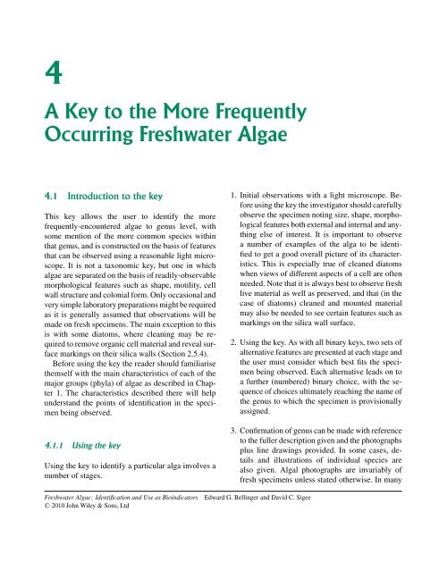
Freshwater Algae: Identification and Use as Bioindicators
Freshwater Algae: Identification and Use as Bioindicators
Freshwater Algae: Identification and Use as Bioindicators

Create successful ePaper yourself
Turn your PDF publications into a flip-book with our unique Google optimized e-Paper software.
4<br />
A Key to the More Frequently<br />
Occurring <strong>Freshwater</strong> <strong>Algae</strong><br />
4.1 Introduction to the key<br />
This key allows the user to identify the more<br />
frequently-encountered algae to genus level, with<br />
some mention of the more common species within<br />
that genus, <strong>and</strong> is constructed on the b<strong>as</strong>is of features<br />
that can be observed using a re<strong>as</strong>onable light microscope.<br />
It is not a taxonomic key, but one in which<br />
algae are separated on the b<strong>as</strong>is of readily-observable<br />
morphological features such <strong>as</strong> shape, motility, cell<br />
wall structure <strong>and</strong> colonial form. Only occ<strong>as</strong>ional <strong>and</strong><br />
very simple laboratory preparations might be required<br />
<strong>as</strong> it is generally <strong>as</strong>sumed that observations will be<br />
made on fresh specimens. The main exception to this<br />
is with some diatoms, where cleaning may be required<br />
to remove organic cell material <strong>and</strong> reveal surface<br />
markings on their silica walls (Section 2.5.4).<br />
Before using the key the reader should familiarise<br />
themself with the main characteristics of each of the<br />
major groups (phyla) of algae <strong>as</strong> described in Chapter<br />
1. The characteristics described there will help<br />
underst<strong>and</strong> the points of identification in the specimen<br />
being observed.<br />
4.1.1 Using the key<br />
Using the key to identify a particular alga involves a<br />
number of stages.<br />
<strong>Freshwater</strong> <strong>Algae</strong>: <strong>Identification</strong> <strong>and</strong> <strong>Use</strong> <strong>as</strong> <strong>Bioindicators</strong> Edward G. Bellinger <strong>and</strong> David C. Sigee<br />
C○ 2010 John Wiley & Sons, Ltd<br />
1. Initial observations with a light microscope. Before<br />
using the key the investigator should carefully<br />
observe the specimen noting size, shape, morphological<br />
features both external <strong>and</strong> internal <strong>and</strong> anything<br />
else of interest. It is important to observe<br />
a number of examples of the alga to be identified<br />
to get a good overall picture of its characteristics.<br />
This is especially true of cleaned diatoms<br />
when views of different <strong>as</strong>pects of a cell are often<br />
needed. Note that it is always best to observe fresh<br />
live material <strong>as</strong> well <strong>as</strong> preserved, <strong>and</strong> that (in the<br />
c<strong>as</strong>e of diatoms) cleaned <strong>and</strong> mounted material<br />
may also be needed to see certain features such <strong>as</strong><br />
markings on the silica wall surface.<br />
2. Using the key. As with all binary keys, two sets of<br />
alternative features are presented at each stage <strong>and</strong><br />
the user must consider which best fits the specimen<br />
being observed. Each alternative leads on to<br />
a further (numbered) binary choice, with the sequence<br />
of choices ultimately reaching the name of<br />
the genus to which the specimen is provisionally<br />
<strong>as</strong>signed.<br />
3. Confirmation of genus can be made with reference<br />
to the fuller description given <strong>and</strong> the photographs<br />
plus line drawings provided. In some c<strong>as</strong>es, details<br />
<strong>and</strong> illustrations of individual species are<br />
also given. Algal photographs are invariably of<br />
fresh specimens unless stated otherwise. In many
138 4 A KEY TO THE MORE FREQUENTLY OCCURRING FRESHWATER ALGAE<br />
c<strong>as</strong>es the photographs are taken under bright field<br />
microscopy, but other techniques such <strong>as</strong> ph<strong>as</strong>e<br />
contr<strong>as</strong>t, differential interference contr<strong>as</strong>t (DIC)<br />
microscopy <strong>and</strong> negative staining are also used<br />
to enhance contr<strong>as</strong>t. Photographs of acid-digested<br />
diatoms are derived from lake sediment samples<br />
(Capstick, 2004).<br />
By working through the key many of the important<br />
features of the specimen will have been used so the<br />
user will already have in their mind the main features<br />
of that organism. Until the user is familiar with the<br />
key there may be occ<strong>as</strong>ions when it is difficult to decide<br />
which set of alternatives is best <strong>and</strong> this could<br />
lead to incorrect identification. It is always best to<br />
note progress through the key by writing down the sequence<br />
of numbers, so that if a mistake occurs steps<br />
can e<strong>as</strong>ily be retraced <strong>and</strong> an alternative route followed.<br />
After each key number is a number in brackets,<br />
referring back to the number you were l<strong>as</strong>t at.<br />
4.1.2 Morphological groupings<br />
Because this is a morphological rather than a taxonomic<br />
key algae from different phyla may be juxtaposed<br />
in the text. The user will find that most specimens<br />
with the following features are found in the<br />
following key groups:<br />
Key nos 5 to23: branched filamentous forms (nondiatom).<br />
Key nos 27 to 29: diatom filaments, str<strong>and</strong>s or ribbons.<br />
Key nos 30 to 42: unbranched filaments but not<br />
blue-greens.<br />
Key nos 43 to 55: filamentous blue-greens.<br />
Key nos 57 to 67: colonial blue-greens.<br />
Key nos 72 to 75: motile colonies.<br />
Key nos 77 to 80: colonial diatoms.<br />
Key nos 81 to 105: other colonial forms (mainly<br />
greens).<br />
Key nos 107 to 125: unicellular flagellates<br />
Key nos 128 to 143: centric diatoms<br />
Key nos 144 to 203: pennate diatoms<br />
Key nos 207 to 212: single-celled green algae<br />
Key nos 214 to 217: some desmids (also 223)<br />
Key nos 218 to 222: remaining single-celled green<br />
algae.<br />
4.2 Key to the main genera <strong>and</strong> species<br />
1 (a) Plants macroscopic, often growing to<br />
>20 cm in length. Thallus differentiated into<br />
nodes bearing whorls of branches <strong>and</strong> internodes.<br />
Erect in habit <strong>and</strong> growing anchored to<br />
the substrate. Chloropl<strong>as</strong>ts discoid <strong>and</strong> numerous...................Charophyta<br />
2<br />
(b) Plants microscopic or if visible to the<br />
naked eye it is normally because they are<br />
present <strong>as</strong> a m<strong>as</strong>s - but still requiring microscopic<br />
observation to determine the more<br />
detailed morphology ...................3<br />
2 (1) (a) Plants coarse to touch, frequently<br />
encrusted with lime. (Common name<br />
Stonewort)........................Chara<br />
(b) Plants not coarse to touch, usually<br />
deep green in colour, not encrusted with<br />
lime.............................Nitella<br />
Members of the Charophyta are essentially<br />
branched filaments with a main axis of large<br />
elongate cells (one per internode) <strong>and</strong> a<br />
whorl of smaller cells forming the branches<br />
at each node. The large internodal cells may<br />
be several centimetres in length. Both Chara<br />
<strong>and</strong> Nitella live anchored to the bottom in
still, clear freshwaters by means of colourless<br />
rhizoids. A few species may occur in<br />
brackish waters. They are probably the only<br />
true members of the rhizobenthos in freshwaters.<br />
Chara particularly favours hard waters<br />
with a pH >7.0 but the pH range of<br />
the whole group is between 5.0 <strong>and</strong> 10.0.<br />
They are not present in polluted waters especially<br />
where sewage is being discharged.<br />
Some species of the Charophyta impart a<br />
garlic like odour to the water. Chlorophyta<br />
(Plate I).<br />
3 (1) (a) Cells grouped together to form a filament,<br />
str<strong>and</strong> or ribbon (see glossary for definition<br />
of these terms). Sometimes filaments<br />
can grow in such profusion to be visible en<br />
m<strong>as</strong>se, or are visible <strong>as</strong> multiseriate rows encrusting<br />
stones.. ........................4<br />
(b) Cells individual or in groups that may be<br />
regular or irregular in shape but not forming<br />
a filament, str<strong>and</strong> or ribbon. . . . .........56<br />
4 (3) (a) Cell pigments localized in chloropl<strong>as</strong>ts * .<br />
Colour when fresh may be gr<strong>as</strong>s green,<br />
pale green, golden to brown, olive green or<br />
(rarely) blueish or reddish . ..............5<br />
(b) Cell pigments not localized in chloropl<strong>as</strong>ts.<br />
Colour when fresh is frequently bluegreenbutmaybeolivegreen...........43<br />
It can sometimes be difficult to decide<br />
whether chloropl<strong>as</strong>ts are present or not <strong>as</strong><br />
with some species they may be large enough<br />
to appear to fill the whole cell leaving no visible<br />
clear are<strong>as</strong>. It is important in such c<strong>as</strong>es<br />
to use other features <strong>as</strong> well such <strong>as</strong> pigment<br />
colour but this is best done with live, fresh<br />
material. It is also useful to look for the range<br />
of features indicated in Table 1.2. The only<br />
group of algae not having chloropl<strong>as</strong>ts are<br />
the Cyanophyta <strong>and</strong> they, for example, do<br />
not have flagella so if the specimen is a flagellate<br />
it cannot be a cyanophyte <strong>and</strong> hence<br />
4.2 KEY TO THE MAIN GENERA AND SPECIES 139<br />
must have chloropl<strong>as</strong>ts. Cross-referencing to<br />
other features in the table can also help arrive<br />
at the correct answer.<br />
5 (4) (a) Filaments branched, sometimes rarely.<br />
False or true branching. Filaments may<br />
branch only occ<strong>as</strong>ionally (in some c<strong>as</strong>es<br />
to generate reproductive structures) so it is<br />
important to examine a re<strong>as</strong>onable length<br />
to determine whether they branch or not.<br />
......................................6<br />
(b) Filaments or ribbons unbranched . . . . 24<br />
6 (5) (a) Branches of filament rejoin to form a<br />
net.......................Hydrodictyon<br />
Hydrodictyon (commonly known <strong>as</strong> the<br />
‘water net’) forms macroscopic nets that<br />
are free floating (occ<strong>as</strong>ionally attached) of<br />
many centimetres in length. Populations<br />
may be large enough to partially block<br />
small streams, especially nutrient-rich nutrient<br />
rich waters. The cells are cylindrical<br />
<strong>and</strong> joined at the ends to form a net. Individual<br />
adult cells can be several millimetres<br />
in length. There is a single parietal, net-like<br />
chloropl<strong>as</strong>t with several pyrenoids within the<br />
cell. Widespread in ponds <strong>and</strong> slow moving<br />
ditches. It can produce very large m<strong>as</strong>ses in<br />
the summer which may be a nuisance blocking<br />
the watercourse. Chlorophyta (Fig. 4.1).<br />
(b) Branches of filaments do not rejoin to<br />
formanet.............................7<br />
7 (6) (a) Each cell is enclosed in a fl<strong>as</strong>k-shaped<br />
lorica which is narrow at one end <strong>and</strong> with a<br />
wide opening at the other. One or two loric<strong>as</strong><br />
may arise from the mouth of the one beneath<br />
forming a forked or dendroid series. The algal<br />
cells within each lorica are biflagellate<br />
with brownish chloropl<strong>as</strong>ts . . . . .Dinobryon<br />
Dinobryon is a mainly colonial alga, the<br />
characteristic feature of which is that the<br />
cells are surrounded by a fl<strong>as</strong>k-shaped lorica.
140 4 A KEY TO THE MORE FREQUENTLY OCCURRING FRESHWATER ALGAE<br />
Plate I 1. Nitella. 2. Chara.<br />
1 2
4.2 KEY TO THE MAIN GENERA AND SPECIES 141<br />
100µm<br />
Figure 4.1 Hydrodictyon. Detail from edge of tubular<br />
net showing 3D intercellular network.<br />
It can be solitary but more frequently occurs<br />
in colonies. These colonies are branched<br />
or dendroid with each new lorica emerging<br />
from the open neck of, <strong>and</strong> attached by<br />
means of a stalk to, the old. Each lorica contains<br />
one biflagellate cell attached to the b<strong>as</strong>e<br />
of the lorica by a fine cytopl<strong>as</strong>mic thread. The<br />
flagella are of unequal lengths (heterokont).<br />
There are typically two chloropl<strong>as</strong>ts per cell<br />
with chrysolaminarin granules at the b<strong>as</strong>e.<br />
An eyespot is also present. The lorica may<br />
be clear or coloured brown, smooth or ornamented<br />
<strong>and</strong> vary in overall shape depending<br />
upon species. The lorica may be up to<br />
100+ µm in length.<br />
Dinobryon is widely recognized <strong>as</strong> a<br />
bacteria-consuming mixotroph (capable of<br />
both autotrophic <strong>and</strong> heterotrophic nutrition).<br />
Very widely distributed in the plankton<br />
of ponds <strong>and</strong> lakes from nutrient-enriched<br />
to nutrient-poor. It may be present in hard or<br />
soft waters. Common in the plankton of cool<br />
waters <strong>and</strong> in mountain regions. H<strong>as</strong> been<br />
50µm<br />
A<br />
Figure 4.2 Dinobryon. Ph<strong>as</strong>e-contr<strong>as</strong>t image of freefloating<br />
dendroid colony. A, Empty lorica; B, lorica containing<br />
biflagellate cell.<br />
<strong>as</strong>sociated with odour probems in drinking<br />
water (Palmer, 1962). Chrysophyta. Plate II<br />
<strong>and</strong> Fig. 4.2.<br />
(b) Cells not in fl<strong>as</strong>k-shaped loric<strong>as</strong> ......8<br />
8 (7) (a) Filaments without cross walls dividing<br />
them into separate cells (siphonaceous<br />
or coenocytic), cross walls only appearing<br />
when reproductive structures produced. Irregularly<br />
branched. ............Vaucheria<br />
Filaments are cylindrical, multinucleate <strong>and</strong><br />
lack cross walls. There are numerous discshaped<br />
to oval chloropl<strong>as</strong>ts with or without<br />
pyrenoids. The usual storage products<br />
are oil <strong>and</strong> fat, very rarely starch. Branching<br />
does occur but is irregular. Filament<br />
20–140 µm wide. The various species are<br />
identified mainly by their reproductive structures.<br />
Widespread <strong>and</strong> may form green mats<br />
of tangled filaments in shallow freshwaters,<br />
on stones <strong>and</strong> damp soil. It may also occur in<br />
B
142 4 A KEY TO THE MORE FREQUENTLY OCCURRING FRESHWATER ALGAE<br />
4a<br />
1<br />
3<br />
4b<br />
Plate II 1. Dinobryon. 2. Lemanea (a) growths on higher plant stem, (b) whole alga. 3. Vaucheria. 4. Batrachospermum,<br />
(a) whole alga, (b) details of branching. 5. Hildenbr<strong>and</strong>ia.<br />
2a<br />
2b<br />
5
200µm<br />
4.2 KEY TO THE MAIN GENERA AND SPECIES 143<br />
20µm<br />
Figure 4.3 Vaucheria. Top: Detail from surface of filament<br />
(DIC image) showing numerous discoid chloropl<strong>as</strong>ts<br />
Bottom: Low power view of filaments showing<br />
absence of cell walls.<br />
salt marshes <strong>and</strong> brackish water muds. Xanthophyta.<br />
Plate II <strong>and</strong> Fig. 4.3.<br />
(b) Filaments with normal cross walls, not<br />
siphonaceous . .........................9<br />
9 (8) (a) Filaments multiseriate, i.e. columns of<br />
cells in many parallel or radiating rows.<br />
Branches may or may not be present . . . . 10<br />
(b) Filaments not multiseriate ..........13<br />
(but see also Coleochaete which, when<br />
older, forms a m<strong>as</strong>s or disc of cells that appear<br />
multiseriate).<br />
10 (9) (a) Filaments branched (although the branching<br />
can be scarce <strong>and</strong> difficult to find – hence<br />
the need to look along several filaments to<br />
confirm situation) with branches arising in<br />
tufts (embedded in mucilage) or <strong>as</strong> less frequent<br />
<strong>and</strong> more tapered bristles . ........11<br />
(b) Filaments encrusting on stones or macrophytes<br />
forming single or packed layers of<br />
cells, green, red or brown in colour. . ....12<br />
11 (10) (a) Filaments loosely embedded in copious<br />
mucilage. Main filament axis with<br />
tufts of branches arranged at regular intervals..................Batrachospermum<br />
Batrachospermum produces large multibranched<br />
filaments in a mucilaginous or<br />
gelatinous m<strong>as</strong>s, similar to frog spawn.<br />
The overall appearance of the filaments<br />
is that they are beaded. There is a main<br />
large axis with regular whorls of smaller<br />
branches. Chloropl<strong>as</strong>ts have no pyrenoids<br />
<strong>and</strong> are ribbon-like (several per cell), olive<br />
brown to reddish or grey-blue in colour. The<br />
whorls may be more than 1 mm in diameter.<br />
Widespread <strong>and</strong> with a characteristic appearance.<br />
Found in streams <strong>and</strong> pools/bogs.<br />
Rhodophyta. Plate II. Fig. 4.4.<br />
(b) Plants without regular tufts of branches.<br />
Thickened are<strong>as</strong> along stem. . . . . . Lemanea<br />
The thallus of Lemanea is cartilaginous <strong>and</strong><br />
thus h<strong>as</strong> an erect posture being attached at<br />
the b<strong>as</strong>e. The bristle-like ‘stems’ can grow<br />
up to 20 cm in length <strong>and</strong> show little to<br />
frequent branching. When growing in water<br />
plants look like stiff tufts of black or brown<br />
hair, each hair having irregular thickenings<br />
along their length. Chloropl<strong>as</strong>ts are numerous,<br />
parietal <strong>and</strong> disc-shaped. Widespread in<br />
moderate to f<strong>as</strong>t flowing streams attached to<br />
stones. Rhodophyta. Plate II.<br />
12 (10) (a) Cells of filaments stacked in vertical<br />
rows to form a pseudoparenchymatous<br />
m<strong>as</strong>s. These filaments are found <strong>as</strong><br />
a thin, flat rose-coloured encrustation on<br />
stones. ...................Hildenbr<strong>and</strong>ia
144 4 A KEY TO THE MORE FREQUENTLY OCCURRING FRESHWATER ALGAE<br />
B<br />
300µm<br />
A<br />
50µm<br />
Figure 4.4 Batrachospermum. Top: Detail showing a<br />
whorl of uniaxial tufts (A) arising from the main axis<br />
(B). Bottom: General view of thallus.<br />
The thalli of Hildenbr<strong>and</strong>ia grow over<br />
stones in rivers <strong>and</strong> streams, frequently in<br />
hard waters. The thalli are crustose, reddish<br />
in colour <strong>and</strong> closely adhere to the substrate.<br />
Cells cubical in vertical rows, each cell up to<br />
16 µm in diameter. Rhodophyta. Plate II.<br />
(b) Filaments attached to a surface, radiating<br />
from a central point forming a flattened disc<br />
or slightly rounded cushion of cells - some<br />
of which have fine hairs or setae which are<br />
sheathed at the b<strong>as</strong>e . .........Coleochaete<br />
Coleochaete is a member of the Chlorophyta.<br />
It is uniseriate when younger or can<br />
grow <strong>as</strong> a m<strong>as</strong>s where the filaments tend<br />
to merge to form a pseudoparenchymatous<br />
m<strong>as</strong>s. Branches may be dichotomous or irregular.<br />
There may be an erect part of the<br />
thallus <strong>as</strong> well <strong>as</strong> the flattened disc-like cushion.<br />
The cells frequently have long bristles<br />
sheathed at their b<strong>as</strong>es. There is a single<br />
parietal plate-like chloropl<strong>as</strong>t with one or<br />
two pyrenoids. Widespread <strong>as</strong> an epiphyte<br />
on aquatic macrophytes <strong>and</strong> other surfaces.<br />
Chlorophyta. Plate III.<br />
13 (9) (a) Some cells along the filament bear hairs<br />
orsetae..............................14<br />
(b)Nohairsorsetaepresent............18<br />
14 (13) (a) Hairs or setae having a bulbous b<strong>as</strong>e . 15<br />
(b) No bulbous b<strong>as</strong>e present with the hairs or<br />
setae.................................16<br />
15 (14) (a) Filaments growing horizontally or prostrate,<br />
epiphytic. Branching irregular or may<br />
be absent. Some cells with one to several<br />
hairs.....................Aphanochaete<br />
Aphanochaete is a prostrate creeping epiphyte<br />
<strong>and</strong> a member of the Chlorophyta.<br />
Cells longer than broad <strong>and</strong> sometimes<br />
slightly swollen in the centre <strong>and</strong> often bearing<br />
one or more long bristles swollen at the<br />
b<strong>as</strong>e. Chloropl<strong>as</strong>ts disc-shaped with one or<br />
more pyrenoids. Widespread distribution, often<br />
reported in nutrient-rich waters <strong>as</strong> an epiphyte<br />
on submerged macrophytes <strong>and</strong> larger<br />
filamentous green algae. Plate III.<br />
(b) Filaments not prostrate, branched, with<br />
cells somewhat broader at the top than the<br />
b<strong>as</strong>e. Many cells have one or more characteristic<br />
colourless chaetae (a terminal hair with<br />
a swollen b<strong>as</strong>e) arising from the top of the<br />
cell........................Bulbochaete.<br />
Bulbochaete is a member of the Oedogoniales.<br />
It h<strong>as</strong> branched filaments <strong>and</strong> is
1<br />
3<br />
5<br />
4.2 KEY TO THE MAIN GENERA AND SPECIES 145<br />
Plate III 1. Coleochaete. 2. Aphanochaete. 3. Bulbochaete. 4. Chaetophora, (a) whole alga, (b) growths on stem of<br />
higher plant. 5. Draparnaldia. 6. Stigeoclonium.<br />
4a<br />
2<br />
6<br />
4b
146 4 A KEY TO THE MORE FREQUENTLY OCCURRING FRESHWATER ALGAE<br />
e<strong>as</strong>ily identified by the hairs which have<br />
distinctly swollen b<strong>as</strong>es. Chloropl<strong>as</strong>t parietal<br />
<strong>and</strong> net-like with several pyrenoids.<br />
Widespread distribution, growing attached<br />
to submerged macrophytes or stones.<br />
Wide pH <strong>and</strong> nutrient range. Chlorophyta.<br />
Plate III.<br />
16 (14) (a) Filaments enclosed in soft watery mucilage<br />
which h<strong>as</strong> no definite shape . .....17<br />
(b) Filaments with a firm mucilage surround<br />
having a definite shape. Often forming<br />
macroscopic attached m<strong>as</strong>ses Chaetophora<br />
Chaetophora filaments are frequently attached<br />
to stones or submerged aquatic plants.<br />
Filaments can be quite long (10+ cm.). Branching<br />
more common towards filament apex.<br />
End cells of filament/branch with a pointed<br />
shape or in the form of a long multicellular<br />
hair. The erect system is well developed<br />
but the prostrate one is not. Fairly<br />
widespread distribution at the edges of shallow<br />
flowing or still waters attached to a substrate<br />
such <strong>as</strong> aquatic plants. Chlorophyta<br />
Plate III.<br />
17 (16) (a) Main filament axis composed of a<br />
single row of larger cells from which<br />
arise tufts of branches composed of much<br />
smaller cells, the whole enclosed in soft<br />
mucilage..................Draparnaldia<br />
Draparnaldia is a member of the Chlorophyta.<br />
The erect filaments are attached to the<br />
substratum by means of rhizoids. Filaments<br />
embedded in soft mucilage. Main filament<br />
cells barrel-shaped (40–100 µm wide) with<br />
net-like or complete b<strong>and</strong>-shaped parietal<br />
chloropl<strong>as</strong>t with several pyrenoids. Branches<br />
arise in whorls from main axis. Branch cells<br />
smaller with single chloropl<strong>as</strong>t <strong>and</strong> up to<br />
three pyrenoids. Branches have either a blunt<br />
terminal cell or form a multicellular hair.<br />
Widely distributed often in phosphorus-poor<br />
waters. Plate III.<br />
(b) Filament main axis cells not markedly<br />
different to branch cells (except those at the<br />
end of branches, which are thinner <strong>and</strong> hairlike).<br />
Branches not usually occurring in distinctwhorls...............Stigeoclonium<br />
Filaments of Stigeoclonium are usually attached<br />
by means of b<strong>as</strong>al cells to a variety<br />
of surfaces. Branches can be opposite,<br />
alternate but rarely whorled <strong>and</strong> terminating<br />
in a tapering series of cells sometimes<br />
forming a terminal multicellular hair. Filaments<br />
surrounded by a thin mucilaginous<br />
layer. Chloropl<strong>as</strong>ts parietal with one or more<br />
pyrenoids. Cells 5–40 µm wide <strong>and</strong> many<br />
times longer than broad. Widespread distribution<br />
usually attached to rocks or stones but<br />
may break away <strong>and</strong> be found free floating.<br />
Several species, in flowing or still waters. S.<br />
tenue is common <strong>and</strong> is often used <strong>as</strong> an<br />
indicator of enriched or organically-polluted<br />
situations (also reported <strong>as</strong> tolerant to heavy<br />
metal pollution). Chlorophyta. Plate III.<br />
Fig. 4.5.<br />
50µm<br />
Figure 4.5 Stigeoclonium. General view of filaments,<br />
with branches tapering to a fine point (arrows). Inset:<br />
Mode of branching.
18 (13) (a) Branched filaments embedded in<br />
mucilage.............................19<br />
(b) Branched filaments not embedded in mucilage................................21<br />
19 (18) (a) Plant forms a gelatinous globular cushion.<br />
Mucilage may be firm <strong>and</strong> encrusted<br />
withlimeinhardwaterlocations.......20<br />
(b) Mucilage covering thin. Usually<br />
attached at b<strong>as</strong>e <strong>and</strong> forming trailing<br />
branched filaments. Not cushion-like or<br />
globular..........Stigeoclonium (see 17)<br />
20 (19) (a) Cushions composed of filaments with<br />
branches tapering to a long multicellular hair<br />
or having a single rounded cell at apex. Mucilage<br />
firm forming cushions 1–2+ cm wide<br />
Chaetophora (see 16)<br />
(b) Cushions generally smaller (>5mm diameter).<br />
Filaments with less branches <strong>and</strong><br />
cells rounded or with swollen ends, often<br />
lime-encrusted . . . . ...........Gongrosira<br />
Gongrosira forms cushion-like colonies that<br />
may be lime-encrusted in hard waters. Filaments<br />
arise from a b<strong>as</strong>al prostrate group of<br />
cells, forming an erect <strong>as</strong> well <strong>as</strong> a prostrate<br />
system. No hairs present <strong>and</strong> terminal cells of<br />
branches bluntly rounded. Cells cylindrical<br />
or swollen. Often with thickened walls. Parietal<br />
chloropl<strong>as</strong>ts with 1 to several pyrenoids.<br />
Frequent in streams, shallow lake margins or<br />
ponds, on stones, often in hard water regions.<br />
Cells 4–30 µm wide, 1 to 3 times longer than<br />
breadth. Chlorophyta. Plate IV.<br />
21 (18) (a) Plants small erect, cells
148 4 A KEY TO THE MORE FREQUENTLY OCCURRING FRESHWATER ALGAE<br />
1<br />
4a<br />
5a 5b<br />
7a<br />
2<br />
6a<br />
Plate IV 1. Gongrosira. 2. Microthamnion. 3. Rhizoclonium: (a) general view; (b) cell detail. 4. Melosira: (a)M.<br />
varians; (b)M. nummuloides; (c)M. dickiei. 5. Tabellaria fenestrata: (a) colony – girdle view; (b) single cell – valve<br />
view 6. Fragilaria: (a)F. crotonensis; (b)F. capucina – single cell, girdle view; (b) colony – valve view. 7. Tabellaria:<br />
(a) T. fenestrata – zigzag colony; (b) T. flocculosa.<br />
4b<br />
3a<br />
6b<br />
7b<br />
6c<br />
4c<br />
3b
100µm<br />
Figure 4.6 Cladophora General view of filaments,<br />
showing newly-formed branches.<br />
be free living or attached to a substrate by<br />
means of a small holdf<strong>as</strong>t. The branches<br />
may be alternate or opposite, dichotomous<br />
or even trichotomous. Cell walls are robust<br />
<strong>and</strong> the chloropl<strong>as</strong>t net-like (reticulate)<br />
<strong>and</strong> parietal with numerous pyrenoids. Cells<br />
50–150 µm broad <strong>and</strong> up to ×10 long <strong>as</strong><br />
broad. Commonly known <strong>as</strong> ‘blanket weed’<br />
it can form extensive, coarse to touch mats.<br />
Frequent in hard or semi-hard waters especially<br />
those enriched with sewage. May<br />
also be found in marine habitats especially<br />
those enriched with sewage. Cladophora<br />
can also produce large growths in water<br />
treatment filtration systems such <strong>as</strong> slow<br />
s<strong>and</strong> filters where severe problems of filter<br />
clogging may result. Chlorophyta. Figs 2.28<br />
<strong>and</strong> 4.6.<br />
(Not to be confused with Pithophora, which<br />
is common in ponds but whose filaments<br />
frequently contain dark akinetes that are<br />
swollen <strong>and</strong> barrel-shaped. Cells 20–100 µm<br />
wide <strong>and</strong> up to 400 µm long. Not illustrated.)<br />
24 (5) (a) Cells with a siliceous wall ..........25<br />
4.2 KEY TO THE MAIN GENERA AND SPECIES 149<br />
To determine whether a silica wall is present<br />
it may be necessary to ‘clean’ the specimen<br />
with a suitable oxidizing or acidifying agent<br />
(see Section 2.5.4). Some indication can be<br />
obtained, however, by sharply focusing on<br />
the cell wall. If a regular pattern of markings<br />
can be made out (e.g. Fig. 1.13) then<br />
it is quite probable that the wall is made of<br />
silica. If an empty cell can be found then<br />
it may be e<strong>as</strong>ier to see any markings. With<br />
some pennate diatoms a distinctive gliding<br />
movement may be observed in live (fresh)<br />
material<br />
(b) Cell wall not made of silica . ........30<br />
25 (24) (a) Cells embedded in a gelatinous tube but<br />
separate from each other within the tube<br />
Frustulia in part see 199<br />
Cymbella <strong>and</strong> Encyonema in part see 160<br />
(b)Cellsnot<strong>as</strong>above.................26<br />
26 (25) (a) Cells joined together to form a continuous<br />
filament not surrounded by extensive mucilage.<br />
Filaments <strong>and</strong> cells circular in cross<br />
section...............................27<br />
(b) Cells forming a ribbon or chain. Cells<br />
elongate <strong>and</strong> not circular in transverse<br />
section...............................28<br />
27 (26) The former genus Melosira, consisting of<br />
filaments of centric cells, h<strong>as</strong> now been subdivided<br />
into Melosira <strong>and</strong> Aulacoseira (see<br />
below)<br />
(a) Cell walls with no obvious markings.<br />
Cells linked in pairs (paired arrangement<br />
may be difficult to see) . .........Melosira<br />
Cells of Melosira do not have distinctive<br />
markings on their surface. They are rectangular<br />
(M. varians) orovoid(M. nummuloides)<br />
in shape (girdle view) with relatively<br />
thin walls or rectangular with quite
150 4 A KEY TO THE MORE FREQUENTLY OCCURRING FRESHWATER ALGAE<br />
thick walls (M. dickiei). Cells of the filament<br />
are linked together in pairs although this can<br />
require careful observation to see. Chloropl<strong>as</strong>ts<br />
are small discs or plates <strong>and</strong> may be<br />
golden-brown to dark-brown. M. varians<br />
(cells 8–35 µm in diameter, 4–14 µm deep)<br />
is common <strong>and</strong> sometimes abundant in shallow,<br />
frequently smaller, eutrophic waters.<br />
M. dickiei (10–2 µm in diameter, 7–10 µm<br />
deep) is found on damp rocks <strong>and</strong> river banks<br />
<strong>and</strong> M. nummuloides (9–42 µm indiameter,<br />
10–14 µm deep) is found in brackish or<br />
marine environments, often attached to the<br />
substratum. M. varians can grow in water<br />
treatment filters open to sunlight <strong>and</strong> cause<br />
clogging problems. Bacillariophyta. Plate IV<br />
<strong>and</strong> Fig. 4.7.<br />
(b) Cell walls with granulated markings <strong>and</strong><br />
often with spines at end (often best seen at<br />
end of a filament). Forming a continuous filament<strong>and</strong>notinpairs........Aulacoseira<br />
In Aulacoseira the cells are rectangular in<br />
shape (girdle view) <strong>and</strong> are linked together to<br />
form long filaments. The surface of the silica<br />
cell wall h<strong>as</strong> characteristic markings of rows<br />
of dots (punctae) <strong>and</strong> the ends of the cells<br />
bear a ring of spines (one or two of which<br />
may be quite long) that link the cells together.<br />
A sulcus can usually be observed between<br />
the mantle <strong>and</strong> girdle b<strong>and</strong>. Chloropl<strong>as</strong>ts<br />
disc-shaped <strong>and</strong> usually golden-brown. Several<br />
species are found in freshwaters. Some<br />
are more common in eutrophic lakes, e.g. A.<br />
ambigua (cells 4–17 × 5–13 µm in size), A.<br />
granulata (cells 4–30 × 5–24 µm in size),<br />
A. granulate var angustissima (h<strong>as</strong> narrow,<br />
long cells with a obvious long spine<br />
protruding from the end cell) <strong>and</strong> A italica<br />
(cells 6–23 × 8–20 µm in size) <strong>and</strong> others in<br />
less enriched waters, e.g. A. isl<strong>and</strong>ica (cells<br />
3–28 ×4–21 µm in size) <strong>and</strong> A. subarctica<br />
(cells 3–15 × 2.5–18 µm in size). A. granulata<br />
can cause filter clogging problems in<br />
water works (see also Melosira). Bacillariophyta.<br />
Figs 1.3, 4.8, 4.9.<br />
10µm<br />
15µm<br />
Figure 4.7 Melosira Top: Single cell showing numerous<br />
yellow-brown discoid chloropl<strong>as</strong>ts. Bottom: Straight,<br />
unbranched filaments typical of this colonial diatom.<br />
Inset: Characteristic paired arrangement of cells along<br />
filament.<br />
28 (26) (a) Cells with internal septa or costae (see<br />
below)...............................29<br />
Costae are silica thickenings on the inside of<br />
the valve, orientated at right angles to the apical<br />
(long) axis. Septa are silica plates that run<br />
across the inside of the cell from the girdle<br />
b<strong>and</strong>s. The septa may protrude for different<br />
lengths into the cell.<br />
(b) Cells without internal septa . ..........<br />
..............................Fragilaria
10µm<br />
4.2 KEY TO THE MAIN GENERA AND SPECIES 151<br />
30µm<br />
Figure 4.8 Aulacoseira Top: Planktonic filament (A.<br />
granulata var. angustissima). Bottom: Fragmented filament<br />
(A. granulata) – acid digest, lake sediment sample.<br />
A terminal spine (arrow) is visible in both c<strong>as</strong>es.<br />
In Fragilaria the pennate or elongate<br />
frustules are joined by their valve faces to<br />
form a ribbon-like chain. Valves fusiform<br />
in shape, sometimes slightly swollen at the<br />
centre (gibbous) or with swollen (capitate)<br />
ends or narrow <strong>and</strong> rectangular in girdle<br />
view. Usually seen in girdle view in non<br />
acid-cleaned material. Valve surface with<br />
fine striae. Two plate-shaped chloropl<strong>as</strong>ts<br />
present. Fragilaria crotonensis can be<br />
found in meso- to eutrophic waters, sometimes<br />
forming blooms. Size, 40–170 µm<br />
long, 2–4 µm wide. F. capucina is also<br />
10µm<br />
Figure 4.9 Aulacoseira granulata. Cells have terminal<br />
long (ls) <strong>and</strong> short (ss) spines.<br />
widespread occurring in lakes <strong>and</strong> rivers,<br />
10–100 µm long, 3.5–4.5 µm wide. Can<br />
impart unwanted odour to drinking waters<br />
(Palmer, 1962). Large numbers can cause<br />
ls<br />
ss
152 4 A KEY TO THE MORE FREQUENTLY OCCURRING FRESHWATER ALGAE<br />
10µm<br />
10µm<br />
Figure 4.10 Fragilaria. Top: Ribbon arrangement of<br />
diatom colony. Fixed sample. Bottom: Single cell showing<br />
fine transverse striae, with central clear area (arrow).<br />
Acid digest, lake sediment sample.<br />
filter clogging problems in water treatment<br />
works. Bacillariophyta. Plate IV. Fig. 4.10.<br />
29 (28) (a) Cells rectangular or tabular in normal<br />
view, sometimes united to form zig-zag<br />
colonies. When seen individually <strong>and</strong> not<br />
<strong>as</strong> part of a chain the valves often show<br />
a strongly swollen middle region. Cells<br />
without costae but have internal septa which<br />
canbeclearlyseen............Tabellaria<br />
Tabellaria cells are rectangular in girdle<br />
view <strong>and</strong> form colonies that may be stellate<br />
or zig-zag. The stellate colonies can be<br />
confused with Asterionella but can be distinguished<br />
by: (1) internal septa in Tabellaria,<br />
<strong>and</strong> (2) more squared-off ends to the<br />
cells in Tabellaria when compared with the<br />
more rounded ends in Asterionella. T. fenestrata<br />
(33–116 µm long, 4–10 µm wide) <strong>and</strong><br />
T. flocculosa (6–130 µm long <strong>and</strong>
(b) Cells rectangular, may unite to form zigzag<br />
colonies. Cells rod-shaped when seen individually<br />
with (at the most) slightly swollen<br />
centres. Transverse costae present within the<br />
cells...........................Diatoma<br />
Diatoma forms zig-zag or ribbon-like<br />
colonies or ribbon-like chains. Internal<br />
costae are present <strong>and</strong> may be seen <strong>as</strong> ridges<br />
or points at the cell margin. Chloropl<strong>as</strong>ts<br />
discoid to plate-like >10 per cell. Cells may<br />
be capitate. Several species occur - the more<br />
common of which are D. vulgaris with<br />
cells (8–75 µm long <strong>and</strong> 7–18 µm wide)<br />
having bluntly rounded ends, common<br />
in the plankton of moderately eutrophic<br />
lakes <strong>and</strong> rivers, <strong>and</strong> D. tenuis (22–120 µm<br />
long <strong>and</strong> >10 m wide) with capitate apices<br />
broader than the rest of the cell. Planktonic<br />
<strong>and</strong> epiphytic. Large numbers of planktonic<br />
species can cause filter clogging problems<br />
in water treatment works. Bacillariophyta.<br />
Fig. 4.12.<br />
30 (24) (a) Chloropl<strong>as</strong>ts form a distinct spiral b<strong>and</strong><br />
withinthecell.................Spirogyra<br />
Spirogyra h<strong>as</strong> cylindrical cells that are<br />
joined end to end to form an unbranched<br />
filament. The cell walls are firm <strong>and</strong> have a<br />
thin film of mucilage on the outside, giving<br />
them a slimy feel. Chloropl<strong>as</strong>ts have<br />
a helical shape <strong>and</strong> there can be up to 15<br />
per cell. Numerous pyrenoids are present.<br />
The nucleus, often visible in live material,<br />
is in the centre of the cell. Cells may be between<br />
10 <strong>and</strong> 160 µm in diameter <strong>and</strong> up to<br />
590 µm long. Filaments fragment e<strong>as</strong>ily at<br />
the cross walls, each fragment growing into<br />
a new filament. Sexual reproduction in Spirogyra<br />
involves conjugation between cells of<br />
different filaments <strong>and</strong> results in the production<br />
of a resistant zygote. It is widely distributed<br />
in shallow ponds <strong>and</strong> ditches where<br />
it can form dense green m<strong>as</strong>ses. Can grow<br />
in large enough amounts in shallow openwater<br />
treatment filters to cause blockage<br />
4.2 KEY TO THE MAIN GENERA AND SPECIES 153<br />
50µm<br />
10µm<br />
Figure 4.12 Diatoma. Top: Star-shaped pattern of<br />
cells at edge of planktonic colony. Bottom: Single cell,<br />
showing transverse costae. Acid digest, lake sediment<br />
sample.<br />
of those filters. Chlorophyta. Plate V.<br />
Fig. 4.13.<br />
(b) Chloropl<strong>as</strong>ts not in the form of a spiral<br />
b<strong>and</strong> . ................................31<br />
31 (30) (a) Filaments are unbranched with the<br />
cells embedded in a prominent broad mucilaginous<br />
hyaline cylindrical envelope or<br />
sheath. Cells are short cylinders with broadly<br />
rounded ends. . . . ..............Geminella<br />
Cells are between 5–25 µm broad <strong>and</strong> up<br />
to twice <strong>as</strong> long <strong>as</strong> they are wide <strong>and</strong> are<br />
surrounded by a thick sheath of mucilage.<br />
Cells often occur in pairs along the filament.<br />
The chloropl<strong>as</strong>t is a parietal plate, roughly<br />
saddle-shaped <strong>and</strong> located near the central<br />
region of the cell. A pyrenoid is usually<br />
present. Reproduction is by fragmentation of
154 4 A KEY TO THE MORE FREQUENTLY OCCURRING FRESHWATER ALGAE<br />
1a<br />
4<br />
1b<br />
7a<br />
2a<br />
1c<br />
Plate V 1. Tabellaria flocculosa: (a) colony; (b) girdle view of cells; (c) single cell – valve view. 2. Spirogyra: (a)<br />
vegetative filament; (b) oospores. 3. Geminella. 4. Spondylosium. 5. Stichococcus. 6. Microspora. 7. Tribonema: (a)T.<br />
viride; (b)T. vulgare.<br />
2b<br />
7b<br />
5<br />
3<br />
6
50µm<br />
4.2 KEY TO THE MAIN GENERA AND SPECIES 155<br />
50µm<br />
Figure 4.13 Spirogyra. Top: Species with multiple<br />
(about 6) spiral chloropl<strong>as</strong>ts <strong>and</strong> simple cross walls. The<br />
chloropl<strong>as</strong>ts have numerous pyrenoids (small dots). Bottom:<br />
Species with two chloropl<strong>as</strong>ts <strong>and</strong> complex (replicate)<br />
cross walls (arrows).<br />
the filament. Occurs in shallow, often slightly<br />
acidic, waters. Chlorophyta. Plate V.<br />
(b) Cells not embedded in a prominent mucilaginous<br />
envelope . ..................32<br />
32 (31) (a) Filament outline with constrictions giving<br />
it a toothed appearance . . . . .........33<br />
(b) Filament outline without constrictions<br />
<strong>and</strong> does not have toothed appearance . . . 34<br />
33 (32) (a) Cells elliptical in shape in one view<br />
but show a deep narrow constriction when<br />
seen from the other view giving a toothed<br />
appearance with the teeth being smoothly<br />
rounded. No gap between adjoining cells.<br />
Spondylosium<br />
Spondylosium is a filamentous desmid.<br />
Each cell resembles an angular or rounded<br />
(depending upon the species of Spondylo-<br />
sum) Cosmarium cell with a deep constriction<br />
- giving the characteristic toothed appearance<br />
to the filament <strong>as</strong> a whole. Cell<br />
walls fairly smooth. There is one chloropl<strong>as</strong>t<br />
with pyrenoid in each semicell. Frequent<br />
around lake margins in upl<strong>and</strong> are<strong>as</strong>.<br />
Chlorophyta. Plate V.<br />
(b) Cells angular, rarely elliptical, in one<br />
view with a small lens-shaped gap between<br />
the cells usually visible. Cells only have a<br />
small constriction so the toothed margin not<br />
so pronounced <strong>and</strong> the ‘teeth’ are more angular........................Desmidium<br />
Desmidium is a filamentous desmid. The<br />
cells can form long twisted filaments enclosed<br />
in a gelatinous envelope. When<br />
viewed in transverse section the filaments<br />
may be triangular. There is one chloropl<strong>as</strong>t<br />
with pyrenoid in each semicell. Cells<br />
26–50 µm broad. Sometimes the space between<br />
the cells can be difficult to see. Common<br />
in the vegetation <strong>and</strong> along the margins<br />
of oligotrophic lakes. Chlorophyta. Fig. 4.14.<br />
34 (32) (a) Filaments very short, often only 2 or 3<br />
cells long (more like a short chain than a<br />
100µm<br />
Figure 4.14 Desmidium. Elongate filaments of this<br />
colonial desmid. Inset: Details of four cells, each composed<br />
of two semicells.
156 4 A KEY TO THE MORE FREQUENTLY OCCURRING FRESHWATER ALGAE<br />
filament). Cells cylindrical with free ends<br />
rounded . . ..................Stichococcus<br />
Stichococcus can occur singly or form very<br />
reduced filaments, just short chains. Cells<br />
1–16 µm broad <strong>and</strong> 4–32 µm long. There is<br />
a single lobed chloropl<strong>as</strong>t but no pyrenoid.<br />
Occurs on damp earth, tree trunks etc. <strong>as</strong> well<br />
<strong>as</strong> in freshwaters. Chlorophyta. Plate V.<br />
(b) Filaments many cells in length, not typicallyshort............................35<br />
35 (34) (a) Large alga with tubular thallus many cells<br />
broad <strong>and</strong> long, macroscopic in size .....<br />
.........................Enteromorpha<br />
Enteromorpha h<strong>as</strong> a tube-like thallus that<br />
may or may not branch. The plant can grow<br />
to many centimetres in length. The tube itself<br />
consists of a layer one cell thick. The<br />
tubes are attached to a substrate by means of<br />
rhizoidal branches <strong>and</strong> an attachment disc.<br />
Enteromorpha h<strong>as</strong> a worldwide distribution<br />
mainly in marine <strong>and</strong> brackish water habitats<br />
but it can stray into freshwater. The two most<br />
common species are E. intestinalis <strong>and</strong> E.<br />
compressa. Chlorophyta. (Not illustrated.)<br />
(b)Alganotalargetubularthallus......36<br />
36 (35) (a) Two star-shaped chloropl<strong>as</strong>ts per cell<br />
..............................Zygnema<br />
Cells of Zygnema are cylindrical <strong>and</strong> have<br />
two, characteristic, star-shaped chloropl<strong>as</strong>ts<br />
separated by a clear area. Each chloropl<strong>as</strong>t<br />
h<strong>as</strong> a pyrenoid. The filaments usually have<br />
a soft mucilage sheath, are unbranched <strong>and</strong><br />
not very long. They may be attached to a substrate<br />
by means of rhizoids. Cells 16–50 µm<br />
in diameter <strong>and</strong> 2 to 3 times long <strong>as</strong> broad.<br />
As members of the Chlorophyta they store<br />
starch. Common in shallow waters where<br />
it can form luxuriant growths. Chlorophyta.<br />
Fig. 4.15.<br />
(b) Chloropl<strong>as</strong>t either single or more than<br />
twopercell...........................37<br />
20µm<br />
Figure 4.15 Zygnema. End of filament showing cells<br />
with two typical stellate chloropl<strong>as</strong>ts.<br />
37 (36) (a) One chloropl<strong>as</strong>t per cell in the form of a<br />
flat plate arranged along the long axis of the<br />
cell. When viewed from one direction the<br />
chloropl<strong>as</strong>t fills most of the cell but when<br />
viewed from the other it is a thin line down<br />
themiddle................... Mougeotia<br />
The chloropl<strong>as</strong>t of Mougeotia is suspended<br />
on cytopl<strong>as</strong>mic str<strong>and</strong>s <strong>and</strong> can move within<br />
the cell depending upon the light. Hence<br />
sometimes it may be seen face-on, sometimes<br />
edge-on <strong>and</strong> sometimes twisted. The<br />
cells form long unbranched free floating filaments.<br />
Cells 3.5–35 µm in diameter <strong>and</strong> between<br />
5 <strong>and</strong> 12 times <strong>as</strong> long <strong>as</strong> broad. There<br />
are several pyrenoids per cell <strong>and</strong> when the<br />
chloropl<strong>as</strong>t is viewed edge-on the pyrenoids<br />
give it a lumpy appearance. Storage product<br />
starch. Common in many habitats. Chlorophyta.<br />
Fig. 4.16.<br />
(b)Chloropl<strong>as</strong>tnot<strong>as</strong>above............38<br />
38 (37) (a) Chloropl<strong>as</strong>t reticulate (see glossary)<br />
.....................................39<br />
(b)Chloropl<strong>as</strong>tnotreticulate...........41<br />
39 (38) (a) Cell walls thick, often lamellate, made up<br />
of two overlapping halves which can break<br />
into H-pieces. End cells usually have Hshaped<br />
ends. Pyrenoids absent . ..........<br />
............................Microspora
50µm<br />
50µm<br />
Figure 4.16 Mougeotia. Detail of separate cells showing<br />
plate-like chloropl<strong>as</strong>t in edge (top) <strong>and</strong> face (bottom)<br />
view. Small refractive pyrenoids can be seen in the<br />
chloropl<strong>as</strong>t (arrows).<br />
Microspora forms unbranched filaments<br />
with a holdf<strong>as</strong>t for substratum attachment.<br />
The cell walls are characteristically lamellate<br />
<strong>and</strong> shaped like an ‘H’ so that each<br />
cell is composed of two H-pieces joined together.<br />
Chloropl<strong>as</strong>t reticulate not very well<br />
defined <strong>and</strong> pyrenoids absent. Storage product<br />
starch. Cells 5–30 µm in diameter <strong>and</strong> 1<br />
to 3 times <strong>as</strong> long <strong>as</strong> broad. Common but not<br />
usually abundant in small freshwater bodies.<br />
More frequent at cooler times of the year.<br />
Chlorophyta. Plate V.<br />
(b) Cell wall not thick <strong>and</strong> lamellate <strong>and</strong> not<br />
composed of H-pieces. Pyrenoids present<br />
.....................................40<br />
40 (39) (a) Cells cylindrical or slightly swollen, cell<br />
walls robust. Cells 20–80 µm broad <strong>and</strong> between<br />
5 <strong>and</strong> 15 times <strong>as</strong> long <strong>as</strong> broad<br />
............................Cladophora<br />
4.2 KEY TO THE MAIN GENERA AND SPECIES 157<br />
(in part see 23)<br />
(b) Cells cylindrical, sometimes slightly<br />
swollen at one end. Cell wall firm but not<br />
very robust. Filaments unbranched. Some<br />
cells along the filament will have ring-like<br />
transverse lines at the swollen end (cap<br />
cells). Cells 10–40 µm wide <strong>and</strong> 2–5 times<br />
<strong>as</strong>long<strong>as</strong>broad............Oedogonium<br />
Oedogonium cells form long, unbranched,<br />
filaments. The chloropl<strong>as</strong>t is parietal <strong>and</strong> netlike.<br />
Cells are rectangular <strong>and</strong> longer than<br />
broad. The chloropl<strong>as</strong>t is parietal <strong>and</strong> netlike<br />
with several pyrenoids. Over 200 species<br />
have been described <strong>and</strong> further identification<br />
is impossible without the reproductive<br />
structures being present. Abundant in still or<br />
gently moving waters where they may form<br />
dense mats Chlorophyta. Fig. 4.17.<br />
41 (38) (a) Chloropl<strong>as</strong>t a ring or is plate-shaped extending<br />
1/3 to most of the way round the cell<br />
(at right angles to the long axis). One or more<br />
pyrenoids. Starch test positive . .........42<br />
50µm<br />
Figure 4.17 Oedogonium. The central cell (cap cell)<br />
h<strong>as</strong> residual cell wall material, seen <strong>as</strong> transverse lines<br />
(arrow).
158 4 A KEY TO THE MORE FREQUENTLY OCCURRING FRESHWATER ALGAE<br />
(b) Cells cylindrical or slightly barrelshaped,<br />
between 2 <strong>and</strong> 6 times <strong>as</strong> long <strong>as</strong><br />
broad. Chloropl<strong>as</strong>ts one to many, curved<br />
discs or plate-shaped. Cell walls may fragment<br />
into H-pieces. Starch test negative<br />
.............................Tribonema<br />
Tribonema forms unbranched filaments.<br />
The cell walls are composed of H-pieces<br />
(compare with Microspora, which it can<br />
be distinguished from <strong>as</strong> it does not store<br />
starch). The presence of these H-pieces is<br />
best seen at the broken end of a filament.<br />
Common in the phytoplankton of lakes <strong>and</strong><br />
reservoirs especially those rich in organic<br />
<strong>and</strong> humic materials. Xanthophyta. Plate V.<br />
Fig. 4.18.<br />
42 (41) (a) Chloropl<strong>as</strong>t saddle-shaped extending<br />
more than half way around the circumference<br />
of the cell . ................Ulothrix<br />
Ulothrix species are composed of unbranched<br />
filaments. The cells are cylindrical<br />
either longer or shorter than broad, depending<br />
upon the species. There is a single<br />
annular or saddle-shaped chloropl<strong>as</strong>t with<br />
one or more pyrenoids. Commonly forms<br />
bright green floating m<strong>as</strong>ses in shallow waters<br />
particularly at cooler times of the year. It<br />
can also be attached to submerged stones or<br />
wood. Common species include U. zonata<br />
(cells 11–37 µm wide <strong>and</strong> usually shorter<br />
than broad with fairly thick walls) <strong>and</strong> U. aequalis<br />
(cells 13–15 µm wide <strong>and</strong> 18–30 µm<br />
long). Can cause filter blocking in water<br />
treatment works. Chlorophyta. Plate VI.<br />
Fig. 4.19.<br />
(b) Chloropl<strong>as</strong>t extending less than half way<br />
round the cell ............Klebsormidium<br />
Cylindrical (may be slightly barrel-shaped)<br />
cells 5–15 µm broad <strong>and</strong> 1–3 times long <strong>as</strong><br />
broad. H-shaped pieces sometimes present<br />
at cross walls. The chloropl<strong>as</strong>t consists of<br />
a parietal b<strong>and</strong> or plate extending part way<br />
10 m<br />
10µm<br />
Figure 4.18 Tribonema. Top: Detail from filament,<br />
showing peripheral discoid chloropl<strong>as</strong>ts. Bottom: Ph<strong>as</strong>econtr<strong>as</strong>t<br />
image showing narrowing at cross walls, giving<br />
cells a ‘barrel shape’.<br />
round the cell. A pyrenoid is usually present.<br />
Common on damp soil <strong>and</strong> in streams.<br />
The filaments fragment e<strong>as</strong>ily. Chlorophyta.<br />
Plate VI.<br />
43 (4) NB. In the following section the terms trichome<br />
<strong>and</strong> filament are both used when<br />
speaking about blue-green algae. The difference<br />
between the two terms is defined <strong>as</strong><br />
follows (John et al., 2002).<br />
A filament is a number of cells united in one<br />
or more rows to form a chain or thread. In the
1<br />
4<br />
4.2 KEY TO THE MAIN GENERA AND SPECIES 159<br />
2<br />
5<br />
Plate VI 1. Ulothrix. 2. Klebsormidium. 3. Stigonema. 4. Tolypothrix. 5. Scytonema. 6. Homoeothrix 7. Calothrix.<br />
3<br />
6<br />
7
160 4 A KEY TO THE MORE FREQUENTLY OCCURRING FRESHWATER ALGAE<br />
10µm<br />
100µm<br />
Figure 4.19 Ulothrix. Top: Detail from filament<br />
showing chloropl<strong>as</strong>t (C) wrapping around the inside of<br />
the cell <strong>and</strong> prominent nuclei (N). Bottom: General view<br />
of floating m<strong>as</strong>s of filaments.<br />
c<strong>as</strong>e of blue-green algae it refers to species<br />
which have a sheath surrounding the cells.<br />
A trichome is a linear arrangement of cells<br />
arranged without a surrounding sheath.<br />
(a) Filaments or trichomes without true<br />
branching. False branching may be<br />
present...............................44<br />
(b) True branching present . ....Stigonema<br />
Stigonema grows with branched trichomes<br />
up to 50 µm wide, the main axis of which<br />
may be multiseriate with the side branches<br />
uniseriate. The branched m<strong>as</strong>s is structured<br />
enough to be called a colony (John et al.,<br />
2002). Young cells at the branch tips can<br />
be transformed into hormogonia. Occ<strong>as</strong>ional<br />
heterocysts may be present. A firm mucila-<br />
C<br />
N<br />
50µm<br />
Figure 4.20 Stigonema. Blue-green alga, showing<br />
uniseriate main axis <strong>and</strong> side branch (true branching).<br />
ginous sheath is present which is often<br />
stained yellow brown in colour. Cells may<br />
be quadrate or cylindrical (depending upon<br />
species). It grows <strong>as</strong> a thin mat over stones,<br />
rocks, damp soil or damp trees but can break<br />
away <strong>and</strong> become free floating. Cyanophyta.<br />
Plate VI. Fig. 4.20.<br />
44 (43) (a) False branching present. ............45<br />
(b) False branching mostly absent (may<br />
rarely be present in Rivularia but this genus<br />
is e<strong>as</strong>y to distinguish because of its tapering<br />
trichomes)............................46<br />
45 (44) (a) False branches arise singly ...........<br />
............................Tolypothrix<br />
In Tolypothrix the trichomes show false<br />
branching where the branches are single<br />
<strong>and</strong> are often with a b<strong>as</strong>al heterocyst. A<br />
brownish (sometimes colourless) sheath is<br />
present. Hormogonia may be produced at the<br />
ends. Filaments 6–18 µm in diameter. Usually<br />
attached or tangled amongst submerged
25µm<br />
Figure 4.21 Tolypothrix. Detail from a web of filaments<br />
showing the false branching in this alga. Cells at<br />
the b<strong>as</strong>e of branches (arrows) have a distinctive flattened<br />
appearance<br />
vegetation but occ<strong>as</strong>ionally free floating, often<br />
in more calcareous waters. Can occur on<br />
damp rocks. Cyanophyta. Plate VI. Fig. 4.21.<br />
NB. In some genera false branching is rare<br />
but may occur occ<strong>as</strong>ionally. This can be true<br />
in Homoeothrix, Calothrix, Gloeotrichia<br />
<strong>and</strong> Rivularia. In this key these are separated<br />
out on the b<strong>as</strong>is that false branching<br />
will not normally be seen.<br />
(b) False branches arise in pairs ...........<br />
.............................Scytonema<br />
Scytonema h<strong>as</strong> false branches that appear<br />
to arise in pairs. This is distinctive<br />
for the genus. The branches usually occur<br />
between heterocysts. Trichomes sheathed.<br />
Cells 5–30 µm wide. Hormogonia may form<br />
at the apex of the trichome. Akinetes may be<br />
present. It occurs on damp rocks <strong>and</strong> soil <strong>and</strong><br />
forms dark tufted mats in lakes or in damp<br />
terrestrial habitats such <strong>as</strong> rocks <strong>and</strong> walls.<br />
Cyanophyta. Plate VI.<br />
4.2 KEY TO THE MAIN GENERA AND SPECIES 161<br />
46 (44) (a) Heterocysts present (although sometimes<br />
infrequent) . ..........................47<br />
(b)Heterocystsabsent.................53<br />
47 (46) (a) Trichomes tapering to a fine point . . . 48<br />
(b) Trichomes not tapering to a fine point<br />
but possibly showing some slight narrowing<br />
towardstheend.......................50<br />
48 (47) (a) Tapered trichomes solitary or in small<br />
tufts..........................Calothrix<br />
Individual trichomes are tapered <strong>and</strong> have a<br />
solitary b<strong>as</strong>al heterocyst. It grows <strong>as</strong> mats or<br />
tufts of trichomes over a surface. The sheath<br />
is firm <strong>and</strong> often dark straw coloured. Trichomes<br />
5–10 µm wide at their b<strong>as</strong>e. False<br />
branching may rarely occur. Hormogonia<br />
may form towards the end of the filament.<br />
Most frequent in hard water streams<br />
attached to a substratum. Cyanophyta.<br />
Plate VI.<br />
(b) Tapered trichomes always in a colony<br />
which may be globular or spherical . . . . . 49<br />
49 (48) (a) B<strong>as</strong>al heterocysts usually with an<br />
akinete immediately above. Usually planktonic<br />
in spherical colonies of radiating<br />
trichomes ..................Gloeotrichia<br />
Colonies of Gloeotrichia are usually<br />
free floating <strong>and</strong> spherical. The radiating<br />
sheathed trichomes are embedded in soft<br />
mucilage. They taper towards the end <strong>and</strong><br />
have a b<strong>as</strong>al heterocyst. An akinete is frequently<br />
present immediately above the heterocyst<br />
<strong>and</strong> is cylindrical in shape. Trichomes<br />
4–10 µm in diameter, colonies up to 5mm in<br />
diameter. Although usually free floating the<br />
colonies may be attached when young before<br />
breaking free. Hormogonia may be fomed at<br />
the trichome ends. Can occur in both brackish<br />
<strong>and</strong> fresh waters. Cyanophyta. Plate VII.<br />
Fig. 4.22.
162 4 A KEY TO THE MORE FREQUENTLY OCCURRING FRESHWATER ALGAE<br />
200µm<br />
Figure 4.22 Gloeotrichia. Single colony showing radiating<br />
filaments. Fixed, iodine stained.<br />
(b) B<strong>as</strong>al heterocysts present on trichome<br />
but no akinetes present. Usually grows <strong>as</strong><br />
attached globular colonies embedded in firm<br />
mucilage......................Rivularia<br />
Rivularia colonies are subspherical to globular<br />
<strong>and</strong> contain numerous filaments that are<br />
attached to a substratum. The colony consists<br />
of tapering filaments each of which h<strong>as</strong> a<br />
b<strong>as</strong>al heterocyst. The tapering trichome often<br />
ends in a long hair. False branching, although<br />
uncommon, may occur. The trichomes are<br />
often radially arranged in firm mucilage.<br />
Hormogonia may be produced at the end<br />
of the trichome. Most frequently found in<br />
hard waters <strong>and</strong> colonies may exhibit some<br />
calcification.<br />
Cyanophyta. Plate VII.<br />
50 (47) (a) Heterocysts terminal, gonidia ellipsoidal<br />
to ovate adjoining the heterocyst<br />
......................Cylindrospermum<br />
Cylindrospermum is characterized by its<br />
terminal heterocyst which may be at one or<br />
both ends of a trichome. A large akinete may<br />
be present, often next to the heterocyst. The<br />
trichomes are usually loosely tangled in soft<br />
mucilage <strong>and</strong> grow on damp soil, stones,<br />
rocks, etc. Occurs in waters or muds forming<br />
dark green patches <strong>and</strong> on damp soils.<br />
Cells 3.5–6 µm in diameter, 4–13 µm long.<br />
Cyanophyta. Plate VII.<br />
(b) Heterocysts not terminal but intercalary<br />
.....................................51<br />
51 (50) (a) Each end of the trichome tapers slightly<br />
<strong>and</strong> is somewhat elongated. Gonidia, when<br />
present, solitary .........Aphanizomenon<br />
Trichomes of Aphanizomenon may be<br />
grouped together like sheaths of wheat <strong>and</strong><br />
form ‘rafts’ visible to the naked eye (A.<br />
flos-aquae) or be solitary (A. gracile, A.<br />
issatschenkoi <strong>and</strong> A. aphanizomenoides).<br />
The trichomes are relatively short <strong>and</strong> show a<br />
slight taper at each end. No sheath is present.<br />
Cells are rectangular, 5–6 µm in diameter<br />
<strong>and</strong> 8–12 µm long with slight constrictions<br />
at the cross walls. Heterocysts cylindrical,<br />
7 µm in diameter <strong>and</strong> 12–29 µm long <strong>and</strong><br />
located in the middle region of the trichome.<br />
Gonidia are also cylindrical <strong>and</strong> in<br />
the mid region but not adjacent to the heterocysts.<br />
They are about 8µm in diameter<br />
<strong>and</strong> 60–70 µm long. Common in the plankton<br />
especially in nutrient-rich lakes where it<br />
can form dense blooms. Aphanizomenon is<br />
one of the genera of blue-green algae that<br />
h<strong>as</strong> been reported to produce toxins in water.<br />
Four such toxins have been <strong>as</strong>sociated with<br />
this genus - saxitoxin, cylindrospermopsin,<br />
anatoxin-a <strong>and</strong> lypopolysaccharides. (Chorus<br />
<strong>and</strong> Bartrum, 1999). Cyanophyta. Plate<br />
VII. Fig. 4.23.<br />
(b) End cells of trichome not narrower than<br />
the rest so trichome does not taper . .....52<br />
52 (51) (a) Trichomes solitary or in a tangled, sometimescoiled,m<strong>as</strong>s.............Anabaena
1a<br />
1b<br />
4b<br />
5 6<br />
4.2 KEY TO THE MAIN GENERA AND SPECIES 163<br />
2b<br />
Plate VII 1. Gloeotrichia: (a) colony; (b) filaments. 2. Rivularia: (a) colony; (b) filaments. 3. Cylindrospermum. 4.<br />
Aphanizomenon: (a) raft of filaments; (b) single filament. 5. Lyngbya. 6. Phormidium. 7. Chamaesiphon.<br />
4a<br />
2a<br />
7<br />
3
164 4 A KEY TO THE MORE FREQUENTLY OCCURRING FRESHWATER ALGAE<br />
100 m<br />
20 m<br />
Figure 4.23 Aphanizomenon. Top: Typical view of<br />
planktonic raft in lake phytoplankton. Bottom: SEM detail<br />
of individual filaments.<br />
There are many species of Anabaena. The<br />
trichomes are fairly e<strong>as</strong>y to recognise <strong>and</strong><br />
are of uniform width throughout with a layer<br />
of amorphous <strong>and</strong> frequently inconspicuous<br />
mucilage on their surface. The filaments<br />
may be straight, curved or coiled depending<br />
upon the species. Some species produce g<strong>as</strong><br />
vacuoles <strong>and</strong> can form blooms. Cells beadlike<br />
to barrel-shaped, 3.5–14 µm wide. Heterocysts<br />
are spherical <strong>and</strong> the akinetes are<br />
rounded or cylindrical, produced adjacent<br />
to the heterocysts, <strong>and</strong> are frequent in natural<br />
populations Many species can only be<br />
identified if the akinetes are present. Cells<br />
are rounded or barrel-shaped giving the filament<br />
the appearance of a string of pearls.<br />
An extremely common species in the plankton<br />
where it frequently forms blooms. Heterocysts<br />
may not be frequent in nitrogenrich<br />
waters. Anabaena can occur in lakes,<br />
ponds <strong>and</strong> ditches. It h<strong>as</strong> been reported<br />
(Palmer, 1962) <strong>as</strong> potentially producing t<strong>as</strong>te<br />
<strong>and</strong> odour in water used for drinking purposes.<br />
When present in large numbers in<br />
the plankton problems of filter clogging can<br />
arise when the water p<strong>as</strong>ses through a treatment<br />
works. Anabaena species are also<br />
able to produce a range of toxins in water.<br />
These include microcystins, anatoxin-a, saxitoxins<br />
<strong>and</strong> lipopolysaccharides (Chorus <strong>and</strong><br />
Bartrum, 1999). Cyanophyta. Figs 1.3, 1.4,<br />
1.5, 2.7, 4.24a, b, c.<br />
Figure 4.24a Anabaena. Top left: Large planktonic<br />
colony. Top right: Detail of olive-brown cells in colony.<br />
Bottom: Small planktonic colony showing filament embedded<br />
in mucilage, <strong>and</strong> having a single heterocyst (arrow).<br />
Indian ink preparation. Comparison of the two<br />
colonies shows the colour variation that can occur in<br />
this alga.
a<br />
h<br />
4.2 KEY TO THE MAIN GENERA AND SPECIES 165<br />
h<br />
10 m<br />
Figure 4.24b Anabaena circinalis. Filament with<br />
spherical vegetative cells <strong>and</strong> akinetes (a) not adjacent<br />
to heterocysts (h).<br />
h<br />
a<br />
10 m<br />
Figure 4.24c Anabaena flos-aquae. Single filament<br />
with sausage-shaped akinetes (a) adjacent to heterocysts<br />
(h).<br />
(b) Trichomes embedded in obvious <strong>and</strong> extensivefirmmucilage.............Nostoc<br />
Cells of Nostoc are similar to Anabaena but<br />
they are embedded in firm, extensive, mucilage<br />
which may be leathery in texture <strong>and</strong><br />
coloured straw or brown. In older colonies<br />
the trichomes tend to be situated towards the<br />
edge. Akinetes may occur in older parts of<br />
the colony <strong>and</strong> are produced between heterocysts<br />
(unlike in Anabaena). Hormogonia<br />
may be occ<strong>as</strong>ionally produced. The cells<br />
are approximately spherical to barrel-shaped<br />
3–6 µm wide. Grows on damp or wet surfaces,<br />
shallow waters. Can be free floating or<br />
attached. It occurs in rice paddies where it is<br />
used to contribute nitrogen to the rice crop.<br />
Nostoc can produce toxins in freshwaters,<br />
e.g. microcystins <strong>and</strong> lipopolysaccharides<br />
(Chorus <strong>and</strong> Bartrum, 1999). Cyanophyta.<br />
Fig. 4.25.<br />
53 (46) (a) Trichomes form a regularly spirallycoiled<br />
cylinder in which the individual cells<br />
may be difficult to distinguish . . . Spirulina<br />
Here the cylindrical trichomes are twisted<br />
into a very regular spiral or helix, where<br />
the cell cross walls are frequently obscure.<br />
G<strong>as</strong> vacuoles may be present. Trichomes<br />
1–8 µm in diameter depending upon species.<br />
Spirulina is widespread in both freshwaters<br />
<strong>and</strong> brackish environments. Cyanophyta.<br />
Fig. 4.26.<br />
(b) Trichomes not forming a regular <strong>and</strong><br />
definite spiral. The individual cells of the<br />
trichome usually more e<strong>as</strong>y to distinguish<br />
.....................................54<br />
54 (53) (a) Trichomes without a sheath . ..........<br />
............................Oscillatoria<br />
Trichomes may be straight or bent, single<br />
or in groups, free floating or attached. They<br />
may be short or quite long. The trichomes<br />
may be capable of a gliding movement <strong>and</strong> a
166 4 A KEY TO THE MORE FREQUENTLY OCCURRING FRESHWATER ALGAE<br />
25 m<br />
200µm<br />
Figure 4.25 Nostoc. Top: Detail from colony, with filaments<br />
embedded in a mucilaginous matrix. Bottom:<br />
Low power view of large globular colony. Indian ink<br />
preparation.<br />
gentle waving (oscillatory movement). The<br />
trichomes may be blue-green, olive green,<br />
reddish or brownish in colour. Free floating<br />
forms commonly have g<strong>as</strong> vacuoles. Cells<br />
1–60 µm wide, either longer or shorter than<br />
wide. The cell cross walls may or may not<br />
be narrowed depending upon species <strong>as</strong> may<br />
the presence or absence of a calyptra on the<br />
end cell of the trichome. The end cell may<br />
be rounded or have a characteristic shape.<br />
Anagnostidis <strong>and</strong> Komarek (1988) separate<br />
several genera from Oscillatoria. The most<br />
15µm<br />
50µm<br />
Figure 4.26 Spirulina. Top: Detail from spiral filament<br />
showing short disc-shaped cells (arrows). Bottom:<br />
Low power view of filaments showing typical spiral<br />
morphology.<br />
notable of these (major species) are Tychonema<br />
bornetii <strong>and</strong> T. bourrellyi (formerly<br />
Oscillatoria bornetii <strong>and</strong> O. bourrellyi),<br />
which are separated mainly on the<br />
b<strong>as</strong>is of the cells containing numerous liquid<br />
filled vacuoles; Planktothrix agardhii, P.<br />
rubescens <strong>and</strong> P. prolifica (formerly Oscillatoria<br />
agardhii, O. rubescens <strong>and</strong> O. prolifica)<br />
are all narrow-celled species. Limnothrix<br />
redekei, (Oscillatoria redekei) h<strong>as</strong><br />
g<strong>as</strong> vacuoles localized either side of the cross<br />
walls Several species are reported to produce<br />
t<strong>as</strong>te <strong>and</strong> odour in water used for drinking
50 m<br />
25 m<br />
Figure 4.27 Oscillatoria. Top: Detail showing cap<br />
cell (arrow) at end of filament. Bottom: Single, free<br />
floating filament. This species h<strong>as</strong> a distinctive purple<br />
coloration.<br />
purposes (Palmer, 1962). When present in<br />
large numbers it can clog filters used in water<br />
treatment processes. Some members of<br />
the Oscillatoria group are known to produce<br />
toxins such <strong>as</strong> microcystins, anatoxina,<br />
lipopolysaccharides <strong>and</strong> aplysiatoxins<br />
(Chorus <strong>and</strong> Bartrum, 1999). Cyanophyta.<br />
Plate VIII. Fig. 4.27.<br />
(b)Trichomes surrounded by a sheath . . . 55<br />
55 (54) (a) Trichomes single or free living. Sheath<br />
delicate but firm (rarely thick) . . . . Lyngbya<br />
Trichomes of Lyngbya are a single row of<br />
cells enclosed in a fairly firm sheath. Trichomes<br />
may be solitary or form a coiled<br />
or tangled m<strong>as</strong>s. Free floating or growing<br />
on a substrate. The sheaths may become<br />
yellowish-brown through staining with<br />
chemicals in the water. Cells 1–24 µmindiameter.<br />
Hormogonia may be formed. Larger<br />
4.2 KEY TO THE MAIN GENERA AND SPECIES 167<br />
15 m<br />
Figure 4.28 Lyngbya. General view of tangled filaments.<br />
N.B. Cultured specimen does not show an obvious<br />
sheath.<br />
species of Lyngbya may be confused with<br />
Oscillatoria, especially if the former h<strong>as</strong> migrated<br />
out of its sheath. Lyngbya can produce<br />
a range of toxins in water such <strong>as</strong><br />
aplysiatoxin, lyngbyatoxin-a, saxitoxin <strong>and</strong><br />
lypopolysaccharides (Chorus <strong>and</strong> Bartrum,<br />
1999). Cyanophyta. Plate VII. Fig. 4.28.<br />
(b) Filaments interwoven in a sticky gelatinous<br />
matrix. Sheaths of filaments become<br />
indistinct<strong>and</strong>sticky.........Phormidium<br />
Phormidium is extremely common on damp<br />
soil, stones, rocks, etc. The trichomes are<br />
cylindrical <strong>and</strong> may taper slightly towards<br />
the ends. Cells shorter than wide. May trichomes<br />
are present within the gelatinous<br />
m<strong>as</strong>s. Trichomes capable of movement.The<br />
end cell of the filament is often characteristic<br />
of the species. Cells 0.6–12 µm wide <strong>and</strong><br />
1.0–10 µm long. Because of the difficulty in<br />
seeing the sheath when preserved it can often<br />
be confused with Oscillatoria. Cyanophyta.<br />
Plate VII. Figs 2.23, 2.29, 4.29.
168 4 A KEY TO THE MORE FREQUENTLY OCCURRING FRESHWATER ALGAE<br />
1<br />
2<br />
4 5<br />
Plate VIII 1. Oscillatoria rubescens. 2. Oscillatoria tenuis. 3. Oscillatoria princeps. 4. Oscillatoria brevis. 5. Oscillatoria<br />
agardhii.<br />
3
4.2 KEY TO THE MAIN GENERA AND SPECIES 169<br />
50 m<br />
Figure 4.29 Phormidium. M<strong>as</strong>s of tangled filaments<br />
forming a dense algal mat.<br />
56 (3) (a) Cell pigments not localized in chloropl<strong>as</strong>ts................................57<br />
(b) Cell pigments localized in chloropl<strong>as</strong>ts<br />
.....................................68<br />
NB some genera, e.g. Botryococcus, have<br />
colonies that are stained densely brown making<br />
it difficult to see cell contents, especially<br />
chloropl<strong>as</strong>ts. This can be made e<strong>as</strong>ier by gently<br />
squ<strong>as</strong>hing the colony under a coverslip to<br />
spread the cells out thus making the contents<br />
of individual cells more visible.<br />
57 (56) (a) Epiphytic, unicellular or at the most a<br />
few-celled colony in which exospores are<br />
produced at the apical end . . . . ............<br />
.........................Chamaesiphon<br />
Chamaesiphon is epiphytic on other aquatic<br />
plants sometimes forming dense aggregates.<br />
Cells normally sausage or club-shaped,<br />
sometimes curved, 10–50 µm long <strong>and</strong><br />
2–7 µm wide. Cells usually surrounded by<br />
a sheath which may be brownish in colour.<br />
Exospores are produced at the apex of the<br />
mother cell <strong>and</strong> are spherical 2–9 µm in<br />
diameter, 5–70 µm long, <strong>and</strong> are produced<br />
in single or multiple rows. Cyanophyta.<br />
Plate VII.<br />
(b) Cells occur <strong>as</strong> isolated individuals or<br />
form colonies which are not epiphytic . . . 58<br />
58 (57) (a) Cells arranged in a rectilinear series, often<br />
in groups of four, forming a plate one cell<br />
thick, often of many cells . . Merismopedia<br />
Merismopedia cells are spherical to oval<br />
forming a colony of a single layer, shaped<br />
<strong>as</strong> a plate or rectangle, within thin structureless<br />
mucilage. Cells are arranged in rows,<br />
sometimes in groups of four. The cells, often<br />
a pale blue in colour, are 0.5–5 µmindiameter<br />
<strong>and</strong> 1–16 µm long. Free floating or resting<br />
on bottom sediments. Cyanophyta. Plate IX.<br />
(b) Cells not <strong>as</strong> above but individual or in<br />
spherical, ovate or irregular colonies but 3dimensional<br />
<strong>and</strong> not a flat plate . . . ......59<br />
59 (58) (a) Cells in distinct colonies which form hollow<br />
spheres with cells arranged regularly<br />
around the periphery ..................60<br />
(b) Cells individual or in colonies that are<br />
not hollow (at most clathrate (see glossary)<br />
with cells distributed throughout . .......63<br />
60 (59) (a) Cells pear-shaped to subspherical, sometimes<br />
with a mucilaginous sheath. Cells<br />
arranged at the ends of branching mucilaginous<br />
str<strong>and</strong>s separate from one another<br />
<strong>and</strong> radiating from the colony centre<br />
.......................Gomphosphaeria<br />
Gomphosphaeria is common in lakes <strong>and</strong><br />
ponds <strong>and</strong> <strong>and</strong> ditches <strong>and</strong> may become the<br />
dominant planktonic alga. Cells 1.5–12 µm<br />
wide <strong>and</strong> 2–16 µm long. Cells have distinct<br />
gelatinized envelopes <strong>and</strong> are often arranged<br />
in clusters within the colony. Reported <strong>as</strong>
170 4 A KEY TO THE MORE FREQUENTLY OCCURRING FRESHWATER ALGAE<br />
20µm<br />
50µm<br />
Figure 4.30 Gomphosphaeria. Top: Detail from edge<br />
of colony showing pear-shaped, granular cells in side<br />
view (arrow). Bottom: Typical lobed colonies, which<br />
have been slightly compressed to accentuate surface mucilage<br />
<strong>and</strong> see peripheral cells. Indian ink preparation.<br />
producing t<strong>as</strong>te <strong>and</strong> odour in waters used for<br />
drinking (Palmer, 1962). Cyanophyta. Plate<br />
IX. Figs 1.3, 4.30.<br />
(b) Cells spherical, not pear-shaped in globular<br />
to spherical colonies.. .............61<br />
61 (60) (a) Cells not on the end of mucilaginous<br />
stalkswithinthecolony..................<br />
........................Coelosphaerium<br />
Cells of Coelosphaerium are spherical to<br />
subspherical or even oval. The cells are<br />
not attached to stalks within the colony,<br />
are closely packed, <strong>and</strong> form a more or<br />
less single layer towards the outside of the<br />
colony. Colonies are common in the plankton<br />
<strong>and</strong> may reach bloom proportions. Cells<br />
2–5 µm broad <strong>and</strong> 2–7 µm long. G<strong>as</strong> vacuoles<br />
may be present. This genus can potentially<br />
produce t<strong>as</strong>te <strong>and</strong> odours in drinking<br />
waters. Colonies 20–180 µm in diameter.<br />
Cyanophyta. Plate IX.<br />
(b) Cells with stalks (sometimes not very<br />
distinct) radiating from the centre of the<br />
colony...............................62<br />
62 (61) (a) Cells spherical to slightly elongated. Arranged<br />
at the ends of branched stalks which<br />
are thin <strong>and</strong> quite e<strong>as</strong>ily seen . . . . Snowella<br />
Cells forming a globular colony embedded<br />
in mucilage. The cells are located towards<br />
the outside of the colony but do<br />
not form a single distinct layer <strong>and</strong> are located<br />
at the ends of distinct branched stalks.<br />
Cells 1.5–3.0 µm in diameter, 2–4 µm long.<br />
G<strong>as</strong> vacuoles uncommon. Colonies up to<br />
80 µm in size. Widespread in the plankton<br />
of eutrophic st<strong>and</strong>ing waters. Cyanophyta.<br />
Plate IX.<br />
(b) Cells spherical to obovoid, stalks indistinct<br />
but quite thick near colony centre.<br />
Stalks branched with cell at the<br />
end of the sometimes numerous branches<br />
..........................Woronichinia<br />
Woronichinia cells are arranged in the outer<br />
region of the colony which may contain<br />
several hundred cells. Colonies are free<br />
floating <strong>and</strong> up to 180 µm in diameter.<br />
Cells 3.5–5.0 µm in diameter <strong>and</strong> 5–7 µm<br />
long. Common in the plankton. Cyanophyta.<br />
Plate IX.<br />
63 (59) (a) Cells solitary or forming small colonies<br />
of 2–4–8 or rarely 16–32 cells sometimes but<br />
not always embedded in mucilage ......64
5<br />
1<br />
8<br />
4.2 KEY TO THE MAIN GENERA AND SPECIES 171<br />
2<br />
6<br />
9<br />
Plate IX 1. Merismopedia. 2. Coelosphaerium. 3. Woronichinia. 4. Gomphosphaeria. 5. Snowella. 6. Synechococcus.<br />
7. Chroococcus. 8. Gloeocapsa. 9. Aphanocapsa. 10. Aphanothece.<br />
3<br />
10<br />
4<br />
7
172 4 A KEY TO THE MORE FREQUENTLY OCCURRING FRESHWATER ALGAE<br />
(b) Mucilaginous colonies composed of<br />
many cells often in the hundreds . .......66<br />
64 (63) (a) Cells spherical or slightly oval ......65<br />
(b) Cells elongate, i.e. longer than broad<br />
sometimescylindrical.....Synechococcus<br />
Synechococcus cells are ovoid to cylindrical,<br />
1–12 µm broad <strong>and</strong> 2–10 times <strong>as</strong> long<br />
<strong>as</strong> broad. Cells either solitary, in twos or<br />
rarely fours, sometimes in short chains. Mucilage<br />
envelope either thin or absent. Frequent<br />
in the plankton <strong>and</strong> on damp surfaces.<br />
Cyanophyta. Plate IX. Fig. 4.31.<br />
65 (64) (a) Cells with a distinct envelope of mucilage<br />
which is lamellate <strong>and</strong> usually thick<br />
............................Gloeocapsa<br />
5 m<br />
Gloeocapsa cells are spherical, between 1<br />
<strong>and</strong> 17 µm in diameter <strong>and</strong> are surrounded<br />
by a sheath which is usually laminate. The<br />
Figure 4.31 Synechococcus. Cultured isolate from a<br />
hyposaline, alkaline lake in Hungary (Somyogi strain<br />
ACT 0616).<br />
30 m<br />
B<br />
Figure 4.32 Gloeocapsa. Two colonies with thick<br />
mucilaginous sheaths. The plates of cells can be seen in<br />
both face (A) <strong>and</strong> side (B) view. Indian ink preparation.<br />
A<br />
sheath can be up to 10 µm thick <strong>and</strong> can vary<br />
in colour. Colonies may grow large enough to<br />
see with the naked eye. Abundant especially<br />
on wet rocks although sometimes epiphytic.<br />
Can produce t<strong>as</strong>te <strong>and</strong> odour in drinking waters<br />
(Palmer, 1962). Cyanophyta. Plate IX.<br />
Fig. 4.32.<br />
(b) Cells nearly spherical. After division<br />
daughter cells occur in groups of 2–4–8–16<br />
in a gelatinous sheath which is often homogenous<br />
with the surrounding mucilage<br />
but may be lamellate in some species<br />
Chroococcus<br />
Chroococcus usually forms small groups of<br />
cells which can either be free floating or<br />
attached. Cells have distinct sheaths which<br />
may be reformed after each cell division resulting<br />
in a multilayered sheath. Only planktonic<br />
species have g<strong>as</strong> vacuoles. Planktonic<br />
species do not tend to have distinct sheaths<br />
since surface layers are often confluent with<br />
the surrounding mucilage. Cells 2–58 µmin
5 m<br />
20 m<br />
Figure 4.33 Chroococcus. Top: High contr<strong>as</strong>t image<br />
of pair of cells, with granular contents <strong>and</strong> central pale<br />
nucleoid. Bottom: Actively growing culture with cells at<br />
various stages of division <strong>and</strong> separation.<br />
diameter. E<strong>as</strong>ily confused with Gloeocapsa.<br />
Cyanophyta. Plate IX. Fig. 4.33.<br />
66 (63) (a) Large mucilaginous colonies. Cells approximately<br />
spherical to globular in shape<br />
.....................................67<br />
(b) Large mucilaginous colonies. Cells cylindrical<br />
to elongate in shape . . . Aphanothece<br />
Aphanothece cells are spherical to ovate<br />
or cylindrical <strong>and</strong> loosely embedded in copious<br />
mucilage. Free floating or sedentary<br />
colonies which may be tens of millimetres in<br />
size. Cells 1–4 µm broad <strong>and</strong> 2–8 µm long.<br />
Cells have a typical blue-green colour <strong>and</strong><br />
the smaller species are e<strong>as</strong>ily confused with<br />
4.2 KEY TO THE MAIN GENERA AND SPECIES 173<br />
bacteria. Common in the plankton or along<br />
the margins of lakes <strong>and</strong> ponds. Cyanophyta.<br />
Plate IX. Fig. 1.3.<br />
67 (66) (a) Cells of colony densely crowded within<br />
themucilage.................Microcystis<br />
Cells of Microcystis are spherical to subspherical<br />
(very slightly elongate) <strong>and</strong> are<br />
usually g<strong>as</strong> vacolate. They form large globular<br />
to irregular mucilaginous colonies (often<br />
containing hundreds of cells) a millimetre<br />
or more in size that are planktonic<br />
<strong>and</strong> are often responsible for nuisance water<br />
blooms. Colonies may be globular, more<br />
elongate or irregular <strong>and</strong> with holes <strong>and</strong> the<br />
mucilage is distinct <strong>and</strong> fairly firm. During<br />
periods of non-active growth the colonies<br />
may rest on the bottom <strong>and</strong> in that state<br />
may loose their g<strong>as</strong> vacuoles. Colonies incre<strong>as</strong>e<br />
by fragmentation. Cells 2.5–6 µm in<br />
diameter. Frequently reported <strong>as</strong> producing<br />
t<strong>as</strong>te <strong>and</strong> odours in drinking water (Palmer,<br />
1962). Because they can grow in large numbers<br />
(producing blooms) they can clog filtration<br />
systems in water treatment works. Microcystis<br />
is well known for producing toxins<br />
such <strong>as</strong> microcystins <strong>and</strong> lipopolysaccharides<br />
in water (Chorus <strong>and</strong> Bartrum, 1999).<br />
Cyanophyta. Figs 1.3, 2.18, 2.21, 4.34.<br />
(b) Cells more spaced out within the mucilage<br />
so that the colonies appear less dense<br />
..........................Aphanocapsa<br />
Aphanocapsa forms globular amorphous<br />
gelatinous colonies that are usually free<br />
floating but can be terrestrial. Cells are<br />
much more spaced out than in a Microcystis<br />
colony <strong>and</strong> also sometimes occur <strong>as</strong> pairs<br />
within the colony <strong>as</strong> a result of cell division.<br />
The mucilage at the outer margin of the<br />
colony is not <strong>as</strong> distinct <strong>as</strong> in Microcystis.<br />
Cells approximately spherical 1–5.5 µm<br />
in diameter depending upon species. Common<br />
in the plankton. Cyanophyta. Plate IX.<br />
Fig. 4.35.
174 4 A KEY TO THE MORE FREQUENTLY OCCURRING FRESHWATER ALGAE<br />
Figure 4.34 Microcystis. Indian ink preparations.<br />
Top: Detail from edge of colony showing granular algal<br />
cells within copious mucilage Bottom: Typical appearance<br />
of entire colony, loosely extending in all directions.<br />
The colour of this alga varies from olive brown (top) to<br />
fresh blue-green (bottom).<br />
68 (56) (a) Cells arranged in colonies of definite<br />
shape . ...............................69<br />
(b) Cells either individual, in pairs or in aggregations<br />
with no definite shape . .....106<br />
69 (68) (a) Cells of the colony with flagella, colony<br />
motile ...............................70<br />
NB it is important to look at fresh specimens<br />
to determine to presence or absence<br />
50 m<br />
Figure 4.35 Aphanocapsa. Low power view of<br />
colony. Olive-brown cells of this alga are widely dispersed<br />
within the copious mucilage. A range of other<br />
algae <strong>and</strong> bacteria are also present. Indian ink preparation.<br />
of flagella <strong>as</strong> they may be difficult to see in<br />
preserved samples.<br />
(b) Cells of colony without flagella, colony<br />
non-motile . ..........................76<br />
70 (69) (a) Each cell is enclosed in a fl<strong>as</strong>k-shaped<br />
lorica, narrow <strong>and</strong> pointed at one end <strong>and</strong><br />
wideattheother..............Dinobryon<br />
(in part see 7)<br />
(b) Cells not located in a lorica . ........71<br />
71 (70) (a) Adjacent cells touching in densely<br />
packed colonies . ......................72<br />
(b) Cells spaced apart within a colourless<br />
mucilagematrix.......................73<br />
72 (71) (a) Chloropl<strong>as</strong>t green <strong>and</strong> cup-shaped, storage<br />
product starch. Cells embedded in
mucilage that obviously extends beyond the<br />
cells at the colony edge. Cells have two<br />
equal-length flagella. . .........P<strong>and</strong>orina<br />
Colonies of P<strong>and</strong>orina are spherical in<br />
shape with 8–32 densely-packed cells. Individual<br />
cells have flattened sides where they<br />
touch their neighbour <strong>and</strong> also slightly flattened<br />
apices. Cells 8–20 µm long On the<br />
outside of the cells is a broad b<strong>and</strong> of mucilage<br />
through which the flagella protrude.<br />
The colony centre is not hollow. Individual<br />
cells may divide to form daughter colonies<br />
which are later rele<strong>as</strong>ed. Colonies swim with<br />
a tumbling motion through the water. Common<br />
in the plankton. Reported to impart a<br />
fishy odour to drinking water (Palmer, 1962).<br />
Chlorophyta. Plate X. Figs 1.3, 4.36.<br />
(b) Chloropl<strong>as</strong>ts brown to golden brown.<br />
Storage product leucosin. No obvious broad<br />
mucilaginous envelope. Each cell h<strong>as</strong> two<br />
flagella of unequal length . . . ......Synura<br />
25 m<br />
Figure 4.36 P<strong>and</strong>orina. Indian ink preparation, showing<br />
surface mucilage. Cells have flattened sides where<br />
they make contact. Flagella are not visible in this bright<br />
field image.<br />
4.2 KEY TO THE MAIN GENERA AND SPECIES 175<br />
Cells of Synura have two golden-brown<br />
chloropl<strong>as</strong>ts <strong>and</strong> are more pear-shaped,<br />
7–17 µm broad. Up to 40 closely packed<br />
cells per colony. The cells are covered with<br />
fine silica scales which are not always obvious.<br />
Common in the plankton. Can produce<br />
both t<strong>as</strong>te <strong>and</strong> odour to drinking waters<br />
(Palmer, 1962). Chrysophyta. Plate X. Figs<br />
1.3, 4.37.<br />
73 (71) (a)Coloniesof64cellsorless..........74<br />
10µm<br />
(b) Colonies with many more cells than 64,<br />
oftenover100........................75<br />
25 m<br />
Figure 4.37 Synura. Top: Edge of colony (lightly<br />
fixed preparation) showing biflagellate golden cells. Bottom:<br />
Live colony, ph<strong>as</strong>e contr<strong>as</strong>t image.
176 4 A KEY TO THE MORE FREQUENTLY OCCURRING FRESHWATER ALGAE<br />
4<br />
1 2<br />
6a 6b<br />
Plate X 1. P<strong>and</strong>orina. 2. Gonium. 3. Eudorina. 4. Synura. 5. Uroglena. 6. Volvox, surface detail of colony: (a) V.<br />
globator; (b)V. tertius.<br />
5<br />
3
74 (73) (a) Colony a flat plate of 4–16 (sometimes<br />
32) ovoid to spherical cells. Cell flagellae are<br />
all directed to the same plane (i.e. the flagella<br />
point outwards from the side which the cells<br />
occupy rather than upwards or downwards).<br />
................................Gonium<br />
Cells within a Gonium colony are arranged<br />
in a flat gelatinous envelope. Each cell h<strong>as</strong><br />
two equal length flagella. The chloropl<strong>as</strong>t<br />
is green <strong>and</strong> cup-shaped with one or more<br />
pyrenoids. Cells 7–20 µm broad. Common in<br />
relatively still waters. Can impart both t<strong>as</strong>te<br />
<strong>and</strong> odour to drinking waters (Palmer, 1962).<br />
Chlorophyta. Plate X. Fig. 4.38.<br />
(b) Colony globular to elliptical composed of<br />
16–32 (sometimes 64) cells. Cells spherical<br />
in shape <strong>and</strong> arranged near to the surface<br />
of the mucilaginous matrix. Each cell h<strong>as</strong><br />
two equal length flagella which point in all<br />
directions from the colony edge . Eudorina<br />
30 m<br />
Figure 4.38 Gonium. Single colony (one cell deep)<br />
showing regular arrangement of cells. Internal detail<br />
(prominent nucleus, cup-shaped chloropl<strong>as</strong>t, orange eyespot)<br />
but not flagella are visible in this DIC image.<br />
4.2 KEY TO THE MAIN GENERA AND SPECIES 177<br />
20µm<br />
25 m<br />
Unlike the cells of a P<strong>and</strong>orina colony, those<br />
of Eudorina are spherical <strong>and</strong> spaced out<br />
within the mucilaginous matrix near to the<br />
edge leaving a clear zone at the centre. In<br />
immature colonies they may be more closely<br />
packed. The chloropl<strong>as</strong>ts are cup-shaped <strong>and</strong><br />
green with one or more pyrenoids. The storage<br />
product is starch. Widely distributed in<br />
the plankton <strong>and</strong> can form dense growths.<br />
Can impart both t<strong>as</strong>te <strong>and</strong> odours to drinking<br />
waters (Palmer, 1962). Chlorophyta. Plate X.<br />
Fig. 4.39.<br />
Figure 4.39 Eudorina. Top: Cells dispersed (not<br />
touching) within the globular colony. Bottom: Colony<br />
slightly compressed to show central cavity <strong>and</strong> mucilaginous<br />
matrix. Indian ink preparation. The flagella are not<br />
visible in either the DIC (top) or BF (bottom) images.
178 4 A KEY TO THE MORE FREQUENTLY OCCURRING FRESHWATER ALGAE<br />
75 (73) (a) Chloropl<strong>as</strong>ts golden brown. Cells pearshaped<br />
with thread-like mucilage str<strong>and</strong>s<br />
connecting them to the colony centre. Storage<br />
product leucosin . ...........Uroglena<br />
Colonies of Uroglena can be quite large,<br />
up to 1mm in diameter, <strong>and</strong> are composed<br />
of many hundreds of cells. Individual cells<br />
are 10–22 µm in length <strong>and</strong> possess one or<br />
two chloropl<strong>as</strong>ts. Cysts are produced which<br />
have smooth walls <strong>and</strong> may not have a collar.<br />
Each cell h<strong>as</strong> two, unequal length, flagella.<br />
Although similar, Uroglena colonies can be<br />
distinguished from Volvox by their golden<br />
brown chloropl<strong>as</strong>ts, pear-shaped cells <strong>and</strong><br />
the unequal flagella. Common in the plankton.<br />
Reported to be able to impart unwanted<br />
odours to drinking waters (Palmer, 1962).<br />
Chrysophyta. Plate X.<br />
(b) Chloropl<strong>as</strong>ts green, storage product<br />
starch. Cells spherical with interconnecting<br />
mucilaginous str<strong>and</strong>s <strong>and</strong> are situated around<br />
the periphery of the colony. Mucilaginous<br />
str<strong>and</strong>s do not radiate from the centre of the<br />
colony. Each cell with two equal length flagella..............................Volvox<br />
Volvox colonies are large <strong>and</strong> spherical,<br />
composed of hundreds or thous<strong>and</strong>s of cells.<br />
Daughter colonies develop from special cells<br />
within the hollow colony from where they<br />
are rele<strong>as</strong>ed. Mature colonies can be up to<br />
2.5 mm in diameter. . Common in the plankton.<br />
May produce a fishy odour in drinking<br />
waters (Palmer, 1962). Chlorophyta. Plate X.<br />
Fig. 4.40.<br />
76 (69) (a) Cells with silica walls which often bear<br />
distinct markings. Storage products mainly<br />
oils that can often be seen <strong>as</strong> small globules<br />
withinthecell,notstarch..............77<br />
(b) Cells do not have silica walls. Main storage<br />
product starch. . . . . ................81<br />
77 (76) (a) Cells cuneate in shape so when joined<br />
together form a fan-shaped colony Meridion<br />
50 m<br />
50µm<br />
Figure 4.40 Volvox. Top: Image focused on the equatorial<br />
cells of the spherical colony. Numerous daughter<br />
colonies (arrows) are present in the central cavity. Bottom:<br />
Indian ink preparation, showing limits of colony.<br />
Flagella not visible in these bright field images.<br />
The cells, or frustules, of Meridion are heteropolar<br />
in girdle view <strong>and</strong> isobilateral in<br />
valve view. Within the frustule are thickened<br />
transparent costae <strong>and</strong> on the frustule
4.2 KEY TO THE MAIN GENERA AND SPECIES 179<br />
25 m<br />
Figure 4.41 Meridion. Fan-shaped colonies of this<br />
colonial diatom. Prepared slide.<br />
surface fine parallel striae. A pseudoraphe is<br />
present on both valves. Individual cells are<br />
usually united to form fan-shaped colonies.<br />
If growth of the colony is vigorous then<br />
a flat spiral-shaped colony develops. Cells<br />
24–45 µm long. Grows attached to surfaces<br />
in shallow waters. Bacillariophyta. Plate XI.<br />
Fig. 4.41.<br />
(b) Cells form stellate or zig-zag colonies<br />
.....................................78<br />
78 (77) (a) Frustules swollen at each end to form a<br />
knob, the inner one at the hub of the starshaped<br />
colony being slightly larger than the<br />
outerone...................Asterionella<br />
Asterionella frustules are long, straight <strong>and</strong><br />
narrow. The narrow central part of the frustule<br />
does have fine transverse striae but<br />
these are difficult to see. There are usually<br />
two or more chloropl<strong>as</strong>ts per cell. Frustules<br />
40–130 µm long <strong>and</strong> up to 20 times <strong>as</strong> long <strong>as</strong><br />
broad. Asterionella can be abundant in the<br />
plankton of lakes especially in the spring <strong>and</strong><br />
to a lesser extent in the autumn. Can impart<br />
a geranium or spicy odour to drinking waters<br />
(Palmer, 1962). Large growths of Asterionella<br />
can cause problems of filter clogging<br />
in water treatment works supplied by lakes<br />
or reservoirs. Bacillariophyta. Plate XI. Figs<br />
1.16, 4.42.<br />
(b) Frustules not swollen at each end,<br />
colonieseitherstellateorzig-zag.......79<br />
100 m<br />
10 m<br />
Figure 4.42 Asterionella. Top: Stellate planktonic<br />
colony, single spiral. Most cells have five chloropl<strong>as</strong>ts.<br />
Bottom: Acid digest, lake sediment sample. Single cell<br />
showing frustule with rounded ends.
180 4 A KEY TO THE MORE FREQUENTLY OCCURRING FRESHWATER ALGAE<br />
1a 1b<br />
4<br />
3<br />
5b<br />
7 8<br />
Plate XI 1. Meridion, showing pl<strong>as</strong>tids: (a) colony, girdle view; (b) single cells, valve view. 2. Asterionella: (a) colony;<br />
(b) detail, girdle view; (c) single cell, valve view. 3. Actin<strong>as</strong>trum. 4. Kirchneriella. 5. Elakatothrix: (a) colony; (b) single<br />
cell. 6. Tetr<strong>as</strong>pora: (a) colony; (b) zoospore. 7. Botryococcus. 8. Dictyosphaerium.<br />
2c<br />
5a<br />
2b<br />
6b<br />
2a<br />
6a
50 m<br />
Figure 4.43 Synedra. Pennate diatom. Elongate<br />
fusiform cell of a common planktonic species.<br />
79 (78) (a) Frustules with neither internal septa nor<br />
costae..........................Synedra<br />
4.2 KEY TO THE MAIN GENERA AND SPECIES 181<br />
(in part see 192)<br />
Synedra h<strong>as</strong> elongate, linear, isopolar<br />
valves. There is a narrow pseudoraphe <strong>and</strong><br />
fine transverse striae. Colonies of Synedra<br />
stellate or short chains but it can also be<br />
present <strong>as</strong> single cells or <strong>as</strong> an attached epiphyte.<br />
Widespread distribution <strong>and</strong> may be<br />
abundant. Can produce both unwanted t<strong>as</strong>te<br />
<strong>and</strong> odour problems in drinking waters <strong>and</strong>,<br />
when in large numbers, clog filters (Palmer,<br />
1962). Bacillariophyta. Plate XXVI. Figs<br />
2.23, 4.43.<br />
(b) Frustules have either internal septa or<br />
costae................................80<br />
80 (79) (a) Frustules with internal longitudinal septa<br />
but no costae. There are small polar <strong>and</strong><br />
larger central swellings when seen in valve<br />
view.........................Tabellaria<br />
(in part see 28)<br />
NB. Although the swollen centre can be<br />
clearly seen in valve view if you are observing<br />
a stellate colony you see the cells<br />
in girdle view <strong>and</strong> the swelling is not visible.<br />
Tabellaria cells, unlike those of Asterionella<br />
with whose colonies they may be con-<br />
fused, do not have such swollen <strong>and</strong> rounded<br />
ends to the cells. However shorter cells <strong>and</strong><br />
colonies of Asterionella do appear (sometimes<br />
less than a quarter the length of normal<br />
cells) <strong>and</strong> these do not have such obvious<br />
swellings at the poles - which can give rise<br />
to confusion.<br />
(b) Frustules without internal septa but with<br />
thick internal costae. Valve ends may be<br />
swollen but no median swelling present.<br />
Cells almost elliptical (albeit elongated in<br />
some species) in valve view Bacillariophyta.<br />
...............................Diatoma<br />
(in part see 29)<br />
81 (76) (a) Cells elongate, cigar-shaped, without silica<br />
walls, radially arranged in a star-shaped<br />
colony with the cells attached to each other<br />
atoneendonly.............Actin<strong>as</strong>trum<br />
Cells of Actin<strong>as</strong>trum join together to form<br />
4–8-16-celled star-shaped colonies. Each<br />
cell is cigar-shaped with a single chloropl<strong>as</strong>t<br />
with pyrenoid <strong>and</strong> are between 10 <strong>and</strong><br />
25 µm long <strong>and</strong> 3–6 µm wide. Common in<br />
the plankton of lakes <strong>and</strong> may occur in rivers.<br />
Chlorophyta. Plate XI. Fig. 4.44.<br />
(b) Cells not elongate like a cigar, often more<br />
spherical, cubical, crescent-shaped or at the<br />
most short cylinders. Can form plate-like<br />
colonies. Stellate colonies are not formed<br />
.....................................82<br />
82 (81) (a) Cells of colonies arranged within a definite<br />
mucilaginous envelope ............83<br />
(b) Cells not within a definite mucilaginous<br />
envelope.............................91<br />
83 (82) (a) cells crescent-shaped or fusiform . . . . 84<br />
(b) Cells spherical, ovoid or other shape<br />
.....................................85
182 4 A KEY TO THE MORE FREQUENTLY OCCURRING FRESHWATER ALGAE<br />
20 m<br />
Figure 4.44 Actin<strong>as</strong>trum. Single colony, with cells radiating<br />
out from a central point.<br />
84 (83) (a) Cells crescent-shaped (lunate), irregularly<br />
arranged in small mucilaginous<br />
colonies...................Kirchneriella<br />
Kirchneriella cells have a parietal chloropl<strong>as</strong>t<br />
with a pyrenoid. Lunate or crescentshaped<br />
cells are often so curved that their<br />
ends almost touch although some may be sigmoid<br />
or slightly spiral. Cells 3–8 µm wide<br />
<strong>and</strong> 10–14 µm long, often enclosed in mucilage,<br />
<strong>and</strong> in groups of 2, 4, 8 or up to 32.<br />
Free floating <strong>and</strong> widely distributed in the<br />
plankton. Chlorophyta. Plate XI.<br />
(b) Cells fusiform or wedge-shaped ovals<br />
...........................Elakatothrix<br />
There is a single chloropl<strong>as</strong>t (sometimes<br />
two) which is parietal <strong>and</strong> may or may not<br />
have one or two pyrenoids. Cells 3–6 µm<br />
broad <strong>and</strong> 15–25 µm long. Cells of Elakatothrix<br />
have a characteristic spindle or<br />
fusiform shape <strong>and</strong> are arranged in pairs or<br />
somewhat irregularly within mucilaginous<br />
colonies. Usually free floating, an occ<strong>as</strong>ional<br />
planktonic species. Chlorophyta. Plate XI.<br />
85 (83) (a) Cells spherical, ovoid or ellipsoidal <strong>and</strong><br />
more or less radially arranged at the ends of<br />
str<strong>and</strong>s of mucilage, <strong>and</strong> embedded in mucilage,<br />
to form a radiating globular colony<br />
.......................Dictyosphaerium<br />
Dictyosphaerium coenobia are composed<br />
of 4 to 64 cells which are spherical to subreniform<br />
<strong>and</strong> are connected by threads radiating<br />
from the colony centre. The whole<br />
is embedded in mucilage. Cells spherical<br />
to ovoid, 3–10 µm in diameter with parietal<br />
cup-shaped chloropl<strong>as</strong>ts with pyrenoids.<br />
Frequent in the plankton of lakes <strong>and</strong> ponds<br />
<strong>and</strong> slow moving waters. Can be found<br />
amongst submerged macrophytes or submerged<br />
objects. May impart an odour to<br />
drinking waters (Palmer, 1962). Chlorophyta.<br />
Plate XI. Fig. 4.45.<br />
20 m<br />
Figure 4.45 Dictyosphaerium. Ph<strong>as</strong>e contr<strong>as</strong>t image<br />
of single colony. Cells are attached to fine mucilaginous<br />
threads (arrows) radiating out from the centre of<br />
the colony.
(b) Cells not attached to mucilage str<strong>and</strong>s<br />
that are radially arranged ..............86<br />
86 (a) Cells of colony very densely packed<br />
<strong>and</strong> often appearing dark brown to black in<br />
colour. Individual cells can be difficult to<br />
distinguish. ................Botryococcus<br />
Cells of Botryococcus are difficult to see<br />
because they are so densely packed. The<br />
colonies may be pale but are often quite<br />
dark <strong>and</strong> look like a piece of organic debris.<br />
Cells may be e<strong>as</strong>ier to see if the colony<br />
is gently squ<strong>as</strong>hed under the microscope<br />
cover slip. Cells roughly spherical in shape,<br />
3–9 µm wide <strong>and</strong> 6–10 µm long. The chloropl<strong>as</strong>t<br />
is parietal with a pyrenoid. The cells<br />
are embedded in a tough oily mucilage.<br />
Widespread in the plankton. Chlorophyta.<br />
Plate XI.<br />
(b) Cells of colony not so densely packed<br />
<strong>and</strong>coloniesnotbrownincolour.......87<br />
87 (86) (a) Cells with pseudocilia (see Glossary)<br />
.............................Tetr<strong>as</strong>pora<br />
Tetr<strong>as</strong>pora forms large irregular gelatinous<br />
colonies that can be visible to the naked eye.<br />
The cells are in groups of 2 or 4 within<br />
the mucilage. Each cell usually h<strong>as</strong> two fine<br />
pseudocilia <strong>and</strong> they are arranged around<br />
the edge of the colony. The chloropl<strong>as</strong>t is<br />
cup-shaped <strong>and</strong> a pyrenoid is present. Cells<br />
6–12 µm in diameter. Common in the plankton,<br />
in shallow waters, ponds <strong>and</strong> ditches<br />
or <strong>as</strong>sociated with submerged macrophytes.<br />
Chlorophyta. Plate XI.<br />
(b) Cells without pseudocilia . ..........88<br />
88 (87) (a) Mucilaginous surrounds to the cells show<br />
marked stratification or layering . .......89<br />
(b) Mucilaginous surrounds to cells more<br />
or less homogenous, no marked stratification..................................90<br />
4.2 KEY TO THE MAIN GENERA AND SPECIES 183<br />
89 (88) (a) Chloropl<strong>as</strong>t dense <strong>and</strong> star-shaped<br />
...........................Asterococcus<br />
Cells of Asterococcus are spherical or<br />
globular, up to 40 µm in diameter. Single<br />
dense star-shaped chloropl<strong>as</strong>t with arms<br />
radiating from the centre is present together<br />
with a centrally located pyrenoid.<br />
Small groups of cells are surrounded by<br />
broad stratified or lamellate hyaline mucilage.<br />
Between 1 <strong>and</strong> 16 cells contained<br />
within the mucilage. Free floating,<br />
common in softer shallow waters usually<br />
amongst other algae <strong>and</strong> moss. Chlorophyta.<br />
Plate XII.<br />
(b) Chloropl<strong>as</strong>t cup-shaped, not starshaped,<br />
<strong>and</strong> nearly fills the whole cell<br />
.............................Gloeocystis<br />
Gloeocystis cells are spherical <strong>and</strong> usually<br />
in groups of 4–8–16 surrounded by stratified<br />
or lamellate mucilage but may be solitary.<br />
There is a single cup-shaped parietal chloropl<strong>as</strong>t<br />
with pyrenoid <strong>and</strong> often many starch<br />
grains. Cells 4–23 µm in diameter. Found in<br />
more stagnant ponds <strong>as</strong> part of the tychoplankton.<br />
Can, if present in large numbers,<br />
impart unwanted odour to drinking waters<br />
(Palmer, 1962). Chlorophyta. Plate XII.<br />
90 (88) (a) Chloropl<strong>as</strong>t cup-shaped with a single<br />
pyrenoid . ......................Palmella<br />
Palmella cells are spherical or rounded<br />
cylinders, 3–15 µm broad. They are surrounded<br />
by an indefinite mucilaginous m<strong>as</strong>s<br />
which may be coloured greenish to reddish<br />
<strong>and</strong> is found on damp surfaces. Some other<br />
species of algae have palmelloid stages in<br />
their life cycle which gives rise to confusion.<br />
The jelly-like m<strong>as</strong>ses can cause blockage of<br />
filters in water treatment processes. Chlorophyta.<br />
Plate XII.<br />
(b) Chloropl<strong>as</strong>t parietal with none to many<br />
pyrenoids . ................Sphaerocystis
184 4 A KEY TO THE MORE FREQUENTLY OCCURRING FRESHWATER ALGAE<br />
5a<br />
4<br />
6<br />
1<br />
7<br />
5b<br />
2<br />
3<br />
5c<br />
5d 5e<br />
Plate XII 1. Asterococcus. 2. Gloeocystis. 3. Palmella. 4. Sphaerocystis. 5. Pedi<strong>as</strong>trum:(a)P. duplex;(b)P. boryanum;<br />
(c) P. boryanum var. cornutum; (d)P. tetr<strong>as</strong>; (e)P. simplex. 6. Westella. 7. Protoderma. 8. Micractinium.<br />
8
50 m<br />
Figure 4.46 Sphaerocystis. Free-floating colony of<br />
cells dispersed throughout a globule of mucilage.<br />
Cells 7–20 µm broad, spherical. Colonies<br />
globular <strong>and</strong> free floating with cells embedded<br />
in stuctureless mucilage. Reproduction<br />
is by the mother cell dividing to form<br />
8–16 daughter cells which form characteristic<br />
microcolonies within the parent colony.<br />
Common <strong>and</strong> sometimes abundant in the<br />
plankton of lakes. Chlorophyta. Plate XII.<br />
Fig. 4.46.<br />
91 (82) (a) Colonies either spherical, oval or a flat<br />
disc..................................92<br />
(b)Coloniesnot<strong>as</strong>above.............101<br />
92 (91) (a)Cellsofcolonyformaflatdisc......93<br />
(b) Cells forming a spherical or ovoid colony<br />
or globular groups of 4–16 cells ........96<br />
93 (92) (a)Colonyfreefloating.......Pedi<strong>as</strong>trum<br />
Pedi<strong>as</strong>trum forms characteristic flat platelike<br />
colonies which are common in lakes,<br />
ponds <strong>and</strong> slow flowing rivers that are<br />
4.2 KEY TO THE MAIN GENERA AND SPECIES 185<br />
20 m<br />
Figure 4.47 Pedi<strong>as</strong>trum. Plate-like colony, one cell<br />
thick – with distinctive peripheral <strong>and</strong> central cells. Cell<br />
walls <strong>and</strong> nuclei are prominent in this DIC image.<br />
nutrient-rich. The cell walls are often quite<br />
tough <strong>and</strong> persist for some time after the<br />
contents have disappeared. There are many<br />
species. Can, if present in large numbers,<br />
impart unwanted odour to drinking waters<br />
(Palmer, 1962). Chlorophyta. Plate XII. Figs<br />
2.18, 4.47.<br />
(b) Colony attached to a surface . .......94<br />
94 (93) (a) At le<strong>as</strong>t some of the cells bear setae (see<br />
glossary).............................95<br />
(b) Cells without setae . . .....Protoderma<br />
Protoderma forms a cushion or disc one<br />
cell thick (sometimes becomes a little thicker<br />
with age) that is actually composed of irregularly<br />
branched filaments. This is more<br />
obvious towards the edge of the disc. Cells<br />
quadrate to cylindrical up to 15 µm in length.<br />
Chloropl<strong>as</strong>ts are parietal with a pyrenoid.<br />
Commonly attached to submerged aquatics.<br />
Chlorophyta. Plate XII.
186 4 A KEY TO THE MORE FREQUENTLY OCCURRING FRESHWATER ALGAE<br />
95 (94) (a) Setae with sheaths, cells up to 40 µm in<br />
length. . . ................... Coleochaete<br />
(in part see 12)<br />
(b) Setae not sheathed, frequent. Cells up to<br />
30 µmlong.................Chaetopeltis<br />
Colonies composed of radiating rectangular<br />
cells forming a flat disc (up to 1 mm<br />
in diameter) on submerged aquatic plants.<br />
Chloropl<strong>as</strong>t cup-shaped. Chlorophyta. (Not<br />
illustrated.)<br />
96 (92) (a) Cells bearing long spines <strong>and</strong> forming<br />
free floating colonies of 4–16 cells<br />
..........................Micractinium<br />
Cells more or less spherical, 3–7 µm indiameter,<br />
forming small colonies, 4 being the<br />
most frequent number. Each cell with 1 to<br />
5 fine tapering spines which are 10–35 µm<br />
long. Chloropl<strong>as</strong>ts parietal with a single<br />
pyrenoid. Occurs frequently in the plankton<br />
of lakes, ponds <strong>and</strong> larger rivers, especially if<br />
enriched with nutrients. Chlorophyta. Plate<br />
XII. Fig. 4.72 (Key no. 207).<br />
(b) Cells without spines................97<br />
97 (96) (a) Cells globose to spherical forming a<br />
tightly packed hollow sphere of up to 100<br />
cells.................................98<br />
(b) Cells oval or with finger-like processes<br />
extending outwards, more loosely arranged.<br />
Oval cells may have thickened poles or may<br />
be within an enc<strong>as</strong>ing envelope . ........99<br />
98 (97) (a) Up to 100 cells per colony, loosely held<br />
together by the mother cell walls from the<br />
previous generation . . ...........Westella<br />
Westella cells form a free floating colony<br />
loosely connected by the original mother<br />
cell walls. Cells 3–9 µm in diameter, chloropl<strong>as</strong>t<br />
parietal with or without a pyrenoid.<br />
Planktonic.<br />
20 m<br />
Figure 4.48 Coel<strong>as</strong>trum. Hollow colony of closely<br />
joined, regularly arranged cells.<br />
NB Some authors regard Westella <strong>as</strong> synonymous<br />
with Dictyosphaerium. Chlorophyta.<br />
Plate XII.<br />
(b) Hollow spherical colonies of up to 64<br />
closely joined cells arranged in a regular<br />
sphere rather like a football . . . Coel<strong>as</strong>trum<br />
Cells of Coel<strong>as</strong>trum are spherical, 8–30 µm<br />
in diameter with a parietal chloropl<strong>as</strong>t <strong>and</strong><br />
pyrenoid. Found in the plankton of lakes <strong>and</strong><br />
slow flowing rivers. Chlorophyta. Plate XIII.<br />
Fig. 4.48.<br />
99 (97) (a) Cells with finger-like or spike-like processes<br />
extending outwards (1 to 4 per cell).<br />
Cells kidney-shaped <strong>and</strong> joined to each other<br />
by a mucilaginous protrubance so <strong>as</strong> to form<br />
aradiatingcolony.............Sor<strong>as</strong>trum<br />
Sor<strong>as</strong>trum is related to Pedi<strong>as</strong>trum but the<br />
cells form a more spherical colony of between<br />
8 <strong>and</strong> 64 cells. Chloropl<strong>as</strong>t parietal<br />
with a pyrenoid. The outer facing surface<br />
of the cell bears 1–4 finger- or spine-like<br />
processes 4–15 µm long. Uncommon, found
20 m<br />
Figure 4.49 Oocystis. Pair of cells, showing discshaped<br />
chloropl<strong>as</strong>ts <strong>and</strong> a polar swelling at each end<br />
of the colony.<br />
in similar locations to Pedi<strong>as</strong>trum. Chlorophyta.<br />
Plate XIII.<br />
(b) Cells ovoid to spherical <strong>and</strong> without<br />
finger-like processes or spines .........100<br />
100 (99) (a) Cells usually oval <strong>and</strong> often having a<br />
clearly observable polar swelling or nodule<br />
at each end. Mother cell wall, when<br />
present, more or less entire <strong>and</strong> surrounds<br />
2–4–8 daughter cells. Cells 4–50 µm<br />
long.........................Oocystis<br />
Plate XIII. Fig. 4.49. (in part see 210)<br />
4.2 KEY TO THE MAIN GENERA AND SPECIES 187<br />
(b) Cells normally spherical, without polar<br />
nodules. Mother cell walls fragmented.<br />
Up to 10+ cells per colony. Cells 3–9 µm<br />
broad........................Westella<br />
(in part see 98)<br />
101 (91) (a) Cells roughly cylindrical (or sausageshaped,<br />
ellipsoidal or fusiform) <strong>and</strong> usu-<br />
15 m<br />
Figure 4.50 Scenedesmus. Four cell colony. This<br />
species (S. quadricauda) h<strong>as</strong> two prominent spines on<br />
each outer cell.<br />
ally more than 2 times long <strong>as</strong> broad.<br />
Colonies of usually single, but sometimes<br />
double rows of 4–16 cells that are joined<br />
alongthelongaxis........Scenedesmus<br />
Scenedesmus is a very common <strong>and</strong><br />
sometimes abundant genus. It is especially<br />
found in eutrophic <strong>and</strong> hypertrophic<br />
waters. Some species bear spines, others<br />
ridges <strong>and</strong> others no ornamentation.<br />
Cell sizes vary greatly from species to<br />
species. When abundant may impart unwanted<br />
odours to drinking waters (Palmer,<br />
1962). Chlorophyta. Plate XIV. Fig. 4.50.<br />
(b) Cells <strong>as</strong> long <strong>as</strong> broad or spherical<br />
to very short cylinders joined end to end<br />
..................................102<br />
102 (101) (a) Cells very short cylinders (2–6 µm<br />
broad <strong>and</strong> 3–20 µm long) joined end to<br />
endin2s,3s,4s,etc.......Stichococcus<br />
(in part see 34)
188 4 A KEY TO THE MORE FREQUENTLY OCCURRING FRESHWATER ALGAE<br />
3a<br />
1a<br />
5a<br />
3b<br />
1b<br />
6a 6b 6c<br />
Plate XIII 1. Coel<strong>as</strong>trum: (a)C. microporum; (b)C. <strong>as</strong>teroideum. 2. Sor<strong>as</strong>trum. 3. Tetr<strong>as</strong>trum: (a)T. triangulare; (b)<br />
T. staurogeniforme. 4. Pleurococcus. 5. Crucigenia: (a)C. tetrapedia; (b)C. fenestrata. 6. Oocystis: (a)O. elliptica; (b)<br />
O. solitaria; (b)O. natans.<br />
5b<br />
4<br />
2
1<br />
3<br />
7<br />
5<br />
4.2 KEY TO THE MAIN GENERA AND SPECIES 189<br />
Plate XIV Species of Scenedesmus. 1. S. quadricauda. 2. S. arcuatus. 3. S. opaliensis. 4. S. ornatus. 5. S. acuminatus.<br />
6. S. dimorphus. 7. S. obliquus. 8. S. obtusus.<br />
2<br />
8<br />
6<br />
4
190 4 A KEY TO THE MORE FREQUENTLY OCCURRING FRESHWATER ALGAE<br />
(b) Cells <strong>as</strong> long <strong>as</strong> broad, spherical or<br />
globose in shape . ..................103<br />
103 (102) (a) Cells with spines or projections<br />
..................................104<br />
(b) Cells without spines or projections<br />
..................................105<br />
104 (103) (a) Cells spherical, in groups of 4–16, each<br />
bearing long tapering spines (many times<br />
<strong>as</strong>long<strong>as</strong>thecell).......Micractinium<br />
(in part see 96)<br />
(b) Cells angular <strong>and</strong> usually in groups of<br />
four but may be solitary. Spines usually<br />
very fine <strong>and</strong> short (not more than 2 times<br />
celldiameter)..............Tetr<strong>as</strong>trum<br />
Cells of Tetr<strong>as</strong>trum are 3–7 µm broad<br />
(excluding the spines) with a cup-shaped<br />
chloropl<strong>as</strong>t with or without a pyrenoid.<br />
Planktonic but not usually abundant.<br />
Chlorophyta. Plate XIII.<br />
105 (103) (a) Cells growing on a damp substrate,<br />
aerial, globular or angular in shape when<br />
touching <strong>and</strong> forming definite clusters<br />
.........................Pleurococcus<br />
Pleurococcus (synonym Apatococcus<br />
<strong>and</strong> Desmococcus). Cells are globular<br />
to angular <strong>and</strong> usually in dense m<strong>as</strong>ses,<br />
sometimes forming short filaments. The<br />
chloropl<strong>as</strong>t is large, parietal <strong>and</strong> lobed,<br />
with or without pyrenoid. Cells 5–20 µm<br />
wide. Frequently found on tree bark,<br />
fences etc..<br />
(b) Cells aquatic, oval or triangular in<br />
shape, usually in groups of four sometimes<br />
sticking to each other by means of a thin<br />
mucilaginous surround. .....Crucigenia<br />
Crucigenia colonies consist of four cells<br />
arranged in a cross so that a gap may be<br />
present at the colony centre. Cells have a<br />
parietal chloropl<strong>as</strong>t with a small pyrenoid,<br />
5–10µm broad. Frequent in the plankton.<br />
Chlorophyta. Plate XIII.<br />
106 (68) (a) Cells with flagella, motile . . .....107<br />
(b) Cells without flagella, non-motile<br />
..................................127<br />
NB some groups of diatoms are able to<br />
move with a gliding action <strong>and</strong> do not have<br />
flagella.<br />
107 (106) (a) Cells covered with delicate scales bearing<br />
long bristles or spines which, at a first<br />
glance, resemble many flagella. There is,<br />
in fact, one single flagellum at the apical<br />
end used for locomotion . . . Mallomon<strong>as</strong><br />
Mallomon<strong>as</strong> is unicellular <strong>and</strong> free swimming.<br />
Its cells are an elongated oval shape<br />
<strong>and</strong> are covered with numerous fine overlapping<br />
plates made of silica. These plates<br />
(which may not be obvious in live material)<br />
bear spines so that the whole organism<br />
is covered with these long spines.<br />
The spines may become detached from the<br />
plates, especially on preservation. There<br />
are two golden-brown parietal chloropl<strong>as</strong>ts.<br />
The food reserve is leucosin. Common<br />
in the plankton. Mallomon<strong>as</strong> can impart<br />
unwanted odours to drinking water<br />
(Palmer, 1962). Chrysophyta. Plate XV.<br />
(b) Cells without spine bearing scales<br />
..................................108<br />
108 (107) (a) Cells with one, two or four flagella<br />
attached at one end only . ...........109<br />
(b) Cells with two flagella attached other<br />
thanatanteriorend................123<br />
NB In dinoflagellates two flagella arise at<br />
the centre of the cell. One trails backwards<br />
<strong>and</strong> is relatively e<strong>as</strong>y to see <strong>and</strong> the other
3a<br />
1<br />
4c<br />
4.2 KEY TO THE MAIN GENERA AND SPECIES 191<br />
3b<br />
7<br />
2a<br />
5<br />
Plate XV 1. Mallomon<strong>as</strong>. 2. Trachelomon<strong>as</strong>: (a)T. hispida; (b)T. superba; (c)T. caudata. 3. Phacus: (a)P. triqueter;<br />
(b) P. longicauda. 4. Euglena: (a)E. viridis; (b)E. mutabilis; (c). Euglenoid movement. 5. Chromulina. 6. Lepocinclis.<br />
7. Pyramimon<strong>as</strong>. 8. Spermatozopsis.<br />
2b<br />
4a<br />
8<br />
2c<br />
6<br />
4b
192 4 A KEY TO THE MORE FREQUENTLY OCCURRING FRESHWATER ALGAE<br />
lies in a furrow around the center of the cell<br />
<strong>and</strong> is more difficult to see (see Fig. 4.56).<br />
109 (108) (a) Cells with single emergent flagellum<br />
..................................110<br />
(b) Cells with two or more emergent flagella...............................114<br />
110 (109) (a) Cells enclosed in a brownish test (see<br />
glossary) of various shapes with the flagellum<br />
emerging from an anterior aperture<br />
......................Trachelomon<strong>as</strong><br />
Trachelomon<strong>as</strong> is free swimming<br />
<strong>and</strong> unicellular. The cells, which are<br />
Euglena-like, are enclosed in a roundish<br />
(although there are many variations on<br />
this shape) test, lorica or theca which<br />
may range from pale to dark brown in<br />
colour <strong>and</strong> is usually opaque. The surface<br />
of the test may be smooth, granulate<br />
or spiny. The single flagellum emerges<br />
from a round aperture at the anterior<br />
end. A red eyespot is present. There are<br />
two to many disc-shaped chloropl<strong>as</strong>ts.<br />
Small paramylum bodies may be present<br />
Found mainly in shallow waters, ditches<br />
<strong>and</strong> ponds, often <strong>as</strong> a tychoplankter, but<br />
may be found in larger water bodies.<br />
Many species separated on the b<strong>as</strong>is of<br />
the shape <strong>and</strong> ornamentation of the test.<br />
Euglenophyta. Plate XV.<br />
(b) Cells not enclosed in a test ......111<br />
111 (110) (a) Cells with pronounced dorsiventral<br />
flattening (flattened side to side so leaflike<br />
in shape) often with part of the cell<br />
twisted........................Phacus<br />
Phacus is solitary <strong>and</strong> free swimming.<br />
Cells are markedly flattened <strong>and</strong> may<br />
be twisted along their length. Broad at<br />
the anterior but pointed at the posterior<br />
end, the point being of variable length.<br />
There are numerous disc-shaped chloro-<br />
pl<strong>as</strong>ts without pyrenoids <strong>and</strong> an eyespot.<br />
Disc-shaped paramylum bodies (one to<br />
many) are present. The pellicle is rigid<br />
<strong>and</strong> longitudinal striations <strong>and</strong> granules<br />
may be visible on the surface. Common<br />
in ponds <strong>and</strong> still waters, tychoplanktonic,<br />
especially where enriched with organic<br />
matter such <strong>as</strong> swamps. Numerous<br />
species. Euglenophyta. Plate XV.<br />
(b) Cells not flattened or leaf-like . . . 112<br />
112 (111) (a) Cells roughly cylindrical or fusiform<br />
often show metaboly (see glossary) .....<br />
.............................Euglena<br />
Euglena cells are solitary <strong>and</strong> free swimming.<br />
They are usually elongated but<br />
may be spindle-shaped or twisted. The<br />
peripl<strong>as</strong>t may be flexible or firm <strong>and</strong> when<br />
firm may, in some species, show spiral<br />
markings or rows of granules. Chloro\<br />
pl<strong>as</strong>ts one to many <strong>and</strong> variously-shaped,<br />
disc-shaped or stellate to b<strong>and</strong>-like, depending<br />
upon the species Sometimes<br />
pyrenoids are present. The emergent flagellum<br />
arises from an apical reservoir.<br />
A prominent eyespot is present. Storage<br />
product is paramylum <strong>and</strong> numerous granules<br />
of variable shape may be present.<br />
Numerous species <strong>and</strong> identification to<br />
species level difficult. Common to abundant<br />
in ponds <strong>and</strong> shallow waters. Some<br />
species found in acid waters. If present<br />
in large numbers Euglena can impart<br />
both t<strong>as</strong>te <strong>and</strong> odour to drinking waters<br />
(Palmer, 1962). Euglenophyta. Plate XV.<br />
Figures 1.6, 4.51.<br />
(b) cells ovoid to pyriform <strong>and</strong> do not show<br />
metaboly .........................113<br />
113 (112) (a) Cells ovoid, peripl<strong>as</strong>t firm with pointed<br />
tail.......................Lepocinclis<br />
Lepocinclis is found in ponds <strong>and</strong><br />
small water bodies, often alongside other
4.2 KEY TO THE MAIN GENERA AND SPECIES 193<br />
15 m<br />
Figure 4.51 Euglena. Single cell with discoid chloropl<strong>as</strong>ts<br />
<strong>and</strong> prominent apical flagellum.<br />
euglenoids. The cells are circular in section<br />
<strong>and</strong> may be spherical to spindleshaped.<br />
The pellicle is rigid with spirally<br />
arranged striations on the surface. There is<br />
sometimes a tapering tail at the b<strong>as</strong>e of the<br />
cell. There are numerous discoid chloropl<strong>as</strong>ts<br />
<strong>and</strong> an anterior eyespot is present.<br />
A single long flagellum (up to twice the<br />
length of the cell) is present. Two annularshaped<br />
paramylum granules are present.<br />
Cells 10–20 µm wide <strong>and</strong> 25–40 µm long.<br />
Euglenophyta. Plate XV.<br />
(b) Cells oval, without pointed tail, show<br />
slight metaboly . ..........Chromulina<br />
Widespread in cleaner waters often<br />
amongst other vegetation, often in the<br />
neuston or plankton. Cells ovoid with one<br />
or two curved plate-like chloropl<strong>as</strong>ts. A<br />
single long flagellum is present. There is<br />
also a leucosin body located at the posterior<br />
end of the cell. Cells 6–7 µm wide <strong>and</strong><br />
9–14 µm long. Chrysophyta. Plate XV.<br />
114 (109) (a) Cells with four flagella . .........115<br />
(b) cells with two flagella. . .........117<br />
115 (114) (a) Cells with a broad crescent shape<br />
......................Spermatozopsis<br />
Only one species found, Spermatozopsis<br />
exsultans. Cells are crescent-shaped or<br />
curved, sometimes S-shaped. Cells have<br />
four flagella <strong>and</strong> a parietal chloropl<strong>as</strong>t.<br />
There are no pyrenoids but an eyespot is<br />
present. Cells 7–12 µm long <strong>and</strong> 3–4 µm<br />
wide. Quite widely distributed although<br />
never abundant in small ponds, nutrientrich<br />
pools or puddles or slow flowing waters.<br />
Chlorophyta. Plate XV.<br />
(b) Cells ovoid, sometimes with a truncatedanteriorend..................116<br />
116 (115) (a) Cell strawberry-shaped, four lobes<br />
forming an indentation at the anterior end<br />
from which the flagella arise ...........<br />
........................Pyramimon<strong>as</strong><br />
The shape of the cells is characteristically<br />
strawberry or subpyramidal. The<br />
chloropl<strong>as</strong>t is cup-shaped <strong>and</strong> often<br />
four lobed towards the anterior end. A<br />
pyrenoid is present towards the b<strong>as</strong>e. Cells<br />
naked, without wall, 12–16 µm broad <strong>and</strong><br />
20–30 µm long. Widespread in ponds <strong>and</strong><br />
still waters frequently at colder times of<br />
the year although it can occur all the year<br />
round. Pr<strong>as</strong>inophyta. Plate XV.<br />
(b) Cells oval or occ<strong>as</strong>ionally heart-shaped<br />
but not lobed . ................Carteria<br />
Carteria cells are virtually identical to<br />
those of Chlamydomon<strong>as</strong> except that<br />
they may be pear-shaped or ellipsoidal<br />
<strong>and</strong> have four, not two, flagella. There<br />
is cup-shaped chloropl<strong>as</strong>t usually without<br />
any pyrenoids but occ<strong>as</strong>ionally one<br />
may be present. Frequent <strong>and</strong> occ<strong>as</strong>ionally
194 4 A KEY TO THE MORE FREQUENTLY OCCURRING FRESHWATER ALGAE<br />
abundant in still waters <strong>and</strong> similar habitats<br />
to Chlamydomon<strong>as</strong>. Cells 9–20 µm<br />
in diameter. Less widespread than Chlamydomon<strong>as</strong>.<br />
Chlorophyta. Plate XVI.<br />
117 (114) (a) Cells fusiform in shape . ...........<br />
.......................Chlorogonium<br />
Cells of Chlorogonium are elongate<br />
fusiform or spindle-shaped tapering at<br />
both ends with two flagella arising from<br />
the narrow apical end. There is a rigid cell<br />
wall. The chloropl<strong>as</strong>t fills most of the cell<br />
<strong>and</strong> pyrenoids are usually present There is<br />
an anterior eyespot. Cells 2–15 µm broad<br />
<strong>and</strong> up to 80 µm long. Can occur in large<br />
numbers in small bodies of water especially<br />
those rich in humic materials.<br />
Chlorophyta. Plate XVI.<br />
(b) Cells not fusiform in shape . .....118<br />
118 (117) (a) Cells with anterior end flattened<br />
obliquely (cut off at an angle in one view<br />
<strong>as</strong> in Plate XVI). Flagella arise from the<br />
oblique face near to the front end <strong>and</strong> are<br />
of slightly unequal length, . .........119<br />
(b) Cells with anterior end rounded or flattened<br />
transversly. Flagella inserted at the<br />
end (apex) of the cell <strong>and</strong> of equal length<br />
..................................121<br />
119 (118) (a) Cells obovoid but strongly curved towards<br />
the rear end to form a hyaline tail<br />
........................Rhodomon<strong>as</strong><br />
Rhodomon<strong>as</strong> cells are small (8–13 µm<br />
long, 3–8 µm wide) <strong>and</strong> fairly delicate<br />
so are often overlooked even though it<br />
is widespread. There is a single reddishgolden<br />
chloropl<strong>as</strong>t with a large pyrenoid<br />
to one side with a definite starch sheath. A<br />
hyaline tail with a b<strong>as</strong>al granule is present<br />
in R. lacustris var nannoplanctica otherwise<br />
the end just tapers. Two slightly<br />
unequal flagella are present. Widespread<br />
A<br />
10µm<br />
Figure 4.52 Rhodomon<strong>as</strong>. Cells appear oval in face<br />
view (A) <strong>and</strong> comma-shaped in side view (B), with a<br />
distinct posterior hyaline tail (arrow). Flagella not visible<br />
in this iodine-stained preparation.<br />
in the plankton. Some reports suggest that<br />
it is able to survive under low lighting conditions<br />
but this is not a prerequisite in its<br />
ecology (Wehr <strong>and</strong> Sheath, 2003). Cryptophyta.<br />
Plate XVI. Fig. 4.52.<br />
(b) Cells not strongly curved. Tail, if<br />
present,nothyaline................120<br />
120 (119) (a) Gullet absent, chloropl<strong>as</strong>t blue to bluegreen<br />
or reddish . .........Chroomon<strong>as</strong><br />
Chroomon<strong>as</strong> <strong>and</strong> Cryptomon<strong>as</strong> are<br />
very similar except that the colour of<br />
the chloropl<strong>as</strong>t is markedly different in<br />
Chroomon<strong>as</strong>. There are two unequal flagella.<br />
The single chloropl<strong>as</strong>t, which is bluegreen<br />
in colour, contains phycocyanin. A<br />
single pyrenoid is present. Cells 7–18 µm<br />
long, 4–8 µm wide. Planktonic, occuring<br />
in ponds <strong>and</strong> shallow waters. Cryptophyta.<br />
Plate XVI.<br />
(b) Gullet present, chloropl<strong>as</strong>ts brown to<br />
olivegreen..............Cryptomon<strong>as</strong><br />
Cells of Cryptomon<strong>as</strong> are slipper to beanshaped<br />
with some dorsiventral flattening.<br />
B
4.2 KEY TO THE MAIN GENERA AND SPECIES 195<br />
1 2<br />
7<br />
5a<br />
6a<br />
8a<br />
5b<br />
Plate XVI 1. Carteria. 2. Chlorogonium. 3. Chroomon<strong>as</strong>. 4. Rhodomon<strong>as</strong>: (a)R. lacustris; (b)R. nanoplanktica. 5.<br />
Cryptomon<strong>as</strong>: (a)C. curvata; (b)C. ovata. 6. Pteromon<strong>as</strong>: (a)P. aequiciliata; (b)P. aculeata. 7. Haematococcus. 8.<br />
Chlamydomon<strong>as</strong>: (a)C. reinhardtii; (b)C. globosa.<br />
3<br />
6b<br />
4a<br />
8b<br />
4b
196 4 A KEY TO THE MORE FREQUENTLY OCCURRING FRESHWATER ALGAE<br />
10 m<br />
10 m<br />
Two nearly equal flagella. One or two reddish<br />
brown chloropl<strong>as</strong>ts per cell with up<br />
to four pyrenoids. Gullet more conspicuous<br />
<strong>as</strong> a longitudinal furrow extending<br />
from the anterior end. Cells up to 80 µmin<br />
length <strong>and</strong> 26 µm wide. Free swimming in<br />
the plankton. Widespread <strong>and</strong> sometimes<br />
in large numbers. H<strong>as</strong> been reported to impart<br />
an odour to drinking water if present<br />
in large numbers (Palmer, 1962). Cryptophyta.<br />
Plate XVI. Fig. 4.53.<br />
Figure 4.53 Cryptomon<strong>as</strong> (cryptophyte). Top: Face<br />
view of live cell (bright field image) containing a pair<br />
of olive-brown chloropl<strong>as</strong>ts (arrows) <strong>and</strong> apical gullet<br />
(G). Bottom: Side view of iodine-stained cell. Two flagella<br />
(arrows) are just visible, inserted close to the gullet<br />
entrance.<br />
G<br />
G<br />
121 (118) (a) Chloropl<strong>as</strong>t in centre of cell <strong>and</strong> suspended<br />
there by str<strong>and</strong>s of cytopl<strong>as</strong>m<br />
......................Haematococcus<br />
50 m<br />
Cells of Haematococcus (sometimes<br />
called Sphaerella) are oval to spherical<br />
<strong>and</strong> between 8–30 µm in diameter. There<br />
is a thick mucilaginous wall. The green<br />
pigments in the chloropl<strong>as</strong>t are often<br />
m<strong>as</strong>ked with red haematochrome so that<br />
the cells appear red. The chloropl<strong>as</strong>t is<br />
cup-shaped with one to four pyrenoids.<br />
There are often protopl<strong>as</strong>mic extensions<br />
into the wide cell wall. The cells are often<br />
noticed in an encysted state giving them a<br />
red colour. Occurs in small water bodies<br />
<strong>and</strong> snow <strong>as</strong> well <strong>as</strong> bird baths which it<br />
may colour red. Chlorophyta. Plate XVI.<br />
Fig. 4.54.<br />
(b) Chloropl<strong>as</strong>t not attached to the cell<br />
wall by means of cytopl<strong>as</strong>mic str<strong>and</strong>s<br />
..................................122<br />
Figure 4.54 Haematococcus. Non-motile cells, ranging<br />
from immature (green) to fully mature cysts filled<br />
with red pigment (haematochrome).
122 (121) (a) Cells with hyaline wing around<br />
the cell but prominent on either side<br />
..........................Pteromon<strong>as</strong><br />
4.2 KEY TO THE MAIN GENERA AND SPECIES 197<br />
Pteromon<strong>as</strong> cells have wing-like extensions<br />
of the cell wall with two flagella<br />
emerging from the anterior end. The cells<br />
are rounded but flattened in side view. The<br />
wings give the cells an angular appearance.<br />
A cup-shaped chloropl<strong>as</strong>t is present<br />
with one to many pyrenoids. Pteromon<strong>as</strong><br />
is widespread in its occurrence but generally<br />
prefers nutrient-rich waters, either<br />
ponds or slow rivers. Plate XVI.<br />
(b) Cells without hyaline wing . ........<br />
.....................Chlamydomon<strong>as</strong><br />
Chlamydomon<strong>as</strong> is a very large genus<br />
with numerous species. Widely distributed<br />
in a range of habitats, especially<br />
small pools <strong>and</strong> ditches, often nutrientrich.<br />
The chloropl<strong>as</strong>t is large <strong>and</strong> cupshaped,filling<br />
much of the cell, with one or<br />
more pyrenoids. Two flagella are present<br />
inserted at the anterior end. A prominent<br />
eye spot is also present at the anterior<br />
end. Non-motile palmelloid stages are<br />
known to occur. If present in large numbers<br />
it can impart odour to drinking water<br />
(Palmer, 1962). Chlorophyta. Plate XVI.<br />
Fig. 4.55.<br />
123 (108) (a) Cells with a long anterior horn <strong>and</strong> two<br />
orthreeposteriorhorns.......Ceratium<br />
Ceratium cells are quite large <strong>and</strong> brownish<br />
in colour with two or three posterior<br />
‘horns’. The shape, with the horns, is characteristic.<br />
There is a narrow but prominent<br />
transverse furrow across the middle of the<br />
cell around which contains a flagellum.<br />
A second flagellum trails backwards from<br />
the mid region. The cell surface is covered<br />
by a number of plates of specific shape<br />
<strong>and</strong> arrangement. The numerous chloropl<strong>as</strong>ts<br />
are discoid <strong>and</strong> golden-brown in<br />
e<br />
10µm<br />
Figure 4.55 Chlamydomon<strong>as</strong>. DIC image showing<br />
(lower cell) central nucleus, parietal chloropl<strong>as</strong>t with eyespot<br />
(e) <strong>and</strong> one of a pair of flagella (arrow).<br />
colour. Characteristic thick walled resistant<br />
cysts are produced towards the end<br />
of the growing se<strong>as</strong>on. Ceratium commonly<br />
imparts a fishy odour <strong>and</strong> biter<br />
t<strong>as</strong>te to drinking waters (Palmer, 1962).<br />
Dinophyta. Plate XVII. Figs 1.3, 1.9, 1.10,<br />
2.7, 4.56.<br />
(b) Cells otherwise shaped . .........124<br />
124 (123) (a) Cell wall thin <strong>and</strong> difficult to see (except<br />
in empty cells) or completely absent<br />
..................................125<br />
(b) Cell wall thick, cellulose plates covering<br />
the cell e<strong>as</strong>ily seen. Transverse furrow<br />
encircling the central area of the cell<br />
..........................Peridinium<br />
Peridinium cells are normally ovoid in<br />
outline <strong>and</strong> free swimming. The cell is<br />
covered with angular plates arranged in<br />
a specific order. The epitheca <strong>and</strong> hypotheca<br />
are approximately equal in size.
198 4 A KEY TO THE MORE FREQUENTLY OCCURRING FRESHWATER ALGAE<br />
1 2<br />
3 4 5<br />
Plate XVII 1. Ceratium. 2. Peridinium. 3. Gymnodinium. 4. Woloszynskia. 5. Glenodinium.
50 m<br />
50 m<br />
Figure 4.56 Ceratium. Top: Two cells in fresh phytoplankton<br />
sample. Bottom: Scanning electron microscope<br />
image showing typical dinoflagellate plated surface<br />
(more clearly visible in Fig. 4.57). Cells have a clear<br />
transverse furrow (arrow).<br />
4.2 KEY TO THE MAIN GENERA AND SPECIES 199<br />
There are numerous brown chloropl<strong>as</strong>ts.<br />
A widespread genus found in a range of<br />
freshwater habitats. Reported to impart<br />
both t<strong>as</strong>te <strong>and</strong> odour to drinking waters<br />
(Palmer, 1962). Dinophyta. Plate XVII.<br />
Figs 1.8, 4.57.<br />
125 (124) (a) Cells without walls, protopl<strong>as</strong>t naked<br />
.......................Gymnodinium<br />
Cells approximately oval but sometimes<br />
flattened dorsiventrally. A median transverse<br />
groove is present. Epicone (above<br />
10 m<br />
15 m<br />
Figure 4.57 Peridinium. Top: Living cell containing<br />
numerous brownish chloropl<strong>as</strong>ts. Bottom: SEM image<br />
showing surface outlines of thecal plates. Both images<br />
show a clear transverse furrow (arrow).<br />
median groove) <strong>and</strong> hypocone (below median<br />
groove) about equal in size. The cell<br />
wall is thin <strong>and</strong> smooth. Cells 7–80 µm<br />
wide <strong>and</strong> 8–118 µm long. Dinophyta.<br />
Plate XVII.<br />
(b) Cells with thin walls with plates but<br />
these are delicate <strong>and</strong> difficult to see unless<br />
cellsempty..............Glenodinium
200 4 A KEY TO THE MORE FREQUENTLY OCCURRING FRESHWATER ALGAE<br />
Cells approximately oval in shape<br />
<strong>and</strong> sometimes dorsiventrally flattened.<br />
Groove around the mid-region bearing<br />
one of the two flagella. Cell wall<br />
thin <strong>and</strong> sometimes lightly ornamented<br />
with about 20 plates. Numerous yellowbrown<br />
chloropl<strong>as</strong>ts, a prominent eyespot<br />
may be present. Cells 13–48 µm<br />
wide <strong>and</strong> 25–50 µm long. Widespread in<br />
ponds <strong>and</strong> swamps. Also found in saline<br />
situations. Epitheca (above the central<br />
groove) <strong>and</strong> hypotheca (below the central<br />
groove) of similar size. Dinophyta.<br />
Plate XVII.<br />
Woloszynskia is a genus with epicone <strong>and</strong><br />
hypocone of nearly equal size <strong>and</strong> with up<br />
to 100 delicate thin plates over the cell<br />
surface. An eyespot may be present. Plate<br />
XVIII.<br />
126 (105) (a) Cells isolated or in groups, wall<br />
siliceous with grooves or dots (punctae)<br />
which form a definite pattern or other<br />
definite markings on the surface. Storage<br />
products mainly lipids <strong>and</strong> droplets may<br />
be visible within the cell. Chloropl<strong>as</strong>ts one<br />
to many, yellow-green or golden to brown<br />
incolour..........................127<br />
(b) Cell wall not made of silica or decorated<br />
with patterned dots or bars. No conspicuous<br />
lipid droplets present . .....204<br />
127 (126) (a) Cells circular in outline in valve view.<br />
Decorations usually arranged in radial<br />
rows or radial segments; sometimes quite<br />
faint or in the form of large processes<br />
(ocelli, see Glossary). Cells often solitary<br />
but can occur in loose chains. .......128<br />
NB Plate XVIII shows some typical examples<br />
of the markings on the valve faces<br />
of centric diatoms to aid the reader <strong>as</strong><br />
to the types of patterns seen. Not all<br />
types are included, see also Plates XIX<br />
<strong>and</strong> XXI.<br />
NB Campylodiscus may appear circular<br />
in one view but is saddle-shaped in another.<br />
This is a distorted form of a pennate<br />
diatom, not a centric one, so will key out<br />
through section (b).<br />
(b) Cells elongate, cigar-, boat-, crescentshaped,<br />
or a distorted version of these.<br />
Decorations arranged bilaterally, although<br />
this is not always obvious in cells having<br />
a crescent or distorted shape . .......139<br />
NB The structure of the diatom wall is<br />
complex (see Chapter 1) <strong>and</strong> some features<br />
may be difficult to see using light<br />
microscopy alone. In many c<strong>as</strong>es scanning<br />
electron microscopy may be required for<br />
positive identification. It is realized that<br />
this facility will not be available to many<br />
users of this text. An attempt h<strong>as</strong> thus been<br />
made to limit the features used in the key<br />
to those which can be seen using a well<br />
set up light microscope. It is always best<br />
to observe both live <strong>and</strong> cleaned diatom<br />
material so that the cell contents <strong>and</strong> external<br />
mucilage can be seen together with<br />
any markings on the silica frustules. Readers<br />
are strongly recommended to consult<br />
the more detailed literature mentioned in<br />
the bibliography.<br />
128 (127) (a) Valve with clear central area devoid<br />
of regular markings (a hyaline area) with<br />
5–7 broad hyaline rays extending to the<br />
cellmargin............Asteromphalus<br />
Asteromphalus is a widely distributed<br />
marine <strong>and</strong> estuarine species common in<br />
cool temperate waters. The valves have<br />
a relatively flat surface <strong>and</strong> between the<br />
hyaline rays there may be fine or coarse<br />
areolae (depending upon the species). Cell<br />
diameter 25–100 µm. Numerous discoid<br />
chloropl<strong>as</strong>ts Bacillariophyta. Plate XXI.<br />
(b) Valve without broad hyaline rays<br />
..................................129
4.2 KEY TO THE MAIN GENERA AND SPECIES 201<br />
1 2 3<br />
4 5<br />
6 7 8<br />
9 10<br />
Plate XVIII 1. Thal<strong>as</strong>siosira. 2. Hyalodiscus. 3. Aulacodiscus. 4. Cyclotella. 5. Coscinodiscus. 6. Stephanodiscus. 7.<br />
Cyclostephanus. 8. Auliscus. 9. Actinoptychus. 10. Actinocyclus.
202 4 A KEY TO THE MORE FREQUENTLY OCCURRING FRESHWATER ALGAE<br />
5<br />
3<br />
1a<br />
1b<br />
7<br />
Plate XIX 1. Thal<strong>as</strong>siosira. (a) Chain of cells, girdle view; (b) single cell, valve view. 2. Stephanodiscus. 3. Auliscus.<br />
4. Aulacodiscus. 5. Hyalodiscus. 6. Cyclotella. 7. Actinocyclus.<br />
4<br />
6<br />
2
129 (128) (a) Markings on valve surface divided into<br />
(usually) six segments that are alternately<br />
raised or level giving an alternating light<br />
<strong>and</strong> dark appearance. ....Actinoptychus<br />
4.2 KEY TO THE MAIN GENERA AND SPECIES 203<br />
Actinoptychus is a cosmopolitan marine,<br />
co<strong>as</strong>tal <strong>and</strong> estuarine species common at<br />
most times of the year. The markings on<br />
the valve are characteristic (Plate XXI).<br />
The segments are covered with coarse areolation.<br />
Chloropl<strong>as</strong>ts plate-like or irregular.<br />
Cells 20–80 µm in diameter. Bacillariophyta.<br />
Plate XXI.<br />
(b)Valvemarkingsnot<strong>as</strong>above.....130<br />
130 (131) (a) Margin or rim of valve with spines<br />
..................................131<br />
NB Coscinodiscus (138) may have spines<br />
(but they may be difficult to see using a<br />
light microscope).<br />
(b) No spines present at rim or margin of<br />
valve.............................134<br />
NB some centric diatoms secrete chitin<br />
fibres or threads that protrude from the<br />
valve margin <strong>and</strong> may superficially look<br />
like spines. These may function to bind<br />
cells together in loose chains or act <strong>as</strong> a<br />
deterrent to grazing animals.<br />
131 (130) (a) Cells usually in chains joined together<br />
by mucilage str<strong>and</strong>s originating from the<br />
valve surface centre or embedded in mucilage.<br />
Valve face gently undulating.with<br />
punctae in a radiating mesh-like pattern<br />
........................Thal<strong>as</strong>siosira<br />
Cells of Thal<strong>as</strong>siosira can be solitary<br />
but are more frequently joined to form<br />
loose chains or even be embedded in mucilage.<br />
Valve face circular, fairly flat to<br />
gently undulating, <strong>and</strong> covered with quite<br />
coarse punctae which may be radial or arranged<br />
in arcs. Small spines may be visible<br />
at valve margin. Chloropl<strong>as</strong>ts small<br />
discs <strong>and</strong> numerous. Very common <strong>and</strong><br />
widely distributed in marine <strong>and</strong> estuarine<br />
habitats. Bacillariophyta. Plates XVIII<br />
<strong>and</strong> XIX.<br />
(b) Cells solitary, or if in loose chains not<br />
embedded in mucilage or with mucilaginous<br />
str<strong>and</strong>s stretching from valve face to<br />
valveface.........................132<br />
132 (131) (a) Valves with small central area bearing<br />
2–5 pores. Distinct striae radiate out<br />
from this central area to the valve margin.<br />
Usually linked to form short chains<br />
by means of small spines. Valve faces then<br />
held close together. . ........Orthoseira<br />
Cells short cylinders with numerous discoid<br />
chloropl<strong>as</strong>ts. Found on damp rocks,<br />
in wet moss or on stream banks, usually in<br />
upl<strong>and</strong> are<strong>as</strong> Bacillariophyta. Plate XXI.<br />
(b)Cellsnot<strong>as</strong>above..............133<br />
133 (132) (a) Valves large (35–130 µm) <strong>and</strong> more<br />
heavily silicified. Many radially arranged<br />
ribs <strong>and</strong> grooves on valve surface<br />
originating from a clear central area.<br />
..........................Ellerbeckia<br />
Ellerbeckia cells are large diameter<br />
short cylinders sometimes joined in<br />
short chains. Numerous small disc-shaped<br />
chloropl<strong>as</strong>ts. Clear differentiation between<br />
the markings on the valve face <strong>and</strong><br />
the mantle, which h<strong>as</strong> a distinctive hatched<br />
pattern. Found in damp habitats. Bacillariophyta.<br />
Plate XX.<br />
(b) Cells usually solitary but sometimes<br />
forming loose chains. Valve surface with<br />
radiating rows of punctae (single towards<br />
the center but often double or more towards<br />
the edge). Central area punctuate<br />
but rows not <strong>as</strong> clearly defined. Valve surface<br />
concentrically undulate. ...........<br />
......................Stephanodiscus
204 4 A KEY TO THE MORE FREQUENTLY OCCURRING FRESHWATER ALGAE<br />
1 2 3<br />
5 6<br />
4<br />
Plate XX 1. Rhizosolenia. 2. Acanthocer<strong>as</strong>. 3. Urosolenia. 4. Chaetoceros. 5. Ellerbeckia. 6. Melosira nummuloides.
4.2 KEY TO THE MAIN GENERA AND SPECIES 205<br />
Cells of Stephanodiscus are disc-shaped<br />
<strong>and</strong> slightly barrel-shaped in girdle view.<br />
The valve face is undulate with the centre<br />
either raised or lower than the margin. The<br />
valve margin h<strong>as</strong> a ring of short spines <strong>and</strong><br />
in some small species long delicate chitin<br />
threads that allow cells to link together<br />
to form loose chains. Numerous discoid<br />
chloropl<strong>as</strong>ts, sometimes appearing to lie<br />
around the margin of the cell. Common<br />
in freshwaters, especially eutrophic ones,<br />
planktonic. Stephanodiscus h<strong>as</strong> been reported<br />
(Palmer, 1962) <strong>as</strong> producing geranium<br />
<strong>and</strong> fishy odours <strong>and</strong> t<strong>as</strong>tes in drinking<br />
waters <strong>and</strong>, when in large numbers,<br />
cause blockage of filters. Bacillariophyta.<br />
Plate XIX. Fig. 4.58.<br />
Cyclostephanus shows features that are<br />
intermediate between Stephanodiscus<br />
<strong>and</strong> Cyclotella.<br />
134 (130) (a) Valve face with two or more circular<br />
processes (ocelli) on the surface. They are<br />
clearly visible <strong>as</strong> obvious large structures<br />
compared with normal punctae. .....135<br />
(b) No obvious large circular structures on<br />
valvesurface......................136<br />
135 (134) (a) Cells broadly elliptical or subcircular<br />
in valve view. Two large ocelli on valve<br />
surface which is also decorated with lines<br />
around the margin <strong>and</strong> radiating from the<br />
central area. ..................Auliscus<br />
Auliscus cells are frequently attached to<br />
s<strong>and</strong> grains, stones or rocks in inshore <strong>and</strong><br />
estuarine habitats. Although bottom living<br />
they may be found in the plankton. Cells<br />
40–80 µm in diameter. The shape of the<br />
cells <strong>and</strong> the large ocelli are characteristic<br />
of the species. Bacillariophyta. Plate XIX.<br />
Fig. 4.59.<br />
(b) Cells circular in valve view. More<br />
than 2 marginal processes. Valve surface<br />
10 m<br />
10 m<br />
Figure 4.58 Stephanodiscus. Centric diatom. Top:<br />
Live planktonic cell., with numerous discoid chloropl<strong>as</strong>ts<br />
<strong>and</strong> a frustule with clear marginal spines (arrows). Bottom:<br />
Acid digest, lake sediment sample – showing radiating<br />
rows of fine punctae.<br />
flat at centre but raised <strong>as</strong> it goes towards<br />
the marginal processes. Marginal<br />
processes may have a furrow running back<br />
towards the centre. Valve surface areolate<br />
.........................Aulacodiscus
206 4 A KEY TO THE MORE FREQUENTLY OCCURRING FRESHWATER ALGAE<br />
50 m<br />
Figure 4.59 Auliscus Surface view of frustule, showing<br />
ocelli. (arrow). Prepared slide.<br />
A marine <strong>and</strong> brackish water species <strong>as</strong>sociated<br />
with sediments where it can be<br />
very common. The processes on the valve<br />
surface are variable in number but normally<br />
range between 3 <strong>and</strong> 8. Cell diameter<br />
60–200 µm. Bacillariophyta. Plate<br />
XIX. Fig. 4.60.<br />
136 (134) (a) Valve surface apparently almost devoid<br />
of markings <strong>and</strong> with large concentric area<br />
around the centre . .........Hyalodiscus<br />
Hyalodiscus cells are either sessile, in<br />
short chains or <strong>as</strong> individuals in the marine<br />
plankton. Valves circular <strong>and</strong> strongly<br />
convex with the centres slightly flattened.<br />
Because of the markedly convex nature of<br />
the valve face only a small area can be<br />
in focus under a light microscope at one<br />
time, hence the apparent lack of markings.<br />
In fact the whole valve surface is covered<br />
with areolae arranged in radial rows.<br />
Chloropl<strong>as</strong>ts numerous <strong>and</strong> rod-shaped.<br />
Cells 12–115 µm in diameter. Bacillariophyta.<br />
Plate XIX. Fig. 4.61.<br />
25 m<br />
Figure 4.60 Aulacodiscus. Valve view showing central<br />
areolate pattern of frustule. Acid digest, lake sediment<br />
sample.<br />
15 m<br />
Figure 4.61 Hyalodiscus Diatom with peripheral<br />
clear area on valve surface. Prepared slide.
4.2 KEY TO THE MAIN GENERA AND SPECIES 207<br />
(b)Valve surface with obvious markings<br />
over whole or a large part of the area<br />
..................................137<br />
137 (136) (a) Valve surface with two distinct are<strong>as</strong><br />
of markings. The middle area is<br />
punctuate <strong>and</strong> the outer striate or ribbed<br />
............................Cyclotella<br />
The cells of Cyclotella are disc-shaped<br />
with circular-shaped valves with a slightly<br />
undulate surface. Valve margins without<br />
spines but in some species small<br />
tubules are present that could be mistaken<br />
<strong>as</strong> spines. The central area is irregularly<br />
punctuate <strong>and</strong> distinct from the<br />
concentric outer area is regularly striate.<br />
Cells frequently solitary but may<br />
be attached in chains by mucilaginous<br />
threads There are several discoid chloropl<strong>as</strong>ts<br />
usually arranged around the cell<br />
margin. Widespread in lakes, rivers, marine<br />
<strong>and</strong> brackish water habitats. H<strong>as</strong><br />
been reported to produce geranium <strong>and</strong><br />
fishy odours <strong>and</strong> t<strong>as</strong>tes in drinking water<br />
(Palmer, 1962). Two of the more common<br />
species in cool temperate waters are<br />
C. menegheniana with cells 10–30 µm<br />
in diameter <strong>and</strong> C. kuetzingiana with<br />
cells 10–40 µm in diameter. The former<br />
h<strong>as</strong> 40–50 rows of radiating striae <strong>and</strong><br />
the latter up to 90 radiating rows. Can<br />
be present in large numbers in reservoirs<br />
<strong>and</strong> lakes <strong>and</strong> cause problems of filter<br />
blocking in water treatment works using<br />
those waters. Bacillariophyta, Plate XIX.<br />
Fig. 4.62.<br />
(b) Valve markings not in two distinct<br />
zones . ............................138<br />
138 (137) (a) Valve surface flat to slightly convex.<br />
Areolae in straight parallel lines (tangential<br />
to the radius) of decre<strong>as</strong>ing length giving<br />
the surface a segmented cross hatched<br />
appearance. . .............Actinocyclus<br />
5 m<br />
Figure 4.62 Cyclotella. Valve view of diatom, with<br />
clear separation of central <strong>and</strong> peripheral are<strong>as</strong>. Acid<br />
digest, lake sediment sample.<br />
Valves of Actinocyclus are circular to<br />
slightly subcircular. There is a small central<br />
hyaline area with irregular punctae.<br />
The valve face is marked with areolae<br />
in regular parallel rows of decre<strong>as</strong>ing<br />
length. Chloropl<strong>as</strong>ts plate-like <strong>and</strong> numerous.<br />
Cells 20–170 µm in diameter.<br />
Common in co<strong>as</strong>tal <strong>and</strong> estuarine plankton<br />
<strong>and</strong> <strong>as</strong> an epiphyte. Bacillariophyta.<br />
Plate XIX.<br />
(b) Valve surface gently undulate. Coarse<br />
punctate markings over surface forming<br />
radial rows or arcs. Punctae circular or<br />
hexagonal ..............Coscinodiscus<br />
The valve markings on Coscinodiscus are<br />
generally coarse <strong>and</strong> often hexagonal in<br />
shape although not always so. They cover<br />
the whole valve face except for a small<br />
central hyaline area which may be present<br />
together with a rosette of larger punctae<br />
(these may be present without the hyaline<br />
area). The valve margin may have
208 4 A KEY TO THE MORE FREQUENTLY OCCURRING FRESHWATER ALGAE<br />
small spines although these are seldom<br />
visible under a light microscope. Choropl<strong>as</strong>ts<br />
numerous <strong>and</strong> plate-like. Free living<br />
<strong>and</strong> abundant in the plankton. Mostly<br />
salt water with only one British freshwater<br />
species, C. lacustris. Cells up to 300 µm<br />
in diameter. Bacillariophyta. Plate XXI.<br />
139 (127) (a) Valves with at le<strong>as</strong>t one long spine. The<br />
spines are <strong>as</strong> long <strong>as</strong>, or longer than, the<br />
cell...............................140<br />
(b) Valves without long spines . .....144<br />
140 (139) (a) Cells forming a typical filament. Cells<br />
many times <strong>as</strong> long <strong>as</strong> broad. One long<br />
spine visible at the end of the filament. . .<br />
..............Aulacoseira granulata v.<br />
angustissima<br />
For main description see key 27<br />
(b)Cellsnot<strong>as</strong>above..............141<br />
141 (140) (a) Cells with two long spines per cell, one<br />
at each end. .......................142<br />
(b) Cells with four long spines per cell,<br />
two at each end . ...................143<br />
142 (141) (a) Cells long <strong>and</strong> thin, up to 200 × 10 µm.<br />
Spines arise from the centre of the valve<br />
end......................Rhizosolenia<br />
Rhizosolenia cells may be solitary or<br />
they may form chains. Numerous small<br />
plate-like/disc-shaped chloropl<strong>as</strong>ts. Frustule<br />
wall only lightly silicified so can<br />
be overlooked, especially in acid-cleaned<br />
specimens when the frustule components<br />
often disintegrate. Planktonic in lakes<br />
<strong>and</strong> slow flowing rivers. Bacillariophyta.<br />
Plate XX.<br />
(b) Cells long, but not <strong>as</strong> long <strong>as</strong> above,<br />
150 × 20 µm. Spines arise from opposite<br />
corners....................Urosolenia<br />
Cells are most frequently solitary with<br />
many disc-shaped chloropl<strong>as</strong>ts. A planktonic<br />
species in lakes which are enriched<br />
<strong>and</strong> of a slightly alkaline nature. Bacillariophyta.<br />
Plate XX.<br />
143 (141) (a) Cells not markedly elongate but square<br />
to rectangular. Frustules have four spines,<br />
one arising from each corner <strong>and</strong> either<br />
projecting in the direction of the long axis<br />
of the cell or at a shallow angle. Many<br />
girdle b<strong>and</strong>s visible. Four discoid to platelikechloropl<strong>as</strong>tspercell...............<br />
........................Acanthocer<strong>as</strong><br />
Acanthocer<strong>as</strong> cells are normally solitary<br />
with one to many plate-like/disc-shaped<br />
chloropl<strong>as</strong>ts. Found in the plankton of enriched<br />
lakes usually in slightly alkaline<br />
conditions. Cells up to 40 × 80 µm in size.<br />
Bacillariophyta. Plate XX.<br />
(b) Cells square to shallow rectangular in<br />
shape. Few girdle b<strong>and</strong>s visible. Spines<br />
project at 45 ◦ angle from each corner. 1 or<br />
2 cup-shaped chloropl<strong>as</strong>ts per cell. Size<br />
up to 15 × 30 µm..........Chaetoceros<br />
Chaetoceros cells are usually joined together<br />
into filaments by the interlocking<br />
of the spines. Found in waters with a high<br />
conductivity, especially in co<strong>as</strong>tal regions,<br />
<strong>and</strong> in brackish waters. Bacillariophyta.<br />
Plate XX.<br />
144 (139) (a) Cells cuneate, heteropolar <strong>and</strong> forming<br />
fan-shaped colonies . .........Meridion<br />
See also key no. 77<br />
(b)Cellsnot<strong>as</strong>above..............145<br />
145 (144) (a) Cells with either costae or septa present<br />
..................................146<br />
(b) Cells without either costae or septa<br />
..................................147
4.2 KEY TO THE MAIN GENERA AND SPECIES 209<br />
1 2<br />
4<br />
6a<br />
Plate XXI 1. Asteromphalus. 2. Actinoptychus. 3. Coscinodiscus. 4. Ellerbeckia (valve view). 5. Orthoseira. 6. Aulacoseira:<br />
(a)A. granulate; (b)A. italica.<br />
5<br />
3<br />
6b
210 4 A KEY TO THE MORE FREQUENTLY OCCURRING FRESHWATER ALGAE<br />
146 (145) (a) Valves swollen in the middle <strong>and</strong><br />
with slightly swollen poles. Isopolar<br />
...........................Tabellaria<br />
Tabellaria form zig-zag, almost linear or<br />
sometimes stellate colonies.. The cells are<br />
usually seen in girdle view making the<br />
prominent septa e<strong>as</strong>y to see. Cells are rectangular<br />
to oblong in shape. In valve view<br />
they are elongate, slightly capitate with a<br />
slightly inflated mid-region. Chloropl<strong>as</strong>ts<br />
elongate <strong>and</strong> lying between the septa. T.<br />
flocculosa h<strong>as</strong> valves 10–100 µm long<br />
<strong>and</strong> T. fenestrata cells 30–120 µm long.<br />
Widespread oligotrophic <strong>and</strong> mesotrophic<br />
in the plankton or attached to stones or<br />
other vegetation.<br />
Can produce fishy odours in drinking waters<br />
<strong>and</strong> block treatment filters (Palmer,<br />
1962). Reported to produce fishy t<strong>as</strong>tes in<br />
drinking water if present in large numbers<br />
(Palmer, 1962).Bacillariophyta. See also<br />
key 29.<br />
(b) Valves isopolar with costae but no<br />
septa........................Diatoma<br />
Diatoma cells often form ribbon or<br />
zig-zag shaped (occ<strong>as</strong>ionally stellate)<br />
colonies. In girdle view the valves are<br />
rectangular to oblong with small plate-like<br />
or discoid chloropl<strong>as</strong>ts. There are distinct<br />
thickened transverse bars (costae) across<br />
the valves. Valve ends sometimes capitate,<br />
rostrate or bluntly rounded. Cells up<br />
to 100 µm long. Epiphytic or planktonic,<br />
in littoral regions of lakes or slow flowing<br />
rivers. Can impart an odour to drinking<br />
water (Palmer, 1962). Bacilariophyta. See<br />
also key 29.<br />
147 (145) (a) Cells triradiate in valve view with a<br />
small chloropl<strong>as</strong>t near to the b<strong>as</strong>e of each<br />
arm......................Centronella<br />
Centronella h<strong>as</strong> characteristically<br />
triradiate-shaped cells with a small<br />
chloropl<strong>as</strong>t at the b<strong>as</strong>e of each arm.<br />
It occ<strong>as</strong>ionally occurs in the plankton<br />
of meso- <strong>and</strong> eutrophic lakes although<br />
it is quite rare. Bacillariophyta. (Not<br />
illustrated.)<br />
(b)Cellsnottriradiate..............148<br />
148 (147) (a) Cells S-shaped (sigmoid) in outline<br />
..................................149<br />
(b) Cells not S-shaped in outline.. . . . 151<br />
149 (148) (a) Cells sigmoid in outline, broad in<br />
the central region <strong>and</strong> narrowing towards<br />
the apices Usually quite large (>70 µm<br />
long). Two large plate-like chloropl<strong>as</strong>ts,<br />
one either side of the longitudinal axis<br />
...........................Gyrosigma<br />
Gyrosigma cells are sigmoid in shape,<br />
although some species are only slightly<br />
so. The poles are rounded. The raphe is<br />
S-shaped. The striae on the valve surface<br />
are both parallel to the raphe <strong>and</strong><br />
also transverse. They are generally fine<br />
but may be coarser in some species.<br />
Two plate-like chloropl<strong>as</strong>ts are present lying<br />
either side of the girdle. Pyrenoids<br />
may be present. G. acuminatum <strong>and</strong><br />
G. attenuatum are widespread species.<br />
Found in flowing or st<strong>and</strong>ing waters on<br />
sediments <strong>and</strong> rocks usually in waters<br />
with high electrolyte content. Bacillariophyta.<br />
Plate XXII. Fig. 4.63 (Pleurosigma<br />
illustrated).<br />
(b) Cells narrow <strong>and</strong> sigmoid, only<br />
slightly tapering towards the ends. Longitudinal<br />
margins of frustule more or less<br />
parallel. One chloropl<strong>as</strong>t or if two one either<br />
side of the centre . . . . ..........150<br />
150 (149) (a) Cells long (up to 200 µm) <strong>and</strong> narrow<br />
(6–9 µm). Single long plate-like chloropl<strong>as</strong>t.<br />
Around the cell margin may be seen<br />
alineofdots............Stenopterobia
1<br />
4c<br />
4.2 KEY TO THE MAIN GENERA AND SPECIES 211<br />
2a 2b 3<br />
4d<br />
4a 4b<br />
Plate XXII 1. Stenopterobia. 2. Gyrosigma: (a) frustule markings; (b) chloropl<strong>as</strong>ts. 3. Nitzschia. 4. Surirella: (a)S.<br />
linearis – frustule markings; (b) S. linearis – chloropl<strong>as</strong>t; (c) S. robusta; (d)S. minuta; (e)S. elegans.<br />
4e
212 4 A KEY TO THE MORE FREQUENTLY OCCURRING FRESHWATER ALGAE<br />
50 m<br />
Figure 4.63 Gyrosigma A solitary diatom with sigmoid<br />
valves. Prepared slide.<br />
Stenopterobia occurs in oligotrophic, often<br />
upl<strong>and</strong>, pools but is rarely abundant.<br />
Its cells are sigmoid in some species (S.<br />
sigmatella)orlinear in S. delicatissima).<br />
Cells have one chloropl<strong>as</strong>t which is divided<br />
into two parts. As with other members<br />
of the Surirellales the raphe is supported<br />
on a raised wing around the margin<br />
of the cell. Bacillariophyta, Plate XXII<br />
(linear form illustrated).<br />
(b) Cells long but broader (>10 µm<br />
broad). Two plate-like chloropl<strong>as</strong>ts, one<br />
above <strong>and</strong> one below the central area<br />
Nitzschia sigmoidea<br />
As with all members of the genus<br />
Nitzschia, N. sigmoidea h<strong>as</strong> its raphe<br />
raised on a keel along one margin. The<br />
keel supports show up along that margin<br />
<strong>as</strong> a series of dots (carinal dots). N.<br />
sigmoidea cells are large, 90–>200 µm<br />
long <strong>and</strong> 8–14 µm broad. Two plate-like<br />
chloropl<strong>as</strong>ts are present, one above <strong>and</strong><br />
one below the central area. Common <strong>and</strong><br />
widespread in meso- to eutrophic waters.<br />
A benthic species. Bacillariophyta.<br />
Plate XXII.<br />
NB Although N. sigmoidea is the most<br />
common of the sigmoid Nitzschia species<br />
others do occur, e.g. N. flexa, N. vermicularis,<br />
N. acula, N. filiformis <strong>and</strong> N.<br />
clausii.<br />
151 (148) (a) Cells circular in outline in one view but<br />
saddle-shaped in another. Striae radiate on<br />
valvesurface...........Campylodiscus<br />
NB It is important to look at a number<br />
of specimens (or turn a single specimen<br />
over) so that a number of views can be<br />
seen in order to observe the saddle shaped<br />
appearance.<br />
Cells of Campylodiscus are quite large,<br />
60–150 µm in diameter (up to 200 µm)<br />
<strong>and</strong> approximately circular in shape (valve<br />
view) or saddle-shaped (girdle view).<br />
One large lobed chloropl<strong>as</strong>t is present.<br />
Widespread in oligo- to eutrophic waters<br />
<strong>and</strong> in brackish <strong>and</strong> marine habitats.<br />
Epipelic. C. hibernicus <strong>and</strong> C. noricus<br />
are the most frequent freshwater species.<br />
Bacillariophyta. Plate XXIII.<br />
(b) Cells not saddle-shaped in one view<br />
..................................152<br />
152 (151) (a) Cells broadly elliptical/oval, one valve<br />
with true raphe, the other with a pseudoraphe.<br />
Cells isopolar. One C-shaped<br />
chloropl<strong>as</strong>t.................Cocconeis<br />
Cells of Cocconeis are isopolar <strong>and</strong> isobilateral<br />
<strong>and</strong> nearly elliptical in shape.<br />
Fine or coarse striations may be visible<br />
on the valve surface running from the<br />
raphe/pseudoraphe to the cell margin. The<br />
raphe-bearing valve slightly concave. A<br />
single C-shaped chloropl<strong>as</strong>t is present.
1a 1b<br />
3a<br />
4<br />
4.2 KEY TO THE MAIN GENERA AND SPECIES 213<br />
5a 5b<br />
Plate XXIII 1. Cocconeis:(a)C. placentula (showing frustule markings/chloropl<strong>as</strong>t); (b) C. pedicula. 2. Diploneis:(a)<br />
frustule markings; (b) chloropl<strong>as</strong>ts. 3. Campylodiscus: (a) valve view, frustule markings; (b) girdle view, chloropl<strong>as</strong>t. 4.<br />
Semiorbis. 5. Eunotia: (a)E. arcus (showing chloropl<strong>as</strong>t); (b) Eunotia serra (markings). 6. Hannea. 7. Reimeria.<br />
6<br />
2a<br />
7<br />
3b<br />
2b
214 4 A KEY TO THE MORE FREQUENTLY OCCURRING FRESHWATER ALGAE<br />
10µm<br />
Figure 4.64 Cocconeis. Valve view of this sedimentary<br />
diatom. Acid digest, lake sediment sample.<br />
Epiphytic <strong>and</strong> epilithic, found on rocks<br />
<strong>and</strong> plants attached by the concave raphe<br />
bearing valve. Very widespread <strong>and</strong> common.<br />
Two common species, C. pediculus<br />
(valves 11–45 µm long, 9–30 µm wide)<br />
with slightly more rhomboid cells <strong>and</strong><br />
C. placentula (cells 10–70 µm long <strong>and</strong><br />
8–35 µm wide). Bacillariophyta. Plate<br />
XXIII. Figs 2.28, 4.64.<br />
(b) Cells more narrowly elliptical, oval or<br />
elongate. Heteropolar or isopolar. One or<br />
more chloropl<strong>as</strong>ts, not C-shaped ....153<br />
153 (152) (a) Cells with bluntly rounded ends,<br />
both valves with a true raphe which<br />
lies within a long thickened ridge.<br />
Two chloropl<strong>as</strong>ts with lobed margins,<br />
one either side of the apical axis<br />
............................Diploneis<br />
The elliptical cells have bluntly-rounded<br />
ends. The raphe is situated between prominent<br />
silica ridges. The two chloropl<strong>as</strong>ts<br />
lie either side of the long (apical) axis.<br />
Striae on the valve surface run from the<br />
raphe to the margin Cells 20–130 µm long,<br />
10–60 µm wide. Widespread in mainly<br />
oligotrophic waters on bottom deposits.<br />
Bacillariophyta. Plate XXIII.<br />
(b) Cells without thickened ridges along<br />
most of the length of the raphe . .....154<br />
154 (153) (a) Cells with a prominent transverse undulate<br />
surface in girdle view <strong>and</strong> b<strong>and</strong>ed<br />
transverse shaded are<strong>as</strong> in valve view.<br />
Isopolar . ...............Cymatopleura<br />
Cells are elliptical with a central ‘waist’.<br />
The valve surface is characteristically undulate<br />
giving a transverse b<strong>and</strong>ed appearance<br />
to the valve surface. The undulations<br />
are very visible in girdle view. The raphe<br />
is on a raised ridge running around the<br />
valve margin. A single large lobed chloropl<strong>as</strong>t<br />
is present. Found on bottom sediments.<br />
Widespread <strong>and</strong> common. Cells<br />
30–>200 µm in length, 20–60 µm broad.<br />
Bacillariophyta. Fig. 4.65.<br />
(b) Cells without undulate surface. Lanceolate<br />
to oval in shape, sometimes eggshaped<br />
or narrowing towards the centre<br />
(slipper-shaped). Iso- or heteropolar..155<br />
155 (154) (a) Cells with permanent rib-like markings<br />
around the margin . . . . .............156<br />
(b)Cellsnot<strong>as</strong>above..............157<br />
156 (155) (a) Cells isopolar .....Surirella group a<br />
(b) Cells heteropolar . . Surirella group b<br />
Surirella cells have rounded to slightly<br />
pointed ends that may be iso or heteropolar.<br />
The raphe runs around the margin of<br />
the cell on a wing supported by ribs which<br />
give the characteristic markings on the<br />
frustule surface. There are two large lobed<br />
<strong>and</strong> plate-like chloropl<strong>as</strong>ts but <strong>as</strong> each lies
A B<br />
4.2 KEY TO THE MAIN GENERA AND SPECIES 215<br />
20 m<br />
Figure 4.65 Cymatopleura elliptica. (A) girdle view;<br />
(B) valve view.<br />
beneath the valve face only one is visible<br />
in valve view. Widespread on sediments<br />
<strong>and</strong> rocks. Some species found in<br />
brackish <strong>and</strong> marine habitats. 20–400 µm<br />
long <strong>and</strong> 10–150 µm broad. Some species<br />
of Surirella, group a above, e.g. S. angusta,<br />
are isopolar whilst others, group b<br />
above, are heteropolar, e.g. S tenera <strong>and</strong><br />
S robusta, with broadly-rounded apices<br />
at one end <strong>and</strong> more narrow <strong>and</strong> slightly<br />
more pointed apices at the other. Bacillariophyta.<br />
Plate XXII.<br />
N.B. Species of Surirella that are not so<br />
broadly oval but are more linear to elliptical,<br />
may be keyed out elsewhere.<br />
157 (155) (a) Cells crescent-shaped (like the segment<br />
of an orange), with one edge strongly<br />
convex <strong>and</strong> the other either concave or<br />
nearly straight, isopolar . ...........158<br />
(b) Cells not crescent-shaped or <strong>as</strong> above<br />
..................................165<br />
158 (157) (a) Cells strongly crescent-shaped or distinctly<br />
curved in valve view (the more<br />
commonview)....................159<br />
(b) Cells more mildly crescent-shaped<br />
with both margins showing more gentle<br />
convexcurves.....................161<br />
159 (158) (a) Cells strongly lunate (moon-shaped)<br />
with parallel sides, raphe difficult to see<br />
other than in girdle view <strong>and</strong> then only towards<br />
the end of the valve. Valve apices<br />
strongly arcuate. Conspicuous transverse<br />
ridges extending into short spines beyond<br />
the cell margin. Two plate-like chloropl<strong>as</strong>ts......................Semiorbis<br />
Semiorbis cells are lunate (shaped like<br />
the first quarter moon) but with the sides<br />
nearly parallel. There are obvious transverse<br />
ridges on the valve face which<br />
extend into small spines at the margin.<br />
Apices rounded. Raphe reduced <strong>and</strong><br />
not conspicuous. Two plate-like chloropl<strong>as</strong>ts<br />
are present. Cells 20–40 µm long,<br />
3.0–5.5 µm wide. Usually found in upl<strong>and</strong><br />
are<strong>as</strong> <strong>and</strong> is not particularly common.<br />
Bacillariophyta. Plate XXIII.<br />
(b) Cells not lunate. . ...............160<br />
160 (159) (a) Cells with swollen area on concave or<br />
ventralside.......................163<br />
(b)Cellsnot<strong>as</strong>above..............161<br />
161 (160) (a) Cells gently curved into a crescent or<br />
nearly straight <strong>and</strong> with bluntly-rounded<br />
apices. Raphe visible towards the poles of<br />
thecell.......................Eunotia
216 4 A KEY TO THE MORE FREQUENTLY OCCURRING FRESHWATER ALGAE<br />
Eunotia cells are not <strong>as</strong> strongly curved <strong>as</strong><br />
in Semiorbis. Sometimes with a slightly<br />
undulate convex margin but often a<br />
smooth curve. Apices bluntly-rounded or<br />
capitate/reflexed. Raphe only visible towards<br />
the cell apices where the polar<br />
nodules are obvious. Two chloropl<strong>as</strong>ts<br />
lying side by side (often only visible<br />
<strong>as</strong> two in valve view). Cells 17–220 µm<br />
long, 1.5–7.0 µm wide. Attached to<br />
other surfaces, particularly plants, by<br />
means of mucilage pads. Found in slow<br />
flowing or st<strong>and</strong>ing waters often poor<br />
in nutrients <strong>and</strong> acidic. Bacillariophyta.<br />
Plate XXIII.<br />
(b)Cellsnot<strong>as</strong>above..............162<br />
162 (161) (a) Cells crescent-shaped with a straight<br />
ventral surface in valve view, oval in girdle<br />
view with squared-off apices. Cell apices<br />
rounded. hyaline area between markings<br />
on central dorsal surface in some species<br />
............................Amphora<br />
Amphora cells usually lie so that they<br />
are seen in girdle view. The valves<br />
are strongly arched so that each raphe,<br />
which are themselves curved, appear on<br />
the same side. They are broadly oval<br />
in girdle view with rounded apices.<br />
There is an H-shaped chloropl<strong>as</strong>t. Cells<br />
30–90 µm long <strong>and</strong> 20–40 µm broad.<br />
Amphora is a benthic form which is<br />
widespread but perhaps more common<br />
in harder richer waters. Often found <strong>as</strong><br />
an epiphyte <strong>and</strong> usually with its concave<br />
face against the substrate. Bacillariophyta.<br />
Plate XXIV.<br />
(b) Cells not shaped <strong>as</strong> above <strong>and</strong> without<br />
hyaline area between markings on dorsal<br />
surface...........................164<br />
163 (160) (a) Cells with no true raphe. Up to<br />
150 µm in length <strong>and</strong> 4–8 µm in breadth<br />
.............................Hannaea<br />
Cells curved (banana-shaped) with a concave<br />
ventral surface but which h<strong>as</strong> a<br />
swollen area in its centre. Either solitary<br />
or in small groups on stones <strong>and</strong> rocks.<br />
Ends of cell slightly capitate or rostrate.<br />
Two lobed chloropl<strong>as</strong>ts present (although<br />
only one may be visible). Only one species<br />
common, H. arcus. Cells 5–150 µm long,<br />
2–50 µm wide. Found in cooler upl<strong>and</strong><br />
streams, sometimes slightly acidic. Bacillariophyta.<br />
Plate XXIII.<br />
(b) Cells with a true raphe. 9–40 µm in<br />
length <strong>and</strong> 3–9 µm in breadth. ........<br />
............................Reimeria<br />
Cells of Reimeria are dorsiventral in<br />
shape (see Glossary) with rounded<br />
(slightly rostrate) apices. An obvious<br />
swollen area in the centre of the concave<br />
margin is present. There is a single<br />
lobed chloropl<strong>as</strong>t. Cells 9–40 µm long,<br />
3–9 µm wide. Widespread on damp rocks,<br />
in mosses <strong>and</strong> in rivers. Bacillariophyta.<br />
Plate XXIII.<br />
164 (162) (a) Cells with strongly curved dorsal surface<br />
<strong>and</strong> a slightly convex, straight or<br />
slightly inflated ventral surface (see Plate<br />
XXIV). Central ends of raphe bent upwards<br />
(towards dorsal surface) <strong>and</strong> polar<br />
ends bent downwards (towards the ventral<br />
surface). ..................Encyonema<br />
Valves strongly arched on the dorsal margin<br />
<strong>and</strong> nearly straight on the ventral margin<br />
giving them the appearance of an orange<br />
segment (see also Cymbella). The<br />
raphe is closer to the ventral margin <strong>and</strong><br />
approximately parallel to it. The outer<br />
ends of the raphe are bent downward (ventrally)<br />
whilst the central ends are bent upwards<br />
(see Plate XXIV). Frustule surface<br />
with very fine striae not e<strong>as</strong>ily seen with a<br />
light microscope. Chloropl<strong>as</strong>t ‘H’-shaped<br />
with a pyrenoid. Cells 20–58 µm long <strong>and</strong><br />
3–30 µm wide. Cells solitary or enclosed
A<br />
B<br />
3b<br />
3a<br />
4a 4b<br />
4.2 KEY TO THE MAIN GENERA AND SPECIES 217<br />
1a 1b<br />
2a 2b<br />
5a 5b 6a 6b<br />
Plate XXIV A/B: general valve view - different orientations of raphe. 1. Encyonema: Type A raphe. (a) frustule<br />
markings; (b) chloropl<strong>as</strong>t. 2. Amphora: (a) valve view – frustule markings; (b) valve view – chloropl<strong>as</strong>t; (c) girdle view<br />
– chloropl<strong>as</strong>t. 3. Cymbella: Type B raphe (a) frustule markings; (b) chloropl<strong>as</strong>t. 4. Rhopalodia: (a) valve view; (b) girdle<br />
view. 5. Peronia: (a) valve view; (b) girdle view. 6. Rhoicosphenia: (a) valve view; (b) girdle view.<br />
2c
218 4 A KEY TO THE MORE FREQUENTLY OCCURRING FRESHWATER ALGAE<br />
in a mucilage tube. A widespread genus in<br />
rivers on rocks <strong>and</strong> other substrata. Bacillariophyta.<br />
Plate XXIV.<br />
(b) Cells with gently curved dorsal surface.<br />
Central raphe ends bent downwards<br />
(towards the ventral surface) <strong>and</strong> polar<br />
ends bent upwards (towards the dorsal surface)<br />
.......................Cymbella<br />
Cymbella cells have a convex dorsal magin<br />
<strong>and</strong> a straight, concave or slightly convex<br />
ventral margin. The raphe is nearly<br />
central with outer ends turned upwards<br />
towards the dorsal margin <strong>and</strong> the central<br />
ends turned downwards towards the<br />
ventral margin. A single H-shaped chloropl<strong>as</strong>t<br />
is present, which h<strong>as</strong> a single central<br />
pyrenoid. Valve surface with striae at right<br />
angles to the raphe. Cells 10–260 µm long<br />
<strong>and</strong> 4–50 µm wide. Cells can be free living<br />
or attached by means of a mucilage pad<br />
to a solid substratum. Can grow in open<br />
water treatment filters <strong>and</strong> reach large<br />
enough numbers to cause filter-blocking<br />
problems. Bacillariophyta. Plate XXIV.<br />
Fig. 4.66.<br />
165 (157) (a) Cells forming star-shaped colonies<br />
.........................Asterionella<br />
See key 78<br />
(b) Cells not forming star-shaped colonies<br />
..................................166<br />
166 (165) (a) Cells wedge-shaped <strong>and</strong> curved along<br />
its apical axis in girdle view. More clubshaped<br />
in valve view. Single H-shaped<br />
chloropl<strong>as</strong>t.............Rhoicosphenia<br />
Cells often attached by a pad of mucilage<br />
at their narrow end to plants <strong>and</strong> other substrata.<br />
Abundant <strong>and</strong> widespread in richer<br />
<strong>and</strong> even brackish waters. Heterovalvar<br />
with the lower valve having a fully developed<br />
raphe with central nodules <strong>and</strong><br />
10 m<br />
Figure 4.66 Encyonema. Valve view of this <strong>as</strong>ymmetric<br />
diatom. Acid digest, lake sediment sample.<br />
the upper valve with an extremely reduced<br />
raphe in the form of short slits near<br />
the poles (only visible using electron microscopy).<br />
Cells 12–75 µm long. Bacillariophyta.<br />
Plate XXIV. Figs 2.29, 4.67.<br />
(b)Cellsnot<strong>as</strong>above..............167<br />
167 (166) (a) Cells heteropolar, wedge-shaped in<br />
one view but shaped like an Egyptian<br />
mummy in other view (see Plate XXV)<br />
sometimes many times <strong>as</strong> long <strong>as</strong> broad<br />
168<br />
(b) Cells not <strong>as</strong> above, isopolar <strong>and</strong> not<br />
shaped like an Egyptian mummy . . . . 170<br />
168 (167) (a) Cells narrow, much longer than broad,<br />
tapering slightly to an acute lower pole,<br />
bluntly-rounded <strong>and</strong> slightly capitate upperpole......................Peronia<br />
Cells of Peronia are narrow <strong>and</strong> relatively<br />
straight compared with their length<br />
(15–70 µm long but only 2.5–5.0 µm
1<br />
4a 4b<br />
5a 5b<br />
4.2 KEY TO THE MAIN GENERA AND SPECIES 219<br />
6a 6b<br />
2a 2b<br />
Plate XXV 1. Didymosphenia. 2. Gomphonema: (a)G. augur – valve view, frustule markings; (b) G. acuminatum –<br />
valve view, chloropl<strong>as</strong>t. 3. Epithemia. 4. Achnanthes: (a) valve view, frustule markings; (b) girdle view, chloropl<strong>as</strong>t. 5.<br />
Tetracyclus: (a) valve view; (b) girdle view. 6. Diatoma hyemalis: (a) valve view, frustule markings; (b) girdle view,<br />
chloropl<strong>as</strong>ts. 7. Diatoma vulgaris. 8. Fragilariaforma: (a) girdle view, chloropl<strong>as</strong>ts; (b) valve view, frustule markings.<br />
7<br />
3<br />
8a<br />
8b
220 4 A KEY TO THE MORE FREQUENTLY OCCURRING FRESHWATER ALGAE<br />
25 m<br />
Figure 4.67 Rhoicosphenia. Epiphytic clusters of this<br />
sedentary diatom with cells attached by stalks (arrows).<br />
Fixed preparation.<br />
wide). They are heteropolar with a subcapitate<br />
or bluntly-rounded apical end <strong>and</strong> a<br />
tapering, narrowly rounded, b<strong>as</strong>e end. One<br />
valve h<strong>as</strong> an obvious raphe but the other<br />
only h<strong>as</strong> a rudimentary one. The valve<br />
surface is finely striated. There are small<br />
spines around the edge of the valve. There<br />
are two chloropl<strong>as</strong>ts, one either side of the<br />
centre. Grows attached to a substratum by<br />
means of a mucilage pad. Found in acidic<br />
oligotrophic waters. Bacillariophyta. Plate<br />
XXIV.<br />
(b) Cells broader with either bluntlyrounded,<br />
capitate or rostrate upper pole<br />
..................................169<br />
169 (168) (a) Cells large (>60 µm) with markedly<br />
capitate upper <strong>and</strong> lower poles, rather<br />
like an Egyptian mummy in shape. ......<br />
......................Didymosphenia<br />
Cells of Didymosphenia have a characteristic<br />
shape like an Egyptian mummy.<br />
They are quite large, 60–140 µm long<br />
<strong>and</strong> 25–43 µm wide. The cell apices are<br />
markedly capitate with the apical end being<br />
larger than the b<strong>as</strong>e. The central cell<br />
area is inflated. The valve face h<strong>as</strong> conspicuous<br />
striae with a clear central area,<br />
in which are 2–4 prominent stigmata on<br />
one side. A single H-shaped chloropl<strong>as</strong>t is<br />
present with a central pyrenoid. Occurs on<br />
damp rocks <strong>and</strong> other substrata attached<br />
by a mucilage stalk. Bacillariophyta. Plate<br />
XXV.<br />
(b) Cells bluntly-rounded or rostrate,<br />
upper pole at most slightly capitate<br />
.......................Gomphonema<br />
Gomphonema cells are heteropolar in<br />
valve view but cuneate in girdle view.<br />
Some species are only slightly heteropolar<br />
(G. angustatum) whilst others are<br />
more strongly heteropolar, somewhat like<br />
an Egyptian mummy (G. truncatum <strong>and</strong><br />
G. acuminatum). The valve surface h<strong>as</strong><br />
fine to obvious striae, parallel in some<br />
species or radiate in others. There is a<br />
single stigma in the clear central area.<br />
A single H-shaped chloropl<strong>as</strong>t is present<br />
with a central pyrenoid. Usually attached<br />
to a surface by a single or branched mucilaginous<br />
stalk. A widespread species in<br />
a range of waters. Some species are reported<br />
<strong>as</strong> being sensitive to pollution (G.<br />
subtile). Bacillariophyta. Plate XXV. Figs<br />
2.29, 4.68.<br />
170 (167) (a) Cells isopolar, linear-lanceolate to<br />
elongate-elliptical in valve view, bent or<br />
curvedingirdleview...............171<br />
(b) Cells isopolar, lanceolate with rounded<br />
or protracted apices but not curved or bent<br />
ingirdleview.....................176<br />
171 (170) (a) Cells with distinct costae extending<br />
from margin to margin. Single much lobed<br />
chloropl<strong>as</strong>t........................172
10 m<br />
Figure 4.68 Gomphonema. Club-shaped diatom with<br />
prominent central raphe (arrow). Acid digest, lake sediment<br />
sample.<br />
4.2 KEY TO THE MAIN GENERA AND SPECIES 221<br />
(b) Costae absent. Cells with single Cshaped<br />
<strong>and</strong> plate-like or two elongate/<br />
plate-likechloropl<strong>as</strong>ts..............173<br />
172 (171) (a) Cells with arched dorsal surface <strong>and</strong><br />
concave ventral surface. Raphe characteristically<br />
V-shaped at centre of concave surface<br />
(see Plate XXV) ........Epithemia<br />
Epithemia is a widely distributed epiphyte<br />
found in freshwater <strong>and</strong> sometimes<br />
in brackish conditions but rarely in more<br />
acid waters. The cells are solitary with a<br />
strongy dorsiventral shape. Distinct transverse<br />
striae are present. The single chloropl<strong>as</strong>t<br />
h<strong>as</strong> distinctly lobed margins. Cells<br />
8–200 µm long <strong>and</strong> 4–35 µm wide. Bacillariophyta.<br />
Plate XXV.<br />
(b) Raphe close to dorsal surface but not<br />
V-shaped. .................Rhopalodia<br />
Rhopalodia cells are solitary <strong>and</strong> either<br />
attached or free living. Freshwa-<br />
ter or brackish water distribution. They<br />
are dorsiventral in shape often having<br />
turned down apices. Chloropl<strong>as</strong>t is single<br />
<strong>and</strong> lobed. Cells 40–150 µm long <strong>and</strong><br />
7–12 µm wide. Coarse transverse striae<br />
are present on the valve surface. The raphe<br />
curves up from the ventral valve surface<br />
in the central ventral area. Bacillariophyta.<br />
Plate XXIV.<br />
173 (171) (a) Cells with a strongly curved appearance<br />
but with margins/sides parallel.<br />
Bluntly rounded ends, raphe hardly visible.<br />
Transverse ridges on the frustule surface<br />
ending in short spines . . . Semiorbis<br />
See also key 159<br />
(b) No prominent transverse ridges or<br />
short spines. Cells slightly but not strongly<br />
curved............................174<br />
174 (173) (a) Cells gently curved with a small<br />
swelling at centre of concave surface.<br />
Slightly capitate ends. Costae <strong>and</strong> septa<br />
absent.......................Hannaea<br />
See also key 163<br />
(b) Cells gently curved or bent at centre<br />
in girdle view. Elliptical to oval or nearly<br />
rectangular in valve view . . . . . .....175<br />
175 (174) (a) Cells gently curved, no small central<br />
swelling. Dorsal or convex margin sometimes<br />
undulate. Ends of raphe visible towardspolesinvalveview......Eunotia<br />
See also key 161<br />
(b) Cells with raphe clearly visible on<br />
one valve. Other valve with pseudoraphe.<br />
Cells rectangular to elliptic in valve view<br />
<strong>and</strong> bent (genuflexed) in girdle view<br />
..........................Achnanthes<br />
Cells of Achnanthes are heterovalvar,<br />
one valve bearing a true raphe <strong>and</strong> the
222 4 A KEY TO THE MORE FREQUENTLY OCCURRING FRESHWATER ALGAE<br />
5 m<br />
Figure 4.69 Planothidium. Acid digest, lake sediment<br />
sample. Face view of valve with central pseudoraphe <strong>and</strong><br />
horse-shoe shaped marking (arrow).<br />
other a pseudoraphe. Cell apices rounded<br />
or variable. Single plate-like chloropl<strong>as</strong>t.<br />
Cells 5–35 µm long <strong>and</strong> 3–10 µm wide.<br />
A widespread <strong>and</strong> common species attached<br />
to a range of surfaces in flowing<br />
waters. Recent revisions of the old genus<br />
Achnanthes h<strong>as</strong> resulted in a number of<br />
new genera being separated out including<br />
Achnanthidium, Eucocconeis, Planothidium,<br />
Kolbesia <strong>and</strong> Psammothidium.<br />
Achnanthes minutissimum, which<br />
is very common, h<strong>as</strong> now been recl<strong>as</strong>sified<br />
<strong>as</strong> Achnanthidium minutissimum.<br />
Bacillariophyta. Plate XXV.<br />
Fig. 4.69.<br />
176 (170) (a) Cells linked to form colonies or chains<br />
..................................177<br />
(b) Cells mostly solitary . . . .........191<br />
This section of the key (177–191) contains<br />
colonial species that can be found <strong>as</strong><br />
single cells if the colony breaks up. It is<br />
important to look at <strong>as</strong> many examples of<br />
a species in a sample to determine whether<br />
it is normally solitary or colonial.<br />
177 (176) (a) Cells joined at corners to form starshaped<br />
or zig-zag colonies . . ........178<br />
Cells joined along valve face to form ribbons<br />
or chains rather than being joined at<br />
corners...........................182<br />
178 (177) (a) Colonies zig-zag in shape . ......179<br />
Colonies star-shaped or like the radiating<br />
spokes of a wheel. .................181<br />
179 (178) (a) Cells without transverse costae or<br />
septa. In valve view cells have a swollen<br />
central area . . . . ....Tabellaria (in part)<br />
See key 29<br />
Cells with strong septa <strong>and</strong> less clearly<br />
defined costae . ....................180<br />
180 (179) (a) Cells with transverse costae, no septa,<br />
elongate <strong>and</strong> rectangular in girdle view.<br />
In valve view with parallel sides <strong>and</strong><br />
bluntly to slightly curved <strong>and</strong> rounded<br />
ends . ...............Diatoma (in part)<br />
See key 29<br />
(b) Cells with both costae <strong>and</strong> septa<br />
present....................Tetracyclus<br />
Tetracyclus cells are small <strong>and</strong> form zigzag<br />
colonies. They are rectangular in girdle<br />
view <strong>and</strong> oval to elliptical in valve<br />
view. Short septa are present <strong>as</strong> are prominent<br />
transverse costae. There are several<br />
disc-shaped chloropl<strong>as</strong>ts per cell. Cells<br />
4–30 µm long <strong>and</strong> 3–12 µm wide. Found<br />
in both the plankton <strong>and</strong> sediments of<br />
lakes <strong>as</strong> well <strong>as</strong> amongst mosses. Bacillariophyta.<br />
Plate XXV.
181 (178) (a) Colonies star-shaped, cells many times<br />
<strong>as</strong> long <strong>as</strong> broad, rectangular in shape<br />
(sqared off ends in girdle view), septate,<br />
valves swollen in centre. .....Tabellaria<br />
4.2 KEY TO THE MAIN GENERA AND SPECIES 223<br />
Tabellaria can form zig-zag or starshaped<br />
colonies which are commonly seen<br />
in girdle view. Valves 12–140 µm long <strong>and</strong><br />
5–16 µm broad. T. fenestrata var. <strong>as</strong>terionelloides<br />
can be confused with Asterionella<br />
but the cell ends are not swollen.<br />
There are distinct septa within the cell.<br />
There are several plate-like chloropl<strong>as</strong>ts<br />
present. Widespead <strong>and</strong> common in the<br />
plankton of neutral to slightly acidic lakes.<br />
Some species can be attached to a substratum.<br />
Can produce odour in drinking<br />
waters <strong>and</strong> block filters if present in large<br />
numbers. Bacillariophyta.<br />
See also keys 29 <strong>and</strong> 146<br />
(b) Colonies star-shaped but valves<br />
not <strong>as</strong> above. Valve apices broader<br />
<strong>and</strong> more rounded, slightly heteropolar<br />
with the inner pole a little larger<br />
than the outer one. Non septate <strong>and</strong><br />
cells hardly or not swollen in the<br />
centre. Several plate-like chloropl<strong>as</strong>ts.<br />
.........................Asterionella<br />
See also key 78<br />
Widespread <strong>and</strong> common in the plankton<br />
of meso- <strong>and</strong> eutrophic lakes. Bacillariophyta.<br />
182 (177) (a) Valves with costae . .............184<br />
(b) Valves without costae . ..........186<br />
183 (182) (a) Valves with costae <strong>and</strong> septa. Cells<br />
square to rectangular in girdle view . . . . .<br />
..........................Tetracyclus<br />
See key 180<br />
(b) Cells without septa . ............184<br />
184 (183) (a) Cells square to rectangular in girdle<br />
view, often linked into loose filaments by<br />
means of mucilage pads . ......Diatoma<br />
mesodon<br />
Diatoma mesodon cells are 10–40 µm<br />
long <strong>and</strong> 6–14 µm wide. They are rectangular<br />
in girdle view being wider than the<br />
cells are long. In valve view the cells are<br />
slightly elliptical or lanceolate. Costae are<br />
present within the valve. The chloropl<strong>as</strong>ts<br />
are small <strong>and</strong> disc-shaped or plate-like. It<br />
occurs in upl<strong>and</strong> flowing waters. Bacillariophyta.<br />
(Not illustrated.)<br />
(b) Cells not square in shape but more<br />
elongate. Costae extend near to apices185<br />
185 (184) (a) Cells linear to lanceolate with<br />
bluntly rounded apices. 30–100 µm long,<br />
7–13 µmwide.......Diatoma hyemalis<br />
Cell apices are rounded <strong>and</strong> somewhat<br />
blunt. Length 30–100 µm in length <strong>and</strong><br />
7–13 µm wide. Chloropl<strong>as</strong>ts are small <strong>and</strong><br />
plate-like or discoid, many per cell. Cells<br />
usually closely attached to one another to<br />
form a chain or ribbon. Found in lakes<br />
<strong>and</strong> ponds, frequently in the littoral zone.<br />
Bacillariophyta Plate XXV.<br />
(b) Cells 12–85 µm long <strong>and</strong> 4–9 µm<br />
wide. Cell apices subcapitate . . Meridion<br />
anceps<br />
Meridion anceps cells are more rectangular<br />
than those of M. circulare which<br />
are cuneate <strong>and</strong> are linear to long elliptical<br />
in girdle view. They also do not tend to<br />
form fan-shaped colonies. Cell apices capitate<br />
to subcapitate, 12–85 µm long <strong>and</strong><br />
4–7 µm wide. Found in more upl<strong>and</strong> waters<br />
low in nutrients. Bacillariophyta. (Not<br />
illustrated.)<br />
186 (182) (a) Cells elongate, >30 µm long . . . . 187
224 4 A KEY TO THE MORE FREQUENTLY OCCURRING FRESHWATER ALGAE<br />
(b) Cells shorter <strong>and</strong> broader . .......189<br />
187 (186) (a) Cells narrow but slightly swollen in<br />
centre. Two chloropl<strong>as</strong>ts per cell ....188<br />
(b) Cells broader, rectangular in girdle<br />
view, several disc-shaped chloropl<strong>as</strong>ts<br />
......................Fragilariaforma<br />
Cells of Fragilariaforma tend to be<br />
broarder in girdle view giving a more<br />
squat rectangular appearance. Cells are<br />
closely linked along their long axes to<br />
form chains or ribbons or sometimes<br />
forming zig-zag colonies. Cells more linear<br />
in valve view with sub-capitate or<br />
rostrate apices. No transverse costae are<br />
present. Chloropl<strong>as</strong>ts discoid <strong>and</strong> fairly<br />
small, many. Found in oligotrophic waters,<br />
especially streams <strong>and</strong> lake margins.<br />
Bacillariophyta Plate XXV.<br />
188 (187) (a) Cells with slightly swollen central<br />
region where adjacent cells touch<br />
................Fragilaria crotonensis<br />
See also key 28<br />
(b) Cells rectangular, touching along entire<br />
length . . . ......Fragilaria capucina<br />
See also key 28<br />
189 (186) (a) Cells rectangular in shape with bluntlyrounded<br />
corners <strong>and</strong> with a single chloropl<strong>as</strong>t.<br />
Valve wall thickened towards the<br />
centre . .....................Diadesmis<br />
Diadesmis h<strong>as</strong> small valves (4–45 µm<br />
long <strong>and</strong> 2–10 µm wide) with a hyaline<br />
area along the edge of the valve <strong>and</strong> a<br />
clear area along the axis either side of the<br />
raphe. Cells can join together at their valve<br />
faces to form small ribbons or chains.<br />
Widespread in distribution on damp rocks,<br />
soils, mosses <strong>and</strong> in ponds <strong>and</strong> springs.<br />
Bacillariophyta. (Not illustrated.)<br />
(b)Cellswithtwochloropl<strong>as</strong>ts......190<br />
190 (189) (a) Cells short, round to elliptical, with<br />
blunt apices. 3–35 µm long <strong>and</strong> 2–12 µm<br />
wide.......................Staurosira<br />
Cells, which are very short <strong>and</strong> elliptical<br />
in shape, can either be attached to a substratum<br />
(e.g. s<strong>and</strong> grains) or be free living.<br />
Can form filaments, which may be zig-zag<br />
in shape. There are two plate-like chloropl<strong>as</strong>ts.<br />
Cells 3–10 µm long <strong>and</strong> 3–6 µm<br />
wide. Bacillariophyta. (Not illustrated.)<br />
(b) Cells larger, lanceolate to rhomboid,<br />
with narrowly-rounded apices<br />
....................Pseudostaurosira<br />
Cells elliptical, lanceolate or more rhomboid<br />
in valve view. Apices narrowlyrounded<br />
to almost rostrate Valve margin<br />
may be slightly undulate. Cells usually<br />
linked to form filaments. Cells 11–30 µm<br />
long <strong>and</strong> 2–12 µm wide. Chloropl<strong>as</strong>t<br />
plate-like. Found in the epipsammon in<br />
a range of waters. Bacillariophyta. (Not<br />
illustrated.)<br />
191 (176) (a) Cells elongate <strong>and</strong> narrow, no true<br />
raphe present on either valve, may be attached<br />
to a surface by a mucilaginous pad.<br />
Two elongate chloropl<strong>as</strong>ts. Isopolar with<br />
apices gently narrowing. No true raphe<br />
present.......................Synedra<br />
Cells of Synedra are elongate needleshape<br />
or fusiform, sometimes with capitate<br />
ends. No raphe is present <strong>and</strong> the axial<br />
clear area is narrow. The central clear area<br />
can be from margin to margin. Fine parallel<br />
striae are present either side of the axial<br />
area. Cells from 500 µm long (S. ulna). Cells are<br />
narrow, ranging from 3–10 µm wide. Two<br />
long chloropl<strong>as</strong>ts are present although<br />
only one is usually visible in valve view.<br />
Some are epiphytic on other algae but
4.2 KEY TO THE MAIN GENERA AND SPECIES 225<br />
other species are free living. Generally<br />
widespread in the plankton of lakes <strong>and</strong> in<br />
slow flowing rivers. Can produce odours<br />
in drinking water <strong>and</strong> block treatment<br />
filters when in large numbers (Palmer,<br />
1962). Bacillariophyta. Plate XXVI.<br />
Figs 2.23, 4.43.<br />
(b) Cells not attached, broader in shape.<br />
True raphe present . ................192<br />
192 (191) (a) Two chloropl<strong>as</strong>ts per cell, one either<br />
side of central axis. There are a series of<br />
dots (carinal dots) visible along one margin.........................Nitzschia<br />
Nitzschia is a large genus whose cells<br />
may be elliptical, narrow linear, spindleshaped<br />
or sigmoid in valve view. In some<br />
species the valve centre may be slightly<br />
constricted. The raphe is displaced to one<br />
margin but the raphe on each valve is diagonally<br />
opposite the other. The raphe structure<br />
itself is a canal supported by bars<br />
which appear <strong>as</strong> carinal dots. There is no<br />
clear central area in the striae which may<br />
be fine or coarse in appearance. Two large<br />
chloropl<strong>as</strong>ts are present, one either end<br />
of the central area. Cells 20–250 µm long<br />
<strong>and</strong> 4.5–16 µm wide. Cells are solitary<br />
<strong>and</strong> may be benthic or planktonic. A very<br />
common <strong>and</strong> widespread genus found in<br />
a variety of water types. Nitzschia sp.<br />
can grow in open water treatment filters<br />
in such numbers <strong>as</strong> to cause blockage of<br />
those filters. Bacillariophyta. Plate XXVI.<br />
Fig. 4.70a, b.<br />
(b) Chloropl<strong>as</strong>ts not <strong>as</strong> above. No carinal<br />
dotspresent.......................193<br />
193 (192) (a) One or two chloropl<strong>as</strong>ts, plate-like or<br />
H–shaped, pyrenoid.may be present or obvious<br />
large droplets on the chloropl<strong>as</strong>t<br />
..................................194<br />
(b)Chloropl<strong>as</strong>tsnot<strong>as</strong>above.......195<br />
5 m<br />
5 m<br />
Figure 4.70a Nitzschia. Top: Live cells, each with two<br />
olive brown chloropl<strong>as</strong>ts. Bottom: Acid digest, lake sediment<br />
sample. The raphe (arrow) with punctae is displaced<br />
to the margin (keel). See also next page.<br />
194 (193) (a) Two plate-like chloropl<strong>as</strong>ts, one above<br />
<strong>and</strong> one below the middle of the cell, often<br />
two conspicuous droplets present. Striations<br />
on valve surface interrupted by one<br />
or more marginal lines. The polar terminals<br />
of the raphe forked, central terminals<br />
hooked/bent in opposite directions<br />
.....................Neidium binodis<br />
N. binodis is a species in the genus<br />
with two H-shaped chloropl<strong>as</strong>ts (the other<br />
species of Neidium having four). Cells<br />
linear to lanceolate with broadly rostrate<br />
ends. The valve surface h<strong>as</strong> fine
226 4 A KEY TO THE MORE FREQUENTLY OCCURRING FRESHWATER ALGAE<br />
1b<br />
1a<br />
1c<br />
4a 4b<br />
2b 2c 2d<br />
5<br />
Plate XXVI 1. Synedra, valveviews:(a)S. ulna – valve view, markings; (b) S. ulna – girdle view, chloropl<strong>as</strong>ts; (c)<br />
S. acus. 2. Nitzschia: (a)N. acicularis; (b)N. palea; (c)N. amphibia; (d) N. dissipata; (e)N. linearis. 3. Stauroneis. 4.<br />
Neidium: (a) valve view, frustule markings; (b) chloropl<strong>as</strong>ts. 5. Caloneis – frustule markings/chloropl<strong>as</strong>ts. 6. Luticola. 7.<br />
Brachysira. 8. Craticula.<br />
6<br />
2e<br />
7<br />
2a<br />
8<br />
3
A<br />
m<br />
4.2 KEY TO THE MAIN GENERA AND SPECIES 227<br />
B<br />
15 m<br />
5 m<br />
Figure 4.70b A. Nitzschia linearis – Straight-sided<br />
alga with small median constriction (m). B. Nitzschia<br />
dubia.<br />
striae which may be interrupted near to<br />
the valve margin. Central ends of raphe<br />
bent in opposite directions. Cells usually<br />
with four chloropl<strong>as</strong>ts, each containing<br />
a pyrenoid. Cells15–120 µm long, <strong>and</strong><br />
4–30 µm wide. A widespread genus found<br />
in oligo- to meso-trophic waters. Bacillariophyta.<br />
Plate XXVI.<br />
(b) Two chloropl<strong>as</strong>ts present, one either<br />
side of the long axis. Central raphe terminals<br />
straight or bent the same way. Clear<br />
marginal lines present. Cells with broad<br />
rostrateapices................Caloneis<br />
Caloneis cells are elliptical to fusiform<br />
with rounded to rostrate or capitate ends.<br />
There is either a single plate-like chloropl<strong>as</strong>t<br />
or two lying either side of the axial<br />
line. The valve face bears fine striae<br />
which are interrupted by a line just inside<br />
the valve margin. The raphe ends are<br />
either straight or both bent to the same<br />
side. Cells 60–125 µm long <strong>and</strong> 25–30 µm<br />
wide. Frequent on rocks <strong>and</strong> stones in<br />
streams <strong>and</strong> amongst mosses. Bacillariophyta.<br />
Plate XXVI.<br />
195 (193) (a) Four chloropl<strong>as</strong>ts per cell . . . Neidium<br />
Neidium cells have four chloropl<strong>as</strong>ts with<br />
a pyrenoid each per cell although N. binodis<br />
(key 194) h<strong>as</strong> two H-shaped chloropl<strong>as</strong>ts.<br />
The valves are linear to lanceolate<br />
in shape with the cell apices broadly rostrate.<br />
The valve is ornamented with rows<br />
of punctae which are interrupted by a series<br />
of small gaps near to the margin. There<br />
is a raphe whose ends are bent in opposite<br />
directions. The cells are relatively<br />
large being 15–200 µm long <strong>and</strong> 4–30 µm<br />
wide. Widespread in nutrient-poor waters.<br />
Bacillariophyta. Plate XXVI.<br />
(b) Less than four chloropl<strong>as</strong>ts per cell . .<br />
..................................196<br />
196 (195) (a) Two plate-like chloropl<strong>as</strong>ts, one either<br />
side of long axis, sometimes with lobed<br />
margins. Stauros (see Glossary) present<br />
..................................197<br />
(b)Cellsnot<strong>as</strong>above..............198<br />
197 (196) (a) Central area of valve broader with<br />
striae missing <strong>and</strong> reaching nearly completely<br />
from side to side, forming a clear<br />
area or stauros. Cells rhomboidal to linearlanceolate<br />
. .................Stauroneis<br />
The valves of Stauroneis are lanceolate<br />
to elliptical with rounded or rostrate ends.<br />
The valve surface h<strong>as</strong> parallel to slightly
228 4 A KEY TO THE MORE FREQUENTLY OCCURRING FRESHWATER ALGAE<br />
radiate striae with a marked clear area or<br />
stauros in the central region which extends<br />
to the valve margin. There are two chloropl<strong>as</strong>ts,<br />
one lying each side of the apical<br />
axis, with one to many pyrenoids. Cells<br />
8–160 µm long <strong>and</strong> 3–20 µm wide. This<br />
is a widespread genus <strong>and</strong> is common on<br />
damp rocks, amongst mosses, with many<br />
species occurring in more oligotrophic<br />
waters. Bacillariophyta. Plate XXVI.<br />
(b) Valves broadly lanceolate, clear central<br />
area not quite reaching the cell margins.<br />
A prominent stigma is present at one side<br />
of the central area . ............Luticola<br />
Valves linear, elliptical or lanceolate with<br />
rounded to slightly capitate apices. Valve<br />
surface with radiate, punctuate, striae. The<br />
central area is broad but does not extend<br />
to the valve edge. A single chloropl<strong>as</strong>t<br />
is present with a central pyrenoid.<br />
Cells 20–50 µm long <strong>and</strong> 5–12 µm wide.<br />
A widespread species occurring on damp<br />
surfaces such <strong>as</strong> rocks, stones or soil.<br />
Bacillariophyta. Plate XXVI.<br />
198 (196) (a) Valve surface covered with fine striae<br />
with irregular gaps. Cells lanceolate in<br />
shape, some with capitate/rostrate apices.<br />
Valve face with thickened ridge along<br />
edge . .....................Brachysira<br />
Brachysira cells are solitary. The genus<br />
is widespread in may waters but especially<br />
in flowing systems. Cells have a single<br />
chloropl<strong>as</strong>t <strong>and</strong> are 14–115 µm long,<br />
4–21 µm wide. Bacillariophyta. Plate<br />
XXVI.<br />
(b)Valvesnot<strong>as</strong>above.............199<br />
199 (198) (a) Striae parallel to raphe <strong>and</strong> cross<br />
hatched. Very small clear central area<br />
which may be completely absent. Valves<br />
broadly lanceolate to slightly elliptical.<br />
Two plate-like chloropl<strong>as</strong>ts . . . Craticula<br />
The genus Navicula is a very large genus<br />
the species of which are sometimes hard<br />
to distinguish. With the use of scanning<br />
electron microscopy, several smaller genera<br />
have been divided off. These include<br />
Craticula, Diadesmis <strong>and</strong> Luticola.<br />
Craticula cells have two plate-like<br />
chloropl<strong>as</strong>ts <strong>and</strong> two large obvious<br />
droplets. Cells 40–150 µm long <strong>and</strong><br />
13–30 µm wide. Common in a range of<br />
electrolyte-rich waters. Bacillariophyta.<br />
Plate XXVI.<br />
(b) Clear central area present, striae not<br />
cross hatched. .....................200<br />
200 (199) (a) Striae rib-like in appearance. ......<br />
...........................Pinnularia<br />
Pinnularia is a large genus. The cells are<br />
linear, lanceolate or even elliptical. The<br />
poles are usually rounded, capitate or rostrate.<br />
Striae are usually coarse (but may be<br />
finer in some species). There is a central<br />
raphe whose middle ends bend in the same<br />
direction. There are usually two plate-like<br />
chloropl<strong>as</strong>ts, one either side of the midline.<br />
Some species may have other shaped<br />
chloropl<strong>as</strong>ts. Cells 13–120 µm long <strong>and</strong><br />
4–16 µm wide. Widespread <strong>and</strong> common<br />
on sediments <strong>and</strong> other substrata <strong>and</strong><br />
may be mixed in moss clumps. Water<br />
types range from nutrient-poor to nutrientrich.<br />
Bacillariophyta. Plate XXVII.<br />
Figs 1.13, 4.71.<br />
(b) Striae finer <strong>and</strong> not rib-like ......201<br />
201 (200) (a) Raphe lying within a thickened ridge<br />
..................................202<br />
(b) Raphe not lying within a thickened<br />
ridge........................Navicula<br />
True Navicula species have lanceolate<br />
valves with a narrow axial area flanked by
1<br />
4.2 KEY TO THE MAIN GENERA AND SPECIES 229<br />
2a 2b<br />
4<br />
Plate XXVII 1. Pinnularia, showing surface markings (left) <strong>and</strong> chloropl<strong>as</strong>t (right). 2. Amphipleura: (a) chloropl<strong>as</strong>t;<br />
(b) surface markings. 3. Diploneis: (a) surface markings; (b) chloropl<strong>as</strong>ts. 4. Frustulia.<br />
3a<br />
3b
230 4 A KEY TO THE MORE FREQUENTLY OCCURRING FRESHWATER ALGAE<br />
100 m<br />
Figure 4.71 Pinnularia. Valve view of this large benthic<br />
diatom. Prepared slide.<br />
fine striae which are slightly radiate at the<br />
centre but parallel towards the cell apices.<br />
The raphe is hooked at the apices with the<br />
ends both pointing the same way. Cells<br />
are often very motile (naviculoid movement).<br />
One H-shaped chloropl<strong>as</strong>t. Cell<br />
apices may be narrowly rounded or subcapitate.<br />
Two plate-like chloropl<strong>as</strong>ts are<br />
present lying either side of the apical axis.<br />
A very widespread <strong>and</strong> common genus.<br />
Found in a range of waters often occurring<br />
in benthic films in streams <strong>and</strong> rivers<br />
<strong>as</strong> well <strong>as</strong> in lakes. Bacillariophyta. Fig.<br />
4.73a, b.<br />
202 (201) (a) Raphe short with thickened ridges can<br />
be seen towards the end of valves only<br />
........................Amphipleura<br />
Amphipleura can be widespread but it is<br />
not very common. The valves are lanceolate<br />
<strong>and</strong> the raphe is very short <strong>and</strong> located<br />
near to each pole edged by a definite rib.<br />
Cells 80–140 µm long <strong>and</strong> 7–9 µm wide.<br />
Found on silt <strong>and</strong> sediments, sometimes<br />
mixed with filamentous algae. More often<br />
reported from alkaline waters. Bacillariophyta.<br />
Plate XXVII.<br />
(b) The thickened area either side of the<br />
raphe extends along the whole length of<br />
thevalve..........................204<br />
203 (202) (a) Cells ovoid, two distinctly lobed<br />
chloropl<strong>as</strong>ts, one either side of the long<br />
axis........................Diploneis<br />
Diploneis cells are linear, rhombic or elliptical.<br />
The poles are blunt <strong>and</strong> rounded.<br />
The raphe is along the mid-line <strong>and</strong><br />
boardered by two ridges. There are different<br />
types of striae on the valve surface.<br />
Cells 10–130 µm long <strong>and</strong> 10–60 µm<br />
wide. Found on bottom deposits <strong>and</strong> silt<br />
in a wide range of waters. Bacillariophyta.<br />
Plate XXVII.<br />
(b) Cells more elongate. Chloropl<strong>as</strong>t not<br />
lobed . ......................Frustulia<br />
Frustulia is a benthic form living on<br />
sediments. It is a widespread genus <strong>and</strong><br />
may be found in a range of pH waters.<br />
Two chloropl<strong>as</strong>ts are present that have a<br />
plate-like appearance. Cells 50–160 µm<br />
long <strong>and</strong> 10–27 µm wide. Bacillariophyta.<br />
Plate XXVII.<br />
204 (127) (a) Cells ovoid to spherical .........205<br />
(b) Cells other shape (note cells with a<br />
median groove, although roughly oval in<br />
outline, are included here). .........210<br />
205 (206) (a) Cells bearing spines ............206<br />
(b) Cells without spines.. ...........209<br />
206 (205) (a) Cells spherical or only very slightly<br />
oval..............................207<br />
(b) Cells oval to elliptical ...........208
207 (206) (a) Cells solitary, spherical, 5–21 µmindiameter.<br />
Numerous fairly long (relative to<br />
thecelldiameter)spines....Golenkinia<br />
A<br />
5 m<br />
C<br />
20 m<br />
4.2 KEY TO THE MAIN GENERA AND SPECIES 231<br />
Golenkinia cells are solitary but may<br />
form false colonies because they fail to<br />
separate after division. There are several<br />
spines per cell (12+) between 24 <strong>and</strong><br />
45 µm long. There is a single cup-shaped<br />
chloropl<strong>as</strong>t with a pyrenoid. Planktonic.<br />
Chlorophyta. Plate XXIX. Fig. 4.72.<br />
(b) Cells sometimes solitary though usually<br />
in colonies of 8–16 cells. Cells ovoid<br />
B<br />
10 m<br />
Figure 4.72 (A) Golenkinia. (B) Lagerheimia. (C)<br />
Micractinium.<br />
to spherical, 3–7 µm diameter. 1–5 spines<br />
per cell each 20–35 µm long . . Micractinium<br />
(see section 92 <strong>and</strong> Fig. 4.72)<br />
208 (206) (a) Cells with two spines arising at each<br />
pole....................Lagerheimia<br />
10 m<br />
The cells of Lagerheimia are ovate,<br />
6–18 µm in diameter <strong>and</strong> up to 23 µm<br />
long. planktonic in lakes <strong>and</strong> ponds<br />
(Fig. 4.72).<br />
(b) Cells with 3–8 spines arising from each<br />
pole.......................Chodatella<br />
10 m<br />
Figure 4.73a Navicula. Top: Group of live cells, each<br />
with two elongate olive-brown chloropl<strong>as</strong>ts. Bottom:<br />
Valve view of diatom frustule, showing striae radiating<br />
out from a central area. Acid digest, lake sediment<br />
sample.
232 4 A KEY TO THE MORE FREQUENTLY OCCURRING FRESHWATER ALGAE<br />
1 2 3a<br />
3b 4<br />
Figure 4.73b Species of Navicula with two plate-like<br />
chloropl<strong>as</strong>ts (1–3) or an ‘H’-shaped (4) chloropl<strong>as</strong>t. 1. N.<br />
rhynchocephala. 2. N. tripunctata. 3a,b. N. bryophila. 4.<br />
N. subtilissima.<br />
The cells of Chodatella are oblong to<br />
ovate, 6–18 µm in diameter <strong>and</strong> 10–21 µm<br />
in length. There are 1–4 chloropl<strong>as</strong>ts with<br />
a pyrenoid per cell. The spines are fine <strong>and</strong><br />
10–24 µm long. Chlorophyta. Planktonic<br />
in lakes <strong>and</strong> ponds. Plate XXVIII.<br />
209 (205) (a) Cells spherical in shape, small,<br />
1<br />
4b<br />
4a<br />
4.2 KEY TO THE MAIN GENERA AND SPECIES 233<br />
3a<br />
7a<br />
2<br />
7b<br />
Plate XXVIII 1. Chodatella. 2. Chlorella. 3. Oocystis. 4. Tetraedron. 5. Pleurotaenium. 6. Tetmemorus. 7. Actinotaenium.<br />
3b<br />
5<br />
6
234 4 A KEY TO THE MORE FREQUENTLY OCCURRING FRESHWATER ALGAE<br />
100 m<br />
Figure 4.75 Pleurotaenium. These large algal cells<br />
have a median constriction (arrow). Chloropl<strong>as</strong>ts are arranged<br />
in longitudinal b<strong>and</strong>s (seen clearly in top cell) <strong>and</strong><br />
have numerous pyrenoids.<br />
210 (204) (a) Cells having a distinct median groove<br />
or isthmus around the whole cell central<br />
region. Sometimes the two sides of the<br />
groove are quite close together so care<br />
must be taken with the observation (see<br />
Figs4.75to4.77)..................215<br />
(b) Cells without a median groove around<br />
theentirecell.....................211<br />
211 (210) (a) Cells angular, 4–5 sided, angles of cells<br />
rounded <strong>and</strong> tipped with a short spine.<br />
One side of the cell with a deep groove<br />
but not extending around the whole cell<br />
..........................Tetraedron<br />
Tetraedron cells are between 8 <strong>and</strong><br />
22 µm in diameter <strong>and</strong> are pentagonal in<br />
shape. Chloropl<strong>as</strong>ts 1 to many <strong>and</strong> usually<br />
with pyrenoids. Common but rarely<br />
abundant in lowl<strong>and</strong> waters. Chlorophyta.<br />
Plate XXVIII.<br />
25 m<br />
Figure 4.76 Staur<strong>as</strong>trum. Single cell, composed of<br />
two prominent semicells.<br />
25 m<br />
Figure 4.77 Cosmarium. The two semicells are joined<br />
by a narrow isthmus (arrow) at the cell equator.
4.2 KEY TO THE MAIN GENERA AND SPECIES 235<br />
(b) Cells not <strong>as</strong> above. Groove hardly or<br />
not present, if present the slight groove is<br />
equal in size on both sides of the cell or<br />
not obvious. There may be a clear area<br />
in the mid-region of the cell between two<br />
chloropl<strong>as</strong>ts........................212<br />
212 (211) (a) Cells with a cylindrical or near cylindrical<br />
shape . ......................213<br />
(b) Cells not cylindrical in shape . . . . 217<br />
Desmids are almost entirely freshwater<br />
with only a few occurring in brackish<br />
waters. Taxonomically they are divided<br />
into two groups; the Saccoderm desmids<br />
which have a relatively simple shape<br />
lacking a median constriction. The second<br />
group are the Placoderm desmids<br />
that do have a definite median constriction<br />
so that the cell is divided into two<br />
semi-cells.<br />
Most desmids are single cells although a<br />
few form filaments. Some aggregate in<br />
m<strong>as</strong>ses held together by a m<strong>as</strong>s of mucilage.<br />
The study of desmids using a microscope<br />
h<strong>as</strong> long been popular because of<br />
their beautiful shapes (e.g. Micr<strong>as</strong>teri<strong>as</strong><br />
Not in key. See Plate XXIX <strong>and</strong> Figure<br />
4.81.).<br />
213 (212) (a) Cells are an elongated straight cylinder,<br />
about ×10 long <strong>as</strong> broad. Shallow<br />
but noticeable constriction in the middle<br />
.......................Pleurotaenium<br />
In Pleurotaenium the median constriction<br />
is not deep <strong>and</strong> a swollen area may<br />
be present on either side of it. Cells<br />
20–650 µm in length <strong>and</strong> up to 75 µm<br />
wide with numerous long b<strong>and</strong>-shaped<br />
chloropl<strong>as</strong>ts with pyrenoids. Often found<br />
in soft, more acid, waters <strong>and</strong> amongst<br />
mosses. Chlorophyta. Plate XXVIII.<br />
Fig. 4.75.<br />
(b) Cells not <strong>as</strong> above but a shorter, more<br />
rounded cylinder ..................214<br />
214 (213) (a) Cells large, 150–240 µm in length,<br />
cylindrical to fusiform in shape with a<br />
distinctive notch at each apex . ..........<br />
.........................Tetmemorus<br />
The cells are rounded cylinders with occ<strong>as</strong>ionally<br />
a slight median constriction.<br />
The cell apices have a characteristic deep<br />
incision. One chloropl<strong>as</strong>t in each semicell<br />
with several pyrenoids. Cells up to<br />
250 µm in length. The cell wall may be<br />
covered with fine granules. Common in<br />
ponds <strong>and</strong> acid pools <strong>and</strong> amongst Sphagnum.<br />
Chlorophyta. Plate XXVIII.<br />
(b) Cells are a short more rounded<br />
cylinder <strong>and</strong> only 60–80 µm in length<br />
.......................Actinotaenium<br />
Actinotaenium cells are relatively short<br />
with only a shallow, wide, constriction in<br />
the centre. Cell wall very finely punctuate.<br />
Chloropl<strong>as</strong>t stellate with a pyrenoid.<br />
Cells 30–85 µm long. Frequent in ponds<br />
amongst other algae. Chlorophyta. Plate<br />
XXVIII.<br />
215 (210) (a) Median groove wide, each semicell<br />
with horn-like extensions or at<br />
le<strong>as</strong>t a polygonal shape. Cell walls may<br />
have granulations <strong>and</strong> some planktonic<br />
species may be surrounded with mucilage<br />
.........................Staur<strong>as</strong>trum<br />
Staur<strong>as</strong>trum is a very large genus of<br />
desmids. The cells are divided into two<br />
semicells with a wide groove or isthmus<br />
between them. The outer angles of each<br />
semicell is extended into long horns or<br />
processes, which may at times be short<br />
<strong>and</strong> stumpy, giving the cells a polygonal<br />
appearance. The ends of the arms may<br />
end in short spines. The cell wall may be<br />
ornamented. In apical view, i.e. looking
236 4 A KEY TO THE MORE FREQUENTLY OCCURRING FRESHWATER ALGAE<br />
down on the cell so the median groove<br />
cannot be seen rather than from the side<br />
so that the medial groove is clearly visible,<br />
the cells are usually triangular but<br />
more arms may be present. Cells may<br />
be between 10 <strong>and</strong> 140 µm in length.<br />
Staur<strong>as</strong>trum is one of the more common<br />
desmids in the plankton. It occurs<br />
in nutrient-poor (most species) <strong>and</strong> moderately<br />
nutrient-rich lakes. Chlorophyta.<br />
Plate XXIX. Fig. 4.76.<br />
(b) Median groove more narrow, or if wide<br />
cell walls smooth <strong>and</strong> with a single spine<br />
atcellapices.......................216<br />
216 (215) (a) Median groove marked, acute to obtuse<br />
in angle. Semicells approximately triangular,<br />
each apex with a single spine<br />
........................Staurodesmus<br />
Staurodesmus cells are triradiate (usually)<br />
with a spine at the end of each<br />
angle. The cells are divided into two<br />
semicels with a wide groove or isthmus<br />
in the centre. A common genus in the<br />
plankton in acid, nutrient-poor waters, although<br />
sometimes reported from nutrientrich<br />
waters, <strong>and</strong> amongst aquatic macrophytes.<br />
Chlorophyta. Plate XXIX.<br />
(b) Median groove very narrow, overall<br />
cell shape is ovoid to rounded. Sometimes<br />
with slightly flattened sides or<br />
ends. No spines or extended processes<br />
present but cell wall may have markings.<br />
..........................Cosmarium<br />
This is the largest desmid genus <strong>and</strong><br />
is very widespread. The cells are normally<br />
rounded (overall shape), divided<br />
into semicells with a narrow groove or<br />
isthmus in the centre between the two<br />
halves. The semicells in some species have<br />
an angular, polygonal appearance. The<br />
cell walls may be ornamented with granules<br />
either scattered over the surface or in<br />
regular rows. Pyrenoids are present in the<br />
chloropl<strong>as</strong>ts. A very widespread <strong>and</strong> common<br />
genus, especially in upl<strong>and</strong> are<strong>as</strong> although<br />
some species do occur in more alkaline<br />
or nutrient-rich are<strong>as</strong>. Chlorophyta.<br />
Fig. 4.77.<br />
217 (212) (a) Cells usually in groups of 2–4–8 or<br />
more, united along some, if not all, of the<br />
lateral walls. Spines sometimes present,<br />
especially on the end cells .............<br />
........................Scenedesmus<br />
(See section 97)<br />
(b)Cellsnotunited<strong>as</strong>above........218<br />
218 (217) (a) Cells polygonal or tetragonal sometimes<br />
with spines at the angles. . ........<br />
..........................Tetraedron<br />
(See also key 211)<br />
Tetraedron h<strong>as</strong> angular cells which may<br />
or may not have spines at the apices. There<br />
is a single chloropl<strong>as</strong>t (sometimes more)<br />
with one to many pyrenoids. Common<br />
but not abundant in the plankton. Chlorophyta.<br />
Plate XXVIII.<br />
(b) Cells elongate, crescent- or cigarshaped.<br />
...........................219<br />
219 (218) (a) Cells with a single chloropl<strong>as</strong>t almost<br />
filling the whole cell, with or without a<br />
pyrenoid. Cells needle-like or fusiform in<br />
shape . ............................220<br />
(b) Cells with two axial chloropl<strong>as</strong>ts, one<br />
in each half of the cell <strong>and</strong> each one having<br />
several pyrenoids in a row along its length<br />
..................................223<br />
220 (219) (a) Cells lunate to arcuate often in aggregatesof4–8–16...........Selen<strong>as</strong>trum<br />
Selen<strong>as</strong>trum cells are strongly curved<br />
<strong>and</strong> frequently occur in aggregations but
1<br />
4.2 KEY TO THE MAIN GENERA AND SPECIES 237<br />
3a 3b<br />
4 5<br />
Plate XXIX 1. Xanthidium. 2. Staurodesmus. 3. Staur<strong>as</strong>trum: (a)S. anatinum; (b)S. arctison. 4. Micr<strong>as</strong>teri<strong>as</strong>. 5.<br />
Eu<strong>as</strong>trum.<br />
2
238 4 A KEY TO THE MORE FREQUENTLY OCCURRING FRESHWATER ALGAE<br />
50 m<br />
10 m<br />
Figure 4.78 Selen<strong>as</strong>trum. Top: DIC image of<br />
crescent-shaped cells. Bottom: General view of culture<br />
showing range of cell size (iodine–stained preparation).<br />
without a mucilaginous surround being<br />
present. Each cell h<strong>as</strong> a single chloropl<strong>as</strong>t<br />
which fills the whole cell. Cells 2–8 µm<br />
wide <strong>and</strong> 13–50 µm long. Common in<br />
the plankton. Chlorophyta. Plate XXX.<br />
Fig. 4.78.<br />
(b) Cells spindle-shaped to fusiform,<br />
straight, curved or sigmoid. Either solitary<br />
or in loose aggregations . ...........221<br />
221 (220) (a) Cells fusiform with ends drawn into<br />
long narrow spines, one of which h<strong>as</strong> a<br />
forkedend....................Ankyra<br />
Cells of Ankyra are fusiform or spindleshaped<br />
with long drawn out apices. There<br />
is a single chloropl<strong>as</strong>t with one to several<br />
pyrenoids. Common in the plankton<br />
of lakes. Chlorophyta. Plate XXX.<br />
(b) Cells do not have one of the spines<br />
forked at the end <strong>and</strong> are not markedly<br />
fusiform..........................223<br />
222 (221) (a) Cells needle-shaped <strong>and</strong> solitary<br />
.....................Monoraphidium<br />
Monoraphidium h<strong>as</strong> needle- or sickleshaped<br />
cells <strong>and</strong> does not form colonies.<br />
Single chloropl<strong>as</strong>t almost filling the cell.<br />
A pyrenoid may be present. Cells 1–5 µm<br />
wide <strong>and</strong> 7–100 µm long. Abundant in the<br />
plankton of eutrophic lakes. Chlorophyta.<br />
Plate XXX. Fig. 4.79.<br />
(b) Cells needle-shaped <strong>and</strong> occurring in<br />
loose, irregular bundles or tangled groups<br />
.....................Ankistrodesmus<br />
The cells of this species are very similar to<br />
Monoraphidium but they occur in groups<br />
or bundles. A mucilage envelope may be<br />
present. Cells a narrow spindle shape <strong>and</strong><br />
may be curved or twisted in shape. A single<br />
parietal chloropl<strong>as</strong>t is present with or<br />
without a pyrenoid. Cells 1–5 µm wide<br />
<strong>and</strong> 20–165 µm long. Very common in<br />
eutrophic lakes <strong>and</strong> slow flowing rivers.<br />
Some species also occur in more acid waters.<br />
Chlorophyta. Plate XXX.<br />
223 (219) (a) Cells straight with rounded or truncated<br />
ends . . . .................Penium<br />
Cells of Penium can be up to 10 times<br />
long <strong>as</strong> broad. They are cylindrical in<br />
shape. A small median constriction may<br />
be present in the truncated apices. The cell<br />
wall is smooth or sometimes punctuate.<br />
The cell is arranged in two halves each of<br />
which h<strong>as</strong> a chloropl<strong>as</strong>t with one to several
3a<br />
1<br />
4.2 KEY TO THE MAIN GENERA AND SPECIES 239<br />
6<br />
2a<br />
4<br />
Plate XXX 1. Ankyra. 2. Monoraphidium (a–c) shape variation. 3. Selen<strong>as</strong>trum (a,b)variationincolonyappearance.<br />
4. Closterium. 5. Penium. 6. Ankistrodesmus.<br />
3b<br />
2c<br />
2b<br />
5
240 4 A KEY TO THE MORE FREQUENTLY OCCURRING FRESHWATER ALGAE<br />
20 m<br />
5µm<br />
Figure 4.79 Monoraphidium. Top: Detailed view of<br />
elongate cells containing dispersed (iodine stained)<br />
starch grains. Bottom: General view of culture.<br />
pyrenoids. Cells 7–26 µm wide <strong>and</strong><br />
10–274 µm long. Common. Chlorophyta.<br />
Plate XXX.<br />
(b) Cells slightly or strongly curved with<br />
tapering ends . .............Closterium<br />
Cells elongate <strong>and</strong> tapering towards the<br />
ends. The outline may be bow-shaped,<br />
sickle-shaped or more or less straight.<br />
The cells are divided into two halves<br />
but there is no median constriction.<br />
There are two chloropl<strong>as</strong>ts, each lying<br />
either side of the central area. There<br />
are several prominent pyrenoids along<br />
each chloropl<strong>as</strong>t. The cell walls may<br />
be smooth or have longitudinal striae.<br />
Cells 35–1000 µm long. Widespread in<br />
waters ranging from acidic nutrient-poor<br />
to alkaline nutrient-rich. It can occur in<br />
the plankton or in amongst moss plants.<br />
Chlorophyta. Plate XXX. Fig. 4.80.<br />
100 m<br />
100 m<br />
Figure 4.80 Closterium. Top: Crescent-shaped<br />
species (C. moniliferum) with two chloropl<strong>as</strong>ts containing<br />
pyrenoids (arrows). Bottom pair: Very narrow<br />
(>500 µm long) celled species (C. aciculare) common<br />
in phytoplankton, showing a pair of cells (lower figure)<br />
<strong>and</strong> chloropl<strong>as</strong>t detail with pyrenoids (arrows – upper<br />
figure).<br />
Figure 4.81 Micr<strong>as</strong>teri<strong>as</strong>. The two semicells of this<br />
desmid are demarcated by a deep median groove (arrow).
4.3 LIST OF ALGAE INCLUDED AND THEIR OCCURRENCE IN THE KEY 241<br />
4.3 List of algae included <strong>and</strong> their<br />
occurrence in the key<br />
The number at which particular genera key out is<br />
given below. Where a particular species of a genus<br />
would key out differently to the main genus these are<br />
listed by species in addition to the genus.<br />
Genus or species Key number<br />
Acanthocer<strong>as</strong> 143<br />
Achnanthes 175<br />
Actin<strong>as</strong>trum 81<br />
Actinocyclus 138<br />
Actinoptychus 129<br />
Actinotaenium 214<br />
Anabaena 52<br />
Amphipleura 202<br />
Amphora 162<br />
Ankistrodesmus 222<br />
Ankyra 221<br />
Aphanizomenon 51<br />
Aphanocapsa 67<br />
Aphanochaete 15<br />
Aphanothece 66<br />
Asterionella 78<br />
Asterococcus 89<br />
Asteromphalus 128<br />
Aulacodiscus 135<br />
Aulacoseira 27<br />
Auliscus 135<br />
Batrachospermum 11<br />
Botryococcus 86<br />
Brachysira 198<br />
Bulbochaete 15<br />
Caloneis 194<br />
Calothrix 48<br />
Campylodiscus 151<br />
Carteria 116<br />
Centronella 147<br />
Ceratium 123<br />
Chaetoceros 143<br />
Chaetopeltis 95<br />
Chaetophora 16<br />
Chamaesiphon 57<br />
Chara 2<br />
Chlamydomon<strong>as</strong> 122<br />
Chlorella 209<br />
Chlorogonium 117<br />
Chodatella 208<br />
Chromulina 113<br />
Chroococcus 65<br />
Chroomon<strong>as</strong> 120<br />
Cladophora 23<br />
Closterium 224<br />
Cocconeis 152<br />
Coel<strong>as</strong>trum 98<br />
Coelosphaerium 61<br />
Coleochaete 12<br />
Coscinodiscus 138<br />
Cosmarium 216<br />
Craticula 199<br />
Crucigenia 105<br />
Cryptomon<strong>as</strong> 120<br />
Cyclotella 137<br />
Cylindrospermum 50<br />
Cymatopleura 154<br />
Cymbella 164<br />
Desmidium 33<br />
Diadesmis 189<br />
Diatoma 29 <strong>and</strong> 146<br />
Diatoma mesodon 184<br />
Diatoma hyemalis 185<br />
Dictyosphaerium 85<br />
Didymosphenia 169<br />
Dinobryon 7<br />
Diploneis 153 <strong>and</strong> 203<br />
Draparnaldia 17<br />
Elakatothrix 84<br />
Ellerbeckia 133<br />
Encyonema 164<br />
Enteromorpha 35<br />
Epithemia 172<br />
Eudorina 74<br />
Euglena 112<br />
Eunotia 161<br />
Fragilaria 28<br />
Fragilaria crotonensis 189<br />
Fragilaria capucina 189<br />
Fragilariformis 187<br />
Frustulia 203<br />
Geminella 31<br />
Glenodinium 125<br />
Gloeocapsa 65<br />
Gloeocystis 89<br />
Gloeotrichia 49<br />
Golewkinia 207<br />
Gomphonema 169<br />
Gomphosphaeria 60<br />
Gongrosira 20<br />
Gonium 74
242 4 A KEY TO THE MORE FREQUENTLY OCCURRING FRESHWATER ALGAE<br />
Gymnodinium 125<br />
Gyrosigma 149<br />
Haematococcus 121<br />
Hannaea 163<br />
Hildenbr<strong>and</strong>ia 12<br />
Hormidium 42<br />
Hyalodiscus 136<br />
Hydrodictyon 6<br />
Kirchneriella 84<br />
Klebsormidium 42<br />
Lagerheimia 208<br />
Lemanea 11<br />
Lepocinclis 113<br />
Lyngbya 55<br />
Mallomon<strong>as</strong> 107<br />
Melosira 27<br />
Meridion 77<br />
Meridion anceps 185<br />
Merismopedia 58<br />
Micractinium 96<br />
Microcystis 67<br />
Microspora 39<br />
Microthamnion 21<br />
Monoraphidium 222<br />
Mougeotia 37<br />
Navicula 201<br />
Neidium 195<br />
Nitella 2<br />
Nitzschia 192<br />
Nitzschia sigmoidea 150<br />
Nostoc 52<br />
Oedogonium 40<br />
Oocystis 100 <strong>and</strong> 209<br />
Orthoseira 132<br />
Oscillatoria 54<br />
Palmella 90<br />
P<strong>and</strong>orina 72<br />
Pedi<strong>as</strong>trum 93<br />
Penium 224<br />
Peridinium 124<br />
Peronia 168<br />
Phacus 111<br />
Phormidium 55<br />
Pinnularia 200<br />
Pithophora 23<br />
Pleurotaenium 213<br />
Pleurococcus 105<br />
Protoderma 94<br />
Pseudostaurosira 190<br />
Pteromon<strong>as</strong> 122<br />
Pyramimon<strong>as</strong> 116<br />
Reimeria 163<br />
Rhizoclonium 23<br />
Rhizosolenia 142<br />
Rhodomon<strong>as</strong> 119<br />
Rhoicosphenia 166<br />
Rhopalodia 172<br />
Rivularia 49<br />
Scenedesmus 101<br />
Scytonema 45<br />
Selen<strong>as</strong>trum 220<br />
Semiorbis 159<br />
Snowella 62<br />
Sor<strong>as</strong>trum 99<br />
Spermatozopsis 115<br />
Sphaerocystis 90<br />
Spirogyra 30<br />
Spirulina 53<br />
Spondylosium 33<br />
Staur<strong>as</strong>trum 215<br />
Staurodesmus 216<br />
Stauroneis 197<br />
Staurosira 190<br />
Stenopterobia 150<br />
Stephanodiscus 133<br />
Stichococcus 34<br />
Stigeoclonium 17<br />
Stigonema 43<br />
Surirella 156<br />
Synechococcus 64<br />
Synedra 79 <strong>and</strong> 191<br />
Synura 72<br />
Tabellaria 29, 146 <strong>and</strong> 181<br />
Tetracyclus 180<br />
Tetraedron 211<br />
Tetmemorus 214<br />
Tetr<strong>as</strong>pora 87<br />
Tetr<strong>as</strong>trum 104<br />
Thal<strong>as</strong>siosira 131<br />
Tolypothrix 45<br />
Trachelomon<strong>as</strong> 110<br />
Tribonema 41<br />
Ulothrix 42<br />
Uroglena 75<br />
Urosolenia 142<br />
Vaucheria 8<br />
Volvox 75<br />
Westella 98<br />
Woronichinia 62<br />
Zygnema 36
4.4 Algal identification: bibliography<br />
This bibliography is provided <strong>as</strong> a supplement to the<br />
main reference list at the end of the book, <strong>and</strong> is<br />
intended to help readers who wish to go into more<br />
detail with identification of both genera <strong>and</strong> species<br />
within the genera.<br />
Anagnostidis, K <strong>and</strong> Komarek, J. (1985) Modern approach<br />
to the cl<strong>as</strong>sification system of cyanophytes 1 – Introduction.<br />
Archiv für Hydrobiologie, Suppl. 71 (Algological<br />
Studies 38–39): 291–302.<br />
Anagnostidis, K <strong>and</strong> Komarek, J. (1988) Modern approaches<br />
to the cl<strong>as</strong>sification system of cyanophytes<br />
3-Oscillatoriales. Archiv für Hydrobiologie, Suppl. 80<br />
(Algological Studies 50–53) 327–472.<br />
Barber, H.G. <strong>and</strong> Haworth, E.Y. (1981) A guide to the morphology<br />
of the diatom frustule. <strong>Freshwater</strong> Biological<br />
Association, Scientific Publication No. 44, Ambleside,<br />
UK.<br />
Bellinger, E.G. (1992) A Key to Common <strong>Algae</strong>, <strong>Freshwater</strong><br />
<strong>and</strong> Some Co<strong>as</strong>tal Species, 4th edition. Institute of<br />
Water <strong>and</strong> Environmental Management, London.<br />
Bourelly, P. (1966) Les Algues D’Eau Douce. Tome<br />
1: les Algues vertes. Editions N. Boubee <strong>and</strong> Cie,<br />
Paris.<br />
Bourelly, P. (1968) Les Algues D’Eau Douce. Tome 2: Les<br />
Algues jaunes et brunes. Chrysophycees, Pheophycees,<br />
Xanthophycees et Diatomees. Editions N. Boubee <strong>and</strong><br />
Cie, Paris.<br />
Bourelly, P. (1970) Les Algues D’Eau Douce. Tome 3: Les<br />
Algues blues et rouges. Les Eugleniens, Peridiniens<br />
et Cryptomonadines. Editions N. Boubee <strong>and</strong> Cie,<br />
Paris.<br />
Brook, A.J. (1981) The Biology of Desmids. Blackwell,<br />
London.<br />
Capstick, M.E. (2004). An <strong>as</strong>sessment of the Bacillariophyceae<br />
(diatoms) present in the sediments of Rostherne<br />
Mere, Cheshire. Life Sciences MSc thesis, University<br />
of Manchester, UK.<br />
Carr, N.G. <strong>and</strong> Whitton, B.A. (Eds) 1982. The Biology of<br />
Cyanobacteria. Blackwell, London.<br />
Chorus, I <strong>and</strong> Bartrum, J. (eds) 2002 Toxic Cyanobacteria<br />
in Water. Spon press, London <strong>and</strong> New York.<br />
Cox, E.J. (1996) <strong>Identification</strong> of <strong>Freshwater</strong> Diatoms from<br />
Live Material. Chapman & Hall, London.<br />
Cro<strong>as</strong>dale, H. <strong>and</strong> Flint, E.A. (1988) Flora of New<br />
Zeal<strong>and</strong>, Desmids vol. II. Botany Division, D.S.I.R.,<br />
Christchurch, New Zeal<strong>and</strong>.<br />
4.4 ALGAL IDENTIFICATION: BIBLIOGRAPHY 243<br />
Desikachary, T.V. (1959) Cyanophyta. Indian Council of<br />
Agricultural Research, New Delhi.<br />
Fritsch, F.E. (1956) The Structure <strong>and</strong> Reproduction of the<br />
<strong>Algae</strong>, volumes 1 <strong>and</strong> 2. Cambridge University Press,<br />
Cambridge, UK.<br />
Hartley, B. (1996) An Atl<strong>as</strong> of British Diatoms b<strong>as</strong>ed on<br />
illustrations by H.G. Barber <strong>and</strong> J.R. Carter, edited by<br />
P.A. Sims. Biopress Ltd, UK.<br />
Hendey, N. Ingram (1964) An Introductory Account of<br />
the Smaller <strong>Algae</strong> of British Co<strong>as</strong>tal Waters. Part V:<br />
Bacillariophyceae (Diatoms). HMSO, London.<br />
Hustedt, F. (1930) Bacillariophyta (Diatomeae) Heft 10. In<br />
A. P<strong>as</strong>cher: Die Sussw<strong>as</strong>ser-Flora Mitteleurop<strong>as</strong>. Gustav<br />
Fischer, Jena.<br />
John, D.M., Whitton, B.A. <strong>and</strong> Brook, A.J. (2002) The<br />
<strong>Freshwater</strong> Algal Flora of the British Isles. Cambridge<br />
University Press, Cambridge, UK.<br />
Kelly, M. (2000) <strong>Identification</strong> of common benthic diatoms<br />
in rivers. Field Studies, 9, 583–700.<br />
Leedale, G.F. (1967) Euglenoid Flagellates. Prentice-<br />
Hall Biological Science series. Englewood Cliffs,<br />
N.J.<br />
Lind, E.M. <strong>and</strong> Brook, A.J. (1980) Desmids of the English<br />
Lake District. <strong>Freshwater</strong> Biological Association<br />
UK. Scientific publication No. 42. FBA, Ambleside,<br />
UK.<br />
National Rivers Authority (1990) Toxic Blue-green <strong>Algae</strong>.<br />
National Rivers Authority, Water Quality Series No 2,<br />
UK.<br />
Palmer, C.M. (1962) <strong>Algae</strong> in Water Supplies. US Department<br />
of Health, Education, <strong>and</strong> Welfare, W<strong>as</strong>hington<br />
DC.<br />
P<strong>as</strong>cher, A. (1913–36) Die Sussw<strong>as</strong>serflora Deutchl<strong>and</strong>s,<br />
Osterreichs und der Schweiz 15 vol.<br />
Patrick, R. <strong>and</strong> Reimer, C.W. (1966, 1975) The Diatoms in<br />
the United States, 2 vols. Monographs of the Academy<br />
of Natural Sciences, Philadelphia.<br />
Pentecost, A. (1984) Introduction to <strong>Freshwater</strong> <strong>Algae</strong>.<br />
Kingprint Ltd, Richmond, UK.<br />
Prescott, G.W. (1951) <strong>Algae</strong> of the Western Great Lakes<br />
Area. Wm. C. Brown, Dubuque, IA. Reprint edition<br />
(1982) by Otto Koeltz Science Publishers, Koenigstein,<br />
Germany.<br />
Prescott, G.W. (1964) The <strong>Freshwater</strong> <strong>Algae</strong>. WM. C.<br />
Brown Publishers, Dubuque, IA, USA.<br />
Round, F.E., Crawford, R.M. <strong>and</strong> Mann, D.G. (1990) The<br />
Diatoms: Biology <strong>and</strong> Morphology of the Genera. Cambridge<br />
University Press, Cambridge, UK.<br />
Smith, G.M. (1950) <strong>Freshwater</strong> <strong>Algae</strong> of the United States.<br />
McGraw Hill, New York.
244 4 A KEY TO THE MORE FREQUENTLY OCCURRING FRESHWATER ALGAE<br />
Van Den Hoek, C., Mann, D.G. <strong>and</strong> Jahns, H.M. (1995)<br />
<strong>Algae</strong>. An Introduction to Phycology. Cambridge University<br />
Press, Cambridge, UK.<br />
Wehr, J.D. <strong>and</strong> Sheath, R.G. (2003) <strong>Freshwater</strong> <strong>Algae</strong> of<br />
North America. Ecology <strong>and</strong> Cl<strong>as</strong>sification. Academic<br />
Press, London.<br />
West, G.S. <strong>and</strong> Fritsch, F.E. (1927) A Treatise on the British<br />
<strong>Freshwater</strong> <strong>Algae</strong>. Cambridge University Press, Cambridge,<br />
UK.<br />
West, W. <strong>and</strong> West, G.S. (1904) A Monograph of the<br />
British Desmidiaceae, Vols I to III. The Ray Society,<br />
London.
Glossary<br />
Acicular Needle-shaped<br />
Acuminate Tapering gradually towards the apex<br />
Akinete Blue-green algal resting spore with thickened<br />
walls, formed from a vegetative cell. Usually<br />
much larger than vegetative cells<br />
<strong>Algae</strong> Phylogenetically-diverse group of simple<br />
(mainly unicellular to colonial) organisms that have<br />
chlorophyll-a <strong>as</strong> their main photosynthetic pigment<br />
<strong>and</strong> lack a sterile covering of cells around reproductive<br />
cells. Carry out oxygenic photosynthesis, <strong>and</strong><br />
include eukaryotes <strong>and</strong> prokaryotes.<br />
Algal bloom Dense population of planktonic algae<br />
that distinctly colours the water <strong>and</strong> may form a scum<br />
on the surface<br />
Algal trophic index Quantitative expression of algal<br />
species counts, providing a me<strong>as</strong>ure of the trophic<br />
(nutrient) status of the aquatic environment<br />
Allochthonous Material, usually organic, produced<br />
outside the particular aquatic system <strong>and</strong> then<br />
entering into it, e.g. tree leaves falling into a stream<br />
Alternating series Group of cells arranged side by<br />
side but with every other cell displaced either up or<br />
down relative to the mid-line (contr<strong>as</strong>t with linear<br />
series)<br />
Annular Shaped like a ring<br />
Anterior Front end, i.e. the end at the front of<br />
motile forms, or top end<br />
Apical axis Axis linking the two poles (ends) of the<br />
cell<br />
Araphid Diatom without a raphe system on either<br />
valve<br />
Arcuate Bow-shaped, strongly curved<br />
Areolae Regular perforated markings on the wall<br />
of a diatom, forming a distinct pattern<br />
Attenuate Narrowing to a point<br />
Autochthonous Material (usually organic) produced<br />
within a water body<br />
Autotroph Organism capable of synthesizing organic<br />
matter by means of photosynthesis (see also<br />
phototroph)<br />
Auxospore Diatom resting spore, formed by sexual<br />
or vegetative activity. Allows size restitution of the<br />
cell to occur<br />
Axial area Clear or hyaline area in diatom frustule,<br />
running along the apical axis <strong>and</strong> usually containing<br />
the raphe<br />
Baeocyte Blue-green algal spore produced by successive<br />
divisions of a mother vegetative cell. These<br />
cells are then rele<strong>as</strong>ed into the surrounding water<br />
Benthos Organisms living at the bottom of an<br />
aquatic system, <strong>as</strong>sociated with the sediments<br />
Bifurcate Forked into two branches<br />
<strong>Freshwater</strong> <strong>Algae</strong>: <strong>Identification</strong> <strong>and</strong> <strong>Use</strong> <strong>as</strong> <strong>Bioindicators</strong> Edward G. Bellinger <strong>and</strong> David C. Sigee<br />
C○ 2010 John Wiley & Sons, Ltd
246 GLOSSARY<br />
<strong>Bioindicators</strong> Particular species or groups of organisms<br />
with distinctive ecological preferences.<br />
Their environmental presence indicates physicochemical<br />
<strong>as</strong>pects such <strong>as</strong> high pH, low inorganic nutrient<br />
level <strong>and</strong> high turbulence<br />
Biseriate Double row of cells or pores on a cell<br />
wall<br />
Biodiversity Range of taxonomic, phenotypic or<br />
genetic characteristics within a taxonomic unit, population<br />
or community<br />
Biofilm Community of microorganisms occurring<br />
at a physical (e.g. water/solid) interface, typically<br />
present within a layer of extracellular polysaccharide<br />
that is secreted by the community<br />
Biom<strong>as</strong>s M<strong>as</strong>s of individual organisms or a set of<br />
organisms, usually expressed <strong>as</strong> dry weight<br />
Biotic index See algal trophic index<br />
Biovolume Volume of single algae <strong>and</strong> algal populations.<br />
Usually expressed for phytoplankton in three<br />
ways – the average volume of organisms in a particular<br />
species (mean unit biovolume), the volume occupied<br />
by single species populations per unit volume<br />
of lake water (species population biovolume) or the<br />
total volume of mixed phytoplankton per unit volume<br />
of lake water (total phytoplankton biovolume)<br />
Bloom See algal bloom<br />
Blue-green algae Phylum Cyanophyta. Major<br />
group of prokaryotic organisms that contain<br />
chlorophyll-a <strong>and</strong> carry out oxygenic photosynthesis.<br />
Also referred to <strong>as</strong> ‘cyanobacteria’<br />
Branching (true) Along the length of a filament<br />
one mother cell gives rise to two or more daughter<br />
cells, one of which continues the main axis whilst<br />
the others grow at an angle to produce an additional<br />
filament (the branch – see Figure 4.20)<br />
(false) Lateral branches occur along a filament but<br />
they are formed by a second filament growing sideways<br />
<strong>and</strong> not by a single cell giving rise to two or<br />
more others which themselves form lateral branches<br />
(see Figure 4.21)<br />
Brown algae Phylum Phaeophyta. Group of attached<br />
brown-pigmented eukaryote algae, poorly represented<br />
in freshwater environments<br />
Bulbous Bulb-like, swollen at one end<br />
Calyptra Hood or cap-like covering usually appearing<br />
on the apical cells of some filaments<br />
Capitate With one or both ends of a cell swollen<br />
into a head<br />
Carinal dots Dots or short bar like markings<br />
spaced evenly along the keel (at one margin) edge<br />
of some diatoms (particularly Nitzschia)<br />
Cell viability Ability of a cell to perform the range<br />
of functions typical of a living entity<br />
Central nodule Central thickening on a diatom cell<br />
wall, between the raphe endings<br />
Centric Diatoms that are radially symmetrical <strong>and</strong><br />
usually circular in outline from the valve view<br />
Chaetae Hair or bristle like growths of the cell wall<br />
Chemical fixation Addition of chemicals (e.g.<br />
aldehyde, alcohol, iodine) to an algal sample to kill<br />
<strong>and</strong> preserve the cells, retaining <strong>as</strong> much of the original<br />
colour <strong>and</strong> morphology <strong>as</strong> possible<br />
Chloropl<strong>as</strong>t Organelle within the cell concerned<br />
with photosynthesis <strong>and</strong> containing the photosynthetic<br />
pigments. Often green in colour but can be<br />
golden to brown (or other colour) depending upon<br />
the combination of pigments present<br />
Chrysolaminarin Polysaccharide storage product<br />
found in several algal groups. Also called leucosin<br />
Chrysophytes Phylum Chrysophyta. Group of<br />
golden-brown eukaryote algae<br />
Cingulum Part of the girdle region of a diatom<br />
<strong>as</strong>sociated with one valve<br />
Clathrate With obvious spaces between the cells<br />
Clavate Club shaped<br />
Coccoid Cells that are rounded or spherical<br />
Coenobium Colony of cells, often arranged in a<br />
specific way <strong>and</strong> of constant number (typically a
multiple of four). The number is determined early<br />
on in its development <strong>and</strong> no further incre<strong>as</strong>e takes<br />
place until a new generation develops.<br />
Coenocytic Having multinucleate cells or structures<br />
(e.g. in Vaucheria) with no division into cells<br />
Colony A discrete group of cells joined together<br />
more or less permanently, or enclosed within the same<br />
mucilage or sheath<br />
Contractile vacuole Small, usually spherical, body<br />
which regularly fills <strong>and</strong> then contracts, expelling<br />
water from the cell. Associated with osmoregulation,<br />
typical of flagellates<br />
Costae Elongate thickening (rib) on a diatom valve<br />
(singular costa)<br />
Crenulate Gently wavy<br />
Crescent-shaped Shaped like the arc of a circle<br />
<strong>and</strong> tapering towards the ends<br />
Cruciform Having four arms <strong>and</strong> shaped like a<br />
cross<br />
Cryptomonads Phylum Cryptophyta. Group of<br />
unicellular motile eukaryote algae<br />
Cuneate Wedge-shaped<br />
Cyanobacteria See blue-green algae<br />
Cyst Thick-walled resting spore whose walls may<br />
be impregnated with silica<br />
Daughter cells Cells formed by division of the<br />
mother cell<br />
Daughter colony New colony arising within a<br />
mother cell or mother colony<br />
Dendroid Tree like, branching like a tree or bush.<br />
<strong>Use</strong>d to describe the colony shape in Dinobryon,<br />
which is not truly filamentous<br />
Diatoms Phylum Bacillariophyta. Group of nonmotile<br />
eukaryote algae, with silica cell walls (see<br />
frustule)<br />
Diatom indices <strong>Use</strong> of diatom species counts to<br />
<strong>as</strong>sess trophic status of a water body (see also algal<br />
trophic index)<br />
GLOSSARY 247<br />
Dichotomous Branching or dividing to form two<br />
equal-sized branches<br />
Dominant species An algal species that is able to<br />
grow actively in a particular situation, out-competing<br />
other algae <strong>and</strong> forming the major part of the phytoplankton<br />
biom<strong>as</strong>s<br />
Dinoflagellates Phylum Dinophyta. Group of unicellular,<br />
highly motile eukaryote algae<br />
Dinospores Flagellated stage in the life cycle of<br />
par<strong>as</strong>itic forms<br />
Dorsal Upper or more convex surface of a cell<br />
(<strong>as</strong> opposed to lower or ventral) e.g. in the diatom<br />
Cymbella<br />
Dorsiventral Cell with two margins showing different<br />
curvature<br />
Dorsiventral flattening Flattened in cross section<br />
(cell), similar to laterally flattened or flattened in side<br />
view<br />
Dystrophic Waters rich in organic matter where<br />
the rate of decay of that organic matter is slow. These<br />
waters are typically coloured brown or yellow <strong>and</strong><br />
have a low pH<br />
Ecosystem Self-regulating biological community<br />
living in a defined habitat<br />
Ellipsoidal Figure with curved margins but elongate<br />
ends which are sharply rounded<br />
Endospores Non-motile spores produced in indefinite<br />
numbers from a parent cell e.g. in Chamaesiphon<br />
Environmental stress factor External change that<br />
impairs biological function at the level of ecosystems,<br />
individual organisms <strong>and</strong> molecular systems<br />
Epicingulum Girdle sections produced from the<br />
parent cell in diatoms<br />
Epicone The upper or anterior part of a dinoflagellate<br />
cell<br />
Epilimnion The surface layer of a stratified water<br />
body<br />
Epilithic Living on rock surfaces<br />
Epipelic Living on fine sediments
248 GLOSSARY<br />
Epiphytic Living on plant surfaces – including<br />
macrophytes, phytoplankton <strong>and</strong> benthic algae (see<br />
also phycosphere)<br />
Epipsammic Occurring on s<strong>and</strong> grains<br />
Epitheca Part of the cell wall of a dinoflagellate<br />
above the transverse furrow or in diatoms the epivalve<br />
+ epicingulum derived from the mother cell<br />
Epivalve Valve derived from the mother cell<br />
Euglenoids Phylum Euglenophyta. Group of unicellular<br />
motile eukaryote algae<br />
Eukaryote Unicellular or multicellular organism,<br />
with cells that contain a membrane-bound nucleus<br />
<strong>and</strong> characteristic cytopl<strong>as</strong>mic membrane-bound organelles<br />
(see also prokaryote)<br />
Euphotic zone See photic zone<br />
Eutrophic Aquatic system rich in inorganic nutrients,<br />
especially nitrogen <strong>and</strong> phosphorus, with high<br />
primary productivity<br />
Eutrophication Incre<strong>as</strong>e in the aquatic concentration<br />
of soluble inorganic nutrients such <strong>as</strong> phosphates<br />
<strong>and</strong> nitrates<br />
Exospore Blue-green algal spore, produced by<br />
budding from another cell<br />
Eye-spot Complex of granules, usually red or<br />
brownish in colour, sensitive to light <strong>and</strong> found at<br />
the anterior end of some motile species<br />
False branching See branching<br />
Fibula In diatoms, a bridge of silica between parts<br />
of the valve either side of the raphe<br />
Filament Series of cells forming one or more rows<br />
in a linear arrangement (cells stacked end to end)<br />
Fl<strong>as</strong>k-like Broad at the b<strong>as</strong>e <strong>and</strong> abruptly narrowing<br />
to a neck<br />
Flow cytometry Automated technique used to<br />
quantify particulate suspensions<br />
Fluorimeter Instrument used to me<strong>as</strong>ure the intensity<br />
<strong>and</strong> wavelength distribution of light emitted <strong>as</strong><br />
fluorescence from chlorophyll <strong>and</strong> other pigments<br />
Food chain Linear progression of feeding from one<br />
major group of organisms to another, describing the<br />
main sequence of biom<strong>as</strong>s transfer<br />
Fossil algae Term used to describe the remains of<br />
algae that persist within (particularly lake) sediments<br />
Frustule Cell wall in diatoms, made of silica <strong>and</strong><br />
composed of two halves (valves) linked together by<br />
a number of b<strong>and</strong>s -collectively called the girdle<br />
Fusiform Elongate shape broadest at the centre <strong>and</strong><br />
tapering at each end (spindle or cigar shaped)<br />
G<strong>as</strong> vacuole G<strong>as</strong>-filled vacuole, found in some<br />
aquatic blue-green algae <strong>and</strong> bacteria, that incre<strong>as</strong>es<br />
buoyancy. It is composed of g<strong>as</strong> vesicles, which are<br />
made of protein<br />
Genuflexed Crooked in the middle, bent at an angle<br />
– similar to a person bowing at the knee<br />
Gibbous Swollen to form a small locally occurring<br />
bump, usually at the centre<br />
Girdle Zone of silica b<strong>and</strong>s linking the two halves<br />
of a diatom frustule<br />
Girdle view Aspect of diatom frustule, with the<br />
girdle or area where the two halves of a diatom cell<br />
being visible (<strong>as</strong> opposed to the valve view when the<br />
girdle generally cannot be seen)<br />
Glaucous Greyish-green or green with a white<br />
overc<strong>as</strong>t<br />
Glycogen Starch-like storage product found in<br />
blue-green algae<br />
Gonidia Spore-like thick-walled reproductive cells<br />
in blue-green algae<br />
Green algae Phylum Chlorophyta. Major group of<br />
eukaryote algae that have predominantly green coloration<br />
Gullet Canal or groove at the anterior end of the<br />
cell, opening at its b<strong>as</strong>e into a chamber. Found in<br />
some flagellates<br />
Habitat The living place of an organism or community,<br />
characterized by its physicochemical <strong>and</strong> biotic<br />
properties
Heterocyst Distinctive enlarged cell present in<br />
some blue-green algae, with thickened walls <strong>and</strong> often<br />
with a highly refractive appearance. Involved in<br />
nitrogen fixation<br />
Heteropolar The two ends (poles) of a diatom cell<br />
are differently shaped<br />
Heterotroph Organism that obtains organic matter<br />
from an external source (see phototroph)<br />
Heterovalvar Each valve of a diatom is a different<br />
shape<br />
H-pieces Sections of the cell wall in a filamentous<br />
alga where the wall is composed of two overlapping<br />
halves that appear shaped like an ‘H’. When the cells<br />
of the filament separate (or break up) they do so at<br />
the centre rather than the end, rele<strong>as</strong>ing the H-pieces<br />
HPLC High-performance liquid chromatography<br />
Holoplanktonic Aquatic organisms which are<br />
present in the water column over most of the annual<br />
cycle<br />
Hormogonium Short part of a filament in bluegreen<br />
algae that can be rele<strong>as</strong>ed <strong>and</strong> form a new filament,<br />
thus serving <strong>as</strong> a reproductive <strong>and</strong> dispersal<br />
mechanism<br />
Hyaline Transparent <strong>and</strong> colourless<br />
Hydrology All <strong>as</strong>pects of water flow connected<br />
with an aquatic system, including inflow <strong>and</strong> outflow<br />
of water.<br />
Hypertrophic Also called Hypereutrophic. Waters<br />
with extremely high levels of dissolved inorganic<br />
nutrients<br />
Hypocone Lower part or posterior portion of a dinoflagellate<br />
cell<br />
Hypotheca Lower half of the cell of a dinoflagellate<br />
Imbricate Overlapping<br />
Integrated depth sample Mixed sample, derived<br />
from different depths in the water column<br />
GLOSSARY 249<br />
Intercalary Arranged within a series of cells rather<br />
than being at the end or alongside, such <strong>as</strong> can occur<br />
with heterocysts in blue-green algae<br />
Inverted microscope Compound microscope with<br />
the objective lens placed below the specimen, facing<br />
upwards<br />
Iodine test The application of weak iodine solution<br />
to stain starch a blue-black colour<br />
Isobilateral Each side of the cell, <strong>as</strong> divided by the<br />
longitudinal axis, is the same shape<br />
Isopolar Both ends (poles) of the cell are the same<br />
shape <strong>and</strong> size<br />
Isthmus The narrow central part of a desmid cell<br />
connecting the two semicells<br />
K-selected species (K-strategist). Organism<br />
adapted to survive, grow <strong>and</strong> reproduce in a crowded<br />
environment, where it is subjected to high levels of<br />
competition from other biota (see also r-selected<br />
species)<br />
Keel The channel or flange, sometimes raised on<br />
ribs or struts, in which the raphe runs. Present in<br />
some diatoms, e.g. Surirella<br />
LAB Long <strong>as</strong> broad, used to compare length <strong>and</strong><br />
breadth dimensions of cells<br />
Lamellate Composed of layers (also laminate)<br />
Lanceolate Lance-shaped, long <strong>and</strong> narrow with<br />
a small incre<strong>as</strong>e in diameter in the mid region <strong>and</strong><br />
tapering towards the ends<br />
Lentic St<strong>and</strong>ing bodies of water such <strong>as</strong> lakes <strong>and</strong><br />
ponds<br />
Lenticular Lens-shaped or hemispherical<br />
Leucosin Whitish food reserve characteristic of<br />
many chrysophytes <strong>and</strong> usually found <strong>as</strong> highly refractive<br />
rounded particles<br />
Limnology Study of aquatic systems contained<br />
within continental boundaries.<br />
Linear series Row of cells arranged side by side in<br />
a straight line (for comparison see alternating series)
250 GLOSSARY<br />
Littoral zone Peripheral shoreline at the edge of<br />
lakes <strong>and</strong> rivers<br />
Lorica Shell or open fl<strong>as</strong>k like structure in which<br />
the organism lies. The shape varies but there is always<br />
an opening at one end, sometimes with a collar<br />
through which a flagellum may p<strong>as</strong>s<br />
Lotic Flowing waters such <strong>as</strong> rivers <strong>and</strong> streams<br />
Lunate Crescent or moon shaped<br />
Macrophyte Large aquatic plant (e.g. water-lily –<br />
Nuphar), <strong>as</strong> opposed to smaller plants such <strong>as</strong> duckweed<br />
(Lemna), macroscopic algae (e.g. Cladophora)<br />
<strong>and</strong> phytoplankton<br />
Mantle Marginal part of a diatom frustule<br />
Marginal lines Lines of markings, or clear are<strong>as</strong><br />
within markings, running parallel to the margin<br />
of a cell. Usually used in descriptions of diatom<br />
morphology<br />
Marginal processes Structures that occur around<br />
the margin of some diatom cells<br />
Matrix Organic material, especially mucilage, surrounding<br />
a cell<br />
Median groove Groove or channel running around<br />
the mid-region of a cell. Present in dinoflagellates<br />
(where one of the two flagella occurs in the median<br />
groove) <strong>and</strong> some desmids (where there is an indentation<br />
between the two semicells)<br />
Meroplanktonic <strong>Algae</strong> with only a limited planktonic<br />
existence in the water column. Most of the annual<br />
cycle is spent on sediments <strong>as</strong> a resting stage<br />
Mesotrophic Waters with moderate levels of inorganic<br />
nutrients <strong>and</strong> moderate primary productivity.<br />
Intermediate state between oligotrophic <strong>and</strong><br />
eutrophic<br />
Metaboly Ability of a unicellular organism (without<br />
a rigid cell wall) to change its shape – <strong>as</strong> in some<br />
species of Euglena<br />
Microalgae Term generally applied to microscopic<br />
algae, planktonic or benthic, with sizes up to about<br />
200 µm<br />
Microenvironment Small-scale habitat occurring<br />
within a general ecosystem.<br />
Microorganism Organism not clearly visible to the<br />
naked eye, requiring a microscope for detailed observation<br />
Microplankton Unicellular <strong>and</strong> multicellular<br />
planktonic organisms in the size range 20–200 µm<br />
Mixotrophy Ability of organisms to combine autotrophic<br />
(using inorganic electron sources) <strong>and</strong> heterotrophic<br />
(organic carbon sources) nutrition. The<br />
term is sometimes used more specifically to describe<br />
algae that can carry out photosynthesis <strong>and</strong> phagotrophy<br />
Molecular probe Short-chain oligonucleotide produced<br />
in the laboratory <strong>as</strong> a diagnostic tool. Specifically<br />
attaches to target nucleotide in algal cells, permitting<br />
in situ identification of species<br />
Mother cell Cell that divides to form two or more<br />
offspring (daughter cells)<br />
Mucilage Organic material, often made of polysaccharides,<br />
which swells in water <strong>and</strong> is slimy to touch<br />
(see also matrix)<br />
Multiseriate Composed of many rows<br />
Nanoplankton Unicellular planktonic organisms<br />
in the size range 2–20 µm<br />
Nephelometer Submerged instrument used to determine<br />
particulate concentration (turbidity) of water<br />
by collecting light from suspended matter<br />
Obliquely flattened Flattened at an angle, neither<br />
horizontally or vertically<br />
Obovoid Approximately oval but with one end<br />
(termed the anterior) broader than the other<br />
Ocelli Singular ocellus. Thickened rimmed plate<br />
of silica with a number of pores on its surface on a<br />
centric diatom valve<br />
Oligotrophic Waters low in dissolved inorganic<br />
nutrients (particularly nitrogen <strong>and</strong> phosphorus) resulting<br />
in low levels of biological productivity
Organotroph Organism that either uses reduced<br />
organic compounds <strong>as</strong> its source of electrons or carries<br />
out organotrophy (osmotrophy)<br />
Osmotrophy Obtaining organic nutrition from an<br />
external source by uptake of soluble organic compounds<br />
over the cell surface. Also referred to <strong>as</strong><br />
organotrophy, saprotrophy<br />
Ovoid Egg shaped<br />
Paramylon Solid starch-like food reserve found in<br />
some euglenoids<br />
Parietal Arranged at or around the wall of a cell<br />
Pelagic Organisms normally present in the water<br />
column of water bodies, divided into plankton <strong>and</strong><br />
nekton<br />
Pelagic zone The main central part of a lake<br />
Pellicle Thin membrane covering a cell that h<strong>as</strong> no<br />
true wall<br />
Pennate Diatoms that are bilaterally symmetrical<br />
about the apical axis<br />
Peptidoglycan Mucopeptide: large cell wall polymer<br />
typical of bacteria <strong>and</strong> blue-green algae,<br />
composed of long chains of alternating<br />
N-acetylglucosamine <strong>and</strong> N-acetylmuramic acid<br />
residues<br />
Perforation With holes or spaces between the cells<br />
Periphyton Community of mainly plant-like organisms<br />
present on underwater substrata – including<br />
algae, bacteria <strong>and</strong> fungi<br />
Peripl<strong>as</strong>t Cell membrane of euglenoids, or other<br />
bounding membrane<br />
Periphyton Algal communities living on submerged<br />
surfaces<br />
Phagotrophy Obtaining organic nutrition from an<br />
external source by ingestion <strong>and</strong> intracellular digestion<br />
of particulate organic matter<br />
Photic zone Upper part of water column in which<br />
net photosynthesis can occur (also known <strong>as</strong> the<br />
euphotic zone). The lower limit is referred to <strong>as</strong> the<br />
photic depth<br />
GLOSSARY 251<br />
Phototroph Organism that uses solar energy to<br />
manufacture organic compounds by photosynthesis<br />
Phycobilins Water soluble pigments present in<br />
blue-green algae, red algae <strong>and</strong> cryptomonads<br />
Phycocyanin Blue-green pigment in the cells of<br />
blue-green algae<br />
Phycosphere Microenvironment occuring within<br />
<strong>and</strong> immediately around the surface of algae<br />
Phytoplankton Free-floating photosynthetic microorganisms,<br />
including algae <strong>and</strong> bacteria<br />
Picoplankton Unicellular planktonic organisms in<br />
the size range 0.2–2 µm. Mainly prokaryotes<br />
Polar nodule Body on the inner wall at the end<br />
(pole) of some diatoms <strong>and</strong> other algae<br />
Posterior Rear end, i.e. the end at the back of motile<br />
forms, or bottom end.<br />
Primary production Synthesis of biom<strong>as</strong>s by photosynthetic<br />
organisms – higher plants, algae <strong>and</strong><br />
photosynthetic bacteria. The first stage of biom<strong>as</strong>s<br />
formation in freshwater ecosystems<br />
Productivity The intrinsic rate of incre<strong>as</strong>e in<br />
biom<strong>as</strong>s (growth rate) in a population of organisms.<br />
Can be expressed <strong>as</strong> net or gross productivity (see<br />
also primary production)<br />
Programmed cell death PCD. Genetically determined<br />
sequence of cellular changes, leading to death<br />
Prokaryote Unicellular or colonial organism, with<br />
cells that lack a membrane-bound nucleus <strong>and</strong> do<br />
not have the membrane-bound organelles seen in<br />
eukaryotes<br />
Prostrate Creeping or growing over a substrate <strong>and</strong><br />
being closely adhering to it<br />
Pseudocilium Flagellum-like structure, but not an<br />
organ of locomotion. Found in the Tetr<strong>as</strong>porales<br />
Pseudoparenchymatous Appearing like parenchyma<br />
but in reality is made up of closely packed<br />
filaments<br />
Pseudoraphe False raphe in diatoms. A clear area<br />
between makings on the valve surface which forms
252 GLOSSARY<br />
an area in the position of a true raphe – but no canal,<br />
visible <strong>as</strong> a line, is present<br />
Punctate Small dots or pores on the surface of<br />
a diatom frustule usually forming a characteristic<br />
pattern<br />
Pyrenoid Protein body in the cell <strong>as</strong>sociated with<br />
the chloropl<strong>as</strong>t. May have a sheath of starch (depending<br />
on genus) which stains darkly with iodine<br />
Pyriform Pear shaped, with a wider b<strong>as</strong>e than top<br />
Quadrate Square or rectangular in shape<br />
r-selected species (r-strategist) Organism adapted<br />
to an uncrowded environment, with low competition<br />
from other biota. Typically pioneer organism (see also<br />
K-selected species)<br />
Raphe Longitudinal canal (visible <strong>as</strong> a line on the<br />
surface) in the wall of some diatoms. Some genera<br />
have a raphe on one valve some on both <strong>and</strong> others<br />
no raphe at all. The raphe is <strong>as</strong>sociated with gliding<br />
movements in some genera<br />
Raphe ribs Longitudinal ribs running alongside<br />
the raphe<br />
Red algae Phylum Rhodophyta. Group of redpigmented<br />
eukaryote algae that are typically attached<br />
(non-planktonic)<br />
Reniform Kidney shaped<br />
Reticulate Like a net in its arrangement<br />
Rhomboid Parallelogram with oblique angles <strong>and</strong><br />
adjacent sides unequal<br />
Ribbon like Large numbers of elongate cells joined<br />
by their sides to form a filament like structure. In<br />
transverse section, however, a ribbon is never circular<br />
(see Fragilaria crotonensis)<br />
Rostrate Narrowing or ending in a beak<br />
Saprobic pollution High concentrations of soluble<br />
organic nutrients.<br />
Saline lakes Sometimes called Salt lakes. These are<br />
lakes where evaporation from the surface greatly exceeds<br />
any inputs so that dissolved chemicals become<br />
concentrated often resulting in white salt ‘crusts’<br />
round their margins<br />
Scalariform Ladder like<br />
Secchi depth Depth within a water column at<br />
which a suspended sectored plate (Secchi disc) can<br />
no longer just be seen. Provides a useful me<strong>as</strong>ure of<br />
water turbidity <strong>and</strong> phytoplankton biom<strong>as</strong>s<br />
Sedgwick–Rafter slide Commonly used counting<br />
chamber for phytoplankton samples<br />
Sedimentation Sinking of non-motile plankton in<br />
the water column due to gravitational forces<br />
Segment shaped Shaped like the segment of an<br />
orange<br />
Semicell One half of a desmid cell<br />
Senescence Series of structural <strong>and</strong> metabolic<br />
changes leading to death<br />
Septum Cross partition either completely or partly<br />
across a diatom cell dividing it into chambers<br />
Seta Hair-like growth arising from a cell<br />
Setiferous Bearing setae or hairs<br />
Sheath A covering envelope (sometimes thin) enclosing<br />
a filament or group of cells<br />
Sickle shaped Acutely curved or crescent shaped<br />
Sigmoid Shaped like an S<br />
Siliceous wall Cell wall impregnated with silica in<br />
diatoms <strong>and</strong> often bearing distinctive markings. Its<br />
presence can be detected by digesting the organic<br />
matter with an oxidizing agent leaving the silica wall<br />
behind. Diatoms have distinctive silica cell walls but<br />
some other algae have small silica scales on the outside<br />
of the cell wall e.g. in some members of the<br />
Chrysophyta<br />
Sinus Deep furrow between the semicells of a<br />
desmid<br />
Siphonaceous Tubular or filamentous-like thallus<br />
having no cross walls e.g. in Vaucheria<br />
Spathulate Broad upper part tapering gradually to<br />
form a stalk
Starch test When a solution of iodine is applied to<br />
the specimen, if starch is present a blue or blue-black<br />
colour is produced (see iodine test)<br />
Stauros Thicker hyaline region on a diatom valve<br />
extending from the central nodule to the margins of<br />
the valve<br />
Stellate Star shaped<br />
Sternum Hyaline region running along the long<br />
axis of a diatom valve in diatoms that have no raphe<br />
(araphid) with striae either side<br />
Stomatocyst Resting stage with a silicified cell<br />
wall found in chrysophytes<br />
Str<strong>and</strong> Group of cells joined side by side to form<br />
a filament-like structure (see also ribbon)<br />
Stratification Vertical structuring of static or very<br />
slow moving water bodies into three distinct layers<br />
– epilimnion, metalimnion <strong>and</strong> hypolimnion. Determined<br />
by temperature <strong>and</strong> circulatory differences<br />
within the water column<br />
Striae Delicate, sometimes long narrow markings<br />
or lines on the valve of a diatom<br />
Sub-pyramidal Shaped like a pyramid but with the<br />
top cut off<br />
Succession Temporal sequence of organisms that<br />
occurs in a developing community such <strong>as</strong> a biofilm<br />
or lake pelagic community<br />
Sulcus Narrow pair of furrows that may be found<br />
in the girdle area of some filamentous diatoms<br />
Test Rigid urn- or pot-shaped surround to a cell<br />
with a space between it <strong>and</strong> the cell inside<br />
Thallus Plant body with little differentiation into<br />
tissues<br />
Theca See test<br />
Transapical At right angles to the apical or long<br />
axis in diatoms<br />
Transfer functions Mathematical models that allow<br />
contemporary phytoplankton data to be applied<br />
to fossil diatom <strong>as</strong>semblages for the quantitative reconstruction<br />
of p<strong>as</strong>t water quality<br />
GLOSSARY 253<br />
Trichome Thread-like series of cells, exclusive of<br />
the sheath, found in blue-green algae<br />
Triradiate With three arms radiating out from a<br />
central position<br />
Trophic Connected with nutrition <strong>and</strong> feeding. The<br />
term is used to describe the inorganic nutrient status<br />
of different water bodies (oligotrophic to eutrophic),<br />
<strong>and</strong> the feeding relationships (trophic interactions) of<br />
freshwater biota<br />
Trophic index See algal trophic index<br />
True branching See branching<br />
Truncate Cell with a flat top or end<br />
Turbidity Cloudiness in water caused by suspended<br />
particulate matter. <strong>Use</strong>d <strong>as</strong> a me<strong>as</strong>ure of<br />
phytoplankton biom<strong>as</strong>s (see Secchi depth <strong>and</strong> nephelometer)<br />
Tychoplankton Organisms circumstantially carried<br />
into the plankton, often from plant or rock surfaces.<br />
Also referred to <strong>as</strong> ‘accidental plankton’ or<br />
‘pseudoplankton’<br />
Undulate With a wavy surface or edge<br />
Unilateral Pertaining to one side only<br />
Uniseriate Formed <strong>as</strong> a single row (of pores in diatoms<br />
or cells in other algae)<br />
Vacuole Space in the cytopl<strong>as</strong>m lined by a membrane,<br />
filled with fluid or occ<strong>as</strong>ionally granules<br />
Valve One of the two halves of a diatom frustule<br />
Valve mantle Margin of a diatom valve, sometimes<br />
with a definite slope or structure<br />
Valve view View of a diatom cell looking at the<br />
valve face <strong>and</strong> not seeing any overlapping portions<br />
(<strong>as</strong> opposed to the girdle view)<br />
Ventral The less convex margin of a dorsiventral<br />
cell, e.g. in the diatom Cymbella<br />
Water bloom See algal bloom<br />
Water column The vertical depth of water in a river<br />
or lake, from surface to bottom (sediments).
254 GLOSSARY<br />
Water quality Range of organic <strong>and</strong> inorganic<br />
chemical characteristics of the water in a particular<br />
aquatic system.<br />
Whorls Several branches arising at the same point<br />
around a main axis e.g. in Chara<br />
Xanthophylls Yellow, carotene derived, photosynthetic<br />
pigments<br />
Yellow-green algae Phylum Xanthophyta. Nonmotile,<br />
single-celled or colonial algae with a yellow<br />
or fresh green coloration<br />
Zooplankton Diverse <strong>as</strong>semblage of invertebrate<br />
planktonic organisms<br />
Zoospore Motile sexual spore<br />
Zygospore Thick-walled resting spore formed<br />
from the fusion of two gametes