


PDF 1.2 MB - PittMed - University of Pittsburgh
PDF 1.2 MB - PittMed - University of Pittsburgh
PDF 1.2 MB - PittMed - University of Pittsburgh

You also want an ePaper? Increase the reach of your titles
YUMPU automatically turns print PDFs into web optimized ePapers that Google loves.
24 PITTMED
TY BAE PUSHES THE ART<br />
AND APPLICATION OF<br />
COMPUTER-AIDED DIAGNOSIS<br />
BY ELAINE VITONE<br />
VISUAL<br />
THINKER<br />
FEATURE<br />
I n the 1960s, Korea’s postwar capital city swelled with<br />
a baby boom and an influx <strong>of</strong> displaced farmers from<br />
the countryside. At one point, a grade school in Seoul<br />
was said to have 12,000 students, more than any other on<br />
the planet. The children <strong>of</strong> this rapidly industrializing city<br />
<strong>of</strong>ten left the classroom for the factories at age 12—that’s<br />
as far as public education went. There was no roll call.<br />
Teachers used a map <strong>of</strong> the desks to track attendance, each<br />
child represented by an assigned number.<br />
PHOTOGRAPH | JIM JUDKIS<br />
Kyongtae (Ty) Bae is an engineer-cum-<br />
radiologist-cum-artist. (He’s shown here<br />
with some <strong>of</strong> his own paintings.) The visual<br />
thinker is now helping other docs decipher<br />
the intricate detail revealed by current<br />
medical imaging technology (which he<br />
helped improve).<br />
WINTER 2012⁄13 25 xx
Number 98 was Kyongtae Bae.<br />
Against the odds, young “Ty” would excel<br />
at his studies. His family could not afford to<br />
send him to middle school—Ty Bae is the<br />
sixth <strong>of</strong> eight children, and his father had died<br />
when he was a teenager. But merit scholarships<br />
enabled him to ultimately become the first in<br />
his family to earn a bachelor’s degree (from<br />
Korea’s prestigious Seoul National <strong>University</strong>).<br />
After he graduated, the Baes immigrated<br />
to Los Angeles. For a year, Bae worked<br />
three jobs—through the days, evenings, and<br />
weekends—pumping gas and mopping floors,<br />
earning minimum wage.<br />
“Like any other immigrants, we had to<br />
adjust to the system. We had no money. We<br />
had borrowed to pay our airfare,” he says<br />
on a recent fall afternoon, standing at his<br />
desk (sitting isn’t really his style) in UPMC<br />
Presbyterian South Tower, where he now heads<br />
the <strong>University</strong> <strong>of</strong> <strong>Pittsburgh</strong> Department <strong>of</strong><br />
Radiology, among the largest academic radiology<br />
departments in the world.<br />
In L.A., Bae longed to return to academia.<br />
He had a chemical engineering degree and<br />
wanted to use it. He contacted a pr<strong>of</strong>essor he<br />
knew at the <strong>University</strong> <strong>of</strong> Iowa and asked for<br />
a reference letter for graduate school. The pr<strong>of</strong>essor<br />
told him to forget the letter and <strong>of</strong>fered<br />
him a graduate position.<br />
Bae had always wanted to invent something.<br />
Early on, he seemed to understand<br />
that if you want to bring something new to<br />
a field, it helps to be able to approach it from<br />
multiple disciplines. He calls this “learning<br />
new languages.” He earned a master’s degree<br />
in chemical engineering while holding down<br />
a research job in the biomedical engineering<br />
department—to prepare himself for a PhD in<br />
that field, and then, ultimately, med school.<br />
“As an engineer I’m trained to solve<br />
problems,” says Bae. “But I know it’s equally<br />
important, if not more important, to define<br />
the good problem. Without that, you end up<br />
getting a solution you don’t really need.<br />
“I figured medicine is the engineering <strong>of</strong><br />
biology.”<br />
As a med student, Bae worked in the<br />
<strong>University</strong> <strong>of</strong> Chicago’s Rassman Lab, a player<br />
in the then-nascent field <strong>of</strong> computer-aided<br />
diagnosis. These early programs aimed to help<br />
physicians flag problem areas in 2-D imaging<br />
scans like X-rays and mammograms. But Bae’s<br />
PhD from the <strong>University</strong> <strong>of</strong> Pennsylvania had<br />
given him the training needed to take things<br />
a step further, into 3-D, using CT scans.<br />
“So that’s what I did,” he says. “It was sort<br />
26 PITTMED<br />
<strong>of</strong> my pet project. I was working weekends<br />
and evenings making a computer program.”<br />
This “pet project,” which kept him crafting<br />
code through many nights, would result in<br />
his first patent; it was filed in his second year<br />
<strong>of</strong> medical school. In 1994, he published the<br />
first paper on computer-aided diagnosis <strong>of</strong><br />
pulmonary nodules from CT images.<br />
It was becoming clear to Bae that radiology<br />
was the ticket for him. It married two <strong>of</strong><br />
his skill sets, technology and medicine. Plus,<br />
he was a born visual person. (Laughing, he<br />
admits to not having much <strong>of</strong> an ear for the<br />
stethoscope.) He has an art studio at home,<br />
and his oil paintings and color photographs<br />
adorn the walls <strong>of</strong> his department. (“I think<br />
maybe they are tolerating them because I am<br />
chair,” he says with a smile.) He might have<br />
pursued an MFA if it had been less <strong>of</strong> a financial<br />
gamble.<br />
In 1992, as Bae began his residency at<br />
Washington <strong>University</strong> in St. Louis, computed<br />
tomography had just taken a giant<br />
step forward with the invention <strong>of</strong> the helical<br />
CT scanner. Previously, to create individual<br />
cross-section images <strong>of</strong> the body, the X-ray<br />
beam and its sensors were mounted to hardware<br />
that made a 180-degree arc across the<br />
patient, then paused to allow the patient<br />
gurney to move ever so slightly in preparation<br />
for the next pass. With the helical model, the<br />
scanner and patient could both move simultaneously<br />
and continuously, the beam moving<br />
through the patient like a spiral slicer through<br />
a ham.<br />
“It used to take several minutes to scan a<br />
body. You would inject the contrast, wait and<br />
have a c<strong>of</strong>fee break, then come back and do<br />
the scan. Now it would take seconds. Because<br />
the scanner is faster, you get much better quality<br />
if you hit it right. But if you miss it, you<br />
won’t get good quality.”<br />
Curious about how he might improve<br />
upon scanning technology and dynamic contrast<br />
agents, he decided to study the literature<br />
on the subject. He found little in this new<br />
field. Here was a good problem.<br />
Bae’s engineering brain kicked in: You have<br />
the blood vessels and organs, and the input<br />
moving through them—the contrast agent.<br />
Depending on how you move your input<br />
through, you get a different product—the<br />
images.<br />
“To me it was like a factory,” he says.<br />
Bae enrolled in a class to brush up<br />
on physiology, then conducted a series <strong>of</strong><br />
experiments on pigs, working out the effect<br />
<strong>of</strong> different rates <strong>of</strong> IV-contrast input on<br />
different rates <strong>of</strong> cardiac output. Once again<br />
burning the midnight oil in his free time, he<br />
created a mathematical model that takes all<br />
<strong>of</strong> the variables into account—the patient’s<br />
height, sex, weight, age, etc.—and projects<br />
how best to image each organ <strong>of</strong> the body.<br />
(After 15 years <strong>of</strong> work in the field, in 2010<br />
he published a landmark article in Radiology<br />
describing the state <strong>of</strong> CT intravenous contrast<br />
medium and timing technology.)<br />
While Bae’s methods weren’t universally<br />
“I figured medicine is the engineering <strong>of</strong> biology.”<br />
adapted, they did get people thinking about<br />
how best to use the tools that were available<br />
to get the most detailed information,<br />
says Vamsi Narra, pr<strong>of</strong>essor <strong>of</strong> radiology at<br />
Washington <strong>University</strong>, who trained alongside<br />
him there. “Just like Google was the one<br />
that started people thinking about indexing.<br />
… I see things coming out now as an <strong>of</strong>fshoot<br />
<strong>of</strong> [Bae’s] work.”<br />
Bae was invited to join the faculty at<br />
Wash. U. and stayed for eight years. Then, in<br />
2006, he joined the <strong>University</strong> <strong>of</strong> <strong>Pittsburgh</strong><br />
as visiting pr<strong>of</strong>essor <strong>of</strong> radiology and bioengineering,<br />
bringing several <strong>of</strong> his research<br />
endeavors with him. Three years later, he<br />
became chair.<br />
It’s an exciting time to be in radiology,<br />
Bae says. He reaches over to his computer<br />
and opens presentation slides (he’s on the<br />
international radiology circuit), comparing<br />
the first CT scan from 1972—a single,<br />
grainy, gray slice <strong>of</strong> the brain that took<br />
20 minutes to scan—to the staggeringly<br />
intricate 3-D renderings that take seconds<br />
to conjure up today. Now we can scan the<br />
entire body, head to toe, in less than a minute<br />
and scrutinize any organ from any angle.<br />
We can spot disease where it lives, as well as<br />
aberrations not yet causing any symptoms<br />
whatsoever.<br />
Therein lies the rub, and Bae’s next big<br />
problem to solve: Yes, we can see the body<br />
in stunning detail, but … now what? At<br />
Pitt’s Imaging Biomarker Lab, which Bae<br />
directs, he and his colleagues are decipher-<br />
COURTESY BAE LAB
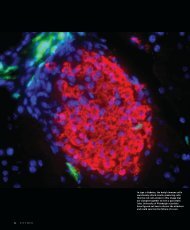
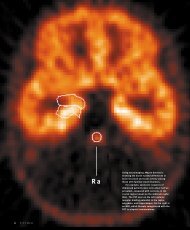
Half <strong>of</strong> patients with polycystic kidney disease progresses quickly to renal failure,<br />
while the other half “is doing fine,” says Bae. Using algorithms on measurements <strong>of</strong><br />
cyst volume, distribution, density, and other characteristics, he has developed a tool to<br />
help doctors determine which path a patient is likely to take.<br />
ing what to do with the massive amount<br />
<strong>of</strong> information that comes with each new<br />
imaging technology. They’re looking for<br />
new imaging biomarkers—visual indicators<br />
<strong>of</strong> diseases—and also evaluating how best to<br />
use the biomarkers that are already known.<br />
For the past 12 years, one focus <strong>of</strong> Bae’s<br />
research has been polycystic kidney disease<br />
(PKD). Characterized by a plague <strong>of</strong> fluid-<br />
filled cysts in the organ, PKD can cause<br />
back pain, high blood pressure, and urinary<br />
tract infections. And, for about half <strong>of</strong> these<br />
patients, the cysts eventually overtake the<br />
organ and cause renal failure. “But the other<br />
half [<strong>of</strong> the patient population] is doing fine,”<br />
says Bae. Right now we have no way to tell at<br />
the onset who’s going to be a rapid progresser,<br />
and who’s going to be okay.<br />
In 1999, the National Institute <strong>of</strong> Diabetes<br />
and Digestive and Kidney Diseases (NIDDK)<br />
formed the Consortium for Radiologic<br />
Imaging Studies <strong>of</strong> Polycystic Kidney Disease<br />
(CRISP), a 10-year prospective study. As chief<br />
radiologist and PI <strong>of</strong> the data center for the<br />
study, Bae has been analyzing MR images<br />
from sites across the country and developing<br />
algorithms that use these images to calculate<br />
the volume, distribution, density, and other<br />
characteristics <strong>of</strong> cysts. He’s finding patterns,<br />
linking various characteristics <strong>of</strong> disease progression<br />
with their outcomes, and developing<br />
a prognostic tool. And in recent years, a<br />
spin<strong>of</strong>f study funded by the NIH has been<br />
putting this new tool to the test. Dubbed<br />
“Halt PKD,” the ongoing trial applies what<br />
has been learned to improving treatments.<br />
Bae is working on similar projects for pulmonary<br />
embolism, emphysema, osteoarthritis,<br />
lung cancer, prostate cancer, breast cancer,<br />
Parkinson’s disease, brain tumor perfusion,<br />
WINTER 2012⁄13 27 xx
COURTESY BAE LAB<br />
Mark Roberts and Bae are finding that not all pulmonary embolism clots are created equal. The<br />
two are developing CT biomarkers to distinguish which patients need less anticoagulant, a potentially<br />
dangerous medication. Here, a pulmonary embolism (left) is completely resolved in the<br />
follow-up scan two months later.<br />
and multiple sclerosis, as well.<br />
Mark Roberts, MD pr<strong>of</strong>essor and chair<br />
<strong>of</strong> the Department <strong>of</strong> Health Policy and<br />
Management in the <strong>University</strong> <strong>of</strong> <strong>Pittsburgh</strong><br />
Graduate School <strong>of</strong> Public Health, calls Bae<br />
forward thinking. “I have been involved in<br />
some other projects back in Boston, where I<br />
came from, where there was this incredible<br />
urgency to find the most accurate test, the<br />
most robust test—not the impact <strong>of</strong> the test on<br />
decisions doctors were making in real patient<br />
care. I personally find that what’s atypical<br />
about Dr. Bae is he’s not just a radiologist;<br />
he’s a doctor first. What he cares about is how<br />
the diagnostic tests he’s using affect patient<br />
outcomes.”<br />
Roberts is collaborating with Bae on a<br />
CT biomarker project involving pulmonary<br />
embolism—blood clots gone rogue.<br />
He explains: When you have an injury,<br />
several processes kick in at roughly the same<br />
28 PITTMED<br />
FOLLOW-UP<br />
COMPLETE RESOLUTION<br />
BASELINE<br />
time. The clotting process, which stops the<br />
bleeding; the repair system, which heals the<br />
vessel; and the clot-dissolution system, which<br />
eats the clot away from the inside out as the<br />
vessel heals so that the normal diameter <strong>of</strong> the<br />
vessel remains intact.<br />
Here’s the problem: The body’s strongest<br />
signal to produce clot is . . . the presence<br />
<strong>of</strong> a clot. And in the case <strong>of</strong> a pulmonary<br />
embolism, unfortunately, the clot didn’t get<br />
there in the first place because it was needed<br />
(because the lung is bleeding); the clot got<br />
there because it formed elsewhere, somehow<br />
became dislodged, and then traveled to this<br />
most precarious spot. Yet there it is, stuck and<br />
sounding alarms—which, in spite <strong>of</strong> their best<br />
intentions, could cause further clotting that<br />
could choke your lung and kill you.<br />
Docs do not take pulmonary embolism<br />
lightly. Right now, an extended course<br />
<strong>of</strong> anticoagulants is recommended for all<br />
cases—though exactly how extended, no<br />
one can agree. Some say six months, others<br />
nine, and still others a full year.<br />
Mind you, anticoagulants don’t dissolve<br />
the clot in your lung. They just allow your<br />
clot-dissolution process to catch up and prevent<br />
further clotting activity. Disabling your<br />
body’s clot production for months on end<br />
comes with its own risks. (You do not want<br />
to cut yourself while you’re on these meds.)<br />
Bae is applying his algorithms to pulmonary-embolism<br />
CTs scanned at sites across<br />
the country. The tool has increased the<br />
team’s ability to predict outcomes by a “nontrivial,”<br />
says Roberts, 10 to 15 percent.<br />
With Pitt’s Donald Yealy, pr<strong>of</strong>essor and<br />
chair <strong>of</strong> emergency medicine, and others,<br />
Bae had submitted a new clinical trial proposal<br />
to the National Heart, Lung, and<br />
Blood Institute; that study will attempt<br />
to answer the nagging question <strong>of</strong> how<br />
long these patients should be given anticoagulants.<br />
The answer, they suspect, is:<br />
It depends. They’re building data stores to<br />
prove that not all clots are created equal;
and, therefore, they do not all require the<br />
same treatment. In fact, the researchers<br />
believe some clots are probably so small they<br />
don’t need treatment at all.<br />
This is one <strong>of</strong> the reasons Bae’s work<br />
is so exciting, Roberts says. “Most <strong>of</strong> the<br />
recommendations we have about diagnostic<br />
testing and treatment are pretty blunt. We<br />
have to understand ways <strong>of</strong> making them<br />
more tailorable, more personalizable. I think<br />
his work can help us do that.”<br />
In doing so, Bae’s research also stands to<br />
help bring down health care costs, improve<br />
reliability and consistency in what happens<br />
with diagnostic imaging, and ultimately help<br />
patients recover faster.<br />
But research is just one <strong>of</strong> the plates Bae<br />
is spinning at any given moment.<br />
This is a guy who has 10 patents to his<br />
name, has secured more than $16 million in<br />
NIH grants in the past 13 years, and still sees<br />
patients and runs a very large department. (In<br />
addition to 220 clinical and research faculty<br />
members, it’s academic home to 57 residents<br />
and 28 fellows at last count.) Bae is an engaged<br />
leader, frequently meeting one-on-one with<br />
mentees and reviewing grant applications and<br />
manuscripts for junior faculty members. In his<br />
relatively short time here, he has overseen the<br />
hiring <strong>of</strong> more than 30 new faculty members.<br />
And in his spare time, just for kicks, he’s<br />
learning Chinese.<br />
None <strong>of</strong> this surprises Narra, who likens<br />
his friend to “some kind <strong>of</strong> movie hero.”<br />
(Apropos <strong>of</strong> nothing, he mentions that he has<br />
seen Bae, who is a lifelong student <strong>of</strong> martial<br />
arts, do thumb push-ups. And one-legged<br />
squats.)<br />
“Ty is one <strong>of</strong> the most brilliant guys I’ve<br />
come across. And he’s not only bright, but he<br />
also has this unending energy and enthusiasm.<br />
It’s amazing. You never hear him say he’s over-<br />
NIGHT SHIFT<br />
They have the graveyard hours. Although, considering their line<br />
<strong>of</strong> work, they prefer to call it the nighthawk shift. Whatever the<br />
name, 10 UPP (<strong>University</strong> <strong>of</strong> <strong>Pittsburgh</strong> Physicians) radiologists<br />
are now staffing a UPMC Emergency and Teleradiology Division<br />
during those hours (5 p.m. to 7 a.m.), when most people have<br />
already called it a day.<br />
The unit started somewhat modestly back in 2008, but<br />
has continued to expand as technology continues to advance.<br />
“Obviously, imaging has evolved and become more complex,<br />
pushing the need for in-house coverage,” says Omar Almusa,<br />
an MD, division chief, and Pitt assistant pr<strong>of</strong>essor <strong>of</strong> radiology.<br />
The need is about having a trained subspecialist (for example,<br />
a neuroradiologist interpreting brain and spinal cord film)<br />
taking an active role in a patient’s trajectory <strong>of</strong> care.<br />
In the not-so-distant past, imaging was considered an adjunct<br />
to patient management at UPMC. But now with the teleradiology<br />
division in place and located on the Presbyterian campus, final<br />
reports are available within an hour (as opposed to 15), and<br />
radiologists are available for consultations in real time.<br />
Almusa sees the radiologist as the doctor’s doctor. A radiologist<br />
might also be thought <strong>of</strong> as the air traffic controller who<br />
assists the ER physician or surgeon in charting the right course.<br />
The concept <strong>of</strong> a 24/7 academic medical center radiolo-<br />
whelmed, or he’s busy. Never! You say, ‘Ty, I<br />
hear you got this [grant application] out,’ and<br />
he says, ‘Yeah, we took care <strong>of</strong> it. There was<br />
a problem, and we got it out, and we got a<br />
paper, and yeah, I got a $5 million grant from<br />
the NIH.’ And it’s all in the past tense!”<br />
Bae, who, after a two-hour interview still<br />
has yet to make use <strong>of</strong> a chair, shrugs it <strong>of</strong>f.<br />
“For me, working hard is no big deal.” He<br />
wonders aloud whether his drive stems from<br />
being one <strong>of</strong> eight kids and always wanting to<br />
distinguish himself by creating something all<br />
his own. Having the chance to do so here in<br />
Bae’s next big problem to solve: Yes, we can see the body in stunning detail, but ...<br />
now what? He and his colleagues are deciphering what to do with the massive<br />
amount <strong>of</strong> information that comes with each new imaging technology.<br />
the States fills him with gratitude. He hopes to<br />
return the favor by realizing his ambitions and<br />
helping others to do the same.<br />
“I appreciate the opportunity,” he says.<br />
“There are a lot <strong>of</strong> people who work in factories<br />
doing the same stuff over and over,<br />
without any additional creative opportunity.<br />
So this is wonderful.” <br />
gy division is not a new one. But, says Almusa, UPP was<br />
an early adopter. These days, its coverage area extends<br />
to UPMC Presbyterian, Shadyside, Magee-Womens, and<br />
Mercy, other UPMC facilities (including its urgent care centers,<br />
Bedford Memorial, Northwest, and Hamot hospitals),<br />
and Trinity Health System in Steubenville, Ohio, as well as<br />
Monongahela Valley Hospital.<br />
Typically, the four overnight radiologists on site will handle<br />
anywhere from 160 to 480 cases; most are related to car<br />
accidents, strokes, and abdominal or chest pains.<br />
“One moment you may have nothing to do,” Almusa says,<br />
“and the next it’s all you can do to keep up.” But he has taken<br />
note <strong>of</strong> predictable patterns <strong>of</strong> behavior, bracing himself for<br />
an onslaught <strong>of</strong> work from 8 p.m. to 2 a.m.; he knows the<br />
summer months will keep him busier than any other time <strong>of</strong><br />
year. (Exceptions are winter days <strong>of</strong> bibulous revelry: New<br />
Year’s Eve, St. Patrick’s Day, and the Super Bowl, especially<br />
if the Steelers are on the field.)<br />
Almusa is looking forward to expanding the division’s<br />
reach to rural areas and its expertise to include more subspecialties<br />
like pediatrics and obstetrics. “It’s very rewarding,”<br />
he says, “to have such an impact on patient care.”<br />
—Barbara Klein<br />
WINTER SPRING 2012⁄13 2007 27 xx 29
![entire issue [pdf 2.79 mb] - Pitt Med - University of Pittsburgh](https://img.yumpu.com/50435398/1/190x231/entire-issue-pdf-279-mb-pitt-med-university-of-pittsburgh.jpg?quality=85)
![entire issue [pdf 6.47 mb] - Pitt Med - University of Pittsburgh](https://img.yumpu.com/50360689/1/190x231/entire-issue-pdf-647-mb-pitt-med-university-of-pittsburgh.jpg?quality=85)
![entire issue [pdf 12.7 mb] - Pitt Med - University of Pittsburgh](https://img.yumpu.com/49831615/1/190x231/entire-issue-pdf-127-mb-pitt-med-university-of-pittsburgh.jpg?quality=85)
![entire issue [pdf 11.3 mb] - Pitt Med - University of Pittsburgh](https://img.yumpu.com/46685830/1/190x231/entire-issue-pdf-113-mb-pitt-med-university-of-pittsburgh.jpg?quality=85)
![entire issue [pdf 12.7 mb] - Pitt Med - University of Pittsburgh](https://img.yumpu.com/44997419/1/190x231/entire-issue-pdf-127-mb-pitt-med-university-of-pittsburgh.jpg?quality=85)