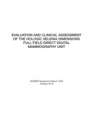
6 | Report<strong>in</strong>g Lesions <strong>in</strong> <strong>the</strong> NHS Bowel Cancer Screen<strong>in</strong>g Programme Figure 4 Examples of tubulovillous adenomas. NHS BCSP September 2007
3.7 Serrated adenomas Report<strong>in</strong>g Lesions <strong>in</strong> <strong>the</strong> NHS Bowel Cancer Screen<strong>in</strong>g Programme | 7 These <strong>lesions</strong> have <strong>the</strong> morphology of hyperplastic polyps, namely a serrated epi<strong>the</strong>lial surface with abundant eos<strong>in</strong>ophilic cytoplasm, but <strong>the</strong>y show def<strong>in</strong>ite dysplasia throughout <strong>the</strong> lesion. 3.8 Mixed hyperplastic/adenomatous polyps These <strong>lesions</strong> have both non-dysplastic hyperplastic type epi<strong>the</strong>lium show<strong>in</strong>g a serrated glandular architecture and areas of adenomatous dysplastic epi<strong>the</strong>lium. The two phenotypes are morphologically dist<strong>in</strong>ct, and, although <strong>the</strong>y may <strong>in</strong>term<strong>in</strong>gle, <strong>in</strong>dividual glandular structures show<strong>in</strong>g both patterns are not present. 3.9 O<strong>the</strong>r types 3.9.1 Inflammatory polyps Experience from NHS BCSP pilot sites has shown that <strong>in</strong>flammatory type polyps are relatively common. Although <strong>the</strong>y are most usually seen as a complication of chronic <strong>in</strong>flammatory <strong>bowel</strong> disease, particularly ulcerative colitis, <strong>the</strong>y are also seen <strong>in</strong> association with diverticulosis, mucosal prolapse and at <strong>the</strong> site of ureterosigmoidostomy. Fur<strong>the</strong>rmore, sporadic s<strong>in</strong>gle <strong>in</strong>flammatory type polyps are well described <strong>in</strong> <strong>the</strong> colorectum. As <strong>the</strong> <strong>report<strong>in</strong>g</strong> pathologist may not know <strong>the</strong> true context of such polyps, we recommend that all such polyps are classified as ‘<strong>in</strong>flammatory polyp’. 3.9.2 Juvenile polyps Classical juvenile polyps are spherical <strong>in</strong> shape, show an excess of lam<strong>in</strong>a propria and have cystically dilated glands. The expanded lam<strong>in</strong>a propria shows oedema and mixed <strong>in</strong>flammatory cells. Experience from <strong>the</strong> NHS BCSP pilot sites suggests that occasional juvenile type polyps are identified, even <strong>in</strong> <strong>the</strong> screen<strong>in</strong>g age group. Juvenile polyps are, of course, most common <strong>in</strong> children, but occasionally examples are seen <strong>in</strong> adults. It rema<strong>in</strong>s uncerta<strong>in</strong> whe<strong>the</strong>r <strong>the</strong> juvenile type polyps identified <strong>in</strong> <strong>the</strong> screen<strong>in</strong>g population are true classical juvenile polyps or whe<strong>the</strong>r <strong>the</strong>y represent <strong>in</strong>flammatory type polyps with mimicry of classical juvenile polyps. We advise that any polyp show<strong>in</strong>g juvenile polyp type features should be classified as a ‘juvenile polyp’ for <strong>the</strong> purposes of diagnostic <strong>report<strong>in</strong>g</strong> <strong>in</strong> <strong>the</strong> NHS BCSP. In classical juvenile polyps, <strong>the</strong>re is often epi<strong>the</strong>lial hyperplasia but dysplasia is very rare, with only a handful of case reports <strong>in</strong> <strong>the</strong> literature. 11 So-called ‘atypical juvenile polyps’ show different morphological features, with a multilobated architecture, <strong>in</strong>tact surface mucosa (usually) and a much more pronounced epi<strong>the</strong>lial component. They are a characteristic feature of juvenile polyposis. It would seem most unlikely, given <strong>the</strong> rarity of juvenile polyposis and <strong>the</strong> age of <strong>the</strong> screen<strong>in</strong>g population, that such polyps might be seen <strong>in</strong> <strong>the</strong> NHS BCSP. Such a polyp should be recorded as represent<strong>in</strong>g ‘juvenile polyp’. They are much more likely to harbour epi<strong>the</strong>lial dysplasia. 12 3.9.3 Peutz–Jeghers polyps Although <strong>the</strong>se polyps are usually seen <strong>in</strong> Peutz–Jeghers syndrome, occasional examples are demonstrated as s<strong>in</strong>gle sporadic polyps <strong>in</strong> <strong>the</strong> colon. There rema<strong>in</strong>s uncerta<strong>in</strong>ty as to whe<strong>the</strong>r ‘<strong>in</strong>flammatory myoglandular polyp’ represents a similar entity. As with juvenile polyposis, it would seem most unlikely, given <strong>the</strong> rarity of <strong>the</strong> syndrome and <strong>the</strong> age of <strong>the</strong> screen<strong>in</strong>g population, that Peutz–Jeghers syndrome would be diagnosed as part of <strong>the</strong> NHS BCSP. Although Peutz–Jeghers polyps are classified as hamartomas, <strong>the</strong>y have a very organised structure. They have a central core of smooth muscle with conspicuous branch<strong>in</strong>g, each branch be<strong>in</strong>g covered by colorectal NHS BCSP September 2007