reporting lesions in the nhs bowel cancer screening programme
reporting lesions in the nhs bowel cancer screening programme
reporting lesions in the nhs bowel cancer screening programme

You also want an ePaper? Increase the reach of your titles
YUMPU automatically turns print PDFs into web optimized ePapers that Google loves.
REPORTING LESIONS IN THE NHS BOWEL<br />
CANCER SCREENING PROGRAMME<br />
Guidel<strong>in</strong>es from <strong>the</strong> Bowel Cancer Screen<strong>in</strong>g Programme Pathology Group<br />
NHS BCSP Publication No 1<br />
September 2007
Published by:<br />
NHS Cancer Screen<strong>in</strong>g Programmes<br />
Fulwood House<br />
Old Fulwood Road<br />
Sheffield<br />
S10 3TH<br />
Tel:0114 271 1060<br />
Fax: 0114 271 1089<br />
Email: <strong>in</strong>fo@<strong>cancer</strong>screen<strong>in</strong>g.<strong>nhs</strong>.uk<br />
Website: www.<strong>cancer</strong>screen<strong>in</strong>g.<strong>nhs</strong>.uk<br />
© NHS Cancer Screen<strong>in</strong>g Programmes 2007<br />
The contents of this document may be copied for use by staff work<strong>in</strong>g <strong>in</strong> <strong>the</strong> public sector<br />
but may not be copied for any o<strong>the</strong>r purpose without prior permission from <strong>the</strong> NHS Cancer<br />
Screen<strong>in</strong>g Programmes.<br />
Fur<strong>the</strong>r copies of this publication are available from <strong>the</strong> Department of Health Publications<br />
Orderl<strong>in</strong>e quot<strong>in</strong>g NHS BCSP Publication No 1.<br />
Tel: 08701 555 455<br />
Fax: 01623 724 524<br />
Email: doh@prolog.uk.com<br />
This publication is also available <strong>in</strong> PDF format on <strong>the</strong> NHS Cancer Screen<strong>in</strong>g Programmes’<br />
website.<br />
Typeset by Prepress Projects Ltd, Perth (www.prepress-projects.co.uk)<br />
Pr<strong>in</strong>ted by Duffield Pr<strong>in</strong>ters
CONTENTS<br />
Report<strong>in</strong>g Lesions <strong>in</strong> <strong>the</strong> NHS Bowel Cancer Screen<strong>in</strong>g Programme | iii<br />
Page No<br />
PREFACE v<br />
1. INTRODUCTION 1<br />
1.1 Background 1<br />
1.2 General issues 1<br />
2. DISSECTION OF SUBMITTED LESIONS 2<br />
3. KEY DIAGNOSTIC FEATURES 3<br />
3.1 Site 3<br />
3.2 Type 3<br />
3.3 Classical adenomas 3<br />
3.4 Villousness 3<br />
3.5 Hyperplastic polyps: serrated adenoma spectrum 5<br />
3.6 Hyperplastic polyps 5<br />
3.7 Serrated adenomas 7<br />
3.8 Mixed hyperplastic/adenomatous polyps 7<br />
3.9 O<strong>the</strong>r types 7<br />
3.10 O<strong>the</strong>r polyps, <strong>in</strong>clud<strong>in</strong>g carc<strong>in</strong>oids and stromal polyps 8<br />
3.11 Shape 8<br />
3.12 Size 8<br />
3.13 Dysplasia 8<br />
3.14 High grade dysplasia 8<br />
4. ADENOCARCINOMA 12<br />
4.1 Def<strong>in</strong>ition of <strong>in</strong>vasion 12<br />
4.2 Epi<strong>the</strong>lial misplacement 12<br />
4.3 Early adenocarc<strong>in</strong>omas (pT1) 12<br />
4.4 Substag<strong>in</strong>g 12<br />
4.5 Tumour grade 14<br />
4.6 Lymphovascular <strong>in</strong>vasion 14<br />
4.7 Marg<strong>in</strong> <strong>in</strong>volvement 14<br />
REFERENCES 15<br />
APPENDIX A: TNM CLASSIFICATION OF COLORECTAL TUMOURS 16<br />
APPENDIX B: SNOMED CODES FOR COLORECTAL TUMOURS 16<br />
NHS BCSP September 2007
iv | Report<strong>in</strong>g Lesions <strong>in</strong> <strong>the</strong> NHS Bowel Cancer Screen<strong>in</strong>g Programme<br />
NHS BCSP September 2007
PREFACE<br />
Report<strong>in</strong>g Lesions <strong>in</strong> <strong>the</strong> NHS Bowel Cancer Screen<strong>in</strong>g Programme | v<br />
These guidel<strong>in</strong>es have been produced by <strong>the</strong> Bowel Cancer Screen<strong>in</strong>g Programme Pathology<br />
Group. Members of <strong>the</strong> panel were:<br />
Professor Frank Carey (Dundee)<br />
Dr Mark Newbold (Coventry)<br />
Professor Phil Quirke (Leeds) 1 (chairman)<br />
Professor Neil Shepherd 2 (Gloucester)<br />
Dr Brian Warren 2,3 (Oxford)<br />
Professor Gera<strong>in</strong>t Williams (Cardiff)<br />
These guidel<strong>in</strong>es have been endorsed by <strong>the</strong> Association of Cl<strong>in</strong>ical Pathologists and <strong>the</strong> Association<br />
of Colproctologists of Great Brita<strong>in</strong> and Nor<strong>the</strong>rn Ireland.<br />
A version of <strong>the</strong>se guidel<strong>in</strong>es with additional examples can also be found at www.virtualpathology.<br />
leeds.ac.uk/nbcs/guidel<strong>in</strong>es.php.<br />
Please send comments/suggestions/feedback to patpq@leeds.ac.uk for consideration for <strong>the</strong> revision<br />
of <strong>the</strong>se guidel<strong>in</strong>es.<br />
1 Royal College of Pathologists<br />
2 British Society of Gastroenterology<br />
3 British Division of <strong>the</strong> International Academy of Pathology<br />
NHS BCSP September 2007
vi | Report<strong>in</strong>g Lesions <strong>in</strong> <strong>the</strong> NHS Bowel Cancer Screen<strong>in</strong>g Programme<br />
NHS BCSP September 2007
Report<strong>in</strong>g Lesions <strong>in</strong> <strong>the</strong> NHS Bowel Cancer Screen<strong>in</strong>g Programme | 1<br />
1. INTRODUCTION<br />
1.1 Background<br />
The NHS Bowel Cancer Screen<strong>in</strong>g Programme (NHS BCSP) will, <strong>in</strong> due course, become <strong>the</strong> largest<br />
<strong>programme</strong> for <strong>bowel</strong> <strong>cancer</strong> screen<strong>in</strong>g <strong>in</strong> <strong>the</strong> world. It offers a unique opportunity to improve<br />
survival <strong>in</strong> this condition as well as clarify<strong>in</strong>g <strong>the</strong> importance of current diagnostic criteria and identify<strong>in</strong>g<br />
<strong>the</strong> biological potential of precursors of colorectal <strong>cancer</strong>.<br />
These guidel<strong>in</strong>es are produced under <strong>the</strong> auspices of <strong>the</strong> NHS BCSP. They have been derived<br />
to answer many of <strong>the</strong> questions that have arisen with<strong>in</strong> <strong>the</strong> pilot screen<strong>in</strong>g centres <strong>in</strong> England<br />
and Scotland, to ensure that key data are collected <strong>in</strong> a consistent manner and to enable fur<strong>the</strong>r<br />
recommendations to be made to provide <strong>the</strong> best possible evidence base for rout<strong>in</strong>e practice. We<br />
welcome feedback and will develop <strong>the</strong>se guidel<strong>in</strong>es as <strong>the</strong> evidence base improves. We are striv<strong>in</strong>g<br />
to ensure consistency across <strong>the</strong> UK and between <strong>the</strong> published recommendations of concerned<br />
professional organisations such as <strong>the</strong> Royal College of Pathologists, <strong>the</strong> British Society of Gastroenterology<br />
and <strong>the</strong> Association of Coloproctology of Great Brita<strong>in</strong> and Ireland. We have also built<br />
on <strong>the</strong> pathology work undertaken dur<strong>in</strong>g <strong>the</strong> Cancer Research UK (CRUK) flexisigmoidoscopy<br />
trial by adopt<strong>in</strong>g many of <strong>the</strong> def<strong>in</strong>itions we developed for that trial. These guidel<strong>in</strong>es are consistent<br />
with <strong>the</strong> dataset produced by <strong>the</strong> Royal College of Pathologists for <strong>report<strong>in</strong>g</strong> colorectal <strong>cancer</strong><br />
(<strong>in</strong>clud<strong>in</strong>g local excision specimens) and will be developed closely with <strong>the</strong>m <strong>in</strong> <strong>the</strong> future.<br />
1.2 General issues<br />
Dysplasia is divided <strong>in</strong>to low and high grade to improve <strong>in</strong>terobserver agreement, with ‘high grade<br />
dysplasia’ equat<strong>in</strong>g to ‘severe dysplasia’ <strong>in</strong> older systems. 1 The term hyperplastic ra<strong>the</strong>r than metaplastic<br />
polyp is recommended; nei<strong>the</strong>r is a good name, but add<strong>in</strong>g a third only confuses matters<br />
fur<strong>the</strong>r. The reasons for recommend<strong>in</strong>g <strong>the</strong> term hyperplastic are that, firstly, it has been used <strong>in</strong><br />
both pilot centres; secondly, true metaplasia (eg squamous islands) can rarely occur <strong>in</strong> dysplastic<br />
adenomas; and, thirdly, <strong>the</strong> term metaplastic is def<strong>in</strong>ed as a change <strong>in</strong> epi<strong>the</strong>lial type from one<br />
mature epi<strong>the</strong>lial type to ano<strong>the</strong>r. Although <strong>the</strong> epi<strong>the</strong>lium of a hyperplastic lesion is abnormal, it is<br />
not of a different epi<strong>the</strong>lial type. Different antigenic patterns have been demonstrated <strong>in</strong> hyperplastic<br />
polyps, but are not those of ano<strong>the</strong>r mature epi<strong>the</strong>lial type. Polyps have been broadly subclassified<br />
<strong>in</strong>to classical, hyperplastic serrated spectrum and o<strong>the</strong>r types of lesion. We have concentrated on<br />
early <strong>in</strong>vasive <strong>lesions</strong> as <strong>the</strong>se have proved challeng<strong>in</strong>g with<strong>in</strong> <strong>the</strong> pilot screen<strong>in</strong>g centres, <strong>the</strong> evidence<br />
base is currently poor and <strong>the</strong> national screen<strong>in</strong>g <strong>programme</strong> will generate many of <strong>the</strong>se<br />
difficult <strong>lesions</strong>. We have also sought to identify <strong>the</strong> serrated spectrum to allow fur<strong>the</strong>r <strong>in</strong>vestigation<br />
<strong>in</strong> this area.<br />
The target for histopathology <strong>report<strong>in</strong>g</strong> is that 90% of <strong>lesions</strong> should be reported with<strong>in</strong> 7 days. This<br />
will allow patients who have had a polyp removed at colonoscopy to be given an appo<strong>in</strong>tment to<br />
be seen <strong>the</strong> follow<strong>in</strong>g week <strong>in</strong> <strong>the</strong> follow-up cl<strong>in</strong>ic.<br />
Pathologists must complete ei<strong>the</strong>r <strong>the</strong> screen<strong>in</strong>g <strong>programme</strong> proforma or its computerised version.<br />
These are to be returned to <strong>the</strong> screen<strong>in</strong>g centre adm<strong>in</strong>istrator for pathology data to be entered<br />
onto <strong>the</strong> <strong>bowel</strong> <strong>cancer</strong> screen<strong>in</strong>g system (BCSS). Pathologists may also wish to provide a free text<br />
report directly to <strong>the</strong> cl<strong>in</strong>ician.<br />
A copy of <strong>the</strong> latest version of <strong>the</strong> proforma can be found at www.virtualpathology.leeds.ac.uk/<br />
nbcs/nbcs.php.<br />
NHS BCSP September 2007
2 | Report<strong>in</strong>g Lesions <strong>in</strong> <strong>the</strong> NHS Bowel Cancer Screen<strong>in</strong>g Programme<br />
2. DISSECTION OF SUBMITTED LESIONS<br />
The material received will be ei<strong>the</strong>r a biopsy of a lesion, an excision of a polyp or a submucosal<br />
resection of a sessile lesion or a larger resection that is ei<strong>the</strong>r a transendoscopic mucosal excision<br />
(TEM) or a full surgical excision. If only a biopsy is received, <strong>the</strong> size of <strong>the</strong> lesion and completeness<br />
of excision will not be assessable by <strong>the</strong> pathologist and <strong>the</strong>se should be recorded as not<br />
assessable (n/a).<br />
Although <strong>the</strong> pr<strong>in</strong>ciples of pathological <strong>report<strong>in</strong>g</strong> are <strong>the</strong> same as <strong>in</strong> major resections, a number<br />
of features require special attention <strong>in</strong> local excisions of (presumed) early <strong>cancer</strong>s with curative<br />
<strong>in</strong>tent because <strong>the</strong>y are used to determ<strong>in</strong>e <strong>the</strong> necessity for more radical surgery. In addition to<br />
<strong>the</strong> assessment of completeness of excision, <strong>the</strong>se <strong>in</strong>clude record<strong>in</strong>g parameters that predict <strong>the</strong><br />
presence of lymph node metastasis <strong>in</strong> early tumours, namely tumour size, poor differentiation, <strong>the</strong><br />
depth of <strong>in</strong>vasion <strong>in</strong>to <strong>the</strong> submucosa and <strong>the</strong> presence of submucosal lymphovascular <strong>in</strong>vasion. 2–7<br />
However, <strong>the</strong>re is only limited evidence and no consensus <strong>in</strong> <strong>the</strong> published literature on exactly<br />
how some <strong>the</strong>se parameters should be assessed, especially <strong>the</strong> depth of submucosal <strong>in</strong>vasion. We<br />
hope to improve this situation from data derived from <strong>the</strong> <strong>bowel</strong> <strong>cancer</strong> screen<strong>in</strong>g <strong>programme</strong>.<br />
Local excisions are undertaken endoscopically or, <strong>in</strong> <strong>the</strong> case of early rectal tumours, under direct<br />
vision. The majority of carc<strong>in</strong>omas arise with<strong>in</strong> pre-exist<strong>in</strong>g adenomas that may be polypoid, sessile<br />
or flat, and <strong>the</strong> best pathological <strong>in</strong>formation is derived when <strong>lesions</strong> are excised <strong>in</strong> <strong>the</strong>ir entirety<br />
to <strong>in</strong>clude both <strong>the</strong> <strong>in</strong>vasive and pre<strong>in</strong>vasive components. 8 Polypoid <strong>lesions</strong> on a narrow stalk can<br />
be fixed <strong>in</strong>tact, whereas sessile <strong>lesions</strong> should be p<strong>in</strong>ned out, mucosal surface upwards, on a<br />
small piece of cork or o<strong>the</strong>r suitable material, tak<strong>in</strong>g pa<strong>in</strong>s to identify <strong>the</strong> narrow rim of surround<strong>in</strong>g<br />
normal tissue before fix<strong>in</strong>g <strong>in</strong>tact. Piecemeal removal of tumours, which is entirely acceptable for<br />
palliative resections, should be avoided because it precludes a reliable assessment of completeness<br />
of excision.<br />
After fixation, polypoid <strong>lesions</strong> may be bisected through <strong>the</strong> stalk if <strong>the</strong>y measure < 10 mm; larger<br />
polyps are trimmed to leave a central section conta<strong>in</strong><strong>in</strong>g <strong>the</strong> <strong>in</strong>tact stalk, and all fragments are<br />
embedded for histology. It is recommended that at least three sections are taken from blocks<br />
conta<strong>in</strong><strong>in</strong>g <strong>the</strong> stalk. The marg<strong>in</strong>s of larger, sessile <strong>lesions</strong> should be identified with appropriate<br />
coloured markers (<strong>in</strong>ks or gelat<strong>in</strong>e), and <strong>the</strong> whole of <strong>the</strong> specimen transversely sectioned <strong>in</strong>to<br />
3 mm slices and submitted for histology <strong>in</strong> sequentially labelled cassettes. When <strong>the</strong> marg<strong>in</strong> of<br />
normal tissue is less than 3 mm, a 10 mm slice conta<strong>in</strong><strong>in</strong>g <strong>the</strong> relevant marg<strong>in</strong> should be made and<br />
fur<strong>the</strong>r sectioned at right angles.<br />
NHS BCSP September 2007
Report<strong>in</strong>g Lesions <strong>in</strong> <strong>the</strong> NHS Bowel Cancer Screen<strong>in</strong>g Programme | 3<br />
3. KEY DIAGNOSTIC FEATURES<br />
3.1 Site<br />
The site of orig<strong>in</strong> of each specimen should be <strong>in</strong>dividually identified by <strong>the</strong> cl<strong>in</strong>ician and provided to<br />
<strong>the</strong> pathologist on <strong>the</strong> request form. The pathologist should record this on <strong>the</strong> pathology proforma.<br />
This is important <strong>in</strong>formation because <strong>the</strong> risk of lymph node metastases from a T1 adenocarc<strong>in</strong>oma<br />
varies depend<strong>in</strong>g on <strong>the</strong> site of <strong>the</strong> lesion. 7<br />
3.2 Type<br />
Three broad groups of lesion are reported, namely classical adenomas, serrated <strong>lesions</strong> and o<strong>the</strong>r<br />
polyps. Classical adenomas are divided <strong>in</strong>to tubular, tubulovillous or villous types. The spectrum<br />
of <strong>lesions</strong> with a serrated growth pattern is subdivided <strong>in</strong>to hyperplastic polyps, mixed dysplastic/<br />
hyperplastic adenomas and serrated adenomas. O<strong>the</strong>r types of polyp <strong>in</strong>clude <strong>in</strong>flammatory polyps,<br />
juvenile polyps, Peutz–Jeghers or o<strong>the</strong>r types which are <strong>in</strong>cluded under not o<strong>the</strong>rwise specified<br />
and described <strong>in</strong> free text.<br />
3.3 Classical adenomas<br />
By def<strong>in</strong>ition, adenomas must show dysplasia. They can be of tubular, tubulovillous or villous types,<br />
and demarcation between <strong>the</strong> three is based on <strong>the</strong> relative proportions of tubular and villous<br />
components accord<strong>in</strong>g to <strong>the</strong> ‘20% rule’ described <strong>in</strong> <strong>the</strong> WHO classification. 8 At least 20% of <strong>the</strong><br />
estimated volume of an adenoma should be villous to classify a polyp as tubulovillous, and 80%<br />
villous to be def<strong>in</strong>ed as a villous adenoma. All o<strong>the</strong>r <strong>lesions</strong> are classified as tubular.<br />
The 20% rule applies only to wholly excised polyps and to <strong>in</strong>tact sections of those <strong>lesions</strong> that are<br />
large enough to provide reliable proportions. For small fragmented <strong>lesions</strong> or superficial polyp<br />
biopsies, <strong>the</strong> presence of at least one clearly identifiable villus merits classification as ‘at least<br />
tubulovillous’.<br />
3.4 Villousness<br />
Although it is accepted that a neoplastic villus almost defies def<strong>in</strong>ition, <strong>the</strong> follow<strong>in</strong>g descriptions<br />
have been developed to help recognize ‘villousness’.<br />
Villous structures may take different forms that, <strong>in</strong> <strong>the</strong>mselves, are not known to have any particular<br />
significance but may assist observer reproducibility. These <strong>in</strong>clude:<br />
• ‘Classical’ villi, which are composed of long, slender, upgrowths of epi<strong>the</strong>lium on a th<strong>in</strong> delicate<br />
stromal core that branches little. These usually have parallel sides (although sometimes a<br />
bulbous tip), and often appear to extend right down to <strong>the</strong> muscularis mucosae.<br />
• ‘Palmate’ villi, which are composed of clustered, broader, branch<strong>in</strong>g, leaf-like structures with<br />
a hand-like configuration. Tubular glands may be present at <strong>the</strong> base of <strong>the</strong>se structures, and<br />
sometimes may even be present with<strong>in</strong> <strong>the</strong> stromal core of villi.<br />
• ‘Foreshortened’ villi, which are composed of slender non-branch<strong>in</strong>g outgrowths with a th<strong>in</strong><br />
stromal core that clearly protrude beyond <strong>the</strong> overall surface contour of an o<strong>the</strong>rwise well<br />
developed tubular lesion.<br />
NHS BCSP September 2007
4 | Report<strong>in</strong>g Lesions <strong>in</strong> <strong>the</strong> NHS Bowel Cancer Screen<strong>in</strong>g Programme<br />
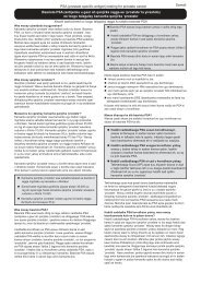
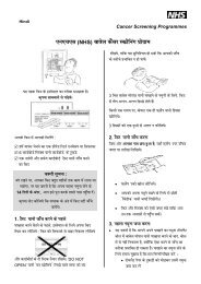
Figure 1 A fragmented biopsy show<strong>in</strong>g a villous component.<br />
Figure 2 An example of classical villi, which should provide little difficulty to a pathologist. The bulbous tip<br />
that can be seen is well demonstrated.<br />
NHS BCSP September 2007
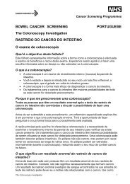
Figure 3 An example of palmate villi.<br />
Report<strong>in</strong>g Lesions <strong>in</strong> <strong>the</strong> NHS Bowel Cancer Screen<strong>in</strong>g Programme | 5<br />
Most diagnostic difficulties will arise with foreshortened villi, particularly <strong>in</strong> dist<strong>in</strong>guish<strong>in</strong>g ‘true’ villi<br />
from exaggerated, axially sectioned, neoplastic crypts with distended lum<strong>in</strong>al open<strong>in</strong>gs. In <strong>the</strong>se situations,<br />
it is better to err on <strong>the</strong> side of underdiagnosis of villous change, especially <strong>in</strong> small (< 1 cm)<br />
adenomas, and to restrict <strong>the</strong> term to <strong>lesions</strong> only for those with conv<strong>in</strong>c<strong>in</strong>g outgrowths.<br />
Villous structures with low grade dysplasia not <strong>in</strong>frequently show a characteristic quality to <strong>the</strong> epi<strong>the</strong>lium,<br />
with rows of regular tall columnar cells with large conspicuous apical muc<strong>in</strong> vacuoles that<br />
are rem<strong>in</strong>iscent of <strong>the</strong> surface epi<strong>the</strong>lium of <strong>the</strong> stomach. This is <strong>in</strong> contrast to <strong>the</strong> typical goblet<br />
cells <strong>in</strong>terspersed with eos<strong>in</strong>ophilic colonocytes conta<strong>in</strong><strong>in</strong>g sparse or no obvious muc<strong>in</strong> that are<br />
characteristic of tubular adenomas. The presence of such a ‘muc<strong>in</strong>ous’ epi<strong>the</strong>lium should sway <strong>the</strong><br />
diagnosis <strong>in</strong> favour of villous histology if it is conspicuous <strong>in</strong> an o<strong>the</strong>rwise <strong>in</strong>determ<strong>in</strong>ate lesion.<br />
3.5 Hyperplastic polyps: serrated adenoma spectrum<br />
The pathology of serrated <strong>lesions</strong> is currently an active area of research, and <strong>the</strong>re is a limited evidence<br />
base for <strong>the</strong> relative importance of some of <strong>the</strong> phenotypes that have been described <strong>in</strong> <strong>the</strong><br />
literature. 9,10 In light of this, <strong>the</strong> current guidel<strong>in</strong>es identify practical categories that may be studied<br />
fur<strong>the</strong>r and subcategorised <strong>in</strong> future when more evidence is available. In <strong>the</strong> spectrum are nondysplastic<br />
hyperplastic polyps, dysplastic <strong>lesions</strong> with a serrated architecture, called here serrated<br />
adenomas, and mixed hyperplastic/adenomatous polyps.<br />
3.6 Hyperplastic polyps<br />
The architecture of <strong>the</strong> glands can vary from normal to grossly distorted. Usually, <strong>in</strong> <strong>the</strong> case of<br />
hyperplastic polyps, <strong>the</strong> glands demonstrate elongated crypts with an excess of columnar absorptive<br />
cells lead<strong>in</strong>g to a tufted, crenated appearance towards <strong>the</strong> surface. A variable degree of epi<strong>the</strong>lial<br />
proliferation can be seen <strong>in</strong> <strong>the</strong> base of <strong>the</strong> crypts. The cells are cytologically regular with<br />
no dysplasia present.<br />
NHS BCSP September 2007
6 | Report<strong>in</strong>g Lesions <strong>in</strong> <strong>the</strong> NHS Bowel Cancer Screen<strong>in</strong>g Programme<br />
Figure 4 Examples of tubulovillous adenomas.<br />
NHS BCSP September 2007
3.7 Serrated adenomas<br />
Report<strong>in</strong>g Lesions <strong>in</strong> <strong>the</strong> NHS Bowel Cancer Screen<strong>in</strong>g Programme | 7<br />
These <strong>lesions</strong> have <strong>the</strong> morphology of hyperplastic polyps, namely a serrated epi<strong>the</strong>lial surface<br />
with abundant eos<strong>in</strong>ophilic cytoplasm, but <strong>the</strong>y show def<strong>in</strong>ite dysplasia throughout <strong>the</strong> lesion.<br />
3.8 Mixed hyperplastic/adenomatous polyps<br />
These <strong>lesions</strong> have both non-dysplastic hyperplastic type epi<strong>the</strong>lium show<strong>in</strong>g a serrated glandular<br />
architecture and areas of adenomatous dysplastic epi<strong>the</strong>lium. The two phenotypes are morphologically<br />
dist<strong>in</strong>ct, and, although <strong>the</strong>y may <strong>in</strong>term<strong>in</strong>gle, <strong>in</strong>dividual glandular structures show<strong>in</strong>g both<br />
patterns are not present.<br />
3.9 O<strong>the</strong>r types<br />
3.9.1 Inflammatory polyps<br />
Experience from NHS BCSP pilot sites has shown that <strong>in</strong>flammatory type polyps are relatively<br />
common. Although <strong>the</strong>y are most usually seen as a complication of chronic <strong>in</strong>flammatory <strong>bowel</strong><br />
disease, particularly ulcerative colitis, <strong>the</strong>y are also seen <strong>in</strong> association with diverticulosis, mucosal<br />
prolapse and at <strong>the</strong> site of ureterosigmoidostomy. Fur<strong>the</strong>rmore, sporadic s<strong>in</strong>gle <strong>in</strong>flammatory type<br />
polyps are well described <strong>in</strong> <strong>the</strong> colorectum. As <strong>the</strong> <strong>report<strong>in</strong>g</strong> pathologist may not know <strong>the</strong> true context<br />
of such polyps, we recommend that all such polyps are classified as ‘<strong>in</strong>flammatory polyp’.<br />
3.9.2 Juvenile polyps<br />
Classical juvenile polyps are spherical <strong>in</strong> shape, show an excess of lam<strong>in</strong>a propria and have cystically<br />
dilated glands. The expanded lam<strong>in</strong>a propria shows oedema and mixed <strong>in</strong>flammatory cells.<br />
Experience from <strong>the</strong> NHS BCSP pilot sites suggests that occasional juvenile type polyps are identified,<br />
even <strong>in</strong> <strong>the</strong> screen<strong>in</strong>g age group. Juvenile polyps are, of course, most common <strong>in</strong> children,<br />
but occasionally examples are seen <strong>in</strong> adults. It rema<strong>in</strong>s uncerta<strong>in</strong> whe<strong>the</strong>r <strong>the</strong> juvenile type polyps<br />
identified <strong>in</strong> <strong>the</strong> screen<strong>in</strong>g population are true classical juvenile polyps or whe<strong>the</strong>r <strong>the</strong>y represent<br />
<strong>in</strong>flammatory type polyps with mimicry of classical juvenile polyps. We advise that any polyp show<strong>in</strong>g<br />
juvenile polyp type features should be classified as a ‘juvenile polyp’ for <strong>the</strong> purposes of diagnostic<br />
<strong>report<strong>in</strong>g</strong> <strong>in</strong> <strong>the</strong> NHS BCSP. In classical juvenile polyps, <strong>the</strong>re is often epi<strong>the</strong>lial hyperplasia but<br />
dysplasia is very rare, with only a handful of case reports <strong>in</strong> <strong>the</strong> literature. 11<br />
So-called ‘atypical juvenile polyps’ show different morphological features, with a multilobated<br />
architecture, <strong>in</strong>tact surface mucosa (usually) and a much more pronounced epi<strong>the</strong>lial component.<br />
They are a characteristic feature of juvenile polyposis. It would seem most unlikely, given <strong>the</strong> rarity<br />
of juvenile polyposis and <strong>the</strong> age of <strong>the</strong> screen<strong>in</strong>g population, that such polyps might be seen <strong>in</strong><br />
<strong>the</strong> NHS BCSP. Such a polyp should be recorded as represent<strong>in</strong>g ‘juvenile polyp’. They are much<br />
more likely to harbour epi<strong>the</strong>lial dysplasia. 12<br />
3.9.3 Peutz–Jeghers polyps<br />
Although <strong>the</strong>se polyps are usually seen <strong>in</strong> Peutz–Jeghers syndrome, occasional examples are<br />
demonstrated as s<strong>in</strong>gle sporadic polyps <strong>in</strong> <strong>the</strong> colon. There rema<strong>in</strong>s uncerta<strong>in</strong>ty as to whe<strong>the</strong>r<br />
‘<strong>in</strong>flammatory myoglandular polyp’ represents a similar entity. As with juvenile polyposis, it would<br />
seem most unlikely, given <strong>the</strong> rarity of <strong>the</strong> syndrome and <strong>the</strong> age of <strong>the</strong> screen<strong>in</strong>g population, that<br />
Peutz–Jeghers syndrome would be diagnosed as part of <strong>the</strong> NHS BCSP. Although Peutz–Jeghers<br />
polyps are classified as hamartomas, <strong>the</strong>y have a very organised structure. They have a central<br />
core of smooth muscle with conspicuous branch<strong>in</strong>g, each branch be<strong>in</strong>g covered by colorectal<br />
NHS BCSP September 2007
8 | Report<strong>in</strong>g Lesions <strong>in</strong> <strong>the</strong> NHS Bowel Cancer Screen<strong>in</strong>g Programme<br />
type mucosa that appears hyperplastic but not dysplastic. As with sporadic juvenile polyps, solitary<br />
Peutz–Jeghers type polyps are most unlikely to demonstrate foci of dysplasia.<br />
3.9.4 Cronkhite–Canada syndrome<br />
We believe that it is most unlikely such cases will present via <strong>the</strong> NHS BCSP, and <strong>the</strong> true diagnosis<br />
may not be recognised by pathological assessment. Such polyps are probably best regarded as<br />
be<strong>in</strong>g of <strong>in</strong>flammatory type.<br />
3.10 O<strong>the</strong>r polyps, <strong>in</strong>clud<strong>in</strong>g carc<strong>in</strong>oids and stromal polyps<br />
Small rectal mucosal nodules, show<strong>in</strong>g <strong>the</strong> characteristic features of h<strong>in</strong>d gut carc<strong>in</strong>oids, are not<br />
uncommon. For pathological <strong>report<strong>in</strong>g</strong> as part of <strong>the</strong> NHS BCSP, we recommend that <strong>the</strong>se are<br />
recorded as ‘o<strong>the</strong>r polyp’ and that <strong>the</strong>ir true nature is recorded <strong>in</strong> free text. Fur<strong>the</strong>rmore, <strong>the</strong>re are<br />
a number of stromal tumours that can also present as polyps. Lipomas and leiomyomas of <strong>the</strong><br />
muscularis mucosae are probably <strong>the</strong> most likely to be seen <strong>in</strong> <strong>the</strong> NHSBCSP, and we recommend<br />
that <strong>the</strong>se are recorded as ‘o<strong>the</strong>r polyp’ and that <strong>the</strong>ir true nature is recorded <strong>in</strong> free text. North<br />
American experience with <strong>bowel</strong> <strong>cancer</strong> screen<strong>in</strong>g <strong>in</strong>dicates that, rarely, o<strong>the</strong>r unusual forms of<br />
stromal tumour can present as polypoid nodules <strong>in</strong> <strong>the</strong> screen<strong>in</strong>g <strong>programme</strong>. Such stromal <strong>lesions</strong><br />
<strong>in</strong>clude ganglioneuroma, neurofibroma, gastro<strong>in</strong>test<strong>in</strong>al stromal tumour (GIST), various forms of<br />
vascular tumour, per<strong>in</strong>eurioma, fibroblastic polyp and epi<strong>the</strong>lioid nerve sheath tumour.<br />
3.11 Shape<br />
The NHS BCSP is not designed to detect flat adenomas because it does not mandate magnify<strong>in</strong>g<br />
endoscopy or chromoendoscopy. Flat adenomas have not been separately identified at this stage,<br />
but this area will be revisited <strong>in</strong> <strong>the</strong> future when more experience has been ga<strong>in</strong>ed. The pilot sites<br />
saw <strong>the</strong>se <strong>lesions</strong> only rarely <strong>in</strong> <strong>the</strong>ir material.<br />
3.12 Size<br />
An accurate measurement is very important and must be to <strong>the</strong> nearest millimetre (and not ‘rounded<br />
up’ to <strong>the</strong> nearest 5 or 10 mm). This will be audited. If possible, <strong>the</strong> maximum size of <strong>the</strong> lesion<br />
should be measured from <strong>the</strong> formal<strong>in</strong> fixed macroscopic specimen. For small <strong>lesions</strong> (5 mm or<br />
less) that fit on one section <strong>in</strong> <strong>the</strong>ir entirety, it is acceptable to measure <strong>the</strong>ir dimensions from <strong>the</strong><br />
glass slide. If a biopsy is received, <strong>the</strong>n n/a should be entered <strong>in</strong> <strong>the</strong> size box.<br />
3.13 Dysplasia<br />
We recommend that high grade dysplasia and low grade dysplasia are used <strong>in</strong>stead of mild, moderate<br />
and severe dysplasia. This will <strong>in</strong>crease <strong>the</strong> <strong>in</strong>terobserver agreement and allow pathologists<br />
to concentrate on <strong>the</strong> important diagnostic criteria.<br />
3.14 High grade dysplasia<br />
The changes of high grade dysplasia should usually <strong>in</strong>volve more than just one or two glands<br />
(except <strong>in</strong> t<strong>in</strong>y biopsies of polyps), sufficient to be identified at low power exam<strong>in</strong>ation. Caution<br />
should be exercised <strong>in</strong> over<strong>in</strong>terpret<strong>in</strong>g isolated surface changes that may be due to trauma, erosion<br />
or prolapse.<br />
High grade dysplasia is diagnosed on architecture, supplemented by an appropriate cytology.<br />
Hence, its presence is nearly always suspected by <strong>the</strong> appearance under low power of complex<br />
NHS BCSP September 2007
Report<strong>in</strong>g Lesions <strong>in</strong> <strong>the</strong> NHS Bowel Cancer Screen<strong>in</strong>g Programme | 9<br />
architectural abnormalities <strong>in</strong> structures whose epi<strong>the</strong>lium looks thick, blue, disorganised and ‘dirty’.<br />
The architectural features are:<br />
• complex glandular crowd<strong>in</strong>g and irregularity (note that <strong>the</strong> word ‘complex’ is important and<br />
excludes simple crowd<strong>in</strong>g of regular tubules that might result from crush<strong>in</strong>g)<br />
• prom<strong>in</strong>ent budd<strong>in</strong>g (note that <strong>the</strong> word ‘prom<strong>in</strong>ent’ is important; <strong>the</strong>re is probably some degree<br />
of glandular budd<strong>in</strong>g, by def<strong>in</strong>ition, <strong>in</strong> all tubular adenomas)<br />
• a cribriform appearance and ‘back to back’ glands<br />
• prom<strong>in</strong>ent <strong>in</strong>tralum<strong>in</strong>al papillary tuft<strong>in</strong>g.<br />
A<br />
B<br />
Figure 5 (A) High grade dysplasia show<strong>in</strong>g architectural changes. (B) Ano<strong>the</strong>r area of high grade<br />
dysplasia from <strong>the</strong> same case show<strong>in</strong>g a lesser degree of architectural abnormalities.<br />
NHS BCSP September 2007
10 | Report<strong>in</strong>g Lesions <strong>in</strong> <strong>the</strong> NHS Bowel Cancer Screen<strong>in</strong>g Programme<br />
Figure 6 (A) and (B) The changes shown here are not those of high grade but are of low grade dysplasia<br />
because of <strong>the</strong> architectural changes. The cytology is less worry<strong>in</strong>g.<br />
NHS BCSP September 2007
Report<strong>in</strong>g Lesions <strong>in</strong> <strong>the</strong> NHS Bowel Cancer Screen<strong>in</strong>g Programme | 11<br />
Although many of <strong>the</strong>se features often coexist <strong>in</strong> high grade dysplasia, <strong>in</strong>dividually <strong>the</strong>y are nei<strong>the</strong>r<br />
necessary nor usually sufficient. Indeed, <strong>the</strong>y may occasionally occur <strong>in</strong> lower grades of dysplasia,<br />
which is why it is necessary to go on to scrut<strong>in</strong>ise <strong>the</strong> cytological features for signs of high grade<br />
dysplasia. The cytological features are:<br />
• loss of cell polarity or nuclear stratification to <strong>the</strong> extent that <strong>the</strong> nuclei are approximately equally,<br />
though haphazardly, distributed with<strong>in</strong> all three thirds of <strong>the</strong> height of <strong>the</strong> epi<strong>the</strong>lium<br />
• markedly enlarged nuclei, often with a dispersed chromat<strong>in</strong> pattern and a prom<strong>in</strong>ent<br />
nucleolus<br />
• atypical mitotic figures<br />
• prom<strong>in</strong>ent apoptosis, giv<strong>in</strong>g <strong>the</strong> lesional epi<strong>the</strong>lium a ‘dirty’ appearance.<br />
Aga<strong>in</strong>, <strong>the</strong>se features usually coexist <strong>in</strong> high grade dysplasia, and caution must be exercised <strong>in</strong><br />
us<strong>in</strong>g just one. It should be emphasised aga<strong>in</strong> that <strong>the</strong>y should occur <strong>in</strong> a background of complex<br />
architectural abnormality. Marked loss of polarity and nuclear stratification sometimes occurs on<br />
<strong>the</strong> surface of small, architecturally regular, tubular adenomas that o<strong>the</strong>rwise have a lower grade of<br />
dysplasia, probably as a result of trauma, and must not be used to classify a lesion as high grade.<br />
The only exception to <strong>the</strong> rule is when <strong>the</strong> specimen consists of just a small biopsy from a polyp, ie<br />
when <strong>the</strong>re is <strong>in</strong>sufficient tissue to assess <strong>the</strong> architecture properly. In this situation, it is permissible<br />
to label florid cytological abnormalities alone as high grade dysplasia, but this will usually lead to<br />
re-excision of <strong>the</strong> whole polyp, when it will be possible to assess <strong>the</strong> whole lesion properly.<br />
NHS BCSP September 2007
12 | Report<strong>in</strong>g Lesions <strong>in</strong> <strong>the</strong> NHS Bowel Cancer Screen<strong>in</strong>g Programme<br />
4. ADENOCARCINOMA<br />
4.1 Def<strong>in</strong>ition of <strong>in</strong>vasion<br />
The recommended def<strong>in</strong>ition of an adenocarc<strong>in</strong>oma is <strong>the</strong> one that is <strong>in</strong> everyday use with<strong>in</strong> <strong>the</strong> UK<br />
of <strong>in</strong>vasion of neoplastic cells through <strong>the</strong> muscularis mucosae <strong>in</strong>to <strong>the</strong> submucosa of <strong>the</strong> <strong>bowel</strong><br />
wall.<br />
This def<strong>in</strong>ition does not allow for <strong>the</strong> diagnosis of <strong>in</strong>tramucosal carc<strong>in</strong>oma, and such cases should<br />
be considered to be high grade dysplasia. Also, <strong>the</strong> def<strong>in</strong>ition does not allow comparison with<br />
Japanese series, <strong>in</strong> which a diagnosis of carc<strong>in</strong>oma can be made on cases of high grade dysplasia<br />
without <strong>in</strong>vasion, but it is compatible with US and European literature.<br />
The TNM classification of colorectal tumours is given <strong>in</strong> Appendix 1, and <strong>the</strong> relevant SNOMED<br />
codes are given <strong>in</strong> Appendix 2.<br />
4.2 Epi<strong>the</strong>lial misplacement<br />
Epi<strong>the</strong>lial misplacement of adenomatous epi<strong>the</strong>lium <strong>in</strong>to <strong>the</strong> submucosa of a polyp is a well recognised<br />
phenomenon. 13 It is commonly seen <strong>in</strong> prolaps<strong>in</strong>g polyps <strong>in</strong> <strong>the</strong> sigmoid colon. Experience<br />
from <strong>the</strong> pilot sites suggests that this will be one of <strong>the</strong> most difficult areas of pathological diagnostic<br />
practice <strong>in</strong> <strong>the</strong> NHS BCSP. These sigmoid colonic polyps are particularly prone to <strong>in</strong>flammation and<br />
ulceration, features which tend to enhance <strong>the</strong> dysplastic changes present. When associated with<br />
epi<strong>the</strong>lial misplacement, <strong>the</strong> potential for misdiagnosis of early carc<strong>in</strong>oma and <strong>the</strong> overall diagnostic<br />
difficulties become much greater.<br />
4.3 Early adenocarc<strong>in</strong>omas (pT1)<br />
Tumours that <strong>in</strong>vade <strong>the</strong> muscularis propria (pT2) usually require fur<strong>the</strong>r surgery and should be<br />
staged accord<strong>in</strong>g to <strong>the</strong> <strong>cancer</strong> m<strong>in</strong>imum dataset.<br />
pT1 tumours will provide many difficulties <strong>in</strong> <strong>the</strong> <strong>programme</strong>, and <strong>the</strong> current evidence base for<br />
<strong>the</strong>ir management is poor. Thus as a priority we have chosen to concentrate on generat<strong>in</strong>g a firm<br />
evidence base for management. This will require a limited number of extra assessments that we will<br />
<strong>the</strong>n ref<strong>in</strong>e on <strong>the</strong> basis of <strong>the</strong> data emerg<strong>in</strong>g from <strong>the</strong> pilot sites and <strong>the</strong> <strong>programme</strong>. In particular,<br />
substag<strong>in</strong>g and differentiation grad<strong>in</strong>g are addressed.<br />
4.4 Substag<strong>in</strong>g<br />
In pT1 tumours, <strong>the</strong> frequency of lymph node metastasis <strong>in</strong> sessile tumours that <strong>in</strong>volve <strong>the</strong><br />
superficial, middle and deep thirds of <strong>the</strong> submucosa (so-called Kikuchi levels sm1, sm2 and sm3<br />
respectively 6 ) has been reported to be 2%, 8% and 23% respectively. 7<br />
In polypoid <strong>lesions</strong>, Haggitt et al 5 identified <strong>the</strong> level of <strong>in</strong>vasion <strong>in</strong>to <strong>the</strong> stalk of <strong>the</strong> polyp as<br />
be<strong>in</strong>g important <strong>in</strong> predict<strong>in</strong>g outcome and found that ‘level 4’ <strong>in</strong>vasion, <strong>in</strong> which tumour extended<br />
beyond <strong>the</strong> stalk of <strong>the</strong> polyp <strong>in</strong>to <strong>the</strong> submucosa but did not <strong>in</strong>vade <strong>the</strong> muscularis propria, was<br />
an adverse factor.<br />
NHS BCSP September 2007
Report<strong>in</strong>g Lesions <strong>in</strong> <strong>the</strong> NHS Bowel Cancer Screen<strong>in</strong>g Programme | 13<br />
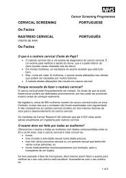
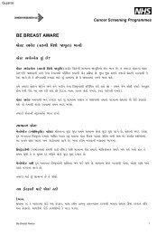
Figure 7A Kikuchi levels of submucosal <strong>in</strong>filtration. From Nascimbeni R, Burgart LJ, Nivatvongs S, Larson<br />
DR. Risk of lymph node metastasis <strong>in</strong> T1 carc<strong>in</strong>oma of <strong>the</strong> colon and rectum. Diseases of <strong>the</strong> Colon &<br />
Rectum, 2002, 45: 200–206. Copyright Spr<strong>in</strong>ger Science and Bus<strong>in</strong>ess Media. Reproduced with k<strong>in</strong>d<br />
permission from Spr<strong>in</strong>ger Science and Bus<strong>in</strong>ess Media.<br />
Figure 7B Haggitt levels of <strong>in</strong>vasion <strong>in</strong> polypoid carc<strong>in</strong>omas. Reproduced with permission from Ma<strong>in</strong>prize<br />
KS, Mortensen NJM, Warren BF. Early colorectal <strong>cancer</strong>: recognition, classification and treatment. British<br />
Journal of Surgery, 1998, 85: 469–476. Copyright British Journal of Surgery Society Ltd. Permission is<br />
granted by John Wiley & Sons Ltd on behalf of <strong>the</strong> BJSS Ltd.<br />
However, nei<strong>the</strong>r <strong>the</strong> Kikuchi (for sessile tumours) nor <strong>the</strong> Haggitt (for polypoid tumours) system is<br />
easy to use <strong>in</strong> practice, especially if <strong>the</strong>re is fragmentation or suboptimal orientation of <strong>the</strong> tissue;<br />
one study found lymph node metastases <strong>in</strong> 6 out of 24 Haggitt level 3 <strong>lesions</strong>. 5 More recently, Ueno<br />
et al 14 proposed that <strong>the</strong> depth of <strong>in</strong>vasion measured <strong>in</strong> microns beyond <strong>the</strong> muscularis mucosae<br />
provides a more objective measure, and this system has been adopted <strong>in</strong> Japan. Each classification<br />
has advantages and disadvantages. The Kikuchi system cannot be used if <strong>the</strong>re is no muscularis<br />
propria; <strong>the</strong> Haggitt system is of no value <strong>in</strong> sessile <strong>lesions</strong>; and measurement depends on a recognisable<br />
submucosa from which to measure. In view of <strong>the</strong> uncerta<strong>in</strong>ty and lack of consensus,<br />
a firm recommendation for one method of assess<strong>in</strong>g local <strong>in</strong>vasion cannot be made, and all three<br />
approaches should be filled <strong>in</strong> on <strong>the</strong> template proforma so that a future analysis can compare <strong>the</strong><br />
value of <strong>the</strong>se substag<strong>in</strong>g methods.<br />
NHS BCSP September 2007
14 | Report<strong>in</strong>g Lesions <strong>in</strong> <strong>the</strong> NHS Bowel Cancer Screen<strong>in</strong>g Programme<br />
4.5 Tumour grade<br />
Poorly differentiated carc<strong>in</strong>omas are identified ei<strong>the</strong>r by <strong>the</strong> presence of irregularly folded, distorted<br />
and often small tubules or by <strong>the</strong> lack of any tubular formation. In <strong>the</strong> absence of good evidence,<br />
we recommend that a grade of poor differentiation should be applied to a polyp <strong>cancer</strong> when any<br />
area of <strong>the</strong> lesion is considered to show poor differentiation. This differs from <strong>the</strong> recommendation<br />
for major colorectal <strong>cancer</strong> resections <strong>in</strong> <strong>the</strong> Royal College of Pathologists’ dataset, <strong>in</strong> which grade<br />
is determ<strong>in</strong>ed on <strong>the</strong> predom<strong>in</strong>ant area. Apply<strong>in</strong>g <strong>the</strong> ‘worst area’ criterion will allow all potentially<br />
poorly differentiated tumours to be identified for research <strong>in</strong>to which of <strong>the</strong> two approaches is better<br />
for identify<strong>in</strong>g T1 <strong>cancer</strong>s at <strong>in</strong>creased risk of lymph node metastases for major resection without<br />
expos<strong>in</strong>g such patients to <strong>the</strong> possibility of undertreatment. An early review of poorly differentiated<br />
pT1 cases will be undertaken.<br />
4.6 Lymphovascular <strong>in</strong>vasion<br />
Def<strong>in</strong>ite <strong>in</strong>vasion of endo<strong>the</strong>lium l<strong>in</strong>ed vascular spaces <strong>in</strong> <strong>the</strong> submucosa is generally regarded<br />
as a significant risk for lymph node or distant metastasis. Sometimes, retraction artefacts around<br />
tumour aggregates can make assessment uncerta<strong>in</strong>, <strong>in</strong> which case this uncerta<strong>in</strong>ty should be<br />
recorded and <strong>the</strong> observation <strong>in</strong>terpreted by <strong>the</strong> multidiscipl<strong>in</strong>ary team <strong>in</strong> light of any o<strong>the</strong>r adverse<br />
histological features.<br />
4.7 Marg<strong>in</strong> <strong>in</strong>volvement<br />
It is important to record whe<strong>the</strong>r <strong>the</strong> deep (<strong>in</strong>tramural) resection marg<strong>in</strong> is <strong>in</strong>volved by <strong>in</strong>vasive<br />
tumour (which may be an <strong>in</strong>dication for fur<strong>the</strong>r surgery) and whe<strong>the</strong>r <strong>the</strong> mucosal resection marg<strong>in</strong><br />
is <strong>in</strong>volved by carc<strong>in</strong>oma or pre-exist<strong>in</strong>g adenoma (<strong>in</strong> which case a fur<strong>the</strong>r local excision may be<br />
attempted).<br />
There has been considerable discussion and controversy <strong>in</strong> <strong>the</strong> literature over <strong>the</strong> degree of clearance<br />
that might be regarded as acceptable <strong>in</strong> tumours which extend close to <strong>the</strong> deep submucosal<br />
marg<strong>in</strong>. It is important that this is measured and recorded <strong>in</strong> <strong>the</strong> report. It is likely that most would<br />
regard a clearance of < 1 mm as an <strong>in</strong>dication for fur<strong>the</strong>r <strong>the</strong>rapy. However, some would use<br />
< 2 mm and a few < 5 mm.<br />
NHS BCSP September 2007
REFERENCES<br />
Report<strong>in</strong>g Lesions <strong>in</strong> <strong>the</strong> NHS Bowel Cancer Screen<strong>in</strong>g Programme | 15<br />
1. Konishi F, Morson MC. Pathology of colorectal adenomas: a colonoscopic survey. Journal of Cl<strong>in</strong>ical Pathology,<br />
1982, 35: 830–841.<br />
2. Coverlizza S, Risio M, Ferrari A, et al. Colorectal adenomas conta<strong>in</strong><strong>in</strong>g <strong>in</strong>vasive carc<strong>in</strong>oma: pathologic<br />
assessment of lymph node metastatic potential. Cancer, 1989, 64: 1937–1947.<br />
3. Cooper HS, Deppisch LM, Gourley WK, et al. Endoscopically removed malignant colorectal polyps:<br />
cl<strong>in</strong>icopathologic correlations. Gastroenterology, 1995, 108: 1657–1665.<br />
4. Volk EE, Goldblum JR, Petra RE, et al. Management and outcome of patients with <strong>in</strong>vasive carc<strong>in</strong>oma aris<strong>in</strong>g <strong>in</strong><br />
colorectal polyps. Gastroenterology, 1995, 109: 1801–1807.<br />
5. Haggitt RC, Glotzbach RE, Soffer EE, Wruble LD. Prognostic factors <strong>in</strong> colorectal carc<strong>in</strong>omas aris<strong>in</strong>g <strong>in</strong><br />
adenomas: implications for <strong>lesions</strong> removed by endoscopic polypectomy. Gastroenterology, 1985, 89: 328–336.<br />
6. Kikuchi R, Takano M, Takagi K, et al. Management of early <strong>in</strong>vasive colorectal <strong>cancer</strong>. Risk of recurrence and<br />
cl<strong>in</strong>ical guidel<strong>in</strong>es. Diseases of <strong>the</strong> Colon and Rectum, 1995, 38: 1286–1295.<br />
7. Nascimbeni R, Burgart LJ, Nivatvongs S, Larson DR. Risk of lymph node metastasis <strong>in</strong> T1 carc<strong>in</strong>oma of <strong>the</strong> colon<br />
and rectum. Diseases of <strong>the</strong> Colon and Rectum, 2002, 45: 200–206.<br />
8. Hamilton SR, Aaltonen LA. Pathology and genetics of tumours of <strong>the</strong> digestive system. World Health Organization<br />
Classification of Tumors, vol. 2. Lyon: IARC Press, 2000.<br />
9. Mak<strong>in</strong>en MJ. Colorectal serrated adenocarc<strong>in</strong>oma. Histopathology, 2007; 50: 131–135.<br />
10. Halvorsen TB, Seim E. Degree of differentiation <strong>in</strong> colorectal adenocarc<strong>in</strong>omas: a multivariate analysis of <strong>the</strong><br />
<strong>in</strong>fluence on survival. Journal of Cl<strong>in</strong>ical Pathology, 1988, 41: 532–537.<br />
11. Haboubi N, Geboes K, Shepherd N, Talbot I. Gastro<strong>in</strong>test<strong>in</strong>al Polyps. London: Greenwich Medical Media Limited,<br />
2002: 95–123.<br />
12. Jass JR, Williams CB, Bussey HJR, Morson BC. Juvenile polyposis: a pre-<strong>cancer</strong>ous condition. Histopathology,<br />
1988, 13: 619–630.<br />
13. Muto T, Bussey HJR, Morson BC. Pseudocarc<strong>in</strong>omatous <strong>in</strong>vasion <strong>in</strong> adenomatous polyps of <strong>the</strong> colon and<br />
rectum. Journal of Cl<strong>in</strong>ical Pathology, 1973, 26: 25–31.<br />
14. Ueno H, Mochizuki H, Hashiguchi Y, et al. Risk factors for an adverse outcome <strong>in</strong> early <strong>in</strong>vasive colorectal<br />
carc<strong>in</strong>oma. Gastroenterology, 2004, 127: 385–394.<br />
NHS BCSP September 2007
16 | Report<strong>in</strong>g Lesions <strong>in</strong> <strong>the</strong> NHS Bowel Cancer Screen<strong>in</strong>g Programme<br />
APPENDIX A:TNM CLASSIFICATION OF<br />
COLORECTAL TUMOURS<br />
pT Primary tumour<br />
pTX Primary tumour cannot be assessed<br />
pT0 No evidence of primary tumour<br />
pT1 Tumour <strong>in</strong>vades submucosa<br />
pT2 Tumour <strong>in</strong>vades muscularis propria<br />
pT3 Tumour <strong>in</strong>vades through muscularis propria <strong>in</strong>to subserosa or non-peritonealised pericolic<br />
or perirectal tissues<br />
pT4 Tumour directly <strong>in</strong>vades o<strong>the</strong>r organs (pT4a) and/or <strong>in</strong>volves <strong>the</strong> visceral peritoneum<br />
(pT4b)<br />
pN Regional lymph nodes<br />
pNX Regional lymph nodes cannot be assessed<br />
pN0 No regional lymph node metastasis<br />
pN1 Metastasis <strong>in</strong> 1–3 regional lymph nodes<br />
pN2 Metastasis <strong>in</strong> four or more regional lymph nodes<br />
pM Distant metastasis<br />
pMX Distant metastasis cannot be assessed<br />
pM0 No distant metastasis<br />
pM1 Distant metastasis<br />
APPENDIX B: SNOMED CODES FOR<br />
COLORECTAL TUMOURS<br />
T codes<br />
T-66000 Appendix<br />
T-67000 Colon<br />
T-68000 Rectum<br />
M codes<br />
M-81400 Adenoma<br />
M-74000 Dysplasia<br />
M-80103 Carc<strong>in</strong>oma<br />
M-81403 Adenocarc<strong>in</strong>oma<br />
M-80703 Muc<strong>in</strong>ous adenocarc<strong>in</strong>oma<br />
M-84903 Signet r<strong>in</strong>g cell adenocarc<strong>in</strong>oma<br />
M-85603 Adenosquamous carc<strong>in</strong>oma<br />
M-80703 Squamous cell carc<strong>in</strong>oma<br />
M-80413 Small cell carc<strong>in</strong>oma<br />
M-80203 Undifferentiated carc<strong>in</strong>oma<br />
M-82433 Adenocarc<strong>in</strong>oid/goblet cell carc<strong>in</strong>oid tumour<br />
NHS BCSP September 2007