Castleman's Disease - Philippine Heart Center
Castleman's Disease - Philippine Heart Center
Castleman's Disease - Philippine Heart Center
You also want an ePaper? Increase the reach of your titles
YUMPU automatically turns print PDFs into web optimized ePapers that Google loves.
Case Report - Thoracic & Cardiovascular Surgery<br />
Castleman’s <strong>Disease</strong><br />
(Giant Lymphnode Hyperplasia of the Mediastinum)<br />
Rafael Martin D. Hilado, MD<br />
Background --- Castleman’s <strong>Disease</strong> is a rare, benign, atypical lymphoproliferative disorder of unknown<br />
etiology characterized by hypervascular lymphoid hyperplasia. It may occur anywhere where there<br />
is lymphoid tissue but is most commonly seen in the anterior mediastinum. It may either be localized<br />
or disseminated and further subdivided into hyaline-vascular, plasma cell, and mixed histopathological<br />
types. Resection of the mass is curative in the majority of cases.<br />
Case --- We present a case of disseminated Castleman’s disease occurring in a 19 years old female patient<br />
who presented with chest pain and chronic non-productive cough, and an intensely enhancing, non-calcified,<br />
lobulated mass in the left anterior mediastinum on Chest CT scan.<br />
Conclusion --- Clinicians should be aware that Castleman’s disease should be considered as one of the<br />
differentials in patients presenting with mediastinal mass. Phil <strong>Heart</strong> <strong>Center</strong> J 2012; 16(2):71-74<br />
C<br />
astleman’s <strong>Disease</strong> (CD) is a disease of<br />
the lymph nodes and is encountered rarely. It<br />
was originally described by Benjamin<br />
Castleman in 1956. 1 Literature has described<br />
three histological variants: hyaline-vascular<br />
(90%), which is observed in young patients;<br />
plasma cell, which affects mainly adults<br />
and elderly people; and the mixed variant<br />
type. Clinically, the disease has been<br />
classified into a localized and a disseminated<br />
form. 2-4 It may occur in various locations but<br />
frequently occurs in the posterior mediastinum.<br />
It usually presents as an asymptomatic, well<br />
circumscribed, solitary mass. The author<br />
reports a Castleman’s disease occurring in the<br />
anterior mediastinum of a 19 years old female.<br />
Case<br />
This is a case of a 19-year old, female<br />
nonsmoker with no history of pulmonary<br />
tuberculosis, who was admitted for evaluation<br />
of a mediastinal mass. The present illness<br />
started few months prior to admission<br />
when the patient experienced chest pains<br />
associate with nonproductive cough. No<br />
Key Words: Castleman’s <strong>Disease</strong> n Mediastinal mass<br />
71<br />
fever, colds, difficulty of breathing or weight<br />
loss were noted. Chest x-ray revealed<br />
presence of a probable mediastinal mass. The<br />
patient was advised further work up and<br />
was subsequently referred to our institution.<br />
Physical examination was unremarkable<br />
except for presence of lymphadenopathies in<br />
the cervical and left supraclavicular areas.<br />
She had symmetrical chest expansion and<br />
auscultation revealed vesicular breath<br />
sounds with no crackles noted. The precordium<br />
was adynamic with the apex beat at the 5th<br />
intercostal space left mid clavicular line.<br />
Neurologic examination was unremarkable.<br />
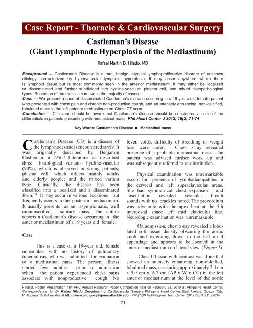
On admission, chest x-ray revealed a lobulated<br />
soft tissue density obscuring the aortic<br />
knob and extending down to the left atrial<br />
appendage and appears to be located in the<br />
anterior mediastinum on lateral view. (Figure 1)<br />
Chest CT scan with contrast was done that<br />
showed an intensely enhancing, non-calcified,<br />
lobulated mass, measuring approximately 2.4 cm<br />
x 5.9 cm x. 6.7 cm (AP x W x CC) in the left<br />
anterior mediastinum at the level of the aortic<br />
Finalist, Poster Presentation 18 th PHC Annual Research Paper Competition held on February 23, 2010 at <strong>Philippine</strong> <strong>Heart</strong> <strong>Center</strong>,<br />
Correspondence to Dr. Rafael Hilado, Department of Cardiovascular Surgery. <strong>Philippine</strong> <strong>Heart</strong> <strong>Center</strong>, East Avenue, Quezon City,<br />
<strong>Philippine</strong>s 1100 Available at http://www.phc.gov.ph/journal/publication copyright by <strong>Philippine</strong> <strong>Heart</strong> <strong>Center</strong>, 2012 ISSN 0018-9034
72 Phil <strong>Heart</strong> <strong>Center</strong> J May - August 2012<br />
Figure 1. Chest X-ray PA and Lateral Views of a 19 y. o. female with mediastinal mass. Chest radiograph shows lobulated<br />
soft tissue density obscuring the aortic knob and extending down to the left atrial appendage, and appears to be located<br />
in the anterior mediastinum on lateral view<br />
Figure 2. Chest CT scan images of the same patient showing an intensely enhancing, non-calcified, lobulated mass,<br />
measuring approximately 2.4 x 5.9 x. 6.7 cm ( AP x W x CC) in the left anterior mediastinum at the level of the aortic arch<br />
extending to the left lateral aspect of the main pulmonary artery.
arch extending to the left lateral aspect of the<br />
main pulmonary artery. (Figure 2)<br />
An initial impression of a thymoma was<br />
made and the patient was scheduled for a<br />
thymectomy. The patient was preoperatively<br />
referred to the Neurology Service to rule out coexistence<br />
of Myasthenia Gravis.<br />
The operative approach was via a median<br />
sternotomy. Intraoperatively, there was a 9 cm<br />
x 7 cm x 4 cm well-circumscribed lobulated<br />
mass adjacent to the left lobe of the<br />
thymus gland extending posteriorly to the<br />
inominate vein and left pulmonary artery.<br />
There was no evidence of infiltration of adjacent<br />
structures. The procedure done was excision<br />
of the anterior mediastinal mass including<br />
the thymus. The patient had an unremarkable<br />
postoperative course. She was immediately<br />
extubated postoperatively and started<br />
on progressive diet. The chest tube was removed<br />
on the second postoperative day and the<br />
patient was eventually discharged<br />
recovered on the 6 th postoperative day.<br />
The final histopathologic examination<br />
revealed nodal tissue with proliferation of<br />
follicles characterized by regressively<br />
transformed germinal centers. There is tight<br />
concentric layering of lymphocytes at the<br />
periphery of the follicles resulting in an onionskin<br />
like appearance. The follicles showed<br />
marked vascular proliferation and hyalinization<br />
of the germinal centers. The intrafollicular<br />
stroma is otherwise prominent, with numerous<br />
hyperplastic vessels and admixture<br />
of plasma cells, eosinophils, and immunoblasts.<br />
Associated vascular proliferation is also<br />
present in the surrounding soft tissue. The<br />
histologic features were compatible with<br />
Castleman’s disease, hyaline vascular type<br />
(angiofollicuar lymph node hyperplasia).<br />
The thymus gland showed features of<br />
involution. There was no evidence of dysplasia<br />
or malignant transformation.<br />
Discussion<br />
Castleman’s disease is a rare, benign<br />
disorder of the lymph nodes that should<br />
Hilado RMD Castleman’s <strong>Disease</strong> 73<br />
be included in the differential diagnosis of<br />
anterior mediastinal masses. It is a lymphoproliferative<br />
disorder and is also known as<br />
“hamartoma, angiofollicular lymph node<br />
hyperplasia, benign giant lymphoma, giant<br />
lymph node hyperplasia, and follicular<br />
lymphoreticuloma.” 5 It is sometimes associated<br />
with other diseases such as human immunodeficiency<br />
virus (HIV) and human herpes<br />
virus 8 (HHV-8). It is sometimes associated<br />
with malignancies such as Kaposi’s sarcoma<br />
(KS), non-Hodgkin’s lymphoma, Hodgkin’s<br />
lymphoma, and POEMS (polyneuropathy,<br />
organomegaly, endocrinopathy, monoclonal<br />
proteinemia and skin) syndrome. 5,9,10<br />
It can occur anywhere in the body<br />
wherever there are lymphnodes but approximately<br />
70% of the cases are located in the thorax,<br />
14% in the neck, 12% in the abdomen and 4%<br />
in the axilla. 14 The lesions are predominantly<br />
of two histologic types: the hyaline vascular<br />
type and the plasma cell type. 5 Majority<br />
of these lesions are of the hyaline vascular<br />
type, which accounts for approximately 90%<br />
of cases and is most often a localized disease.<br />
Radiologic studies show that these masses<br />
typically appear as well-circumscribed mass<br />
in the visceral compartment of the mediastinum.<br />
These patients tend to be younger (median<br />
age, 23.5 years), to be asymptomatic, and<br />
to have a benign clinical course. 6 “Surgical excision<br />
is curative, with a 5-year survival<br />
rate of 100%, although close follow- up is<br />
recommended due to reports of recurrence.” 6<br />
The plasma cell variant, characterized by<br />
relatively few capillaries and the presence of<br />
mature plasma cells between the hyperplastic<br />
and germinal centers, usually presents with<br />
anemia, fever, fatigue, polyclonal hypergammaglobulinemia<br />
and bone marrow plasmacytosis.<br />
7-8<br />
Castleman’s disease can also be classified<br />
into two clinical forms, namely localized and<br />
multicentric. Those with localized disease<br />
have only one mediastinal compartment<br />
involvement, with no evidence of disease<br />
in an extrathoracic site. If more than one<br />
mediastinal compartment or if there is<br />
evidence of disease in an extrathoracic site,<br />
it is classified as disseminated Castleman’s
74 Phil <strong>Heart</strong> <strong>Center</strong> J May - August 2012<br />
disease. 11 Our patient presented with a multicentric<br />
form of the disease since there were<br />
lymphadenopathies noted in the neck and left<br />
supraclavicular area.<br />
The etiology of Castleman’s disease remains<br />
unknown. It may be due to infection (HHV-<br />
8), Autoimmunity or Cytokine dysregulation<br />
(IL-6). 6<br />
The histologic diagnosis of Castleman’s<br />
<strong>Disease</strong> is usually made after the mass is<br />
excised; needle biopsy is not usually done<br />
because of low diagnostic accuracy, whereas,<br />
in thorascopic biopsy, there is a risk of<br />
bleeding due to the tumor’s hypervascularity .12-13<br />
The treatment for unicentric disease can<br />
either be surgery or radiotherapy if resection<br />
was incomplete or chemotherapy. For<br />
multicentric disease, treatment options include<br />
Steroids (60-70% ORR, 15% CR, usually not<br />
durable), Chemotherapy with Rituximab, or<br />
Auto BMT, Antivirals, and Anti-IL-6.<br />
References<br />
1. Castleman B, Iverson L, Menendez VP. Localized Mediastinal<br />
Lymph-Node Hyperplasia Resembling Thymoma.<br />
Cancer 1956, 9:822-830.<br />
2. Palestro G, Turrini F, Pagano M, Chiusa L. Castleman’s<br />
disease. Adv Clin Path, 3: 11-22, 1999.<br />
3. Bowne WB, Lewis JJ, Filippa DA, Niesvizky R, Brooks<br />
AD, Burt ME, Brennan MF. The management of unicentric<br />
and multicentric Castleman’s disease. Cancer,<br />
85: 706-717, 1999.<br />
4. Shroff VJ, Gilchrist BF, De Luca FG, McCombs HL,<br />
Wesselhoeft CW: Castleman’s disease presenting as a<br />
pediatric surgical problem. J Pediatr Surg, 30: 745-<br />
747, 1995.<br />
5. Keller AR, Hocholzer L, Castleman B. Hyaline vascular<br />
and plasma cell types of giant lymph node hyperplasia<br />
of the mediastinum and other locations. Cancer 1972;<br />
29: 670–82.<br />
6. Kim JH, Jun TG, Sung SW, et al. Giant lymph node<br />
hyperplasia (Castleman’s disease) in the Chest. Ann<br />
Thorac Surg 1995; 59:1162–1165.<br />
7. J ohkoh T, Mueller NL, Ichikadoh K, Nishimoto<br />
N,Yoshizaki K, et al. Intrathoracic Multicentric Castleman’s<br />
disease: CT findings in 12 patients. Radiology<br />
1998; 209:477-481.<br />
8. Weisenbruger DD,Athawani BN, Winndberg CD, Rappaport<br />
H. Multicentric angiofollicular lymph node hyperplasia;<br />
a clinicopathologic study of 16 cases. Hum<br />
Pathol 1985;2:162-172.<br />
9. Flendrig JA, Schillings PHM. Benign giant lymphoma:<br />
the clinical signs and symptoms. Folia Med Neerl 1969;<br />
12:119.<br />
10. Gaba AR, Stein RS, Sweet DL, Variakojis D. Multicentric<br />
giant lymph node hyperplasia. Am J Clin Pathol<br />
1978; 69(1): 86- 90.<br />
11. A Ahluwalia, K Saggar, P Sandhu, V Kalia. Chest: Castleman<br />
disease of thorax. Ind J Radiol Imag 2005<br />
15:2:232.<br />
12. Ottavio Rena, Caterina Casadio, Giuliano Maggi. Castleman’s<br />
disease: unusual intrathoracic localization.<br />
Eur J Cardio-thorac Surg 2001; 19: 519–21.<br />
13. Peter A. Seirafi, Eric Ferguson, Fred H. Edwards. Thoracoscopic<br />
resection of Castleman’s disease. Chest<br />
2003; 123: 280–82.<br />
14. Frizzera G, Massarelli G, Banks PM, Rosai J. A systemic<br />
lymphoproliferative disorder with morphological<br />
features of Castleman’s <strong>Disease</strong>; Pathological findings<br />
in 15 patients. Am J Surg Pathol 1983; 7: 212–31.


