Blood Blood Vessels Tissue fluid - BiologyMad A-Level Biology
Blood Blood Vessels Tissue fluid - BiologyMad A-Level Biology
Blood Blood Vessels Tissue fluid - BiologyMad A-Level Biology

You also want an ePaper? Increase the reach of your titles
YUMPU automatically turns print PDFs into web optimized ePapers that Google loves.
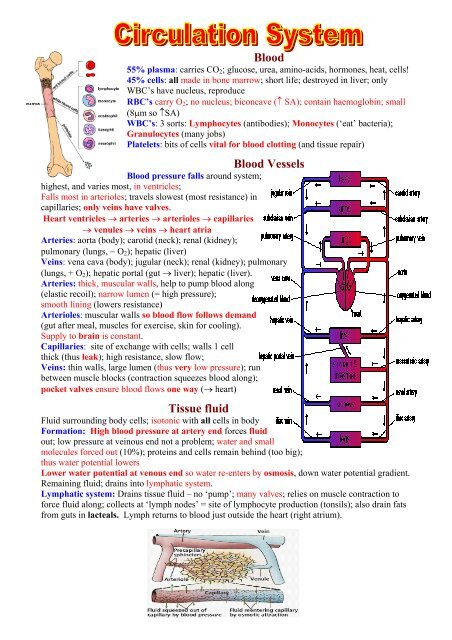
<strong>Blood</strong><br />
55% plasma: carries CO2; glucose, urea, amino-acids, hormones, heat, cells!<br />
45% cells: all made in bone marrow; short life; destroyed in liver; only<br />
WBC’s have nucleus, reproduce<br />
RBC’s carry O2; no nucleus; biconcave (↑ SA); contain haemoglobin; small<br />
(8µm so ↑SA)<br />
WBC’s: 3 sorts: Lymphocytes (antibodies); Monocytes (‘eat’ bacteria);<br />
Granulocytes (many jobs)<br />
Platelets: bits of cells vital for blood clotting (and tissue repair)<br />
<strong>Blood</strong> <strong>Vessels</strong><br />
<strong>Blood</strong> pressure falls around system;<br />
highest, and varies most, in ventricles;<br />
Falls most in arterioles; travels slowest (most resistance) in<br />
capillaries; only veins have valves.<br />
Heart ventricles → arteries → arterioles → capillaries<br />
→ venules → veins → heart atria<br />
Arteries: aorta (body); carotid (neck); renal (kidney);<br />
pulmonary (lungs, − O2); hepatic (liver)<br />
Veins: vena cava (body); jugular (neck); renal (kidney); pulmonary<br />
(lungs, + O2); hepatic portal (gut → liver); hepatic (liver).<br />
Arteries: thick, muscular walls, help to pump blood along<br />
(elastic recoil); narrow lumen (= high pressure);<br />
smooth lining (lowers resistance)<br />
Arterioles: muscular walls so blood flow follows demand<br />
(gut after meal, muscles for exercise, skin for cooling).<br />
Supply to brain is constant.<br />
Capillaries: site of exchange with cells; walls 1 cell<br />
thick (thus leak); high resistance, slow flow;<br />
Veins: thin walls, large lumen (thus very low pressure); run<br />
between muscle blocks (contraction squeezes blood along);<br />
pocket valves ensure blood flows one way (→ heart)<br />
<strong>Tissue</strong> <strong>fluid</strong><br />
Fluid surrounding body cells; isotonic with all cells in body<br />
Formation: High blood pressure at artery end forces <strong>fluid</strong><br />
out; low pressure at veinous end not a problem; water and small<br />
molecules forced out (10%); proteins and cells remain behind (too big);<br />
thus water potential lowers<br />
Lower water potential at venous end so water re-enters by osmosis, down water potential gradient.<br />
Remaining <strong>fluid</strong>; drains into lymphatic system.<br />
Lymphatic system: Drains tissue <strong>fluid</strong> – no ‘pump’; many valves; relies on muscle contraction to<br />
force <strong>fluid</strong> along; collects at ‘lymph nodes’ = site of lymphocyte production (tonsils); also drain fats<br />
from guts in lacteals. Lymph returns to blood just outside the heart (right atrium).
Dual pump; all 4 chambers have same volume; myogenic (does not need nerves to stimulate)<br />
Diastole: = filling chamber (low pressure); Systole = contracting chamber (high pressure)<br />
Right side – deoxygenated, blood from vena cava to lungs; lower pressure (short artery, no gravity)<br />
Left side: oxygenated, blood from lungs to body;<br />
highest pressure (long trip, problem of gravity)<br />
<strong>Blood</strong> flows: right atrium → right ventricle → lungs<br />
→ left atrium → left ventricle → body (aorta)<br />
Valves: semi-lunar valves between ventricles and<br />
main arteries; open at start of ventricular systole;<br />
close at end of ventricular systole (pressure in<br />
ventricle < artery)<br />
Atrio-ventricular (a-v) valves are between atria and<br />
ventricles (L = bicuspid, R = tricuspid)<br />
A-V valves open when atria contract (systole);<br />
(pressure > than ventricles);<br />
A-V valves close when ventricular systole begins<br />
(pressure > that in atria)<br />
Diastole<br />
<strong>Blood</strong> returning from the body flows<br />
into the right atrium, and oxygenrich<br />
blood flowing from the lungs<br />
flows into the left atrium.<br />
Atrial systole<br />
The right and left atria contract to<br />
push blood into the ventricles. The<br />
semi-lunar valves close to stop the<br />
blood flowing back into the heart.<br />
Ventricular systole<br />
The ventricles contract to push blood<br />
out of the heart through semi-lunar<br />
valves. Both sets of AV valves close to<br />
prevent backflow.<br />
Control: regulated by autonomic nerves (vagus↓, cardiac↑) and by hormones (adrenalin, insulin)<br />
Nerve impulse arrives at sino-atrial node (SAN); impulse travels over atria, causing contraction; to<br />
Atrio-ventricular node (AVN); DELAY (allows time for ventricles to fill);<br />
impulse → down Bundle of His; causes ventricles to contract from bottom (thus fully emptying)<br />
Cardiac Output = stroke volume x heart rate (= pulse rate)<br />
Heart rate affected by: stress; exercise; drugs (caffeine); hormones; volume of blood returning