Functions of Blood System - BiologyMad A-Level Biology
Functions of Blood System - BiologyMad A-Level Biology
Functions of Blood System - BiologyMad A-Level Biology

You also want an ePaper? Increase the reach of your titles
YUMPU automatically turns print PDFs into web optimized ePapers that Google loves.
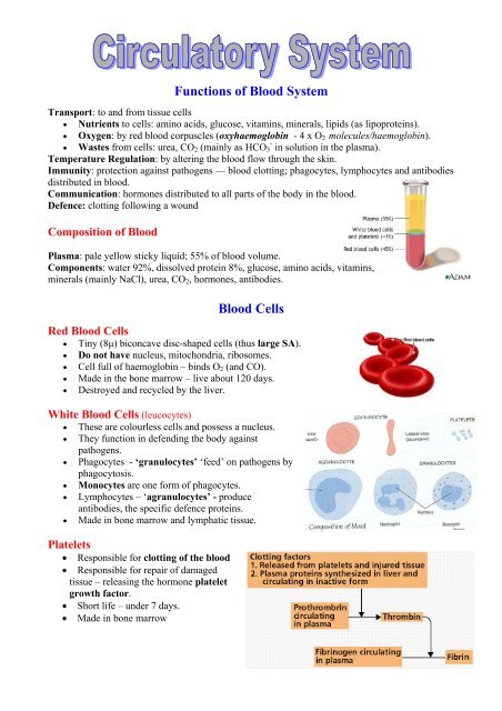
<strong>Functions</strong> <strong>of</strong> <strong>Blood</strong> <strong>System</strong><br />
Transport: to and from tissue cells<br />
• Nutrients to cells: amino acids, glucose, vitamins, minerals, lipids (as lipoproteins).<br />
• Oxygen: by red blood corpuscles (oxyhaemoglobin - 4 x O2 molecules/haemoglobin).<br />
• Wastes from cells: urea, CO2 (mainly as HCO3 - in solution in the plasma).<br />
Temperature Regulation: by altering the blood flow through the skin.<br />
Immunity: protection against pathogens — blood clotting; phagocytes, lymphocytes and antibodies<br />
distributed in blood.<br />
Communication: hormones distributed to all parts <strong>of</strong> the body in the blood.<br />
Defence: clotting following a wound<br />
Composition <strong>of</strong> <strong>Blood</strong><br />
Plasma: pale yellow sticky liquid; 55% <strong>of</strong> blood volume.<br />
Components: water 92%, dissolved protein 8%, glucose, amino acids, vitamins,<br />
minerals (mainly NaCl), urea, CO2, hormones, antibodies.<br />
<strong>Blood</strong> Cells<br />
Red <strong>Blood</strong> Cells<br />
• Tiny (8µ) biconcave disc-shaped cells (thus large SA).<br />
• Do not have nucleus, mitochondria, ribosomes.<br />
• Cell full <strong>of</strong> haemoglobin – binds O2 (and CO).<br />
• Made in the bone marrow – live about 120 days.<br />
• Destroyed and recycled by the liver.<br />
White <strong>Blood</strong> Cells (leucocytes)<br />
• These are colourless cells and possess a nucleus.<br />
• They function in defending the body against<br />
pathogens.<br />
• Phagocytes - ‘granulocytes’ ‘feed’ on pathogens by<br />
phagocytosis.<br />
• Monocytes are one form <strong>of</strong> phagocytes.<br />
• Lymphocytes – ‘agranulocytes’ - produce<br />
antibodies, the specific defence proteins.<br />
• Made in bone marrow and lymphatic tissue.<br />
Platelets<br />
• Responsible for clotting <strong>of</strong> the blood<br />
• Responsible for repair <strong>of</strong> damaged<br />
tissue – releasing the hormone platelet<br />
growth factor.<br />
• Short life – under 7 days.<br />
• Made in bone marrow
<strong>Blood</strong> Vessels<br />
Artery v. vein<br />
• The wall <strong>of</strong> the artery is thicker: thicker connective tissue<br />
layer, thicker mixed layer <strong>of</strong> muscle and elastic tissue.<br />
• The lumen <strong>of</strong> the artery is much narrower.<br />
• Arteries do not have valves along their length, veins do.<br />
• Valves in the veins prevent the backflow <strong>of</strong> blood so the<br />
flow is in one correct direction towards the heart.<br />
• <strong>Blood</strong> flows away from the heart in arteries; blood flows<br />
towards the heart in veins.<br />
• <strong>Blood</strong> pressure in arteries is higher and so also the speed <strong>of</strong> blood flow<br />
• Pulsed flow in an artery, steady flow in a vein.<br />
• Many tissues, thus both are organs<br />
Arterioles have muscular walls which control how much blood flows to a particular organ – e.g.<br />
• guts after meal,<br />
• skin for temperature regulation<br />
• muscle when working hard Note: <strong>Blood</strong> supply to brain is constant!<br />
Capillaries<br />
• Capillaries are the link between arteries and veins – where exchange with tissues occurs.<br />
• The capillary wall is one cell thick and somewhat porous — ideal to allow materials to pass<br />
in and out.<br />
• All tissue cells very close to a capillary so exchange is very efficient.<br />
• Exchange at the capillaries is by diffusion, mass flow and active transport.<br />
• <strong>Blood</strong> flow in capillaries is slow giving enough time for effective exchange.<br />
• One type <strong>of</strong> cell, thus a tissue.<br />
Closed <strong>System</strong> <strong>of</strong> <strong>Blood</strong> Vessels<br />
• The blood does not make direct contact with the tissue cells.<br />
• The blood is retained in the blood vessels.<br />
• A closed system is very responsive to the change needs <strong>of</strong> the organs and is highly efficient.<br />
Double Circulation<br />
The double circuit prevents mixing <strong>of</strong> oxygenated and deoxygenated blood. Therefore oxygen<br />
supply is highly efficient.<br />
Pulmonary Circulation: deoxygenated blood flows from the heart to the lungs<br />
• oxygen is taken on and carbon dioxide is excreted,<br />
• oxygenated blood flows from the lungs back to the heart.<br />
<strong>System</strong>ic Circulation: oxygenated blood flows from the heart to the organ systems <strong>of</strong> the body.<br />
• oxygen is delivered and carbon dioxide is taken on,<br />
• deoxygenated blood flow from the organs systems back to the heart.<br />
Portal <strong>System</strong><br />
• A portal blood vessel has a set <strong>of</strong> capillaries at each end.<br />
• The hepatic portal vein carries blood rich in absorbed nutrients from the capillaries in the<br />
gut to capillaries in the liver.<br />
• Hepatic artery supplies liver with O2<br />
• Hepatic vein takes all the blood away from the liver, back to heart.
The Heart<br />
• The heart is a double pump.<br />
• The right atrium collects deoxygenated blood from all parts<br />
(vena cava).<br />
• The right ventricle pumps deoxygenated blood to the lungs<br />
(for gas exchange = ↑O2, ↓CO2) (pulmonary artery)<br />
• The left atrium collects oxygenated blood from the lungs<br />
(pulmonary vein)<br />
• The left ventricle pumps oxygenated blood to all parts (aorta).<br />
• The right and left side fill and empty in unison.<br />
• Each chamber pumps the same volume <strong>of</strong> blood.<br />
• The wall <strong>of</strong> the left ventricle is about three times thicker than that <strong>of</strong> the right ventricle.<br />
• The left ventricle needs more cardiac muscle to give the blood a much stronger push.<br />
• <strong>Blood</strong> pressure therefore highest in left ventricle<br />
• Cardiac output = stroke volume x pulse rate<br />
Heart action<br />
• <strong>Blood</strong> enters the atria, filling them. (= atrial diastole)<br />
• The sino-atrial node (= SAN) in the right atrium generates a nerve impulse causing the atria<br />
to contract (atrial systole)<br />
• The sympathetic nerve impulses increase heart rate<br />
• The vagus nerve impulses decrease heart rate<br />
• <strong>Blood</strong> pressure in atria rises above that in ventricles<br />
• <strong>Blood</strong> forced into the ventricles (AV valves open).<br />
• The impulse arrives at the atrio-ventricular node (= AVN)<br />
• Impulse delayed (0.2 s) giving time for ventricles to fill (ventricular diastole).<br />
• The impulse enters the ventricles and travels through the Bundle <strong>of</strong> His (in septum).<br />
• The ventricles contract and force the blood out <strong>of</strong> the pulmonary artery and aorta<br />
(ventricular systole).<br />
• The AV valves close preventing blood returning to the atria.<br />
• The semi-lunar valves are pushed open by the higher blood pressure in the ventricles.<br />
• The elastic artery walls expand (=a pulse).<br />
• When the blood pressure falls the arteries recoil squeezing the blood away from the heart.<br />
• Ventricular blood pressure falls, closing the semilunar valves.<br />
• This prevents blood flowing back into the ventricles from the arteries.
Cardiac cycle<br />
Diastole: relaxed cardiac muscle — the heart fills with blood under low pressure from the veins.<br />
Systole: cardiac muscle contracting — the chambers <strong>of</strong> the heart are emptying <strong>of</strong> blood.<br />
• Atrial Systole: contraction and emptying <strong>of</strong> the atria supplying extra blood to the ventricles.<br />
• Ventricular Systole: contraction and emptying <strong>of</strong> the ventricles ejecting blood from the heart<br />
into the arteries.<br />
Cardiac Muscle<br />
• The muscle making up the heart is called cardiac muscle.<br />
• It is myogenic, i.e., stimulates itself to contract — does not need external stimulation.<br />
• It is an involuntary, strong muscle that does not fatigue (no anaerobic respiration).<br />
The SAN (pacemaker)<br />
• A small area <strong>of</strong> cardiac muscle in the wall <strong>of</strong> the right atrium, near entry <strong>of</strong> vena cava.<br />
• Its automatic rhythmic contraction starts each cardiac cycle.<br />
• Two nerves from the medulla oblongata connect to it influencing its rate <strong>of</strong> contraction.<br />
• One nerve accelerates the heart rate and the other reduces it back to resting rate.<br />
Factors affecting heart rate:<br />
Since the heart can only pump out the blood that is returned to it, the primary cause <strong>of</strong> increased<br />
cardiac output when exercising, is ↑ blood to the heart caused by the muscles squeezing the veins.<br />
• Increase: exercise, increased body temperature, stress, mental excitement, infection.<br />
• Decrease: increased physical fitness, sleep, and mental relaxation.<br />
Coronary circulation<br />
• The blood flowing through the heart does not supply the heart with blood<br />
• Coronary artery supplies heart muscle with blood<br />
• No coronary vein – directly drains back in diastole.<br />
Major blood vessels:<br />
Arteries: Aorta; Pulmonary (↑CO2, ↓O2); Carotid (neck); Renal;<br />
Hepatic<br />
Veins: Vena Cava; Pulmonary (↓CO2, ↑O2); Renal; Jugular;<br />
Hepatic; Hepatic Portal;<br />
<strong>Blood</strong> Pressure<br />
• The pressure varies along the circuit – measured with<br />
sphygmomanometer<br />
• Pressure decreases in the following order:<br />
o ventricle > artery > arteriole > capillary > venule ><br />
vein > atrium.<br />
• Standard healthy readings: 80 mm Hg diastolic, 120 mm Hg systolic.
Formation <strong>of</strong> Tissue Fluid<br />
• As the blood enters the capillaries the high hydrostatic pressure forces some <strong>of</strong> the plasma<br />
out through the wall.<br />
• The escaped fluid (tissue fluid) is similar in composition to plasma, but lower in protein.<br />
• Therefore the remaining blood has a lower water potential<br />
• And a lower hydrostatic pressure<br />
• At the venule end the hydrostatic pressure is lower<br />
• Thus water returns to blood down water potential gradient.<br />
• The 10% excess tissue fluid must be drained away – by the lymphatic system.<br />
The Lymphatic <strong>System</strong><br />
• A collection <strong>of</strong> special drainage vessels receiving excess tissue fluid.<br />
• Once the tissue fluid enters the lymphatic capillaries it is called lymph.<br />
• Lymph nodes (e.g. tonsils) filter the lymph and produce lymphocytes.<br />
• The lymph vessels have many valves, but low pressure.<br />
• The lymph is moved along by the squeezing action <strong>of</strong>:<br />
o the skeletal muscles,<br />
o pressure changes in the thorax during breathing and<br />
o by the rhythmic contraction <strong>of</strong> the lymph vessel walls.<br />
• Lymph re-enters the blood just before the right atrium.<br />
<strong>Functions</strong> <strong>of</strong> the Lymphatic <strong>System</strong>:<br />
Circulatory role<br />
• Return the excess tissue fluid to the blood: this maintains blood volume, pressure and<br />
concentration.<br />
• Collect and deliver the absorbed lipids from the small intestine to the blood<br />
Defence role<br />
• The lymph nodes filter out pathogens in the lymph.<br />
• Production and ‘export’ <strong>of</strong> lymphocytes to the blood system for general distribution.<br />
• Detection <strong>of</strong> antigens and production <strong>of</strong> specific antibodies.<br />
© IHW March 2005