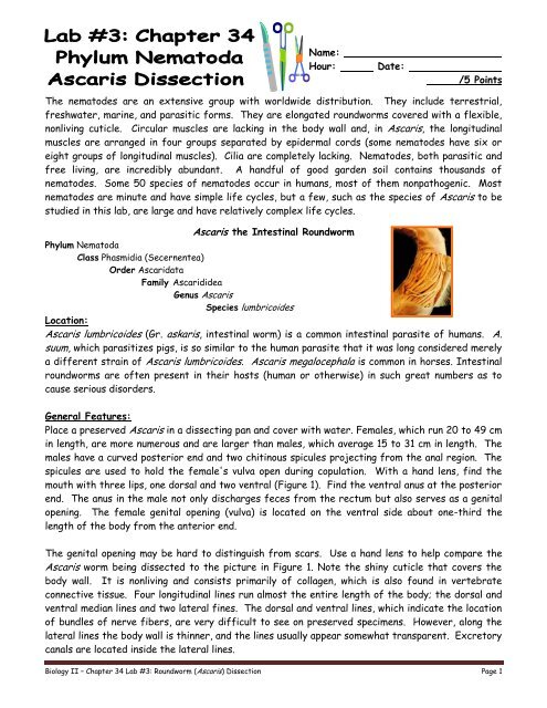
Phylum Nematoda Ascaris Dissection
Phylum Nematoda Ascaris Dissection
Phylum Nematoda Ascaris Dissection

Create successful ePaper yourself
Turn your PDF publications into a flip-book with our unique Google optimized e-Paper software.
Name:<br />
Hour: Date:<br />
/5 Points<br />
The nematodes are an extensive group with worldwide distribution. They include terrestrial,<br />
freshwater, marine, and parasitic forms. They are elongated roundworms covered with a flexible,<br />
nonliving cuticle. Circular muscles are lacking in the body wall and, in <strong>Ascaris</strong>, the longitudinal<br />
muscles are arranged in four groups separated by epidermal cords (some nematodes have six or<br />
eight groups of longitudinal muscles). Cilia are completely lacking. Nematodes, both parasitic and<br />
free living, are incredibly abundant. A handful of good garden soil contains thousands of<br />
nematodes. Some 50 species of nematodes occur in humans, most of them nonpathogenic. Most<br />
nematodes are minute and have simple life cycles, but a few, such as the species of <strong>Ascaris</strong> to be<br />
studied in this lab, are large and have relatively complex life cycles.<br />
<strong>Ascaris</strong> the Intestinal Roundworm<br />
<strong>Phylum</strong> <strong>Nematoda</strong><br />
Class Phasmidia (Secernentea)<br />
Order Ascaridata<br />
Family Ascarididea<br />
Genus <strong>Ascaris</strong><br />
Species lumbricoides<br />
Location:<br />
<strong>Ascaris</strong> lumbricoides (Gr. askaris, intestinal worm) is a common intestinal parasite of humans. A.<br />
suum, which parasitizes pigs, is so similar to the human parasite that it was long considered merely<br />
a different strain of <strong>Ascaris</strong> lumbricoides. <strong>Ascaris</strong> megalocephala is common in horses. Intestinal<br />
roundworms are often present in their hosts (human or otherwise) in such great numbers as to<br />
cause serious disorders.<br />
General Features:<br />
Place a preserved <strong>Ascaris</strong> in a dissecting pan and cover with water. Females, which run 20 to 49 cm<br />
in length, are more numerous and are larger than males, which average 15 to 31 cm in length. The<br />
males have a curved posterior end and two chitinous spicules projecting from the anal region. The<br />
spicules are used to hold the female's vulva open during copulation. With a hand lens, find the<br />
mouth with three lips, one dorsal and two ventral (Figure 1). Find the ventral anus at the posterior<br />
end. The anus in the male not only discharges feces from the rectum but also serves as a genital<br />
opening. The female genital opening (vulva) is located on the ventral side about one-third the<br />
length of the body from the anterior end.<br />
The genital opening may be hard to distinguish from scars. Use a hand lens to help compare the<br />
<strong>Ascaris</strong> worm being dissected to the picture in Figure 1. Note the shiny cuticle that covers the<br />
body wall. It is nonliving and consists primarily of collagen, which is also found in vertebrate<br />
connective tissue. Four longitudinal lines run almost the entire length of the body; the dorsal and<br />
ventral median lines and two lateral fines. The dorsal and ventral lines, which indicate the location<br />
of bundles of nerve fibers, are very difficult to see on preserved specimens. However, along the<br />
lateral lines the body wall is thinner, and the lines usually appear somewhat transparent. Excretory<br />
canals are located inside the lateral lines.<br />
Biology II – Chapter 34 Lab #3: Roundworm (<strong>Ascaris</strong>) <strong>Dissection</strong> Page 1
Figure 1 a. female b. male<br />
Internal Structure:<br />
Select a female specimen, place the worm in a dissecting pan, and cover it with water. Locate the<br />
lateral lines, where the body wall seems somewhat thinner. Now find the anus and vulva on the<br />
ventral side. This should help you identify the opposite, or mid-dorsal line. Now, with a razor<br />
blade, slit open the body wall along the mid-dorsal line, being careful to avoid injuring the internal<br />
structures. Pin back the body wall to expose the viscera, slanting the pins outward to allow room<br />
for dissection.<br />
Body wall and pseudocoel:<br />
Note the body cavity. Note the fluffy masses lining the body wall. These are the large nucleated<br />
cell bodies of the longitudinal muscle cells, whose fibers extend longitudinally in the body wall.<br />
With a pin or pointed probe, tease out some of the fibers from the cut edge of the wall. Examine<br />
fibers and cells under the microscope. Absence of circular muscles accounts for the thrashing<br />
movements of these animals. Note the absence of muscle cells along the lateral lines.<br />
Excretory System:<br />
Excretory canals located in the lateral lines unite just back of the mouth to empty ventrally<br />
through an excretory pore. The canals are largely osmoregulatory in function. Excretion also<br />
occurs through the cuticle. Flame cells are lacking in <strong>Ascaris</strong> and other nematodes, although they<br />
are found in some other pseudocoelomate phyla.<br />
Biology II – Chapter 34 Lab #3: Roundworm (<strong>Ascaris</strong>) <strong>Dissection</strong> Page 2
Digestive System:<br />
The mouth empties into a short muscular pharynx; which sucks food into the ribbon-like intestine<br />
(Figure 2). The intestine is thin walled for absorption of digested food products into the<br />
pseudocoel. Trace it to the anus. Digestion is begun extracellularly in the lumen of the intestine<br />
and is completed intracellularly in the cells of the intestinal wall. There are no respiratory or<br />
circulatory organs. Oxygen is obtained mainly from the breakdown of glycogen within the body,<br />
and distribution is handled by the pseudocoelomic fluid.<br />
Reproductive System:<br />
The female reproductive system fills most of the pseudocoel. The system is a Y-shaped set of<br />
long, convoluted tubes. Unravel them carefully with a probe. The short base of the Y, the vagina,<br />
opens to the outside at the vulva. The long arms of the inverted Y are the uteri. These extend<br />
posteriorly and then double back as slender, much coiled oviducts, which connect the uteri with the<br />
threadlike terminal ovaries. Eggs pass from the ovaries through the oviducts to the uteri, where<br />
fertilization occurs and shells are secreted. Then they pass through the vagina and vulva to the<br />
outside. The uteri of an <strong>Ascaris</strong> may contain up to 27 million eggs at a time, with as many as<br />
200,000 eggs being laid per day.<br />
The male reproductive system is essentially a single, long tube made up of a threadlike testis,<br />
which continues as a thicker vas deferens. Both are much coiled. The vas deferens connects with<br />
the wider seminal vesicle, which empties by a short, muscular ejaculatory duct into the anus. Thus<br />
the male anus serves as an outlet for both the digestive system and the reproductive system and is<br />
often called a cloaca. Spicules secreted by and contained in spicule pouches may be extended<br />
through the anus. In copulation the male inserts the copulatory spicules into the vulva of the<br />
female and discharges spermatozoa through the ejaculatory duct into the vagina.<br />
Figure 2- Transverse sections:<br />
Study a prepared stained slide, at first under low power.<br />
Biology II – Chapter 34 Lab #3: Roundworm (<strong>Ascaris</strong>) <strong>Dissection</strong> Page 3
If both female and male cross sections are present, examine first the larger female cross section.<br />
Note the thick non-cellular cuticle on the outside of the body wall. Below the cuticle is the thinner<br />
syncytial epidermis, which contains nuclei but few cell walls. The longitudinal muscles making up<br />
most of the body wall appear as fluffy, irregular masses dipping into the pseudocoel, with the tips<br />
of the cells directed toward the nearest nerve cord. Muscle continuity is interrupted by the<br />
longitudinal lines. Look for excretory canals in the lateral lines and look for the dorsal and ventral<br />
nerve cords in the dorsal and ventral lines. The lateral lines appear free of muscle cells. In the<br />
pseudocoel of the female the large uteri are filled with eggs enclosed in shells and in cleavage<br />
stages. The thin-walled oviducts also contain eggs, whereas the wheel-shaped ovaries are<br />
composed of tall epithelial cells and have small lumens. The intestine is composed of a single layer<br />
of tall columnar cells (endodermal). The pharynx and the rectal region of the intestine are lined<br />
with cuticle.<br />
Examine now the male cross section. It is similar to the female in all respects except for the<br />
reproductive system. You should see several rounded sections of testes packed with<br />
spermatogonia (precursors to male reproductive cells). There may also be several sections of vas<br />
deferens containing numerous spermatocytes, and possibly a section of a large seminal vesicle filled<br />
with mature spermatozoa. Note that the male reproductive structures visible in the cross section<br />
of the roundworm will depend on the level at which the cross section is taken.<br />
1. What is the scientific name of the intestinal roundworm found in humans?<br />
2. Is the male or female roundworm generally longer?<br />
3. What substance is found in the cuticle of a roundworm that is also found in human connective<br />
tissue?<br />
4. Why is the cavity of the roundworm called a pseudocoel? How does it differ from a true<br />
coelom?<br />
5. Considering the cuticle, why do you think <strong>Ascaris</strong> is not digested in the human intestine?<br />
Biology II – Chapter 34 Lab #3: Roundworm (<strong>Ascaris</strong>) <strong>Dissection</strong> Page 4