Chapter 8 - BGM Jamieson, ultrastructure, reproductive biology ...
Chapter 8 - BGM Jamieson, ultrastructure, reproductive biology ...
Chapter 8 - BGM Jamieson, ultrastructure, reproductive biology ...

You also want an ePaper? Increase the reach of your titles
YUMPU automatically turns print PDFs into web optimized ePapers that Google loves.
Non-leech Clitellata 299<br />
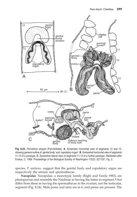
Fig. 8.23. Parvidrilus strayeri (Parvidrilidae). A. Schematic horizontal view of segments 12 and 13,<br />
showing general outline of ‘genital body’ and ‘copulatory organ’. B. Somewhat horizontal view of segments<br />
11-13 of a paratype. C. Somewhat lateral view of segments 11-13 of a further paratype. Relabeled after<br />
Erséus, C. 1999. Proceedings of the Biological Society of Washington 112(2): 327-337, Fig. 2.<br />
species, P. spelaeus, suggest that the genital body and copulatory organ are<br />
respectively the atrium and spermathecae.<br />
Narapidae. Narapidae, a monotypic family (Righi and Varela 1983), are<br />
plesioporous and resemble the Naidinae in having the testes in segment 5 but<br />
differ from these in having the spermathecae in the ovarian, not the testicular,<br />
segment (Fig. 8.24). Male pores and atria are in 6, and penes are present. The
300 Reproductive Biology and Phylogeny of Annelida<br />
Fig. 8.24. Narapa bonettoi (Narapidae). A. Lateral view of segments 6-8. B. Transverse section of atrium.<br />
C. Lateral view of male ducts. Relabelled after Righi, G. and Varela, M. E. 1983. Revista De La<br />
Asociacion De Ciencias Naturales Del Litoral 14(1): 7-15, Figs. 4-6.<br />
atria are covered by diffuse gland cells. Whereas the testes and efferent ducts<br />
are paired, the ovary, in 7, is unpaired. There is a pair of spermathecae in 7.<br />
Like the Randiellidae and Propappidae, there is a gonad-less segment<br />
between the testicular and ovarian segments. This might represent a<br />
proandric condition derived from former holandry.<br />
8.2.11.3 Subclass Lumbriculata<br />
We will here deal only with the oligochaetous members, the Lumbriculidae.<br />
Other taxa here included are the Branchiobdellida, Acanthobdellida and<br />
Euhirudinea which are discussed in <strong>Chapter</strong> 9.<br />
Lumbriculidae. The Lumbriculidae is an Holarctic family with extension<br />
into West Asia. Some species have become widely distributed, including the<br />
Southern Hemisphere. The <strong>reproductive</strong> system is very variable. There are<br />
one to four pairs of testes, in variable locations. Atria are one to four pairs,<br />
located between segments 7 and 15, paired or unpaired, always in a testisbearing<br />
segment, each being associated with one or two pairs of testes (Figs.<br />
8.4B, 8.25). There are commonly two pairs of testes in adjacent segments, both<br />
with funnels and vasa deferentia feeding a single pair of atria in the same<br />
segment as the posterior pair of testes. Sometimes the anterior testes and<br />
ducts are absent, leaving a single pair of atria, testes and vasa deferentia<br />
within one segment, and then often with this arrangement serially repeated.<br />
There are one or two pairs of ovaries beginning one, or rarely two, segments<br />
behind the most posterior testis-bearing segments. Spermathecae are variable
Non-leech Clitellata 301<br />
Fig. 8.25. Bythonomus mirus (Lumbriculidae). Atrium and posterior male gonoduct. After Chekanovskaya,<br />
O.V. 1981. Aquatic Oligochaeta of the USSR, United States Department of the Interior and the National<br />
Science Foundation, Washington, D.C., Amerind Publishing Co. Pvt. Ltd., New Delhi, pp. 513, Fig. 232.<br />
in number and either anterior or posterior to the testicular segments (Pinder<br />
and Brinkhurst 1994).<br />
Michaelsen (1928-32) (see Michaelsen 1928) brilliantly foreshadowed the<br />
findings of molecular phylogenetics when he illustrated (Fig. 8.26) a pathway<br />
from lumbriculid organization to that of hirudinid leeches. The progressive<br />
stages were exemplified by 1) the lumbriculid Rhynchelmis, with compact<br />
testes but long seminal vesicles, extending through several segments; 2) the<br />
lumbriculid Agriodrilus vermivorus, in which a chain of testes has developed<br />
within the elongate seminal vesicles, though still with a single pair of
302 Reproductive Biology and Phylogeny of Annelida<br />
Fig. 8.26. Hypothetical scheme suggesting that the testicular sacs of leeches corrrespond to the seminal<br />
vesicles of lumbriculids. A. Rhynchelmis. B. Agriodrilus vermivorus offers an intermediate in which a long<br />
series of testicular portions has developed in the original seminal vesicles. In an abnormal condition one<br />
of these was seen to be isolated like a testis in the chain of testes occurring in leeches. C. The leech<br />
condition: a chain of postovarian testes. Modified from Michaelsen, W. 1928. Oligochaeta. In W. Kukenthal<br />
and T. Krumbach (eds). Handbuch der Zoologie 2, Fig. 93.<br />
seminal funnels, and in which the coelom is constricted; 3) Hirudo in which<br />
each testicular chamber has acquired its own pair of seminal funnels and the<br />
coelom has been reduced to a system of sinuses. The leech distinction from<br />
oligochaetes, extension of testes posterior to the ovaries, was thus explained<br />
in terms of modification of pre-existing seminal vesicles.<br />
8.2.11.4 Subclass Diplotesticulata<br />
The validity of recognizing the Diplotesticulata is discussed in 8.1.4 above.<br />
Superorder Haplotaxidea. Order Haplotaxida sensu stricto. The<br />
haplotaxid <strong>reproductive</strong> system usually has two pairs of testes, in segments<br />
10 and 11 (rarely in 9 and 10); the anterior pair is rarely absent. There are<br />
one or two pairs of ovaries in the segments following the testicular segments.<br />
The male ducts are simple and lead to ventrolateral or lateral male pores.<br />
However there is a large glandular mass between the male pores in<br />
Hologynus hologynus, in which both pairs of male pores lie in the same<br />
segment, the posterior vasa deferentia being reflexed forward. The two pairs<br />
of vasa deferentia also open into a single segment in Pelodrilus violaceus but
Non-leech Clitellata 303<br />
in that case the anterior vasa penetrate more than one segment, discharging<br />
near the posterior pair in segment 12.<br />
In Adenodrilus denticulatus there are four pairs of large copulatory glands<br />
which open externally near the ventral setae and are not directly associated<br />
with the male ducts (Chekanovskaya 1981) (Fig. 8.27). These glands are<br />
reminiscent of those of Sparganophilus, a genus which has in the past been<br />
placed in the Haplotaxidae (Tétry 1934), but the molecular study (<strong>Jamieson</strong><br />
et al. 2002) indicates that at least the type-species, Haplotaxis gordioides, is<br />
genetically distant from Sparganophilus. One species, H. brinkhursti, has lost<br />
the anterior pair of ovaries and therefore is unique in the known<br />
Haplotaxidae in having the metagynophoran condition.<br />
8.27. Adenodrilus denticulatus (Haplotaxidae). Lateral view of genital organs, showing large copulatory<br />
glands. After Chekanovskaya, O. V. 1981. Aquatic Oligochaeta of the USSR, United States Department<br />
of the Interior and the National Science Foundation, Washington, D. C., Amerind Publishing Co. Pvt. Ltd,<br />
New Delhi. pp. 513, Fig. 204.<br />
Tiguassuidae. The Tiguassuidae was recognized as a family by <strong>Jamieson</strong><br />
(1988b) and by Brinkhurst (1988) in morphocladistic analyses for Tiguassu<br />
reginae which Righi et al. (1978) had placed in the Hapolotaxidae. In the<br />
analysis of <strong>Jamieson</strong> (1988b) Tiguassu proved paraphyletic relative to the<br />
Haplotaxidae sensu lato and formed the plesiomorphic sister-taxon of the<br />
Metagynophora. Its sole autapomorphy was restriction of the hearts to<br />
segment 10. The large proboscis-like prostomium (not computed) was a<br />
unique apomorphy in the entities included but is known homoplasically in<br />
the glossoscolecid Enantiodrilus bilolleyi Cognetti and is approached in some<br />
naids and lumbriculids. In its <strong>reproductive</strong> system (Fig. 8.28) Tiguassu<br />
provides evidence of reduction from two pairs of testes, and possibly from an<br />
ocotogonadal condition, in having two pairs of seminal funnels (in 10 and<br />
11) of which those in 10 are vestigial in the absence of testes. Well developed<br />
testes are present in 11. The female system is progynous, as in most<br />
haplotaxids, with a single pair of ovaries in 12 immediately succeeding a<br />
testis-segment. There are no atria or other modifications of the male ducts,<br />
presumably as plesiomorphic conditions. There are two pairs of small,
304 Reproductive Biology and Phylogeny of Annelida<br />
Fig. 8.28. Tiguassu reginae (Tiguassuidae). Reconstruction of the anterior 15 segments based on serial<br />
sections, showing the <strong>reproductive</strong> system with vestigial anterior seminal funnels. The smaller figure<br />
shows the proboscis-like prostomium. After Righi, G. et al. 1978. Acta Amazonica 8 (3 Supplement 1):<br />
1-49, Figs. 3, 1.<br />
adiverticulate spermathecae, in segments 9 and 10. The ova have a diameter<br />
of 40-50 µm (Righi et al. 1978). The co-occurrence of spermathecae with male<br />
funnels in 10 is a condition also seen in the Tubificidae, as is the great<br />
elongation of the seminal vesicle, but ovisacs were not found.<br />
Superorder Metagynophora. Loss of the anterior ovaries of a<br />
hypothetical octogonadal set, with retention of ovaries in segment 13 so that<br />
a segment lacking gonads intervenes between the posterior testes and the<br />
ovaries, or two segments in proandric taxa such as alluroidids, diagnoses all<br />
oligochaetes above the tubificid-enchytraeid assemblage and the<br />
Lumbriculidae, i.e. from the Moniligastridae through the Megascolecidae,<br />
loosely termed ‘megadriles’. This synapomorphy characterizes the<br />
Metagynophora of <strong>Jamieson</strong> (1988b) (Fig. 8.4). These are equivalent to the<br />
Lumbricida of Brinkhurst (1982). As the most plesiomorphic representatives,<br />
the Moniligastridae, Alluroididae and Syngenodrilidae, have not been<br />
sequenced for DNA, monophyly of the Metagynophora awaits confirmation<br />
from molecular analysis.<br />
Order Moniligastrida. Moniligastridae. Reproductive features among<br />
unambiguous synapormorphies for the Moniligastrida, as represented by<br />
Desmogaster and Moniligaster, are: ovaries in septal chambers; testis-sacs<br />
suspended on the posterior septum of the testicular segment; seminal<br />
vesicles absent; prostates capsular; spermathecae with non-seminal<br />
diverticula.<br />
Brinkhurst and <strong>Jamieson</strong> (1971) had already recognized the<br />
Moniligastrida as a separate order. <strong>Jamieson</strong> (1977b) re-interpreted the long<br />
debated nature of the testis-sacs, showing that they were neither reduced<br />
segments, as proposed by Stephenson (1922, 1930), nor intraseptal cavities,<br />
as argued by Gates (1962), but that they were normal testis-sacs which, with<br />
their enclosed testes, had become detached from the original testis-bearing<br />
septa (Fig. 8.29). It was recognized that moniligastrids are extraordinarily<br />
primitive in retaining a plesiopore condition, the extremely plesiomorphic
Non-leech Clitellata 305<br />
Fig. 8.29. Distribution of genital organs in relation to existing segmentation and hypothetical segmental<br />
homologues in Moniligastridae. Origin of the ‘intraseptal’ testis-sacs from premoniligastrid sacs attached,<br />
with their testes, to the anterior septa of segments 10 and 11, as in other metagynophorans, is<br />
hypothesized. From <strong>Jamieson</strong>, B. G. M. 1977. Evolutionary Theory 2: 95-114, Fig. 3.<br />
state (in Desmogaster) of 2 pairs of male pores in consecutive segments (both<br />
conditions seen elsewhere only in the Haplotaxidae) and a single layered<br />
clitellum with large yolked eggs. The moniligastrid clitellum is here shown<br />
to consist of a single layer of tall, slender modified epidermal cells with basal<br />
nuclei and dense granular secretory contents which discharge at the outer<br />
surface of each cell (Fig. 8.30A,B). They contrast with the wider, more robust<br />
goblet cells (putative large orthochromatic mucous cells) which predominate<br />
in the general epidermis (Fig. 8.30C).<br />
The <strong>reproductive</strong> system of a moniligastrid is here exemplified by that of<br />
Moniligaster troyi described by <strong>Jamieson</strong> (1977b). Details of the system are
306 Reproductive Biology and Phylogeny of Annelida<br />
Fig. 8.30. Light micrographs of the <strong>reproductive</strong> anatomy of Moniligaster troyi (Moniligastridae). A.<br />
Longitudinal section of the clitellum confirming that is consists of a single layer of cells. B. Same, through<br />
an intersegmental furrow. C. Longitudinal section of the general epidermis, showing the goblet cells. D.<br />
Longitudinal section of a testis-sac showing that it has anterior and posterior portions suspended in a<br />
septum. E. Morulae of spermatids in a testis-sac; a spermatid is labeled in a layer of spermatids encircling<br />
and attached to the cytophore. Abbreviations: c.m, circular muscle; clit.gl, secretory contents of a cell of<br />
the clitellum; cu, cuticle; cy, cytophore; go.c, goblet cell; l.m, longitudinal muscle; mo, morula of<br />
spermatids; sep, septum; spd, spermatid (in a layer of spermatids encircling and attached to the cytophore);<br />
te.s, testis-sac wall. From <strong>Jamieson</strong>, unpublished.
Non-leech Clitellata 307<br />
illustrated from previously unpublished light micrographs (Figs. 8.30, 8.31).<br />
Testes and putative funnels are enclosed in a pair of diaphanous iridescent<br />
testis-sacs. Each sac is suspended in a septum so that it has pre- and postseptal<br />
portions (Figs. 8.29, 8.30D). The vas deferens from each testis-sac joins<br />
the sac ventrally at the anterior face of the supporting septum and passes into<br />
the anterior segment (segment 9) abutting the septum; it is very long and<br />
much coiled in this segment; numerous coils nearest the sac are narrow and<br />
iridescent but by far the greater length is wider and non-iridescent, with<br />
many hair-pin bends, and forms a large cluster. The vas deferens continues<br />
posteriorly to join the glandular portion of the prostates, in segment 11,<br />
considerably ectal of the ental end of the gland, and is straight in this<br />
segment. Immediately within the testis-sac the vas deferens gives rise to<br />
several iridescent ribbons which pass posteriorly for the entire length of the<br />
sac and were interpreted (<strong>Jamieson</strong> 1977b) as a backwardly directed sperm<br />
funnel. The testis-sac contains developmental stages of spermatozoa,<br />
including morulae of spermatids and free spermatozoa (Fig. 8.30D,E); it thus<br />
functions as a testis-sac and seminal vesicle.<br />
Each prostate extends from its pore, at 10/11 to intersegment 13/14; it<br />
has a clavate, superficially slightly lobulated glandular portion and a shorter,<br />
narrow duct which is poorly differentiated from the gland; the duct forms a<br />
muscular swelling at the pore which houses the base of the combined male<br />
and prostatic porophore (<strong>Jamieson</strong> 1977b) (Figs. 8.15, 8.31H,J). The wall of the<br />
gandular portion of the prostate consists of an outer thick longitudinal<br />
muscle layers, a thinner, though still thick, intermediate circular muscle layer,<br />
and an inner epithelium which contains gland cells (Fig. 8.31G,I).<br />
The ovary consists of folded (fan-like) laminae (Fig. 8.31F,H) on the<br />
anterior septum of its segment (11?). Oviducal funnels have yet to be<br />
recognized but large elongate ovisacs extend into segment 13 though<br />
arising from septum 11/12 against which septum 13/14 is adpressed; some<br />
lobules each contain a large-yolked egg (putative primary oocyte) with<br />
conspicuous nucleus.<br />
Moniligaster troyi has one pair of spermathecae, each with a large,<br />
elongate-ovoid ampulla in segment 8, its duct is long and much coiled in this<br />
segment but almost straight (Fig. 8.31C) on passing into segment 7 where it<br />
joins the apex of the wide, muscular ectal spermathecal duct (Figs. 8.14,<br />
8.31A,B). The latter duct has two branches or horns, one on each side of the<br />
apex, each of which bears a large lobulated gland, the dichotomous gland;<br />
with the ectal spermathecal duct this constitutes the spermathecal atrium,<br />
discharging at intersegment 7/8 on each side. The spermathecal ampulla, in<br />
its ectal half, and its duct are exceptional for oligochaetes in being internally<br />
ciliated (Fig. 8.31A,B). The dichotmous gland consists of many blind tubules,<br />
opening into a common lumen; each tubule consists of a tall, glandular<br />
epithelium (Fig. 8.31C-E).<br />
Order Opisthopora. All remaining oligochaetes, above the<br />
Moniligastridae, from the Alluroididae to the Megascolecidae, form a<br />
convincing clade, the Opisthopora (see 8.1).
308 Reproductive Biology and Phylogeny of Annelida<br />
Fig. 8.31 contd
Non-leech Clitellata 309<br />
Suborder Alluroidina. Superfamily Alluroidoidea. Syngenodrilidae.<br />
Apomorphies of the syngenodrilid <strong>reproductive</strong> system include presence of<br />
longitudinal tubercula pubertatis, intrasegmental testis-sacs, tubular<br />
prostate-like glands, all of which occur in other taxa, and a unique location<br />
of prostate pores in segments 11, 12 and 13. The male pores are lateral, and<br />
separate from the prostate pores, in segment 13. Genital and penial chaetae<br />
are present or absent. The clitellum begins in segment 11 and intraclitellar<br />
tubercula are present. Female pores lie in segment 14. Spermathecal pores are<br />
two pairs, posteriorly in segments 7 and 8. Testes are two pairs in 10 and 11<br />
(rarely on segment more posterior), enclosed in the testis-sacs. The seminal<br />
vesicles are of a microdrile type, extending posteriad within the ovisacs<br />
through several segments.<br />
Alluroididae. Like the Syngenodrilidae, alluroidids represent an<br />
evolutionary transition in that they have the microdrile characteristic of a<br />
single layered clitellum but have attained the most plesiomorphic<br />
opisthoporan condition of male pores in segment 13, as in Righiella jamiesoni<br />
(Fig. 8.32A).<br />
In alluroidids the unilayered clitellum (Fig. 8.32D) commences on<br />
segment 12 or 13. Male pores are ventral to lateral in the chaetal arc of<br />
segment 13 or 14. The pair of female pores lies at or near the anterior border<br />
of segment 14. Spermathecal pores are paired, lateral, or are single, middorsal,<br />
in segments 6-9, maximally in three of these segments, never in line<br />
with the male pores. The male gonads are proandric, with testes in segment<br />
10. In Kathrynella guyanae, all gonads are homeotically displaced one segment<br />
further posteriorly so that the testes are in 11. The sperm funnels have their<br />
mouths directed anterodorsally. Seminal vesicles project into the segment<br />
behind that of the testes or are absent; in the latter case spermatogenesis<br />
occurs in the testis-segment. Prostates (atria) are tubular or bulbous, receiving<br />
the male ducts, or discharging with the latter but separately from them, into<br />
a terminal chamber; they consist of an internal epithelium surrounded by a<br />
Fig. 8.31 contd<br />
Fig. 8.31. Light micrographs of the <strong>reproductive</strong> anatomy of Moniligaster troyi (Moniligastridae), continued.<br />
A. Longitudinal section (LS) of a spermathecal ampulla. B. Same, showing internal ciliation. C. Passage<br />
of the straight region of the spermathecal duct through septum 7/8 into segment 7, where it joins (not<br />
shown) the dichotomous gland. Note lobes of the gland. D. Cross section through the dichotomous gland.<br />
E. Cross section through a single tubule of the dichotomous gland. F. Longitudinal section of the ovary,<br />
showing stages of oogenesis with terminal putative primary oocytes. G. Section through the prostate gland,<br />
showing the three layers of its wall, with inner gland cells. H. Section showing all regions of the prostates<br />
in segment 11: glandular region, duct and muscular swelling containing the common prostatic and male<br />
porophore. The ovary is visible (top left) in the following segment. I. Section of the glandular part of the<br />
prostate. J. Approximately horizontal section of the muscular swelling containing the common prostatic and<br />
male porophore. Abbreviations: ci, ciliation; c.m, circular muscle; di.g, dichotomous gland; gl.c, gland cell;<br />
l.m, longitudinal muscle; ov, ovary; p.o, putative primary oocyte; pr.d, prostate duct; pr. ep, internal<br />
epithelium of prostate gland; pr.g, prostate gland; pr. lu, lumen of prostate gland; pr.po, common prostatic<br />
and male porophore; sep, septum; sp.amp, spermathecal ampulla; sp.d, straight part of spermathecal duct.<br />
From <strong>Jamieson</strong>, unpublished.
310 Reproductive Biology and Phylogeny of Annelida<br />
Fig. 8.32 contd
Non-leech Clitellata 311<br />
muscular sheath outside which prostatic (atrial gland) cells are usually<br />
present. Ductules from the atrial gland cells penetrate the muscular sheath<br />
of the atrium, as in Alluroides brinkhursti brinkhursti (Fig. 8.32E,G), to reach the<br />
atrial lumen; an elongate penis, terminally containing a spermatozoal mass,<br />
may be present (Fig. 8.32F). Genital or penial chaetae are present or absent.<br />
Ovisacs extend posteriorly from the ovarian segment through several<br />
segments.<br />
Suborder Crassiclitellata. Crassiclitellate relationships are discussed<br />
under molecular phylogeny in 8.1.4 above (see also Fig. 8.4A,B).<br />
Biwadrilidae.The <strong>reproductive</strong> apparatus of Biwadrilus bathybates,<br />
illustrated by Nagase and Nomura (1937) (Fig. 8.33) lacks spermathecae, an<br />
absence shared with Criodrilus, in the Almidae (sensu <strong>Jamieson</strong> 1988b) and<br />
with Ocnerodrilus. In the case of Ocnerodrilus, at least, this appears to be a<br />
homoplasy. The male system is holandric, with testes in 10 and 11; testis-sacs<br />
are absent; seminal vesicles are two pairs, in segments 11 and 12. Vasa<br />
deferentia are intraparietal for much of their lengths, uniting only at the<br />
base of the conical male porophore, on each side on segment 13. The ventral<br />
chaetae of 13 are replaced by bifid genital chaetae. Prostate glands<br />
consisting of numerous lobules with branched ducts, bundles of ducts, and<br />
common ducts open into a male slit just ventral to each male pore. A large<br />
single ‘copulation gland’, resembling the chaetal gland of Microchaetus, is<br />
present on each side in 13, opening into the male slit ventrally to the prostate<br />
pores and just external to the genital chaetae; each gland has a terminal duct<br />
and a glandular portion consisting of outer peritoneum, a middle muscularvascular<br />
layer, an inner glandular layer with three types of cells, and a<br />
simple lumen. The lobed ovaries occupy the metagynophoran location of<br />
segment 13, with pores in 14. Ovisacs are restricted to segment 14 but an<br />
extensive subenteric (non-genital?) septal pouch (also seen in Microchaetus)<br />
may be present, arising further anteriorly.<br />
The location of the male pores, in segment 13, in Biwadrilus, only one<br />
segment behind the plesioporous location, is the most plesiomorphic<br />
condition for the Opisthopora and for the Crassiclitellata. It is shared with<br />
Fig. 8.32 contd<br />
Fig. 8.32. Righiella jamiesoni (Alluroididae). A. Diagram showing arrangement of genital organs and<br />
vascular commissures. B. Transverse section (TS) of spermthecal duct. C. TS of prostate. After Omodeo,<br />
P. and Coates, K.A. 2000. Hydrobiologia 463(39): 39-47, Fig. 6. D-F. Alluroides brinkhursti brinkhursti. D.<br />
Transverse section (TS) of clitellum, showing single cell layer; the cells with conspicuous secretory<br />
granules and each with a basal nucleus. E. TS through the wall of the atrium, showinga group of atrial<br />
gland cells with ductule penetrating the muscular sheath of the atrium. F. Longitudinal section through the<br />
male pore, showing the ectal end of the atrium, which forms a penis with muscular sheath, ciliated<br />
epithelium and rope of spermatozoa in the lumen, forming in the ectal chamber a sperm mass. G. Alluroides<br />
pordagei. Oblique section through the atrial bulb, containing a large sperm mass, and the associated atrium.<br />
D-G. From <strong>Jamieson</strong>, unpublished figures from the study of <strong>Jamieson</strong>, B. G. M. 1971a. Alluroididae. Pp.<br />
708-722. In R. O. Brinkhurst and B. G. M. <strong>Jamieson</strong> (eds), Aquatic Oligochaeta of the World, Oliver and<br />
Boyd, Edinburgh.
312 Reproductive Biology and Phylogeny of Annelida<br />
the non-crassiclitellate Alluroididae and the male genital systems of the two<br />
families show considerable similarities. Testing of phylogenetic proximity<br />
from molecular sequences would be desirable.<br />
Glossoscolecidae. The glossoscolecid clitellum is usually saddle-shaped<br />
and occupies as many as 15 segments, beginning near or shortly behind the<br />
female pores. Male pores are inconspicuous, one pair, rarely two pairs,<br />
intraclitellar or (Opisthodrilus) postclitellar. The female pores have the normal<br />
crassiclitellate location in segment 14 or exceptionally (Enantiodrilus) there<br />
are two pairs, in segments 13 and 14. The spermathecal pores are<br />
pretesticular, rarely extending into or behind the testis-segments; and in each<br />
intersegment occupied are usually a pair, though sometimes multiple,<br />
sometimes absent. Testes are one or two pairs, in segment 10 or segments 10<br />
and 11; testis-sacs are present or absent. Copulatory sacs are present or<br />
absent. Spermathecae are absent (Glossoscolex, Fimoscolex, Goiascolex) or,<br />
usually, are present, when they extend freely into the coelom and are well<br />
differentiated into duct and ampulla or are intraparietal and poorly<br />
differentiated; they usually lack diverticula (Gates 1972; <strong>Jamieson</strong> 1971c;<br />
Righi 1995; Sims 1982).<br />
Tumakidae. Tumak hammeni (Fig. 8.35C,D) has a saddle-shaped clitellum<br />
commencing in segment 14 and occupies 9 segments. Male and female pores<br />
are microscopic, the female in the usual crassiclitellate location of segment<br />
14, the male in 18 on the tubercula. Genital papillae surround the ventral<br />
setae (a and b separately) in segment 12 and throughout the clitellar region<br />
except in 17-20 where there is one pair of rectangular, tumid glandular pads<br />
in each segment; the pads on each side collectively considered to probably<br />
be homologous with the puberal bands (here termed tubercula pubertatis) of<br />
glossocolecids. We may also note the striking resemblance of the genital field<br />
to that of the microchaetid Michalakus (Fig. 8.35A,B). Testes and male funnels<br />
occupy segments 10 and 11, lacking testis-sacs; seminal vesicles are paired<br />
in 11 and 12. Tumak differs from the Glossoscolecidae in having intraparietal<br />
male ducts. The ovaries are large, folded and fan-shaped. Prostates and<br />
copulatory chambers are absent. The spermathecae are post-testicular,<br />
simple, two pairs in each of segments 12-14, lacking diverticula or seminal<br />
chambers and opening by microscopic pores in the corresponding anterior<br />
intersegments (Righi 1995).<br />
Eudrilidae. In eudrilids the male pores lie segment in 17, as is also<br />
typical of Ocnerodrilidae. Eudrilids differ from the Megascolecidae in having<br />
euprostates (Fig. 8.34), i.e. tubular prostates through which the male ducts<br />
discharge and which appear to be reflexed modifications of these ducts, thus<br />
more resembling the atria of lumbriculids and monilgastrids than the<br />
separate prostates (metaprostates) of megascolecids. However, the ectal ends<br />
of the vasa deferentia in some ocnerodriles are enlarged and somewhat<br />
resemble euprostates, though accompanied by tubular metaprostates.<br />
Eudrilids further differ from megascolecids, and ocnerodrilids, in migration<br />
of the spermathecae from the basic earthworm anterior location (in and/or<br />
anterior to segment 9) to the vicinity of the ovaries (in 13; sometimes posterior
Non-leech Clitellata 313<br />
Fig. 8.33. Biwadrilus bathybates (Biwadrilidae). Diagrammatic dorsal view of genitalia. Relabelled from<br />
<strong>Jamieson</strong> 1971c. Glossoscolecidae. Pp. 147-199. In R. O. Brinkhurst and B. G. M. <strong>Jamieson</strong> (eds), The<br />
Aquatic Oligochaeta of the World, Oliver and Boyd, Edinburgh, Toronto, Fig. 15.12A, After Nagase and<br />
Nomura.<br />
to the male pore) and in the development in many of internal fertilization,<br />
foreign sperm passing internally from the spermathecae to ovisacs, on the<br />
oviducts, internally (see, for instance, <strong>Jamieson</strong> 1967, 1969; Sims 1967; Zicsi<br />
1997). Elsewhere in the Oligochaeta, only the Phreodrilidae are suspected of<br />
having internal fertilization.<br />
The <strong>reproductive</strong> system in the Eudrilinae is more complex than that of<br />
the Pareudrilinae. In the latter transitions are seen in Stuhlmannia from
314 Reproductive Biology and Phylogeny of Annelida<br />
Fig. 8.34 contd
Non-leech Clitellata 315<br />
ovaries free in the ovarian segment, presumably with fertilization in the<br />
cocoon, to ovaries enclosed within the spermathecal system and with<br />
presumed internal fertilization. Penetration of the wall of the spermatheca by<br />
sperm from the partner, thus gaining access to the ovisacs has been<br />
demonstrated in Stuhlmannia variabilis by <strong>Jamieson</strong> (1958, 1967). Transition<br />
from free to enclosed ovaries is also seen in the pareudrilines Chuniodrilus<br />
and Scolecillus (see <strong>Jamieson</strong> 1969) (Fig. 8.34).<br />
Microchaetidae. In microchaetids the single pair of male pores is<br />
intraclitellar, behind segment 16, and female pores are on segment 14. The<br />
clitellum is saddle-shaped, beginning on segment 11 to 14 and occupying as<br />
many as 44 segments though sometimes a more modest six segments.<br />
Spermathecal pores are immediately postesticular or also occupy the last<br />
testis segment and are paired or multiple in each intersegment. Testes are<br />
in segments 10 and 11 or 10 only, in testis-sacs. Copulatory sacs and<br />
prostates are absent. The spermathecae do not project far into the coelom but<br />
are sometimes sinuous tubes. Tubercula pubertatis and/or genital papillae<br />
are present and have been illustrated by Plisko in several papers (e.g. Plisko<br />
1996a,b) (see, for instance, Michalakus, Fig. 8.35).<br />
Lumbricidae. Lumbricidae, native in the Holarctic, are readily<br />
distinguished by location of the male pores, on 15, as in Lumbricus terrestris<br />
(Figs. 8.36, 8.53) or exceptionally 11, 12 or 13, well anterior to the clitellum.<br />
The clitellum is usually saddle-shaped, commencing between segments 17<br />
and 52, and occupying 4-32 segments (Fig. 8.53). The spermathecal pores are<br />
preclitellar and usually paired, in two to eight of furrows 5/6-19/20,<br />
commonly in 9/10 and 10/11. There are two pairs of testes (Fig. 8.36, 8.37A)<br />
rarely one pair, in segments 10 and 11, usually free but occasionally in<br />
suboesophageal or perioesophageal testis-sacs. The vasa deferentia are<br />
Fig. 8.34 contd<br />
Fig. 8.34. Genital anatomy of some Eudrilidae (Pareudrilinae), showing transition from free to enclosed<br />
ovaries with internal fertilization. A-E. Spermathecal and female genital systems in Chuniodrilus and<br />
Scolecillus arranged in order of increasing modification. A. C. ghabbouri. B. C. zielae. C. C. vuattouxi. D.<br />
C. compositus. E. S. tantillus. Note, in B-E, asymmetry of the oviducal system, with the ovisac vestigial<br />
on the left side (contrast Stuhlmannia). F, G. Stuhlmannia variabilis. Dorsal and lateral view of female<br />
<strong>reproductive</strong> system, respectively. Ovaries in the ‘coelomic tube’ discharge eggs into the ovisac on the<br />
right side, that of the left side being vestigial. Allosperm received into the spermatheca pass through the<br />
wall of the spermathecal atrium into the oviducal system where they are presumed to effect internal<br />
fertilization. H. Stuhlmannia asymmetrica. Here the oviducal system is developed on the left side only,<br />
having been totally suppressed on the right side. Sperm do not have to penetrate the wall of the<br />
spermatheca to reach the oviducal system as there is a wide, ciliated portal between the two. I.<br />
Stuhlmannia variabilis. Spermatophore redrawn after Beddard. A-E. After <strong>Jamieson</strong>, B. G. M. 1969.<br />
Journal of Natural History 3: 41-51, Fig. 1, After Omodeo 1958. Mémoires de l’Institut Français d’Afrique<br />
Noire 53: 1-109, and Wasawo, D. and Omodeo, P. 1963. Memorie del Museo Civico di Storia Naturale<br />
di Verona 11: 211-223. F-I. After <strong>Jamieson</strong>, B. G. M. 1967. Journal of Zoology, London 152: 79-126, Figs.<br />
2, 3, 7 and 4 respectively. Abbreviations: cd, coelomic diverticulum. cs, coelomic sac; ct, coelomic tube;<br />
fp, female pore; o, ovary; oca, ovarian capsule; od, oviduct; of, oviducal funnel; ol, oviducal loop; os,<br />
ovisac; sa, spermathecal atrium; sam, spermathecal ampulla; sch, seminal chamber; sdiv, spermathecal<br />
diverticulum.
316 Reproductive Biology and Phylogeny of Annelida<br />
Fig. 8.35. A,B. Genital field of Michalakus initus (Microchaetidae). After Plisko, J. D. 1996. Michalakus,<br />
a remarkable new genus of microchaetid earthworm from South Africa (Oligochaeta: Microchaetidae).<br />
Annals of the Natal Museum 37: 287-293, Figs. 1,2. C,D. Tumak hammeni. C. Ventral view of segments<br />
10 to 24, showing genital field. D. Spermathecae of segments 13 and 14. After Righi, G. 1995. Studies on<br />
Tropical Andean Ecosystems 4: 485-607, Fig. 201A,E.<br />
extraparietal and sometimes coiled behind the seminal funnels to form<br />
epididymides. There are two to four pairs of seminal vesicles. Spermathecae<br />
are adiverticulate; they lack a distinct duct and are intraparietal, sessile or<br />
pedunculate. The ovaries, in segment 13, have a single egg string; each<br />
oviduct, discharging at a paired female pore in segment 14, bears a small<br />
ovisac (Bouché 1972; Gates 1976; Sims 1980).<br />
Kynotidae. The clitellum is annular or saddle-shaped in the region of<br />
segments 18-47. Tubercula pubertatis are absent. Male pores (clasper pores)<br />
are preclitellar, very conspicuous, on segment 16 or, rarely, 15, on a flat area<br />
or, on erection, on everted copulatory sacs (Fig. 8.37B). The spermathecal<br />
pores are post-testicular in the region of intersegments 13/14-16/17 and<br />
multiple in each row. Distinctive tubular prostate-like glands are associated<br />
with the copulatory sacs and with the follicles of preclitellar genital chaetae<br />
(Fig. 8.37C). The adiverticulate spermathecae are spherical to tubular (see<br />
review in <strong>Jamieson</strong> 1971c).<br />
Hormogastridae. In hormogastrids the male pores are intraclitellar, in the<br />
posterior half of segment 15, as in the type-species Hormogster redii (Fig.<br />
8.39B), or, rarely, discharge on the tubercula pubertatis on 22 (as also in<br />
Ailoscolex). The clitellum is annular or saddle shaped, commencing on or<br />
near segments 12 or 14 and extends posteriorly for about 17 segments. The<br />
spermathecae are paired or multiple, in two to four intersegments, at the level
Non-leech Clitellata 317<br />
Fig. 8.36. Lumbricus terrestris (Lumbricidae). Anatomy revealed by sagittal bisection. Original.
318 Reproductive Biology and Phylogeny of Annelida<br />
Fig. 8.37. A. Lumbricus terrestris (Lumbricidae). Diagram of the <strong>reproductive</strong> organs in dorsal view.<br />
Relabelled after Jepson, M. 1951. Biological Drawings. Part II. John Murray, London, p. 32. B, C. Kynotus<br />
cingulatus (Kynotidae). B. Ventral surface of segment 13-16, showing the pores of three pairs of prostates;<br />
a fourth pair discharges at the male pores. The clasper is shown evaginated through the left male pore<br />
(clasper pore). C. Internal view of the four pairs of prostates and the bursa propulsoria which contains the<br />
clasper. Each prostate is a convoluted tube enveloped in a sac. After Stephenson 1930. The Oligochaeta.<br />
Oxford. Figs. 145,146, from Benham.<br />
of the genital segments. Testes are two pairs, in 10 and 11, or a pair in 11 only.<br />
Testis-sacs are absent but there are two pairs of seminal vesicles, in segments<br />
11 and 12. Copulatory sacs and prostates are absent. Female pores lie in<br />
segment 14 (Bouché 1972; Sims 1980).
Non-leech Clitellata 319<br />
Lutodrilidae. Lutodrilus multivesiculatus is unique in the earthworms in<br />
having ten pairs of testes, in segments 12-21. It is considered to have<br />
interpolated ten segments, the last eight of them testicular, anterior to the<br />
normal megadrile location of testes in segments 10 and 11 (<strong>Jamieson</strong> 1978b).<br />
Lutodrilus stands apart from other almoids in having single-stringed ovaries,<br />
a feature clearly over-valued by Gates (1976) in aligning Lutodrilus with<br />
Lumbricus in his Lumbricoidea.<br />
The male pores are in segment 32; the pores discharge on a tumescence<br />
that encloses both ventral chaetal couples on 32 and 33. There is one pair<br />
of female pores, on segment 24. The clitellum is annular, only slightly<br />
swollen, and covers 37-51 segments, between segments 20 to 71. Alae about<br />
1.5-3 mm high, extend through 16-32 segments, through segments 22 to 53<br />
(Fig. 8.38A). Similar alae are also seen in the almoids Glyphidrilus (Fig. 8.38B),<br />
and, as segmentally less extensive claspers, in Drilocrius alfari (Fig. 8.38C)<br />
and Alma (Fig. 8.11A,B). Genital tumescences surround the ventral chaetal<br />
pairs in some of segments 13-51. The male tumescence is an elevated<br />
flattened area on the ventrum of 32-33, sometimes also 31 and/or 34.<br />
The ten pairs of testes are not enclosed in testis-sacs; each has several<br />
strings; vasa deferentia are intraparietal and prostates are absent. Seminal<br />
vesicles are largest in 14-22, attached to the posterior facesof their respective<br />
septa with the exception of the vesicles of 11 and 12 which attach to the<br />
anterior faces of septa 11/12 and 12/13 respectively. The ovaries are paired<br />
in segment 23, each with a single egg-string. The spermathecae are ovoidal<br />
and intraparietal in 2-5 of intersegments 15/16-25/26, that is, commencing<br />
in the gonadal region; they are multiple in each row; the external pores are<br />
not recognizable (McMahan 1976,1979). If the ten interpolated sections are<br />
deducted, comparison with other megadriles is facilitated and its closest<br />
relationship of seen to be with the Oriental Glyphidrilus and Ethiopian<br />
Callidrilus.<br />
Almidae. The <strong>reproductive</strong> anatomy of the Alminae will here be<br />
considered separately from that of the Criodrilinae.<br />
Alminae. In the Alminae genital chaetae, if present, are little if at all<br />
modified, except when on claspers. The male pores are one pair, on segments<br />
15-30, always inconspicuous, intraclitellar or preclitellar. Female pores are on<br />
segment 14 but Glyphidrilus kukenthali is one of only three megadrile species<br />
known to have two pairs of female pores, in 13 and 14. Spermathecal pores<br />
are post-testicular (as in microchaetids), but are rarely continued into and<br />
anterior to the testis segments; they are sometimes (some Alma species)<br />
translocated into the hindbody; and are usually (with the spermathecae)<br />
multiple in an intersgement. Testes are paired in segments 10 and 11 or<br />
(Areco) 11 only. Prostate-like glands are rarely present. The paired<br />
intraceolomic parietal glands described by Righi et al. (1978) in some<br />
segments in Areco, although seen in some other almids, are reminiscent of the<br />
prostate-like glands of Sparganophilus. This endorses the view (<strong>Jamieson</strong><br />
1971b) that sparganophilids have a morphology close to that which might<br />
be attributed to proto-almids. A close relationship between Sparganophilus
320 Reproductive Biology and Phylogeny of Annelida<br />
Fig. 8.38. Genital fields in Lutodrilidae and Almidae. A. Lutodrilus multivesiculatus (Lutodrilidae). Anterior<br />
end, with genital region, in ventral view, showing alae. After McMahan, M. L. 1979. Proceedings of the<br />
Biological Society of Washington 92(1): 84-97, Fig. 1. B. Glyphidrilus kukenthali (Almidae). Anterior end,<br />
with genital region, in ventral view, showing alae. C. Drilocrius alfari (Almidae). Anterior end, with genital<br />
region, in ventral view, showing claspers. C and D after <strong>Jamieson</strong>, B. G. M. 1971. Glossoscolecidae.<br />
Pp. 147-199. In R. O. Brinkhurst and B. G. M. <strong>Jamieson</strong> (eds), The Aquatic Oligochaeta of the World,<br />
Oliver and Boyd, Edinburgh, Toronto, Figs. 15.4B, 15.10A.
Non-leech Clitellata 321<br />
and almoids (represented by Criodrilus and Lutodrilus) is not refuted by<br />
molecular data (Figs. 8.1, 8.6).<br />
Almines are notable for extensions of the body wall in the vicinity of or<br />
including the male pores. These extensions may be mere protuberances, as<br />
in some Drilocrius species; or involve a greater extent of the body wall, as<br />
in Glyphidrilocrius, or take the form of wing or keel-like structures (alae) in<br />
Glyphidrilus (Fig. 8.38B) or paddle-shaped claspers in Drilocrius alfari (Fig.<br />
8.38C) and all species of Alma (Fig. 8.38A,B). In D. alfari, the male pores lie<br />
near the bases of the claspers but in Alma they are near the tips of the<br />
claspers which are furnished with genital chaetae and sucker-like structures<br />
(Figs. 8.11, 8.56).<br />
The structure of the clitellum of Alma emini has been described by Grove<br />
(1931) (Fig. 8.8D) and corresponds closely with that observed by the same<br />
author in the glossoscolecid Diachaeta exul, in the almids, Callidrilus<br />
ugandaensis by <strong>Jamieson</strong> (1971b), Glyphidrilus annandalei by Nair (1938) and<br />
Alma nilotica (Fig. 8.8E) by Khalaf El Duweini (1951) and in the<br />
sparganophilid Sparganophilus tamesis by <strong>Jamieson</strong> (1971b) (Fig. 8.8B). The<br />
clitellum of the biwadrilid Biwadrilus is similar but has, in addition to the<br />
fine- and coarse-grained cells, club-shaped peripheral cells with fine or<br />
coarse granules (Nagase and Nomura, 1937). In Criodrilus lacuum, Benham<br />
(1887) observed only glandular cells with small spherical globules.<br />
In Alma nilotica (Fig. 8.8E) the shortest (outermost) cells are normal<br />
epidermal supporting cells with a few sensory cells and mucin-secreting cells<br />
irregularly distributed amongst them. The cells that appear to make the<br />
middle layer are glandular cells which are fairly numerous and are<br />
irregularly distributed. They contain large granules and there is evidence that<br />
they secrete the cuticle and membrane of the cocoon. The apparent third layer<br />
is composed of cells appearing to form several tiers and arranged in groups<br />
that are separated from one another by thin lamellae of connective tissue.<br />
These contain fine granules of an albuminous secretion (Khalaf El Duweini<br />
1951).<br />
The latter author, as did Grove (1931) for Alma emini and Grove and<br />
Cowley (1927) for Eisenia, presents evidence that the fine-granule cells<br />
secrete the albuminous contents of the cocoon. Grove (1931) considered this<br />
relative abundance of mucin-secreting cells in the clitellum of A. emini to<br />
indicate secretion of a copulatory slime tube. Their paucity in the clitellum<br />
of A. nilotica corresponds with the absence of a slime tube in this species.<br />
Criodrilinae. The Criodrilinae contain a single genus, Criodrilus (Fig.<br />
8.19), including two species, the type-species Criodrilus lacuum and little<br />
know species inquirendae, C. ochridensis. In C. lacuum the ventralmost chaetae<br />
of at least segments 12, 13, 16-18 are modified as genital chaetae: terminally<br />
bearing four deep longitudinal grooves the proximal ends of which grade<br />
into irregular transverse jagged tooth-like ridges. The clitellum is indistinctly<br />
delimited anteriorly and posteriorly, annular, embracing 14, 15, 16 to 45, 47<br />
(=30, 32, 34 segments). It consists histologically of an outer columnar<br />
epidermis continuous with that of the general body surface and three or four
322 Reproductive Biology and Phylogeny of Annelida<br />
layers of club-shaped glandular cells with basal nucleus and filled with<br />
highly refractive small spherical globules. Male porophores are very strongly<br />
protuberant, transversely placed, ellipsoidal mounds filling, and widening,<br />
segments 15 and 16 longitudinally. Each male pore is a transverse cleft<br />
deeply bisecting the summit of the male porophore. There may be one to<br />
several spermatophores: curved, horn-shaped, hard but flexible structures<br />
approximately 1 mm long and maximally about 0.4 mm wide, at the<br />
expanded base; attached in the vicinity of the genital field. The female pores<br />
are each a small transverse slit in intersegmental furrow 14/15. Spermathecal<br />
pores are absent.<br />
The testes are free, digitate, or delicate, transversely slightly plicate lobes<br />
in segments 10 and 11; posterior to each is a much convoluted sperm funnel.<br />
Seminal vesicles are four pairs, in segments 9-12. The vasa deferentia are<br />
concealed deeply in the unusually thick body wall musculature, emerging in<br />
the coelom of segment 15 where that of each side of the body joins the anterodorsal<br />
aspect of a large hemispherical male bursa or prostate gland which<br />
is restricted to that segment. The gland consisting of cells similar to and<br />
continuous with those forming the epidermis of the clitellum; the muscular<br />
layers of the body wall covering the inner surface of the gland are thin; the<br />
vas deferens is continuous through the substance of the gland to the male<br />
pore. Each ovary is a solid, tongue-like or paddle-shaped lobe showing few<br />
external indications of oocytes, almost filling the length of segment 13.<br />
Oviducal funnels form small rosettes. Ovisacs, in segment 14, at maturity are<br />
at least as large as the ovaries and contain large oocytes; they project into 14<br />
from septum 13/14 and are closely associated with but apparently not<br />
directly connected with the funnels (<strong>Jamieson</strong> 1971c).<br />
Ailoscolecidae. The male pores of Ailoscolex are intraclitellar, discharging<br />
on the tubercula pubertatis anteriorly in segment 22. The clitellum is annular,<br />
on segments 14-23 though incomplete ventrally in the first three segments.<br />
The tubercula pubertatis each consist of a gutter bordered dorsally by a pad<br />
and ventrally by the chaetal papillae, in segments 22-24. In Ailoscolex<br />
lacteospumosus the chaetal papillae form a row of contiguous tubercles from<br />
14-24, of which the last three pairs are fused with the ventral aspect of the<br />
large tuberculum pubertatis (Bouché 1972) (Fig. 8.39A).<br />
Testes are paired, in 10 and 11; testis-sacs are absent; seminal vesicles lie<br />
in 11 and 12. Spermathecae are simple, very large, intracoelomic, pedunculate<br />
and globose, in segment 9 and 10. Prostate-like glands occur on the body<br />
wall, associated with the tubercula pubertatis, and radiate about a point of<br />
maximum density situated on intersegments 21/22-23/24. Ovaries are in<br />
segment 13, and large ovisacs in 14 (Bouché 1972). Ailoscolex appears to have<br />
close affinities with the family Komarekionidae (see below), which was<br />
subsumed in it by Sims (1980, 1982) and with the Sparganophilidae.<br />
Komarekionidae. This family is known from a single, terrestrial species,<br />
Komarekiona eatoni (Gates 1974), from North America. (Sims 1980, 1982)<br />
included Komarekiona in the Ailoscolecidae. There are striking similarities<br />
between the two entities, including the unusual location of male pores on
Non-leech Clitellata 323<br />
Fig. 8.39. Anterior ends, showing genital fields of A. Ailoscolex lactospumosus (Ailoscolecidae). B.<br />
Hormogaster redii insularis (Hormogastridae). After Bouché, M. B. 1972. Lombriciens de France: Écologie<br />
et Systématique, Institut National de la Recherche Agronomique, Vol. 72, Fig. 19.<br />
segment 22; the long, saddle-shaped clitellum; tubercula pubertatis on the<br />
clitellum; the dorsolateral location of the spermathecal pores; the large<br />
number of tubular prostate-like glands associated with ventral chaetae; and<br />
the adiverticulate spermathecae. However, Komarekiona shows important<br />
differences from Ailoscolex which collectively are here considered to caution<br />
against synoymy in the Ailoscolecidae, although it must be admitted that<br />
variation of a similar magnitude occurs within other families, for instance the<br />
Megascolecidae. These differences are numbers of gizzards (single in<br />
segment 6, two, in 6-7 and 8-9, in Ailoscolex); absence of nephridial caeca and<br />
an intestinal typhlosole; a pretesticular (not testicular) location of the<br />
spermathecae; and presence of two pairs of latero-oesophgeal vessels which<br />
are not seen in Ailoscolex.<br />
In Komarekiona the clitellum is saddle-shaped, in segments 19-25 or 26,<br />
and bears ridge-like tubecula pubertatis. The male pores are inconspicuous,<br />
near the equator of segment 22. Spermathecae are adiverticulate, with pores
324 Reproductive Biology and Phylogeny of Annelida<br />
in 6/7-8/9. There are two pairs of testes, in segments 10 and 11. The vasa<br />
deferentia are supraparietal. Prostates are absent but prostate-like tubular<br />
glands, resembling those of Sparganophilus, are associated with the ventral<br />
chaetae in any of segments 7 to 26, those in 9-11 are larger. Additional,<br />
intraclitellar paired glands occur between the ventral chaetal pairs in some<br />
or all of segments 20-26. The ovaries have a single, terminal egg-string.<br />
Sparganophilidae. The male pores in Sparganophilus are one pair,<br />
inconspicuous in intersegmental furrow 18/19 or anteriorly in segment 19<br />
(Fig. 8.40). The saddle-shaped clitellum is extensive, occupying eight to<br />
twelve segments in the region of segments 15-19. Tubercula pubertatis, in the<br />
Fig. 8.40. Sparganophilus tamesis (=eiseni) (Sparganophilidae). A. Dorsal dissection. B. Anterior end,<br />
with genital region, in ventral view. After <strong>Jamieson</strong>, B. G. M. 1971. Glossoscolecidae. Pp. 147-199. In<br />
R. O. Brinkhurst and B. G. M. <strong>Jamieson</strong> (eds), The Aquatic Oligochaeta of the World, Oliver and Boyd,<br />
Edinburgh, Toronto, Figs.15.13C and B.
Non-leech Clitellata 325<br />
clitellar region, are ridge like or a series of paillae, lateral to the ventral<br />
chaetal couples. Female pores are inconspicuous, in front of the ventral<br />
chaetal couples of segment 14. Spermathecal pores are inconspicuous, and<br />
dorsolateral, in 6/7-8/9, or 5/6 also, a single pair or four pairs per<br />
intersegment. Pores of postate-like glands, if these are present, are minute in<br />
the vicinity of the ventral chaetae in several segments in the clitellar region<br />
and sometimes in a variable number of more anterior segments. Testes and<br />
funnels are free in segments 10 and 11; seminal vesicles two pairs, in 11 and<br />
12. Vasa deferentia are intraparietal. Ovaries are of the lumbricid type, i.e.<br />
with a single egg string, in 14; small ovisacs are present. Spermathecae are<br />
adiverticulate, paired or multiple, and extend far into the coelom (<strong>Jamieson</strong><br />
1971c). Resemblances to the Ailoscolecidae are noted under that family,<br />
above.<br />
Megascolecoidea. Families Ocnerodrilidae and Megascolecidae. This<br />
grouping is strongly supported by molecular data (<strong>Jamieson</strong> et al. 2002) (Fig.<br />
8.1).<br />
Ocnerodrilidae. Relationship of ocnerodriles to the Megascolecidae has<br />
been widely accepted but they have been given subfamilial or familial status<br />
or even dispersed within the Megascolecidae (see <strong>Jamieson</strong> 1971d).<br />
Molecular analyses (<strong>Jamieson</strong> 2000; <strong>Jamieson</strong> et al. 2002) indicate that they<br />
are the plesiomorph sister-group of the Megascolecidae (Acanthodriliinae +<br />
Megascolecinae) (Fig. 8.1). They are divisible into two groups, ranking as<br />
subfamilies if ocnerodriles are given familial rank: the Ocnerodrilinae and<br />
a small group, the Malabarinae. The Ocnerodrilinae have extramural<br />
calciferous glands (esophageal diverticula) in segment 9; they occur from<br />
near the Tropic of Cancer in western North America through Central America<br />
and some Caribbean Islands into South America near the Tropic of Capricorn<br />
and throughout Africa from the Nile Valley and south of the Sahara, into<br />
Madagascar and the Seychelles. The Malabarinae lack extramural calciferous<br />
glands; they occur in the Indian subcontinent and Burma (<strong>Jamieson</strong> 1971d;<br />
Sims 1980, 1982).<br />
Ocnerodriles closely resemble Megascolecidae but differ from these in<br />
that calciferous glands, which are frequently absent from megascolecid<br />
species, are restricted to segment 9, or, in Malabarinae, 9 and 10. They are<br />
plesiomorphic relative to megascolecids in origin of the intestine in<br />
segment 12 (sometimes 13 or 14) and in not having added hearts behind<br />
segment 11 (<strong>Jamieson</strong> 1971d).<br />
With regard to <strong>reproductive</strong> anatomy, there are one to three pairs of<br />
tubular prostates with pores in the region of segments 16-21, of which one<br />
or two pairs are sometimes united with the male pores. Penial chaetae if,<br />
rarely, present are little modified. In some genera, including Eukerria (Fig. 8.9)<br />
(<strong>Jamieson</strong> 1970) the male pores and prostates are in the acanthodrilin<br />
arrangement which is typical of the megascolecid subfamily<br />
Acanthodrilinae. This genus alone was represented in the molecular<br />
analysis which confirmed sister-group relationship with the Megascolecidae<br />
(<strong>Jamieson</strong> 2000; <strong>Jamieson</strong> et al. 2002) (Fig. 8.6) and it would be desirable in
326 Reproductive Biology and Phylogeny of Annelida<br />
further analyses to include species with the typical ocnerodrile arrangement<br />
of a pair of united prostatic and male pores on segment 17.<br />
The clitellum in Ocnerodrilidae usually occupies up to seven segments,<br />
between 12 to 18, but in Nematogenia it is 13 segments long and extends to<br />
segment 26. Spermathecal pores are, as in megascolecids, pretesticular but,<br />
unlike the latter, rarely bear diverticula. Whereas in Pygmaeodrilus<br />
nabugaboensis the spermathecal diverticula are inseminated (<strong>Jamieson</strong> 1957),<br />
in P. montiskenyae the ampulla receives the sperm (<strong>Jamieson</strong> 1965) (Fig. 8.17).<br />
Megascolecidae. Megascolecids usually have male pores on segment 18,<br />
fused with or near a pair of prostate glands, or prostates in 17 and 19 with<br />
male pores intermediate or fused with one pair of prostate pores. Different<br />
arrangements are shown and named in Fig. 8.8 Hoplochaetella is exceptional<br />
in having two pairs of male pores. Prostate glands are tubular to racemose<br />
(the latter with branched internal ducts, as in Pheretima). The vasa deferentia<br />
do not usually enter the glandular part of the prostate and they are therefore<br />
metaprostates and not euprostates. Spermathecae are usually diverticulate,<br />
rarely (Fig. 8.18) multiple.<br />
The evidence of the Ocnerodrilidae, which may have from one to three<br />
or more pairs of prostates, suggests that more than one pair of prostates<br />
were present in ancestral megascolecids. In the Megascolecidae, two pairs are<br />
still seen in the acanthodrilin condition, in which two pairs of prostate pores<br />
lie on segments 17 and 19, and the male pores are on segment 18, or the<br />
homeotic equivalent of these segments. Correspondingly, there are usually<br />
two pairs of spermathecal pores, at intersegments 7/8 and 8/9. This<br />
condition of the male terminalia is typical, though not constant, for the<br />
Acanthodrilinae (Fig. 8.8) and is seen, and probably of common derivation,<br />
in the Ocnerodrilidae such as Eukerria (Figs. 8.9, 8.54A). It is well exemplified<br />
by the genus Diplotrema, in which, as is usual for the acanthodrilin condition,<br />
the two prostate pores of each side communicate with the male pores by a<br />
seminal groove. Michaelsen (e.g. 1928) may well have been correct in<br />
proposing the acanthodrilin arrangement as basic to the Acanthodrilinae<br />
and that the microscolecin condition (Figs. 8.8, 8.54B) resulted by loss of the<br />
posterior prostates (in segment 19) and migration of the male pores into the<br />
vicinity of the anterior prostate pores (on segment 17), but a sexprostatic<br />
(with prostates in segment 18 also, as in Dichogaster damonis) or<br />
multiprostatic precursor (as in some ocnerodriles, see <strong>Jamieson</strong> 1958) cannot<br />
be ruled out. The microscolecin condition is seen, for instance, in Rhododrilus<br />
and, with complete fusion of male and prostate pores on segment 17, in<br />
Kayarmacia and in the circummundane parthenogenetic Microscolex dubius.<br />
The less common balantin condition (Figs. 8.8, 8.54C) was putatively<br />
derived by migration of the male pores onto segment 19 where they<br />
approached the single remaining, posterior, pair of prostate pores. This<br />
condition with male pores at intersegment 18/19 (and correspondingly a<br />
single pair of spermathecal pores, in 7/8) is seen in the Yucatan acanthodrile<br />
Balanteodrilus and was so derived by Pickford (1937) and is also seen in the<br />
New Zealand genus Sylvodrilus, in which the male pore remain on segment
Non-leech Clitellata 327<br />
18 (Lee 1959). The most extreme balantin reduction is seen in the<br />
acanthodrile Torresiella from Horn Island, Torres Strait, Australia, in which<br />
the male pores and those of the single pair of prostates are united on segment<br />
19 (with spermathecal pores in 7/8) and, as a further profound apomorphy,<br />
the nephridia are wholly meronephric (Dyne 1997).<br />
The strongly protuberant nature of the male pores in some acanthodriles<br />
suggests that in the acanthodrilin arrangement, the seminal grooves serve to<br />
pass prostatic secretion to the male pores rather than sperm to the prostate<br />
pores. However, the usual correlation of the number of pairs of spermathecal<br />
pores with the number of prostate pores might suggest the latter, commonly<br />
accepted, alternative.<br />
The megascolecin condition of the male pores (Figs. 8.8, 8.54D) is<br />
characteristic of the subfamily Megascolecinae. In the megascolecin<br />
condition, the male pores are united with the pores of a single pair of<br />
prostates on segment 18 or, in a presumably more plesiomorphic condition<br />
seen only in the New Caledonian genus Eudiplotrema, are near but not fused<br />
with the prostate pores (<strong>Jamieson</strong> and Bennett 1979). Michaelsen (1913)<br />
debated, and in Michaelsen (1928) remained equivocal, as to whether the<br />
megascolecin condition was acquired by migration of the remaining<br />
anterior or posterior pair of prostates of a former acanthodrilin condition<br />
onto segment 18. However, the possibility exists that the prostates of the<br />
megascolecin arrangement are plesiomorphically and intrinsically of that<br />
segment and, though not necessitated by this proposition, that they are<br />
persistent from a longer segmental series of prostates, possibly from a<br />
sexprostatic condition, with a pair of prostates in each of 17, 18 and 19. The<br />
latter, sexprostatic condition, though exceedingly rare, is known in the<br />
ocnerodrile, Diaphorodrilus doriae Cognetti (1910), the acanthodrile Pickfordia<br />
magnisetosa Omodeo (1958) and supposedly in the inadequately described<br />
Dichogaster damonis Beddard (1889b), from Fiji, the type species of Dichogaster,<br />
and has been reported in other Fijian and also in Caribbean Dichogasters<br />
(James, pers. com.; <strong>Jamieson</strong> et al., 2002) (Figs. 8.1, 8.6).<br />
The prostates are predominantly tubular in the Acanthodrilinae but they<br />
are tubuloracemose or even racemose in Dipotrema scheltingai and are<br />
racemose in Exxus Gates 1959.<br />
8.3 OOGENESIS<br />
Oogenesis in oligochaetes is intraovarian (<strong>Jamieson</strong> 1988a) (though<br />
considered extraovarian by Eckelbarger 1988) in that the germ cells are not<br />
released from the ovary into the coelom or a diverticulum of this, the ovisac,<br />
until they are ripe eggs (metaphase primary oocytes) and have completed<br />
vitellogenesis. Here they remain in metaphase of the first meiotic division<br />
and are released in this state from the female pores into the cocoon in which<br />
they are fertilized (see review by <strong>Jamieson</strong> 1981c). The size of the egg differs<br />
greatly between microdriles and crassiclitellates. Thus in microdriles the<br />
primary oocyte is very large, ranging from 300 µm to 1 mm, well within the
328 Reproductive Biology and Phylogeny of Annelida<br />
size range for so-called lecithotrophic eggs of polychaetes and other<br />
invertebrates, whereas in lumbricid earthworms primary oocytes reach 120 µm<br />
(see references in <strong>Jamieson</strong> 1988a), roughly in the range of planktotrophic<br />
eggs of polychaetes. The smaller size in megadriles is correlated with what<br />
is considered to be secondary acquisition of a multilayered clitellum,<br />
secretions of which, in the cocoon, reduce the necessity for reserves in the egg<br />
(<strong>Jamieson</strong> 1971b, 1988b).<br />
Intercellular bridges. Whereas connections between oocytes are rare in<br />
polychaetes (Eckelbarger 1988), in earthworms (Eisenia fetida) the oogonia<br />
and premeiotic primary oocytes are interconnected, each group developing<br />
from a single oogonium. The bridges each have the form of a fuzzy-coated<br />
zonula collaris as seen in spermatogenesis and at their confluence they<br />
constitute the cytophore. Homology of such structures between developing<br />
eggs and sperm is attributed to the hermaphroditic condition. It is inferred<br />
that the bridges permit synchronization of development of the gametes but<br />
the mechanism of information exchange is unknown (<strong>Jamieson</strong> 1988a). As<br />
shown for Enchytraeus and Tubifex, when the primary oocyte of an oligochaete<br />
is released into the coelom it is detached from the cytophore (see review by<br />
<strong>Jamieson</strong> 1981c).<br />
Vitellogenic Phase. Oligochaete vitellogenesis is already underway in<br />
the primary oocytes and in Enchytraeus albidus is said to begin in the third<br />
stage oogonium (Dumont 1969). It appears to be both autosynthetic and<br />
heterosynthetic; the distinction between the two being somewhat arbitrarily<br />
defined by the size of imported molecules. Heterosynthesis involves<br />
endocytosis (pinocytosis) as in polychaete eggs but some transference of yolk<br />
to the egg by chloragocytes occurs in enchytraeids (references in <strong>Jamieson</strong><br />
1981c). Such mechanisms for rapid incorporation of yolk precursors are<br />
characteristic of species having semi-continuous <strong>reproductive</strong> periods with<br />
short periods of oogenesis and frequent egg-laying (Eckelbarger 1988), as is<br />
true of oligochaetes, as opposed to monotelic species. For evidence for autoand<br />
hetero-synthesis of vitelline materials see (<strong>Jamieson</strong> 1981c, 1988a;<br />
Siekierska 2003).<br />
Cortical granules. Conspicuous cortical granules seen in some<br />
polychaete eggs, are not characteristic of oligochaete eggs. The cortical zone<br />
of the Tubifex primary oocyte in the ovisac is 2-3 µm thick, containing<br />
mitochondria and RER, ribosomes, minute vesicles, multivesicular bodies<br />
(often open to the surface) and other components in a finely particulate<br />
matrix devoid of granules and lipid droplets. Yolk granules and lipid<br />
droplets are confined to the endoplasm (Shimizu 1976).<br />
Egg envelopes. Oolemmal microvilli first appear in Eisenia in the primary<br />
oocytes where they project into a newly acquired acellular sheath, the zona<br />
pellucida (ZP). The ZP is regarded as a thickened oocyte membrane, the<br />
chorion, by Lechenault (1968) and, with the microvilli, would therefore<br />
constitute a primary envelope sensu Eckelbarger (1988). However, the origin<br />
of the ZP requires further investigation. The ZP and microvilli may jointly be<br />
termed the vitelline envelope. The microvilli in the mature ovarian oocytes are
Non-leech Clitellata 329<br />
much shorter in microdriles (Enchytraeus albidus, Tubifex tubifex) than in<br />
crassiclitellates (Fig. 8.41). Their length has been shown in lumbricids to be<br />
strongly correlated with the length of the acrosome. They are illustrated for<br />
Allolobophora chlorotica in Fig. 8.41F,G and for Lumbricus rubellus in Figs.<br />
8.41H. In tangential section of the ZP of A. chlorotica (Fig. 8.41F) it is seen that<br />
the microvilli are interrupted by circular fenestrae (<strong>Jamieson</strong> et al. 1983); these<br />
are perhaps equivalent to multiple micropyles. There is no ZP in tubificid<br />
oocytes but an indistinct fibrillar ‘vitelline membrane’ is traversed by the<br />
short microvilli. After fertilization the vitelline membrane of the Tubifex egg,<br />
becomes a trilaminar fertilization membrane overlying a perivitelline space.<br />
The oolemma loses its microvilli. The detached microvilli reach outwards<br />
only to the middle layer, suggesting (unless they have retracted) that the outer<br />
layer of the fertilization membrane is added from extrinsic sources, possibly<br />
from the cocoon fluid (Shimizu 1976, see <strong>Jamieson</strong> 1981c).<br />
Nurse cells (as sterilized oocytes), seen in polychaetes (Eckelbarger<br />
1988), have not been reported in oligochaetes, excepting in a recent paper by<br />
Siekierska (2003). As only one or two embryos commonly develop in<br />
earthworm cocoons with sixteen or more eggs, the infertile eggs are<br />
effectively vitelline cells equivalent to those of neoophoran plathyhelminthes<br />
(<strong>Jamieson</strong> 1981c). Siekierska (2003), for the lumbricid Dendrobaena veneta<br />
reported the presence of nurse cells (trophocytes): the ovarian stroma is<br />
composed of somatic cells, the processes of which are connected to each other<br />
via numerous desmosomes; the somatic cells (identified as follicle cells sensu<br />
<strong>Jamieson</strong>) and their processes envelop the germ cells tightly and play a<br />
supportive role; oogonia, oocytes and trophocytes are arranged in distinct<br />
zones in the ovary; trophocytes form chains of cells, which are interconnected<br />
by intercellular bridges and contain numerous microtubules; the oocytes are<br />
distally arranged in the ovary. The function of the nurse cells in D. veneta has<br />
not been elucidated. There is evidence that trophocytes are mainly<br />
responsible for RNA (mainly rRNA) synthesis (references in Siekierska 2003),<br />
RNA being partly or fully synthesized in these cells and then transported to<br />
oocytes. They do not seem to be involved in vitellogenesis in D. veneta; the<br />
connections between oocytes and trophocytes no longer exist in ovarian zone<br />
III (mature oocytes) as the trophocytes degenerate (Siekierska 2003). That<br />
these trophocytes are distinct from follicle cells requires further investigation.<br />
Follicle cells. In Enchytraeus several layers of squamous epithelial cells<br />
(termed follicle cells by <strong>Jamieson</strong> 1981c but presumed to be modified<br />
peritoneal cells) cover the distal surfaces of the stage I and stage II oogonia.<br />
The stage III oogonium becomes covered distally and laterally by a thin,<br />
electron-dense layer derived from this epithelium by attenuation. Pillars of<br />
epithelial cytoplasm project towards the oogonial surface.<br />
Follicle cells also occur in lumbricids. In Eisenia fetida, they surround the<br />
primary oocyte at least while it retains its connection to the ovary. They are<br />
very much branched, with long slender processes forming several beds<br />
around the ZP, the more internal projecting into the latter. The projections in<br />
Enchytraeus and Eisenia do not establish connections with the egg. As
330 Reproductive Biology and Phylogeny of Annelida<br />
Fig. 8.41 contd
Non-leech Clitellata 331<br />
observed by Eckelbarger (1988) such follicle cells may have a supportive<br />
rather than nutritive role. In Enchytraeus, when the oocyte has entered the<br />
ovisac it has lost its follicle cells and this is presumably the case for the<br />
lumbricid egg (see reviews by <strong>Jamieson</strong> 1981c, 1988a, 1992).<br />
Oogenesis and phylogeny. The modes of oogenesis and vitellogenesis<br />
are of little value in determining the phylogenetic position of the Oligochaeta<br />
within the Annelida but they do not appear to show specializations which<br />
exclude regarding oligochaetes as being near the stem of the Annelida<br />
(<strong>Jamieson</strong> 1988a).<br />
8.4 SPERMATOGENESIS AND SPERMATOZOAL ULTRASTRUCTURE<br />
(MARCO FERRAGUTI AND BARRIE G.M. JAMIESON)<br />
8.4.1 Spermatogenesis<br />
Spermatogenesis refers to the process of cell division and differentiation that<br />
commences with the primordial germ cells (protogonia) and ends with<br />
production of the mature spermatozoa. The latter stage of the process,<br />
whereby the spermatids differentiate without division to produce<br />
spermatozoa, is distinguished as spermiogenesis. The first spermatogonial<br />
divisions occur in the testis. Division is synchronous and results in the<br />
development of groups of cells interconnected by cytoplasmic bridges (collars<br />
in Ferraguti and Lanzavecchia 1971; bridges in Martinucci et al. 1977;<br />
zonulae collaris in <strong>Jamieson</strong> 1978a) to form morulae (e.g. <strong>Jamieson</strong> 1981c,<br />
1992), also termed cysts (Ferraguti 1999). Collars surrounding the bridges are<br />
illustrated for mature spermatids of Haplotaxis ornamentus (Fig. 8.42H,I) and<br />
Eudrilus eugeniae (Fig. 8.43D). Further spermatogonial divisions, and<br />
spermiogenesis, may be limited to the coelom of the testis segment, as in<br />
Phreodrilidae, but usually occur in diverticula of the septa which project into<br />
adjacent segments and constitute the seminal vesicles. Development may also<br />
occur in specialized compartments of the testis segments, termed testis-sacs,<br />
present in combination with seminal vesicles, as in lumbricids and many<br />
megascolecids. The many variations on these themes are beyond the scope<br />
of this volume but details are given by <strong>Jamieson</strong> (1981c, 1992). Light<br />
microscopical observations on spermatogenesis in the megascolecids<br />
Amynthas hawayanus, A. morrisi and Metaphire californica are given by de Majo<br />
(2002a,b).<br />
The spermatogonia differ from oogonia in lacking smooth ER (present,<br />
however, in spermatids, see Boi et al. 2001), which is mostly situated<br />
Fig. 8.41 contd<br />
Fig. 8.41. A-E. Acrosome and proximal portion of the nucleus of spematozoa of Lumbricidae in longitudinal<br />
section. A. Eisenia fetida. B. Lumbricus castaneus. C. Allolobophora longa. D. L. rubellus. E. A.<br />
chlorotica. F-H. Zona pellucida of unfertilized primary oocytes of Lumbricidae. F. Allolobophora chlorotica.<br />
In near-tangential section, showing fenestrae between the microvilli of the ZP. G, H. In vertical section,<br />
showing microvilli and portion of adjacent oocyte. G. Allolobophora chlorotica. H. Lumbricus rubellus. Based<br />
on the study of <strong>Jamieson</strong> B. G. M. et al. 1983. Gamete Research 8: 149-169.
332 Reproductive Biology and Phylogeny of Annelida<br />
Fig. 8.42 contd
Non-leech Clitellata 333<br />
peripherally in the oogonia. The number of nuclei will increase stage by stage<br />
following powers of two. Thus morulae with 4, 8, 16, 32, 64, 128 (sometimes<br />
more) cells will be found. When the spermatogonial morula or follicle has<br />
reached the 8-32 cell stage, the region of confluence becomes a common<br />
cytoplasmic mass, the early cytophore. The spermatogonial morulae released<br />
from the testes into the coelom, or into modifications of this such as testissacs<br />
or seminal vesicles, possess only small cytophores (see Eudrilus eugeniae,<br />
Fig. 8.43A,B). In microdriles (Branchiura sowerbyi) (Hirao 1973) and<br />
earthworms (Lumbricus terrestris) (Walsh 1954) division of spermatogonia of<br />
the 16 cell morula gives the 32 primary spermatocytes. These are<br />
distinguished from spermatogonia, whose divisions are mitotic, in<br />
subsequently undergoing a meiotic reductional division, the first maturation<br />
division. In oligochaetes spermatocytes are smaller than spermatogonia, as<br />
there is a precocious enlargement of late spermatogonia. The products of<br />
reductional division of the diploid primary spermatocytes (usually 32) are<br />
haploid secondary spermatocytes (usually 64) (Hirao 1973). The cytophore<br />
further enlarges.<br />
The second non-reductional meiotic division of the haploid secondary<br />
spermatocytes produces similarly haploid spermatids, 128 in most of the<br />
oligochaete species studied (<strong>Jamieson</strong> 1981c), but 256 in two different<br />
lumbriculids (Ferraguti 1999) and more than 400 in some cysts, considered<br />
atypical, of the phreodrilid Astacopsidrilus (=Phreodrilus) (<strong>Jamieson</strong> 1981b).<br />
The spermatids transform into spermatozoa without further divisions<br />
(details in <strong>Jamieson</strong> 1981c, 1992; Ferraguti 1999). After detachment of the<br />
spermatozoa (Martinucci et al. 1977) or shortly before this in the tubificid<br />
Limnodriloides (<strong>Jamieson</strong> and Daddow 1979) and in Astacopsidrilus (<strong>Jamieson</strong><br />
1981b), the cytophore becomes globular (see Eudrilus eugeniae, Fig. 8.43C) and<br />
disintegrates.<br />
Functions of the cytophore. These are deduced to include support and<br />
synchronization of germinal cells through the interconnecting bridges. Boi<br />
et al. (2001) have shown that the bridges are kept open by actin rings. When<br />
the actin is de-polymerized by the end of spermiogenesis, some nuclei slip<br />
Fig. 8.42 contd<br />
Fig. 8.42. Haplotaxis ornamentus. Spermiogenesis by transmission electron microscopy. A. Transverse<br />
section (TS) through condensing nucleus of spermatid, surrounded by the micrtoubular manchette. B. Golgi<br />
apparatus of spermatid, showing a Golgi lamella contributing to the developing acrosome vesicle.C.<br />
Acrosome developing an acrosome tube. D. TS distal centriole with satellite rays of anchoring apparatus.<br />
E. TS axoneme showing tetragon arrangement at central singlets. F. Developing acrosome and midpiece<br />
accompanied by Golgi apparatus. G. Spermatid nucleus surrounded by microtubular manchette. H.<br />
Longitudinal section of a younjg spermatid, showing collar attaching it to cytophore, Golgi apparatus,<br />
undondensed nucleus, proximal and distal centrioles, and flagellum. I. Part of a cytophore, containing<br />
rough endoplasmic reticulum (RER) stacks, and attached spermatids. J. Detail of RER. Abbreviations: at,<br />
acrosome tube; av, acrosome vesicle; co, collar attaching spermatid to cytophore; cy, cytophore; dc,<br />
distal centriole; f, flagellum; g, Golgi apparatus; gl, Golgi lamella; m, mitochondria of midpiece; mt,<br />
microtubules; n, nucleus; nm, nuclear membrane; pc, proximal centriole; rer, rough endoplasmic reticulum.<br />
From <strong>Jamieson</strong>, unpublished.
334 Reproductive Biology and Phylogeny of Annelida<br />
Fig. 8.43 contd
Non-leech Clitellata 335<br />
inside the cytophore. The bridges possibly also play an active role in<br />
migration of cell elements into the cytophore as evidenced by the abundant<br />
cytoskeletal elements and mitochondria, as energy transducers, on the<br />
cytophore side. This includes selective intake of cell organelles not required<br />
for further spermiohistogenesis such as ribosomes, chromatoid bodies,<br />
fibrillar clumps, smooth endoplasmic reticulum, the Golgi body after it has<br />
completed its secretory phase, multivesicular and lysosomal bodies, and<br />
mitochondria other than the midpiece set. Nuclear projections into the<br />
cytophore may provide a means of discarding nucleoplasm or, it is<br />
speculated (<strong>Jamieson</strong> 1981c), transmitting genetic instructions into the<br />
cytophore. The large number of mitochondria in the cytophore suggests that<br />
it has an important function in providing energy to the developing<br />
spermatids as it seems unlikely that the mitochondria of the midpiece<br />
precursor fulfill the total energy requirements of these cells. The cytophore<br />
at the second meiotic division has a volume which is at least twenty-two<br />
times that of the spermatogonial stage (Eudrilus eugeniae, Fig. 8.43C). This is<br />
taken as evidence of much endogenous synthesis of materials. Organelles<br />
are, clearly, synthesized. Some of them, including RER, remain in the<br />
cytophore but it has been suggested that materials involved in cell<br />
morphogenesis (precursors of manchette microtubules and of sperm tails)<br />
and in energy production (glycogen of sperm tail) probably countermigrate<br />
into the spermatids. It is also suggested that the great increase in size, and<br />
synthesis of materials, of the cytophore are mediated by transcription<br />
products emitted from the nucleus in the cell cytoplasm at earlier,<br />
protogonial and spermatogonial, stages (Martinucci and Felluga 1975;<br />
Martinucci et al. 1977). The transcription products are possibly represented<br />
by the chromatoid bodies and fibrillar mitochondria-associated material<br />
Fig. 8.43 contd<br />
Fig. 8.43. Eudrilus eugeniae (Eudrilidae). Stages in spermatogenesis by TEM. A. Morula of primary<br />
spermatocytes in metaphase. B. Morula of young spermatids, with some enlargement of the cytophore. C.<br />
Morula of elongated spermatids, with rounded cytophore (penetrated by a spermatozoon); spermatids with<br />
condensed nuclei in each of which an endonuclear canal has developed. D. Detail of late morula showing<br />
spermatid attached to the cytophore by a collar, microtubules surrounding the nucleus with its endonuclear<br />
canal. E. Transverse sections of nuclei of advanced spermatids and of a midpiece, both structures<br />
surrounded by microtubules. F. Longitudinal section (LS) of an elongating spermatid. The developing<br />
acrosome, being secreted by the Golgi apparatus, is still basal, near the distal centriole which has satellite<br />
rays. The mitochondria are assembling at the base of the nucleus. G. LS of an earlier spermatid. The<br />
acrosome rudiment shows a short acrosome tube and, within this, a shorter secondary tube. Mitochondria<br />
are assembling as the midpiece. The satellite apparatus of the distal centriole is visible as a ‘muff’ at the<br />
base of the axoneme. H. Later acrosome adjacent to the Golgi apparatus which has dense secretions at<br />
the ends of its lamellae. Beneath the dome-shaped acrosome vesicle there are two dense granules and a<br />
slender secondary acrosome tube. Outside the latter is the elongating acrosome tube. I. More advanced<br />
acrosome with more elongated acrosome tube. Abbreviations: at, acrosome tube; av, acrosome vesicle;<br />
ch, chromosomes; co, collar; cy, cytophore; dc, distal centriole; ec, endonuclear canal; f, flagellum; g,<br />
Golgi apparatus; m, mitochondria of midpiece; mt, manchette; n, nucleus; nm, nuclear membrane; sc,<br />
primary spermatocyte; sec, secondary tube; st, spermatid. From <strong>Jamieson</strong>, unpublished.
336 Reproductive Biology and Phylogeny of Annelida<br />
(Martinucci et al. 1977). Finally, the cytophore may play a part in separation<br />
of the spermatozoa and its own lysis (see reviews by <strong>Jamieson</strong> 1981c; 1992;<br />
Ferraguti 1999).<br />
Origin of early acrosome rudiments. The history of the complex and<br />
sometimes confused terminology of the components of the acrosome and of<br />
their development is treated in detail by <strong>Jamieson</strong> (1981c). Only a brief<br />
outline can be given here.<br />
The earliest anlage of the acrosome vesicle in oligochaetes originates<br />
from the Golgi apparatus of the spermatid, as the proacrosome. It becomes<br />
a small cap-shaped (concavoconvex) vesicle containing electron-dense<br />
material, the acrosome vesicle, as in Haplotaxis ornamentus (Fig. 8.42B,C,F).<br />
In all species studied it moves to the outer side of the Golgi under the<br />
plasmalemma, the latter becoming convex as a ‘’bleb”, over it. Additional<br />
Golgi vesicles may fuse with it during this migration (see review by <strong>Jamieson</strong><br />
1981c). Figure 8.42B is unique among published micrographs in showing an<br />
entire Golgi lamella fusing with the developing acrosome vesicle in H.<br />
ornamentus; this probably involves active movement of the lamellae.<br />
The second early anlage of the acrosome consists of one to several<br />
electron densities or granules, which develop immediately below the<br />
acrosome vesicle.<br />
A third structure which appears very early in development of the<br />
acrosome is the acrosome tube, as in Haplotaxis ornamentus (Fig. 8.42C,F) and<br />
Eudrilus eugeniae (Fig. 8.43G,H,I). This is a diagnostic autapomorphy of<br />
clitellates. It develops below the acrosome vesicle and, while growing in<br />
length, migrates with the vesicle to the tip of the nucleus which has<br />
undergone elongation and condensation. Its origin has been attributed,<br />
from micrographs, to the subacrosome granule(s), and/or directly to<br />
lamellae or vesicles originating from the Golgi apparatus (as in H.<br />
ornamentus, Fig. 8.42) and Limnodriloides or, questionably, to the proximal<br />
centriole (for a detailed discussion see <strong>Jamieson</strong> 1981c).<br />
A fourth major component of the acrosome complex termed the secondary<br />
tube (periaxial sheath), appears shortly before the axial rod makes its<br />
appearance. This is a short sleeve-like tube which appears in Lumbricus to<br />
develop from the base of the dense subacrosomal granule. It is attached as<br />
its apical end to the encircling rim formed by the bounding membrane of the<br />
primary acrosome vesicle and lies within the acrosome tube.<br />
<strong>Jamieson</strong> (1981c) proposed that in oligochaetes the acrosome vesicle, the<br />
dense granules, the acrosome tube and (indirectly) the secondary tube are all<br />
products of the Golgi apparatus but that the sequence of development of the<br />
granules and tube is variable.<br />
In the final stages of acrosome morphogenesis the acrosome vesicle is<br />
withdrawn except for its terminal domed tip, into the acrosome tube, or in<br />
enchytraeids, capilloventrids, and some tubificid species remains external.<br />
Differentiation of a fifth major component, the acrosome rod (axial rod), the<br />
putative perforatorium also occurs. This rod is already present inside the<br />
basal invagination of the vesicle in the earthworms but in the lumbriculid
Non-leech Clitellata 337<br />
oligochaete Bythonomus lemani (see description below) differentiation of the<br />
rod occurs when the vesicle is already withdrawn. The rod probably<br />
develops from the dense granule(s) in all oligochaetes (see <strong>Jamieson</strong> 1981c,<br />
1992; Ferraguti 1983, 1999).<br />
Migration of the acrosome from its origin at the Golgi apparatus to its<br />
emplacement on the tip of the nucleus occurs outside the microtubular<br />
manchette and without any association with microtubules (MTs). However,<br />
after its emplacement on the nucleus the manchette, which surrounds the<br />
nucleus, extends anteriorly to ensheath the acrosome tube and elongation of<br />
the acrosome occurs within the manchette. It seems possible that the MTs<br />
play a part in the later morphogenesis of the acrosome (see <strong>Jamieson</strong> 1981c).<br />
Some details of acrosome structure in the investigated oligochaete<br />
families are given below in 8.4.2.<br />
Nuclear morphogenesis. The nucleus of clitellate male germ cells is a<br />
long, filiform structure in the mature spermatozoa, but is rounded, as is the<br />
cell, in early, so-called isodiametric spermatids. Nuclear morphogenesis<br />
consists in the transformation of the nucleus from the spheroidal to the<br />
elongate, cylindroid form, and in the condensation of its chromatin,<br />
accompanied by major modifications of the microtubules present in the<br />
spermatid cytoplasm. The microtubules first appear, during early<br />
spermiogenesis in the vicinity of the distal centriole and later surround the<br />
nucleus as the manchette which is at first circumferentially discontinuous<br />
but becomes continuous (see Haplotaxis ornamentus, Fig. 8.42A,G; Eudrilus<br />
eugeniae, Fig. 8.43D,E). That the manchette controls nuclear elongation and<br />
chromatin condensation has been the subject of much debate (see, for<br />
instance, Fawcett et al. 1971; Ferraguti and Lanzavecchia 1971; Lora Lamia<br />
Donin and Lanzavecchia 1974; Webster and Richards 1977; <strong>Jamieson</strong> and<br />
Daddow 1979; Martinucci and Felluga 1979; Troyer 1980; <strong>Jamieson</strong> 1981c;<br />
Ferraguti and Ruprecht 1992; Ferraguti 1999). This subject cannot be revisited<br />
here. Suffice it to say that with the progress of spermiogenesis, the diameter<br />
of the nucleus decreases, as does the number of microtubules forming the<br />
manchette (<strong>Jamieson</strong> and Daddow 1979; Hodgson and <strong>Jamieson</strong> 1992). The<br />
microtubules discarded from the manchette are found in the cytoplasm of the<br />
spermatids, and are later discarded with the residual cytoplasm.<br />
Origin of the midpiece. Posterior to the nucleus, and approximately as<br />
wide as its base, are the mitochondria of the midpiece. The mitochondria are<br />
cristate but are radially adpressed; where several occur, they form a cartwheel<br />
configuration in cross section (Haplotaxis ornamentus, Fig. 8.42F; Eudrilus<br />
eugeniae, Fig. 8.43E). The fact that in ontogeny of the spermatozoon the<br />
midpiece originates as two to 11 separate, rounded mitochondria and that<br />
little elongation occurs in most tubificids suggests that a short untorted<br />
midpiece is plesiomorphic. There are seven mitochondria in the<br />
spermatozoon of Branchiobdella, and there is one in that of leeches. As four<br />
is the most common number in “primitive” sperm this has been somewhat<br />
arbitrarily assumed to be the basic number for oligochaete sperm, with<br />
divergence to two in Tubifex and a maximum of 11 in Capilloventer (see
338 Reproductive Biology and Phylogeny of Annelida<br />
<strong>Jamieson</strong> 1981c; Ferraguti 1999). However, as Capilloventer appears to be the<br />
most basal oligochaete it is possible that many parallel mitochondria is the<br />
plesiomophric condition.<br />
Transformation of the centrioles. As reveiwed by Ferraguti (1999), two<br />
centrioles with the conventional <strong>ultrastructure</strong> of nine microtubular triplets<br />
are present only in the very early euclitellate spermatids (<strong>Jamieson</strong> 1981c),<br />
as here shown for Haplotaxis ornamentus (Fig. 8.42H). Soon after, the proximal<br />
centriole disappears. A remnant of the proximal centriole is said to make<br />
contact with the base of the nucleus just before the clustering of the midpiece<br />
mitochondria (Gatenby and Dalton 1959). The fate of the proximal centriole<br />
has never been followed in detail, but its topographical, if not causational,<br />
importance as a center for aggregation of the midpiece mitochondria has been<br />
repeatedly demonstrated (Ferraguti and <strong>Jamieson</strong> 1984).<br />
Very early in spermiogenesis the distal centriole produces the flagellum<br />
when it is connected to the plasma membrane of the spermatid through a<br />
complex nine-rayed structure, as in Haplotaxis ornamentus (Fig. 8.42D) and<br />
Eudrilus eugeniae (Fig. 8.43F), identical with the anchoring apparatus of<br />
‘primitive’ spermatozoa, i.e. those fertilizing in sea water. The anchoring<br />
apparatus disappears progressively as spermiogenesis continues. It has been<br />
supposed that the anchoring apparatus in mature spermatozoa loses its<br />
function, since the basal body is constrained by the midpiece (Ferraguti<br />
1984a,b).<br />
In mature sperm the remnants of the anchoring apparatus assume the<br />
shape of a ring or a cylinder of dense material placed under the plasma<br />
membrane (annuloid in <strong>Jamieson</strong> 1982) which may involve the basal body<br />
microtubules, as in tubificid oligochaetes and in branchiobdellids or be<br />
discontinuous and irregular as in leeches or in megascolecid oligochaetes<br />
(<strong>Jamieson</strong> 1978a).<br />
The basal body never shows a conventional triplet appearance in the<br />
mature oligochaete spermatozoon. Tannic acid treatment reveals<br />
microtubular doublets surrounded by dense material (Ferraguti and Gelder<br />
1991). In the oligochaetes the basal cylinder is a structure with a diameter<br />
ranging from 60 to 100 nm and a length from 0.1 to 0.3 µm from which the<br />
central apparatus of the flagellum emerges (Ferraguti 1999). It makes its<br />
appearance in the early spermiogenetic stages and elongates to fill, to a<br />
variable extent, the basal body (Ferraguti 1984a). In branchiobdellids,<br />
hirudineans, and acanthobdellids, all lacking a basal cylinder, the basal<br />
body is progressively penetrated by the central apparatus of the axoneme<br />
from the early spermiogenetic stages. The central apparatus reaches the<br />
mitochondrion in acanthobdellids and leeches, or even penetrates into the<br />
midpiece axis, as in branchiobdellids.<br />
Modifications of the flagellum. The flagellum of the early euclitellate<br />
spermatids has a conventional 9+2 appearance. During spermiogenesis, the<br />
basic structure is modified by the appearance of glycogen granules<br />
surrounding the axonemal doublets, and by the transformations of the<br />
central apparatus, as in Haplotaxis ornamentatus (Fig. 8.42E). The glycogen
Non-leech Clitellata 339<br />
granules appear in the mid-spermatids to surround the axoneme for almost<br />
its complete length, so that only the terminal portion of the euclitellate<br />
flagella has the conventional 9+2 appearance. The central apparatus of the<br />
flagellum may be modified by ‘tetragon fibers’ (two fibers at right angles to<br />
the two central singlets) (Fig. 8.42E) or by the prominent central sheath. In<br />
some oligochaete species, the tetragon fibers grow into the central apparatus<br />
of mid-spermatids (<strong>Jamieson</strong> 1981a); in others, the tetragon fibers are<br />
replaced (or embedded?) in later stages by the prominent central sheath. This<br />
process has been followed in detail in the tubificid Monopylephorus limosus<br />
(Ferraguti 1999) and proceeds from the basal towards the distal portion of the<br />
flagellum. In some oligochaete species (for instance in the tubificid<br />
Coralliodrilus rugosus, see Erséus and Ferraguti 1995) a mixture of the two<br />
modifications of the central apparatus is present in the mature spermatozoa:<br />
the basal portion of the flagellum shows a prominent central sheath<br />
appearance, whereas a more distal portion shows the tetragon fibers, and a<br />
short final tract has a 9+2 appearance without any modification of the central<br />
apparatus. The branchiobdellids, acanthobdellids, and leeches, show the<br />
progressive involvement of the central tubules by the prominent central<br />
sheath (Ferraguti and Lanzavecchia 1977).<br />
8.4.2 Mature Spermatozoa<br />
The Clitellata (Oligochaeta sensu lato) possess only introsperm (terminology<br />
of Rouse and <strong>Jamieson</strong> 1987), i.e., sperm that are not introduced into the<br />
ambient water if the species is aquatic and are usually (obligatorily in<br />
leeches) internally fertilizing, though in most oligochaetes fertilization occurs<br />
external to the body, in the cocoon.<br />
The spermatozoa of the Clitellata are filiform cells characterized by (1)<br />
the presence of an acrosome tube containing and/or supporting the<br />
acrosome vesicle which is often withdrawn into it; (2) interpolation of the<br />
mitochondria between the nucleus and the basal body of the flagellum; (3)<br />
peculiar modifications of the central apparatus of the axoneme and (4) two<br />
glycogen granules per doublet cross section (<strong>Jamieson</strong> 1981c). The<br />
oligochaetes sensu stricto are diagnosed by the presence of a basal cylinder<br />
situated within the basal body which is lost in brachiobdellids and leeches<br />
(Ferraguti 1984a,b; Ferraguti and Erséus 1999); the branchiobdellidans by an<br />
apical, conical indentation of the nucleus and by a helical marginal fiber<br />
coiled around the tail; the acanthobdellids by a dense sheath and accessory<br />
fibers surrounding the axoneme; the euhirudineans by an anterior<br />
prolongation of the acrosome tube (<strong>Jamieson</strong> 1978c, 1981b,c, 1982, 1983a,b,c,<br />
1984, 1986, 1987, 1988a ,b, 1992; <strong>Jamieson</strong> et al. 1978, 1982, 1987; Ferraguti<br />
1983, 1999; Ferraguti and Erséus 1999). Some caveats apply to these<br />
characteristic features. The acrosome tube though highly distinctive, being<br />
unknown in polychaetes, occurs convergently, though only superficially<br />
similar, in nematomorphs (Valvassori et al. 1999). It is reduced or possibly<br />
absent in Bathydrilus formosus, a phallodriline tubificid (Ferraguti et al. 1989).<br />
Interpolated mitochondria also occur in Onychophora, with possible
340 Reproductive Biology and Phylogeny of Annelida<br />
phylogenetic implications (<strong>Jamieson</strong> 1986) and, with no phylogenetic<br />
significance, in Chondrichthyes (<strong>Jamieson</strong> 1991) and in some gastrotrichs<br />
(Ferraguti et al. 1995). Similarly distributed glycogen is seen, convergently, in<br />
the polychaete Micromaldane (Rouse and <strong>Jamieson</strong> 1987).<br />
The clitellate spermatozoal synapomorphies of possession of an<br />
acrosome tube and interpolation of the midpiece between nucleus and distal<br />
centriole do not occur in Aeolosoma and Potamodrilus, thus confirming that<br />
aphanoneurans are not clitellates (Bunke 1985, 1986). A combined<br />
immunohistochemical and ultrastructural investigation of the central<br />
nervous system and the sense organs in Aeolosoma hemprichi (Hessling and<br />
Purschke 2000) also indicated that aphanoneurans could not be included in<br />
the Clitellata. Some molecular cladograms based on three combined genes,<br />
two nuclear (18S, 28S) and one mitochondrial (COI), gees placed Aeolosoma<br />
as the sister-taxon of the Clitellata (Hugall et al. unpublished) (Fig. 8.5).<br />
However, Struck et al. (2002) found that 18S rDNA sequences did not<br />
unequivocally support a sister-group relationship of Aeolosoma sp. and the<br />
Clitellata. Instead, depending on the algorithms applied, Aeolosoma clustered<br />
in various clades within the polychaetes, for instance, together with<br />
eunicidan species, the Dinophilidae, Harmothoe impar or Nereis limbata.<br />
Although spermaozoal <strong>ultrastructure</strong> asserts monophyly of the Clitellata,<br />
it does not prove monophyly of the oligochaetes sensu stricto and, though not<br />
conflicting with monophyly of the latter, it could equally support the view<br />
that oligochaetes sensu stricto are a paraphyletic congeries, as indicated by<br />
molecular data (see 8.1.2 above). The constantly present basal cylinder,<br />
though distinctive of oligochaetes, is presumably plesiomorphic. A<br />
generalized, plesiomorphic oligochaete spermatozoon is illustrated in Fig.<br />
8.44 to aid in understanding of the components of a mature spermatozoon.<br />
Regarding major departures from general oligochaete sperm<br />
characteristics, only the eudrilid Eudrilus eugeniae (<strong>Jamieson</strong> and Daddow<br />
1992) and the tubiificid Rhizodrilus russus (Ferraguti et al. 1994) are known<br />
to have an endonuclear canal. In members of the subfamily Tubificinae, and<br />
some Limnodriloidinae, a double sperm line produces euspermatozoa and<br />
paraspermatozoa (Braidotti et al. 1980; Braidotti and Ferraguti 1982, 1983;<br />
Ferraguti et al. 1983, 1988, 1989, 1994, 2002b; Ferraguti and Ruprecht 1992;<br />
Boi et al. 2001; Marotta et al. 2003).<br />
An account follows on the <strong>ultrastructure</strong> of spermatozoa in those<br />
oligochaete families for which it has been investigated.<br />
Enchytraeidae. Lumbricillus rivalis has a simple, short acrosome, with the<br />
vesicle external to the tube, a well developed secondary tube, a stout rod, a<br />
basal chamber, and a limen (Webster and Richards 1977) (Fig. 8.45A). These<br />
characters have been considered to make the enchytraeid acrosome the most<br />
primitive oligochaete spermatozoon examined (<strong>Jamieson</strong> 1983a), though this<br />
status is now questionable in view of molecular phylogeny discussed above<br />
in 8.1.4. The nucleus is apically flanged and basally straight; the four<br />
mitochondria (six in one Mesenchytraeus species, Ferraguti and Fender<br />
unpublished) are twisted; the flagellum has a long basal cylinder and an
Non-leech Clitellata 341<br />
Fig. 8.44. Diagrammatic longitudinal section of generalized, plesiomorphic oligochaete sperm to illustrate<br />
chief components. From <strong>Jamieson</strong>, B. G. M. et al. 1987. Cladistics 3(2): 145-155.Fig. 3.<br />
axoneme with a tract with a prominent central sheath followed by one with<br />
tetragon fibers (Fig. 9 in Webster and Richards 1977).<br />
A profound comparative study on 19 different populations and species<br />
of Enchytraeus (Westheide et al. 1991) has confirmed the above description,<br />
except for the absence or extreme reduction of the basal chamber in the<br />
acrosome and for the nucleus being flanged (or corkscrew-shaped) for the<br />
whole length. Significant metric differences allowed the identification of the<br />
various populations and species.
342 Reproductive Biology and Phylogeny of Annelida<br />
Fig. 8.45. Longitudinal section of a spermatozoon by TEM of A. An enchytraeid (Lumbricillus rivalis). B.<br />
A tubificid (Rhyacodrilus arthingtonae). 1. acrosome vesicle, 2. subvesicular space, 3. acrosome tube, 4.<br />
secondary tube, 5. axial rod (perforatorium), 6. nuclear pad, 7. nucleus, 8. capitulum, 9. connective, 10.<br />
midpiece, 11. proximal core of axoneme. From <strong>Jamieson</strong>, B.G. M. 1983. Zoologica Scripta 12: 107-14,<br />
Fig. 1.
Non-leech Clitellata 343<br />
The morphometric and qualitative spermatozoal data were used<br />
(Westheide et al. 1991) as morphologic taxonomic characters, as discussed in<br />
8.1.5. Their analysis confirmed the value of sperm <strong>ultrastructure</strong> for solving<br />
taxonomical problems at the species level (Westheide et al. 1991).<br />
Capilloventridae. The acrosome of the spermatozoon of the Australian<br />
freshwater Capilloventer australis (Fig. 8.46) is similar to that of enchytraeids,<br />
and putatively plesiomorphic, in having the vesicle completely external to the<br />
tube, but differs in lacking a secondary tube and a limen. The nucleus is<br />
again similar to that of enchytraeids, but C. australis has the condition,<br />
known in no other clitellate, of eleven mitochondria arranged longitudinally<br />
in parallel (not radially) in the midpiece. It is uncertain whether this is<br />
autapomorphic or plesiomorphic. The basal cylinder is very long, as in<br />
Enchytraeidae, but the tail shows only the prominent central sheath pattern<br />
(Ferraguti et al. 1996).<br />
Phreodrilidae. The spermatozoon (Fig. 8.47A) of Astacopsidrilus<br />
(=Phreodrilus) jamiesoni, epizoic on an Australian freshwater crayfish<br />
(<strong>Jamieson</strong> 1981b), has a long undulating acrosome tube, with the vesicle only<br />
partly withdrawn, a small acrosome rod (perforatorium), a short secondary<br />
tube, a large basal chamber, a putative nuclear pad, an undulating nucleus,<br />
six helical mitochondria forming the unusually long (9 µm) midpiece, and a<br />
tail with prominent central sheath basally and tetragon fibers distally. The<br />
acrosome resembles that of some crassiclitellates, thus falling among them in<br />
the phylogenetic analysis. The sperm of Insulodrilus bifidus is similar in most<br />
respects: even longer midpiece; nuclear shape; six mitochondria; prominent<br />
central sheath and and tetragon fibres but the acrosome tube is less that 1<br />
µm long, slightly bent to one side, has a limen, and a short basal chamber<br />
and the vesicle is completely withdrawn (Marotta, pers. comm.). Phreodrilids<br />
are suspected of having internal fertilization, which is rare among<br />
oligochaetes, occurring elsewhere in Eudrilidae (<strong>Jamieson</strong> et al. 1987), and<br />
this possibly accounts for the unusual acrosomal morphology in both taxa.<br />
Tubificidae. This account is drawn from the review of Ferraguti (1999).<br />
The Tubificidae is the most speciose microdrile family (about 600 species)<br />
and shows the greatest diversity in sperm morphologies. Variations<br />
concerning all the characters were reviewed in detail for 17 species by<br />
Erséus and Ferraguti (1995) and Ferraguti et al. (1994). A list of all examined<br />
species is given in Ferraguti (1999).<br />
A generalized tubificid sperm based on Limnodriloides australis is<br />
illustrated in Fig. 8.45B. The acrosome tube may be straight as in Tubificinae<br />
and Limnodriloidinae, or bent, as in the rhyacodriline Monopylephorus<br />
limosus and in the gutless phallodrilines, or twisted, as in the phallodriline<br />
Thalassodrilus prostatus. The acrosome tube has, in M. limosus, a crystalline<br />
appearance in negative staining (period of 4.7 nm). The vesicle can be<br />
completely external, as in Thalassodrilus prostatus or in Limnodriloides sp.,<br />
partly withdrawn, as in Rhyacodrilus arthingtonae (<strong>Jamieson</strong> et al. 1978) or in<br />
the Tubificinae, or completely withdrawn, as in M. limosus. A large basal<br />
chamber is present, as in Thalassodrilus prostatus or absent, as in M. limosus.
344 Reproductive Biology and Phylogeny of Annelida<br />
Fig. 8.46. Capilloventer australis (Capilloventridae). Ultrastructure of the spermatozoon. From Ferraguti et<br />
al. 1996. The spermatozoon of Capilloventer australis and the systematic position of the Capilloventridae<br />
(Annelida: Oligochaeta). Australian Journal of Zoology 44(5): 469-478, Fig. 1.
Non-leech Clitellata 345<br />
Fig. 8.47. Longitudinal section of a spermatozoon by TEM of A. A phreodrilid (Astacopsidrilus<br />
(=Phreodrilus) jamiesoni). B. A haplotaxid (Haplotaxis ornamentus). C. A sparganophilid (Sparganophilus<br />
tamesis). D. A hormogastrid (Hormogaster redii). E. A lumbricid (Lumbricus rubellus). F. A megascoelcid<br />
(Fletcherodrilus unicus). G. A megascolecid (Amynthas gracilis). 1. acrosome vesicle, 2. subvesicular<br />
space, 3. acrosome tube, 4. secondary tube, 5. axial rod (perforatorium), 6. nuclear pad, 7. nucleus, 8.<br />
capitulum, 9. connective, 10. midpiece, 11. proximal core of axoneme. From <strong>Jamieson</strong>, B. G. M. 1983.<br />
Zoologica Scripta 12: 107-14, Fig. 2.<br />
Some acrosome tubes have a limen, others are simple. In Tubifex tubifex<br />
eusperm, and in M. limosus, the plasma membrane at the level of the tube<br />
shows a characteristic array of particles.<br />
In tubificid sperm, the nucleus is never straight for its whole length, but<br />
can be in part straight, or twisted, or corkscrew-shaped, or flanged , or with<br />
regions with different shapes. The midpiece mitochondria vary in number<br />
from two (Tubifex tubifex) to five (all the Phallodrilinae), and in shape from<br />
hemispherical (in T. tubifex) to radial sectors of a cylinder, as in Clitellio<br />
arenarius. When elongated, the mitochondria can also be helical, as in<br />
Pectinodrilus molestus or in Coralliodrilus rugosus. The axoneme can have the<br />
prominent central sheath, or the tetragon fibers, or both in sequence (Erséus
346 Reproductive Biology and Phylogeny of Annelida<br />
and Ferraguti 1995). The plasma membrane of tubificid sperm tails is<br />
characterized by the possession of zipper lines, i.e. nine parallel double rows<br />
of particles at the level of the axonemal doublets (Ferraguti et al. 1991).<br />
Besides the zipper lines all rhyachodriline sperm tails, except in Rhizodrilus<br />
russus, also have “muffs” (Ferraguti et al. 1991, and Marotta, unpublished),<br />
double rows of particles running perpendicular to the zipper lines. The<br />
interplay between zipper lines, muffs, and other membrane structures gives<br />
rise to a complex model of the periaxonemal area.<br />
The considerable variation of sperm models within tubificids may be the<br />
consequence of the great species differentiation within the family. However,<br />
no other microdrile family has been analyzed in such detail. A high amount<br />
of structural homoplasy was detected in a cladistic analysis (Erséus and<br />
Ferraguti 1995).<br />
Naidinae. The spermatozoa of the naidines Paranais litoralis, Nais<br />
communis and Slavina appendiculata (Gluzman 1998, 1999), Stylaria lacustris<br />
and two species of Paranais (Ferraguti et al. 1999) have the usual oligochaete<br />
sperm components Paranais litoralis has a straight nucleus, five midpiece<br />
mitochondria and a complex flagellum with prominent central sheath. Nais<br />
communis, on the other hand, has a twisted nucleus and, questionably, only<br />
one mitochondrion. The spermatozoon of Slavina appendiculata has a<br />
‘corkscrew’ nucleus, 5 or 6 mitochondria in the usual radial arrangement.<br />
The axonemes have the tetragon fiber configuration (Gluzman 1998, 1999).<br />
In Stylaria lacustris (and two species of Paranais, see Ferraguti et al. 1999)<br />
the acrosome is short and straight, with the vesicle withdrawn, no secondary<br />
tube, and the acrosome rod barely visible. The nucleus is twisted for most of<br />
its length, but basally straight. There are five parallel mitochondria, and a tail<br />
with prominent central sheath basally and tetragon fibers distally. This<br />
description is confirmed for Unicnais uncinata (Marotta, pers. comm.). The<br />
<strong>ultrastructure</strong> of the naid spermatozoon is consistent with inclusion of<br />
Naididae within the Tubificidae (Erséus 1990), as the Naidinae (Erséus and<br />
Gustavsson 2002).<br />
Lumbriculidae. Ultrastructural descriptions exist for the spermatozoa of<br />
Bythonomus lemani (Ferraguti and <strong>Jamieson</strong> 1987) (Fig. 8.48), Kinkaidiana sp.,<br />
Rhynchelmis limosella, R. alyonae (Martin et al. 1998), R. brachicephala (Ferraguti<br />
et al. 1999) and an undetermined species from Lake Baikal (Ferraguti 1999).<br />
The acrosome is short, bent to one side, with the vesicle deeply withdrawn,<br />
well developed rod, secondary tube with possible connections to the rod, no<br />
basal chamber, no limen, and possibly the tube closed at its base (or a nuclear<br />
pad fused with the tube?). The nucleus is twisted for the whole length<br />
(Bythonomus and Rhynchelmis alyonae) or apically corkscrew-shaped and<br />
basally straight (Kinkaidiana), or completely straight as in R. brachicephala. In<br />
all Rhynchelmis species examined so far there is a deep apical concavity at<br />
the apex of the nucleus. In Kinkaidiana sp. there is also an apical concavity<br />
reminiscent of that present in most Branchiobdellidae and, possibly, in some<br />
leeches, a feature consistent with molecular phylogeny. There are six, highly<br />
twisted mitochondria in the lumbriculid midpiece, and a prominent central
Non-leech Clitellata 347<br />
Fig. 8.48. Bythonomus lemani (Lumbriculidae). Longitudinal section of a spermatozoon by TEM. From<br />
Ferraguti, M. and <strong>Jamieson</strong>, B. G. M. 1987. Hydrobiologia 155: 123-134, Fig. 23.
348 Reproductive Biology and Phylogeny of Annelida<br />
sheath in Rhynchelmis and Kinkaidiana, but a sequence of prominent central<br />
sheath and tetragon fibers in the axoneme in Bythonomus lemani.<br />
Haplotaxidae. The spermatozoa of the Tasmanian Haplotaxis ornamentus<br />
(<strong>Jamieson</strong> 1982) (Fig. 8.47B) and Pelodrilus leruthi, a subterranean species<br />
living in the Pyrenées, are substantially similar to each other (Ferraguti 1999).<br />
There is a long acrosome tube, and a comparatively small vesicle, only partly<br />
withdrawn into the tube. A thin acrosome rod with an apical enlargement<br />
(capitulum sensu <strong>Jamieson</strong> 1978a) is housed in the subacrosomal space,<br />
leaving a large basal chamber. A secondary tube is connected to the rod in<br />
Haplotaxis, but not so evidently in Pelodrilus. The tube ends with a limen in<br />
Pelodrilus, but is possibly closed in Haplotaxis. A flat nuclear pad separates<br />
the acrosome tube from the straight nucleus. Six mutually parallel<br />
mitochondria follow, and a flagellum with tetragon fibers. The two species<br />
examined are distant in space and phylogeny, thus the haplotaxid sperm<br />
models appear remarkably uniform.<br />
Sparganophilidae. The acrosome tube of the spermatozoon of<br />
Sparganophilus tamensis, investigated by <strong>Jamieson</strong> et al. (1982) (Fig. 8.47C), is<br />
characterized by a long basal chamber ending proximally against a large<br />
limen and distally with the basal extremity of the vesicle. This vesicle is only<br />
partly withdrawn into the tube, projecting anteriorly as a bleb. The acrosome<br />
rod is contained within the subacrosomal space and partly projects outside<br />
the tube anteriorly. A short secondary tube surrounds the base of the rod. In<br />
Sparganophilus, as in the ‘higher’ families described below, the secondary<br />
tube has two clearly distinct parts: a cylinder surrounding the proximal<br />
extremity of the rod, and an oblique connection between the cylinder and the<br />
proximal extremity of the acrosome vesicle.<br />
A nuclear pad with a central boss separates the acrosome from the<br />
straight nucleus. The midpiece is formed by eight parallel mitochondria,<br />
two of which are in line, one over the other. The flagellum has the typical<br />
tetragon fibers of the megadriles, as well as the usual glycogen granules.<br />
Ocnerodrilidae. The spermatozoon of the peregrine ocnerodrilid species<br />
Nematogenia panamensis is 35 µm long and shows the conventional clitellate<br />
sequence of acrosome, nucleus, middle piece and tail. The acrosome is<br />
asymmetric, showing an acrosome rod crossing the vesicle to nearly touch<br />
the tube, and re-crossing the vesicle anteriorly. This condition is unique<br />
among investigated euclitellates, as is the structure of the acrosome tube,<br />
which seems to be decorated by longitudinal spiral furrows. The secondary<br />
tube and the limen are similar to those of other earthworms. The nucleus is<br />
straight as are the six midpiece mitochondria. The flagellum has one of the<br />
two usual oligochaete arrangements: a 9+2 axoneme with two central<br />
tetragon fibers, surrounded for most of its length by glycogen granules. The<br />
secondary tube and the limen are similar to those of earthworms (Bondi et<br />
al. 1993; Ferraguti et al. 1999). While the general features of Nematogenia<br />
spermatozoon are undoubtedly of crassiclitellate type, characters such as the<br />
shortness of the acrosome and the basal chamber were considered to indicate<br />
a plesiomorphic condition within the group.
Non-leech Clitellata 349<br />
Eudrilidae. Only a single species of the Eudrilidae has been examined<br />
for spematozoal <strong>ultrastructure</strong>, the circummundane Eudrilus eugeniae<br />
(<strong>Jamieson</strong> and Daddow 1992) (Fig. 8.49). Spermatogenesis in this species is<br />
illustrated for the first time in Fig. 8.43). This is an internally fertilizing<br />
species and investigation of species which extrude spermatozoa from<br />
spermathecae into the cocoon in the usual oligochaete mode is desirable. The<br />
Fig. 8.49. Eudrilus eugeniae (Eudrilidae). Highly diagrammatic representation of the <strong>ultrastructure</strong> of the<br />
spermatozoon by TEM. From <strong>Jamieson</strong>, B. G. M. and Daddow, L. 1992. Journal of Submicroscopic<br />
Cytology and Pathology 24(3): 323-333, Fig. 2.
350 Reproductive Biology and Phylogeny of Annelida<br />
Eudrilus spermatozoon shows the following unique or unusual features<br />
relative to other oligochaetes: 1) The acrosome tube bears externally a spiral<br />
ridge or flange (also seen in some other oligochaetes, e.g. Coralliodrilus,<br />
Ferraguti and Erséus, 1999). The tube and its extension also bear helical<br />
ridges in leeches and some branchiobdellidans; it has a spiral tendency in<br />
lumbriculid and phreodrilid sperm (<strong>Jamieson</strong> 1981c; Ferraguti and Gelder<br />
1991). The undulating nucleus and helical midpiece are unusual among<br />
earthworms. 2) The acrosome tube is greatly elongated, at 7.7 µm, though<br />
shorter than the maximum in other oligochaetes of 12.7 µm, reported for<br />
Allolobophora chlorotica by <strong>Jamieson</strong> et al. (1983). The tube is shorter in most<br />
leeches but in the rhynchobdellid leech Theromyzon tessulatum reaches the<br />
remarkable length of 55 µm, and 66 µm in the branchiobdellid Cambarincola<br />
pamelae (Gelder and Ferraguti 2001). 3) the acrosome tube is thickened<br />
around the subacrosomal space by oblique ribs. 4) Presence of an<br />
endonuclear canal containing the axial rod (an endonuclear canal is present<br />
in the tubificid Rhizodrilus russus, see Ferraguti et al. 1994, but does not<br />
contain a rod). 5) Presence of a subplasmalemmal sheath of dense material<br />
in the basal portion of the tail, reminiscent of that of the fish leech<br />
Acanthobdella peledina. 6) Presence of a wide band of cytoplasm beneath the<br />
plasma membrane of the anterior region of the axoneme. 7) Replacement of<br />
the two glycogen granules usually associated with each axonemal doublet in<br />
clitellates with radial rows of glycogen granules which occupy a wide band<br />
of cytoplasm peripheral to the axonemal doublets. The large amount of<br />
glycogen in a broad cytoplasmic zone resembles the condition in tubificine<br />
parasperm though it is there γ-glycogen (see 8.4.3).<br />
As noted by <strong>Jamieson</strong> and Daddow (1992), it is not possible<br />
categorically to state which unique features of the sperm of Eudrilus are<br />
adaptations to the requirements of internal fertilization. Examination of<br />
sperm of externally (cocoon) fertilizing eudrilids is needed. However, all are<br />
apomorphies not seen in other oligochaete eusperm and coexist with highly<br />
apomorphic modifications of the <strong>reproductive</strong> system for internal<br />
fertilization in Eudrilus. The <strong>reproductive</strong> system rivals that of the similarly<br />
internally fertilizing leeches in its complexity. The apomorphies may<br />
therefore reasonably be considered to be adaptations for aspects of internal<br />
fertilization, presumably including specific requirements of altered sperm<br />
metabolism (increased glycogen storage), migration and storage of of sperm<br />
within the female system and peculiarities of sperm-egg interaction. The<br />
significance of elongation of the acrosome together with the extraordinary<br />
elongation of the axial rod, which varies between approximately 14 and 20 µm<br />
in length, are uncertain beyond its presumed relationship to internal<br />
fertilization. The length of the acrosome tube was shown in other<br />
oligochaetes to be highly correlated with that of the microvilli which<br />
constitute the zona pellucida of the egg (<strong>Jamieson</strong> et al. 1983). The egg of<br />
Eudrilus has yet to be studied.<br />
Microchaetidae. Spermatogenesis in Microchaetus pentheri follows the<br />
familiar pattern known for other oligochaetes (Hodgson and <strong>Jamieson</strong> 1992).
Non-leech Clitellata 351<br />
As usual, spermatogenic stages develop around an anucleate cytophore from<br />
which they separate as mature spermatozoa. During spermiogenesis the<br />
nucleus elongates and becomes surmounted by a complex, elongate<br />
acrosome: the flagellar axoneme develops from the distal centriole. The<br />
centriole is positioned posterior to the midpiece. Microchaetus shows many<br />
plesiomorphic features in the structure of its acrosome, which are also seen<br />
in two other taxa of the Diplotesticulata, Haplotaxis (Haplotaxidae) and<br />
Sparganophilus (Spartganophilidae).<br />
The spermatozoon has a long (3.8 µm) acrosome, with a tube only<br />
partly containing the acrosome vesicle, which projects anteriorly in a<br />
spheroidal bleb (terminal bulb in Hodgson and <strong>Jamieson</strong> 1992). The vesicle<br />
is deeply introflected at its basis, delimiting a large subacrosomal space<br />
extending up to the bulb. In the subacrosomal space there is a 1 µm long<br />
rod entirely contained in the tube. The rod is surrounded basally by a nodelike<br />
sheath probably homologous to the secondary acrosome tube. A large<br />
basal chamber is delimited distally by the end of the acrosome vesicle and<br />
proximally by the limen terminating the nuclear end of the tube. A thin<br />
nuclear pad separates the acrosome from the rectilinear, 24 µm long, nucleus.<br />
Six parallel, radial mitochondria form the midpiece. A conventional<br />
megadrile flagellum follows, with tetragon fibers and glycogen granules<br />
(Hodgson and <strong>Jamieson</strong> 1992).<br />
Hormogastridae. The Sardinian species Hormogaster redii has a typical<br />
megadrile (metagynophoran) spermatozoon (Fig. 8.47D). The acrosome is<br />
2.7 µm long, with the vesicle completely withdrawn into the tube. An axial<br />
rod is housed in the basal invagination of the vesicle. A short secondary tube<br />
surrounds the base of the rod. There is a limen at the base of the tube, and<br />
a pad with central boss separates the acrosome from the nucleus. The straight<br />
nucleus is followed by a midpiece with six (or seven) straight mitochondria<br />
(Ferraguti and <strong>Jamieson</strong> 1984).<br />
Lumbricidae. Comprehensive descriptions exist for spermatozoa of<br />
Lumbricus terrestris (Cameron and Fogal 1963; Anderson and Ellis 1968;<br />
Anderson et al. 1967, 1968; Anderson and Curgy 1969; Lanzavecchia and<br />
Lora Lamia Donin 1972; Shay 1972; Bergstrom and Henley 1973; Henley<br />
1973) and of Allolobophora sp. (Troyer 1980; Troyer and Cameron 1980); see<br />
also an interpretation of their structure by <strong>Jamieson</strong> (1978a, 1981c) (Fig.<br />
8.47E). The acrosome has been described and measured in several lumbricid<br />
species (<strong>Jamieson</strong> et al. 1983): Eisenia fetida; Lumbricus castaneus; Allolobophora<br />
longa; L. rubellus; Allolobophora chlorotica (Fig. 8.41A-E); Dendrobaena octaedra,<br />
Eiseniella tetraedra and Aporrectodea caliginosa. The observations point to a<br />
uniform sperm model for the family, with variations affecting the length of<br />
different portions. The acrosome has a length variable from 2.33 µm in L.<br />
rubellus to 12.7 µm in A. chlorotica (<strong>Jamieson</strong> et al. 1983), with the tube<br />
completely enclosing the vesicle, a 1 µm long rod in the basal invagination<br />
of the acrosome vesicle, and a short secondary tube around the base of the<br />
rod. The acrosome tube ends basally with a limen. There is a thin nuclear<br />
pad between the acrosome and the straight nucleus. The nucleus is 14.3 µm
352 Reproductive Biology and Phylogeny of Annelida<br />
long in Allolobophora sp. (Troyer 1980), and 9 µm in Lumbricus terrestris<br />
(Henley 1973). Six straight, 2 µm long, radial mitochondria follow, then a<br />
flagellum with tetragon fibers and glycogen granules. The tail plasma<br />
membrane shows two modifications: a flagellar necklace, i.e. a triple row of<br />
parallel particles at the base of the flagellum, and “long rows and aggregates<br />
of individual particles” longitudinally arranged (Bergstrom and Henley<br />
1973) reminscent of the zipper lines of tubificids already mentioned.<br />
Megascolecidae. The <strong>ultrastructure</strong> of the spermatozoa of six species of<br />
megascolecid was described by <strong>Jamieson</strong> (1978a); data discussed also in<br />
<strong>Jamieson</strong> (1981c) and reviewed by Ferraguti (1999): Fletcherodrilus unicus (Fig.<br />
8.47F), Cryptodrilus sp., Digaster longmani, Spenceriella sp., Amynthas<br />
(=Pheretima) sp., Amynthas corticis (=diffringens) and that of Amynthas<br />
rodericensis was illustrated in a discussion of spermathecal function by<br />
<strong>Jamieson</strong> (1992) (Fig. 8.47G). There are variations in the size of the different<br />
organelles in the various species examined, but the general scheme is<br />
uniform. The acrosome is 1.7 (Amynthas) to 2.6 µm (Digaster) long, thus being<br />
much shorter than that of lumbricids. The acrosome vesicle is completely<br />
withdrawn and deeply invaginated at its base. The acrosome rod lies within<br />
the invagination, only its basal portion being external to it and surrounded<br />
by a distinctive secondary tube very close to it and obliquely connected to the<br />
posterior rim of the acrosome vesicle. The node-like form of this secondary<br />
tube is diagnostic of the investigated Megasolecidae. Posterior to this there<br />
is a short basal chamber. The acrosome tube terminates with an obvious<br />
medianly directed shelf-like exension, the limen, surmounting a thin nuclear<br />
pad and the domed extremity of the nucleus. The nucleus is straight and<br />
about 10 µm long. A midpiece follows, with six parallel mitochondria, of<br />
variable length (0.5-1.4 µm). The tail has tetragon fibers and glycogen<br />
granules. The low level of ATP in oligochaete (Amynthas hawayanus) sperm is<br />
discussed by Teisaire and Del (1989).<br />
8.4.3 Double Spermatogenesis in Oligochaetes<br />
The literature on oligochaete spermatogenesis contains many reports on the<br />
existence of “atypical” spermatozoa, reviewed by Fain-Maurel (1966) and<br />
Christensen (1980), uniformly interpreted as degenerating cells, without<br />
any genetic role in fertilization.<br />
In was not until 1980 that two functional types of spermatozoa were<br />
found in Limndodrilus hoffmeisterii, a species belonging to the tubificid<br />
subfamily Tubificinae (Block and Goodnight 1980) and their function<br />
interpreted in another member of the same subfamily, Tubifex tubifex, as<br />
joining to form the spermatozeugmata (defined above) (Braidotti et al. 1980).<br />
They were the large rods (up to 2 mm long) already described in tubificids<br />
in the 19th century (Claparède 1861; Lankester 1871).<br />
The spermatozeugmata. The first modern ultrastructural studies on<br />
spermatozeugmata were made on Tubifex tubifex, and showed that they<br />
contain two regions: an inner axial cylinder, and an outer cortex (Fig. 8.50I)<br />
(Braidotti and Ferraguti 1982; Ferraguti et al. 1988). In the axial cylinder the
Non-leech Clitellata 353<br />
fertilizing eusperm, resembling the conventional oligochaete spermatozoa are<br />
orientated in parallel. In the cortex a large number of paraspermatozoa is<br />
arranged with the nuclei facing the interior and the tails spirally coiled<br />
around and tightly connected by cell junctions (Fig. 8.50H). Only the<br />
extremities of the sperm tails are free (Fig. 8.50I) (Ferraguti et al. 1988). This<br />
organization proved to be valid, with minor variations, for other tubificine<br />
species examined (see below, and Ferraguti 1999).<br />
The structure of Tubifex tubifex spermatozeugmata provides hints as to its<br />
functions: the spermatozeugmata hold together, by means of the junctional<br />
complex which connect parasperm tails, a large amount of euspermatozoa<br />
“ready for use”; the parasperm form the cortex of the spermatozeugmata and<br />
envelope the eusperm; the free ends of the parasperm tails are able to move,<br />
forming a metachronal wave; this probably transports fertilizing sperm<br />
towards the opening of the spermathecae at the moment of fertilization<br />
(Ferraguti et al. 1988). Other functions for parasperm cannot, however, be<br />
excluded: the parasperm may filter or process substances passing from the<br />
spermathecal lumen to the axial cylinder; the parasperm could also be a lowcost<br />
material to fill up the spermatheca of the partner, thus preventing further<br />
copulations (the ‘eunuch effect’ proposed for lepidopteran parasperm by<br />
Silberglied et al. 1984).<br />
When the study of spermatozeugmata was extended to other tubificine<br />
species it was found that, while the presence of a cortex produced by<br />
parasperm and of an axial cylinder produced by eusperm was a common<br />
feature in all tubificines, the shape of parasperm nuclei and the presence of<br />
cell junctions varied in the different species. In Tubifex tubifex and in Clitellio<br />
arenarius, the parasperm nuclei change their shapes in the spermatozeugmata:<br />
in the first species they degenerate visibly even showing myelin<br />
figures, whereas in the latter the nuclei are coiled on themselves, leaving only<br />
the acrosomes outside the skein. In Isochaetides arenarius (Ferraguti et al.<br />
2002b), in members of Tubificoides (Ferraguti et al. 1989, and unpublished),<br />
and in Heterochaeta costata (unpublished) the parasperm nuclei in the<br />
spermatozeugmata maintain their rectilinear shapes.<br />
Cell junctions of two different types connect parasperm tails of Tubifex<br />
tubifex in the spermatozeugmata: septate junctions in the main tract, and<br />
scalariform junctions in a more distal portion (Ferraguti et al. 1988). In the<br />
second model of spermatozeugma studied, that of Clitellio arenarius (Ferraguti<br />
and Ruprecht 1992) the junctions were completely absent, whereas in species<br />
of Tubificoides only a limited number of septate junctions were present<br />
(Ferraguti et al. 1989, and unpublished). Heterochaeta costata, Psammoryctides<br />
barbatus (unpublished), and Isochaetides arenarius (Ferraguti et al. 2002b)<br />
showed a large number of septate junctions. As noted by Ferraguti et al.<br />
(2002b), it is possible that there is a connection between a freshwater habitat<br />
of a species and the production of large numbers of septate junctions.<br />
Species of the sister subfamily to Tubificinae, the fully marine<br />
Limnodriloidinae, also produce spermatozeugmata (Marotta et al. 2003). In<br />
the genus Limnodriloides the five species examined have ‘tubificine-type’
354 Reproductive Biology and Phylogeny of Annelida<br />
Fig. 8.50. A, B. Tubifex tubifex eusperm. A. Apical portion of the head. B. Basal portion of the nucleus,<br />
mitochondrial midpiece and proximal portion of the flagellum. Note the reduced size of the mitochondria and<br />
the ‘conventional’ aspect of the flagellar plasma membrane (× 20 000). C. Eusperm acrosome of T. tubifex<br />
fixed in the presence of tannic acid. The acrosome vesicle (paler) is almost completely withdrawn into the<br />
acrosome tube (× 55 000). D. Transition area between axial cylinder and cortex of a spermatozeugma of<br />
Fig. 8.50 contd
Non-leech Clitellata 355<br />
spermatozeugmata, formed by eusperm surrounded by parasperm (those last<br />
are not connected by septate junctions, however), whereas members of<br />
Smithsonidrilus have spermatozeugmata of two types, each one formed by euor<br />
para-sperm only. Spermatozeugmata were also found in members of the<br />
other limnodriloidine genera examined, Doliodrilus and Thalassodrilides.<br />
Species of both genera produce eusperm only, but their spermatozeugmata<br />
differ significantly: a parsimony analysis indicates that these<br />
spermatozeugmata may even have arisen independently (Marotta et al. 2003).<br />
The same analysis suggests that, despite some morphological differences, the<br />
spermatozeugmata composed of both eusperm and parasperm may be<br />
homologous in the Tubificinae and Limnodriloides and that the simpler<br />
spermatozeugmata observed in Smithsonidrilus may be the result of an<br />
apomorphic secondary transformation of tubificine-like spermatozeugmata<br />
(Marotta et al. 2003).<br />
The paraspermatozoa. The two types of sperm (eusperm and<br />
parsperm) differ in all their parts (Fig. 8.50). Table 8.3 lists the differences<br />
discovered to date in Tubifex tubifex. The other models studied in some<br />
detail in both tubificines and limnodriloidines showed the same type of<br />
differences, with species-specific features. In general it may be said that in<br />
paraspermatozoa:<br />
1. Acrosomes are reduced in size and contents, or even absent (as in all<br />
Limnodriloidinae) (Fig. 8.50E,C)<br />
2. Nuclei are much shorter (up to one tenth those of the eusperm in<br />
tubificines) and slender (Fig. 8.50A-B,G). Chromatin shows<br />
uncondensed areas in all tubificines<br />
3. Mitochondria are always fewer in number then in euspermatozoa (only<br />
T. tubifex has two mitochondria in both sperm types). In the tubificines,<br />
however, the volume of the parasperm midpiece is about double that of<br />
eusperm (Fig. 8.50B,G).<br />
4. Plasma membrane surrounding the flagellum is largely separated from<br />
the axoneme in all tubificines and most limnodriloidines (Fig.<br />
8.50D,G,H).<br />
Fig. 8.50 contd<br />
Isochaetides arenarius. In this area, cross-sections of different regions of both eusperm and parasperm are<br />
visible (× 20 000). E. Parasperm acrosome of T. tubifex. The acrosome tube appears ‘empty’ and the<br />
vesicle is completely external to the tube (× 55 000). F. Ciliated funnel of T. tubifex: both parasperm and<br />
eusperm are visible among the cilia (× 3 000). G. An entire parasperm head of T. tubifex. Note the plasma<br />
membrane largely separated from the nucleus and from the axoneme, and the large mitochondria (× 20<br />
000). H. Cross section of the cortex of an I. arenarius spermatozeugma. Prominent septate junctions<br />
connect parasperm tails (× 45 000). I. Spermatozeugma of T. tubifex broken and seen under scanning<br />
electron microscope. Part of the axial cylinder with eusperm is visible, as well as part of the cortex formed<br />
by parasperm. In the main portion of the cortex, parasperm tails are tightly packed, only their extremities<br />
are free, and form a metachronal wave (× 2 500). Abbreviations: a, acrosome complex; c, cilia; ce,<br />
ciliated epithelium; f, flagellum; m, mitochondria; n, nucleus. Arrowheads point to some eusperm sections;<br />
arrows point to some parasperm sections.
356 Reproductive Biology and Phylogeny of Annelida<br />
Table 8.3 Differences between the characters of euspermatozoa and paraspermatozoa in Tubifex<br />
tubifex.<br />
Sperm components<br />
Acrosome<br />
Eusperm Parasperm<br />
Acrosome tube containing the acrosome vesicle thinner: acrosome vesicle external<br />
Acrosome vesicle with dense contents apparently empty<br />
Acrosome rod present absent<br />
Secondary tube present absent<br />
Membrane particles on<br />
plasma membrane<br />
Nucleus<br />
regular array absent<br />
Length about 30 µm about 3 µm<br />
Shape cylinder basally straight and comma-shaped, elliptical in crossapically<br />
twisted-column-shaped sections<br />
DNA content 1C approx. one eighth of that of the<br />
eusperm<br />
Chromatin<br />
Shape in<br />
fully condensed with uncondensed areas<br />
spermatozeugmata maintained lost<br />
Cytoplasm<br />
Mitochondria<br />
virtually absent small amount present<br />
Number two, hemispherical two, hemispherical<br />
Volume<br />
Tail<br />
normal double<br />
Basal body extremely short; no microtubules<br />
visible<br />
longer; doublets visible<br />
Basal cylinder present irregular or absent<br />
Axoneme with ‘tetragon fibers’ in the central<br />
apparatus<br />
with a conventional central apparatus<br />
Glycogen 18 β-glycogen granules around the large amount of γ-glycogen between<br />
axoneme axoneme and plasma membrane<br />
Flagellar plasma<br />
membrane<br />
close to the axoneme largely separated from the axoneme<br />
Particles arrangement interrupted zipper-lines forming septate and scalariform<br />
on flagellar plasma<br />
membrane<br />
junctions in spermatozeugmata<br />
We do not know the biological meaning of the differences observed<br />
between the parasperm of the tubificine and limnodriloidine species, but,<br />
equally, this is not known for eusperm. It may be speculated that the<br />
reduction or absence of the acrosome and the reduced size of the nuclei and,<br />
at least as far as Tubifex tubifex is concerned, the reduction of their DNA<br />
content shown by Ferraguti et al. (1987) is related to their not being built for<br />
fertilization.<br />
Genesis of the two sperm types in Tubificinae. In tubificids, as in all<br />
the oligochaetes studied (see 8.4.1), the cysts consist of a central cytoplasmic<br />
mass, the cytophore, to which the cells are connected through a narrow collar
Non-leech Clitellata 357<br />
(zonula collaris) (Fig. 8.51A). The cysts pass into the seminal vesicles where,<br />
in the eusperm line they undergo a series of nuclear divisions without<br />
cytoplasmic divisions (Fig. 8.52A), and finally undergo meiosis. Cysts at<br />
different developmental stages are mixed in the seminal vesicles thus the<br />
problem of distinguishing between cysts belonging to the two spermatogenic<br />
lines arises. At the spermatid stage the task is easy: the cysts of<br />
paraspermatids are much more numerous than those of euspermatids (see a<br />
discussion in Braidotti and Ferraguti 1982). Secondly, the parasperm cysts<br />
consist of several hundreds (1250 ± 900; n=114 in Tubifex tubifex, Ferraguti<br />
et al. 2002a) of small cells (Fig. 8.52G-H), whereas the eusperm cysts contain<br />
a smaller number (128 in T. tubifex: Fig. 8.52B-E) of larger spermatids<br />
(Ferraguti et al. 1983). Furthermore, pycnotic nuclei are always present in the<br />
central cytophore of the paraspermatid cysts (Fig. 8.51) whereas nuclei are<br />
absent from the eusperm cytophore (reviewed in Ferraguti 1999). Many<br />
ultrastructural details of spermatids and spermiogenesis also differ: in<br />
particular the parasperm nuclei have, from the early spermiogenetic stages,<br />
an irregular shape; chromatin condensation is also irregular; the<br />
microtubular manchette is incomplete (pointing to some kind of relationship<br />
between microtubular manchette, chromatin condensation and nuclear<br />
morphogenesis, see Ferraguti and Ruprecht 1992). The tail shows, from early<br />
spermiogenesis, wide separation of the plasma membrane from the<br />
axonemes.<br />
The early spermatogenetic stages of both lines are easily recognized by<br />
counting the nuclei in each cyst (Fig. 8.52A). However, we could not<br />
distinguish between the two lines by their DNA content, whether by<br />
measuring single cells per cyst with a traditional method, Feulgen stain and<br />
densitometry (Ferraguti et al. 1987), or by measuring DNA content of the<br />
whole cysts under the confocal microscope (Boi et al. 2001). In other words,<br />
it was not possible to identify, in terms of DNA content, a line of<br />
spermatogenic cells containing a DNA amount being two or four times that<br />
of parasperm. Furthermore, DNA content of parasperm cysts (i.e. those<br />
having more than 128 cells), although extremely variable, is less or equal to<br />
that of the euspermatid cysts (i.e. those with 128 cells) (Boi et al. 2001;<br />
different figures were reported in Ferraguti 1999: for a discussion of the<br />
discrepancy see Boi et al. 2001). This rules out the possibility that parasperm<br />
are produced through an increased number of cell divisions. How is the<br />
commitment of the two sperm lines achieved? Two explanations were<br />
possible: either there is an early commitment of the two developmental<br />
lines, but this is not in terms of DNA content, or the spermatogenetic pathway<br />
is common in the two lines until the spermatocytes I stage (32 cell cysts), then<br />
some peculiar process occurs to produce paraspermatids.<br />
Laboratory cohort cultures of Tubifex tubifex with a constant check of<br />
spermatogenesis have revealed that the production of parasperm begins<br />
before that of eusperm (Boi and Ferraguti 2001). However, since “...<br />
euspermatid and paraspermatid cysts and their precursors (i.e. meiotic cysts<br />
and fragmenting cysts) are present in the seminal vesicles at the same time,
358 Reproductive Biology and Phylogeny of Annelida<br />
Fig. 8.51. A-E. The process of fragmentation during paraspermiogenesis in T. tubifex is here represented<br />
for a small portion of the cyst. For a description, see text and Boi et al. 2001 from which this figure is<br />
reproduced (with permission). F. Contrasted spermiogenesis in an oligochaete with only one sperm line,<br />
Bythonomus lemani (Lumbriculidae). Modified after Ferraguti, M. and <strong>Jamieson</strong>, B. G. M. 1987.<br />
Hydrobiologia 155: 123-134, Fig. 1.
Non-leech Clitellata 359<br />
Fig. 8.52. Spermiogenesis and paraspermiogenesis in Tubifex tubifex. A-G. Optical micrographs<br />
(fluorescent microscope) of Feulgen-stained whole mounts of sperm cysts from the seminal vesicles. A.<br />
Nine premeiotic cysts (16 and 32 cells) (× 390). B. Euspermatid cyst at the beginning of<br />
spermiohistogenesis: the 128 nuclei are still roundish (× 390). C-E. progressive elongation of nuclei during<br />
euspermiogenesis. In E a (probably) 64 cells cyst is visible in the lower right corner (× 390). F. A<br />
(seemingly) 32 cells cyst undergoes the fragmentation process. At the upper left a four cells cyst (× 390).<br />
G. A paraspermatid cyst during the elongation process (× 280). H. A paraspermatid cyst at the same stage<br />
as the one in G as seen under phase contrast microscope shows an enormous number of flagella (× 280).<br />
Arrowheads point to two eusperm; arrow points to a parasperm.<br />
the two sperm productions overlap. This leads us to exclude a sequential<br />
commitment due to hormone production during development, as is the case,<br />
for instance, of Lepidoptera (Friedländer 1997)” (Boi and Ferraguti 2001).<br />
Independently from the presence of an early commitment, a peculiar<br />
process of cell division in Tubifex tubifex has been identified for which the<br />
term ‘fragmentation’ has been coined (Boi et al. 2001). The following account<br />
will be mainly based on these findings.<br />
Fragmentation is a nuclear division which does not entail the formation<br />
of a spindle and a regular migration of equal portions of DNA into the<br />
daughter cells. The process of fragmentation is extremely complex and only<br />
partly understood (Fig. 8.51A-E). However, we were able to identify a<br />
population of cysts resembling the 32 cell cysts (euspermatocytes I), but<br />
characterized by the collapse of the collars connecting the single cells to the<br />
central cytophore (Fig. 8.52F) and by a process of de novo mass production<br />
of centrioles. The collapse of the collars is probably caused by a
360 Reproductive Biology and Phylogeny of Annelida<br />
depolymerization of the ring-forming actin (Boi et al. 2001) which in turn lets<br />
the nuclei ‘slide’ into the cytophore (Figs. 8.51B-D, 8.52F).<br />
This phenomenon is accompanied by an impressive multiplication of the<br />
centrioles (Ferraguti et al. 2002a) due to the high number of spermatids<br />
produced by each paraspermatid cysts: each newly-formed centriole will, in<br />
fact, become the basal body of a parasperm. Curiously enough,<br />
multiplication of centrioles occurs through the model of de novo formation<br />
(deuterosomal mode), a model never before observed in an uniflagellated<br />
spermatozoon, but followed in the production of basal bodies in the<br />
multiciliated spermatozoa, like that of the termite Mastotermes darwiniensis<br />
(Baccetti and Dallai 1978) and the paraspermatozoa of certain gastropod<br />
molluscs (Healy and <strong>Jamieson</strong> 1981). It is, however, interesting to remember<br />
that in tubificine spermatozeugmata the cortex of parasperm will, in fact,<br />
behave as a multiciliated cell (Ferraguti et al. 1988).<br />
The next stage of paraspermiogenesis is the formation of irregular<br />
chromatin lumps in each nucleus, which will become the paraspermatid<br />
nuclei (Fig. 8.51D-E). The last step of paraspermiogenesis is the migration of<br />
the newly-formed centrioles to the periphery of the cytophore where they will<br />
grow a flagellum (Figs. 8.51E, 8.52G-H). In the same area one of the ‘lumps’<br />
of chromatin, now detached from the nuclei, migrates, accompanied by two<br />
mitochondria. Finally, actin re-polymerizes, the collars are re-formed, and the<br />
paraspermatid cyst assumes the typical final aspect with a large cytophore<br />
at the center, and hundreds of small paraspermatids at the periphery each<br />
with its own flagellum (Fig. 8.51E). The mechanism of irregular nuclear<br />
fragmentation produces a considerable variability of DNA content in the<br />
parasperm and explains the presence of degenerating nuclei in the common<br />
central cytoplasmic mass of the cytophore. We may suppose that the<br />
information for the production and working of the ‘functional’ parts of the<br />
parasperm (flagella, cell junctions, mitochondria) is already present in the<br />
common cytoplasm before fragmentation.<br />
It is difficult, in our present state of knowledge, to speculate on a possible<br />
evolutionary origin of the dichotomous spermatogenesis in the tubificinelimnodriloidine<br />
assemblage. It seems pertinent to report that alterations of<br />
cell divisions have been described in the past during spermiogenesis in the<br />
oligochaete Pheretima heterochaeta (Cognetti de Martiis 1925) and that in<br />
Tubifex tubifex parthenogenesis occurs through a deep alteration of meiosis,<br />
the premeiotic doubling model (Christensen 1984; Baldo and Ferraguti 2005)<br />
as in oogenesis in some earthworms (see 8.5, below).<br />
Among annelids there is only one other example of dichotomous<br />
spermiogenesis: that described in 13 Protodrilus species belonging to 22<br />
different populations of the polychaete Protodrilus by von Nordheim (1987,<br />
1989). Spermiogenesis has been followed with particular detail in<br />
Protodrilus oculifer (von Nordheim 1987). There were no evident differences<br />
between the two developmental lines at the stage of spermatogonia and<br />
spermatocytes, whereas euspermatids and paraspermatids were clearly<br />
recognizable.
Non-leech Clitellata 361<br />
It is tempting to establish a parallel between the tubificine and<br />
protodriline dichotomous spermatogenesis. In both cases, parasperm and<br />
eusperm jointly form sperm bundles which may be interpreted as a transport<br />
device for the fertilizing eusperm. In both cases, the parasperm of the different<br />
species resemble each other much more than do eusperm, thus suggesting<br />
some sort of ‘arrest’ of spermiogenesis. Finally, in both cases there seem to<br />
be no differences between the two developmental lines at spermatogonia and<br />
spermatocyte stages, whereas spermatids of the two types are easily<br />
distinguishable. However, this similarity may be ascribed to homoplasy as a<br />
close relationship of tubificines and protodrilines cannot readily be<br />
postulated.<br />
8.5 MATING AND COITION (BARRIE G. M. JAMIESON)<br />
Mating refers to the events surrounding insemination and in oligochaetes<br />
involves coition (copulation). The present account greatly augments the most<br />
recent review of coition in earthworms, that of Benham (1950).<br />
Although hermaphrodite, oligochaetes are usually amphimictic, with<br />
copulation. Uniparental reproduction is, however, known and involves<br />
either self-fertilization, as in the Enchytraeus buchholzi and E. bulbosus (Dozsa<br />
1995) or parthenogenesis, widespread in the megascolecid Amynthas (Gates<br />
1972). Some populations, at least, of the cosmopolitan enchytraeids, E.<br />
buchholzi, and E. bulbosus have obligate uniparental reproduction. Presence of<br />
sperm in the spermathecae is attributed to entry from the cocoon as it passes<br />
over the spermathecal pores. In contrast, E. coronatus and E. irregularis<br />
reproduce only biparentally (Dozsa 1995). Parthenogenesis, as in<br />
earthworms results in diploid zygotes, without fertilization, owing to<br />
premeiotic doubling of the chromosome number in primary oocytes. Elegant<br />
investigations of this phenomenon were reported for the lumbricids Eiseniella<br />
tetraedra, Allolobophora rosea, A. caliginosa and Octolasion lacteum by Omodeo<br />
(1951, 1952, 1955).<br />
Early limitation of knowledge of oligochaete anatomy to the Lumbricidae<br />
resulted in acceptance of the mode of copulation in lumbricids as the norm<br />
for oligochaetes. In lumbricids the male pores (usually on segment 15) are<br />
apposed in copulation to the clitellum of the partner and the exuded<br />
spermatozoa move in external seminal grooves to its spermathecae located<br />
anterior to the cliellum (Fig. 8.53). Seminal grooves are also utilized in some<br />
ocnerodriles and megascolecids. They are seen in the ocnerodrilid Eukerria<br />
which has two pairs of prostate pores connected by seminal grooves to the<br />
pair of male pores, the acanthodrilin arrangement (Figs. 8.9, 8.54A). They are<br />
also seen in the balantin reduction seen in the acanthodriline megascolecid<br />
Balanteodrilus (Fig. 8.54C) as in Torresiella Dyne (1997). In the vast majority of<br />
oligochaetes, however, the male pores are apposed directly to the<br />
spermathecal pores of the partner and are often located on permanent or<br />
transient protrusions, forming distinct porophores or ‘penes’, which are<br />
inserted into the spermathecal pores. This form of copulation is illustrated
362 Reproductive Biology and Phylogeny of Annelida<br />
Fig. 8.53. Lumbricus terrestris (Lumbricidae). A. External features of worm turned slightly to one side to<br />
show genital pores, seminal groove and clitellum. B. Two worms in coition. The slime tube encloses the<br />
clitellum and apposed spermathecal pores. Relabelled after Jepson, M. 1951. Biological Drawings. Part II.<br />
John Murray, London, p. 31.<br />
diagrammatically for the megascolecid (Megascolecinae) Spenceriella (Fig.<br />
8.54, top) and Metaphire (Fig. 8.54D), which have the megascolecin<br />
arrangement with male and prostate pores united on segment 18, and is also<br />
exemplified by Microscolex dubius (Acanthodrilinae, Megascolecidae) (Fig.<br />
8.54C) which has the microscolecin arrangement, with male and prostatic<br />
pores united on segment 17. Penetration may be aided by insertion of penial<br />
chaetae into the spermathecal orifices. This mode of insemination, with or<br />
without penial chaetae, is normal for the Megascolecidae. Coition has been<br />
described in detail for the megascolecid Eutyphoeus waltoni by Bahl (1927).<br />
This species is unusual for its family in copulating above ground. The most<br />
striking feature is the ‘male cup’ on segment 17 in the centre of which is a<br />
true penis from the tip of which protrudes a penial chaeta. Further forwards,<br />
on segment 7?, is a pair of spermathecal pores. In coition the two worms<br />
appose their ventral surfaces, with anterior ends pointing in opposite<br />
directions. Each male cup fits over the spermathecal papilla of the partner<br />
and the penis, and penial chaeta, is inserted into the duct of the spermatheca.<br />
Benham (1950) recognized four external organs or structures employed<br />
in coition:
Non-leech Clitellata 363<br />
(1) Genital markings, including the tubercula pubertatis of lumbricids.<br />
These have been discussed in 8.2.4 above.<br />
(2) ‘Coupling chaetae’, here termed genital chaetae and including penial<br />
and spermathecal chaetae; have been discussed in 8.2.3 above.<br />
Lumbricus terrestris uses 40 needle-like copulatory chaetae situated<br />
ventrally on segments 10, 26, and the clitellar segments, 31 to 38, to<br />
inject a substance into the mating partner from the chaetal glands.<br />
Compared to the normal (crawling) chaetae, these chaetae are longer<br />
and grooved. It has been proposed that the chaetal glands may<br />
produce an allohormone that manipulates the <strong>reproductive</strong> physiology<br />
of the mating partner (Koene et al. 2002). Some penial chaetae of<br />
megascolecids are illustrated in Fig. 8.55.<br />
(3) Claspers, with which we may include the alae of Glyphidrilus and<br />
Lutodrilus. Claspers are best developed in the almid genus Alma for<br />
which their interspecific variation is illustrated in Fig. 8.11A,B. They are<br />
discussed in 8.2.4. Detail for Alma tazelaari is shown in Fig. 8.56.<br />
(4) A true penis. Male porophores which act as what may be considered<br />
true penes are discussed for Spenceriella and Eutyphoeus in this section,<br />
above. The large penis like structure, accompanied by a seminal<br />
groove, of Stuhlmannia variabilis is illustrated in Fig. 8.10; it is<br />
presumed that this is inserted into the spermathecal aperture.<br />
(5) Seminal grooves (added here) of various types. These include the semnal<br />
grooves of lumbricids, which run from the male pore, usually on<br />
segment 15 to the far posterior clitellum (Fig. 8.53) and that of the<br />
eudrilid Stuhlmannia variabilis, which runs from the male pore to the<br />
tip of the penis (Fig. 8.10). Other examples are the seminal grooves of<br />
Eukerria (Ocnerodrilinae) (Fig. 8.9) and of most Acanthodrilinae<br />
(Megascolecidae).<br />
Precopulatory behaviour. Mating may be preceded by precopulatory<br />
behaviour.<br />
Thus, mating of Lumbricus terrestris involves a pre-copulation behaviour<br />
sequence during which prospective partners visit each others burrows. Mate<br />
searching involves trail-following on the soil surface. This is followed by a<br />
series of, usually reciprocated, burrow visits. A burrow visit typically<br />
consisted of anterior segments insertion, for a period of 30 to 50 seconds, but<br />
also deeper burrow-penetrations, which sometimes lasted several minutes.<br />
Resident worms, when visited, either withdraw below ground completely or<br />
remain at the surface, with the first few anterior segments in view. Visiting<br />
worms normally retain their posterior segments in their own burrows.<br />
Partners often maintain close contact while moving back and forth between<br />
the burrow openings and the pre-copulation phase appears to be a specific<br />
courtship behaviour. Uninterrupted, the pre-copulation behaviour sequences<br />
lasted from 11 to 90 minutes. After a pre-copulation sequence, pairs adopt a<br />
static ‘s’-shaped copulation position of close ventral contact. Copulations<br />
lasts from 69 to 200 minutes (median 135 minutes). If other individuals touch<br />
the copulating pair, matings are shorter (Nuutinen and Butt 1997).
Colour<br />
Figure<br />
364 Reproductive Biology and Phylogeny of Annelida<br />
Fig. 8.54 contd
Non-leech Clitellata 365<br />
8.55. Genital chaetae. A-I. Australian Megascolecinae. A-C. Heteroporodrilus mediterreus. D. Notoscolex<br />
camdenensis. E-F. Cryptodrilus polynephricus polynephricus. G-I. Digaster armifera. From <strong>Jamieson</strong>,<br />
B. G. M. 2000. The Native Earthworms of Australia (Megascolecidae Megascolecinae). Science<br />
Publishers, Inc.: Enfield, New Hamphshire, Fig. 0.12, after various papers of <strong>Jamieson</strong>. J-N. New<br />
Caledonian Acanthodrilus (Megascoelcidae, Acanthodrilinae). J, K. A. cavaticus. Spermathecal setae. L.<br />
A. cavaticus. Penial chaeta. M, N. A. chevalieri. Penial chaeta. Unpublished, from the study of <strong>Jamieson</strong>,<br />
B. G. M. and Bennett, J. D. 1979. Bulletin du Muséum National d’Histoire Naturelle Zoologie Ser 1: 353-<br />
403.<br />
Fig. 8.54 contd<br />
Fig. 8.54. Top. Coition in Spenceriella (Megascolecidae). After <strong>Jamieson</strong>, B.G.M. 1994. <strong>Chapter</strong> 39.<br />
Annelids, Arthropods and Molluscs. Pp. 855-878. In B. Knox, P. Ladiges and B. Evans (eds), Biology,<br />
McGraw-Hill Book Company, Sydney, Fig. 39.8. Bottom. The copulatory apparatus of a mating couple<br />
of Ocnerodrilidae and Megascolecidae. A. The acanthodrilin condition in Eukerria saltensis<br />
(Ocnerodrilidae) B. The microscolecin reduction in Microscolex (Acanthodrilinae). C. The balantin<br />
reduction in Balantodrilus (Acanthodrilinae). D. The megascolecin reduction in Metaphire saigonensis<br />
(Megascolecidae). Note that only in the megascolecin reduction do the vasa deferentia open into the<br />
prostate ducts, whereas in other cases the male pores are independent of the prostate pores, to which<br />
they are connected by narrow seminal grooves. A-D slightly modified after Omodeo, P. 2000. Italian<br />
Journal of Zoology 67: 179-201, Fig. 9.
366 Reproductive Biology and Phylogeny of Annelida<br />
Fig. 8.56. Alma tazelaari (Almidae). Detail of claspers. After <strong>Jamieson</strong>, B. G. M. 1971. Glossoscolecidae.<br />
Pp. 41-72. In R. O. Brinkhurst and B. G. M. <strong>Jamieson</strong> (eds), Aquatic Oligochaeta of the World. Oliver<br />
and Boyd, Edinburgh, Toronto, Fig. 15.8G (lapsus for H).<br />
In the Spanish endemic earthworm Hormogaster elisae, spermatogenesis<br />
ceases during summer months but spermatozoa are retained in the seminal<br />
funnels and spermathecae through the year, allowing copulation at any time,<br />
whenever conditions allow. H. elisae has two pairs of spermathecae variable<br />
in both shape and size; sperm storage is mainly in the second pair (Garvin<br />
et al. 1999).<br />
Mated individuals of Lumbricus terrestris produced cocoons for up to<br />
12 months after mating, while unmated individuals produced no cocoons.<br />
Hatchability of cocoons decreased to 11% in the sixth month after mating and<br />
zero thereafter. Median total production of viable cocoons is 5 per individual<br />
(range 0-21). There is no discernible relationship between cocoon production<br />
and length of copulation, individual longevity, or individual mass at mating.<br />
Both partners usually contribute to the production of viable cocoons, but<br />
within mating pairs there was a median difference of 4 cocoons. Median<br />
survival time after an experimental mating period was 9 and 11 months for<br />
mated and unmated earthworms, respectively (Butt and Nuutinen 1998).<br />
8.6 FERTILIZATION, CLEAVAGE AND DEVELOPMENT<br />
8.6.1 Sperm Entry, Polarity and Meiosis<br />
For a molecular perspective on development, see <strong>Chapter</strong> 5.<br />
In Tubifex, sperm entry is restricted to the vegetal hemisphere, especially<br />
near the vegetal pole. A fertilization cone is formed (Shimizu 1982, and
Non-leech Clitellata 367<br />
literature therein). Two deformation movements then result in extrusion of the<br />
two successive polar bodies. The second deformation movement results in the<br />
formation of optically dense, yolk-free cytoplasm at the animal and vegetal<br />
poles, the so-called pole plasms mentioned below (see review by <strong>Jamieson</strong><br />
1981c; 1988a).<br />
The meiotic apparatus (MA) of meiosis I is located away from the egg<br />
surface at the time of oviposition. The position of the animal pole is<br />
subsequently marked by attachment of one pole of the MA to the Tubifex egg<br />
cortex as an optically bright spot (Shimizu 1981, 1982). A centriole is found<br />
in the inner aster of the MA. Later a bulge develops at this site and is<br />
extruded as the first polar body. The second meiotic apparatus if formed at<br />
the site of extrusion and is tethered to the surface by the microfilamentous<br />
cortical layer. Eggs remain at metaphase II (for 90 minutes) and then enter<br />
anaphase, followed by extrusion of the second polar body. The deformation<br />
movements which occur during polar body formation are dependent on<br />
actin-containing microfilaments (Shimizu 1982).<br />
8.6.2 Evidence for Polarity in the Primary Oocyte<br />
A central problem in Tubifex development is the relationship between the pole<br />
plasm localization and cell determination. It is known that the pole plasms<br />
are segregated first to the D-cell, subsequently to the 2d- and 4d-cell (see 8.6.3,<br />
Fate maps, below), and finally to germ band cells (Shimizu 1982). During<br />
meiosis of the Tubifex primary oocyte, biosomatic elements (endoplasmic<br />
vesicles and mitochondria) and nutritive elements (lipid droplets and yolk<br />
granules) migrate within the cell by streaming movements (see review by<br />
<strong>Jamieson</strong> 1981c). Yolk granules may be concerned with organization as much<br />
as nutrition (Eckelbarger 1988). As a result of these segregational movements<br />
the components become arranged before the first cleavage division in a<br />
specific morphogenetic pattern of which the most conspicuous element is the<br />
concentration of mitochondria and endoplasmic vesicles at the animal and<br />
vegetal poles to form the two pole plasms. There is evidence that actin-like<br />
microfilaments in the cortex are responsible for accumulation of pole plasm<br />
(see review by Shimizu 1982).<br />
8.6.3 Embryogenesis<br />
The embryology of oligochaetes was reviewed and reinterpreted by Anderson<br />
(1971, 1973) and development in Tubifex was comprehensively studied and<br />
reviewed by Shimizu (1982). These studies form the basis of the present,<br />
albeit brief, review. Additional studies on the development of teloblasts in<br />
Tubifex (Goto et al. 1999a,b; Arai et al. 2000; Kitamura and Shimizu 2000a,b;<br />
Nakamoto et al. 2000; Shimizu et al. 2001) will also be summarized.<br />
Fate maps. The fate maps of the blastulae of oligochaetes and<br />
polychaetes are basically similar (Anderson 1971, 1973) (Fig. 8.57) (see also<br />
chapter 5). In both groups the 1st, 2nd and 3rd quartets of micromeres give<br />
ectoderm, with adult epidermis developing from 2d; 4d gives mesoderm; and<br />
macromeres 3 A, B and D give the midgut. In oligochaetes the process of
368 Reproductive Biology and Phylogeny of Annelida<br />
cleavage, in terms of cell lineages, which segregate the areas from one<br />
another and deformation movements, varies greatly according to the degree<br />
of yolkiness of the eggs which is least in the crassiclitellates (Anderson 1971,<br />
1973).<br />
Yolk content and cleavage. In the Enchytraeidae, Tubificidae sensu stricto<br />
and Lumbriculidae the egg is large, ranging in diameter from approximately<br />
300 to 500 µm in Enchytraeus albidus and Tubifex to 1 mm in Rhynchelmis<br />
(references in Anderson 1973; <strong>Jamieson</strong> 1988a). The albumen which fills the<br />
cocoon and bathes the embryos appears to be of little significance as a source<br />
of food for development as this is provided by the internal yolk. The large<br />
(yolky) egg is deduced to be basic (plesiomorphic) in euclitellates. The size<br />
Fig. 8.57. Embryology of an oligochaete (Tubifex) embryo. A. 8-cell stage, anterodorsal view. B.<br />
Presumptive areas, lateral view. After Anderson, D.T. In R.O. Brinkhurst and B. G. M. <strong>Jamieson</strong> (eds),<br />
Aquatic Oligochaeta of the World. Oliver and Boyd, Edinburgh, Toronto, Figs. 2.1C, 2.2C.<br />
and degree of yolkiness of the egg are imperfectly known for many<br />
oligochaete families but there is a reduction of yolk in the naids (now placed,<br />
as a subfamily, in the Tubificidae) and it is further reduced, presumably<br />
independently, in crassiclitellate families. In crassiclitellates, and to a lesser<br />
extent in naids, the eggs are smaller and the ambient albumen in the cocoon<br />
is exploited, in what is termed albumenotrophy, as a major source of nutrition<br />
during embryonic development.<br />
The mesolecithal egg in microdriles gastrulates by epiboly and the lateforming<br />
archenteron is empty whereas the oligolecithal egg of megadriles<br />
(crassiclitellates) gastrulates by emboly and forms a large archenteron which<br />
engulfs the albumen contained in the cocoon (see Anderson 1971; Omodeo<br />
2000).<br />
Whether the egg be large or small, total, spiral cleavage, leading to a<br />
spherical blastula persists, as in polychaetes, but unlike the latter, the<br />
trochophore and, therefore, such regions as the presumptive prototroch are<br />
absent and there is no trace of larval organs or of metamorphosis.
Non-leech Clitellata 369<br />
Gastrulation is prolonged and is accompanied by formation of numerous<br />
segments from the posterior growth zone, leading to direct development of<br />
the adult organization.<br />
Cleavage in the naidines Stylaria and Chaetogaster shows major modifications<br />
although they retain eggs of considerable diameter, 350 to 400 µm in<br />
Stylaria, and 400 to 500 µm in Chaetogaster. The blastula is attained through<br />
fewer cell divisions than in Tubifex and consequently has a large 3d cell<br />
instead of a large 4d cell (Anderson 1971, 1973).<br />
Earthworms (crassiclitellates) have small eggs (in lumbricids 70 µm in<br />
Dendrobaena subrubicunda and 100 to 120 µm in Eisenia fetida; references in<br />
<strong>Jamieson</strong> (1988a) exhibiting a more marked reduction in yolk than that seen<br />
in naidids. Their cleavage is correspondingly modified relative to that of<br />
Tubifex. The D quadrant is emphasized to an even greater degree than in<br />
naids but resemblance of the blastula to that of oligochaetes with yolky eggs<br />
is retained and the usual 4d cell is present posteroventrally (Anderson 1971,<br />
1973).<br />
Cleavage is least modified in the large yolky eggs of tubificids and<br />
lumbriculids and we will take development in Tubifex as described by<br />
Shimizu (1982) as an example.<br />
Developmental stages in Tubifex are illustrated in Fig. 8.58. The Tubifex<br />
egg is fertilized at metaphase of the first meiosis, that is, as a primary oocyte.<br />
After extrusion of polar bodies, pole plasms comprising endoplasmic<br />
reticulum and mitochondria accumulate around the animal and vegetal<br />
poles. The developmental stages of polar body formation are characterized by<br />
a dynamic shape change called deformation movement. The first cleavage<br />
produces the smaller AB- and larger CD-cell. The second cleavage gives rise<br />
to four cells: A, B, C and D; the D-cell is larger than the other three cells.<br />
Thereafter, these four cells divide in a spiral cleavage pattern, producing<br />
micromeres and yolky macromeres. The pole plasms are segregated into 2dand<br />
4d-cells resulting from divisions of the D-cell. The descendant cell (2d 111 )<br />
of 2d-cell and 4d-cell exclusively participate in teloblastogenesis. The stages<br />
of teloblastogenesis as seen by by SEM are illustrated in Fig. 8.59. Four<br />
ectoblasts and a mesoblast are located on either side of the embryo, and bud<br />
off small cells forming germ bands. Gastrulation movement consists of two<br />
events: 1) ventral shift and ensuing coalescence of germ bands, and 2)<br />
epibolic expansion of a micromere-derived epithelial sheet over the<br />
endodermal cells (Shimizu 1982). In naidines gastrulation is wholly<br />
eliminated; the presumptive midgut, M cells and the ectoteloblast cells attain<br />
their definitive positions directly as a result of cleavage and proceed directly<br />
into organogenetic activity. In earthworms, in contrast, gastrulation by<br />
invagination (emboly) occurs as a secondary development relative to<br />
gastrulation by overgrowth of the large presumptive midgut by definitive<br />
ectoderm which occurs in yolky clitellate embryos (Anderson 1971, 1973).<br />
Organogenesis begins during the gastrula stage. The ectodermal germ bands<br />
are responsible for the ventral nerve cord and circular muscle layer. The<br />
mesodermal organs are exclusively derived from the stem cells produced by
370 Reproductive Biology and Phylogeny of Annelida<br />
Fig. 8.58 contd
Non-leech Clitellata 371<br />
the mesoblasts. Gastrulation is followed by elongation of the embryo. It<br />
finally changes its shape to become vermiform. The embryonic period lasts<br />
for two weeks at 18°C, and is divided into 19 stages (Shimizu 1982).<br />
Significance of pole plasm. It appears that the acquisition of both pole<br />
plasms accounts for the totipotency of the D-cell quadrant and for<br />
formation of organs, as opposed merely to endoderm and ectodermal<br />
epithelium produced by isolated A, B and C cells. The pole plasm is devoid<br />
of yolk granules. Its major components are mitochondria and its different<br />
metabolism may be responsible for the asynchronous Tubifex cleavage<br />
which is led by the D-quadrant. Differences in cleavage patterns of D cells<br />
compared with other cells may be governed by the presence of pole plasm as<br />
this determines the position of the MA. The somatoblasts of the early embryo<br />
thus differ from each other in the different proportions of primary<br />
constituents of the egg cytoplasm (mitochondria and ER) and in the distinct<br />
patterns of spatial distribution of these (see review by Shimizu 1982).<br />
Origin of ectoderm and mesoderm (Teloblasts and their fate). The<br />
following account of the fate of teloblasts in Tubifex is based chiefly on that<br />
of Shimizu et al. (2001) and the findings of Arai et al. (2000); Goto et al.<br />
(1999a,b); Kitamura and Shimizu (2000a,b) and Nakamoto et al. (2000) which<br />
they review. The relevant events of development, as summarized by Shimizu<br />
et al. (2001) are illustrated in Fig 8.60A-H.<br />
As in other clitellates, embryonic development in Tubifex is characterized<br />
by the generation of five bilateral pairs of teloblasts (designated M, N, O, P<br />
and Q), which serve as embryonic stem cells to produce germ bands on either<br />
side of the embryo (Goto et al. 1999a; Shimizu et al. 2001 ) (Fig. 8.60C,D). Each<br />
teloblast divides repeatedly to produce primary blast cells which are<br />
arranged in a coherent longitudinal column or bandlet (Fig. 8.60D). Four of<br />
the five bandlets on each side of the embryo join together to form an<br />
ectodermal germ band, while the remaining bandlet becomes a mesodermal<br />
germ band underlying that of the ectoderm (Fig. 8.60H). A large part of the<br />
tissues comprising body segments has been assigned to the progenies of the<br />
teloblasts. Goto et al. (1999a) followed the fate of the progenies of each<br />
teloblast using horseradish peroxidase tracers. M teloblasts give rise to nearly<br />
all of the mesodermal tissues, which included circular and longitudinal<br />
muscles, coelomic walls, nephridia (in segments VII and VIII) and primordial<br />
Fig. 8.58 contd<br />
Fig. 8.58. Diagrammatic illustration of developmental stages in Tubifex. Stages 1a-12c, animal pole view;<br />
stages 13-15, side and ventral view; stages 16-18, side view. N, O, P, and Q denote ectoblasts; NOPQ,<br />
OPQ, and OP denote precursor cells of ectoblasts. Mesoblasts (M) are located posteriorly behind<br />
ectoblasts (stage 12). Anteroposterior (Ar-Pr) and dorsoventral (DI-VI) axes are indicated for stages 13-15.<br />
Double arrowheads (stages 16-18) point to the anterior end of the embryo. E, endodermal cell; GB,<br />
ectodermal germ band; PB, Polar body; PP, pole plasm; S, chaeta; Sg, segment. After Shimizu, T. 1982.<br />
Development in the freshwater oligochaete Tubifex. Pp. 283-316. In F. W. Harrison, and R. R. Cowden<br />
(eds), Developmental Biology of Freshwater Invertebrates. Alan R. Liss, Inc., New York, Fig. 3.
372 Reproductive Biology and Phylogeny of Annelida<br />
Fig. 8.59 contd
Non-leech Clitellata 373<br />
germ cells (in segments X and XI). Although few in number, M teloblasts also<br />
contributed cells to the ventral ganglion. Similarly, each of the ectoteloblasts,<br />
N, O, P and Q, made a topographically characteristic contribution to the<br />
ectodermal tissues such as the nervous system (i.e. ganglionic cells and<br />
peripheral neurones) and epidermis, all of which exhibited a segmentally<br />
repeated distribution pattern (Goto et al. 1999a).<br />
In tubificids and enchytraeids, products of the proliferation of primordial<br />
germ cells (PGCs) spread forward through the mass of yolky midgut cells<br />
during gastrulation; in each genital segment they proliferate to form the testes<br />
and ovaries projecting into the coelom and covered by somatic peritoneum.<br />
However, it is reported that in Eisenia the primordial germ cells which, as in<br />
other earthworms, first become recognizable in the walls of the genital<br />
segments, cannot be traced to the products of division of the pair of cells cut<br />
off as the first products of the mesoteloblasts as these products regress late<br />
in gastrulation (see Anderson 1971, 1973). As stated by Shimizu (1982) it has<br />
yet to be firmly established that PGCs undergo migration.<br />
Ectodermal bands and segmentation. Segmentation of the ectoderm in<br />
Tubifex is a process of separation of 50-µm-wide blocks of cells from the<br />
initially continuous ectodermal germ band (GB), a cell sheet consisting of<br />
four bandlets of blast cells derived from ectoteloblasts (N, O, P and Q). The<br />
initially linear array of blast cells in each ectodermal bandlet gradually<br />
changes its shape in a lineage-specific manner. These morphogenetic<br />
changes result in the formation of distinct cell clumps, which are separated<br />
from the bandlet to serve as segmental elements (SEs). SEs in the N and Q<br />
lineages each consist of clones of two consecutive primary blast cells. In<br />
contrast, in the O and P lineages, individual blast cell clones are distributed<br />
across SE boundaries; each SE is, therefore, a mixture of a part of the<br />
preceding anterior clone and a part of the next posterior clone (Shimizu et al.<br />
2001).<br />
The P and Q teloblasts uniquely give rise to additional ectodermal<br />
tissues, namely ventral and dorsal chaetal sacs, respectively. Furthermore, O<br />
teloblasts make a contribution to the nephridiopores in segments VII and<br />
VIII as well. Ectoteloblasts and mesoteloblasts are the main source of<br />
ectodermal and mesodermal segmental tissues, respectively, but all of the<br />
teloblasts produce more types of tissue than has previously been thought.<br />
Fig. 8.59 contd<br />
Fig. 8.59. A-D. CSFM Micrographs illustrating the sequence of teloblastogenesis. A. Stage 11. B. Stage<br />
12a. C. Stage 12b. D. Stage 12c. Ectoblasts are designated N, O, P, and Q; NOPQ, and OP denote<br />
Precursor cells of ectoblasts. M, mesoblast. Mesoblasts in C and D are covered with epithelium and<br />
indicated by ‘M.’ ed, endoderm; inic, cells derived from micromeres. × 120. E,F. Higher magnification of<br />
C and D, showing details of initiation of ectodermal gerrn band formation. Bars indicate relations between<br />
ectoblasts and their offspring. × 210. After Shimizu, T. 1982. Development in the freshwater oligochaete<br />
Tubifex. Pp. 283-316. In F. W. Harrison, and R. R. Cowden (eds), Developmental Biology of Freshwater<br />
Invertebrates. Alan R. Liss, Inc., New York, Fig. 10.
374 Reproductive Biology and Phylogeny of Annelida<br />
Fig. 8.60. Summary of Tubifex development. A and B. Posterior view with dorsal to the top. C and D.<br />
Dorsal view with anterior to the top. E-G. Side view with anterior to the left and dorsal to the top. A. A 22cell<br />
stage embryo. Cells 2d 11 , 4d and 4D all come to lie in the future midline. B. 4d divides bilaterally into<br />
left and right mesoteloblasts (MI and Mr); 2d 111 derived from 2d 11 divides into a bilateral pair of<br />
ectoteloblast precursors (NOPQl and NOPQr), and 4D divides into a pair of endodermal precursor cells<br />
E D . C. An embryo at about 30 h after the bilateral division of 4d. Only teloblasts are depicted. NOPQ on<br />
each side of the embryo has produced ectoteloblasts N, O, P and Q. D. A two-day-old embryo following<br />
the bilateral division of 4d. Only teloblasts and associated structures are depicted. At this stage, a short<br />
ectoderrnal germband (EGB) extending from the ectoteloblasts N, O, P and Q is seen on either side of the<br />
embryo. A mesodermal germ band (MGB) extending from the M teloblast is located under the ectodermal<br />
germ band. E-G. Morphogenesis of the ectodermal germ band. Embryos are shown in E 2.5, F 4, and G<br />
6 days after the 4d cell division. E. The germ band (EGB) is associated, at its anterior end, with an<br />
anteriorly located cluster of micromeres (called a micromere cap; MC), and it is initially located at the<br />
dorsal side of the embryo. F. The germ bands (EGB) on both sides of the embryo elongate and gradually<br />
curve round toward the ventral midline and finally coalesce with each other along the ventral midline. G.<br />
The coalescence is soon followed by dorsalward expansion of the edge of the germ band, Pr, prostomium.<br />
H. Longitudinal section showing the relative positions of the endoderm (end) and bandlets extending from<br />
teloblasts M and O. Anterior is to the left and posterior is to the right. The bandlet (germ band) derived from<br />
the M teloblast is overlain by the O-bandlet and is underlain by the endoderm. Asterisks indicate the<br />
presence of a single primary blast cell in each block of the bandlet. The remaining blocks individually<br />
represent a cell cluster, which is derived from a single primary blast cell. After Shimizu, T. et al. 2001.<br />
Hydrobiologia 463(123): 123-131, Fig. 1.
Non-leech Clitellata 375<br />
Without the underlying mesoderm, separated SEs fail to space themselves at<br />
regular intervals along the anteroposterior axis (Nakamoto et al. 2000;<br />
Shimizu et al. 2001). Nakamoto et al. (2000) suggest that ectodermal<br />
segmentation in Tubifex consists of two stages: first, autonomous<br />
morphogenesis of each bandlet leading to generation of segmental elements<br />
and, secondly, the ensuing mesoderm-dependent alignment of separated<br />
segmental elements (Nakamoto et al. 2000; Shimizu et al. 2001). Some of the<br />
epidermis of the Tubifex embryo is reported to derive from the temporary yolk<br />
sac (Anderson 1971, 1973).<br />
Mesodermal segmentation. Using lineage tracers, in Tubifex Goto et al.<br />
(1999b) showed that segmental organization arises sequentially in the<br />
anterior-to-posterior direction along the longitudinal axis of the mesodermal<br />
germ band, a coherent column of primary blast cells that are produced from<br />
the mesodermal teloblast. Shortly after its origin, each primary blast cell<br />
undergoes a spatiotemporally stereotyped sequence of cell divisions to<br />
generate three classes of cells (in terms of cell size), which together give rise<br />
to a distinct cell cluster, a mesodermal compartment (Fig. 8.61). Each cluster<br />
is composed of descendants of a single primary blast cell; there is no<br />
intermingling of cells between adjacent clusters. Relatively small-sized cells in<br />
each cluster become localized at its periphery to form coelomic walls including<br />
an intersegmental septum thus establishing individuality of segments.<br />
Ablation experiments show that these features of mesodermal<br />
segmentation are not affected by the absence of the overlying ectodermal germ<br />
band, that each primary blast cell serves as a founder cell of each mesodermal<br />
Fig. 8.61. Schematic summary of pattern and sequence of divisions in mesodermal blast cells. Inequality<br />
and direction of divisions are reflected by position and orientation of mitotic spindles in dividing cells. The<br />
mesodermal germ band (GB) extending from the mesoteloblast (M) cell is illustrated in the lower part of the<br />
figure; each block in the GB represents a cell cluster. Arrows indicate the approximate position, along the<br />
GB, where each division occurs. A—anterior; D—dorsal; P—posterior; V—ventral. After Shimizu, T. et<br />
al. 2001. Hydrobiologia 463(123): 123-131, Fig. 4, adapted from Goto A. et al. 1999. International Journal<br />
of Developmental Biology. July 43(4): 317-327, Fig. 4.
376 Reproductive Biology and Phylogeny of Annelida<br />
segment and that the boundary between segments is determined<br />
autonomously. In contrast with development of the ectoderm (see below), the<br />
metameric body plan of Tubifex thus arises from an initially simple<br />
organization (i.e., a linear series) of a segmental founder cell for each segment<br />
(Goto et al. 1999b; Shimizu et al. 2001). Using alkaline phosphatase activity<br />
as a biochemical marker for segments VII and VIII it appears that segmental<br />
identities in primary M-blast cells are determined according to the<br />
genealogical position in the M lineage and that the M teloblast possesses a<br />
developmental program through which the sequence of blast cell identities<br />
is determined (Shimizu et al. 2001).<br />
8.6.4 Organogenesis<br />
Somite formation. On each side of the embryo a somite derives from a single<br />
mesodermal stem cell produced by the mesoblast (Shimizu 1982). The mode<br />
of segmentation of the mesoderm, and the overlying ectoderm has been<br />
outlined above.<br />
Coelomic walls. The coelomic walls of the somite are differentiated into<br />
lateral somatopleure (somatic mesoderm), median splanchnopleure<br />
(splanchnic mesoderm) and the transverse epithelium covering the septa.<br />
The longitudinal muscle of the body wall differentiates from the somatic<br />
mesoderm. The splanchnic mesoderm is differentiated into the gut<br />
musculature and the overlying splanchnic peritoneum which on the midgut<br />
(and elsewhere) usually forms the cholagogen tissue (Shimizu et al. 2001).<br />
Prostomium. Shimizu et al. (2001) state, for Tubifex, that the prostomium<br />
and the cerebral ganglia (brain) originate from cells that are not the progeny<br />
of teloblasts. The prostomium, at least, derives from a ‘micromere cap’<br />
(Fig. 8.60E-G). This contrasts with the view (Anderson 1971, 1973) that the<br />
prostomium originates from the anterior ends of the ectoblast bands and their<br />
underlying mesodermal bands and that the cerebral ganglia develop from the<br />
ventral neuroblast components at the extreme anterior end of each ectoblast<br />
band.<br />
Peristomium. The oligochaete peristomium appears to be a single<br />
segment formed by fusion of the first, bilateral pair of segmental somites, and<br />
overlying segmental ectoderm, immediately behind the prostomial rudiment.<br />
The neuroblast components of the segmental ectoderm giving rise within it<br />
to a single pair of ventral ganglia (Anderson 1971, 1973).<br />
Stomodeum. In yolky (lecithotrophic) euclitellate embryos, as part of the<br />
development of the gut, the stomodeum eventually grows back through the<br />
first segments of the segmenting embryo, develops a lumen and differentiates<br />
as the lining epithelium of the pharynx. Continuity between the buccal and<br />
pharyngeal lumen is established. Albumenotrophic embryos, in contrast,<br />
develop a precociously functional embryonic pharynx lined by cilia. In<br />
earthworms this is developed during gastrulation while that of naidines<br />
arises as an independent albumenotrophic invagination. It is later<br />
transformed into or (earthworms) replaced by the definitive pharynx. In<br />
Tubifex, the large mass of yolky cells, or in Rhynchelmis a syncytium, filling
Non-leech Clitellata 377<br />
the interior of the embryo develops more or less directly into a midgut<br />
epithelium by developing a central split. In naidines a provisional midgut<br />
sac becomes connected with the provisional pharynx and presumably acts<br />
temporarily in feeding on the ambient albumen. The walls of this sac later<br />
merge and become syncytial before resorption of the central part of the<br />
syncytium and differentiation of the definitive midgut. In earthworms the<br />
midgut sac formed as a result of invaginate gastrulation is already connected<br />
with the provisional pharynx and takes over the albumenotrophic role<br />
played in cleavage by albumenotrophic cells. The proctodeum forms, like the<br />
pygidial cells which surround it, from cells of the temporary yolk sac<br />
ectoderm. The formation of the proctodeum in naidines, in which the<br />
provisional ectoderm does not contribute to the definitive ectoderm, requires<br />
elucidation (Anderson 1973).<br />
Blood vessels. The ventral blood vessel (VBV) develops in Tubifex by<br />
separation of the apposed walls of the ventral mesentery and the segmental<br />
commissural vessels by separation of the apposed walls of the intersegmental<br />
septa; thus the blood vascular system occupies the site of the former<br />
blastocoel. In Criodrilus and Eisenia, however, the VBV is first apparent<br />
between the ventral mesentery and the floor of the midgut. In Tubifex the<br />
dorsal vessel develops precociously, as a paired vessel between the upper<br />
edges of the somites and the lateral surfaces of the yolky midgut. Later, as the<br />
edges of the somites come together in the dorsal midline, the two half-vessels<br />
combine into a single, dorsal longitudinal vessel in the resulting mesentery<br />
(Anderson 1971, 1973). In contrast, Shimizu (1982) states that all blood<br />
vessels develop between the endoderm and splanchnopleure of the somites.<br />
Nephridia. That nephridia are ectodermal derivatives, as is often stated,<br />
has been controversial. Origin from a single nephridioblast is generally<br />
accepted (Anderson 1971, 1973). Anderson noted that the septal location of<br />
the nephridioblast had been taken as evidence that the cell has a mesodermal<br />
origin but he considered the possibility of an earlier derivation from<br />
ectoderm. Goto et al. (1999a) demonstrated that embryonic nephridia of<br />
Tubifex are mesodermal, being descended from M teloblasts. These<br />
provisional protonephridia develop anteriorly in Tubifex, Rhynchelmis, and<br />
earthworms but were considered to be absent in naidines (Anderson 1973).<br />
However, Bunke (2003) has convincingly demonstrated origin of<br />
metanephridia of the naidine Dero digitata from three nephroblast cells in the<br />
frontal epithelium of a septum, suggesting its mesodermal origin. One cell<br />
produces the rudiment of the canal; another the ciliated mantle cell, and the<br />
third produces a flame of cilia that beats into the canal lumen, the so-called<br />
flame cell. In Tubifex hattai each nephridium-like structure was traced back,<br />
by histochemical staining, to a single cell (detected by alkaline phosphatase<br />
staining) that emerges in the mesodermal territory in the ventrolateral region<br />
of each of segments 7 and 8 at the late gastrula stage (Kitamura and Shimizu<br />
2000b).<br />
Gonoducts. Where development of the gonoducts has been investigated<br />
in clitellate embryos, they have been identified as coelomoducts. In
378 Reproductive Biology and Phylogeny of Annelida<br />
oligochaetes each gonoduct arises as a thickening of the coelomic epithelium<br />
opposite a gonad. The thickening develops as a funnel while the base of the<br />
thickening grows out as a duct. A small ectodermal invagination establishes<br />
the opening to the exterior. A detailed study of the development of the<br />
gonoducts and prostates in Tubificidae by Gustavsson and Erséus (1997,<br />
1999) has been discussed in 8.2.11.2.<br />
8.7 ACKNOWLEDGMENTS<br />
The work of <strong>BGM</strong>J was made possible by previous grants from the Australian<br />
Research Council, Australian Biological Resources Study, and University of<br />
Queensland Research Grants. Provision of facilities by the School of<br />
Integrative Biology, University of Queensland is also gratefully<br />
acknowledged. Mrs. Lina Daddow is especially to be thanked for aid in<br />
ultrastructural studies. Andrew Hugall is thanked for invaluable<br />
collaboration in molecular analyses and for kindly making his unpublished<br />
MCMC analyses available. Christer Erséus and Emilia Rota kindly provided<br />
some literature. Two PhD students of MF, Silvia Boi and Roberto Marotta,<br />
have done most of the work on the double sperm line respectively of Tubifex,<br />
and the limnodriloidines; Giulia Mazzoleni, and Samuele Cerea have done<br />
most of the cutting; Christer Erséus supplied the marine species; Patrick<br />
Martin contributed the Lake Baikal species; Umberto Fascio has helped with<br />
the confocal microscope. The research was supported by a grant from MIUR<br />
(Rome).<br />
8.8 LITERATURE CITED<br />
Adiyodi, K. G. 1988. Annelida. Pp. 189-250. In K. G. Adiyodi and R. G. Adiyodi<br />
(eds), Reproductive Biology of Invertebrates, John Wiley and Sons, New York.<br />
Anderson, D. T. 1971. Embryology. Pp. 73-103. In R. O. Brinkhurst and B. G. M.<br />
<strong>Jamieson</strong> (eds), Aquatic Oligochaeta of the World, Oliver and Boyd, Edinburgh.<br />
Anderson, D. T. 1973. Embryology and Phylogeny in Annelids and Arthropods.<br />
Pergamon Press, New York.<br />
Anderson, W. A. and Curgy, J. J. 1969. RNA synthesis in nuclei and mitochondria<br />
during spermiogenesis of Lumbricus terrestris. Journal of Submicroscopic<br />
Cytology 1: 25-34.<br />
Anderson, W. A. and Ellis, R. A. 1968. Acrosome morphogenesis in Lumbricus<br />
terrestris. Zeitschrift fur Zellforschung und Mikroskopische Anatomie 85: 398-<br />
407.<br />
Anderson, W. A., Weissman, A. and Ellis, R. A. 1967. Cytodifferentiation during<br />
spermiogenesis in Lumbricus terrestris. Journal of Cell Biology 32: 11-26.<br />
Anderson, W. A., Weissman, A. and Ellis, R. A. 1968. Intranuclear crystalline<br />
inclusions in spermatozoa of Lumbricus terrestris. Protoplasma 66: 21-26.<br />
Arai, A., Nakamoto, A. and Shimizu, T. 2000. Specification of ectodermal teloblast<br />
lineages in embryos of the oligochaete annelid Tubifex: Involvement of novel<br />
cell-cell interactions. Development Cambridge 128(7): 1211-1219.<br />
Baccetti, B. and Dallai, R. 1978. The spermatozoon in the termite Mastotermes<br />
darwiniensis. Journal of Cell Biology 76: 569-576.
Non-leech Clitellata 379<br />
Baldo, L. and Ferraguti, M. 2005. Mixed <strong>reproductive</strong> strategies in Tubifex tubifex<br />
(Oligochaeta, Tubificidae)?. Journal of Experimental Zoology, 303A, 168-177.<br />
Bahl, K. N. 1927. On the <strong>reproductive</strong> processes of earthworms: Pt. I. The process<br />
of copulation and exchange of sperms in Eutyphoeus waltoni Mich. Quarterly<br />
Journal of Microscopical Science 71: 481-502.<br />
Beauchamp, K. A., Kathman, R. D., McDowell, T. S., Hedrick, R. P., Braun, E. L. and<br />
Ligon, J. D. 2001. Molecular phylogeny of tubificid oligochaetes with special<br />
emphasis on Tubifex tubifex (Tubificidae). Molecular Phylogenetics and Evolution<br />
19(2): 216-224.<br />
Beddard, F. E. 1889. On the structure of three new species of earthworms, with<br />
remarks on certain points in the morphology of the Oligochaeta. Quarterly<br />
Journal of Microscopical Science 29: 103-131.<br />
Beddard, F. E. 1901. On some earthworms from British East Africa; and on<br />
spermatophores of Polytoreutus and Stuhlmannia. Proceedings of the Zoological<br />
Society of London 1: 336-365.<br />
Benham, W. B. 1887. Studies on earthworms. III. Criodrilus lacuum, Hoffmeister.<br />
Quarterly Journal of Microscopical Science 27: 561-#.<br />
Benham, W. B. 1950. A review of certain external structures employed in coition of<br />
earthworms. Proceedings of the Zoological Society of London 119(4): 905-916.<br />
Bergstrom, B. H. and Henley, C. 1973. Flagellar necklaces: freeze-etch observations.<br />
Journal of Ultrastructure Research 42: 551-553.<br />
Block, E. M. and Goodnight, C. J. 1980. Spermatogenesis in Limnodrilus hoffmeisteri<br />
(Annelida, Tubificidae): a morphological study of the development of two sperm<br />
types. Transactions of the American Microscopical Society 99: 368-384.<br />
Boi, S., and Ferraguti, M. 2001. Temporal pattern of the double sperm line<br />
production in Tubifex tubifex (Annelida, Oligochaeta). Hydrobiologia 463: 103-<br />
106.<br />
Boi, S., Fascio, U. and Ferraguti, M. 2001. Nuclear fragmentation characterises<br />
paraspermiogenesis in Tubifex tubifex (Annelida, Oligochaeta). Molecular<br />
Reproduction and Development 59(4): 442-450.<br />
Bondi, M., Ferraguti, M. and Scari, G. 1993. Sperm <strong>ultrastructure</strong> in Nematogenia<br />
panamensis (Annelida, Oligochaeta, Ocnerodrilidae): A phylogenetic approach.<br />
Invertebrate Reproduction and Development 23(1): 51-57.<br />
Bouché, M. B. 1972. Lombriciens de France: Écologie et Systématique, Institut National<br />
de la Recherche Agronomique, Vol. 72. 671 pp.<br />
Braidotti, P. and Ferraguti, M. 1982. Two sperm types in the spermatozeugmata of<br />
Tubifex tubifex (Annelida, Oligochaeta). Journal of Morphology 171: 123-136.<br />
Braidotti, P., Ferraguti, M. and Fleming, T. P. 1980. Cell junctions between<br />
spermatozoa flagella within the spermatozeugmata of Tubifex tubifex (Annelida:<br />
Oligochaeta). Journal of Ultrastructure Research 74: 299-309.<br />
Brinkhurst, R. O. 1964. A taxonomic revision of the Alluroididae (Oligochaeta).<br />
Proceedings of the Zoological Society of London 142: 527-536.<br />
Brinkhurst, R. O. 1982. Evolution in the Annelida. Canadian Journal of Zoology<br />
60(5): 1043-1059.<br />
Brinkhurst, R. O. 1988. A taxonomic analysis of the Haplotaxidae (Annelida,<br />
Oligochaeta). Canadian Journal of Zoology 66(10): 2243-2252.<br />
Brinkhurst, R. O. 1989. A phylogenetic analysis of the Lumbriculidae (Annelida,<br />
Oligochaeta). Canadian Journal of Zoology 67(11): 2731-2739.<br />
Brinkhurst, R. O. and Gelder, S. R. 1989. Did the lumbriculids provide the ancestors<br />
of the branchiobdellidans, acanthobdellidans and leeches? Hydrobiologia 180:<br />
7-15.
380 Reproductive Biology and Phylogeny of Annelida<br />
Brinkhurst, R. O. and <strong>Jamieson</strong>, B. G. M. 1971. Aquatic Oligochaeta of the World. Oliver<br />
and Boyd, Edinburgh. 462 pp.<br />
Brinkhurst, R. O. and Nemec, F. L. 1986. A comparison of phenetic and phylogenetic<br />
methods applied to the systematics of the Oligochaeta. Hydrobiologia 155:<br />
65-74.<br />
Bunke, D. 1985. Ultrastructure of the spermatozoon and spermiogenesis in the<br />
interstitial annelid Potamodrilus fluviatilis. Journal of Morphology 185(2): 203-216.<br />
Bunke, D. 1986. Ultrastructural investigations on the spermatozoon and its genesis<br />
in Aeolosoma litorale with considerations on the phylogenetic implications for the<br />
Aeolosomatidae (Annelida). Journal of Ultrastructure and Molecular Structure<br />
Research 95(1-3): 113-130.<br />
Bunke, D. 2003. Early development of metanephridia in the caudal budding zone of<br />
a clitellate annelid, Dero digitata (Naidida): an electron microscopical study. Acta<br />
Zoologica 84(2): 87-97.<br />
Butt, K. R. and Nuutinen, V. 1998. Reproduction of the earthworm Lumbricus<br />
terrestris Linné after the first mating. Canadian Journal of Zoology 76(1): 104-<br />
109.<br />
Cameron, M. L. and Fogal, W. H. 1963. The development and structure of the<br />
acrosome in the sperm of Lumbricus terrestris. Canadian Journal of Zoology 41:<br />
753-761.<br />
Chekanovskaya, O. V. 1966. English translation 1981. Aquatic Oligochaeta of the USSR,<br />
United States Department of the Interior and the National Science Foundation,<br />
Washington, D. C., Amerind Publishing Co. Pvt. Ltd, New Delhi. 513. pp.<br />
Christensen, B. 1980. Annelida. Pp. 1-81. In B. John (ed), Animal Cytogenetics,<br />
Borntraeger, Berlin, Stuttgart.<br />
Christensen B. 1984. Asexual propagation and <strong>reproductive</strong> strategies in aquatic<br />
Oligochaeta. Hydrobiologia 115: 91-95.<br />
Christensen, B. and Theisen, B. F. 1998. Phylogenetic status of the family Naididae<br />
(Oligochaeta, Annelida) as inferred from DNA analyses. Journal of Zoological<br />
Systematics and Evolutionary Research 36(4): 169-172.<br />
Claparède, E. 1861. Études anatomiques sur les annelides, turbellariés, opalines et<br />
grégarines observés dans les Hébrides. Mémoires de la Sociéte de Physique et<br />
d’Histoire Naturelle de Genève 16: 71-164.<br />
Coates, K. A. 1986. Redescription of the oligochaete genus Propappus and diagnosis<br />
of the new family Propappidae (Annelida: Oligochaeta). Proceedings of the<br />
Biological Society of Washington 99(3): 417-428.<br />
Cognetti de Martiis, L. 1910. Contibuto all conoscenza della fecondazione negli<br />
Oligocheti. Atti Academia della Scienze, Torino 45: 493-506.<br />
Cognetti de Martiis, L. 1925. La prespermatogenesi negli oligocheti (Terricoli) e la<br />
formazione del citoforo. Bollettino dei Musei di Zoologia ed Anatomia<br />
comparata della R. Università di Torino 40: 1-38.<br />
Cook, D. G. 1968. The genera of the family Lumbriculidae and the genus Dorydrilus<br />
(Annelida, Oligochaeta). Journal of Zoology (London) 156: 273-289.<br />
Cook, D. G. 1971a. Family Dorydrilidae. Pp. 647-653. In R. O. Brinkhurst and B. G.<br />
M. <strong>Jamieson</strong> (eds), Aquatic Oligochaeta of the World, Oliver and Boyd, Edinburgh,<br />
Toronto.<br />
Cook, D. G. 1971b. Family Opistocystidae. Pp. 640-653. In R. O. Brinkhurst and B.<br />
G. M. <strong>Jamieson</strong> (eds), Aquatic Oligochaeta of the World, Oliver and Boyd,<br />
Edinburgh, Toronto.<br />
Cuadrado, S. and Martinéz-Ansemil, E. 2001. External structures used during<br />
attachment and sperm transfer in tubificids (Annelida, Oligochaeta).<br />
Hydrobiologia 463: 107-113.
Non-leech Clitellata 381<br />
de Majo, A. M. A. 2002a. Gametogenesis masculina en Oligochaeta, Megascolecidae.<br />
I: Espermatogenesis normal de Amynthas hawayanus (Rosa, 1891), Amynthas<br />
morrisi (Beddard, 1892) y Metaphire californica (Kinberg, 1867) de la provincia de<br />
Tucuman, Argentina. Acta Zoologica Lilloana 2002 46: 5-21.<br />
de Majo A. M. A. 2002b. Gametogenesis masculina en Oligochaeta, Megascolecidae.<br />
II: Espermatogenesis en ejemplares no clitelados de Amynthas hawayanus (Rosa,<br />
1891), Amynthas morrisi (Beddard, 1892) y Metaphire californica (Kinberg, 1867) de<br />
la provincia de Tucuman, Argentina. Acta Zoologica Lilloana 2002 46: 67-70.<br />
Dixon, G. C. 1915. Tubifex. Liverpool Marine Biological Committee Memoirs on<br />
Typical British Marine Plants and Animals 33: 1-93.<br />
Dozsa, F. K. 1995. Self-fertilization: An adaptive strategy in widespread<br />
enchytraeids. European Journal of Soil Biology 31(4): 207-215.<br />
Dumont, J. N. 1969. Oogenesis in the annelid Enchytraeus albidus with special<br />
reference to the origin and cytochemistry of yolk. Journal of Morphology 129:<br />
317-344.<br />
Dyne, G. R. 1997. Two new genera of Acanthodrilinae (Megascolecidae,<br />
Oligochaeta) from Cape York Peninsula and the Torres Strait. Memoirs of the<br />
Queensland Museum 42(1): 139-157.<br />
Eckelbarger, K. J. 1988. Oogenesis and female gametes. Pp. 281-307. In W.<br />
Westheide and C. O. Hermans (eds), The Ultrastructure of Polychaeta, Microfauna<br />
Marina, P. Ax (ed.) 4, Gustav Fischer, Stuttgart, New York.<br />
Erséus, C. 1997. A record of Randiella from New Caledonia, the first known<br />
occurrence of the marine interstitial family Randiellidae (Annelida; Oligochaeta)<br />
in the South Pacific Ocean. Journal of Natural History. Dec. 31(12): 1745-1750.<br />
Erséus, C. 1979. Bermudrilus peniatus n.g., n.sp. (Oligochaeta, Tubificidae) and two<br />
new species of Adelodrilus from the North-west Atlantic. Transactions of the<br />
American Microscopical Society 98: 418-427.<br />
Erséus, C. 1981. Taxonomic studies of Phallodrilinae (Oligochaeta, Tubificidae) from<br />
the Great barrier Reef and Comoro Island with descriptions of ten new species<br />
and one new genus. Zoologica Scripta 10: 15-31.<br />
Erséus, C. 1990. Cladistic analysis of the subfamilies within the Tubificidae<br />
(Oligochaeta). Zoologica Scripta 19: 57-63.<br />
Erséus, C. 1997. A record of Randiella from New Caledonia, the first known<br />
occurrence of the marine interstitial family Randiellidae (Annelida; Oligochaeta)<br />
in the South Pacific Ocean. Journal of Natural History. 31(12): 1745-1750.<br />
Erséus, C. 1999. Parvidrilus strayeri, a new genus and species, an enigmatic<br />
interstitial clitellate from underground waters in Alabama. Proceedings of the<br />
Biological Society of Washington 112(2): 327-337.<br />
Erséus, C. 2003. Phylogeny of oligochaetous Clitellata. Hydrobiologia 535(1): 357-<br />
372.<br />
Erséus, C. and Baker, H. R. 1982. New species of the gutless marine genus<br />
Inanidrilus (Oligochaeta, Tubificidae) from the Gulf of Mexico and Barbados.<br />
Canadian Journal of Zoology 60: 3063-3067.<br />
Erséus, C. and Ferraguti, M. 1995. The use of spermatozoal <strong>ultrastructure</strong> in<br />
phylogenetic studies of Tubificidae (Oligochaeta). Mémoires du Muséum<br />
National d’Histoire Naturelle 166(0): 189-201.<br />
Erséus, C. and Gustavsson, L. 2002. A proposal to regard the former family<br />
Naididae as a subfamily within the Tubificidae (Annelida, Clitellata).<br />
Hydrobiologia 485: 253-256.<br />
Erséus, C. and Källersjö, M. 2003. 18S rDNA phylogeny of basal groups of Clitellata<br />
(Annelida). Zoologica Scripta: 33(2): 187-196.
382 Reproductive Biology and Phylogeny of Annelida<br />
Erséus, C., Kallersjo, M., Ekman, M. and Hovmoler, R. 2002. 18S rDNA phylogeny<br />
of the Tubificidae (Clitellata) and its constituent taxa: dismissal of the Naididae.<br />
Molecular Phylogenetic and Evolution 22(3): 414-422.<br />
Erséus, C. and Strehlow, D. R. 1986. Four new interstitial species of Oligochaeta<br />
representing a new family. Zoologica Scripta 15: 53-60.<br />
Erséus, C., Prestegaard, T. and Kallersjo, M. 2000. Phylogenetic Analysis of<br />
Tubificidae (Annelida, Clitellata) Based on 18S rDNA Sequences. Molecular<br />
Phylogenetics and Evolution 15(3): 381-389.<br />
Fain-Maurel, M. A. 1966. Acquisitions récentes sur les spermatogenèses atypiques.<br />
Année Biologique 5: 513-564.<br />
Fawcett, D. W., Anderson, W. A. and Phillips, D. M. 1971. Morphogenetic factors<br />
influencing the shape of the sperm head. Developmental Biology 26: 220-251.<br />
Feldkamp, J. 1924. Untersuchungen über die Geschlechtsmerkmale und die<br />
Begattung der Regenwürmer. Zoologisches Jahrbücher, Anatomie 46: 609-<br />
632.<br />
Fernandez, I. and Benito, J. 1987. Fine structure of the clitellum of Lumbricus friendi<br />
Cognetti 1904 (Annelida, Oligochaeta). Boletin De La Real Sociedad Espanola De<br />
Historia Natural Seccion Biologica 83(1-4): 79-92.<br />
Fernandez, J., Goater, T. M. and Esch, G. W. 1991. Population dynamics of<br />
Chaetogaster limnaei limnaei (Oligochaeta) as affected by a trematode parasite in<br />
Helisoma anceps (Gastropoda). American Midland Naturalist 125(2): 195-205.<br />
Ferraguti, M. 1983. Annelida-Clitellata. Pp. 343-376. In K. G. Adiyodi and R. G.<br />
Adiyodi (eds), Reproductive Biology of Invertebrates, John Wiley and Sons, New<br />
York.<br />
Ferraguti, M. 1984a. Slanted centriole and transient anchoring apparatus during<br />
spermiogenesis of an oligochaete (Annelida). Biology of the Cell 52(2): 175-180.<br />
Ferraguti, M. 1984b. The comparative <strong>ultrastructure</strong> of sperm flagella central sheath<br />
in Clitellata reveals a new Autapomorphy of the group. Zoologica Scripta 13:<br />
201-207.<br />
Ferraguti, M. 1999. Euclitellata. Pp. 125-182. In B. G. M. <strong>Jamieson</strong> (ed), Reproductive<br />
Biology of Invertebrates. Series editors K. G. and R. G. Adiyodi, John Wiley,<br />
Chichester.<br />
Ferraguti, M., Balsamo, M., and Fregni, E. 1995. The spermatozoa of three species of<br />
Xenotrichulidae (Gastrotricha: Chaetonotida). The two “dunne Nebengeisseln”<br />
of spermatozoa in Heteroxenotrichula squamosa are peculiar para-acrosomal<br />
bodies. Zoomorphology 115(2): 151-160.<br />
Ferraguti, M., Bernardini, G., Melone, G. and Dallai, E. 1988. Structure and function<br />
of the metachronal wave in Tubifex tubifex spermatozeugmata (Annelida,<br />
Oligochaeta). Journal of Ultrastructure and Molecular Structure Research 99(1):<br />
79-95.<br />
Ferraguti, M., Bondi, M. and Lanzavecchia, S. 1991. Ultrastructural analysis of sperm<br />
flagella in two clitellates (Annelida), plasma membrane and periaxonemal area.<br />
Tissue and Cell 23(4): 537-546.<br />
Ferraguti, M., Braidotti, P. and Trigari, A. 1983. Two different lines in Tubifex tubifex<br />
spermiogenesis. Pp. 446-449. In J. André (ed), The Sperm Cell, M. Nijhoff, The<br />
Hague.<br />
Ferraguti, M. and Erséus, C. 1999. Sperm types and their use for a phylogenetic<br />
analysis of aquatic clitellates. Hydrobiologia. May 402(0): 225-237.<br />
Ferraguti, M., Erséus, C. and Pinder, A. 1996. The spermatozoon of Capilloventer<br />
australis and the systemic position of the Capilloventridae (Annelida:<br />
Oligochaeta). Australian Journal of Zoology 44(5): 469-478.
Non-leech Clitellata 383<br />
Ferraguti, M., Erséus, C., Kaygorodova, I. and Martin, P. 1999. New sperm types in<br />
Naididae and Lumbriculidae (Annelida: Oligochaeta) and their possible<br />
phylogenetic implications. Hydrobiologia . July 1999(406): 213-222.<br />
Ferraguti, M., Fascio, U. and Boi, S. 2002a. Mass production of basal bodies in<br />
paraspermiogenesis of Tubificinae (Annelida, Oligochaeta). Biology of the Cell<br />
94: 109-115.<br />
Ferraguti, M., Marotta, R. and Martin, P. 2002b. The double sperm line in<br />
Isochaetides (Annelida, Clitellata, Tubificidae). Tissue and Cell 34: 305-314<br />
Ferraguti, M., Redi, C. A. and Garagna, S. 1987. DNA amount in the double<br />
spermatogenetic line of Tubifex tubifex (Annelida, Oligochaeta). Pp. 357-363. In<br />
H. Mohri (ed). New Horizons in Sperm Cell Research, Japan Scientific Societies<br />
Press, Tokyo.<br />
Ferraguti, M. and Gelder, S. R. 1991. The comparative <strong>ultrastructure</strong> of spermatozoa<br />
from five branchiobdellidans (Annelida: Clitellata). Canadian Journal of Zoology<br />
69: 1945-1956.<br />
Ferraguti, M., Gelder, S. R. and Bernardini, G. 1986. On helices, sperm morphologies<br />
and phylogeny: the case of branchiobdellids. Pp. 199-204. In M. Cresti and R.<br />
Dallai (eds), Biology of Reproduction and Cell Motility in Plants and Animals,<br />
Università di Siena, Siena.<br />
Ferraguti, M., Grassi, G. and Erséus, C. 1989. Different models of tubificid<br />
spermatozeugmata. Hydrobiologia 180: 73-82.<br />
Ferraguti, M. and <strong>Jamieson</strong>, B. G. M. 1984. Spermiogenesis and spermatozoal<br />
<strong>ultrastructure</strong> in Hormogaster (Hormogastridae, Oligochaeta, Annelida). Journal<br />
of Submicroscopic Cytology 16: 307-316.<br />
Ferraguti, M. and <strong>Jamieson</strong>, B. G. M. 1987. Spermiogenesis in Bythonomus lemani<br />
and the phylogenetic position of the Lumbriculidae (Oligochaeta, Annelida).<br />
Hydrobiologia 155: 123-134.<br />
Ferraguti, M. and Lanzavecchia, G. 1971. Morphogenetic effects of microtubules.<br />
I—Spermiogenesis in Annelida Tubificidae. Journal of Submicroscopic Cytology<br />
3: 121-137.<br />
Ferraguti, M. and Lanzavecchia, G. 1977. Comparative electron microscopic studies<br />
of muscle and sperm cells in Branchiobdella pentodonta Whitman and Bythonomus<br />
lemani Grube (Annelida Clitellata). Zoomorphologie 88: 19-36.<br />
Ferraguti, M. and Ruprecht, D. 1992. The double sperm line in the tubificid Clitellio<br />
arenarius (Annelida, Oligochaeta). The Italian Journal of Zoology 59: 349-362.<br />
Ferraguti, M., Ruprecht, D., Erséus, C. and Giere, O. 1994. An ultrastructural<br />
overview of tubificid spermatozoa. Hydrobiologia 278(1-3): 165-178.<br />
Ferraguti, M., Erséus, C., Kaygorodova, I. and Martin, P. 1999. New sperm types in<br />
Naididae and Lumbriculidae (Annelida: Oligochaeta) and their possible<br />
phylogenetic implications. Hydrobiologia. July 1999(406): 213-222.<br />
Garvin, M. H., Trigo, D. and Diaz, C. D. J. 1999. Some data on the <strong>reproductive</strong><br />
<strong>biology</strong> of Hormogaster elisae Alvarez 1977 (Oligochaeta, Hormogastridae).<br />
Pedobiologia 43(6): 830-834.<br />
Gatenby, J. B., and Dalton, A. J. 1959. Spermiogenesis in Lumbricus herculeus. An<br />
electron microscopic study. Journal of Biophysical and Biochemical Cytology 6:<br />
45-52.<br />
Gates, G. E. 1959. On a taxonomic puzzle and the classification of the earthworms.<br />
Bulletin of the Museum of Comparative Anatomy, Harvard 121(6): 229-261.<br />
Gates, G. E. 1962. On some Burmese earthworms of the moniligastgrid genus<br />
Drawida. Bulletin of the Museum of Comparative Zoology, Harvard 27: 295-<br />
374.
384 Reproductive Biology and Phylogeny of Annelida<br />
Gates, G. E. 1972. Burmese earthworms. Transactions of the American Philosophical<br />
Society (new series) 62(7): 1-326.<br />
Gates, G. E. 1974. Contributions on North American earthworms (Annelida) No. 9.<br />
On a new species of earthworm in a southern portion of the United States.<br />
Bulletin of the Tall Timbers Research Station 15: 1-13.<br />
Gates, G. E. 1976. On earthworm ovaries and their importance in megadrile<br />
systematics. Megadrilogica 2(12): 1-4.<br />
Gelder, S. R. and Ferraguti, M. 2001. Diversity of spermatozoal morphology in two<br />
families of Branchiobdellids (Annelida: Clitellata) from North America. Canadian<br />
Journal of Zoology 79(8): 1380-1393.<br />
Gluzman, C. 1998. Ultrastructural study of the spermiogenesis in two species of<br />
Naididae, Paranais litoralis and Nais communis (Annelida, Oligochaeta). Biocell.<br />
Aug. 22(2): 141-148.<br />
Gluzman, C. 1999. The spermatozoon of Slavina appendiculata (Annelida,<br />
Oligochaeta). Biocell. 23(2): 97-102.<br />
Goto, A., Kitamura, K., Arai, A. and Shimizu, T. 1999. Cell fate analysis of teloblasts<br />
in the Tubifex embryo by intracellular injection of HRP. Development Growth<br />
and Differentiation. 41(6): 703-713.<br />
Goto, A., Kitamura, K. and Shimizu, T. 1999. Cell lineage analysis of pattern<br />
formation in the Tubifex embryo. I. Segmentation in the mesoderm.<br />
International Journal of Developmental Biology 43(4): 317-327.<br />
Grove, A. J. 1931. The structure of the clitellum of Alma emini Mich. Quartely Journal<br />
of Microscopical Science 74: 223-233.<br />
Grove, A. J. and Cowley, L. F. 1927. The relation of the glandular elements of the<br />
clitellum of the brandling worm (Eisenia foetida, Sav.) to the secretion of the<br />
cocoon. Quarterly Journal of Microscopical Science 71: 32-45.<br />
Gruffydd, L. l. 1965. Evidence for the existence of a new subspecies of Chaetogaster<br />
limnaei (Oligochaeta), in Britain. Journal of Zoology 146: 175-196.<br />
Gupta, S., Saxena, P. N. and Sundararaman, V. 1999. An ultrastructural study of the<br />
prostate gland of megascolicid earthworm Lampito mauritii (Kinberg). Tissue and<br />
Cell 31(4): 397-402.<br />
Gustavsson, L. M. and Erséus, C. 1997. Morphogenesis of the genital ducts and<br />
spermathecae in Clitellio arenarius, Heterochaeta costata, Tubificoides benedii<br />
(Tubificidae) and Stylaria lacustris (Naididae) (Annelida, Oligochaeta). Acta<br />
Zoologica (Stockholm) 78(1): 9-31.<br />
Gustavsson, L. M. and Erséus, C. 1999. Morphology and phylogenetic implications<br />
of oesophageal modifications in the Limnodriloidinae (Oligochaeta, Tubificidae).<br />
Journal of Zoology London 248(4): 467-482.<br />
Healy, J. M. and <strong>Jamieson</strong>, B. G. M. 1981. An ultrastructural examination of<br />
developing and mature paraspermatozoa in Pyrazus ebeninus (Mollusca,<br />
Gastropoda, Potamididae). Zoomorphology (Berlin) 98: 101-119.<br />
Henley, C. 1973. Ultrastructure of the spermatozoon of the earthworm as revealed<br />
by negative staining. Journal of Morphology 140: 197-214.<br />
Hessling, R. and Purschke, G. 2000. Immunohistochemical (cLSM) and<br />
ultrastructural analysis of the central nervous system and sense organs in<br />
Aeolosoma hemprichi (Annelida, Aeolosomatidae). Zoomorphology Berlin 120(2):<br />
65-78.<br />
Hirao, Y. 1973. Spermatogenesis in a freshwater oligochaete, Branchiura sowerbyi<br />
Beddard. Memoirs of the Wakayama Prefecture University of Medicine 3: 55-62.<br />
Hodgson, A. N. and <strong>Jamieson</strong>, B. G. M. 1992. Spermatogenesis in the earthworm<br />
Microchaetus pentheri (Oligochaeta, Microchaetidae). Zoomorphology (Berlin)<br />
112(1): 57-66.
Non-leech Clitellata 385<br />
Jaana, H. 1982. The <strong>ultrastructure</strong> of the epithelial lining of the male genital tract<br />
and its role in spermatozeugma formation in Tubifex hattai Nomura (Annelida,<br />
Oligochaeta). Zoologisches Anzeiger 209: 159-176.<br />
<strong>Jamieson</strong>, B. G. M. 1957. Some species of Pygmaeodrilus (Oligochaeta) from East<br />
Africa. Annals and Magazine of Natural History, Series 12(10): 449-470.<br />
<strong>Jamieson</strong>, B. G. M. 1958. Taxonomic Studies on the Ocnerodrilinae and of the Pareudriline<br />
Genus Stuhlmannia. Unpublished Ph.D. Thesis. Bristol, UK, 691 pp.<br />
<strong>Jamieson</strong>, B. G. M. 1965. Two Ocnerodrilinae (Megascolecidae, Oligochaeta) from<br />
East Africa: Nematogenia lacuum (Beddard 1893) and Pygmaeodrilus montiskenyae<br />
sp. n. Annals and Magazine of Natural History 8: 95-107.<br />
<strong>Jamieson</strong>, B. G. M. 1967. A taxonomic review of the African megadrile genus<br />
Stuhlmannia (Eudrilidae, Oligochaeta). Journal of Zoology (London) 152: 79-126.<br />
<strong>Jamieson</strong>, B. G. M. 1968a. Macquaridrilus: a new genus of Tubificidae (Oligochaeta)<br />
from Macquarie Island. University of Queensland papers. Department of<br />
Zoology 3: 55-69.<br />
<strong>Jamieson</strong>, B. G. M. 1968b. A taxonometric investigation of the Alluroididae<br />
(Oligochaeta). Journal of Zoology (Lond.) 155: 55-86.<br />
<strong>Jamieson</strong>, B. G. M. 1969. A new Egyptian species of Chuniodrilus (Eudrilidae,<br />
Oligochaeta) with observations on internal fertilization and parallelism with the<br />
genus Stuhlmannia. Journal of Natural History 3: 41-51.<br />
<strong>Jamieson</strong>, B. G. M. 1970. A taxonomic revision of the oligochaete genus Eukerria<br />
Michaelsen 1935 (Ocnerodrilinae, Megascolecidae). Bulletin of the British<br />
Museum (Natural History) Zoology 20: 131-172.<br />
<strong>Jamieson</strong>, B. G. M. 1971a. Alluroididae. Pp. 708-722. In R. O. Brinkhurst and B. G. M.<br />
<strong>Jamieson</strong> (eds), Aquatic Oligochaeta of the World, Oliver and Boyd, Edinburgh,<br />
Toronto.<br />
<strong>Jamieson</strong>, B. G. M. 1971b. Anatomy: Glossoscolecidae. Pp. 41-72. In R. O. Brinkhurst<br />
and B. G. M. <strong>Jamieson</strong> (eds), Aquatic Oligochaeta of the World, Oliver and Boyd,<br />
Edinburgh, Toronto.<br />
<strong>Jamieson</strong>, B. G. M. 1971c. Glossoscolecidae. Pp. 147-199. In R. O. Brinkhurst and B.<br />
G. M. <strong>Jamieson</strong> (eds), The Aquatic Oligochaeta of the World, Oliver and Boyd,<br />
Edinburgh, Toronto.<br />
<strong>Jamieson</strong>, B. G. M. 1971d. A review of the megascolecoid earthworm genera<br />
(Oligochaeta) of Australia. Part I—Reclassification and checklist of the<br />
megascolecoid genera of the world. Proceedings of the Royal Society of<br />
Queensland 82: 75-86.<br />
<strong>Jamieson</strong>, B. G. M. 1971e. A review of the megascolecoid earthworm genera<br />
(Oligochaeta) of Australia. Part II—The subfamilies Ocnerodrilinae and<br />
Acanthodrilinae. Proceedings of the Royal Society of Queensland 82(8): 95-108.<br />
<strong>Jamieson</strong>, B. G. M. 1971f. A review of the megascolecoid earthworm genera<br />
(Oligochaeta) of Australia. Part III—The subfamily Megascolecinae. Memoirs of<br />
the Queensland Museum 16(1): 69-102.<br />
<strong>Jamieson</strong>, B. G. M. 1974. The zoogeography and evolution of Tasmanian<br />
Oligochaeta. Pp. 195-228. In W. Williams (ed), Biogeography and Ecology in<br />
Tasmania, Dr. W. Junk, The Hague.<br />
<strong>Jamieson</strong>, B. G. M. 1977a. Marine meiobenthic Oligochaeta from Heron and Wistari<br />
Reefs (Great Barrier Reef) of the genera Clitellio, Limnodriloides and Phallodrilus<br />
(Tubificidae) and Grania (Enchytraeidae). Zoological Journal of the Linnean<br />
Society 61: 329-349.<br />
<strong>Jamieson</strong>, B. G. M. 1977b. On the phylogeny of the Moniligastridae with description<br />
of a new species of Moniligaster (Oligochaeta, Annelida). Evolutionary Theory 2:<br />
95-114.
386 Reproductive Biology and Phylogeny of Annelida<br />
<strong>Jamieson</strong>, B. G. M. 1978a. Rhyacodrilus arthingtonae. A new species of freshwater<br />
Oligochaeta (Tubificidae) from North Stradbroke Island, Queensland.<br />
Proceedings of the Royal Society of Queensland 89: 39-43.<br />
<strong>Jamieson</strong>, B. G. M. 1978b. Phylogenetic and phenetic systematics of the opisthoporous<br />
Oligochaeta (Annelida: Clitellata). Evolutionary Theory 3: 195-233.<br />
<strong>Jamieson</strong>, B. G. M. 1978c. A comparison of spermiogenesis and spermatozoal<br />
<strong>ultrastructure</strong> in megascolecid and lumbricid earthworms (Oligochaeta :<br />
Annelida). Australian Journal of Zoology 26: 225-240.<br />
<strong>Jamieson</strong>, B. G. M. 1980. Preliminary discussion of an Hennigian analysis of the<br />
phylogeny and systematics of opisthoporous oligochaetes. Revue d’Ecologie et<br />
Biologie du Sol 17: 261-275.<br />
<strong>Jamieson</strong>, B. G. M. 1981a. Historical biogeography of Australian Oligochaeta. Pp.<br />
887-921. In A. Keast (ed), Ecological Biogeography of Australia, Dr. W. Junk, The<br />
Hague, Boston, London.<br />
<strong>Jamieson</strong>, B. G. M. 1981b. Ultrastructure of spermatogenesis in Phreodrilus<br />
(Phreodrilidae, Oligochaeta, Annelida). Journal of Zoology (Lond.) 194: 393-<br />
408.<br />
<strong>Jamieson</strong>, B. G. M. 1981c. The Ultrastructure of the Oligochaeta, Oliver and Boyd,<br />
Edinburgh, Toronto.<br />
<strong>Jamieson</strong>, B. G. M. 1982. The <strong>ultrastructure</strong> of the spermatozoon of Haplotaxis<br />
ornamentus (Annelida, Oligochaeta, Haplotaxidae) and its phylogenetic<br />
significance. Zoomorphology (Berl.) 100: 177-188.<br />
<strong>Jamieson</strong>, B. G. M. 1983a. Spermatozoal <strong>ultrastructure</strong>: evolution and congruence<br />
with a holomorphological phylogeny of the Oligochaeta (Annelida). Zoologica<br />
Scripta 12: 107-114.<br />
<strong>Jamieson</strong>, B. G. M. 1983b. Spermiogenesis in the oligochaetoid polychaete Questa<br />
(Annelida, Questidae). Zoologica Scripta 12: 179-186.<br />
<strong>Jamieson</strong>, B. G. M. 1983c. The <strong>ultrastructure</strong> of the spermatozoon of the<br />
oligochaetoid polychaete Questa sp. (Questidae, Annelida) and its phylogenetic<br />
significance. Journal of Ultrastructure Research 84: 238-251.<br />
<strong>Jamieson</strong>, B. G. M. 1984. A phenetic and cladistic study of spermatozoal<br />
<strong>ultrastructure</strong> in the Oligochaeta (Annelida). Hydrobiologia 115: 3-13.<br />
<strong>Jamieson</strong>, B. G. M. 1986. Onychophoran-euclitellate relationships: evidence from<br />
spermatozoal <strong>ultrastructure</strong>. Zoologica Scripta 15: 141-155.<br />
<strong>Jamieson</strong>, B. G. M. 1987. The Ultrastructure and Phylogeny of Insect Spermatozoa,<br />
Cambridge University Press, Cambridge. 320 pp.<br />
<strong>Jamieson</strong>, B. G. M. 1988a. Oligochaete <strong>ultrastructure</strong>: some comparisons with the<br />
Polychaeta. Pp. 397-428. In W. Westheide and C. O. Hermans (eds), The<br />
Ultrastructure of Polychaeta, Gustav Fischer Verlag, New York.<br />
<strong>Jamieson</strong>, B. G. M. 1988b. On the phylogeny and higher classification of the<br />
Oligochaeta. Cladistics 4(4): 367-402.<br />
<strong>Jamieson</strong>, B. G. M. 1991. Fish Evolution and Systematics: Evidence from Spermatozoa,<br />
Cambridge University Press, Cambridge.<br />
<strong>Jamieson</strong>, B. G. M. 1992. Oligochaeta. Pp. 217-322. In F. W. Harrison and S. L.<br />
Gardiner (eds), Microscopic Anatomy of Invertebrates, Wiley-Liss, New York.<br />
<strong>Jamieson</strong>, B. G. M. 2000. The Native Earthworms of Australia (Megascolecidae<br />
Megascolecinae). Science Publishers, Inc., Enfield, New Hamphshire. (CD ROM).<br />
<strong>Jamieson</strong>, B. G. M. and Bennett, J. D. 1979. New species of Acanthodrilinae and a<br />
new genus of Perionychini (Oligochaeta, Megascolecidae) from New Caledonia,<br />
their phylogeny and zoogeography. Bulletin du Muséum National d’Histoire<br />
Naturelle Zoologie Ser 1: 353-403.
Non-leech Clitellata 387<br />
<strong>Jamieson</strong>, B. G. M. and Daddow, L. 1979. An ultrastructural study of microtubules<br />
and the acrosome in spermiogenesis of Tubificidae (Oligochaeta). Journal of<br />
Ultrastructure Research 67: 209-224.<br />
<strong>Jamieson</strong>, B. G. M. and Daddow, L. 1992. The spermatozoon of Eudrilus eugeniae<br />
(Oligochaeta: Annelida): The first record of an endonuclear canal in oligochaete<br />
spermatozoa. Journal of Submicroscopic Cytology and Pathology 24(3): 323-333.<br />
<strong>Jamieson</strong>, B. G. M., Daddow, L. and Bennett, J. D. 1978. Ultrastructure of the<br />
tubificid acrosome (Annelida, Oligochaeta). Zoologica Scripta 7: 115-118.<br />
<strong>Jamieson</strong>, B. G. M., Erséus, C. and Ferraguti, M. 1987. Parsimony analysis of the<br />
phylogeny of some Oligochaeta (Annelida) using spermatozoal <strong>ultrastructure</strong>.<br />
Cladistics 3(2): 145-155.<br />
<strong>Jamieson</strong>, B. G. M., Fleming, T. P. and Richards, K. S. 1982. An ultrastructural study<br />
of spermatogenesis and spermatozoal morphology in Sparganophilus tamesis<br />
(Sparganophilidae, Oligochaeta, Annelida). Journal of Zoology, London 196: 63-<br />
79.<br />
<strong>Jamieson</strong>, B. G. M., Richards, K. S., Fleming, T. P. and Erséus, C. 1983. Comparative<br />
morphometrics of oligochaete spermatozoa and egg-acrosome correlation.<br />
Gamete Research 8: 149-169.<br />
<strong>Jamieson</strong>, B. G. M., Tillier, S., Tillier, A., Justine, J. -L., Ling, E., James, S., McDonald,<br />
K. and Hugall, A. F. 2002. Phylogeny of the Megascolecidae and Crassiclitellata<br />
(Annelida, Oligochaeta): combined versus partitioned analysis using nuclear<br />
(28S) and mitochondrial (12S, 16S) rDNA. Zoosystema 24(4): 707-734.<br />
Khalaf-El-Duweini, A. 1951. On the clitellum and cocoons of Alma nilotica Grube.<br />
Proceedings of the Egyptian Academy of Science 6(1950): 52-62.<br />
Kitamura, K. and Shimizu, T. 2000a. Analyses of segment-specific expression of<br />
alkaline phosphatase activity in the mesoderm of the oligochaete annelid Tubifex:<br />
Implications for specification of segmental identity. Developmental Biology<br />
219(2): 214-223.<br />
Kitamura, K. and Shimizu, T. 2000b. Embryonic expression of alkaline phosphatase<br />
activity in the oligochaete annelid Tubifex. Invertebrate Reproduction and<br />
Development 37(1): 69-73.<br />
Koene, J. M., Sundermann, G. and Michiels, N. K. 2002. On the function of body<br />
piercing during copulation in earthworms. Invertebrate Reproduction and<br />
Development 41: 35-40.<br />
Kojima, S. 1998. Paraphyletic status of Polychaeta suggested by phylogenetic<br />
analysis based on the amino acid sequences of elongation factor-1alpha.<br />
Molecular Phylogenetics and Evolution. April 9(2): 255-261.<br />
Lankester, E. R. 1871. On the structure and origin of the spermatophors, or spermropes,<br />
of two species of Tubifex. Quarterly Journal of Microscopical Science 11:<br />
180-187.<br />
Lanzavecchia, G. and Lora Lamia Donin, C. 1972. Morphogenetic effects of<br />
microtubules. II. Spermiogenesis in Lumbricus terrestris. Journal of<br />
Submicroscopic Cytology 4: 247-260.<br />
Lechenault, H. 1968. Etude cytochimique et ultrastructurale de l’ovocyte d’Eisenia<br />
foetida (Sav.). Zeitschift fur Zellforschung und Mikroskopische Anatomie 90: 96-<br />
112.<br />
Lee, K. E. 1959. The Earthworm Fauna of New Zealand. New Zealand Department of<br />
Scientific and Industrial Research. Wellington. Bulletin of New Zealand Scientific<br />
and Industrial Research 130: 1-486.<br />
Livanow, N. 1931. Die Organisation der Hirudineen und die Beziehungen dieser<br />
Gruppe zu den Oligochäten. Ergebnisse Fortschritte der Zoologie 7: 378-484.
388 Reproductive Biology and Phylogeny of Annelida<br />
Lockhart, P. J., Steel, M. A., Hendy, M. D. and Penny, D. 1994. Recovering<br />
evolutionary trees under a more realistic model of sequence evolution.<br />
Molecular Biology Evolution 11: 605-612.<br />
Lora Lamia Donin, C. and Lanzavecchia, G. 1974. Morphogenetic effects of<br />
microtubules. III. Spermiogenesis in Annelida Hirudinea. Journal of<br />
Submicroscopic Cytology 6: 245-259.<br />
Lufty, R. 1965. Studies on the skin of oligochaetes. 2. The clitellum of Pheretima<br />
hawayana (Rosa). Proceedings of the Egyptian Academy of Sciences 19: 83-89.<br />
Mann, T. R. R. 1984. Spermatophores: Development, Structure, Biochemical Attributes<br />
and Role in the Transfer of Spermatozoa. Springer-Verlag, Berlin, New York.<br />
Marotta, R., Ferraguti, M. and Erséus, C. 2003. A phylogenetic analysis of<br />
Tubificinae and Limnodriloidinae (Annelida, Clitellata, Tubificidae) using sperm<br />
and somatic characters. Zoologica Scripta 32(3): 255-278.<br />
Martin, P., Kaygorodova, I., Sherbakov, D. Y. and Verheyen, E. 2000. Rapidly<br />
evolving lineages impede the resolution of phylogenetic relationships<br />
among Clitellata (Annelida). Molecular Phylogenetics and Evolution 15(3):<br />
355-368.<br />
Martin, P., Ferraguti, M. and Kaygorodova, I. 1998. Description of two new species<br />
of Rhynchelmisi (Oligochaeta, Lumbriculidae) from Lake Baikal (Russia) using<br />
classical morphology and <strong>ultrastructure</strong> of spermatozoa. Annales de Limnologie<br />
34(3): 283-293.<br />
Martin, P. 2001. On the origin of the Hirudinea and the demise of the Oligochaeta.<br />
Proceedings of the Royal Society Biological Sciences Series B. 268(1471): 1089-<br />
1098.<br />
Martin, P. and Giani, N. 1995. Two new species of Epirodrilus (Oligochaeta,<br />
Tubicificidae) from Lake Nyasa and Tanganyika (East Africa) with redescriptions<br />
of E. slovenicus and E. michaelseni. Zoologica Scripta 24: 13-19.<br />
Martinez-Ansemil, E., Sambugar, B. and Giani, N. 2002. First record of Parvidrilidae<br />
(Annelida, Oligochaeta) in Europe with a description of a new species (Parvidrilus<br />
speleaus sp. nov.) and comments on the family and its phyletic relationships.<br />
Journal of Zoology, London 256: 495-503.<br />
Martinucci, G. B. and Felluga, B. 1975. Early development of the cytophorus in<br />
premeiotic male gonial cells of Eisenia foetida (Sav.). Bolletino di Zoologia 42: 271-<br />
273.<br />
Martinucci, G. B. and Felluga, B. 1979. Mitochondria-mediated chromatin<br />
condensation and nucleus reshaping, during spermiogenesis in Lumbricidae.<br />
Journal of Submicroscopic Cytology 11: 221-228.<br />
Martinucci, G. B., Felluga, B. and Carli, S. 1977. Development and degeneration of<br />
cytophorus during spermiohistogenesis in Eisenia foetida (Sav.). Bolletino di<br />
Zoologia 44: 383-398.<br />
McHugh, D. 2000. Molecular phylogeny of the Annelida. Canadian Journal of<br />
Zoology 78(11): 1873-1884.<br />
McMahan, M. L. 1976. Preliminary notes on a new megadrile species, genus and<br />
family from the southeastern United States. Megadrilogica 2(11): 6-8.<br />
McMahan, M. L. 1979. Anatomical notes on Lutodrilus multivesiculatus (Annelida:<br />
Oligochaeta). Proceedings of the Biological Society of Washington 92(1): 84-97.<br />
Michaelsen, W. 1913. Die Oligochäten von Neu-Caledonien und den beachbarten<br />
Inselngruppen. Nova Caledonia 1: 173-280.<br />
Michaelsen, W. 1918. Die Lumbriciden mit besonderer Berücksichtung der bisher<br />
also Familie Glossoscolecidae zusammengefassten Unterfamilien. Zoologisches<br />
Jahrbucher 41 Heft 1-3: 1-398.
Non-leech Clitellata 389<br />
Michaelsen, W. 1928. Oligochaeta. Pp. 1-118. In W. Kukenthal and T. Krumbach<br />
(eds), Handbuch der Zoologie. 2. (Plus appendices dated 1930 1932), Walter de<br />
Gruyter and Co, Berlin.<br />
Moon, S. Y., Kim, C. B., Gelder, S. R. and Kim, W. 1996. Phylogenetic positions of the<br />
aberrant branchiobdellidans and aphanoneurans within the Annelida as derived<br />
from 18S ribosomal RNA gene sequences. Hydrobiologia 324(3): 229-236.<br />
Nagase, I. and Nomura, E. 1937. On the Japanese aquatic Oligochaeta Criodrilus<br />
miyashitai n. sp. Scientific Reports of Tohoku Imperial University 11: 361-402.<br />
Nakamoto, A., Arai, A. and Shimizu, T. 2000. Cell lineage analysis of pattern<br />
formation in the Tubifex embryo. II. Segmentation in the ectoderm. International<br />
Journal of Developmental Biology 44(7): 797-805.<br />
Nordheim, H. von 1989. Vergleichende Ultrastrukturuntersuchungen der Eu- und<br />
Paraspermien von 13 Protodrilus-Arten (Polychaeta, Annelida) und ihre taxonomische<br />
und phylogenetische Bedeutung. Helgoländer Meersuntersuchungen<br />
43:113-156.<br />
Nordheim, H. von 1990. Anatomie, Ultrastruktur und Systematik der gattung<br />
Protodrilus (Annelida, Polychaeta). Ph. D. Thesis, Universität Osnabrück,<br />
Osnabrück, Germany.<br />
Nuutinen, V. and Butt, K. R. 1997. The mating behaviour of the earthworm<br />
Lumbricus terrestris (Oligochaeta: Lumbricidae). Journal of Zoology London<br />
242(4): 783-798.<br />
Nylander, J. A. A., Erséus, C. and Kallersjo, M. 1999. A test of monophyly of the<br />
gutless Phallodrilinae (Oligochaeta, Tubificidae) and the use of a 573-bp region of<br />
the mitochondrial cytochrome oxidase I gene in analysis of annelid phylogeny.<br />
Zoologica Scripta 28(3-4): 305-315.<br />
Omodeo, P. 1951. Raddoppiamento del corredo cromosomico nelle cellule<br />
germinali femminili di alcuni lombrichi. Atti Accademia Fisiocritici Siena 19: 1-<br />
4.<br />
Omodeo, P. 1952. Cariologia dei Lumbricidae. Caryologia 4: 173-275.<br />
Omodeo, P. 1955. Cariologia dei Lumbricidae. II contributo. Caryologia 8: 135-178.<br />
Omodeo, P. 1958. Oligochètes, In: La réserve intégrale du Mont Nimba. Mémoires<br />
de l’Institut Français d’Afrique Noire 53: 1-109.<br />
Omodeo, P. 1996. Kathrynella, a new oligochaete genus from Guyana.<br />
Hydrobiologia 334(1-3): 11-15.<br />
Omodeo, P. 1998. History of Clitellata. Italian Journal of Zoology 65: 51-73.<br />
Omodeo, P. 2000. Evolution and biogeography of megadriles (Annelida, Clitellata).<br />
Italian Journal of Zoology 67: 179-201.<br />
Omodeo, P. and Coates, K. A. New alluroidids (Annelida, Clitellata) from Guyana.<br />
Hydrobiologia 463(39): 39-47.<br />
Pickford, G. E. 1937. A Monograph of the Acanthodriline Earthworms of South Africa,<br />
Cambridge, UK.<br />
Pinder, A. M. and Brinkhurst, R. O. 1994. A preliminary guide to the identification<br />
of the microdrile Oligochaeta of Australian inland waters Cooperative Research<br />
Centre for Freshwater Ecology, Ellis Street, Thurgoona, Albury, New South<br />
Wales 2640.<br />
Plisko, J. D. 1996a. Michalakus, a remarkable new genus of microchaetid earthworm<br />
from South Africa (Oligochaeta: Microchaetidae). Annals of the Natal Museum<br />
37: 287-293,<br />
Plisko, J. D. 1996b. Six new earthworm species of the southern African genus<br />
Proandricus Plisko 1992 (Oligochaeta: Microchaetidae). Annals of the Natal<br />
Museum 37: 295-307.
390 Reproductive Biology and Phylogeny of Annelida<br />
Pop, A. A., Wink, M. and Pop, V. V. 2003. Use of 18S, 16S rDNA and cytochrome c<br />
oxidase sequences in earthworm taxonomy (Oligochaeta, Lumbricidae).<br />
Pedobiologia 47: 428-433.<br />
Purschke, G., Westheide, W., Rohde, D. and Brinkhurst, R. O. 1993. Morphological<br />
reinvestigation and phylogenetic relationship of Acanthobdella peledina (Annelida,<br />
Clitellata). Zoomorphology Berlin 113(2): 91-101.<br />
Qiu, J. -P. and Bouché, M. B. 1998. Classified list of earthworms (Oligochaeta:<br />
Lumbricoidea), after the study of three fifth[s] of them. Documents<br />
Pédozoologiques et Intégrologiques 4(17): 181-200.<br />
Ramisch, H. and Graff, O. 1985. The cocoon chambers of some earthworms<br />
(Lumbricidae: Oligochaeta) from the Brunswick area (Lower Saxony) (West<br />
Germany). Braunschweiger Naturkundliche Schriften 2(2): 299-308.<br />
Righi, G. 1995. Colombian earthworms. Studies on Tropical Andean Ecosystems 4:<br />
485-607.<br />
Righi, G., Ayres, I. and Bittencourt, C. R. 1978. Oligochaeta (Annelida) do<br />
Insituto nacional de Pesquisas da Amazonia. Acta Amazonica 8(3 Supplement<br />
1): 1-49.<br />
Righi, G. and Varela, M. E. 1983. Narapa bonettoi, gen. nov. sp. nov. (Oligochaeta,<br />
Narapidae, fam. nov.) de agua doce da Argentina. Revista De La Asociacion De<br />
Ciencias Naturales Del Litoral 14(1): 7-15.<br />
Rota, E. and Brinkhurst, R. O. 2000. Mesenchytraeus antaeus, a new giant enchytraeid<br />
(Annelida, Clitellata) from the temperate rainforest of British Columbia, Canada,<br />
with a revised diagnosis of the genus Mesenchytraeus. Journal of Zoology<br />
London 252(1): 27-40.<br />
Rota, E. and Omodeo, P. 1992. Phylogeny of Lumbricina: re-examination by an<br />
authoritative process. Soil Biology and Biochemistry 24: 1263-1277.<br />
Rota, E., Martin, P. and Erséus, C. 2001. Soil-dwelling polychates: enigmatic as ever?<br />
Some hints on their phylognetic relationships as suggested by a maximum<br />
parsimony analysis of 18S rDNA gene sequences. Contributions to Zoology 70:<br />
127-138.<br />
Rouabah Sadaoui, L. and Marcel, R. 1995a. Analysis of proteinic nutrients in<br />
clitellum and cocoon’s albumen in Eisenia fetida Sav (Annelida Oligochaeta).<br />
Evidence for a vitellogenin-like glycolipoprotein. Reproduction Nutrition<br />
Development 35(5): 491-501.<br />
Rouabah Sadaoui, L. and Marcel, R. 1995. Glucids and lipids of clitellum and<br />
cocoon’s albumen in Eisenia fetida Sav (Annelida Oligochaeta). Reproduction<br />
Nutrition Development 35(5): 537-548.<br />
Rouse, G. W. and <strong>Jamieson</strong>, B. G. M. 1987. An ultrastructural study of the<br />
spermatozoa of the polychaetes Eurythoe complanata (Amphinomidae), and<br />
Clymenella sp., and Micromaldane sp. (Maldanidae), with definition of sperm<br />
types in relation to <strong>reproductive</strong> <strong>biology</strong>. Journal of Submicroscopic Cytology<br />
19: 573-584.<br />
Schmelz, R. M. 2003. Taxonomy of Fridericia (Oligochaeta, Enchytraeidae). Revision of<br />
species with morphological and biochemical methods. Abhandlungen des<br />
Naturwissenschaftlichen Vereins in Hamburg. Vol. 38. O. Kraus (ed.). Goecke and<br />
Evers, Keltern-Weiler, Germany. 415+73 plates.<br />
Shay, J. W. 1972. Ultrastructural observations on the acrosome of Lumbricus<br />
terrestris. Journal of Ultrastructure Research 41: 572-578.<br />
Shimizu, T. 1976. The fine structure of the Tubifex egg before and after<br />
fertilization. Journal of the Faculty of Science of Hokkaido University Series IV<br />
20: 253-262.
Non-leech Clitellata 391<br />
Shimizu, T. 1981. Cortical differentiation of the animal pole during maturation<br />
division in fertilized eggs of Tubifex (Annelida, Oligochaeta): I. Meiotic apparatus<br />
formation. Developmental Biology 85: 65-76.<br />
Shimizu, T. 1982. Development in the freshwater oligochaete Tubifex. Pp. 283-316.<br />
In F. W. Harrison and R. R. Cowden (eds), Developmental Biology of Freshwater<br />
Invertebrates. Alan R. Liss, Inc., New York.<br />
Shimizu, T., Kitamura, K., Arai, A. and Nakamoto, A. 2001. Pattern formation in<br />
embryos of the oligochaete annelid Tubifex: Cellular basis for segmentation and<br />
specification of segmental identity. Hydrobiologia 463(123): 123-131.<br />
Siddall, M. E. 1998. Success of parsimony in the four-taxon case: Long-branch<br />
repulsion by likelihood in the Farris Zone. Cladistics, 14: 209-220.<br />
Siddall, M. E. and Burreson, E. M. 1998. Phylogeny of leeches (Hirudinea) based on<br />
mitochondrial cytochrome c oxidase subunit I. Molecular Phylogenetics and<br />
Evolution. Feb. 9(1): 156-162.<br />
Siddall, M. E., Apakupakul, K., Burreson, E. M., Coates, K. A., Erséus, C., Gelder, S.<br />
R., Kallersjo, M. and Trapido, R. H. 2001. Validating Livanow: Molecular data<br />
agree that leeches, branchiobdellidans, and Acanthobdella peledina form a<br />
monophyletic group of oligochaetes. Molecular Phylogenetics and Evolution<br />
21(3): 346-351.<br />
Siekierska, E. 2003. The structure of the ovary and oogenesis in the earthworm,<br />
Dendrobaena veneta (Annelida, Clitellata).Tissue and Cell. 35(4): 252-259.<br />
Silberglied, R. E., Shepherd, J. G. and Dickinson, J. L. 1984. Eunuchs: the role of<br />
apyrene sperm in Lepidoptera. American Naturalist 123: 255-265.<br />
Sims, R. W. 1966. The classification of the megascolecid earthworms: an<br />
investigation of oligochaete systematics by computer techniques. Proceedings<br />
of the Linnean Society of London 177: 125-141.<br />
Sims, R. W. 1967. Earthworms (Acanthodrilidae and Eudrilidae) from Gambia.<br />
Bulletin of the British Museum of Natural History 16: 1-43.<br />
Sims, R. W. 1980. A classification and the distribution of earthworms, suborder<br />
Lumbricina (Haplotaxida: Oligochaeta). Bulletin of the British Museum of<br />
Natural History (Zoology) 39(2): 103-124.<br />
Sims, R. W. 1982. Lumbricina. Pp. 55-61. In S. P. Parker (ed), Synopsis and<br />
Classification of Living Organisms. McGraw-Hill, New York.<br />
Stacey, D. F. and Coates, K. A. 1996. Oligochaetes (Naididae, Tubificidae,<br />
Opistocystidae, Enchytraeidae, Sparganophilidae and Alluroididae) of Guyana.<br />
Hydrobiologia 334(1-3): 17-29.<br />
Stephenson, J. 1922. Contributions to the morphology classification, and<br />
zoogeography of Indian Oligochaeta. Proceedings of the Zoological Society of<br />
London 1922: 109-148.<br />
Stephenson, J. 1930. The Oligochaeta. Clarendon Press, Oxford.<br />
Struck, T., Hessling, R. and Purschke, G. 2002. The systematic position of two<br />
enigmatic oligochaete-like taxa of the Polychaeta based on molecular data:<br />
Aeolosomatidae and Parergodrilidae. Journal of Zoological Systematics and<br />
Evolutionary Research 40(3): 155-163.<br />
Takashima, Y. and Mawatari Shunsuke, F. 1998. Mitinokuidrilus excavatus n. g., n.<br />
sp., a marine tubificid (Oligochaeta) with a unique mode of reproduction.<br />
Zoological Science Tokyo 15(4): 593-597.<br />
Teisaire, E. S. and Del, C. Q. M. 1989. Determination of the ATP concentration in<br />
spermatozoa stored in the spermathecae of Amynthas hawayanus (Oligochaeta:<br />
Megascolecidae). Comunicaciones Biologicas 8(2): 115-120.
392 Reproductive Biology and Phylogeny of Annelida<br />
Teisaire, E. S. and Roldan, I. A. 1995. Ultrastructure and histochemistry of the<br />
spermatheca of Amynthas Kinberg and Metaphire Sims and Easton (Oligochaeta:<br />
Megascolecidae). Comunicaciones Biologicas 13(2): 169-181.<br />
Tétry, A. 1934. Description d’une espèce française du genre Pelodrilus (Oligochètes).<br />
Contes Rendues Hebdomadaires des Séances de l’Académie, Paris 199: 322.<br />
Troyer, D. 1980. Spermiogensis in Lumbricid earthworms revisited. II—Elongation<br />
and shortening of the spermatid nucleus and the roles of microtubules and<br />
chromatin in organelle shaping. Biologie Cellulaire 37: 287-292.<br />
Troyer, D. and Cameron, M. L. 1980. Spermiogensis in Lumbricid earthworms<br />
revisited. I—Function and fate of centrioles, fusion of organelles and organelle<br />
movements. Biologie Cellulaire 37: 273-286.<br />
Valvassori, R., de Eguileor, E. M., Grimaldi, A. and Lanzavecchia, G. 1999.<br />
Nematomorpha. Pp. 271. In B. G. M. <strong>Jamieson</strong> (ed), Reproductive Biology of<br />
Invertebrates. Progress in Male Gamete Ultrastructure and Phylogeny, John Wiley &<br />
Sons, Chichester.<br />
Van Praagh, B. D. 1995. Reproductive <strong>biology</strong> of Megascolides australis McCoy<br />
(Oligochaeta : Megascolecidae). Australian Journal of Zoology 43: 489-507.<br />
Walsh, M. P. 1954. Spermatogenesis of Lumbricus terrestris (L.). Transactions of the<br />
American Microscopical Society 73: 59-65.<br />
Wasawo, D. and Omodeo, P. 1963. Some arboricolous Oligochaeta from Ivory<br />
Coast. Memorie del Museo Civico di Storia Naturale di Verona 11: 211-223.<br />
Webster, P. M. and Richards, K. S. 1977. Spermiogenesis in the enchytraeid<br />
Lumbricillus rivalis (Oligochaeta: Annelida). Journal of Ultrastructure Research 61:<br />
62-77.<br />
Welsch, U., Storch, V. and Richards, K. S. 1984. Annelida: Epidermal cells. Pp. 269-<br />
296. In J. Bereiter-Hahn, A. G. Matoltsy and K. S. Richards (eds), Biology of the<br />
Integument, Springer-Verlag, New York.<br />
Westheide, W. 1988. Genital organs. Pp 263-279. In W. Westheide and C. O.<br />
Hermans (eds), The Ultrastructure of the Polychaeta, Microfauna Marina (ed. P. Ax),<br />
4. Gustav Fischer, Stuttgart, New York.<br />
Westheide, W. 1999. Ultrastructure and functional significance of intestinojunctional<br />
spermathecae in enchytraeids (Oligochaeta, Annelida). Hydrobiologia 406: 199-<br />
211<br />
Westheide, W., Purschke, G. and Middendorf, K. 1991. Spermatozoal <strong>ultrastructure</strong><br />
of the taxon Enchytraeus (Annelida, Oligochaeta) and its significance for species<br />
discrimination and identification. Zeitschrift Fuer Zoologische Systematik und<br />
Evolutionsforschung 29(5-6): 323-342.<br />
Winnepenninckx, B. M. H., Van, D. P. Y. and Backeljau, T. 1998. Metazoan<br />
relationships on the basis of 18S rRNA sequences: A few years later. American<br />
Zoologist. Dec. 38(6): 888-906.<br />
Yamaguchi, H. 1953. Studies on the aquatic Oligochaeta of Japan VI. A systematic<br />
report with some remarks on the classification and phylogeny of the<br />
Oligochaeta. Journal of the Faculty of Science of Hokkaido Imperial University<br />
Series VI. Zoology 11: 277-342.<br />
Zicsi, A. 1997. Contribution to the knowledge to the earthworm fauna of East<br />
Africa (Oligochaeta: Eudrilidae), with description of a new species of Polytoreutus.<br />
Revue Suisse de Zoologie. 104 (4): 807-820.