Chromosome structure - BiologyMad A-Level Biology
Chromosome structure - BiologyMad A-Level Biology
Chromosome structure - BiologyMad A-Level Biology

You also want an ePaper? Increase the reach of your titles
YUMPU automatically turns print PDFs into web optimized ePapers that Google loves.
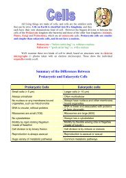
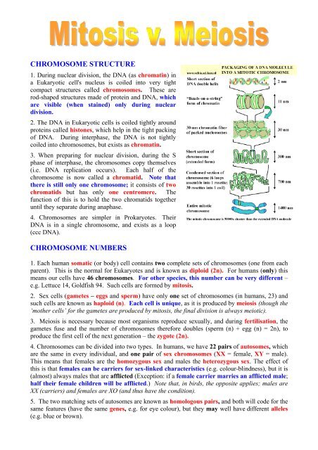
CHROMOSOME STRUCTURE<br />
1. During nuclear division, the DNA (as chromatin) in<br />
a Eukaryotic cell's nucleus is coiled into very tight<br />
compact <strong>structure</strong>s called chromosomes. These are<br />
rod-shaped <strong>structure</strong>s made of protein and DNA, which<br />
are visible (when stained) only during nuclear<br />
division.<br />
2. The DNA in Eukaryotic cells is coiled tightly around<br />
proteins called histones, which help in the tight packing<br />
of DNA. During interphase, the DNA is not tightly<br />
coiled into chromosomes, but exists as chromatin.<br />
3. When preparing for nuclear division, during the S<br />
phase of interphase, the chromosomes copy themselves<br />
(i.e. DNA replication occurs). Each half of the<br />
chromosome is now called a chromatid. Note that<br />
there is still only one chromosome; it consists of two<br />
chromatids but has only one centromere. The<br />
function of this is to hold the two chromatids together<br />
until they separate during anaphase.<br />
4. <strong>Chromosome</strong>s are simpler in Prokaryotes. Their<br />
DNA is in a single chromosome, and exists as a loop<br />
(ccc DNA).<br />
CHROMOSOME NUMBERS<br />
1. Each human somatic (or body) cell contains two complete sets of chromosomes (one from each<br />
parent). This is the normal for Eukaryotes and is known as diploid (2n). For humans (only) this<br />
means our cells have 46 chromosomes. For other species, this number can be very different –<br />
e.g. Lettuce 14, Goldfish 94. Such cells are formed by mitosis.<br />
2. Sex cells (gametes – eggs and sperm) have only one set of chromosomes (in humans, 23) and<br />
such cells are known as haploid (n). Each cell is unique, as it is produced by meiosis (though the<br />
‘mother cells’ for the gametes are produced by mitosis, the final division is always meiotic).<br />
3. Meiosis is necessary because most organisms reproduce sexually, and during fertilisation, the<br />
gametes fuse and the number of chromosomes therefore doubles (sperm (n) + egg (n) = 2n), to<br />
produce the first cell of the next generation – the zygote (2n).<br />
4. <strong>Chromosome</strong>s can be divided into two types. In humans, we have 22 pairs of autosomes, which<br />
are the same in every individual, and one pair of sex chromosomes (XX = female, XY = male).<br />
This means that females are the homozygous sex and males the heterozygous sex. The effect of<br />
this is that females can be carriers for sex-linked characteristics (e.g. colour-blindness), but it is<br />
(almost) always males that are afflicted (Exception: if a female carrier marries an afflicted male;<br />
half their female children will be afflicted.) Note that, in birds, the opposite applies; males are<br />
XX (carriers) and females are XO (and thus have the condition).<br />
5. The two matching sets of autosomes are known as homologous pairs, and both will code for the<br />
same features (have the same genes, e.g. for eye colour), but they may well have different alleles<br />
(e.g. blue or brown).
PROKARYOTES<br />
CELL DIVISION<br />
1. Prokaryotic cells divide by binary fission, in which the DNA<br />
replicates and then the cell divides in two. That’s all – no mitosis, no<br />
spindle, no nothing!<br />
EUKARYOTES<br />
1. A cell typically goes through three phases during its life, beginning with growth before it can<br />
divide. This is known as the cell cycle and consists of three phases:<br />
A.<br />
B.<br />
C.<br />
Interphase<br />
Mitosis (nuclear division, producing 2 identical nuclei)<br />
Cytokinesis (cell division, producing 2 roughly equal cells)<br />
A. INTERPHASE<br />
1. This is the longest phase of the cell cycle and can<br />
last from a few hours to many years. It used to be<br />
known as the ‘resting phase’, which is completely<br />
misleading, as it is during this time of normal<br />
metabolic activity that the cell performs all its normal<br />
functions, beginning with growth and development.<br />
2. Interphase can be divided into 3 stages:<br />
I. G1 Phase - A period of normal metabolic activity – the number of cell organelles<br />
increases to normal levels and the volume of cytoplasm increases too, eventually reaching<br />
mature size. A cell can remain in this phase indefinitely.<br />
II. S Phase - The DNA and chromosomes replicate. The cell is now committed to division.<br />
III. G2 Phase - Structures directly involved with mitosis are formed. The new DNA is<br />
checked for errors and the substances that will be needed during mitosis are synthesised.<br />
B. MITOSIS<br />
1. Mitosis is only the division of the nucleus into two identical nuclei. It is a continuous process,<br />
but has been artificially divided into 4 steps. These can be remembered as I Pee MAT:<br />
I. Prophase<br />
II. Metaphase<br />
III. Anaphase<br />
IV. Telophase
I. Prophase<br />
1. The chromatin condenses into chromosomes by<br />
dehydrating and coiling. The chromosomes consist of<br />
two identical sister chromatids, joined together by a<br />
centromere. For the first time, they can be seen with<br />
alight microscope.<br />
2. The nucleolus and nuclear envelope disappear, and<br />
the centriole (animal cells only) divides into two<br />
centrosomes, which move apart, creating the spindle.<br />
This eventually fills the whole of the cell and is made of<br />
the protein tubulin which forms spindle fibres – a form<br />
of microtubule.<br />
II. Metaphase<br />
The chromosomes are moved to the equator of the cell,<br />
and the centromeres are attached to the spindle fibres, so<br />
that the sister chromatids line up in the centre of the cell.<br />
III. Anaphase<br />
The centromeres of each chromosome divide and are<br />
pulled apart by the contraction of the spindle fibres, thus<br />
moving the chromosomes (as they must now be<br />
called) to opposite poles of the cell.<br />
IV. Telophase<br />
1. After the chromosomes reach the poles, the spindle<br />
disappears and the chromosomes return to their<br />
functional chromatin state by rehydrating and uncoiling.<br />
2. A new nuclear envelope begins to form around the<br />
chromosomes at each end of the cell, each with its own<br />
nucleolus. With mitosis now over, cytokinesis can<br />
begin.<br />
C. CYTOKINESIS<br />
1. Following mitosis, cytokinesis completes the process of cell division. The two cells formed are<br />
roughly equal in size. It is rather different in animal and plant cells:<br />
I. Animal cells. The cytoplasm divides by the pinching of the cell in the middle.<br />
II. Plant cells. In a plant cell, a new cell wall begins to form across the centre of the<br />
cell, along<br />
the line of the cell plate (or middle lamella), which is made of pectin. A new cellulose cell<br />
wall is then laid down on both side of this, and new cell membranes form inside that,<br />
leaving gaps through which the cytoplasm of one cell links to that of its sister. These gaps<br />
are known as plasmodesma (pl. plasmodesmata).
MEIOSIS<br />
Meiosis is a nuclear division that reduces the number of chromosomes in new cells to half the<br />
original number (diploid, 2N → haploid, N). This counteracts the fusion of cells in fertilisation<br />
(haploid, N → diploid, 2N). Crucially, it also produces unique gametes, which means that sexual<br />
reproduction results in variation, the raw material for evolution. Whilst mitosis can occur<br />
anywhere in an animal, meiosis is confined to the gonads (sex organs).<br />
1. Most organisms reproduce sexually, by the fusion of separate male (= sperm) and female (= egg<br />
or ovum) gametes. Eggs are much larger and non-motile; sperm are the smallest human cell and<br />
swim by means of a flagellum. Reproduction therefore depends on water.<br />
2. Meiosis is the second type of nuclear division, but in this case the number of chromosomes is<br />
halved. As in mitosis, meiosis is followed by cytokinesis. It is otherwise very different:<br />
I. Meiosis produces daughter cells with half the number of chromosomes of the parent cell, i.e.<br />
the cell goes from diploid to haploid.<br />
II. Meiosis consists of two nuclear divisions, resulting in four haploid daughter cells.<br />
III. The cells produced by meiosis are not all alike. Each cell is unique and this variation is<br />
produced by two processes – independent segregation of the chromosomes and by<br />
crossing-over. Both take place during Meiosis I.
STAGES OF MEIOSIS<br />
Meiosis consists of two divisions:<br />
A. During meiosis I homologous pairs of chromosomes are separated and cells become haploid.<br />
B. During meiosis II the sister chromatids of each chromosome are separated (as in mitosis).<br />
Meiosis I<br />
1. At the start of meiosis, each chromosome consists of two sister chromatids, connected at the<br />
centromere. However, meiosis differs from mitosis, in that homologous pairs of chromosomes<br />
come together at the start of the meiosis, in a process called synapsis, forming a tetrad.<br />
Prophase I.<br />
1. Just as in mitosis, the nucleolus and nuclear membrane disappear, and the centriole divides and<br />
forms the spindle. As in mitosis, the chromosomes dehydrate and become thick and visible, but<br />
with the chromosomes of each homologous pair tangled together. There are thus four chromatids<br />
in each pair of chromosomes (N.B. and so only half as many ‘sets’ visible in diagrams).<br />
2. Portions of each chromatid may break off and reattach to an<br />
adjacent chromatid on the homologous chromosome. The places<br />
where this happens are called chiasma (pl. chiasmata). The whole<br />
process is called crossing-over and it results in unique combination<br />
of alleles on each chromatid. Crossing-over can only take place<br />
between genes. This is an essential part of the genetic<br />
recombination that takes place in meiosis.<br />
Metaphase I. Homologous pairs are still together and arranged on<br />
the equator of the cell.<br />
Anaphase I. The homologous pairs of chromosomes separate from each other, as the spindle fibres<br />
pull one of each pair to opposite ends of the cell. The random separation of the homologous<br />
chromosomes that this produces is called independent assortment, and results in 2 23 different<br />
gametes being possible from any one human being – a couple could have 2 46 different children!<br />
Telophase I. Cytokinesis takes place; and each new cell is haploid, containing one chromosome<br />
from each pair.<br />
MEIOSIS II<br />
1. In males, this follows immediately. In females, meiosis I takes place before birth, but meiosis II<br />
takes place only after the sperm has fused with the egg (i.e. immediately before fertilisation). The<br />
chromosomes do not replicate before this, second, division, so the gametes formed will have only<br />
half the DNA of a normal cell.<br />
2. The second division of meiosis is essentially the same as mitosis. Often Telophase I is omitted<br />
and, after cell division, both daughter cells are already at the end of prophase II.<br />
N.B. When answering questions about the stage of meiosis (in a diagram)<br />
or when a particular event occurs (in words), you MUST state if it is the<br />
first or second division. So ‘Prophase I’ is usually the correct answer,<br />
but ‘Prophase’ is ALWAYS WRONG! Suggestion: when in doubt, the answer is<br />
NORMALLY Prophase I, but ALWAYS in division I, as division II looks the same as mitosis.<br />
© IHW April 2006