Horizontal Section Through Neck
Horizontal Section Through Neck
Horizontal Section Through Neck

Create successful ePaper yourself
Turn your PDF publications into a flip-book with our unique Google optimized e-Paper software.
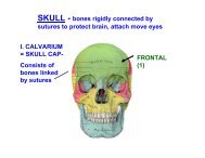
I. OVERVIEW OF NECK - neck is compartmentalized<br />
LAT.<br />
POST.<br />
Plane of section<br />
ANT.<br />
HORIZONTAL SECTION THROUGH NECK<br />
1. Posterior<br />
Compartment -<br />
Vertebrae and muscles<br />
which support and<br />
move head & neck<br />
2. Anterior<br />
Compartment- Viscera<br />
and rostral continuation<br />
GI & Respiratory<br />
Systems<br />
3. Lateral Compartment-<br />
Blood vessels & nerve
BACK<br />
+ SUBOCCIP<br />
MUSCLES<br />
Post side - Deep<br />
Muscles (like<br />
back)- extensor<br />
& Suboccipital<br />
Muscles<br />
1. POSTERIOR COMPARTMENT<br />
- muscles move head and neck<br />
SCALENE<br />
MUSCLES<br />
Lateral side -<br />
Scalene<br />
muscles - flex<br />
neck laterally<br />
PRE-<br />
VERTEBRAL<br />
MUSCLES<br />
Anterior side -<br />
Prevertebral Muscles -<br />
directly anterior to<br />
vertebrae - flex head &<br />
neck
2. ANTERIOR COMPARTMENT - VISCERA<br />
Esophagus<br />
Trachea<br />
In thorax,<br />
trachea is<br />
anterior to<br />
esophagus
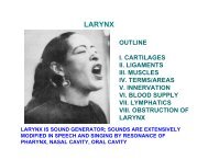
ANTERIOR COMPARTMENT - VISCERA<br />
Anterior<br />
Compartment -<br />
Larynx is part of<br />
upper end of<br />
respiratory systemspecialized<br />
for<br />
sound production;<br />
also acts as<br />
‘sphincter’ of<br />
respiratory system-<br />
Thyroid cartilage is<br />
Adam's apple<br />
Larynx<br />
Trachea
SAY<br />
AAHH!<br />
PHARYNX
Nasal<br />
Cavity<br />
Oral<br />
cavity<br />
Nose<br />
Larynx<br />
Trachea<br />
Pharynx<br />
ANTERIOR<br />
COMPARTMENT -<br />
VISCERA<br />
Esophagus<br />
1) Larynx &<br />
Esophagus<br />
open into<br />
pharynx<br />
2) Pharynx - a<br />
tube of<br />
muscles &<br />
fascia that<br />
opens to nasal<br />
and oral<br />
cavities
HYOID BONE- parts: body, greater & lesser horns – All<br />
Infrahyoid & Suprahyoid attach to Body of Hyoid<br />
(except Sternothyroid-> thyroid cartilage)<br />
Palpable<br />
in neck<br />
Hyoid means<br />
"U" shaped<br />
GREATER<br />
HORNS<br />
LESSER<br />
HORNS<br />
BODY
ANTERIOR COMPARTMENT - moveable, changes<br />
shape in swallowing, speech<br />
Hyoid Bone –<br />
attached to<br />
larynx, pharynx &<br />
tongue; free<br />
floating; attached<br />
by ligaments and<br />
moved by<br />
muscles<br />
trachea<br />
Hyoid- means "U"<br />
shaped<br />
HYOID BONE<br />
Pharynx<br />
esophagus
TONGUE<br />
Hyoid<br />
Bone<br />
Larynx<br />
HYOID BONE<br />
- muscles that<br />
move hyoid<br />
bone move<br />
larynx &<br />
tongue, for<br />
Swallowing,<br />
Talking
3. LATERAL COMPARTMENT - CAROTID SHEATH<br />
Lateral Compartmentlateral<br />
and posterior to<br />
pharynx<br />
Contained in Carotid<br />
Sheath<br />
1) Common and<br />
Internal Carotid<br />
arteries; 2) Int. jugular<br />
vein, 3) Vagus nerve
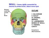
OUTLINE<br />
II. MUSCLES<br />
III. NERVES<br />
IV. ARTERIES<br />
V. VEINS<br />
VI. FASCIA<br />
VII. LYMPHATICS
II. MUSCLES OF NECK
A. MUSCLES OF NECK - NOT ATTACHED TO HYOID -<br />
move head & neck<br />
1. STERNO-<br />
CLEIDOMASTOID<br />
0 - Two heads 1)<br />
manubrium of sternum<br />
2) clavicle- medial 1/3<br />
I - mastoid process of<br />
temporal bone<br />
Act - bilateral - flex head<br />
unilateral rotate head,<br />
face to directed opposite<br />
side<br />
Inn - CN XI Accessory n.<br />
TORTICOLLIS –<br />
Contracture of<br />
Sternocleidomastoid
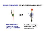
MUSCLES OF NECK - NOT ATTACHED TO HYOID<br />
2. SCALENUS<br />
ANTERIOR AND<br />
SCALENUS MEDIUS<br />
O - vertebraetrans<br />
processes<br />
upper cervical<br />
I - rib 1<br />
A - flex neck &<br />
elevate rib 1<br />
Inn - ventral<br />
rami of cervical<br />
spinal nerves<br />
THESE MUSCLES ARE IMPORTANT<br />
LANDMARKS IN NECK
B. INFRAHYOID MUSCLES - all depress hyoid<br />
1. OMOHYOID (omo = greek<br />
for shoulder) - Two bellies -<br />
Inf. Belly- Scapula- medial to<br />
suprascapular notch
INFRAHYOID MUSCLES - all depress hyoid<br />
NOSE<br />
2. STERNOHYOID<br />
O- Manubrium &<br />
clavicle<br />
1. OMOHYOID<br />
intermediate<br />
tendon to<br />
clavicle, rib 1;<br />
Sup. belly to<br />
hyoid<br />
deeper<br />
4. THYROHYOID -O -<br />
thyroid cartilage; also<br />
elevates larynx<br />
3. STERNOTHYROID --<br />
O - manubrium I -<br />
thyroid cartilage; also<br />
depresses larynx
SUPRAHYOID MUSCLES - all elevate hyoid
SUPRAHYOID MUSCLES - all elevate hyoid<br />
1. DIGASTRIC - two bellies / two cranial nerves -<br />
insert to hyoid via intermediate tendon<br />
Post Belly-<br />
Temp. Bone,<br />
mastoid<br />
notch<br />
(medial to<br />
mastoid<br />
process)<br />
Inn - CN VII<br />
Act- Depress mandible;<br />
- MAJOR EFFECT is to OPEN Mouth<br />
NOSE<br />
Ant. Belly-<br />
Mandible<br />
Inn- CN V
SUPRAHYOID MUSCLES - all elevate hyoid<br />
2. STYLOHYOID<br />
O-styloid<br />
process of temp<br />
bone<br />
tendon splits to<br />
surround<br />
digastric tendon<br />
Inn - CN VII
SUPRAHYOID MUSCLES - all elevate hyoid<br />
3. MYLOHYOID - forms muscular floor of mouth<br />
O - mylohyoid line on<br />
inner side of mandible<br />
Act - Elevates floor of<br />
mouth in swallowing<br />
Inn - CN V - from V3
SUPRAHYOID MUSCLES - all elevate hyoid<br />
4. GENIOHYOID -<br />
O - inner side of<br />
mandible<br />
above mylohyoid<br />
A - Elevates hyoid<br />
and draws forward<br />
Inn - C1 branch<br />
hitch-hiking with<br />
Hypoglossal nerve<br />
(CN XII)
III. NERVES OF NECK<br />
A. CERVICAL PLEXUS<br />
from C2-C4<br />
ventral primary<br />
rami
1) Lesser Occipital<br />
C2 behind ear<br />
4) Supraclavicular -<br />
C3, C4 lower neck &<br />
shoulder<br />
A. CERVICAL PLEXUS NOSE<br />
2) Great Auricular -<br />
(C2, C3) skin over<br />
parotid, inf. to ear<br />
3) Transverse<br />
Cervical -C2, C3<br />
ant. neck<br />
emerge from post<br />
border of<br />
sternocleidomastoid<br />
m.
CERVICAL PLEXUS<br />
NOSE
B. ANSA CERVICALIS<br />
- fibers from C1 join<br />
Hypoglossal Nerve<br />
(XII)<br />
- some leave & join<br />
fibers of C2 & C3 to<br />
form ANSA (loop)<br />
Cervicalis<br />
- other fibers continue<br />
with XII to innervate<br />
Thyrohyoid &<br />
Geniohyoid<br />
(Looks like XII<br />
innervates neck<br />
muscles; actually C1-<br />
C3 do)
CN XII<br />
Receives<br />
hitchhiking<br />
fibers<br />
ANSA CERVICALIS
IV. ARTERIES OF HEAD AND NECK<br />
SUBCLAVIAN A.<br />
A. SUBCLAVIAN<br />
ARTERY<br />
At root of neckpasses<br />
to arm -<br />
becomes Axillary a.<br />
( rib 1)<br />
- Scalenus Anterior<br />
muscle divides<br />
Subclavian into 3<br />
parts
SUBCLAVIAN ARTERY - divided into 3 parts by<br />
scalenus ant. muscle<br />
Part 2- post to<br />
scal. ant.<br />
1) Costocervical<br />
trunk - branches<br />
a) Superior<br />
intercostal a. first<br />
two int spaces;<br />
b) Deep cervical<br />
a. - deep neck<br />
Part 3 - lat to scalenus ant. No Branches<br />
Part 1- medial to<br />
scal. ant,<br />
1) Vertebral a.<br />
2) Int. thoracic a.<br />
3) Thyrocervical<br />
trunk: branches -<br />
a) Inf. thyroid<br />
b) Trans. cervical<br />
c) Suprascapular
NOSE<br />
Sternocleidomastoid
Transverse<br />
cervical<br />
artery<br />
Suprascapular<br />
artery<br />
Phrenic n.<br />
Scalenus Ant.<br />
M.
B. EXTERNAL CAROTID ARTERY<br />
5. OCCIPITAL A-<br />
POST SCALP<br />
6. POST.<br />
AURICULAR A-<br />
POST TO EAR<br />
4. FACIAL A- BELOW<br />
THEN ON SURFACE<br />
OF MANDIBLE<br />
3. LINGUAL A-<br />
TONGUE<br />
2. ASCENDING<br />
PHARYNGEAL A-<br />
ASCENDS TO<br />
PHARYNX<br />
1. SUPERIOR<br />
THYROID A-<br />
DESCENDS TO<br />
THYROID
Superficial temporalscalp<br />
& temporalis<br />
Post auricular- post. ear &<br />
scalp<br />
Occipital- post.<br />
scalp<br />
EXTERNAL CAROTID ARTERY<br />
Ascending pharyngealpharynx<br />
Maxillary<br />
Facial<br />
Lingual- tongue<br />
Superior thyroid- br. is Sup<br />
laryngeal
EXTERNAL CAROTID ARTERY<br />
Reflect sternocleidomastoid<br />
Common<br />
carotid divides<br />
-> int & ext<br />
carotid at upper<br />
border thyroid<br />
cartilage
Post side<br />
POST.<br />
AURICULAR<br />
OCCIPITAL<br />
Ant side<br />
SUP.<br />
TEMPORAL<br />
MAXILLARY<br />
FACIAL<br />
LINGUAL<br />
SUP.<br />
THYROID
Post.<br />
Auricular<br />
External<br />
Jugular<br />
Sup.<br />
Temp.<br />
PD<br />
V. VEINS OF NECK<br />
RM<br />
AD<br />
Max<br />
Common<br />
facial<br />
Ant<br />
jug<br />
Facial<br />
1. Superficial Temporal &<br />
Maxillary vv. form<br />
Retromandibular V. (RM)<br />
2. Retromand. V. Divides Ant.<br />
(AD) and Post. (PD) divisions<br />
3. Ant. Division joins Facial V.<br />
to form Common Facial V. -><br />
Int. jugular V.<br />
4. Post. Division joins Post.<br />
Auricular V. to form External<br />
Jugular V-> Subclavian V.<br />
5. Ant. Jugular from veins<br />
below mandible -> Ext. Jugular<br />
above clavicle
Pattern of<br />
Venous<br />
Drainage<br />
VEINS OF NECK
A. Superficial<br />
fascia:<br />
- connective<br />
tissue below<br />
dermis<br />
- completely<br />
surrounds neck -<br />
thin and hard to<br />
demonstrate<br />
- contains<br />
Platysma &<br />
Superficial veins<br />
VI. FASCIA OF NECK
B. Deep Cervical<br />
fascia- one layer<br />
surrounds neck,<br />
other layers form<br />
tubes (names poorly<br />
chosen)<br />
2. Prevertebral<br />
Layer<br />
FASCIA OF NECK<br />
1. Investing layer<br />
4. Carotid<br />
sheath<br />
3. Pre-tracheal<br />
layer
N<br />
O<br />
S<br />
E<br />
Carotid<br />
Sheath<br />
Prevertebral<br />
layer<br />
FASCIA OF NECK<br />
Pretracheal<br />
layer<br />
1. Investing layer of deep cervical<br />
fascia- surrounds neck, splits<br />
around sternocleid., trap, supra &<br />
infrahyoid<br />
2. Prevertebral Layer- surrounds<br />
vert. column & muscles back of<br />
neck, prevertebral, lateral<br />
vertebral and suboccipital m.<br />
3. Pretracheal Layer- surrounds<br />
trachea, esophag. & thyroid<br />
continues to mediastinum<br />
4. Carotid Sheath- surrounds<br />
common & int carotid, int jugular<br />
and X (not: Symp. Chain)<br />
Retropharyngeal Space- between<br />
PreTrach & Pre Vert layers -<br />
infection from head (tonsillitis) can<br />
spread to mediastinum
RETROPHARYNGEAL ABSCESS<br />
Infection in retropharyngeal<br />
space can spread unimpeded<br />
to mediastinum
FASCIA OF NECK
O<br />
VII. LYMPHATICS OF HEAD AND NECK<br />
RA<br />
Deep<br />
Cerv.<br />
Chain<br />
P<br />
SMan<br />
B<br />
RP<br />
SMen<br />
PT<br />
three groups (two arranged as<br />
rings; drain to chain)<br />
A. Superficial Ring;<br />
Submental, Submandibular,<br />
Buccal, Parotid, Retroauricular<br />
& Occipital nodes<br />
B. Deep Ring: Pretracheal,<br />
Retropharyngeal nodes<br />
C. Deep cervical chainalong<br />
Internal Jugular vein;<br />
receive lymph from all<br />
above nodes<br />
D. Jugular lymph trunk -to<br />
Right lymphatic duct or<br />
Thoracic duct