Shortness of breath while lying down - Canadian Medical ...
Shortness of breath while lying down - Canadian Medical ...
Shortness of breath while lying down - Canadian Medical ...

Create successful ePaper yourself
Turn your PDF publications into a flip-book with our unique Google optimized e-Paper software.
CMAJ<br />
What is your call?<br />
<strong>Shortness</strong> <strong>of</strong> <strong>breath</strong> <strong>while</strong> <strong>lying</strong> <strong>down</strong>: a woman<br />
with orthopneic asthma<br />
Chou-Han Lin, Mong-Wei Lin, Jin-Shing Chen, Chong-Jen Yu<br />
See related practice article by Lee and Cheng, page 80<br />
A36-year-old woman with a six-month<br />
history <strong>of</strong> worsening shortness <strong>of</strong><br />
<strong>breath</strong> was admitted to hospital for<br />
investigation. She had received a diagnosis <strong>of</strong><br />
bronchial asthma six months earlier, for which<br />
treatment with steroids and bronchodilators had<br />
been ineffective. Her history included allergic<br />
rhinitis and an eight pack-year history <strong>of</strong> smoking,<br />
which she had stopped since her diagnosis<br />
<strong>of</strong> asthma.<br />
On admission, the patient was in mild respiratory<br />
distress. She had a temperature <strong>of</strong><br />
36.5°C, a pulse rate <strong>of</strong> 90 beats/min, a respiratory<br />
rate <strong>of</strong> 22 <strong>breath</strong>s/min and a blood pressure<br />
<strong>of</strong> 116/64 mm Hg. She had diffuse wheezing on<br />
expiration and during the first half <strong>of</strong> inspiration.<br />
We noticed that she had orthopnea and<br />
had to use three pillows for relief. She had not<br />
mentioned this symptom to her treating physicians.<br />
Her distress when <strong>lying</strong> <strong>down</strong> occurred<br />
even though her oxygen saturation remained<br />
above 98% on 2 L <strong>of</strong> oxygen per minute via<br />
nasal cannula. A pulmonary function test<br />
showed a forced expiratory volume in the first<br />
Inspiratory flow, L/s Expiratory flow, L/s<br />
8<br />
6<br />
4<br />
2<br />
0<br />
2<br />
4<br />
6<br />
8<br />
10<br />
1<br />
2 3 4 5<br />
Volume, L<br />
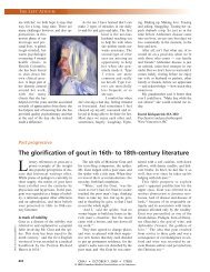
Figure 1: Flow–volume loop from a 36-year-old<br />
woman with orthopnea, showing a plateau phase<br />
on the expiratory arm.<br />
second <strong>of</strong> expiration (FEV 1) <strong>of</strong> 17.5% <strong>of</strong> predicted,<br />
a forced vital capacity (FVC) <strong>of</strong> 75.7%<br />
and an FEV 1/FVC ratio <strong>of</strong> 20.4% <strong>of</strong> predicted.<br />
Her flow–volume loop showed slowing and<br />
flattening in the expiratory limb (Figure 1),<br />
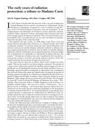
which led us to re-examine her chest radiograph<br />
taken on admission (Figure 2). The tracheal air<br />
column was not deviated, and no conspicuous<br />
lesions were noted.<br />
What is your diagnosis?<br />
a. Upper airway obstruction<br />
b. Left-sided heart failure (cardiac asthma)<br />
c. Status asthmaticus<br />
d. Hepatopulmonary syndrome<br />
e. Exacerbation <strong>of</strong> chronic obstructive pulmon -<br />
ary disease<br />
Figure 2: Posteroanterior chest radiograph, showing<br />
a straight tracheal air column (arrow) without<br />
narrowing or deviation.<br />
Practice<br />
Competing interests: None<br />
declared.<br />
This article has been peer<br />
reviewed.<br />
Correspondence to:<br />
Dr. Chong-Jen Yu,<br />
jefferycjyu@ntu.edu.tw<br />
CMAJ 2011. DOI:10.1503<br />
/cmaj.081801<br />
© 2011 <strong>Canadian</strong> <strong>Medical</strong> Association or its licensors CMAJ, January 11, 2011, 183(1) 77
Practice<br />
78 CMAJ, January 11, 2011, 183(1)<br />
What is your next step?<br />
a. Give magnesium sulfate along with standard<br />
combination <strong>of</strong> oxygen, bronchodilators and<br />
steroids<br />
b. Give antibiotics along with standard combination<br />
<strong>of</strong> oxygen, bronchodilators and steroids<br />
c. Order a lateral chest radiograph and a computed<br />
tomography (CT) scan <strong>of</strong> the chest<br />
d. Arrange contrast echocardiography<br />
e. Administer intravenous nitroglycerin with an<br />
angiotensin-converting-enzyme inhibitor<br />
The diagnosis is (a) upper airway obstruction,<br />
and the next step is (c) order a lateral chest radio -<br />
graph and a CT scan. We made the diagnosis<br />
with the help <strong>of</strong> the flow–volume loop, which<br />
was consistent with intrathoracic upper airway<br />
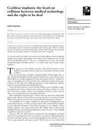
obstruction. Her lateral chest radiograph showed<br />
a tumour-like opacity in the trachea (Figure 3).<br />
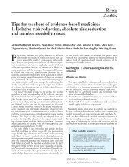
The CT scan showed a tracheal tumour with<br />
almost total obstruction <strong>of</strong> the tracheal lumen<br />
(Figure 4). Bronchoscopy showed a whitish<br />
bosselated tumour arising from the anterior wall<br />
<strong>of</strong> the trachea 6 cm below the vocal cords.<br />
The patient underwent segmental resection <strong>of</strong><br />
the trachea with anastomosis. A whitish elastic<br />
tumour (2 × 2 × 2 cm) was found 3 cm above the<br />
carina, extending from the anterior tracheal wall<br />
(Figure 5). Histopathology results were consistent<br />
with a pleomorphic adenoma. After resection, her<br />
wheezing and orthopnea disappeared.<br />
Figure 3: Lateral chest radiograph, showing an<br />
opacity (arrow) arising from the anterior tracheal<br />
wall with a patent posterior tracheal lumen.<br />
Discussion<br />
Orthopnea is <strong>of</strong>ten associated with congestive<br />
heart failure, chronic obstructive pulmonary disease,<br />
anterior mediastinal tumours and diaphragmatic<br />
weakness. Tracheal tumours, as in our<br />
patient, are a rare cause <strong>of</strong> orthopnea. 1 Because<br />
our patient’s tumour stalk emerged from the anterior<br />
tracheal wall, part <strong>of</strong> the tumour approached<br />
the opposing membranous portion and occluded<br />
the lumen when she lay <strong>down</strong> (Figure 4).<br />
The posteroanterior chest radiograph is an<br />
appropriate initial imaging study for assessing<br />
dyspnea. However, it is a relatively poor tool for<br />
identifying tumours situated distally in the anterior<br />
or posterior tracheal wall. A lateral chest<br />
radio graph is helpful for assessing tracheal<br />
tumours in these locations. Most pleomorphic<br />
adenomas are located in the upper third <strong>of</strong> the trachea.<br />
2 However, the intrathoracic location <strong>of</strong> our<br />
patient’s tumour lowered its visibility on the<br />
postero anterior view <strong>of</strong> the chest radiograph. A<br />
lateral view provides better exposure, without<br />
interference from the thoracic spine and sternum. 3<br />
Computed tomography is useful in detecting<br />
and assessing tracheal tumours, given that not all<br />
s<strong>of</strong>t-tissue lesions are visible on chest radiography.<br />
As in our case, pulmonary function testing<br />
can be helpful in diagnosing tracheal lesions, with<br />
confirmation provided by direct visualization<br />
using bronchoscopy.<br />
Differential diagnosis<br />
Orthopnea occurs most frequently in patients<br />
with heart failure. These patients find relief in an<br />
upright position because this posture reduces the<br />
demand on their pulmonary circulation. A constellation<br />
<strong>of</strong> clinical symptoms and signs such as<br />
paroxysmal nocturnal dyspnea, distention <strong>of</strong> the<br />
veins in the neck and lung crackles can also be<br />
found in patients with heart failure. The subacute<br />
course and lack <strong>of</strong> these features in our patient<br />
ruled out this disease as a cause <strong>of</strong> her orthopnea.<br />
Figure 4: Chest computed tomography scan, showing<br />
a pedunculated tumour (arrow) nearly obliterating<br />
the tracheal lumen.
Figure 5: Tracheal tumour attached to the anterior<br />
<strong>of</strong> the tracheal ring.<br />
Also, her chest radiograph did not show cardiomegaly<br />
or pulmonary edema, which are <strong>of</strong>ten<br />
associated with heart failure.<br />
Hepatopulmonary syndrome develops in the<br />
setting <strong>of</strong> chronic liver disease. The resulting<br />
dilated pulmonary capillaries create intrapulmonary<br />
shunts. Patients with this condition <strong>of</strong>ten<br />
exhibit platypnea (shortness <strong>of</strong> <strong>breath</strong> relieved<br />
with <strong>lying</strong> <strong>down</strong>) or orthodeoxia (deoxygenation<br />
in the sitting or standing position) rather than<br />
orthopnea.<br />
Occasionally, orthopnea is also found in<br />
patients with chronic obstructive lung disease<br />
and asthma, since the upright position may be<br />
associated with reduced pooling <strong>of</strong> lung secretions<br />
and improved diaphragmatic excursion.<br />
Orthopnea tends to be a late manifestation <strong>of</strong><br />
chronic lung disease. Wheezing is commonly<br />
observed in chronic obstructive pulmonary disease<br />
and asthma, and subacute or chronic upper<br />
airway obstruction can mimic diseases <strong>of</strong> peripheral<br />
airflow obstruction. Our patient’s wheezing<br />
pattern, however, which extended from expiration<br />
to inspiration, would be unusual in these<br />
conditions.<br />
Pulmonary function testing<br />
Not only is spirometry a diagnostic tool for<br />
chronic obstructive lung disease and asthma, it<br />
can also help identify upper airway obstruction<br />
(Figure 6). 4 FEV 1, the spirometric parameter<br />
used for assessing chronic obstructive pulmonary<br />
disease and asthma, is less sensitive in<br />
FVC<br />
FEV 1<br />
FEV 1/FVC<br />
Flow–volume<br />
loop<br />
Small airway obstruction Upper airway obstruction<br />
COPD Asthma attack<br />
N to<br />
N to<br />
Variable<br />
intrathoracic<br />
diagnosing upper airway obstruction. 5 For this<br />
condition, the ratio <strong>of</strong> maximal expiratory to<br />
maximal inspiratory flow at 50% <strong>of</strong> vital capacity<br />
(MEF 50:MIF 50 ratio) is more useful. The<br />
MEF 50:MIF 50 ratio, normally between 0.9 and<br />
1.0, will decrease to less than 0.2 in patients with<br />
an intrathoracic obstruction or increase to more<br />
than 1.0 in those with an extrathoracic obstruction.<br />
6 As shown in Figure 6, direct inspection <strong>of</strong><br />
the flow–volume loop is helpful when interpreting<br />
spirometric data and can be used along with<br />
specific flow calculations to make the diagnosis<br />
<strong>of</strong> upper airway obstruction. 6<br />
References<br />
1. Gaissert HA, Grillo HC, Shadmehr B, et al. Uncommon primary<br />
tracheal tumors. Ann Thorac Surg 2006;82:268-73.<br />
2. Aribas OK, Kanat F, Avunduk MC. Pleomorphic adenoma <strong>of</strong><br />
the trachea mimicking bronchial asthma: report <strong>of</strong> a case. Surg<br />
Today 2007;37:493-5.<br />
3. Li W, Ellerbroek NA, Libshitz HI. Primary malignant tumors <strong>of</strong><br />
the trachea; a radiologic and clinical study. Cancer 1990;66: 894-9.<br />
4. Hyatt RE, Scanlon PD, Nakamura M. Interpretation <strong>of</strong> pulmonary<br />
function tests: a practical guide. 3rd ed. Philadelphia:<br />
Lippincott Williams & Wilkins; 2009. Figure 2-7.<br />
5. Miller RD, Hyatt RE. Obstructing lesions <strong>of</strong> the larynx and trachea:<br />
clinical and physiologic characteristics. Mayo Clin Proc<br />
1969;44:145-61.<br />
6. Lunn WW, Sheller JR. Flow volume loops in the evaluation <strong>of</strong><br />
upper airway obstruction. Otolaryngol Clin North Am 1995;28:<br />
721-9.<br />
Affiliations: From the Division <strong>of</strong> Chest Medicine, Department<br />
<strong>of</strong> Internal Medicine (C.-H. Lin), Far Eastern Memorial<br />
Hospital, Taipei, Taiwan; the Division <strong>of</strong> Thoracic Surgery,<br />
Department <strong>of</strong> Surgery (M.-W. Lin, Chen), National Taiwan<br />
University Hospital, Taipei, Taiwan; and the Division <strong>of</strong><br />
Chest Medicine, Department <strong>of</strong> Internal Medicine (Yu),<br />
National Taiwan University Hospital, Taipei, Taiwan<br />
Practice<br />
Variable<br />
extrathoracic Fixed<br />
N N N<br />
Figure 6: Typical findings <strong>of</strong> pulmonary function tests in patients with small<br />
(lower) airway and upper airway obstruction. COPD = chronic obstructive pulmonary<br />
disease, FVC = forced vital capacity, FEV 1 = forced expiratory volume in<br />
the first second <strong>of</strong> expiration. Reprinted, with permission, from Hyatt et al. 4<br />
Copyright © 2009 Lippincott Williams & Wilkins (http://lww.com).<br />
CMAJ, January 11, 2011, 183(1) 79<br />
N<br />
N