Rapid Maxillary Expansion: A Unique Treatment Modality in ... - JCDR
Rapid Maxillary Expansion: A Unique Treatment Modality in ... - JCDR
Rapid Maxillary Expansion: A Unique Treatment Modality in ... - JCDR

You also want an ePaper? Increase the reach of your titles
YUMPU automatically turns print PDFs into web optimized ePapers that Google loves.
906<br />
Review Article<br />
<strong>Rapid</strong> <strong>Maxillary</strong> <strong>Expansion</strong>: A <strong>Unique</strong><br />
<strong>Treatment</strong> <strong>Modality</strong> <strong>in</strong> Dentistry<br />
S. Arv<strong>in</strong>d KumAr, deepA GurunAthAn, muruGAnAndhAm, ShivAnGi ShArmA<br />
ABSTRACT<br />
<strong>Rapid</strong> <strong>Maxillary</strong> expansion or palatal expansion as it is sometimes<br />
called, occupies unique niche <strong>in</strong> dentofacial therapy. <strong>Rapid</strong><br />
<strong>Maxillary</strong> expansion is a skeletal type of expansion that<strong>in</strong>volves<br />
the separation of the mid-palatal suture and movement of the<br />
maxillary shelves away from each other. An objective approach to<br />
the design of a suitable appliance should be made by prepar<strong>in</strong>g<br />
InTRoduCTIon<br />
<strong>Rapid</strong> maxillary expansion (RME) is a dramatic procedure with<br />
a long history. <strong>Rapid</strong> <strong>Maxillary</strong> expansion or palatal expansion<br />
as it is sometimes called, occupies unique niche <strong>in</strong> dentofacial<br />
therapy. <strong>Rapid</strong> <strong>Maxillary</strong> expansion or Split palate is a skeletal<br />
type of expansion that <strong>in</strong>volves the separation of the mid-palatal<br />
suture and movement of the maxillary shelves away from each<br />
other.<br />
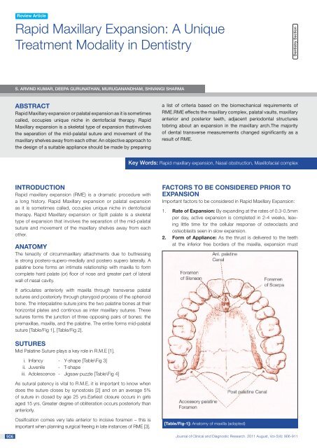
AnATomy<br />
The tenacity of circummaxillary attachments due to buttress<strong>in</strong>g<br />
is strong postero-supero-medially and postero supero laterally. A<br />
palat<strong>in</strong>e bone forms an <strong>in</strong>timate relationship with maxilla to form<br />
complete hard palate (or) floor of nose and greater part of lateral<br />
wall of nasal cavity.<br />
It articulates anteriorly with maxilla through transverse palatal<br />
sutures and posteriorly through pterygoid process of the sphenoid<br />
bone. The <strong>in</strong>terpalat<strong>in</strong>e suture jo<strong>in</strong>s the two palat<strong>in</strong>e bones at their<br />
horizontal plates and cont<strong>in</strong>ous as <strong>in</strong>ter maxillary sutures. These<br />
sutures forms the junction of three oppos<strong>in</strong>g pairs of bones: the<br />
premaxillae, maxilla, and the palat<strong>in</strong>e. The entire forms mid-palatal<br />
suture [Table/Fig 1], [Table/Fig 2].<br />
SuTuRES<br />
Mid Palat<strong>in</strong>e Suture plays a key role <strong>in</strong> R.M.E [1].<br />
i. Infancy - Y-shape [Table\Fig 3]<br />
ii. Juvenile - T-shape<br />
iii. Adolescence - Jigsaw puzzle [Table\Fig 4]<br />
As sutural patency is vital to R.M.E, it is important to know when<br />
does the suture closes by synostosis [2] and on an average 5%<br />
of suture <strong>in</strong> closed by age 25 yrs.Earliest closure occurs <strong>in</strong> girls<br />
aged 15 yrs. Greater degree of obliteration occurs posteriorly than<br />
anteriorly.<br />
Ossification comes very late anterior to <strong>in</strong>cisive foramen – this is<br />
important when plann<strong>in</strong>g surgical free<strong>in</strong>g <strong>in</strong> late <strong>in</strong>stances of RME [3].<br />
a list of criteria based on the biomechanical requirements of<br />
RME.RME effects the maxillary complex, palatal vaults, maxillary<br />
anterior and posterior teeth, adjacent periodontal structures<br />
tobr<strong>in</strong>g about an expansion <strong>in</strong> the maxillary arch.The majority<br />
of dental transverse measurements changed significantly as a<br />
result of RME.<br />
Key Words: <strong>Rapid</strong> maxillary expansion, Nasal obstruction, Maxillofacial complex<br />
FACToRS To BE ConSIdEREd pRIoR To<br />
ExpAnSIon<br />
Important factors to be considered <strong>in</strong> <strong>Rapid</strong> <strong>Maxillary</strong> <strong>Expansion</strong>:<br />
1. Rate of <strong>Expansion</strong>: By expand<strong>in</strong>g at the rates of 0.3-0.5mm<br />
per day, active expansion is completed <strong>in</strong> 2-4 weeks, leav<strong>in</strong>g<br />
little time for the cellular response of osteoclasts and<br />
osteoblasts seen <strong>in</strong> slow expansion.<br />
2. Form of Appliance: As the thrust is delivered to the teeth<br />
at the <strong>in</strong>ferior free borders of the maxilla, expansion must<br />
[Table/Fig-1]: Anatomy of maxilla (adopted)<br />
Journal of Cl<strong>in</strong>ical and Diagnostic Research. 2011 August, Vol-5(4): 906-911<br />
dentistry Section
www.jcdr.net S. Arv<strong>in</strong>d Kumar et al., <strong>Rapid</strong> <strong>Maxillary</strong> <strong>Expansion</strong><br />
[Table/Fig-2]: Sutures <strong>in</strong> the maxillofacial region<br />
[Table/Fig-3]: Mid palant<strong>in</strong>e suture <strong>in</strong> <strong>in</strong>fancy (adopted)<br />
[Table/Fig-4]: Mid palant<strong>in</strong>e suture <strong>in</strong> early adolescence (adopted)<br />
reach to the basal portions. The form of appliance will play an<br />
important role <strong>in</strong> this effort, accord<strong>in</strong>g to its rigidity or flexibility,<br />
i.e. anchorage or control of tipp<strong>in</strong>g.<br />
3. Age and Sex of the patient: The <strong>in</strong>creas<strong>in</strong>g rigidity of the<br />
facial skeleton with advanc<strong>in</strong>g age restricts bony movements<br />
remote from the appliance of expansion, which differs <strong>in</strong> both<br />
sexes.<br />
4. discrepancy between maxillary and mandibular first<br />
molars & bicuspid width is 4mm or more RME <strong>in</strong>dicated.<br />
5. Severity of cross bite i.e number of teeth <strong>in</strong>volved.<br />
6. Initial angulation of molars or premolars: When the<br />
maxillary molars are buccally <strong>in</strong>cl<strong>in</strong>ed, conventional expansion<br />
will tip them further <strong>in</strong>to the buccal musculature and if the<br />
Journal of Cl<strong>in</strong>ical and Diagnostic Research. 2011 August, Vol-5(4): 906-911<br />
mandibular molars are l<strong>in</strong>gually <strong>in</strong>cl<strong>in</strong>ed, the buccal movement to<br />
upright them will <strong>in</strong>crease the need to widen the upper arch.<br />
7. Assessment of roots of deciduous tooth<br />
8. Physical availability of space for expansion.<br />
9. nasal obstruction: All patients considered for RME should<br />
be exam<strong>in</strong>ed for nasal obstruction and, if obstruction is<br />
found, they should be referred to an otolaryngologist before<br />
orthodontic treatment.<br />
10. medical history: S<strong>in</strong>ce the efficacy of maxillary expansion<br />
depends on suture patency and the flexibility of craniofacial<br />
com pelex to adapt to mechanical changes hence medical<br />
conditions alter<strong>in</strong>g these should be considered.<br />
11. meatbolic disorders: Many metabolic disorders are found<br />
associated with suture synotoses which <strong>in</strong>clude hyperthyroidism,<br />
hypophosphatemic vitam<strong>in</strong> D-resistant rickets, and mucopolysaccharidoses<br />
and mucolipidoses. These disorders are<br />
mostly associated with bone metabolism. <strong>Maxillary</strong> expansion<br />
would be futile even <strong>in</strong> young patients if they are suffer<strong>in</strong>g from<br />
any of these diseases.<br />
12. periodontal Type: It is essential to record the thickness of the<br />
g<strong>in</strong>gival tissues dur<strong>in</strong>g cl<strong>in</strong>ical evaluation of the periodontium.<br />
This is especially important because a th<strong>in</strong> and delicate<br />
g<strong>in</strong>giva might be prone to recession after traumatic, surgical,<br />
or <strong>in</strong>flammatory <strong>in</strong>juries [4].<br />
13. mucog<strong>in</strong>gival Health: Orthodontic tooth movement has<br />
significant effect on the mucog<strong>in</strong>gival tissues and hence it is<br />
important to asses the periodontal health of the patient before<br />
perform<strong>in</strong>g OME.<br />
IndICATIonS FoR RmE [5]<br />
Patients who have lateral discrepancies that result <strong>in</strong> either<br />
unilateral or bilateral posterior crossbites <strong>in</strong>volv<strong>in</strong>g several teeth are<br />
candidates for RME.<br />
Anteroposterior discrepancies are cited as reasons to consider<br />
RME. For example, patients with skeletal Class II, Division 1 malocclusions<br />
with or without a posterior crossbite, patients with Class<br />
III malocclusions, and patients with borderl<strong>in</strong>e skeletal and pseudo<br />
Class III problems are candidates if they have maxillary constriction<br />
or posterior crossbite.<br />
[Table/Fig-5] shows the various factors responsible for constricted<br />
maxillary arches.<br />
1. Habits-thumb suck<strong>in</strong>g<br />
2. Obstructive sleep apnea<br />
3. Iatrogenic (cleft repair)<br />
4. Palatal dimensions and <strong>in</strong>heritance<br />
5. Muscular<br />
6. Syndromes<br />
7. Klippel-Feil syndrome<br />
8. Cleft lip and palate<br />
9. Congenital nasal pyriform aperture stenosis<br />
10. Marfan syndrome<br />
11. Craniosynostosis (Apert’s, Crouzon’s disease, Carpenter’s)<br />
12. Osteopatia striata<br />
13. Treacher Coll<strong>in</strong>s<br />
14. Duchenne muscular dystrophy<br />
15. Nonsyndromic palatal synostosis<br />
[Table/Fig 5]: Etiology for maxillary constriction <strong>in</strong>dicat<strong>in</strong>g RME<br />
907
908<br />
S. Arv<strong>in</strong>d Kumar et al., <strong>Rapid</strong> <strong>Maxillary</strong> <strong>Expansion</strong> www.jcdr.net<br />
ConTRAIndICATIonS oF RmE<br />
Patients who have anterior open bites, steep mandibular planes,<br />
and convex profiles are generally not well suited to RME.<br />
Patients who have skeletal asymmetry of the maxilla or mandible,<br />
and adults with severe anteroposterior and vertical skeletal<br />
discrepancy.<br />
HAZARdS oF RmE<br />
• Oral hygiene<br />
• Length of fixation<br />
• Dislodgement and breakage<br />
• Tissue damage<br />
• Infection<br />
• Failure of suture to open<br />
<strong>Rapid</strong> maxillary expansion can be of two types<br />
1. Tissue borne: Haas type expansion.<br />
2. Tooth borne : Banded – Hyrax or Biedermann type.<br />
Bonded maxillary expansion.<br />
M<strong>in</strong>ne Expander or Isaacson type.<br />
diagnostic Aids<br />
Case History, Cl<strong>in</strong>ical exam<strong>in</strong>ation, study models, radiographs -<br />
maxillary occlusal, P.A. cephalogram.<br />
1. A radiologically visible midpalatal suture corresponds histologically<br />
to a predom<strong>in</strong>antly straight runn<strong>in</strong>g oronasal suture,<br />
which projects largely <strong>in</strong>to the saggital X-ray path<br />
2. Radiological <strong>in</strong>visible suture corresponds histologically to a<br />
relatively large area of <strong>in</strong>terdigitation, an oblique runn<strong>in</strong>g suture<br />
course <strong>in</strong> relation to X-ray path or bone structures project<strong>in</strong>g<br />
above the suture course. Percentage of suture obliteration to<br />
be expected is also low <strong>in</strong> this group.<br />
3. A radiologically <strong>in</strong>visible suture is not histologically equivalent<br />
of fused suture.<br />
dESIGn<br />
An objective approach to the design of a suitable appliance should<br />
be made by prepar<strong>in</strong>g a list of criteria based on the biomechanical<br />
requirements of RME.<br />
1. Rigidity ( Resistance to Rotation): An RME is most likely<br />
to be “applied to the permanent dentition when there is<br />
considerable resistance to maxillary separation, the resistance<br />
is found ma<strong>in</strong>ly <strong>in</strong> those very areas where expansion is<br />
required, i.e., <strong>in</strong> the basal portion of the maxillae, yet the force<br />
is applied remotely, to teeth at the free lower border.<br />
2. Tooth utilization: (no. of teeth <strong>in</strong>cluded <strong>in</strong> appliance)<br />
(a) Load distribution: As the entire lower portions of the<br />
maxilla are to be moved laterally, it would be best to<br />
<strong>in</strong>cor porate as many teeth as possible & thus spread the<br />
load over the entire alveolar length <strong>in</strong>stead of apply<strong>in</strong>g it<br />
only at a few isolated po<strong>in</strong>ts<br />
3. <strong>Expansion</strong>: (dilat<strong>in</strong>g unit & action): The dilat<strong>in</strong>g mechanism<br />
can be a spr<strong>in</strong>g (or) a screw but a spr<strong>in</strong>g reduces the rigidity<br />
& control. A screw is far better but should have a thread<br />
of sufficient length to complete the expansion without<br />
<strong>in</strong>terruption.<br />
4. Economy:<br />
(a). Time: The use of cap spl<strong>in</strong>ts keep the cl<strong>in</strong>ical time to a<br />
m<strong>in</strong>imum with good laboratory backup. Chairside work<br />
is limited to tak<strong>in</strong>g of impressions & bite registration.<br />
b. material: The appliance which makes the least <strong>in</strong>trusion<br />
<strong>in</strong>to the oral space will be best tolerated by patient. Here the<br />
banded appliances have a dist<strong>in</strong>ct advantage over the bulky<br />
capspl<strong>in</strong>ts<br />
5. Hygiene: The form which produces the m<strong>in</strong>imal cover<strong>in</strong>g of<br />
the dental and palatal mucosal tissues consists of bands and<br />
less amount of <strong>in</strong>terconnect<strong>in</strong>g material. But this design as<br />
the <strong>in</strong>herent disadvantage of too much flexibility.<br />
Cap spl<strong>in</strong>ts should be fixation of choice, especially where rigidity<br />
is important & bands have their place, where there are difficulties<br />
<strong>in</strong> retention.<br />
In order to simplify <strong>in</strong>structions patients have been classified <strong>in</strong>to 3<br />
age groups [ 6].<br />
1. upto age 15 years<br />
• 180° daily rotation can be met with turn of 90° both<br />
morn<strong>in</strong>g & even<strong>in</strong>g.<br />
• Patient recalled after one week.<br />
2. Age 15 to 20years<br />
• Increas<strong>in</strong>g resistance for maxillary separation may cause<br />
a force buildup & pa<strong>in</strong> to patients <strong>in</strong> this age group with<br />
turns of 90°.<br />
• Patients are asked to return after one week.<br />
3. over age 25 years<br />
• The mid palatal suture often is opened surgically which<br />
relives much of the tension. Here it may not be necessary<br />
to reduce the overall rate of expansion <strong>in</strong> these patients.<br />
• Revisit with<strong>in</strong> 3-4 days.<br />
• Pa<strong>in</strong> to be reviewed dur<strong>in</strong>g active RME, before cont<strong>in</strong>u<strong>in</strong>g<br />
with patient management dur<strong>in</strong>g subsequent visits.<br />
4. pa<strong>in</strong> dur<strong>in</strong>g RmE: Completion of the desired expansion<br />
<strong>in</strong> the short time allotted requires strong forces which often<br />
produce pa<strong>in</strong>ful effects.The cl<strong>in</strong>ician monitor<strong>in</strong>g treatment by<br />
rate of expansion has only the modality of pa<strong>in</strong> as a monitor<br />
and <strong>in</strong>dication of excessive force buildup that may lead to<br />
possible tissue damage.<br />
5. Instructions: (Subsequent)<br />
• First ask the patient & person turn<strong>in</strong>g the screw if there<br />
were any difficulties. This <strong>in</strong>formation may be volunteered<br />
as any persistent pa<strong>in</strong> certa<strong>in</strong>ly will be.<br />
• Then check the central <strong>in</strong>cisors for diastema.<br />
• Then exam<strong>in</strong>e the screw to see how much thread is<br />
exposed, which <strong>in</strong>dicates regularity <strong>in</strong> turn<strong>in</strong>g.<br />
• The patients who compla<strong>in</strong>ts of pa<strong>in</strong> when the screw<br />
is turned should be asked how long it lasts; it generally<br />
disappears if the suture is open. Advice that 2nd 45° turn<br />
of screw not be made before the pa<strong>in</strong> generated by the<br />
first has dissipated.<br />
• With patients overage 20 years it is difficult to differentiate<br />
b/w the pa<strong>in</strong> from on unopened suture & that from skeletal<br />
rigidity. In event of non open<strong>in</strong>g of suture, surgical free<strong>in</strong>g<br />
should be considered.<br />
• Should difficulties (or) m<strong>in</strong>or illnesses arise dur<strong>in</strong>g the active<br />
expansion phase, it may be stopped & resumed later.<br />
6. How much to expand: <strong>Expansion</strong> should stop when the<br />
maxillary palatal cusps are level with the buccal cusps of the<br />
mandibular teeth [6-7]. Young grow<strong>in</strong>g patients – two turns<br />
each day for the first 4 to 5 days, one turn each day for the<br />
rema<strong>in</strong>der of RME treatment. In adult (non-grow<strong>in</strong>g) patients –<br />
because of <strong>in</strong>creased skeletal resistance, two turns each day<br />
for the first 2 days, one turn each day for the next 5 to 7<br />
Journal of Cl<strong>in</strong>ical and Diagnostic Research. 2011 August, Vol-5(4): 906-911
www.jcdr.net S. Arv<strong>in</strong>d Kumar et al., <strong>Rapid</strong> <strong>Maxillary</strong> <strong>Expansion</strong><br />
days, and one turn every other day for the rema<strong>in</strong>der of RME<br />
treatment.<br />
7. Integration:<br />
• Malocclusion often has a different appearance & its<br />
easier to treat after RME as result of changed maxillomandibular<br />
relationship.<br />
• Extractions also should be left until after RME, not that<br />
much relief will ga<strong>in</strong>ed from crowd<strong>in</strong>g & will elim<strong>in</strong>ate<br />
extractions only <strong>in</strong> mild cases but expansion may help <strong>in</strong><br />
better clarification of this issue.<br />
• A palate cover<strong>in</strong>g reta<strong>in</strong>er is satisfactory but may be some<br />
what awkward <strong>in</strong> comb<strong>in</strong>ation with a fixed appliance to<br />
align the teeth as 1st stage of treatment proceeds.<br />
• When functional appliances are to be used, the cl<strong>in</strong>ician<br />
must be sure that it has been fully accepted before<br />
discard<strong>in</strong>g the retention plate.<br />
• With fixed appliances, the palatal arch must be used.<br />
EFFECTS oF RmE on THE mAxILLARy<br />
CompLEx<br />
<strong>Rapid</strong> maxillary expansion occurs when the force applied to the<br />
teeth and the maxillary alveolar processes exceed the limits needed<br />
for orthodontic tooth movement.. The appliance compresses the<br />
periodontal ligament, bends the alveolar processes, tips the anchor<br />
teeth, and gradually opens the mid-palatal suture [Table/Fig 6].<br />
mAxILLARy HALVES<br />
It is seen that the two halves of the maxilla rotated <strong>in</strong> both the sagittal<br />
and frontal planes. The maxilla was found to be more frequently<br />
[Table/Fig-6]: Effects of RME on mid palat<strong>in</strong>e suture (adopted)<br />
Journal of Cl<strong>in</strong>ical and Diagnostic Research. 2011 August, Vol-5(4): 906-911<br />
displaced downward and forward [8]. Haas suggested when the<br />
midpalatal suture opens, the maxilla always moves forward and<br />
downward. Skeletal changes <strong>in</strong> vertical and anterior displacement<br />
of maxilla with bonded rapid palatal expansion appliances us<strong>in</strong>g<br />
the lateral cephalograms showed that downward and anterior<br />
displacement of the maxilla may be m<strong>in</strong>imized or negated with the<br />
use of the bonded appliance.<br />
pALATAL VAuLT<br />
The palat<strong>in</strong>e processes of the maxilla were lowered as a result of<br />
the outward tilt<strong>in</strong>g of the maxillary halves, also the palatal vault<br />
height decreased significantly dur<strong>in</strong>g RME. Palatal height returned<br />
to pretreatment values one year after expansion and <strong>in</strong>creased an<br />
average of 0.5mm two years after treatment.<br />
ALVEoLAR pRoCESS<br />
It has been seen<strong>in</strong> studies that s<strong>in</strong>cebone is resilient, lateral bend<strong>in</strong>g<br />
of the alveolar processes occurs early dur<strong>in</strong>g RME [6].<br />
BIoLoGIC RESponSE oF mId-pALATAL<br />
SuTuRE To mAxILLARy ExpAnSIon<br />
The immediate effect of apply<strong>in</strong>g force to the suture results <strong>in</strong><br />
trauma. Small, localized tears occurred with<strong>in</strong> the suture from<br />
the localized blood vessels. These small defects were filled with<br />
exudate, a few extravasated red blood cells, scattered filaments<br />
of fibr<strong>in</strong> and a few f<strong>in</strong>e collagen fibrils [9]. A transient polymorph<br />
response was noted <strong>in</strong> the region of the defects <strong>in</strong> the first 12<br />
hours and thereafter was not seen aga<strong>in</strong>. Follow<strong>in</strong>g the polymorph<br />
response, an <strong>in</strong>flux of macrophages and pioneer fibroblasts <strong>in</strong>to<br />
the defect occurred by 24 hours.<br />
With<strong>in</strong> 3 to 4 days, bone formation had begun at the marg<strong>in</strong>s of the<br />
suture achieved by the pre-exist<strong>in</strong>g and undamaged osteoblasts.<br />
These formed successive lamellae along the suture marg<strong>in</strong>. The<br />
collagen fibers and cells were aligned transversely across the<br />
suture correspond<strong>in</strong>g to levels of tension. New bone formation<br />
now occurred along the same axis as trabeculae formed at right<br />
angles to the lamellae deposited <strong>in</strong>itially at the suture marg<strong>in</strong>s.<br />
With dim<strong>in</strong>ution and cessation of the expansion force (2 to 3<br />
weeks), remodel<strong>in</strong>g of both the bone and the suture occurred<br />
by the osteocytic and fibrocytic cell series until normal sutural<br />
dimensions were achieved..<br />
The m<strong>in</strong>eral content with<strong>in</strong> the suture rose rapidly dur<strong>in</strong>g the first<br />
month after the completion of suture open<strong>in</strong>g. The m<strong>in</strong>eral content<br />
<strong>in</strong> the bone beside the suture decreased rapidly <strong>in</strong> the first month<br />
but returned to its <strong>in</strong>itial level with<strong>in</strong> 3 months [10] .<br />
mAxILLARy AnTERIoR TEETH<br />
From the patient’s po<strong>in</strong>t of view, one of the most spectacular<br />
changes accompany<strong>in</strong>g RME is the open<strong>in</strong>g of a diastema<br />
between the maxillary central <strong>in</strong>cisors. It is estimated that dur<strong>in</strong>g<br />
active suture open<strong>in</strong>g, the <strong>in</strong>cisors separate approximately half the<br />
distance the expansion screw has been opened. Follow<strong>in</strong>g this<br />
separation, the <strong>in</strong>cisor crowns converge and establish proximal<br />
contact. If a diastema is present before treatment, the orig<strong>in</strong>al<br />
space is either ma<strong>in</strong>ta<strong>in</strong>ed or slightly reduced. The mesial tipp<strong>in</strong>g<br />
of the crowns is due to the elastic recoil of the transseptal fibers.<br />
Once the crowns contact, the cont<strong>in</strong>ued pull of the fibers causes<br />
the roots to converge toward their orig<strong>in</strong>al axial <strong>in</strong>cl<strong>in</strong>ations. This<br />
cycle generally takes about 4 months [Table/Fig-7, 8, 9].<br />
909
910<br />
S. Arv<strong>in</strong>d Kumar et al., <strong>Rapid</strong> <strong>Maxillary</strong> <strong>Expansion</strong> www.jcdr.net<br />
[Table/Fig-7]: Effects of RME on anterior teeth (adopted)<br />
[Table/Fig-8]: Effects of appliance on midl<strong>in</strong>e diastema (adopted)<br />
[Table/Fig-9]: Radiograph show<strong>in</strong>g appliance for RME (adopted)<br />
mAxILLARy poSTERIoR TEETH<br />
With the <strong>in</strong>itial alveolar bend<strong>in</strong>g and compression of the periodontal<br />
ligament, there is a def<strong>in</strong>ite change <strong>in</strong> the long axis of the posterior<br />
teeth. Teeth show buccal tipp<strong>in</strong>g and believed to extrude to a<br />
limited extent [11] [Table/Fig-10].<br />
[Table/Fig-11]: Effect of RME on mandibular teeth (adopted)<br />
[Table/Fig-10]: Effect of RME on maxillary posterior teeth (adopted)<br />
EFFECTS oF RmE on THE mAndIBLE<br />
The greatest <strong>in</strong>crease <strong>in</strong> upright<strong>in</strong>g of the buccal segments was<br />
<strong>in</strong> the bonded RME case for the lower arch. RME could lead to<br />
a concurrent expansion of the lower arch as much as 4 mm <strong>in</strong><br />
<strong>in</strong>ter-can<strong>in</strong>e width and 6 mm <strong>in</strong> <strong>in</strong>ter-molar width [12] [Table/Fig-11]<br />
(figure to be <strong>in</strong>serted after this l<strong>in</strong>e i.e 11)].<br />
EFFECTS oF THE RmE on AdJACEnT<br />
FACIAL STRuCTuRES<br />
All craniofacial bones directly articulat<strong>in</strong>g with the maxilla were<br />
displaced except the sphenoid bone.The cranial base angle rema<strong>in</strong>ed<br />
constant. Displacement of the maxillary halves was asymmetric,<br />
the sphenoid bone, and not the zygomatic arch, was the<br />
ma<strong>in</strong> buttress aga<strong>in</strong>st maxillary expansion.<br />
Journal of Cl<strong>in</strong>ical and Diagnostic Research. 2011 August, Vol-5(4): 906-911
www.jcdr.net S. Arv<strong>in</strong>d Kumar et al., <strong>Rapid</strong> <strong>Maxillary</strong> <strong>Expansion</strong><br />
EFFECTS oF RmE on nASAL VoLumE<br />
CHAnGES<br />
The use of maxillary expansion has been extended to nasal<br />
obstruction , as it has been suggested that nasal width and volume<br />
<strong>in</strong>creases by RME [13]. A 5.1 percent <strong>in</strong>crease <strong>in</strong> nasal volume has<br />
been reported <strong>in</strong> patients after RME accord<strong>in</strong>g to a study by Deeb<br />
W <strong>in</strong> Pubmed.<br />
EFFECT oF RmE on SoFT TISSuE<br />
Accord<strong>in</strong>g to a study by, the effect of RME on soft tissues, the<br />
nose tip and soft tissue Po<strong>in</strong>t A followed the anterior movement of<br />
the maxilla and maxillary <strong>in</strong>cisors. Nihat Kilic and et al , concluded<br />
<strong>in</strong> their study that the soft tissue facial angle decreases and the H<br />
angle and profile convexity <strong>in</strong>creases after RME. Also the H angle<br />
and profile convexity were statistically significant for their study [14].<br />
AdVAnCEmEnTS In TREATmEnT<br />
The most recent method used <strong>in</strong> the treatment of maxillary<br />
transverse deficiency (MTD) is Surgically Assisted <strong>Rapid</strong> Palatal<br />
<strong>Expansion</strong> (SARPE). Orthopedic <strong>Maxillary</strong> expansion (OME), <strong>in</strong><br />
mature patient has been found associated with laterally tipp<strong>in</strong>g of<br />
teeth , extrusion , periodontal membrane compression, buccal root<br />
resorption, alveolar bone bend<strong>in</strong>g, fenestration of buccal cortex,<br />
palatal tissue necrosis, pa<strong>in</strong> and <strong>in</strong>stability of expansion. Because<br />
of the complications of OME SARPE has been recommended as a<br />
treatment of choice<br />
Recent advances <strong>in</strong> molecular biology has identified the underly<strong>in</strong>g<br />
mechanism <strong>in</strong> suture fusion which is an important criteria for<br />
successful long term maxillary expansion. Increased rate of cell<br />
numbers and cell differentiation can cause the formation of a bony<br />
obliteration <strong>in</strong> between the sutures.<br />
RoLE oF LITHIum<br />
Effect of Lithium has also been studied related to RME by Tang H<br />
and et al,they found out that lithium treatment could aid to improve<br />
stability of ortho treatment like RME because beta caten<strong>in</strong> formation<br />
enhances new bone formation.<br />
RETEnTIon And RELApSE oF RmE<br />
<strong>Expansion</strong> through maxillary suture widen<strong>in</strong>g by rapid maxillary<br />
expanders has been claimed to promote stability after retention.<br />
Stability has been attributed to the skeletal component of<br />
arch enlargement obta<strong>in</strong>ed by the expansion appliance as<br />
opposed to dental expansion as a result of edgewise appliance<br />
mechanotherapy.<br />
AuthOr(S):<br />
1. Dr. S.Arv<strong>in</strong>d Kumar<br />
2. Dr. Deepa Gurunathan<br />
3. Dr. Muruganandham<br />
4. Dr. Shivangi Sharma<br />
pArtiCuLArS OF COntriButOrS:<br />
1. Correspond<strong>in</strong>g Author.<br />
2. Sr.Lecturer, Department of Pedodontics and Preventive<br />
Dentistry, Saveetha Dental College and Hospitals.<br />
3. PG Student, Department of Orthodontics,<br />
Saveetha Dental College and Hospitals.<br />
4. Shivangi Sharma, B.D.S.<br />
Journal of Cl<strong>in</strong>ical and Diagnostic Research. 2011 August, Vol-5(4): 906-911<br />
The causes of Relapse are:<br />
• High stress accumulated between the articulations of the<br />
craniofacial complex.<br />
• Tension produced <strong>in</strong> the palatal mucosa.<br />
• Imbalance between the buccal and l<strong>in</strong>gual pressures, which is<br />
created as a result of maxillary expansion.<br />
• The application of a fixed reta<strong>in</strong>er immediately and subsequent<br />
to rapid maxillary expansion, then followed by an <strong>in</strong>termittent<br />
removable retention appliance is highly recommended..<br />
ConCLuSIon<br />
The majority of dental transverse measurements changed significantly<br />
as a result of RME. The maturity of the maxilla-facial structures<br />
determ<strong>in</strong>e the time and success rate of the treatment with RME.<br />
REFEREnCE<br />
[1] Melson B. Palatal growth study on human autopsy material: A<br />
histologic micro radiographic study. Am J Orthod 1975 ; 68: 42-54.<br />
[2] Persson M, Thilander B. Palatal suture closure <strong>in</strong> man from 15 to 35<br />
years of age. Am J Orthod 1977;72:42-52<br />
[3] Bjork A and Skieller V. Growth <strong>in</strong> width of the maxilla by the implant<br />
method. Scand J Plast Reconst Surgery 1974;8-22-33.<br />
[4] Suri and Taneja, Surgically assisted rapid palatal expansion:A literature<br />
review, American Journal of Orthodontics and Dentofacial Orthopedics<br />
Volume 133, Number 2 776-780.<br />
[5] Haas, A. J.: <strong>Rapid</strong> <strong>Expansion</strong> of the <strong>Maxillary</strong> Dental Arch and Nasal<br />
Cavity by Open<strong>in</strong>g the Midpalatal Suture, Angle Ortho., 31:73-89, 1961.<br />
[6] Isaacson RJ,Ingram AH. Forces produced by rapid maxillary expansion.<br />
Part II. forces present dur<strong>in</strong>g treatment. Angle Orthod 1964;<br />
34:261-9.<br />
[7] Zimr<strong>in</strong>g JF, Isaacson RJ. Forces produced by rapid maxillary expansion.<br />
III. Forces present dur<strong>in</strong>g retention. Angle Orthod 1965;35:178-86.<br />
[8] Haas, A. J. The treatment of maxillary deficiency by open<strong>in</strong>g the<br />
midpalatal suture, Angle Orthodont., 35: 200-217, 1965<br />
[9] Ten Cate AR, Freeman E, Dick<strong>in</strong>son JB. Sutural development: structure<br />
and its response to rapid expansion. Am J Orthod 1977;71:622-36<br />
[10] Ekstrm. C, Henrickson CO and Jeensen R. M<strong>in</strong>eralization <strong>in</strong> the<br />
midpalatal suture after orthodontic expansion. Am J Orthod 1977;<br />
71:449-55.<br />
[11] Hicks EP. Slow maxillary expansion: a cl<strong>in</strong>ical study of the skeletal vs<br />
dental response <strong>in</strong> low magnitude force. Am J Orthod 1978;73:121-41.<br />
[12] Sandstrom RA, Klaper L, and Papaconstant<strong>in</strong>ou S.<strong>Expansion</strong> of the<br />
lower arch concurrent with rapid maxillary expansion. Am J Orthod<br />
1988;94: 296-302.<br />
[13] Doruk Cenk, Comparison of nasal volume changes dur<strong>in</strong>g rapid<br />
maxillary expansion us<strong>in</strong>g acoustic rh<strong>in</strong>ometry and computed<br />
tomography, European Journal of Orthodontics,2007:29;251–255<br />
[14] Nihat Kiliç, Effects of rapid maxillary expansion on Holdaway soft<br />
tissue measurements, European Journal of Orthodontics, 1998, Vol<br />
30, Issue 3 ;239-243.<br />
[15] Vardimon AD, Graber TM and Pitarn S. Repair process of external root<br />
resorption subsequent to palatal expansion treatment. Am J Orthod<br />
1993;103:120-130<br />
nAme, AddreSS, teLephOne, e-mAiL id OF the<br />
COrreSpOnd<strong>in</strong>G AuthOr:<br />
Dr. S. Arav<strong>in</strong>d Kumar<br />
Department of Orthodontics<br />
Saveetha Dental College<br />
Chennai 600077.<br />
Mobile: 9841299939<br />
Phone: 04426801581-85<br />
deCLArAtiOn On COmpet<strong>in</strong>G <strong>in</strong>tereStS:<br />
No compet<strong>in</strong>g Interests.<br />
Date of Submission: may 03, 2011<br />
Date of Peer Review: may 18, 2011<br />
Date of Acceptance: may 31, 2011<br />
Onl<strong>in</strong>e First: Jun 25, 2011<br />
911