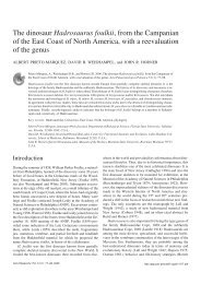
The skull of Velociraptor - Acta Palaeontologica Polonica
The skull of Velociraptor - Acta Palaeontologica Polonica
The skull of Velociraptor - Acta Palaeontologica Polonica

Create successful ePaper yourself
Turn your PDF publications into a flip-book with our unique Google optimized e-Paper software.
<strong>The</strong> <strong>skull</strong> <strong>of</strong> <strong>Velociraptor</strong> (<strong>The</strong>ropoda) from<br />
the Late Cretaceous <strong>of</strong> Mongolia<br />
RINCHEN BARSBOLD and HALSZKA OSMOLSKA<br />
Barsbold, R. & Osmdlska, H. 1999. <strong>The</strong> <strong>skull</strong> <strong>of</strong> <strong>Velociraptor</strong> (<strong>The</strong>ropoda) from the Late<br />
Cretaceous <strong>of</strong> Mongolia. - <strong>Acta</strong> <strong>Palaeontologica</strong> <strong>Polonica</strong> 44,2, 189-219.<br />
<strong>The</strong> well preserved material <strong>of</strong> the Late Cretaceous dromaeosaurid, <strong>Velociraptor</strong> mon-<br />
goliensis, has allowed us to supplement earlier descriptions <strong>of</strong> the <strong>skull</strong> in this species.<br />
<strong>The</strong> <strong>skull</strong> <strong>of</strong> I? mongoliensis is similar to that <strong>of</strong> Deinonychus antirrhopus, but differs<br />
from the latter by: (1) laterally convex supratemporal arcade resulting in short, rounded<br />
supratemporal fenestra; (2) depressed nasal; (3) longer maxillary process <strong>of</strong> premaxilla;<br />
(4) lack <strong>of</strong> separate prefrontal, and (5) convex ventral border <strong>of</strong> the dentary. <strong>The</strong>se differ-<br />
ences, especially that in the structure <strong>of</strong> the temporal region, support generic distinction<br />
<strong>of</strong> Deinonychus and <strong>Velociraptor</strong>. Skulls <strong>of</strong> other dromaeosaurids are compared.<br />
Key words: Dinosauria, <strong>The</strong>ropoda, Dromaeosauridae, <strong>Velociraptor</strong>, <strong>skull</strong>, mandible,<br />
Late Cretaceous, Gobi Desert, Mongolia.<br />
Rinchen Barsbold [barsgeodin@magicnet,mn], Institute <strong>of</strong> Geology, Mongolian Academy<br />
<strong>of</strong> Sciences, Enkh Taivani Gudamji, Ulan Bator 210351, Mongolia.<br />
Halszka Osmdlska [osm@ twarda.pan.pl], Znstytut Paleobiologii PAN, ul. Twarda 51/55,<br />
PL-00-818 Warszawa, Poland.<br />
Introduction<br />
Dromaeosaurids were small to medium size theropods, except for Utahraptor and an<br />
undetermined dromaeosaurid from Japan (Azuma & Currie 1995), which were rela-<br />
tively large animals. <strong>The</strong>y were cursorial, moderately fast carnivores with large,<br />
rostrolaterally facing eyes. <strong>The</strong>re is taphonomic evidence (Ostrom 1990; Maxwell &<br />
Ostrom 1995) that at least Deinonychus antirrhopus may have hunted in packs. On the<br />
other hand, <strong>Velociraptor</strong> mongoliensis may have also been a carrion feeder (Osm6lska<br />
1993; but see Kielan-Jaworowska & Barsbold 1972; Unwin et al. 1994; Fastovsky et<br />
al. 1997 for alternative interpretations). <strong>The</strong> Dromaeosauridae are Cretaceous mani-<br />
raptoran theropods, and eight monotypic genera: Adasaurus Barsbold, 1983, Deino-<br />
nychus Ostrom, 1969, Dromaeosaurus Matthew & Brown, 1922, Hulsanpes Osm61-<br />
ska, 1982, Omithodesmus Seeley, 1887, Sauromitholestes Sues, 1978, Utahraptor
190 Skull <strong>of</strong> <strong>Velociraptor</strong>: BARSBOLD & OSMOLSKA<br />
Kirkland, Burge, & Gaston, 1993, and <strong>Velociraptor</strong> Osborn, 1924 are presently as-<br />
signed to this family. Three <strong>of</strong> these genera (Adasaurus, Hulsanpes, and Ornitho-<br />
desmus), are based exclusively on incomplete postcrania.<br />
<strong>The</strong> most peculiar dromaeosaurid feature is the opisthopubic pelvis (Barsbold<br />
1976), which distinguishes these dinosaurs from other theropods, except for the dis-<br />
tantly related therizinosauroids; the extremely long caudal zygapophyses and chev-<br />
rons are probably also common to all dromaeosaurids, but caudal vertebrae are known<br />
only in Deinonychus, <strong>Velociraptor</strong>, and Saurornitholestes (Dr P.J. Currie's personal<br />
communication 1999)<br />
<strong>The</strong> <strong>skull</strong> <strong>of</strong> V mongoliensis has been known for more than 70 years. Over this time,<br />
it has been described, illustrated or commented by several authors, among them Sues<br />
(1977a), Barsbold (1983), Paul (1988), and Ostrom (1969b, 1990). Up to now, Veloci-<br />
raptor is represented by the most complete and most numerous <strong>skull</strong>s and postcrania<br />
among dromaeosaurids (in addition to the here described material, there are numerous<br />
still not described specimens recently collected by the AMNH Asiatic Expeditions).<br />
It was Ostrom (1969a), who first recognised the close relationship <strong>of</strong> <strong>Velociraptor</strong><br />
with the North American forms, Dromaeosaurus and Deinonychus, and assigned it to<br />
the Dromaeosauridae (= Dromaeosaurinae Matthew & Brown 1922). <strong>The</strong> <strong>skull</strong>s are<br />
largely complete in the two latter genera, whereas <strong>skull</strong>s <strong>of</strong> Saurornitholestes and<br />
Utahraptor are represented by a few bones each. <strong>The</strong> following description <strong>of</strong> the <strong>skull</strong><br />
in V mongoliensis supplements the earlier descriptions by Osborn (1924), Sues<br />
(1977a), and Barsbold (1983). <strong>The</strong> <strong>skull</strong> data for D. antirrhopus, Dromaeosaurus<br />
albertensis, and Saurornitholestes langstoni used in the comparisons below are re-<br />
spectively from Ostrom (1969b), Colbert & Russell (1969), Currie (1995), Sues<br />
(1977a), and Witmer & Maxwell (1996), unless stated otherwise.<br />
Institutional abbreviations: AMNH, American Museum <strong>of</strong> Natural History, New<br />
York; GIN, Institute <strong>of</strong> Geology, Mongolian Academy <strong>of</strong> Sciences, Ulan Bator;<br />
PIN, Museum <strong>of</strong> Palaeontology, Russian Academy <strong>of</strong> Sciences, Moscow; ROM,<br />
Royal Ontario Museum, Toronto; TPM, Royal Tyrrell Museum <strong>of</strong> Palaeontology,<br />
Drumheller; ZPAL, Institute <strong>of</strong> Paleobiology, Polish Academy <strong>of</strong> Sciences, Warsaw.<br />
Systematic palaeontology<br />
<strong>The</strong>ropoda Marsh, 1881<br />
Maniraptora Gauthier, 1986<br />
Family Dromaeosauridae Matthew & Brown, 1922<br />
Subfamily <strong>Velociraptor</strong>inae Barsbold, 1983<br />
<strong>Velociraptor</strong> Osborn, 1924<br />
Type species by monotypy: <strong>Velociraptor</strong> mongoliensis Osborn, 1924.<br />
<strong>Velociraptor</strong> mongoliensis Osborn, 1924<br />
Figs 1-8.<br />
Holotype: AMNH 6515, <strong>skull</strong>, mandible, manual digit I.
ACTA PALAEONTOLOGICA POLONICA (44) (2) 191<br />
Type horizon and locality: Djadokhta Formation (?early Campanian), Bayn Dzak, Ornnogov prov-<br />
ince, Gobi Desert, Mongolia.<br />
Material. -<strong>The</strong> present description <strong>of</strong> the <strong>skull</strong> <strong>of</strong> V: mongoliensis is based on several<br />
<strong>skull</strong>s pertaining to more or less complete skeletons from the Upper Cretaceous sand-<br />
stone deposits <strong>of</strong> the Mongolian Gobi. <strong>The</strong> specimens studied were found by the<br />
Polish-Mongolian <strong>Palaeontologica</strong>l Expeditions (specimens GIN 100125 and ZPAL<br />
MgD-U97, see Kielan-Jaworowska & Barsbold 1972; Gradziliski et al. 1977), by the<br />
Soviet-Mongolian <strong>Palaeontologica</strong>l Expeditions (specimen PIN 3 14318), by a Mongo-<br />
lian expedition (specimen GIN 100124, see Barsbold 1983) and by the Mongolian-<br />
-Japanese <strong>Palaeontologica</strong>l Expeditions (specimen GIN 10012000). Description <strong>of</strong> the<br />
postcrania preserved with these <strong>skull</strong>s will be published at a later date (Barsbold &<br />
Osm6lska in preparation).<br />
<strong>The</strong> following specimens derive from the Djadokhta Formation (?early Campanian,<br />
see Kielan-Jaworowska & Hurum 1997), TugrrE;zn-Shire, Omnogov, Mongolia:<br />
GIN 100124 - consists <strong>of</strong> an almost complete, articulated, but dorsoventrally flat-<br />
tened <strong>skull</strong>, both mandibular rami, and a few fragmentary postcranial bones. <strong>The</strong><br />
premaxillae are damaged rostrally, as are the caudal ends <strong>of</strong> the nasals and the rostral<br />
tips <strong>of</strong> the frontals along their mutual contact; the shaft and the caudal end <strong>of</strong> the left<br />
lacrimal are missing; jugals, quadratojugals and quadrates are fragmentary; pterygoids<br />
and palatines are partly damaged, those on the left side having shifted caudally from<br />
their natural position; vomers are not exposed. <strong>The</strong> dentaries are positioned between<br />
the left and right maxillae and premaxillae; the postdentary portion <strong>of</strong> the right man-<br />
dibular ramus is fragmentary, the left surangular lacks its rostrodorsal part; the dentary<br />
teeth and articular region are not exposed.<br />
GIN 100125 - includes skeletons <strong>of</strong> two dinosaurs - V: mongoliensis and Proto-<br />
ceratops andrewsi. Remains <strong>of</strong> these dinosaurs are preserved in a position suggestive<br />
<strong>of</strong> combat and for that reason this specimen is widely known as one <strong>of</strong> the 'fighting di-<br />
nosaurs'. <strong>The</strong> V mongoliensis skeleton is complete and articulated. It includes the<br />
<strong>skull</strong>, adducted mandibles and postcranium. <strong>The</strong> snout and mandible are somewhat<br />
compressed laterally and the dentaries are forced under the <strong>skull</strong> obscuring the palate<br />
and rostral part <strong>of</strong> the basicranium.<br />
GIN 10012000 - is represented by the complete skeleton <strong>of</strong> a young individual. It<br />
includes <strong>skull</strong> with mandible and postcranium.<br />
PIN 314318 - almost complete <strong>skull</strong> with left mandibular ramus, lacking the right<br />
temporal region; tip <strong>of</strong> the rostrum is severely damaged.<br />
<strong>The</strong> Barun Goyot Formation (?late Campanian: Kielan-Jaworowska & Hurum<br />
1997), Khulsan, Bayankhongor, Mongolia, yielded one specimen ZPAL MgD-U97 -<br />
included is the left, rostral half <strong>of</strong> the <strong>skull</strong> (exposed from the medial side) but lacks the<br />
premaxilla and tips <strong>of</strong> the vomers. Both mandibular rami are placed between the<br />
maxillae (as in all described V mongoliensis specimens with the mandible preserved);<br />
the left ramus lacks the caudal articular region and the tip <strong>of</strong> the dentary; the fragmen-<br />
tary right ramus includes the splenial and an incomplete dentary. Associated with the<br />
<strong>skull</strong> was a distal part <strong>of</strong> the left hind limb.<br />
<strong>The</strong> new, abundant <strong>Velociraptor</strong> material recently collected in Mongolia by the Mon-<br />
golian Academy <strong>of</strong> Sciences - American Museum <strong>of</strong> Natural History Expeditions<br />
(Norell & Makovicky 1998) has not been studied by the present authors.
192 Skull <strong>of</strong> <strong>Velociraptor</strong>: BARSBOLD & OSMOLSKA<br />
Revised diagnosis (based upon <strong>skull</strong> characters). - Skull shallow with long snout,<br />
preorbital length constituting 60% <strong>of</strong> total <strong>skull</strong> length (estimated at about 50% in<br />
Dr. albertensis and D. antirrhopus; proportions unknown in other drornaeosaurids);<br />
supratemporal fossa (and fenestra) subcircular, bound by laterally convex supra-<br />
temporal arcade (elongate, with straight arcade in D. antirrhopus; shape <strong>of</strong> fossa un-<br />
known in other drornaeosaurids); frontal long, almost four times longer than wide<br />
across the orbital portion, and almost four times as long as parietal [wider in S.<br />
langstoni, frontal length at most three times the width across the orbital portion; in Dr.<br />
albertensis frontal shorter, about twice as long as wide; parietal/frontal length ratio un-<br />
known in both these species; in D. antirrhopus, frontal only three times longer than<br />
wide and twice as long as parietal (Dr L.M. Witmer's personal communication 1999)l;<br />
rostral border <strong>of</strong> internal antorbital fenestra broadly rounded (subtriangular in D.<br />
antirrhopus; shape unknown in other drornaeosaurids); premaxilla with long max-<br />
illary process reaching well beyond caudal margin <strong>of</strong> external naris (not extending be-<br />
yond naris in D. antirrhopus; unknown or incomplete in other drornaeosaurids); nasal<br />
depressed, deepest just behind the external naris (not depressed in D. antirrhopus; un-<br />
known in other drornaeosaurids); maxilla with longitudinal ridge dorsal to a row <strong>of</strong><br />
neuro-vascular foramina, which are arranged in one row (no ridge in D. antirrhopus;<br />
maxilla unknown in other drornaeosaurids); no separate prefrontal (prefrontal is sepa-<br />
rate in D. antirrhopus and probably also in Dr. albertensis); dentary very shallow, its<br />
depth constituting one-eighth to one-seventh <strong>of</strong> its length, ventral margin convex (den-<br />
tary relatively deeper and with straight ventral margin in other dromaeosaurids); first<br />
and second premaxillary teeth larger than third and fourth.<br />
Because <strong>of</strong> deficiency <strong>of</strong> the <strong>skull</strong> data for some dromaeosaurid taxa, all the above<br />
characters are equivocal synapomorphies <strong>of</strong> V mongoliensis.<br />
Occurrence. - Upper Cretaceous, ?lower-?upper Campanian, Djadokhta and Barun<br />
Goyot formations; Ornnogov and Bayankhongor provinces, Gobi Desert, Mongolia.<br />
Description and comparisons<br />
Skull as a whole<br />
<strong>The</strong> <strong>skull</strong> has a long, narrow and shallow snout, which constitutes 60% <strong>of</strong> the <strong>skull</strong> length (Table 1).<br />
In caudal view, the <strong>skull</strong> is almost as wide as deep (Figs 2A, 5); the rostral view shows the orbits<br />
rostrolaterally oriented for a stereoscopic vision (Norell & Makovicky 1998). In lateral view (Figs<br />
1 A, B, 3A, 4A), the <strong>skull</strong> pr<strong>of</strong>ile weakly rises caudally; from about the mid-length <strong>of</strong> the nasals for-<br />
wards, the rostral part <strong>of</strong> the snout is somewhat elevated above the narial opening and depressed<br />
behind it. <strong>The</strong> naris is large and oval, and set in a depression. <strong>The</strong> elongate subnarial foramen is<br />
pronounced. <strong>The</strong> antorbital fossa is long and shallow and occupies slightly more than a half <strong>of</strong> the<br />
snout length. Only the rostral and rostrodorsal portion <strong>of</strong> its rim is well developed, while the ventral<br />
part <strong>of</strong> the rim is indistinct. More than a half <strong>of</strong> the antorbital fossa is occupied by the internal<br />
antorbital fenestra, the rostral margin <strong>of</strong> which is broadly rounded. <strong>The</strong> maxillary fenestra is much<br />
smaller, the interfenestral strut separating it from the internal antorbital fenestra is wide. <strong>The</strong><br />
promaxillary fenestra is divided into two small openings in GIN 100125, although it seems slit-like<br />
in other <strong>skull</strong>s. <strong>The</strong> orbit is almost circular, only slightly longer than high. <strong>The</strong> infratemporal<br />
fenestra is about three times higher than long, and is slightly inclined caudodorsally. Caudal to this<br />
fenestra, there is a tall, wide paraquadratic foramen that, because <strong>of</strong> its great size, is well exposed
ACTA PALAEONTOLOGICA POLONICA (44) (2) 193<br />
both laterally and caudally. In dorsal view, the <strong>skull</strong> has a striking appearance, due to the narrow-<br />
ness <strong>of</strong> the snout, the width <strong>of</strong> which equals only about a third <strong>of</strong> the postorbital <strong>skull</strong> width. <strong>The</strong><br />
snout is also long in comparison to the short frontoparietal part <strong>of</strong> the <strong>skull</strong> ro<strong>of</strong> between the<br />
supratemporal fenestra (Figs 2B, 3B, 4B). <strong>The</strong> lateral apices <strong>of</strong> the lacrimals extend somewhat be-<br />
yond the jugals. <strong>The</strong> supratemporal arcades are laterally convex making the supratemporal fossae<br />
(and fenestrae) almost circular. <strong>The</strong> <strong>skull</strong> is about twice as wide across the temporal arcades as be-<br />
tween the orbits. <strong>The</strong> supraorbital fossae are bounded caudally by a steep, sharp nuchal crest. <strong>The</strong><br />
nuchal crest is confluent medially with the sagittal crest, which is relatively low and short, and ex-<br />
tends along the open sutural interparietal contact.<br />
Table 1. Skull and mandible measurements (in mrn) <strong>of</strong> <strong>Velociraptor</strong> mongoliensis (GIN 100125)<br />
Maximum length <strong>of</strong> <strong>skull</strong> (paroccipital process-tip <strong>of</strong> snout)<br />
230<br />
Medial length <strong>of</strong> <strong>skull</strong> (transverse nuchal crest-tip <strong>of</strong> snout) 2 13<br />
Width across supratemporal arcades 1 77 1<br />
Width across occiput 1 69 1<br />
Width across lateral tips <strong>of</strong> lacrimals 1 Xi 1<br />
Width across parietals (at about midlength)<br />
Maximum depth<br />
27<br />
66<br />
Length <strong>of</strong> snout (rostra1 margin <strong>of</strong> orbit-tip) 1 140 1<br />
Width <strong>of</strong> snout (in front <strong>of</strong> rostra1 ends <strong>of</strong> lacrimals)<br />
Depth <strong>of</strong> snout (in front <strong>of</strong> orbit)<br />
Length <strong>of</strong> mandible<br />
Maximum depth <strong>of</strong> mandible (behind external fenestra)<br />
22<br />
45<br />
210<br />
23<br />
<strong>The</strong> distinctive appearance <strong>of</strong> the <strong>skull</strong> in <strong>Velociraptor</strong>, with its short temporal re-<br />
gion, rounded temporal fenestrae and very long, narrow snout has not been noticed so<br />
far. <strong>The</strong> <strong>skull</strong> proportions, with the length <strong>of</strong> the preorbital region exceeding almost 4.5<br />
times the length <strong>of</strong> the postorbital region, distinguish <strong>Velociraptor</strong> from Dromaeo-<br />
saurus and Deinonychus, in which the preorbitallengths are about 2.5-3.5 times these<br />
<strong>of</strong> the postorbital regions. Among theropods, such a long snout occurs only in the<br />
ornithomimids. <strong>The</strong> subcircular shape <strong>of</strong> the temporal fenestra is the character that dis-<br />
tinctly differs <strong>Velociraptor</strong> from Deinonychus. <strong>The</strong> right temporal arcade is complete<br />
in YPM 5210 specimen <strong>of</strong> D. antirrhopus and it seems subparallel to the medial line <strong>of</strong><br />
the <strong>skull</strong>. As a result, the supratemporal fenestra is elongate and relatively narrow in<br />
Deinonychus, and its width (measured across its caudal portion) constitutes about 30%<br />
<strong>of</strong> the length <strong>of</strong> the fenestra. <strong>The</strong> temporal arcade is laterally convex in <strong>Velociraptor</strong>,<br />
and the width <strong>of</strong> the fenestra is more than 50% <strong>of</strong> its length. Due to the shape <strong>of</strong> the<br />
supratemporal arcade, the supratemporal fenestra is wider in <strong>Velociraptor</strong>, and it pro-<br />
vided more space for the adductor muscles than the fenestra in Deinonychus. At the<br />
same time, the sagittal extent <strong>of</strong> the adductor origins was relatively greater in the<br />
American than in the Mongolian genus. It might result in a more oblique direction <strong>of</strong><br />
some adductor fibres in Deinonychus. <strong>The</strong> unique specimen <strong>of</strong> Dromaeosaurus en-<br />
tirely lacks the temporal arcade, as well as most <strong>of</strong> the parietal; thus the shape <strong>of</strong> the
194 Skull <strong>of</strong> <strong>Velociraptor</strong>: Barsbold & Osm6lska<br />
Fig. 1. Skull with mandible <strong>of</strong> <strong>Velociraptor</strong> mongoliensis GIN 100125. A. Left lateral view. B. Right lateral<br />
view, stereophotograph. C. Ventral view. D. Left caudoventral view, showing contacts <strong>of</strong> quadrate heads<br />
with squamosals. Scale bar 2 cm.<br />
supratemporal fenestra cannot be determined in this genus. Colbert & Russell (1969)<br />
reconstructed it as elongate, listing the long supratemporal arcade in Dromaeosaurus<br />
among differences to <strong>Velociraptor</strong>, while Currie's (1995) reconstruction shows<br />
a straight, but relatively shorter arcade and a rather rounded fenestra. <strong>The</strong> mode <strong>of</strong><br />
preservation <strong>of</strong> the <strong>skull</strong>s at our disposal, and the loose contacts between some bones<br />
within the snout, show that the <strong>skull</strong> and mandible were not as rigid as in most other<br />
theropods.
ACTA PALAEONTOLOGICA POLONICA (44) (2) 195<br />
Fig. 2. Skull with mandible <strong>of</strong> <strong>Velociraptor</strong> mongoliensis GIN 100125. A. Occipital view, stereophoto-<br />
graph. B. Dorsal view, stereophotograph, GIN 100125. C. Tooth <strong>of</strong> <strong>Velociraptor</strong> mongoliensis, ZPAL<br />
MgD-I/97a. Scale bars 2 cm for A and B, 2 mrn for C.<br />
Snout<br />
Premaxilla. - <strong>The</strong> main body <strong>of</strong> the premaxilla is longer than high. A long, thin maxillary process<br />
<strong>of</strong> the premaxilla is wedged between the maxilla and nasal, reaching caudally to or close to the rostral<br />
margin <strong>of</strong> the antorbital fossa, and it separates the maxilla from the naris. In the extreme case (GIN<br />
100/25), on the right side <strong>of</strong> the <strong>skull</strong>, this process extends even farther, to a point above the fifthlsixth<br />
maxillary teeth. <strong>The</strong> nasal process has a stout base, but its internarial portion is thin, and its end fits<br />
into a medial groove present along the tip <strong>of</strong> the nasal. A shallow depression in the premaxilla out-<br />
lines the rostral and ventral margins <strong>of</strong> the narial opening. At the contact with the maxilla, the<br />
caudoventral border <strong>of</strong> the premaxilla is slightly embayed to mark the subnarial foramen. <strong>The</strong> surface<br />
<strong>of</strong> the rostral and alveolar parts <strong>of</strong> the premaxilla bears small, irregularly spaced foramina. In palatal<br />
view, the rostral portion <strong>of</strong> the premaxilla is steep, and only below the naris is there a narrow palatal<br />
shelf. <strong>The</strong> premaxillary shelves contact each other on the mid-line. <strong>The</strong>y separate caudally, leaving<br />
a triangular space below the narial openings, where tip <strong>of</strong> the fused vomers is inserted.
196 Skull <strong>of</strong> <strong>Velociraptor</strong>: BARSBOLD & OSMOLSKA<br />
Maxilla. - <strong>The</strong> lateral body <strong>of</strong> the maxilla has a low triangular outline. <strong>The</strong> nasal process <strong>of</strong> the<br />
maxilla is shorter than the jugal process. Along the alveolar margin, there is a row <strong>of</strong> small<br />
neuro-vascular foramina, above which extends a low, longitudinal ridge. <strong>The</strong> antorbital fossa is<br />
weakly delimited. It occupies almost two-thirds <strong>of</strong> the ventral length <strong>of</strong> the maxilla and the internal<br />
antorbital fenestra takes up about a third <strong>of</strong> that length. <strong>The</strong> rostral half <strong>of</strong> the fossa is shallow<br />
lateromedially and bears a small, teardrop-shaped maxillary fenestra. This fenestra is located half-<br />
way between the antorbital fenestra and the rostral margin <strong>of</strong> the antorbital fossa, and it leads into the<br />
maxillary sinus. Close to the rostral margin <strong>of</strong> the antorbital fossa, two small, rostrally directed open-<br />
ings (probably homologous to the promaxillary fenestra) penetrate the bone to enter the promaxillary<br />
sinus. This double promaxillary fenestra has been only noticed in GIN 100125, while in AMNH 65 15<br />
(holotype), and possibly also in PIN 3 14318, the promaxillary fenestra is slit-like.<br />
<strong>The</strong> maxillary recesses are exposed medially in ZPAL MgD-U97 (Figs 7E, 8A). As preserved,<br />
they resemble the structures illustrated by Witmer (1997a: fig. 30) in Albertosaurus, with well pro-<br />
nouncedpila postantralis, antrum maxillaris, and several recessipneumatici interalveolares; the re-<br />
gion <strong>of</strong> the promaxillary recess is poorly preserved. Owing to the damage <strong>of</strong> the rostralmost region <strong>of</strong><br />
the maxilla in ZPAL MgD-LJ97, it cannot be stated whether a vestibular bulla was present in<br />
<strong>Velociraptor</strong>. <strong>The</strong> left palatal shelf <strong>of</strong> the maxilla is partly preserved in ZPAL MgD-U97, its rostral<br />
portion lacking. It is moderately wide rostrally (at least 7 mm), and has a smooth, upturned medial<br />
border. As preserved, a part <strong>of</strong> this border contacts the vomer along the region rostral to pila<br />
interfenestralis. <strong>The</strong> shelf is inclined dorsomedially-ventrolaterally. This inclination gradually de-<br />
creases rostrally, and the shelf was probably almost horizontal at the contact with the premaxilla.<br />
Caudally, the palatal shelf underlies the medial surface <strong>of</strong> the jugal, close to the contact <strong>of</strong> the latter<br />
with the lacrimal shaft. Rostra1 to the lacrimal the medial border <strong>of</strong> the palatal shelf is in contact with<br />
the palatine. However, this contact is located more caudally than that <strong>of</strong> Albertosaurus (Witmer<br />
1997a: fig. 30d). <strong>The</strong> position <strong>of</strong> the last maxillary tooth is opposite the mid-length <strong>of</strong> the internal<br />
antorbital fenestra.<br />
Nasal. -<strong>The</strong> nasal is L-shaped in cross section and its dorsal portion is very narrow along more than<br />
its rostral half. In the rostral portion <strong>of</strong> the snout, the dorsal surfaces <strong>of</strong> each nasal incline slightly me-<br />
dially to produce a groove along the internasal contact. Judging from a vertical displacement along<br />
the internasal suture visible in the holotype, as well as in all other specimens at our disposal, this con-<br />
tact was loose. <strong>The</strong> caudalmost portion <strong>of</strong> the nasal is broken <strong>of</strong>f in all our specimens, but the exten-<br />
sive caudal extent <strong>of</strong> this bone is evidenced by the presence <strong>of</strong> the longitudinally ridged dorsal sur-<br />
face on the rostralmost portion <strong>of</strong> the frontal in PIN 314318 (see below). Approximately from the<br />
fourth maxillary tooth position to the end <strong>of</strong> the maxillary fenestra, the nasal contacts the maxilla and<br />
this contact looks loose in the specimens at our disposal, as well as in the holotype. Contrary to that,<br />
the contact between the nasal and the maxillary process <strong>of</strong> the premaxilla seems more firm. Along its<br />
caudal third, the nasal contacts the lacrimal laterally. <strong>The</strong> nasal deepens rostrally and is the deepest<br />
behind the caudal boundary <strong>of</strong> the narial opening, producing a distinct elevation <strong>of</strong> the snout pr<strong>of</strong>ile<br />
above the nares. <strong>The</strong> outer surface <strong>of</strong> the nasal is covered by irregularly spaced, small foramina.<br />
Kirkland et al. (1993) drew attention to differences in shapes <strong>of</strong> the premaxillae in the<br />
dromaeosaurids, expressed as the length-to-depth ratio <strong>of</strong> the main body <strong>of</strong> the pre-<br />
maxilla. This index is the highest in <strong>Velociraptor</strong>: 164 in the holotype and 170 in GIN<br />
100125, while it is much lower in all other dromaeosaurids: 86 in Dromaeosaurus, about<br />
90 in Deinonychus, and 101 in Utahraptor. Except in <strong>Velociraptor</strong>, the maxillary process<br />
is not completely preserved in any dromaeosaurid. In Deinonychus this process is not<br />
complete (Ostrom 1969b), and has been reconstructed as not extending beyond the cau-<br />
dal border <strong>of</strong> the external naris. On the illustrations <strong>of</strong> the holotype <strong>of</strong> V mongoliensis<br />
<strong>skull</strong> in Osborn's (1924) and Sues' (1977a) papers, this process is short and ends just be-<br />
hind the naris, but in GIN 100125 and 100124 it extends much farther caudally. This re-<br />
gion <strong>of</strong> the <strong>skull</strong> is not well preserved in the holotype and its interpretation by these au-
ACTA PALAEONTOLOGICA POLONICA (44) (2) 197<br />
Fig. 3. A, B. Deformed <strong>skull</strong> and mandible <strong>of</strong> <strong>Velociraptor</strong> mongoliensis, GIN 100124, in right lateral (A)<br />
and dorsal (B) views. Scale bar 2 cm.<br />
thors may be incorrect. <strong>The</strong> respective regions <strong>of</strong> maxillae are damaged in Dromaeo-<br />
saurus. To our knowledge, such a long and shallow maxillary process occurs only in the<br />
Ornithomimidae (a long process is also present in the Oviraptoridae, but its form is in-<br />
comparable to that in the dromaeosaurids: Barsbold, Maryariska, & Osm6lska in prepa-<br />
ration). <strong>The</strong> maxillary process <strong>of</strong> the premaxilla separates the maxilla from the narial<br />
border only in some theropods, among them in the earliest ones, the Triassic Eoraptor<br />
Sereno et al., 1993 and Herrerasaurus Reig, 1963 (Sereno & Novas 1992), but also in<br />
the Late Jurassic Ornitholestes Osbom, 1903, Sinraptor Currie & Zhao 1994) and the<br />
Cretaceous tyrannosaurids. <strong>The</strong> maxilla forms a part <strong>of</strong> the narial margin in Compso-<br />
gnathus, the ceratosaws, allosaurids, therizinosaurids (Erlikosaurus), troodontids, and<br />
Archaeopteryx. <strong>The</strong> symphyseal edge <strong>of</strong> the premaxilla is rostroventrally inclined in<br />
Deinonychus whereas it is perpendicular to the long axis <strong>of</strong> the <strong>skull</strong> in <strong>Velociraptor</strong>.<br />
<strong>The</strong> maxilla in Deinonychus is relatively deeper caudally than that in <strong>Velociraptor</strong>, and<br />
the rostral margin <strong>of</strong> the internal antorbital fenestra is subtriangular, whereas this<br />
fenestra is broadly rounded rostrally in <strong>Velociraptor</strong>. Deinonychus and Dromaeosaurus<br />
lack the longitudinal ridge which runs above the alveolar border in <strong>Velociraptor</strong>. <strong>The</strong><br />
outer surfaces <strong>of</strong> the maxillae in both North American genera are marked by more nu-<br />
merous neuro-vascular foramina, which are less regularly spaced, whereas these foram-<br />
ina are arranged in a single row in <strong>Velociraptor</strong>. <strong>The</strong> promaxillary fenestra is slit-like in<br />
Deinonychus, but not in all <strong>skull</strong>s <strong>of</strong> <strong>Velociraptor</strong>, but otherwise the maxillary recesses<br />
seem comparable to those in <strong>Velociraptor</strong>. <strong>The</strong> nasal <strong>of</strong> <strong>Velociraptor</strong> resembles that <strong>of</strong><br />
Deinonychus in being relatively long, and in having the lateral and dorsal surfaces at<br />
right angles to each other, the dorsal one very narrow. However, in Deinonychus, the<br />
pr<strong>of</strong>ile <strong>of</strong> the nasal slopes uniformly towards the rostral extremity <strong>of</strong> the snout, whereas
198 Skull <strong>of</strong> <strong>Velociraptor</strong>: BARSBOLD & OSMOLSKA<br />
in <strong>Velociraptor</strong>, the nasal pr<strong>of</strong>ile slopes only along about two thirds <strong>of</strong> its caudal length<br />
and it rises from about the level <strong>of</strong> the rostral boundary <strong>of</strong> the antorbital fossa. This results<br />
in the so called depressed 'nasal'. It is worth mentioning that the angles between the<br />
'sloped' caudal and the 'raised' rostral parts <strong>of</strong> the nasal differ in <strong>skull</strong>s GIN 100124,25,<br />
PIN 3 14318, and A . 6515. <strong>The</strong> angle is insignificant in the first <strong>of</strong> these <strong>skull</strong>s (Fig.<br />
3A), is small in the second (Fig. lA, B) and third, and is most conspicuous in the<br />
holotype (Sues 1977a: pl. 16). This suggests that the nasals might be flexible in this region.<br />
<strong>The</strong> internasal contact seems weak rostrally in <strong>Velociraptor</strong> and Deinonychus,<br />
contrary to the contact between the nasal and maxillary process <strong>of</strong> the premaxilla, which<br />
seems firm in both genera.<br />
Skull ro<strong>of</strong>, orbital and temporal region<br />
Frontal. - <strong>The</strong> frontal is three times longer than wide at the level <strong>of</strong> the mid-length <strong>of</strong> the orbit. <strong>The</strong><br />
interfrontal suture is easily discernible. At the caudodorsal comer <strong>of</strong> the orbit, the frontal abruptly<br />
widens into a rather narrow, transversely elongated postorbital process. Rostra1 to this process, the<br />
frontal gently narrows rostrally. On the outer surface <strong>of</strong> the frontal, there is a large, shallow depres-<br />
sion in PIN 314318, which is weakly marked in other specimens. This depression extends along the<br />
mid-length <strong>of</strong> the frontal, and is separated from its fellow by a narrow and low ridge along the<br />
interfrontal suture, formed by the somewhat elevated medial margins <strong>of</strong> both frontals. <strong>The</strong> frontal is<br />
rough where it forms the caudal half <strong>of</strong> the dorsal margin <strong>of</strong> the orbit. Rostrolaterally, there is<br />
a subrectangular platform on the dorsal surface <strong>of</strong> the frontal for the extensive, overlapping contact<br />
with the caudal process <strong>of</strong> the lacrimal. <strong>The</strong> overlapping nasal-frontal contact was extensive judging<br />
by a longitudinally ridged surface exposed rostrally on the frontal in PIN 3 14318. l ks surface occu-<br />
pies about a fifth <strong>of</strong> the frontal length. In the region <strong>of</strong> the supratemporal fossa, there is a distinct<br />
S-shaped ridge extending caudomedially from the postorbital contact towards the interfrontal suture.<br />
<strong>The</strong> ridge marks the rostral extent <strong>of</strong> the fossa and <strong>of</strong> the temporal musculature. Caudal to that ridge,<br />
the frontal slopes towards the parietal and the slope is relatively steep laterally. Close to the contact<br />
with the postorbital, the frontal has a deep depression on its dorsal surface. <strong>The</strong> frontoparietal suture<br />
is interdigitate and tight. <strong>The</strong> ventral wing <strong>of</strong> the frontal is medially inclined and bounds laterally the<br />
rostral part <strong>of</strong> the brain cavity. Farther rostrally, it forms the thick lateral wall to the olfactory tract.<br />
<strong>The</strong> caudal margin <strong>of</strong> the ventral wing has an extensive sutural contact with the laterosphenoid, that<br />
extends medially from the tip <strong>of</strong> the transversely directed postorbital process <strong>of</strong> the frontal. In lateral<br />
view, the frontals gently rise towards the suture with the parietals and attain their highest level just in<br />
front <strong>of</strong> that contact.<br />
Parietal. -<strong>The</strong> length <strong>of</strong> parietals equals to about a half <strong>of</strong> the combined (smallest) transverse width<br />
<strong>of</strong> both these bones. <strong>The</strong> parietals are suturally joined to each other along a sharp sagittal crest that<br />
rises slightly caudally. In lateral view, the parietals slope caudoventrally from their contact with the<br />
frontals, but close to the margin <strong>of</strong> the <strong>skull</strong> they turn abruptly dorsally to form a thin and high trans-<br />
verse nuchal crest. <strong>The</strong> crest caudally bounds the supratemporal fossae and is medially continuous<br />
with the sagittal crest. <strong>The</strong> dorsolateral apex <strong>of</strong> the parietal sets on the dorsal surface <strong>of</strong> the caudal<br />
process <strong>of</strong> the squamosal. On the occiput, the parietal occupies a relatively wide region lateral to the<br />
supraoccipital, where it overlies the caudal surface <strong>of</strong> the caudal process <strong>of</strong> the squamosal.<br />
Lacrimal (+prefrontal?). - In all the specimens at our disposal, as well as in the holotype, there is<br />
definitely only one element present in the rostrodorsal region <strong>of</strong> the orbit. For this reason it is here de-<br />
scribed as the lacrimal. However, this element may represent the fused prefrontal and lacrimal. <strong>The</strong><br />
lacrimal is T-shaped in lateral view. Its dorsal portion is triangular, flat and horizontal, with a pointed<br />
lateral apex. <strong>The</strong> external margin is rough. <strong>The</strong> tip <strong>of</strong> the narrow, long rostral process <strong>of</strong> the lacrimal is<br />
wedged between the nasal and maxilla and dorsally bounds more than the half <strong>of</strong> the antorbital fossa.<br />
<strong>The</strong> caudal portion is short, wider than the rostral; it has the subrectangular shape and bounds dor-<br />
sally the rostral half <strong>of</strong> the orbit. This portion <strong>of</strong> the lacrimal overlaps a relatively large lateral portion
ACTA PALAEONTOLOGICA POLONICA (44) (2) 199<br />
<strong>of</strong> the frontal. As preserved in GIN 100124 and 25, a small caudolateral angle <strong>of</strong> the lacrimal extends<br />
outwards, slightly beyond the orbital margin <strong>of</strong> the frontal. As seen on the broken surface <strong>of</strong> the lacri-<br />
mal shaft in the ZPAL MgD-U97, at least its dorsal portion was pneumatized, and there is a small<br />
ventral aperture at the base <strong>of</strong> the horizontal portion. This aperture is located above the much larger,<br />
funnel-like 'lacrimal canal' visible on the caudal surface <strong>of</strong> the shaft. <strong>The</strong> lacrimal recess seems to ex-<br />
tend also into the base <strong>of</strong> the rostral process <strong>of</strong> the dorsal horizontal portion <strong>of</strong> the lacrimal. As seen<br />
from the side, the shaft is straight for most <strong>of</strong> its length. In the caudal view, however, it is arched<br />
dorsomedially. <strong>The</strong> shaft is narrow in the sagittal direction and expanded transversely. Its surface<br />
facing the antorbital fossa is excavated. This excavation extends dorsally and rostrally along the ro<strong>of</strong><br />
<strong>of</strong> the antorbital fossa. Ventrally, the concavity on the shaft becomes deeper, and close to the contact<br />
with the jugal it passes into a deep, funnel-like recess, which penetrates the base <strong>of</strong> the lacrimal<br />
ventrocaudally. <strong>The</strong> dorsal lacrimal-maxilla contact within the fossa is indistinct in all our speci-<br />
mens. <strong>The</strong> lacrimal shaft is well exposed medially in ZPAL MgD-1/97. This specimen shows that the<br />
ventral extremity <strong>of</strong> the shaft extends caudolaterally-rostromedially. <strong>The</strong> ventral end <strong>of</strong> the shaft has<br />
an extensive contact with the jugal and an inclined, triangular facet medially for contact with the pala-<br />
tine. In this specimen, there is no ventral contact with the maxilla, the maxillary process <strong>of</strong> the jugal<br />
separating these two bones ventrally.<br />
Postorbital. - <strong>The</strong> triradiate postorbital forms most <strong>of</strong> the caudal boundary <strong>of</strong> the orbit in GIN<br />
100125, but not in PIN 3 14318, in which the jugal bounds most <strong>of</strong> the caudal orbital margin. <strong>The</strong> fron-<br />
tal process <strong>of</strong> the postorbital is directed dorsally and medially, and its end contacts the frontal above<br />
and the laterosphenoid below. Contact with the frontal is much more extensive than with the<br />
laterosphenoid. <strong>The</strong> medial flexion <strong>of</strong> the frontal process is almost at right angles to the squamosal<br />
process. <strong>The</strong> latter process deviates caudally 20"-30" from the longitudinal axis <strong>of</strong> the <strong>skull</strong> to help<br />
form the short, laterally bowed supratemporal arcade.<br />
Squamosal. - <strong>The</strong> squamosal has four prominent processes: the ventral (= prequadratic) process,<br />
the caudal (= paroccipital) process, the rostral (= postorbital) process and the medial (= parietal) pro-<br />
cess. <strong>The</strong> prequadratic process is relatively short and subtriangular. Its extensive, oblique caudo-<br />
ventral edge contacts a triangular rostrolateral flange <strong>of</strong> the quadrate (Figs lA, B, 4A). <strong>The</strong> ventral<br />
apex <strong>of</strong> this process also has a short contact with the ascending process <strong>of</strong> the quadratojugal (see be-<br />
low). On the occipital surface <strong>of</strong> the <strong>skull</strong>, the caudal process is well exposed dorsal to the opisthotic.<br />
It slopes slightly caudoventrally in lateral view. Rostrally, the postorbital process diverges from the<br />
long axis <strong>of</strong> the <strong>skull</strong> at an angle <strong>of</strong> about 35". <strong>The</strong> parietal process is directed rostromedially and<br />
slightly ventrally, and invades a narrow sulcus on the parietal, above the ventral contact <strong>of</strong> the latter<br />
bone with the prootic. It forms the ventral part <strong>of</strong> the steep caudal wall to the supratemporal fossa.<br />
<strong>The</strong> angle between the parietal and postorbital processes is about 80". <strong>The</strong> cotyla for the head <strong>of</strong> the<br />
quadrate is not well exposed. A sharp crest extends along the lateral surface <strong>of</strong> the squamosal, which<br />
caudally transforms into a shelf overhanging the prequadratic process and extends lateral to the<br />
quadrate cotyla.<br />
Quadrate. - <strong>The</strong> quadrate seems not pneumatic and has a single-headed otic process. In lateral as-<br />
pect, the ventral third <strong>of</strong> the quadrate shaft is perpendicular to the ventral margin <strong>of</strong> the <strong>skull</strong>. More<br />
dorsally, the shaft inclines somewhat backwards. Close to mid-height, the rostrolateral edge <strong>of</strong> the<br />
shaft expands into a large, triangular flange directed rostrally and slightly medially. <strong>The</strong> rostrodorsal<br />
margin <strong>of</strong> this flange contacts, along its almost entire extent, the prequadratic process <strong>of</strong> the<br />
squarnosal, except rostrally where a tip <strong>of</strong> the quadratojugal inserts between these bones. <strong>The</strong> head <strong>of</strong><br />
quadrate is narrow. In caudal view, the shaft <strong>of</strong> the quadrate is bowed to produce a concave lateral<br />
edge which forms the medial boundary to the large, tall paraquadratic (= quadrate-quadratojugal) fo-<br />
ramen. <strong>The</strong> mandibular process is transversely expanded and is divided into mandibular condyles by<br />
a shallow groove. <strong>The</strong> lateral condyle is larger than the medial one, and bears the mediolaterally ex-<br />
tended articular surface. <strong>The</strong> articular surface on the medial condyle is oriented obliquely (rostro-<br />
medially-caudolaterally) to the median axis <strong>of</strong> the <strong>skull</strong>. <strong>The</strong> mandibular articulation projects a little<br />
below the alveolar margin <strong>of</strong> the maxilla.
Skull <strong>of</strong> <strong>Velociraptor</strong>: BARSBOLD & OSM~LSKA<br />
rostrolateral<br />
-\\ " I \<br />
-\ paraquadratic<br />
foramen<br />
promaxillary fenestra maxillary fenestra palatine cultriforrn process<br />
Fig. 4. Reconstruction <strong>of</strong> <strong>skull</strong> in <strong>Velociraptor</strong> mongaliensis in lateral (A) and dorsal (B) views. Scale bar<br />
4 cm. Drawn by K. Sabath.<br />
Quadratojugal. - <strong>The</strong> quadratojugal has the shape on an inverted T, which is characteristic <strong>of</strong> the<br />
dromaeosaurids (Paul 1988). It has two widely separated contacts (the dorsal and the ventral one)<br />
with the quadrate. <strong>The</strong> ascending process <strong>of</strong> the quadratojugal is slender and bounds almost<br />
two-thirds <strong>of</strong> the infratemporal fenestra. <strong>The</strong> dorsal tip <strong>of</strong> this process fits between the prequadratic<br />
process <strong>of</strong> the squamosal and the triangular, rostrolateral flange <strong>of</strong> the quadrate. <strong>The</strong> ascending pro-<br />
cess delimits rostrally the paraquadratic foramen. Because <strong>of</strong> the slenderness <strong>of</strong> the ascending pro-<br />
cess, this forken is well exposed also laterally, not only caudally as is the case in most theropods. As<br />
in all dromaeosaurids, the quadrate (= caudal) process <strong>of</strong> the quadratojugal is stouter than the ascend-<br />
ing and jugal processes. Its contact with the quadrate is limited: in fact, only the rounded end <strong>of</strong> this<br />
process bears medially a flat articular surface which adheres to the mandibular process <strong>of</strong> the<br />
quadrate, just above the lateral condyle. <strong>The</strong> jugal process <strong>of</strong> the quadratojugal is thin, and wedges<br />
deeply into the jugal.<br />
Jugal. - In lateral view, the ventral margin <strong>of</strong> the jugal is horizontal, continuing the line <strong>of</strong> the alveo-<br />
lar border <strong>of</strong> the maxilla. Along the rostral three quarters <strong>of</strong> the orbit, the ventral margin <strong>of</strong> the jugal<br />
flares out, to cover the surangular when the mandible is adducted. <strong>The</strong> suborbital margin <strong>of</strong> the jugal<br />
is slightly concave in GIN 100125, but rather straight in GIN 100124 and PIN 3 14318. <strong>The</strong> postorbital<br />
process <strong>of</strong> the jugal inclines strongly caudodorsally. Its length is unknown in GIN 100124 and 25, in<br />
which the postorbital processes are damaged distally. In PIN 3 14318, the complete left postorbital<br />
process (presently, its tip is broken <strong>of</strong>f) reaches the supratemporal arcade, and forms the entire rostral<br />
margin <strong>of</strong> the infratemporal fenestra. <strong>The</strong> long and relatively shallow maxillary process dorso-<br />
medially overlaps the jugal process <strong>of</strong> the maxilla, ending just in front <strong>of</strong> the ventral extremity <strong>of</strong> the<br />
lacrimal, forming a small portion <strong>of</strong> the ventral border to the antorbital fossa. <strong>The</strong> rostral apex <strong>of</strong> the<br />
maxillary process is entirely occupied on its lateral surface by the jugal pneumatic recess (Witmer<br />
1997a), which deepens caudally and seems to penetrate the suborbital ramus <strong>of</strong> the jugal. <strong>The</strong><br />
quadratojugal process is relatively long and shallow, and caudally has a wedge-like incision for the
ACTA PALAEONTOLOGICA POLONICA (44) (2) 201<br />
quadratojugal. <strong>The</strong> medial surface <strong>of</strong> the jugal (exposed in ZPAL MgD-1/97) is angled along and be-<br />
low the ventral margin <strong>of</strong> the orbit to produce a strong ridge, which continues caudodorsally along<br />
the orbital margin <strong>of</strong> the postorbital process <strong>of</strong> the jugal. Rostrally, the maxillary process <strong>of</strong> the jugal<br />
has an extensive medial contact with the ventral extremity <strong>of</strong> the lacrimal. Part <strong>of</strong> this contact is also<br />
overlapped medially by the maxillary ramus <strong>of</strong> the palatine. Below the palatine-jugal contact, the<br />
suborbital ramus <strong>of</strong> the jugal is underlain by the tapering caudal extremity <strong>of</strong> the maxilla. <strong>The</strong> caudal<br />
extent <strong>of</strong> h s contact is along the rostral third <strong>of</strong> the orbital margin. Rostral to the base <strong>of</strong> the<br />
postorbital process <strong>of</strong> the jugal, there is a large depression on the medial surface. At the rostral limit<br />
<strong>of</strong> this depression, the jugal process <strong>of</strong> the ectopterygoid abuts against the jugal.<br />
<strong>The</strong> complete <strong>skull</strong> ro<strong>of</strong> is known only in <strong>Velociraptor</strong> and Deinonychus, but<br />
frontals are known also in Saurornitholestes and Dromaeosaurus. <strong>The</strong> frontals are rel-<br />
atively short and broad between the orbits in Dromaeosaurus, and the orbital portion<br />
<strong>of</strong> the frontal is subequal in length to the postorbital one. <strong>The</strong> adorbital part <strong>of</strong> the fron-<br />
tal is more elongate in <strong>Velociraptor</strong>. In Dromaeosaurus, and possibly also in Sauro-<br />
rnitholestes (fide Currie 1987), there is a slot in the rostrolateral margin <strong>of</strong> the frontal<br />
for the contact with the lacrimal, whereas the articular surface is wide and extensively<br />
overlapped by the lacrimal in <strong>Velociraptor</strong>. <strong>The</strong> holotype <strong>of</strong> S. langstoni, which in-<br />
cludes both frontals, represents an individual <strong>of</strong> similar size to GIN 100125 (the maxi-<br />
mum widths across the frontals are somewhat over 60 mm in both specimens). <strong>The</strong><br />
interorbital width <strong>of</strong> the paired frontals (measured just behind lacrimal contacts) is<br />
about 70% <strong>of</strong> the maximum frontal length in Saurornitholestes, but is only about 50%<br />
in <strong>Velociraptor</strong>. <strong>The</strong> caudal portion <strong>of</strong> the frontal is somewhat bulbous centrally in<br />
Sauronitholestes, but it is only very weakly convex in all <strong>skull</strong>s <strong>of</strong> <strong>Velociraptor</strong> at our<br />
disposal.<br />
As in Dromaeosaurus (Currie 1995), the nasal in <strong>Velociraptor</strong> extensively overlaps<br />
the frontal, differing in this respect from Saurornitholestes, in which the nasal-frontal<br />
contact seems shorter. Parietals are fragmentary in Dromaeosaurus. A striking feature<br />
<strong>of</strong> the lacrimal in <strong>Velociraptor</strong> is its slender and greatly elongated rostral process (it<br />
reaches almost to the mid-length <strong>of</strong> the long nasal). Although the external edge <strong>of</strong> the<br />
lacrimal seems smooth in the holotype <strong>of</strong> V mongoliensis, in all <strong>skull</strong>s at our disposal<br />
the orbital margin <strong>of</strong> the lacrimal is somewhat thickened and rough. <strong>The</strong> lacrimal in<br />
Deinonychus also has the long rostral process (Dr L.M. Witmer's personal communi-<br />
cation 1999). <strong>The</strong> shape <strong>of</strong> the lacrimal attributed to Utahraptor is distinctive in dorsal<br />
aspect, because it lacks the lateral projection, and is rather rectangular (not triangular<br />
as in <strong>Velociraptor</strong> and Deinonychus). In Dromaeosaurus only fragments <strong>of</strong> the lacri-<br />
mal are known. However, also in this genus the lacrimal does not contact the maxilla<br />
ventrally. Lack <strong>of</strong> the ventral lacrimal-maxilla contact is a common character <strong>of</strong><br />
theropods, the rare exceptions known to us being Eoraptor (Sereno et al. 1993),<br />
Syntarsus rhodesiensis (fide Colbert 1989), Ceratosaurus (Gilmore 1920), Allosaurus<br />
(Madsen 1976) and Compsognathus (Ostrom 1978). <strong>The</strong> prefrontal is absent (or com-<br />
pletely fused with the lacrimal) in <strong>Velociraptor</strong>, while it is a separate bone in Deino-<br />
nychus and probably also in Dromaeosaurus. In the presumably embryonic dro-<br />
maeosaurid <strong>skull</strong>s (probably representing <strong>Velociraptor</strong>) preliminary described by<br />
Norell et al. (1994), the prefrontal is absent, which according to these authors contra-<br />
dicts the idea that the prefrontal was fused to the lacrimal.<br />
<strong>The</strong> squamosal process <strong>of</strong> the postorbital is almost parallel to the long axis <strong>of</strong> the<br />
<strong>skull</strong> in Deinonychus showing but negligible outward inflexion, while this process dis-
202 Skull <strong>of</strong> <strong>Velociraptor</strong>: BARSBOLD & OSMOLSKA<br />
tinctly deviates in <strong>Velociraptor</strong>. This results in a different shape <strong>of</strong> the supratemporal<br />
fenestrae in both genera (see above). As noticed by Paul (1988), the dorsally directed<br />
frontal process <strong>of</strong> the postorbital is the character occurring both in <strong>Velociraptor</strong> and<br />
Deinonychus. Although the postorbital is unknown in Dromaeosaurus, the shape <strong>of</strong><br />
the frontal suggests (Currie 1995) that the frontal process <strong>of</strong> the postorbital was dor-<br />
sally directed, as in Deinonychus and <strong>Velociraptor</strong>. <strong>The</strong> axis <strong>of</strong> the frontal process is at<br />
a distinct angle to the axis <strong>of</strong> the jugal process in <strong>Velociraptor</strong>, this angle is much<br />
greater in Deinonychus. <strong>The</strong> pronounced dorsal direction <strong>of</strong> the frontal process is a<br />
character occurring in a few theropods (e.g., in oviraptorids, see Barsbold, Maryafiska,<br />
& Osm6lska in preparation), but seems extreme in the dromaeosaurids.<br />
<strong>The</strong> squamosal is generally similar in Deinonychus to that in <strong>Velociraptor</strong>, but<br />
shows a few differences: in Deinonychus, the prequadratic process is longer and more<br />
slender, the caudal process is more inclined ventrally, the occipital exposure <strong>of</strong> the<br />
squamosal above the paroccipital process is shallower, and the angle between the axes<br />
<strong>of</strong> the parietal and postorbital processes is only 60" (80" in <strong>Velociraptor</strong>). <strong>The</strong> squa-<br />
mosal in Dromaeosaurus has a much shorter caudal process than those <strong>of</strong> <strong>Velociraptor</strong><br />
and Deinonychus. <strong>The</strong> far caudal extension <strong>of</strong> this process produces in <strong>Velociraptor</strong><br />
(and probably also in Deinonychus) a shelf built <strong>of</strong> the squamosal and opisthotic,<br />
which extends behind, as well as laterally, to the cotyla for the quadrate head. It seems<br />
that this shelf was less extensive in Dromaeosaurus (Currie 1995: fig. la). <strong>The</strong><br />
prequadratic process in Dromaeosaurus is longer and narrower than in <strong>Velociraptor</strong>,<br />
and has a more extensive contact with the ascending process <strong>of</strong> the quadratojugal. In<br />
this respect, the prequadratic processes are similar in Dromaeosaurus and Deino-<br />
nychus. <strong>The</strong> distinctly quadriradiate shape <strong>of</strong> the dromaeosaurid squamosal is peculiar,<br />
due to its widely separate prequadratic and caudal processes, the axes <strong>of</strong> which are at<br />
about right angle to each other. In most theropods, these two processes are at an angle<br />
<strong>of</strong> less than 90°, or are subparallel. <strong>The</strong> prequadratic process <strong>of</strong> a similar shape occurs<br />
also in Troodon (Currie 1985), but, unlike dromaeosaurids, it does not contact the as-<br />
cending process <strong>of</strong> the quadratojugal (Russell & Dong 1994).<br />
<strong>The</strong> quadrate <strong>of</strong> Deinonychus is similar to that <strong>of</strong> <strong>Velociraptor</strong> (Dr. L.M. Witmer's<br />
personal communication 1999). In Dromaeosaurus, there is only a slight rostra1 exten-<br />
sion <strong>of</strong> the lateral edge <strong>of</strong> the shaft (Currie 1995: fig. 4b) instead <strong>of</strong> the large, triangular<br />
rostrolateral flange characteristic <strong>of</strong> <strong>Velociraptor</strong>. Colbert & Russell (1969) empha-<br />
sised that the mandibular articulation was well below the alveolar margin <strong>of</strong> the<br />
maxilla. This has been confirmed by Currie's reconstruction <strong>of</strong> the <strong>skull</strong> (1995: fig. la).<br />
<strong>The</strong> ventral projection <strong>of</strong> the mandibular articulation seems shallower in <strong>Velociraptor</strong>.<br />
<strong>The</strong> preserved portion <strong>of</strong> the left quadrate <strong>of</strong> Saurornitholestes shows a slight differ-<br />
ence in the more oblique course <strong>of</strong> the intercondylar groove. <strong>The</strong> troodontid quadrate<br />
is poorly known. According to the schematic illustration <strong>of</strong> Sinornithoides youngi pub-<br />
lished by Russell & Dong (1994: fig. 3), the rostrolateral flange is absent in this<br />
troodontid species. Unlike <strong>Velociraptor</strong>; the quadrate is pneumatic in troodontids (Cur-<br />
rie & Zhao 1994). Within the Dromaeosauridae, the quadratojugal, especially its as-<br />
cending process, is stoutest in Dromaeosaurus. In this genus, the ascending process<br />
has more extensive contacts with the quadrate process <strong>of</strong> the squamosal and the<br />
rostrolateral flange <strong>of</strong> the quadrate. In Deinonychus, the ascending process is about as
ACTA PALAEONTOLOGICA POLONICA (44) (2) 203<br />
slender as in <strong>Velociraptor</strong>, but its contact with the rostrolateral flange <strong>of</strong> the quadrate is<br />
less extensive.<br />
Note that in Ostrom 1969b: fig. 11, the quadratojugal is erroneously oriented and<br />
should be turned about 90" counter-clockwise on fig. 1 la, and clockwise on fig. 1 lb. In<br />
fact, fig. 11 shows the right quadratojugal, not the right reversed as stated in the expla-<br />
nation. <strong>The</strong> process on which the articular surface for the jugal is indicated represents<br />
the ascending process <strong>of</strong> the quadratojugal, and this surface is not for the jugal but for<br />
the contact with the rostrolateral flange <strong>of</strong> the quadrate. Consequently, the process di-<br />
rected upwards on the figure in question is the jugal process <strong>of</strong> the quadratojugal.<br />
<strong>The</strong> nature <strong>of</strong> contact <strong>of</strong> the ascending process <strong>of</strong> the quadratojugal with the<br />
squamosal and quadrate is peculiar in <strong>Velociraptor</strong>, because it looks as if it were not<br />
rigid. Ostrom (1969b: p. 24) has also drawn attention to the fact that in this place the<br />
contact between the two bones 'does not appear to be a particularly solid union', al-<br />
though, due to his erroneous orientation <strong>of</strong> the quadratojugal, he regarded this contact<br />
as the 'clasping junction7 <strong>of</strong> the quadratojugal and jugal. Because <strong>of</strong> the slenderness <strong>of</strong><br />
the ascending process, the paraquadratic foramen is larger in <strong>Velociraptor</strong> than in<br />
Dromaeosaurus (and probably also Deinonychus). As observed by Paul (1988), the<br />
large paraquadratic ('quadrate') foramen is characteristic <strong>of</strong> the Dromaeosauridae. In<br />
most theropods the quadratojugal has a more extensive contact with the quadrate. In<br />
Dromaeosaurus, the end <strong>of</strong> the quadratojugal twists around the quadrate onto its cau-<br />
dal surface, as it does in most theropods. This is not the case in <strong>Velociraptor</strong>. In the<br />
troodontids, the quadratojugal is L-shaped and its ascending process does not reach the<br />
prequadratic process <strong>of</strong> the squamosal (Russell & Dong 1994). In the ornithomirnids,<br />
the quadratojugal, as well as the entire infratemporal region, are not comparable with<br />
those <strong>of</strong> dromaeosaurids. Although the quadratojugal is also slender and reduced in the<br />
oviraptorids, and has a contact with the squamosal, it is rather L-shaped, and its as-<br />
cending process attaches to the caudolateral margin <strong>of</strong> the quadrate, leaving only<br />
a small and caudally facing paraquadratic foramen. <strong>The</strong> contact <strong>of</strong> the quadratojugal<br />
with the quadrate is <strong>of</strong> the cotyla-condyle type in the oviraptorids (Maryafiska &<br />
Osm6lska 1997). <strong>The</strong> jugal <strong>of</strong> <strong>Velociraptor</strong> is similar to those <strong>of</strong> Deinonychus and<br />
Dromaeosaurus. However, in the latter genus, the ventral margin slopes slightly<br />
caudoventrally, and unlike <strong>Velociraptor</strong> does not continue the horizontal line <strong>of</strong> the al-<br />
veolar border <strong>of</strong> the maxilla as does the ventral border <strong>of</strong> the jugal in <strong>Velociraptor</strong> (the<br />
maxilla and jugal are disarticulated in Deinonychus). Presence <strong>of</strong> the jugal pneumatic<br />
recess in Deinonychus has also been reported by Witmer (1997a).<br />
Occiput and braincase<br />
Most <strong>of</strong> the bones forming the occiput and basicranium are fused. <strong>The</strong> occipital condyle is round and<br />
about as wide as the foramen magnum (Figs 2A, 5), although its depth is about a half <strong>of</strong> the vertical di-<br />
ameter <strong>of</strong> the foramen magnum. <strong>The</strong> major part <strong>of</strong> the condyle is formed by the basioccipital. <strong>The</strong> ven-<br />
tral articular surface <strong>of</strong> the condyle is well developed. <strong>The</strong> condylar neck is short. <strong>The</strong> basal tubers are<br />
well separated from each other by a deep broad sulcus. Damage to one <strong>of</strong> the tubers in GIN 100125<br />
shows that the basioccipital is pneumatic. <strong>The</strong> lateral and dorsomedial contacts <strong>of</strong> the supraoccipital<br />
with the parietals is well defined. This bone is roughly subrectangular and abuts dorsally against the<br />
caudal surface <strong>of</strong> the upturned parietals, participating in the formation <strong>of</strong> the central part <strong>of</strong> the trans-<br />
verse nuchal crest. Some distance above the level <strong>of</strong> the foramen magnum the supraoccipital bears<br />
a pair <strong>of</strong> vertical, crescent grooves. <strong>The</strong>y apparently do not lead to any foramen. Sutures between the
asipte jgoid<br />
process<br />
Skull <strong>of</strong> <strong>Velociraptor</strong>: BARSBOLD & OSMOLSKA<br />
- quadratoji<br />
. paraquadr<br />
foramen<br />
Fig. 5. Reconstruction <strong>of</strong> <strong>skull</strong> in <strong>Velociraptor</strong> mongoliensis in occipital view. Scale bar 4 cm. Drawn by<br />
K. Sabath.<br />
supraoccipital and exoccipitals are obscure. <strong>The</strong> paroccipital process is stout, ends bluntly and is ori-<br />
ented almost entirely laterally with only a slight downward inclination. Distally, the end <strong>of</strong> paroccipital<br />
process is twisted and faces dorsocaudally. <strong>The</strong> dorsal contact <strong>of</strong> the exoccipital with the parietal and<br />
squamosal is distinct, but the sutures with the basioccipital and opisthotic are not discernible. Lateral to<br />
the foramen magnum three foramina pierce the exoccipital: two are the exits <strong>of</strong> cranial nerve XU, the<br />
third is probably for cranial nerve X (and most likely IX and XI).<br />
Basisphenoid + parasphenoid. - Most <strong>of</strong> the basisphenoid contacts are not discernible, and the<br />
parasphenoid is fused with the basisphenoid. In front <strong>of</strong> the basioccipital tubera, there is a deep basin<br />
ventrally - the basisphenoid recess - that is bounded rostrally by a massive crest <strong>of</strong> bone from which<br />
extends rostrally the parasphenoid. Caudal to the basisphenoid recess, a pair <strong>of</strong> openings is visible in<br />
GIN 100/24 (this region is poorly displayed in GIN 100/25), which lead deep into the basisphenoid.<br />
<strong>The</strong>y resemble the auditory tube foramina <strong>of</strong> birds, but their far caudal position would speak against<br />
such an interpretation. <strong>The</strong> crest which bounds rostrally the basisphenoid recess is continuous later-<br />
ally with the basipterygoid processes, which project rostroventrally and laterally. <strong>The</strong> basipterygoid<br />
process is stout, and ends bluntly. Its medial side is entirely covered by a rough articular surface for<br />
the palatal ramus <strong>of</strong> the pterygoid, while the rostrodorsal surface contacts the quadrate ramus. On the<br />
dorsal surface <strong>of</strong> the base <strong>of</strong> the basipterygoid process there is an extensive basipterygoid recess<br />
(Witmer 1997b). More dorsally, there are two smaller excavations close to base <strong>of</strong> the parasphenoid<br />
rostrum (Fig. 6B). <strong>The</strong> more caudal <strong>of</strong> the two might have contained the entrance to the carotid canal,<br />
but preservation <strong>of</strong> this region is too poor to be sure whether there was an opening within the excava-<br />
tion. <strong>The</strong> parasphenoid rostrum is broken <strong>of</strong>f distally in all specimens at our disposal. Its preserved<br />
proximal portion bears a shallow, longitudinal sulcus dorsally, whereas ventrally, at the base <strong>of</strong> the<br />
rostrum, there is a concavity - the subsellar recess (Witmer 1997b).<br />
<strong>The</strong> braincase is short but deep in <strong>Velociraptor</strong>. Its lateral wall is not well displayed in specimens<br />
GIN 100/25 and 24. As the brain cavity could not be studied in the specimens in our disposal, the fol-<br />
lowing interpretation <strong>of</strong> the openings on the lateral wall <strong>of</strong> the braincase is tentative (Fig. 6). As in the<br />
occipital region, only a few contacts between the bones are distinguishable, but include those <strong>of</strong> the<br />
laterosphenoid. <strong>The</strong> rostrodorsal process <strong>of</strong> the laterosphenoid forms a laterally curved cone, and its<br />
tip has rostrally a lateromedially elongate, well finished and convex articular surface. This process,<br />
along with the frontal, participates in the lateromedially elongate fossa for the postorbital. <strong>The</strong> bot-<br />
tom <strong>of</strong> this fossa is smooth. <strong>The</strong> nature <strong>of</strong> the dorsal articulation between the postorbital and the <strong>skull</strong><br />
ro<strong>of</strong> speaks in favour <strong>of</strong> a certain mobility <strong>of</strong> the postorbital at this contact. Caudally, the latero-<br />
sphenoid-frontal suture is continuous with the laterosphenoid-parietal suture. Farther caudally, the<br />
rate
ACTA PALAEONTOLOGICA POLONICA (44) (2)<br />
recess<br />
recess<br />
occipital condyler -. :%\ .W<br />
k7;:4,3i2?<br />
fenestra vestibularis + ";$-!:<br />
fenestra pseudorotunda I ot&phenoidal crest<br />
basisphenoid<br />
basisphenoid w<br />
Fig. 6. Schematic drawing <strong>of</strong> braincase in <strong>Velociraptor</strong> mongoliensis. A. Lateral view, GIN 100125.<br />
B. Dorsolateral view, GIN 100124. C. Rostral view, GIN 100124. I-V, VII - nerve exits. Not to scale. Drawn<br />
by K. Sabath.<br />
latter suture is continued by the parietal-prootic suture, which slopes ventrally towards the back <strong>of</strong><br />
the braincase. <strong>The</strong> caudal contact <strong>of</strong> the laterosphenoid is with the dorsal portion <strong>of</strong> the prootic along<br />
a distinct dorsoventral, zigzag suture. Just behind its contact with the laterosphenoid, the dorsal wing<br />
<strong>of</strong> the prootic bears a large, elongate depression. This depression is present in GIN 100125 and 24 and<br />
it probably represents the dorsal tympanic recess. <strong>The</strong> rostroventral contact <strong>of</strong> the laterosphenoid<br />
with the prootic is not clearly marked, and it is unknown how far rostrally this contact continues. <strong>The</strong><br />
otosphenoid crest is well pronounced. Below it, three openings pierce the prootic in GIN 100125. <strong>The</strong><br />
most rostral and dorsal one represents the exit <strong>of</strong> the trigeminal nerve; it is large, placed within a shal-<br />
low concavity (there is a pair <strong>of</strong> openings in this position on the left side <strong>of</strong> the braincase), and is<br />
bounded dorsally by the laterosphenoid and caudally by the prootic. A horizontal groove runs rostral<br />
to this opening, which might conduct the ophthalmic branch <strong>of</strong> the trigeminal nerve (Currie 1995).<br />
A vertical groove runs ventrally from the exit <strong>of</strong> the fifth nerve. Caudoventral to this opening, an-<br />
other, much smaller one is visible in GIN 100125. It is probably for the exit <strong>of</strong> the facial nerve. This<br />
opening is much larger in GIN 100124 and may represent the prootic recess (Witmer 1997b) which<br />
probably contained the exit <strong>of</strong> the facial nerve. <strong>The</strong> third <strong>of</strong> these openings is most ventral and most<br />
caudal in position. It is large and vertically elongated and may correspond to the fenestra vestibularis<br />
+ fenestra cochlearis. This opening seems to be bounded ventrally by a narrow tongue <strong>of</strong> the<br />
basisphenoid, rostrodorsally by the prootic and caudodorsally by the opisthotic. In GIN 100125, on<br />
the rostral surface <strong>of</strong> the paroccipital process and at its base, ventral and somewhat medially to the
206 Skull <strong>of</strong> <strong>Velociraptor</strong>: BARSBOLD & OSMOLSKA<br />
quadrate cotyla, the opisthotic is pierced by a large, longitudinally extended fenestra, the caudal tym-<br />
panic recess. In GIN 100124, there is only a depression in this position. As preserved, the exits <strong>of</strong> op-<br />
tic nerves are confluent with that for the olfactory stalk to form a large, rostrally facing fenestra. <strong>The</strong><br />
laterosphenoids meet rostrally on the mid-line <strong>of</strong> the braincase to form a dorsal portion <strong>of</strong> the caudal<br />
wall <strong>of</strong> the pituitary fossa. On each side, this wall is pierced by three small foramina (Fig. 6C). <strong>The</strong><br />
most lateral <strong>of</strong> them, placed rostrally to the exit <strong>of</strong> the trigeminal nerve, may represent the exit for the<br />
abducens nerve (Currie 1995). Two others may correspond to exits <strong>of</strong> the trochlear and oculomotor<br />
nerves.<br />
<strong>The</strong> occiput and the lateral braincase wall are known in Dromaeosaurus. <strong>The</strong><br />
condylar neck is more pronounced in this genus than in <strong>Velociraptor</strong>, and the basal<br />
tubera are separated less distinctly. <strong>The</strong> tubera were considered by Colbert & Russell<br />
(1969) as lacking sinuses, but these are present in <strong>Velociraptor</strong>. <strong>The</strong> proportions <strong>of</strong> the<br />
braincase seem to be similar in both genera, although compared to the total <strong>skull</strong><br />
length, the basicranium is shorter in <strong>Velociraptor</strong>. <strong>The</strong> basipterygoid processes extend<br />
ventrally only to the level <strong>of</strong> the basal tubera in Drornaeosaurus. In <strong>Velociraptor</strong>, they<br />
extend farther ventrally, being well exposed in the occipital view. <strong>The</strong> dorsal surfaces<br />
<strong>of</strong> basipterygoid processes are not preserved in Dromaeosaurus (Colbert & Russell<br />
1969: fig. 5), thus it is impossible to state whether the basipterygoid recesses were<br />
present, but the subsellar recess, as well as the basisphenoid recess, are pronounced in<br />
this genus; the basisphenoid recess is much deeper than this in <strong>Velociraptor</strong>. <strong>The</strong> de-<br />
pression on the prootic, here interpreted as the dorsal tympanic recess, seems homolo-<br />
gous to the depression on the prootic in Archaeopteryx (see also Witmer 1997b).<br />
Palate<br />
<strong>The</strong> palate is not seen in GIN 100125 and is poorly displayed in GIN 100124, because it is partly ob-<br />
scured in both specimens by the mandibles forced into the <strong>skull</strong>. <strong>The</strong> palate has also been crushed in<br />
the holotype and strongly damaged in PIN 3 14318. Only in ZPAL MgD-U97 are some remains <strong>of</strong> the<br />
palate visible (Figs 7E, 8), although their mutual position is somewhat distorted due to the lateral flat-<br />
tening <strong>of</strong> the snout. <strong>The</strong> secondary palate is narrow and relatively highly vaulted, due to the inclina-<br />
tions <strong>of</strong> the palatal shelves <strong>of</strong> maxillae. This inclination increases caudally and so does the vault <strong>of</strong><br />
the palate. Most probably, the palatal shelves <strong>of</strong> the maxillae did not suture to each other, being sepa-<br />
rated by the vomers. Any reconstruction <strong>of</strong> the shape <strong>of</strong> the exochoanal fenestra is impossible be-<br />
cause <strong>of</strong> the lateral flattening <strong>of</strong> the snout in ZPAL MgD-U97.<br />
Pterygoid. -<strong>The</strong> pterygoid is a thin bone that is broken in the available specimens. It does not differ<br />
from other theropods. <strong>The</strong> basipterygoid notch is deep, narrow and faces caudodorsally.<br />
Ectopterygoid. - <strong>The</strong> ectopterygoids are exposed dorsally in GIN 100125, and the left ecto-<br />
pterygoid has been found articulated in ZPAL MgD-U97. This bone has a typical theropod shape,<br />
with a pocketed medial.portion, and a slender, arched jugal process. <strong>The</strong> pterygoid margin <strong>of</strong> the<br />
ectopterygoid is thin and long, and the ectopterygoid-pterygoid contact is extensive. <strong>The</strong> caudal ex-<br />
tremity <strong>of</strong> the ectopterygoid is overlapped laterally by the caudal end <strong>of</strong> the palatine. In ZPAL<br />
MgD-U97, due to lateral compression <strong>of</strong> the <strong>skull</strong>, the ectopterygoid seems to be pushed medially<br />
onto the lateral surface <strong>of</strong> the pterygoid, without damage to the delicate adjoining bones. It has also<br />
been shifted slightly caudally and its caudomedial corner has been displaced and moved opposite the<br />
region <strong>of</strong> the basipterygoid articulation. <strong>The</strong> pocketed portion <strong>of</strong> the ectopterygoid has its caudal<br />
margin broken. <strong>The</strong> pocket (ventral ectopterygoid recess, see Witmer 1997a) is elongate and includes<br />
two depressions separated by a wide but low elevation. As preserved, the recess faces medially, al-<br />
though originally, it might have faced more ventrally. <strong>The</strong> dorsal ectopterygoid recess has not been<br />
found in any <strong>of</strong> our specimens, but its absence may be due to inadequate preservation. <strong>The</strong> jugal<br />
ramus contacts the jugal behind mid-length <strong>of</strong> the ventral orbital margin.
ACTA PALAEONTOLOGICA POLONICA (44) (2) 207<br />
Fig. 7. <strong>Velociraptor</strong> mongoliensis, ZPAL MgD-W7. A. Right mandible with fragment <strong>of</strong> maxilla, labial<br />
view. B. <strong>The</strong> same, lingual view. C. Caudal fragment <strong>of</strong> left mandible, lingual view. D. <strong>The</strong> same, labial<br />
view. E. Medial view <strong>of</strong> left half <strong>of</strong> fragmentary <strong>skull</strong> with mandible; fragments <strong>of</strong> right maxilla and denta-<br />
ry articulated and visible in lateral view. F. Same specimen, ventrolateral view; proximal surface <strong>of</strong> a pedal<br />
ungual visible to the right. Scale bar 2 cm.<br />
Palatine. - <strong>The</strong> palatine is tetraradiate and thin, except for its hatchet-shaped choanal process,<br />
which is more massive and has a thickened exochoanal margin (Fig. 8B, C). <strong>The</strong> palatine is steeply<br />
inclined, raising medially, the choanal process extending well dorsal to the pterygoid (Figs lA, 4A).<br />
As a result, the exochoanal fenestra lies also in an inclined plane. Dorsal margins <strong>of</strong> the choanal pro-<br />
cesses <strong>of</strong> the opposite palatines meet well dorsal to the palatal rami <strong>of</strong> pterygoids. <strong>The</strong> maxillary pro-<br />
cess <strong>of</strong> the palatine is short, and bounds laterally less than the caudal half <strong>of</strong> the exochoanal fenestra.<br />
Close to its caudal end, the palatine abuts against the well pronounced subtriangular surface present<br />
on the medial surface <strong>of</strong> the lacrimal shaft, just above the contact <strong>of</strong> this latter bone with the jugal.<br />
Medially and dorsally to the maxillary contact, there is a relatively large palatine recess (Witmer
208 Skull <strong>of</strong> <strong>Velociraptor</strong>: BARSBOLD & OSMOLSKA<br />
1997a) on the dorsal surface <strong>of</strong> the palatine, which is confluent with the floor <strong>of</strong> the antorbital fossa.<br />
This recess is bounded rostrally and caudally by thickened ridges that converge into the roller-like<br />
ridge reinforcing the caudomedial part <strong>of</strong> the exochoanal margin. Caudal to the lacrimal contact, the<br />
palatine protrudes into a long pterygoid process, the end <strong>of</strong> whch covers the ectopterygoid dorsally.<br />
Both the suborbital and pterygopalatine fenestrae are present, although they are relatively narrow,<br />
because <strong>of</strong> the narrowness and strong vaulting <strong>of</strong> the mouth cavity.<br />
Vomer. - <strong>The</strong> vomer is fragmentary in all specimens at our disposal. Its rostral section is visible in<br />
ZPAL MgD-U97 (Fig. 8A), although the most rostral tip <strong>of</strong> the vomer is broken <strong>of</strong>f in this specimen.<br />
In this specimen, the vomer is distinctly paired along its preserved rostral part. Each half extends<br />
slightly laterally, but medially they tightly adhere to each other, forming together a stout, elongate<br />
structure, that is ventrally flat and plough-like in lateral view. In ventral view, this structure has a<br />
subrhomboidal shape and bears a distinct sagittal suture, whereas dorsally there is a deep, longitudi-<br />
nal groove between both halves <strong>of</strong> the vomer. Caudally, the pterygoid rami <strong>of</strong> the vomer become less<br />
tightly joined, shallower and flatten transversely. As found, the rostral portion <strong>of</strong> the vomer adheres<br />
to the medial margins <strong>of</strong> the palatal shelves <strong>of</strong> the maxillae, closing medially the hard palate up to the<br />
level <strong>of</strong> the rostral end <strong>of</strong> the maxillary fenestra. <strong>The</strong> vomer-maxilla contact was not firm, because,<br />
during preparation <strong>of</strong> ZPALMgD-U97, the right maxilla was easily detached from the vomer, with no<br />
damage being done to the bones. Moreover, the articular surfaces <strong>of</strong> these bones are well preserved<br />
and smooth. <strong>The</strong> right vomer is displayed in GIN 100124, but is fragmentary.<br />
<strong>The</strong> poorly displayed palate in the <strong>skull</strong>s at our disposal makes it difficult to com-<br />
pare it with the palates <strong>of</strong> other dromaeosaurids. <strong>The</strong> pterygoid is fragmentary, but it<br />
does not seem to differ significantly from that in Deinonychus. Ostrom (1969b) sug-<br />
gested that the dorsally expanded sheet <strong>of</strong> bone on the palatal ramus was in this genus<br />
an articular surface for the vomer. Our <strong>Velociraptor</strong> <strong>skull</strong>s show that this surface is<br />
rather for a free contact with the palatine. This seems to be confirmed by the fact, that<br />
once the Deinonychus palatine (YPM 5210) is placed in a steeply inclined position<br />
(similar to the palatine position in <strong>Velociraptor</strong>) its choanal process is located almost<br />
exactly opposite the above mentioned dorsal extension <strong>of</strong> the palatal ramus <strong>of</strong> the<br />
pterygoid. Ectopterygoids are known in Deinonychus, Drornaeosaurus, and Sauro-<br />
rnitholestes. <strong>The</strong>y are generally similar to that in <strong>Velociraptor</strong>. However, in the Mon-<br />
golian genus, the pterygoid flange <strong>of</strong> the ectopterygoid lies in the same plane as the<br />
base <strong>of</strong> the jugal process, while both parts are at an angle to each other in Deinonychus<br />
and Dromaeosaurus. It does not seem that this difference is due to a deformation <strong>of</strong> the<br />
ectopterygoid in <strong>Velociraptor</strong>, but it may reflect the more steep position <strong>of</strong> the<br />
pterygoid flange. <strong>The</strong> medial margin <strong>of</strong> the ectopterygoid is longer in the Mongolian<br />
genus than in Drornaeosaurus and Deinonychus, due to its longer rostral (palatine)<br />
process. <strong>The</strong> ventral pocket (= ventral ectopterygoid recess: Witmer 1997a) seems to<br />
be also shallower than in the compared genera. This difference may be related to the<br />
smaller size. (?younger age) <strong>of</strong> our individuals. <strong>The</strong> dorsal ectopterygoid recess, which<br />
was described by Sues (1978) in Saurornitholestes and which is also present in<br />
Deinonychus (Witrner 1997a), is missing in <strong>Velociraptor</strong> and Drornaeosaurus. As<br />
compared with the palatine in Deinonychus (Witmer 1997a: fig. 32), the exochoanal<br />
margin is thicker in the Mongolian genus, due to the presence <strong>of</strong> the reinforcing ridge,<br />
and the palatine recess is bounded also rostrally in <strong>Velociraptor</strong>. Because <strong>of</strong> the steep<br />
inclination <strong>of</strong> the palatine, its rostral portion is visible through the internal antorbital<br />
fenestra in all <strong>Velociraptor</strong> <strong>skull</strong>s at our disposal, except for the dorsoventrally flat-<br />
tened <strong>skull</strong> in GIN 100124. <strong>The</strong> steep position <strong>of</strong> the palatine (Osm6lska 1985) and the<br />
dorsal contact between the margins <strong>of</strong> the opposite choanal processes are observed in
ACTA PALAEONTOLOGICA POLONICA (44) (2)<br />
antorbital fenestra<br />
Fig. 8. <strong>Velociraptor</strong> mongoliensis, ZPALMgD-1/97 (see also Fig. 7). A. Drawing <strong>of</strong> left half <strong>of</strong> fragmentary<br />
<strong>skull</strong> in medial view; surface for lacrimal contact <strong>of</strong> palatine delimited by broken line. B. Drawing <strong>of</strong> left<br />
palatine in dorsolateral view. C. Schematic reconstruction <strong>of</strong> palate in dorsal view. Scale bar 2 cm for A and<br />
B, C not to scale. Drawn by K. Sabath.<br />
many theropods, resulting in a highly vaulted palate. Bones <strong>of</strong> the palate are disarti-<br />
culated in Deinonychus. As reconstructed by Ostrom (1969b: fig. 3, the palatines are<br />
placed horizontally, contacting pterygoids andfor vomers with their medial margins.<br />
As a result, the palate in Deinonychus was reconstructed as being broad and flat. How-<br />
ever, the very similar shapes <strong>of</strong> the palatine (and <strong>of</strong> other palate elements) in Deino-<br />
nychus and <strong>Velociraptor</strong>, as well as the presence <strong>of</strong> the vertically extended contact sur-<br />
face on the palatal ramus <strong>of</strong> the pterygoid (possibly for the palatine, see above), pro-<br />
vide evidence that the palatine was also inclined in Deinonychus. Thus, the palate was<br />
in fact vaulted and the <strong>skull</strong> much narrower than shown in the published reconstruction<br />
<strong>of</strong> this American genus. In perhaps all non-avian theropods, the vomers are indistin-<br />
guishably fused rostrally, whereas in <strong>Velociraptor</strong> they are distinctly separate along<br />
their entire length. According to Ostrom (1969b), the paired construction <strong>of</strong> the rostral<br />
part <strong>of</strong> the vomer was marked by 'narrow dorsal and ventral grooves'. Although the<br />
most rostral (premaxillary) portion <strong>of</strong> the palate is not exposed in any <strong>of</strong> our specimens<br />
<strong>of</strong> <strong>Velociraptor</strong>, we assume that the vomers probably did not reach as far rostrally as in<br />
Deinonychus Vide Ostrom l969b).<br />
Mandible<br />
<strong>The</strong> mandibles are slightly damaged rostrally in GIN 100125 but both rarni seem to be naturally artic-<br />
ulated in GIN 100124 (see Barsbold 1983: fig. 13a). In all the specimens at our disposal, the mandi-<br />
bles are adducted and placed between the maxillae and premaxillae. In ventral view, the caudal ex-
210 Skull <strong>of</strong> <strong>Velociraptor</strong>: BARSBOLD & OSMOLSKA<br />
tremities <strong>of</strong> the mandibular rami curve towards each other, while the dentaries are almost parallel to<br />
each other. For most <strong>of</strong> their length the mandibular rami are close to each other. This is only partly<br />
due to a secondary lateral flattening, because the inter-ramal distance is also very small in the<br />
dorsoventrally flattened <strong>skull</strong> <strong>of</strong> GIN 100124. In ZPAL MgD-I/97 (Fig. 7A-D, F), the mandibles<br />
have well exposed lingual sides, but lack rostral and caudal extremities. In PIN 3 14318, the left man-<br />
dible is preserved to the very end, but bones are incomplete in many places. A relatively complete and<br />
well preserved lower jaw is known only in the holotype. It was described by Sues (1977a) and the de-<br />
scription <strong>of</strong> bones that follows supplements Sues' data. At the tip, the mandibles lie inside the<br />
premaxillae in all specimens at our disposal. <strong>The</strong> mandible is slender and about twice as deep cau-<br />
dally as rostrally. In lateral view, the ventral mandibular margin is almost straight caudally, some-<br />
what concave in the mid-length (along the splenial), and convex along the dentary. <strong>The</strong> external man-<br />
dibular fenestra is shallow and elongate; its length equals about one-sixth <strong>of</strong> the total mandibular<br />
length. Caudal to this fenestra, there is a relatively large, longitudinally oval surangular foramen.<br />
Along the surangular, the dorsal mandibular border is flat, almost horizontal and wide; due to a me-<br />
dial eversion <strong>of</strong> the dorsal part <strong>of</strong> the surangular, the surangular ridge is prominent along the caudal<br />
third <strong>of</strong> the surangular length, and it overhangs the labial surface along the region <strong>of</strong> the surangular<br />
foramen. On the lingual side, there is a large Meckelian fenestra. In the mid-length, the mandible is<br />
divided by the intramandibular joint. <strong>The</strong> articular, angular, surangular and coronoid are incorpo-<br />
rated into the caudal structural unit, while the splenial and dentary form the rostral one.<br />
Dentary. - <strong>The</strong> dentary is shallow, its depth constituting only about 13% <strong>of</strong> its length. Both dentaries<br />
are parallel and close to each other for most <strong>of</strong> their length. In GIN 100124, in which the symphyseal re-<br />
gion is best preserved, the symphyseal articular surface extends almost in the longitudinal plane <strong>of</strong> the<br />
dentary and a narrow slit is left between the two opposing surfaces. <strong>The</strong>re are two longitudmal rows <strong>of</strong><br />
foramina on the dentary. <strong>The</strong> caudal margin <strong>of</strong> the dentary is oblique for most <strong>of</strong> its length, except dor-<br />
sally where its short caudodorsal process contacts the surangular. <strong>The</strong> much longer caudoventral pro-<br />
cess is concave along its contact with the splenial ventrally, whereas dorsally it makes about a half <strong>of</strong> the<br />
rostroventral margin <strong>of</strong> the external mandibular fenestra. <strong>The</strong> tip <strong>of</strong> the caudoventral process fits into a<br />
shallow groove on the labial surface <strong>of</strong> the angular, just above the ventral contact <strong>of</strong> the latter bone with<br />
the splenial. On the lingual side, the alveolar margin is about on the same level as on the labial side. <strong>The</strong><br />
lingual side <strong>of</strong> the dentary is well exposed in ZPAL MgD-1/97. It shows the Meckelian groove, which is<br />
rather wide, shallow, and runs close to the lower margin <strong>of</strong> the dentary. Just below the alveolar margin<br />
there runs a thin furrow, which fades away near the mid-length <strong>of</strong> the dentary; the rostral section <strong>of</strong> this<br />
furrow bears a row <strong>of</strong> tiny openings. No interdental plates were noticed. <strong>The</strong> lingual lamina <strong>of</strong> the den-<br />
tary is damaged close to its caudal end in this specimen, but this region is broadly covered by a thin<br />
wing <strong>of</strong> the splenial in the holotype. A short portion <strong>of</strong> the ventral contact between the dentary and<br />
splenial is visible on the lingual side <strong>of</strong> the mandible; caudally, it passes onto the labial side, a narrow<br />
tongue <strong>of</strong> the splenial being visible here below the dentary and angular.<br />
Splenial. - As in other dromaeosaurids, the splenial wraps ventrally around the angular, makes<br />
a short medial portion <strong>of</strong> the ventral mandibular margin and is exposed labially. This labial part <strong>of</strong> the<br />
splenial is narrow, subtriangular, and its rostral half dorsally contacts the dentary whereas the caudal<br />
one contacts the angular. On the lingual side <strong>of</strong> the mandible, the splenial is deep rostrally. Caudally,<br />
the lingual sheet <strong>of</strong> the splenial is deeply incised by a V-shaped notch resulting in formation <strong>of</strong><br />
a short, thin dorsal flange and a long, stout, caudally tapering ventral (= angular) process. Most <strong>of</strong> the<br />
dorsal surface <strong>of</strong> the latter process is slightly concave transversely, but towards its end, this surface<br />
becomes convex, displaying two articular facets for contact with the angular. <strong>The</strong>se facets are at an<br />
angle to each other; the medial one is narrow and steep, whereas the lateral is broad and only slightly<br />
inclined lateroventrally. <strong>The</strong> dorsal flange <strong>of</strong> the splenial covers medially the caudodorsal portion <strong>of</strong><br />
the dentary and its margin reaches up to the bases <strong>of</strong> the last 2-3 teeth. <strong>The</strong> splenial-dentary contact is<br />
extensive here. <strong>The</strong> splenial-prearticular contact is not visible in GIN 100125 and damaged in ZPAL<br />
MgD-I/97. However, this region looks almost intact in GIN 100124, and it shows that margins <strong>of</strong><br />
these two bones come very close to each other, but do not contact (contrary to Sues 1977a).
ACTA PALAEONTOLOGICA POLONICA (44) (2) 211<br />
Angular. - On the labial side, the angular is shallow below the extemal mandibular fenestra, consti-<br />
tuting here less than a quarter <strong>of</strong> the total caudal depth <strong>of</strong> the mandible. It deepens immediately be-<br />
hind the fenestra, narrowing again along its caudal extremity. <strong>The</strong> angular-surangular suture is easily<br />
visible. <strong>The</strong> stout, lateromedially extended splenial process <strong>of</strong> the angular rises rostrodorsally and its<br />
end bears laterally a shallow groove for the tip <strong>of</strong> dentary. On its ventromedial surface this process<br />
has a broad, shallow and relatively long concavity that accommodates the angular process <strong>of</strong> the<br />
splenial. This articular surface is smooth, broader and longer than the respective process <strong>of</strong> the<br />
splenial; its lateral border is somewhat elevated. On the lingual side, the angular is shallow and is ex-<br />
posed below the prearticular. Its rostral portion overlaps the latter bone medially, and more caudally it<br />
underlies the ventral border <strong>of</strong> the prearticular. Mutual relations between the articular surfaces <strong>of</strong> the<br />
splenial and angular might allow some passive rotation <strong>of</strong> the tooth-bearing portion <strong>of</strong> the lower jaw.<br />
Rostrocaudal sliding was probably also possible; flexion <strong>of</strong> the rostral segment <strong>of</strong> the mandible at the<br />
angular-splenial joint does not seem likely, because contact between the two bones seems too long.<br />
Prearticular. - Prearticulars are visible in all V mongoliensis mandibles at our disposal. In ZPAL<br />
MgD-1/97 they are best displayed, but their caudal extremities are broken <strong>of</strong>f. In this latter specimen,<br />
the rostral margin <strong>of</strong> the thin, vertical prearticular blade is deeply embayed along the caudal boundary<br />
<strong>of</strong> the Meckelian fenestra. <strong>The</strong> edge <strong>of</strong> the bone along the embayment is thin, unfinished and might be<br />
continued by an unossified membrane (Barsbold 1983). <strong>The</strong> contact with the angular seems to be<br />
loose rostrally, because, within both mandibular rarni the thin, vertical blade has its ventral margin<br />
slightly displaced laterally, and separated from the dorsomedial edge <strong>of</strong> the angular by a narrow space<br />
filled with sediment. Close to its caudal end, the prearticular curves medially and it seems to be fused<br />
with the articular in GIN 100124 and 25.<br />
Coronoid. - <strong>The</strong> small, triangular coronoid is visible in ZPAL MgD-1/97. It has a sharp dorsal margin<br />
and is sandwiched between the prearticular and a thin plate <strong>of</strong> bone, which may be either a portion <strong>of</strong><br />
the broken dorsal margin <strong>of</strong> the surangular or the caudodorsal end <strong>of</strong> the dentary. <strong>The</strong> rostral comer <strong>of</strong><br />
the coronoid has a narrow long furrow, parallel to the dorsal margin <strong>of</strong> the prearticular.<br />
Surangular. - <strong>The</strong> maximum length <strong>of</strong> the surangular is only somewhat less than that <strong>of</strong> the dentary.<br />
<strong>The</strong> dorsal half <strong>of</strong> the caudal margin <strong>of</strong> the external mandibular fenestra is formed by the surangular. At<br />
the end <strong>of</strong> the mandible, the surangular does not cover its entire labial surface and a narrow portion <strong>of</strong><br />
the prearticular is visible just above the ventral margin. <strong>The</strong> surangular is pierced by a relatively large<br />
surangular foramen. <strong>The</strong> dorsal rim <strong>of</strong> the surangular is inflected medially along the dorsal boundary <strong>of</strong><br />
the adductor fossa. It results in a flat, or even slightly concave broadening <strong>of</strong> the mandibular border.<br />
More rostrally, the dorsal surangular margin becomes sharp. Contact with the dentary along the dorsal<br />
mandibular border is not seen in our specimens. This contact is not visible in the holotype either (Sues<br />
1977a: fig. 2).<br />
Articular. -<strong>The</strong> articular region is either poorly seen or lost in our specimens, but it shows the short<br />
retroarticular process with a concave, weakly caudoventrally inclined dorsal surface. <strong>The</strong> very end <strong>of</strong><br />
the retroarticular process is vertical, lateromedially wide and slightly concave. <strong>The</strong> dorsomedial ver-<br />
tical process seems short and dorsomedially inclined.<br />
<strong>The</strong> exact size <strong>of</strong> the extemal mandibular fenestra is not known in Deinonychus, but<br />
it was probably similarly long. <strong>The</strong> fenestra is much smaller, only about one tenth <strong>of</strong> the<br />
mandibular length, in Dromaeosaurus. <strong>The</strong> resemblance between the lower jaws in<br />
<strong>Velociraptor</strong> and Deinonychus has been noticed by Paul (1988).<strong>The</strong> dentary in these two<br />
genera is less robust than that in Dromaeosaurus. <strong>The</strong> <strong>Velociraptor</strong> dentary differs from<br />
Deinonychus and Dromaeosaurus dentaries in having a convex ventral margin, but the<br />
Saurornitholestes dentary is also weakly convex (Sues 1977b: fig. 1). Splenials in<br />
<strong>Velociraptor</strong>, Deinonychus, Saurornitholestes and Dromaeosaurus are similar in every<br />
respect. <strong>The</strong> caudoventral (= angular process) <strong>of</strong> the splenial is concave along the articu-<br />
lar surface for the angular in all dromaeosaurids, in which it resembles the respective
212 Skull <strong>of</strong> <strong>Velociraptor</strong>: BARSBOLD & OSMOLSKA<br />
process in Herrerasaurus. In Dromaeosaurus, the caudal margin <strong>of</strong> the caudodorsal ex-<br />
tension <strong>of</strong> the splenial dorsally contacts the rostral margin <strong>of</strong> the prearticular, just as it<br />
does in <strong>Velociraptor</strong> (Barsbold 1983: fig. 13a). Contacts <strong>of</strong> the caudodorsal and<br />
caudoventral flanges <strong>of</strong> the splenial in Deinonychus is erroneously reconstructed by<br />
Ostrom (1969b: fig. 16), among others, the Meckelian fenestra not being marked. How-<br />
ever, judging from the incised rostral margin <strong>of</strong> the splenial, identical with those in<br />
Drornaeosaurus and <strong>Velociraptor</strong>, this fenestra had to be present also in Deinonychus.<br />
Thus differences with the <strong>Velociraptor</strong> mandible may be less significant than implied by<br />
Ostrom's reconstruction. <strong>The</strong> angular in Deinonychus is similar, although the contact<br />
surface for the splenial and the floor <strong>of</strong> the Meckelian canal are shallower. In Dromaeo-<br />
saurus, the angular is relatively shallower: it forms at most a third <strong>of</strong> the height <strong>of</strong> entire<br />
labial wall <strong>of</strong> the mandible. In this respect, it resembles more the angular in some<br />
carnosaurs. <strong>The</strong> most rostral portion <strong>of</strong> the prearticular is not preserved in Deinonychus.<br />
In Dromaeosaurus, the rostral embayment <strong>of</strong> the prearticular is less deep and the<br />
splenial process more stout. <strong>The</strong> coronoid is very similar in Dromaeosaurus and<br />
<strong>Velociraptor</strong>, although the furrow along the rostral margin seems to be lacking in the for-<br />
mer. <strong>The</strong> coronoid is unknown in Deinonychus. <strong>The</strong> surangular in Deinonychus is repre-<br />
sented by an isolated caudal fragment and it differs from this bone in <strong>Velociraptor</strong> in the<br />
presence <strong>of</strong> a ridge that projects medially along the dorsal border <strong>of</strong> the adductor fossa.<br />
<strong>The</strong> marginal portion <strong>of</strong> the surangular is well exposed medially in ZPAL MgD-1/97 and<br />
it is flat, devoid <strong>of</strong> any ridge. In Dromaeosaurus, the surangular is relatively deeper be-<br />
hind the external mandibular fenestra and it forms caudally a larger portion <strong>of</strong> the ventral<br />
margin. <strong>The</strong> surangular foramen is much smaller in this genus than that in <strong>Velociraptor</strong>.<br />
<strong>The</strong> articular in <strong>Velociraptor</strong> seems similar to the articular in Dromaeosaurus. A com-<br />
parison <strong>of</strong> casts <strong>of</strong> the right surangular (YPM 5234) and the right articular (YPM 5232)<br />
<strong>of</strong> Deinonychus (the two bones may come from the same mandible, because they articu-<br />
late perfectly with each other) with our specimens <strong>of</strong> <strong>Velociraptor</strong> shows that the articu-<br />
lar on Ostrom's figure (1969b: fig. 22) is incorrectly oriented. <strong>The</strong> 'medial' surface <strong>of</strong><br />
the bone (fig. 22a) is actually the ventral one, while that determined as lateral (fig. 22b)<br />
appears to represent, in fact, the slanting caudal extremity <strong>of</strong> the bone. <strong>The</strong> surface indi-<br />
cated on this figure as the 'area overlain by the surangular' was located dorsal to the tu-<br />
ber labeled by Ostrom as the insertion site <strong>of</strong> rn. depressor mandibulae; presumably it<br />
faced caudodorsally, as in <strong>Velociraptor</strong>. This surface occurs in many theropods, e.g., in<br />
the Tyrannosauridae (Osborn 1912: fig. 4) and Bagaraatan, although it is rather vertical<br />
in the tyrannosaurids, and slants slightly in Bagaraatan (Osm6lska 1996: fig. 2e, f).<br />
Dentition<br />
<strong>The</strong>re are four premaxillary teeth in all our specimens, and they are weakly curved. <strong>The</strong> two rostralrnost<br />
teeth preserved in GIN 100124 are <strong>of</strong> about equal size and nearly twice as long as the third and fourth<br />
teeth (Fig. 3A). <strong>The</strong> maxilla bears 11 teeth and within the rostral half <strong>of</strong> the tooth row, every second<br />
tooth is longer than the neighbouring teeth. As a result, the tips <strong>of</strong> the functioning teeth are separated by<br />
large spaces. <strong>The</strong> maxillary teeth are slender and caudally curved. Carinae <strong>of</strong> these teeth are poorly pre-<br />
served and it is only possible to note that serrations on the mesial carina are distinctly smaller than those<br />
on the distal carina. <strong>The</strong> denticles on the mesial carina are strongly worn and cannot be measured.<br />
Those on the distal carina are better preserved, and there are nine denticles per 2 rnm along the medial<br />
section <strong>of</strong> the carina. <strong>The</strong> dentary teeth cannot be counted in specimens at our disposal, because mandi-<br />
bles are forced under the <strong>skull</strong>. However, in the holotype the number <strong>of</strong> dentary teeth is 14 to 15.
ACTA PALAEONTOLOGICA POLONICA (44) (2) 213<br />
In the Djadokhta Fm sediments at Bayn Dzak (= Shabarakh Usu), the type locality <strong>of</strong> V<br />
mongoliensis, numerous shed dromaeosaurid teeth were found that most probably are teeth <strong>of</strong> this<br />
species. All are between 9 and 14 mm (usually 10 mm) long and 3-5 mm (usually 5 rnm) wide across<br />
the base <strong>of</strong> the crown. Some <strong>of</strong> them (Fig. 2C) have preserved serrations on the mesial carina, but<br />
these are most easily visible along the distal portion <strong>of</strong> the crown; close to the tip, the serrations are<br />
worn <strong>of</strong>f. In these teeth, the average number <strong>of</strong> denticles on the mesial carinae is 7 per 1 mm. <strong>The</strong> dis-<br />
tal carina is thin and serrated, the number <strong>of</strong> denticles being, on the average, 5 per 1 mm. Denticles are<br />
somewhat more crowded close to the base <strong>of</strong> the crown. Some <strong>of</strong> these teeth have a wear facet on one<br />
side, which is located near the tip <strong>of</strong> the crown.<br />
<strong>The</strong> interdental plates are indistinguishable in <strong>Velociraptor</strong>, their presence being probably obscured<br />
by fusion, as in other dromaeosaurids. However, the lingual margin <strong>of</strong> the tooth sockets is as high as the<br />
labial margin, which, as suggested by Currie (1995), may indicate that the plates are present.<br />
In Deinonychus, there are four premaxillary and 15 maxillary teeth, and a maxi-<br />
mum <strong>of</strong> 16 dentary teeth. <strong>The</strong> discrepancy between numbers <strong>of</strong> teeth in the upper and<br />
lower jaws is smaller (or, maybe, none) in <strong>Velociraptor</strong>, 13-15 teeth being present in<br />
the upper jaw and 14-15 teeth in the mandible. Sues (1977a) has counted only three<br />
premaxillary teeth in the holotype <strong>of</strong> V mongoliensis. However, on all other specimens<br />
there are four premaxillary teeth as in other dromaeosaurids and in most other<br />
theropods. According to Sues (1977a) there are nine or ?ten maxillary teeth in the<br />
holotype <strong>of</strong> V mongoliensis. Nine teeth are present in the maxilla <strong>of</strong> Dromaeosaurus.<br />
Discussion<br />
Skulls in Deinonychus antirrhopus and <strong>Velociraptor</strong> mongoliensis, as well as in<br />
Saurornithoides langstoni were considered by Paul (1988) as much more similar to<br />
each other than either is to the <strong>skull</strong> in Dromaeosaurus albertensis, and consequently<br />
he assumed that Deinonychus and Saurornitholestes are junior synonyms <strong>of</strong> Velo-<br />
ciraptor. This opinion has not been supported by Witmer & Maxwell (1996), who have<br />
studied new, more complete material <strong>of</strong> D. antirrhopus, and have drawn attention to<br />
some previously unknown features <strong>of</strong> this species. For example, they stated that the<br />
<strong>skull</strong> was more robust in D. antirrhopus than in V mongoliensis. <strong>The</strong>se new data on D.<br />
antirrhopus, as well as the redescription <strong>of</strong> Dr. albertensis by Currie (1995), have ren-<br />
dered invalid some evidence quoted by Paul in favour <strong>of</strong> his hypothesis, whereas oth-<br />
ers appear to be dromaeosaurid synapomorphies. For example, the nasal is not 'de-<br />
pressed' in Deinonychus (it is unknown in Dromaeosaurus and other dromaeosaurids),<br />
the maxillary alveolar border is also slightly convex in Dromaeosaurus (Currie 1995:<br />
figs 1,2), the cross-section <strong>of</strong> the lacrimal shaft is also U-shaped in Utahraptor (and in<br />
some other theropods; the shaft is not preserved in Dromaeosaurus; the lacrimal is un-<br />
known in Saurornitholestes), the quadratojugal also has the inverted T shape in<br />
Dromaeosaurus, and, as deduced by Currie (1995), the frontal process <strong>of</strong> the post-<br />
orbital was also upturned in Dromaeosaurus.<br />
Our present study has farther increased the number <strong>of</strong> differences between V mon-<br />
goliensis and D. antirrhopus. In the latter species, the maxilla is more robust, the<br />
antorbital fenestra is longer and distinctly triangular, the supratemporal fenestra is nar-<br />
rower but longer, and is bounded laterally by the straight supratemporal arcade. In<br />
V mongoliensis, the supratemporal arcade is laterally bowed and the fenestra is sub-
214 Skull <strong>of</strong> <strong>Velociraptor</strong>: BARSBOLD & OSMOLSKA<br />
circular; this difference in shape most probably reflects a difference in the action <strong>of</strong> ad-<br />
ductors in D. antirrhopus and V. mongoliensis. This seems to us <strong>of</strong> taxonomic impor-<br />
tance, speaking for the separation <strong>of</strong> the two species at the generic level. <strong>The</strong> taxo-<br />
nomic status <strong>of</strong> S. langstoni remains unclear until the description <strong>of</strong> better material.<br />
Consequently, we agree with Witmer & Maxwell (1996) that Deinonychus is a valid<br />
genus. <strong>The</strong> greater number <strong>of</strong> maxillary teeth in Deinonychus (15, versus 11 in<br />
<strong>Velociraptor</strong>) may be an equivocal character, because the so far known Deinonychus<br />
<strong>skull</strong>s are larger than those in <strong>Velociraptor</strong>. However, it is unknown whether the tooth<br />
count correlated with size in dromaeosaurids.<br />
Two subfamilies, the Dromaeosaurinae (with Dromaeosaurus, Adasaurus and<br />
Deinonychus) and the monotypic <strong>Velociraptor</strong>inae, have been recognised by Barsbold<br />
(1983) within the Dromaeosauridae. Later, basing upon distribution <strong>of</strong> 27 characters<br />
among the dromaeosaurids and some other theropod taxa, Currie (1995) concluded<br />
that Deinonychus, Saurornitholestes, and tentatively Utahraptor, should also be as-<br />
signed to the <strong>Velociraptor</strong>inae. His opinion on the velociraptorine affinity <strong>of</strong> Deino-<br />
nychus has been supported by Witmer & Maxwell (1996) and we have not found any<br />
evidence to falsify this hypothesis.<br />
Currie (1995: pp. 587-588) recognised 15 dromaeosaurid autapomorphies among<br />
the <strong>skull</strong> and mandible characters (eighteen autapomorphic <strong>skull</strong> and mandible charac-<br />
ters have been listed on pp. 590-591), but a few <strong>of</strong> them appear equivocal to us. <strong>The</strong>se<br />
are: long and shallow maxillary process <strong>of</strong> the premaxilla (Currie 1995: p. 587 - char-<br />
acter 1) - it is a widespread feature among theropods (this character is not included in<br />
the matrix on p. 591); absence <strong>of</strong> the ventrally extended pterygoid flange (1.c.: p. 587 -<br />
character 9; p. 591 - character 16) - the ventral extension, such as that found in the<br />
'large' theropods, is missing in many coelurosaurs (e.g., in the ornithomimids, troo-<br />
dontids, oviraptorids and Avimimus); presence <strong>of</strong> a palatine-ectopterygoid contact<br />
(1.c.: p. 588 - character 10; p. 591 - character 17) - the palatine contacts the<br />
ectopterygoid in at least the ornithomimids and oviraptorids; presence <strong>of</strong> the caudal<br />
tympanic recess (1.c.: p. 587 - character 7; p. 591 - character 15) - Clark et al. (1994)<br />
rightly quote this character among tetanuran synapomorphies. <strong>The</strong> following apo-<br />
morphic character states are absent in <strong>Velociraptor</strong> and cannot be considered as<br />
synapomorphies <strong>of</strong> the Dromaeosauridae: relatively short basipterygoid processes, not<br />
extending ventrally beyond the level <strong>of</strong> the basal tubera (1.c.: p. 587 - character 8) -<br />
these processes extend well below the basal tubera in <strong>Velociraptor</strong>; slot-like fron-<br />
tal-lacrimal contact (1.c.: p. 591 - character 8) - there is no vertical slot on this contact<br />
in <strong>Velociraptor</strong>, instead, the lacrimal extensively overlaps the frontal dorsally; tall and<br />
labiolingually thin dentary (1.c.: p. 588 -character 11; 591 -character 19) -the dentary<br />
is thick in relation to height in <strong>Velociraptor</strong>.<br />
Geographic and stratigraphic record <strong>of</strong> the<br />
Dromaeosauridae<br />
<strong>The</strong> only dromaeosaurid material determinable on the generic level comes from the<br />
Upper Cretaceous deposits in the southern Gobi (Mongolia and China) and from west-<br />
ern North America (Alberta, Montana, Oklahoma, Wyoming, Utah). Some indetermi-
ACTA PALAEONTOLOGICA POLONICA (44) (2) 215<br />
nate, or doubtful, dromaeosaurid remains have been also reported (see: Weishampel<br />
1990) from the Lower or Upper Cretaceous deposits <strong>of</strong> the United States (Utah, Colo-<br />
rado, New Mexico, Texas), Mexico (Estado de Baja California Norte, Estado de<br />
Coahuila), Uzbekistan and Kazakhstan (Nessov 1995). Dromaeosaurid or dromaeo-<br />
saurid-like teeth, with denticles differing in size between the mesial and distal carinae,<br />
have been reported from the presumably Lower Cretaceous deposits (Xinminbao For-<br />
mation) <strong>of</strong> the Gansu Province in China (Dong 1997). <strong>The</strong>se may constitute the earliest<br />
dromaeosaurid record, not supported as far by any bone material. Supposedly dro-<br />
maeosaurid teeth, the more precise taxonomic assignment <strong>of</strong> which is at the moment<br />
impossible, have been found also in the European Upper Cretaceous deposits (France:<br />
Buffetaut et al. 1986; Romania: Grigorescu 1984; Csiki & Grigorescu 1998; Spain:<br />
Sanz & Frances in: Pol et al. 1992). As demonstrated by Howse & Milner (1993), the<br />
holotype sacrum <strong>of</strong> Ornithodesmus cluniculus Seeley, 1887 (England), determined by<br />
Seeley (1901) as belonging to a pterosaur, represents in fact a theropod. Contrary to<br />
Howse & Milner, who assigned 0. cluniculus to the Troodontidae, Norell & Mako-<br />
vicky (1997), believe it should be placed within the dromaeosaurids.<br />
Some small dromaeosaurid teeth have been reported from Alaska (Clemens &<br />
Nelms 1993) and Sudan (Rauhut & Werner 1995). <strong>The</strong> Sudan teeth are accompanied<br />
by some pedal phalanges and unguals; these remains constitute the first Gondwanan<br />
record <strong>of</strong> the Dromaeosauridae.<br />
<strong>The</strong> stratigraphically oldest so far described dromaeosaurid species are known<br />
from North America (and from Europe, if the Barremian 0. cluniculus is a dromaeo-<br />
saurid, as suggested by Norell & Makovicky 1997). <strong>The</strong>y are: the Barremian (Cedar<br />
Mountain Formation) Utahraptor ostrommaysorum Kirkland et al., 1 993, which is<br />
based upon fragmentary <strong>skull</strong> and postcranial material, and the Aptian-Albian (Clo-<br />
verly Formation) Deinonychus antirrhopus Ostrom, 1969. <strong>Velociraptor</strong> mongoliensis<br />
Osborn, 1924 from Asia is younger, being found in deposits <strong>of</strong> the ?early and ?late<br />
Campanian age (Djadokhta, Bayan Mandahu and Barun Goyot formations, and proba-<br />
bly also the Minhe Formation). Two North American species, Dromaeosaurus alber-<br />
tensis Matthew & Brown, 1922 and Saurornitholestes langstoni Sues, 1978 (both from<br />
the late Campanian Dinosaur Park Formation, see Eberth & Hamblin 1993), are<br />
roughly contemporaneous with V mongoliensis, while the poorly known Mongolian<br />
Adasaurus mongoliensis Barsbold, 1983 (the ?early Maastrichtian Bugeen Tsav beds;<br />
contemporaneous with the tyrannosaurid Tarbosaurus bataar) represents the stratigra-<br />
phically youngest dromaeosaurid so far known. If the determination <strong>of</strong> the strati-<br />
graphic age <strong>of</strong> the Bugeen Tsav beds is correct, the Dromaeosauridae persisted in Asia<br />
until at least the early Maastrichtian. It should be added, that Dr. P. J. Currie (personal<br />
communication 1995) kindly informed us <strong>of</strong> unquestionable dromaeosaurid teeth<br />
which are common in the Maastrichtian beds <strong>of</strong> North America. Another Asian spe-<br />
cies, Hulsanpes perlei Osm6lska, 1982, sometimes assigned to the Dromaeosauridae<br />
(Ostrom 1990), is <strong>of</strong> the late Campanian age (Barun Goyot Formation). However, this<br />
species is so far known exclusively from a single, slender metatarsus <strong>of</strong> an immature<br />
individual, and may represent either a dromaeosaurid or a troodontid (Osm6lska<br />
1982). Even if its assignment to the Dromaeosauridae is correct, it may represent a ju-<br />
venile V mongoliensis, both occurring in the same locality and strata. It should be men-<br />
tioned, that among the still undescribed Asian theropod materials, there are remains
216 Skull <strong>of</strong> <strong>Velociraptor</strong>: BARSBOLD & OSMOLSKA<br />
(including the <strong>skull</strong>) <strong>of</strong> a new dromaeosaurid species from Mongolia (Dr. A. Perle's<br />
personal communication 1992) and <strong>of</strong> a giant Lower Cretaceous dromaeosaurid from<br />
Japan (Azuma & Currie 1995).<br />
Phaedrolosaurus ilikensis Dong, 1973, from the Chinese Lower Cretaceous<br />
Tugulo series <strong>of</strong> the Junggar Basin, was originally assigned to the Dromaeosauridae<br />
but has been considered a nomen dubium by Norman (1990). <strong>The</strong> second author saw in<br />
the IVPP collection a fragmentary femur attributed by Dong (1973) to Ph. ilikensis.<br />
This femur has a well developed wing-like anterior trochanter, different from the fin-<br />
ger-like dromaeosaurid anterior trochanter (see also comments upon the status <strong>of</strong> Ph.<br />
ilikensis by Sues in 1977a).<br />
Acknowledgements<br />
We are greatly indebted to Dr. Philip J. Currie and Dr. Lawrence M. Witmer, who reviewed the manu-<br />
script <strong>of</strong> the present paper, generously provided unpublished data on the dromaeosaurids, and made<br />
some useful comments. Dr. Philip J. Currie has also read and commented an earlier version <strong>of</strong> this pa-<br />
per. We are grateful to Dr. J0rn Hurum for his comments on the dromaeosaurid mandibles. Thanks are<br />
due to Dr. E. N. Kurochkin (Moscow), Dr. M.A. Norell (New York), and Dr. H-D. Sues (Toronto) for<br />
providing access to specimens in the PIN, AMNH, and ROM collections, and to Mr Karol Sabath<br />
who drew figures 4-6, and 8. This study was partly financed by a grant from the State Committee for<br />
Scientific Research (KBN No. 407259101) to Halszka Osm6lska.<br />
References<br />
Azuma, Y. & Currie, P.J. 1995. A new giant dromaeosaurid from Japan. -Journal <strong>of</strong> Vertebrate Paleon-<br />
tology 15, 17A.<br />
Barsbold, R. 1976. On the evolution and systematics <strong>of</strong> the late Mesozoic dinosaurs [in Russian]. -<br />
Sovmestnad Sovetsko-Mongolskad Paleontologi~eskad Ekspedicid) Trudy 3,68-75.<br />
Barsbold, R. 1983. Carnivorous dinosaurs from the Cretaceous <strong>of</strong> Mongolia [in Russian]. - Sovmestnab<br />
Sovetsko-Mongolskad Paleontologic'eskad Ekspedicid, Trudy 19, 1-1 17.<br />
Buffetaut, E., Marandat, B., & SigC, B. 1986. DCcouverte de dents de Deinonychosaures (Saurischia,<br />
<strong>The</strong>ropoda) dans le CrCtacC supCrieur du Sud de la France. - Comptes Rendues de 1 'Acadkmie des Sci-<br />
ences) Paris 303, 1393-1396.<br />
Clark, J.M., Perle, A., & Norell, M.A. 1994. <strong>The</strong> <strong>skull</strong> <strong>of</strong> Erlikosaurus andrewsi, a Late Cretaceous<br />
'segnosaur' (<strong>The</strong>ropoda: <strong>The</strong>rizinosauridae) from Mongolia. -American Museum Novitates 3115,l-<br />
39.<br />
Clemens, W.A. & Nelrns, L.G. 1993. Paleoecological implications <strong>of</strong> Alaskan terrestrial vertebrate fauna in<br />
latest Cretaceous time at high paleolatitudes. - Geology 21, 503-506.<br />
Colbert, E.H. 1989. <strong>The</strong> Triassic dinosaur Coelophysis. -Bulletin <strong>of</strong> the Museum <strong>of</strong>Northern Arizona 57,<br />
1-160.<br />
Colbert, E.H. & Russell, D.A. 1969. <strong>The</strong> small Cretaceous dinosaur Dromaeosaurus. -American Museum<br />
Novitates 2380, 149.<br />
Csiki, Z. & Grigorescu, D. 1998. Small theropods from the Late Cretaceous <strong>of</strong> the Hateg Basin (western Ro-<br />
mania) - an unexpected diversity at the top <strong>of</strong> the food chain. - Oryctos 1, 87-104.<br />
Currie, P.J. 1985. Cranial anatomy <strong>of</strong> Stenonychosaurus inequalis (Saurischia, <strong>The</strong>ropoda) and its bearing<br />
on the origin <strong>of</strong> birds. - Canadian Journal <strong>of</strong> Earth Sciences 22, 1643-1658.<br />
Currie, P.J. 1987. <strong>The</strong>ropods <strong>of</strong> the Judith River Formation <strong>of</strong> Dinosaur Provincial Park, Alberta. In:<br />
P.J. Cume & E.H. Koster (eds), Fourth Symposium on Mesozoic Terrestrial Ecosystems, 52-60. Tyr-<br />
re11 Museum <strong>of</strong> Palaeontology, Drumheller.
ACTA PALAEONTOLOGICA POLONICA (44) (2) 217<br />
Cume, P.J. 1995. New information on the anatomy and relationships <strong>of</strong> Dromaeosaurus albertensis<br />
(Dinosauria: <strong>The</strong>ropoda). -Journal <strong>of</strong> Vertebrate Paleontology 15,576-591.<br />
Currie, P.J. & Zhao, X. 1994. A new carnosaur (Dinosauria, <strong>The</strong>ropoda) from the Jurassic <strong>of</strong> Xinjiang, Peo-<br />
ple's Republic <strong>of</strong> China. - Canadian Journal <strong>of</strong> Earth Sciences 30,2037-208 1.<br />
Dong, Z. 1973. Dinosaurs from Wuerho [in Chinese]. -Memoirs <strong>of</strong> the Institute <strong>of</strong> Vertebrate Paleonto-<br />
logy and Paleoanthropology, Academia Sinica 11,45-52.<br />
Dong, 2. 1997. On small theropods from Mazongshan Area, Gansu Province, China. In: Z. Dong (ed.),<br />
Sino-Japanese Silk Road Dinosaur Expedition, 13-18. China Ocean Press, Beijing.<br />
Eberth, D.A. & Hamblin, A.P. 1993. Tectonic, stratigraphic, and sedimentologic significance <strong>of</strong> a regional<br />
discontinuity in the upper Judith River Group (Belly River wedge) <strong>of</strong> southern Alberta, Saskatchewan,<br />
and northern Montana. - Canadian Journal <strong>of</strong> Earth Sciences 30,174-200.<br />
Fastovsky, D.E., Badamgarav, D., Ishimoto, H., Watabe, M., & Weisharnpel, D.B. 1997. <strong>The</strong> paleoenviron-<br />
ments <strong>of</strong> Tugrikin-Shireh (Gobi Desert, Mongolia) and aspects <strong>of</strong> the taphonomy and paleoecology <strong>of</strong><br />
Protoceratops (Dinosauria: Ornithischia). - Palaios 12,59-70.<br />
Gauthier, J. 1986. Saurischian monophyly and the origin <strong>of</strong> birds. In: K. Padian (ed.), <strong>The</strong> Origin <strong>of</strong> Birds<br />
and the Evolution <strong>of</strong> Flight. -Memoirs <strong>of</strong> the California Academy <strong>of</strong> Sciences 8, 1-55.<br />
Gilmore, H.W. 1920. Osteology <strong>of</strong> the carnivorous Dinosauria in the United States National Museum, with<br />
special reference to the genera Antrodemus (Allosaurus) and Ceratosaurus. - Bulletin <strong>of</strong> the United<br />
States National Museum 110, 1-1 54.<br />
Gradzidski, R., Kielan-Jaworowska, Z., & Maryariska, T. 1977. Upper Cretaceous Djadokhta, Barun Goyot<br />
and Nemegt formations <strong>of</strong> Mongolia, including remarks on previous subdivisions. - <strong>Acta</strong> Geologica<br />
<strong>Polonica</strong> 27,281-3 18.<br />
Grigorescu, D. 1984. New tetrapod groups in the Maastrichtian <strong>of</strong> the Hateg Basin: coelurosaurians and<br />
multituberculates. In: W-E. Reif & F. Westphal (eds), Third Symposium on Mesozoic Terrestrial Eco-<br />
systems, Short Papers, 99-104. Tiibingen.<br />
Howse, S.C.B. & Milner, A.R. 1993. Ornithodesmus - a maniraptoran theropod dinosaur from the Lower<br />
Cretaceous <strong>of</strong> the Isle <strong>of</strong> Wight, England. - Palaeontology 36,425-437.<br />
Kielan-Jaworowska, Z. & Barsbold, R. 1972. Narrative <strong>of</strong> the Polish-Mongolian <strong>Palaeontologica</strong>l Expedi-<br />
tions 1967-197 1. - Palaeontologia <strong>Polonica</strong> 27,5-13.<br />
Kielan-Jaworowska, Z. & Hurum, J. 1997. Djadochtatheria - a new suborder <strong>of</strong> multituberculate mammals.<br />
- <strong>Acta</strong> <strong>Palaeontologica</strong> <strong>Polonica</strong> 42,201-242.<br />
Kirkland, J.I., Burge, D., & Gaston, R. 1993. A large dromaeosaur (<strong>The</strong>ropoda) from the Lower Cretaceous<br />
<strong>of</strong> eastern Utah. - Hunteria 2,2-16.<br />
Madsen, J.H. 1976. Allosaurus fragilis: a revised osteology. -Bulletin <strong>of</strong> the Utah Geological and Miner-<br />
alogical Survey 109, 1-163.<br />
Maryadska, T. & Osmdska, H. 1997. <strong>The</strong> quadrate <strong>of</strong> oviraptorid dinosaurs. - <strong>Acta</strong> <strong>Palaeontologica</strong><br />
<strong>Polonica</strong> 42,361-371.<br />
Matthew, W.D. & Brown, B. 1922. <strong>The</strong> family Deinodontidae, with notice <strong>of</strong> a new genus from the<br />
Creataceous <strong>of</strong> Alberta. - Bulletin <strong>of</strong> the American Museum <strong>of</strong> Natural History 46, 367-385.<br />
Maxwell, W.D. & Ostrom, J.H. 1995. Taphonomy and paleobiological implications <strong>of</strong> Tenontosau-<br />
rus-Deinonychus associations. -Journal <strong>of</strong> Vertebrate Paleontology 15,707-712.<br />
Nessov, L.A. 1995. Dinosaurs <strong>of</strong> Northern Eurasia: New Data About Assemblage, Ecology and Palaeo-<br />
biogeography. 156 pp. University <strong>of</strong> Saint-Petersburg, Institute <strong>of</strong> Earth Crust, Saint-Petersburg.<br />
Norell, M.A., Clark, J.M., Dashzeveg, D., Barsbold, R., Chiappe, L.M., Davidson A.R, McKenna, M.C.,<br />
Perle, A., & Novacek, M.J. 1994. A theropod dinosaur embryo and the affinities <strong>of</strong> the Flaming Cliffs<br />
dinosaur eggs. - Science 266,779-782.<br />
Norell, M.A. & Makovicky, P.J. 1997. Important features <strong>of</strong> the dromaeosaur skeleton: information from a<br />
new specimen. -American Museum Novitates 3215, 1-28.<br />
Norell, M.A. & Makovicky, P. 1998. A revised look at the osteology <strong>of</strong> dromaeosaurs: evidence from new<br />
specimens <strong>of</strong> <strong>Velociraptor</strong>. - Journal <strong>of</strong> Vertebrate Paleontology 18,66A.<br />
Norman, D.B. 1990. Problematic theropoda: 'Coelurosaurs'. In: D.B. Weishampel, P. Dodson, & H. Os-<br />
m6lska (eds), <strong>The</strong> Dinosauria, 280-305. University <strong>of</strong> California Press, Berkeley.<br />
Osborn, H.F. 1903. Ornitholestes hermani, a new compsognathoid dinosaur from the Upper Jurassic. -<br />
Bulletin <strong>of</strong> the American Museum <strong>of</strong> Natural History 19,459464.
218 Skull <strong>of</strong> <strong>Velociraptor</strong>: BARSBOLD & OSM~LSKA<br />
Osborn, H.F. 1912. Crania <strong>of</strong> Tyrannosaurus and Allosaurus. -Memoirs <strong>of</strong> the American Museum <strong>of</strong>Natu-<br />
ral History 1, 1-30.<br />
Osborn, H.F. 1924. Three new <strong>The</strong>ropoda, Protoceratops zone, central Mongolia. -American Museum<br />
Novitates 144, 1-12.<br />
Osmdska, H. 1982. Hulsanpesperlei n.g., n. sp. (Deinonychosauria, Saurischia, Dinosauria) from the Up-<br />
per Cretaceous Barun Goyot Formation <strong>of</strong> Mongolia. - Neues Jahrbuchfur Geologie und Pala-<br />
ontologie, Monatshefte 1982,440-448.<br />
Osmblska, H. 1985. Antorbital fenestra <strong>of</strong> archosaurs and its suggested function. In: H.-R. Duncker & G.<br />
Fleischer (eds), Functional Morphology in Vertebrates, 159-162. Gustav Fischer Verlag, Stuttgart.<br />
Osmdlska, H. 1993. Were the Mongolian 'fighting dinosaurs' really fighting? - Revue de Pal&obiologie 7,<br />
161-162.<br />
Osm6lska, H. 1996. An unusual theropod dinosaur from the Late Cretaceous Nemegt Formation <strong>of</strong> Mongo-<br />
lia. - <strong>Acta</strong> <strong>Palaeontologica</strong> <strong>Polonica</strong> 41, 1-38.<br />
Ostrom, J.H. 1969a. A new theropod dinosaur from the Lower Cretaceous <strong>of</strong> Montana. - Postilla 128,<br />
1-17.<br />
Ostrom, J.H. 1969b. Osteology <strong>of</strong> Deinonychus antirrhopus, an unusual theropod from the Lower Creta-<br />
ceous <strong>of</strong> Montana. -Bulletin <strong>of</strong> the Peabody Museum 30, 1-165.<br />
Ostrom, J.H. 1978. <strong>The</strong> osteology <strong>of</strong> Compsognathus longipes Wagner. - Zitteliana 4,73-118.<br />
Ostrom, J.H. 1990. Dromaeosauridae. In: D.B. Weishampel, P. Dodson, & H. Osm6lska (eds), <strong>The</strong><br />
Dinosauria, 269-270. University <strong>of</strong> California Press, Berkeley.<br />
Paul, G.S. 1988. <strong>The</strong> small predatory dinosaurs <strong>of</strong> the mid-Mesozoic: the homed theropods <strong>of</strong> the Morrison<br />
and Great Oolite - Orni?holestes and Proceratosaurus - and the sickle-claw theropods <strong>of</strong> the Cloverly,<br />
Djadokhta and Judith River - Deinonychus, <strong>Velociraptor</strong> and Saurornitholestes. - Hunteria 2, 1-9.<br />
Rauhut, O.W.M. &Werner, Ch. 1995. First record <strong>of</strong> the family Dromaeosauridae (Dinosauria: <strong>The</strong>ropoda)<br />
in the Cretaceous <strong>of</strong> Gondwana (Wadi Milk Formation, northern Sudan). - Palantologische Zeit-<br />
schrift 69,475489.<br />
Reig, 1963. La presencia de dinosaurios saurisquios en 10s 'Estratos de Ischigualasto' (MesotriBsico supe-<br />
rior) de las provincias de San Juan y la Rioja (Repfiblico Argentina). - Ameghiniana 3,3-20.<br />
Russell, D.A. &Dong, 1994. A nearly complete skeleton <strong>of</strong> a new troodontid dinosaur from the Early Creta-<br />
ceous <strong>of</strong> the Ordos Basin, Inner Mongolia, People's Republic <strong>of</strong> China. - Canadian Journal <strong>of</strong> Earth<br />
Sciences 30,2 163-2 173.<br />
Sanz, J.L. & FrancCs, V. In: Pol, C., Buscalioni, A.D., Carballeira, J., Frances, V., Ldpez Martinez, N.,<br />
Marandat, B., Moratalla, J.J., Sanz, J.L., SigC, B., & Villatte, J. 1992. Reptiles and mammals from the<br />
Late Cretaceous new locality Quintanilla del Coco (Burgos Province, Spain). - Neues Jahrbuchfur<br />
Geologie und Palaontologie, Abhandlungen 184,279-3 14.<br />
Seeley, H.G. 1887. On a sacrum apparently indicating a new type <strong>of</strong> Bird, Ornithodesmus cluniculus. -<br />
Quarterly Journal <strong>of</strong> Geological Society 43,206.<br />
Seeley, H.G. 1901. Dragons <strong>of</strong> the Air. An Account <strong>of</strong> Extinct Flying Reptiles. 239 pp. Methuen and Co.,<br />
London.<br />
Sereno, P.C., Forster, C.A., Rogers R.R., & Monetta, A.M. 1993. Primitive dinosaur skeleton from Argen-<br />
tina and the early evolution <strong>of</strong> Dinosauria. -Nature 361,64-66.<br />
Sereno, P.C. & Novas, F.E. 1994. <strong>The</strong> <strong>skull</strong> and neck <strong>of</strong> the basal theropod Herrerasaurus ischiguala-<br />
stensis. - Journal <strong>of</strong> Vertebrate Paleontology 13,45 1-476.<br />
Sues, H.-D. 1977a. <strong>The</strong> <strong>skull</strong> <strong>of</strong> <strong>Velociraptor</strong> mongoliensis, a small Cretaceous theropod dinosaur from<br />
Mongolia. - Palaontologische Zeitschrift 51, 173-184.<br />
Sues, H.-D. 1977b. Dentaries <strong>of</strong> small theropods from the Judith River Formation (Campanian) <strong>of</strong> Alberta,<br />
Canada. - Canadian Journal <strong>of</strong> Earth Sciences 14,587-592.<br />
Sues, H.-D. 1978. A new small theropod dinosaur from the Judith River Formation (Campanian) <strong>of</strong> Alberta,<br />
Canada. -Zoological Journal <strong>of</strong> the Linnean Society 62,381400.<br />
Unwin, D.M., Perle, A., & Trueman, C. 1995. Protoceratops and <strong>Velociraptor</strong> preserved in association:<br />
evidence for predatory behaviour in dromaeosaurid dinosaurs. - Journal <strong>of</strong> Vertebrate Paleontology<br />
15,57A.<br />
Weishampel, D.B. 1990. Dinosaurian Distribution. In: D.B. Weishampel, P. Dodson, & H. Osmdlska (eds),<br />
<strong>The</strong> Dinosauria, 63-139. University <strong>of</strong> California Press, Berkeley.
ACTA PALAEONTOLOGICA POLONICA (44) (2) 219<br />
Witmer, L.M. 1997a. <strong>The</strong> evolution <strong>of</strong> the antorbital cavity <strong>of</strong> archosaurs: A study in s<strong>of</strong>t-tissue reconstruc-<br />
tion in the fossil record with and analysis <strong>of</strong> the function <strong>of</strong> pneumaticity. -Memoirs <strong>of</strong> the Society <strong>of</strong><br />
Vertebrate Paleontology 3, 1-73.<br />
Witmer, L.M. 1977b. Crani<strong>of</strong>acial air sinus systems. In: P.J. Currie & K. Padian (eds), Encyclopedia <strong>of</strong>Di-<br />
nosaurs, 15 1-159. Academic Press, San Diego.<br />
Witmer, L.M. & Maxwell, W.D. 1996. <strong>The</strong> <strong>skull</strong> <strong>of</strong> Deinonychus (Dinosauria: <strong>The</strong>ropoda): new insights<br />
and implications. -Journal <strong>of</strong> Vertebrate Paleontology 16,73A.<br />
Czaszka drapiehego dinozaura <strong>Velociraptor</strong> mongoliensis<br />
z p6inej kredy Mongolii<br />
IUNCHEN BARSBOLD i HALSZKA OSMOLSKA<br />
S treszczenie<br />
W pracy opisano czaszkq i zuchwq niewielkiego, mierzqcego okolo 2 m dlugoSci, dra-<br />
pieznego dinozaura z rodziny Dromaeosauridae, <strong>Velociraptor</strong> mongoliensis Osborn,<br />
1924, oraz przedstawiono rozprzestrzenienie geograficzne i stratygraficzne tej ro-<br />
dziny. Badany material pochodzi z osad6w kampanu (g6rna kreda) pustyni Gobi<br />
(Mongolia) i zostal zebrany przez mongolskie, polsko-mongolskie, radziecko-mon-<br />
golskie i japohsko-mongolskie ekspedycje paleontologiczne w latach 1970-1997.<br />
Dromeozaurydy zyly w okresie kredowym w Ameryce P6lnocnej i Azji ~rodkowej.<br />
Z szeSciu do oSmiu monotypowych rodzaj6w zaliczanych do tej rodziny, tylko<br />
mongolski <strong>Velociraptor</strong> jest reprezentowany przez do66 liczne i kompletne szkie-<br />
lety, pozostale rodzaje oparte sq na pojedynczych i bardzo fragmentarycznych<br />
szczqtkach, zaS czaszki (znacznie mniej kompletne) znane sq tylko u dw6ch p6l-<br />
nocnoamerykariskich rodzaj6w Deinonychus i Dromaeosaurus. Przeprowadzona w<br />
pracy analiza anatomiczna kilku czaszek V. mongoliensis i por6wnanie ich z czaszkq<br />
wczesnokredowego Deinonychus antirrhopus Ostrom, 1969 wykazaly, ze nie ma<br />
dostatecznych podstaw do uznania nazwy rodzajowej Deinonychus za mlodszy<br />
synonim nazwy <strong>Velociraptor</strong>, co bylo sugerowane przez niekt6rych badaczy (Paul<br />
1988). Potwierdzono, ze oba te rodzaje sq ze sobq blizej spokrewnione niz z innymi<br />
przedstawicielami dromeozauryd6w.