Blood Transfusion (PDF) - The Leukemia & Lymphoma Society
Blood Transfusion (PDF) - The Leukemia & Lymphoma Society
Blood Transfusion (PDF) - The Leukemia & Lymphoma Society

You also want an ePaper? Increase the reach of your titles
YUMPU automatically turns print PDFs into web optimized ePapers that Google loves.
<strong>Blood</strong> Cancer Treatment Series<br />
<strong>Blood</strong><br />
<strong>Transfusion</strong><br />
Revised 2011
Table of Contents<br />
1 Introduction<br />
1 Here to Help<br />
3 <strong>Blood</strong> Donation<br />
4 <strong>The</strong> <strong>Blood</strong><br />
7 Preparing <strong>Blood</strong> Components<br />
Pooled Platelets<br />
Pheresis Platelets<br />
8 Safety of <strong>Blood</strong> <strong>Transfusion</strong>s<br />
Autologous and Directed Donations<br />
Donor Screening and Collection<br />
Testing for Carriers of Infectious Disease<br />
Removing White Cells<br />
11 <strong>Transfusion</strong>s for Patients with <strong>Blood</strong> Cancer<br />
Red Cells and Platelets<br />
White Cells<br />
<strong>Transfusion</strong> of Red Cells<br />
Iron Overload<br />
<strong>Transfusion</strong> of Platelets<br />
<strong>Transfusion</strong> of Granulocytes<br />
<strong>Transfusion</strong> of Plasma and Cryoprecipitate<br />
Use of Intravenous Gamma Globulin (IVIG)<br />
<strong>Transfusion</strong> of Albumin<br />
Palliative Care and <strong>Transfusion</strong>s<br />
16 Complications of <strong>Blood</strong> <strong>Transfusion</strong>s<br />
Reactions That Damage or Destroy Red Cells<br />
Reactions That Cause Fever<br />
Reactions That Cause Hives<br />
<strong>The</strong> Patient Makes Antibodies to the Donor’s <strong>Blood</strong><br />
Transmission of Viral Infections<br />
Transmission of Cytomegalovirus (CMV)<br />
Transmission of Bacterial Infections<br />
Graft-Versus-Host Disease (GVHD)<br />
Effect on a Patient’s Immune System<br />
19 More Information
Introduction<br />
Each year more than 14½ million units of whole blood are collected and 5 million<br />
patients are transfused with blood components in the United States. (Source: <strong>The</strong><br />
2007 National <strong>Blood</strong> Collection and Utilization Survey Report; 2007 is the most<br />
recent year for which data are available.) Each unit is generally divided into three<br />
components: red cells, platelets and plasma. Most of the red cells are transfused<br />
to patients undergoing surgical procedures. Patients with leukemia, lymphoma,<br />
myeloma, myelodysplastic syndromes and myeloproliferative neoplasms (blood<br />
cancers) frequently receive platelets and some red cells; they may require more<br />
blood components overall than surgical patients because their need is likely to<br />
continue over a period of weeks or longer. In addition, most patients who undergo<br />
marrow or blood stem cell transplantation will be transfused.<br />
<strong>The</strong> most frequently asked questions about blood transfusion relate to<br />
{ { <strong>The</strong> safety of the blood supply (see page 8)<br />
{ { Diseases that can be transmitted by blood components (see page 9)<br />
{ { Other complications that may occur following blood transfusion and what is<br />
being done to reduce those risks (see page 16).<br />
Here to Help<br />
This booklet will be helpful when you talk to your doctor about blood transfusions.<br />
We encourage you to take the lead in asking questions and discussing your fears<br />
and concerns. This will give members of your healthcare team the opportunity to<br />
answer your questions, extend emotional support and provide any needed referrals.<br />
LLS Has Ways to Help. LLS offers free information and patient services for<br />
individuals and families touched by blood cancers.<br />
Speak to an Information Specialist. Information Specialists are master’s level<br />
oncology professionals. <strong>The</strong>y provide accurate, up-to-date disease and treatment<br />
information and are available to speak with callers Monday through Friday, from<br />
9 a.m. to 6 p.m. ET at (800) 955-4572. You can email infocenter@LLS.org or chat<br />
live with a Specialist at www.LLS.org.<br />
Language Services. Free language services are available when you speak with an<br />
Information Specialist. Let your doctor know if you want a professional healthcare<br />
interpreter who speaks your native language or uses sign language to be present<br />
during your visit. Many times, this is a free service.<br />
<strong>Blood</strong> <strong>Transfusion</strong> I page 1
Información en Español. LLS has a number of resources available in Spanish for<br />
patients, caregivers and healthcare professionals. You can read and download these<br />
resources online at www.LLS.org/espanol, order printed copies by mail or by phone.<br />
Other Helpful Organizations. Our website, www.LLS.org/resourcedirectory,<br />
offers an extensive list of resources for patients and families about financial<br />
assistance, counseling, transportation, summer camps and other needs.<br />
Chapter Programs and Services. LLS chapter offices around the United States<br />
and Canada offer support and education. Your chapter can arrange for peer-to-peer<br />
support through the Patti Robinson Kaufmann First Connection Program.<br />
<strong>The</strong> Patient Financial Aid program offers a limited amount of financial aid for<br />
qualified patients. Find your chapter by calling (800) 955-4572 or by visiting<br />
www.LLS.org/chapterfind.<br />
Co-Pay Assistance Program. This program offers assistance for financially<br />
eligible patients with certain blood cancer diagnoses to help pay for private or<br />
public health insurance premiums and/or co-pay costs for prescription medications.<br />
Check www.LLS.org/copay or call (877) 557-2672 to speak to a Co-Pay Assistance<br />
Program specialist for more eligibility information.<br />
Clinical Trials. Our Information Specialists help patients work with their doctors<br />
to find out about specific clinical trials. Information Specialists conduct clinical trial<br />
searches for patients, family members and healthcare professionals. You can also<br />
use TrialCheck®, an online clinical trial search service supported by LLS that offers<br />
patients and caregivers immediate access to listings of blood cancer clinical trials.<br />
Please see www.LLS.org/clinicaltrials.<br />
Free Materials. LLS publishes many free education and support materials<br />
for patients and healthcare professionals. <strong>PDF</strong> files can be either read online or<br />
downloaded. Free print versions can be ordered. Visit www.LLS.org/resourcecenter.<br />
Telephone/Web Education Programs. LLS provides a number of free, live<br />
telephone and web education programs presented by experts for patients, caregivers<br />
and healthcare professionals.<br />
Reach Out. You and your loved ones can reach out for support in several ways.<br />
For example:<br />
{ { LLS offers online <strong>Blood</strong> Cancer Discussion Boards as well as online chats at<br />
www.LLS.org/getinfo.<br />
{ { Local or Internet support groups and blogs can provide forums for support.<br />
{<br />
{ Patients with cancer often become acquainted with one another, and these<br />
friendships provide support.<br />
page 2 I 800.955.4572 I www.LLS.org
Information for Veterans. Veterans with certain blood cancer diagnoses who<br />
were exposed to Agent Orange while serving in Vietnam may be able to get help<br />
from the United States Department of Veterans Affairs. For more information<br />
call the Department of Veterans Affairs at (800) 749-8387 or visit<br />
www.publichealth.va.gov/exposures/agentorange.<br />
Depression. Treatment for depression has proven benefits for people living<br />
with cancer. Depression is an illness that should be treated even when a person is<br />
undergoing cancer treatment. Seek medical advice if your mood does not improve<br />
over time—for example, if you feel depressed every day for a two-week period.<br />
Contact LLS or ask your healthcare team for guidance and referrals to other<br />
sources of help, such as counseling services or community programs. For more<br />
information you can contact the National Institute of Mental Health (NIMH) at<br />
www.nimh.nih.gov and enter “depression” in the search box at the top of the web<br />
page, or call the NIMH toll-free at (866) 615-6464.<br />
We’d Like to Hear From You. We hope this booklet helps you. Please tell us<br />
what you think at www.LLS.org/publicationfeedback. Click on “LLS Disease &<br />
Treatment Publications—Survey for Patients, Family and Friends.”<br />
<strong>Blood</strong> Donation<br />
<strong>The</strong> need for blood transfusions for patients with blood cancers never takes a<br />
holiday. Every day thousands of blood components are transfused to patients. <strong>Blood</strong><br />
cannot be made artificially; thus, patients’ lives literally depend on volunteers who<br />
give blood on a regular basis. Volunteers have the option to donate platelets alone<br />
versus donating whole blood. As the population gets older and more sophisticated<br />
medical practices are developed, the need for blood component therapy will grow.<br />
In many areas of the country, blood centers have had difficulty keeping up with the<br />
need, and as a result there have been frequent shortages.<br />
About 60 percent of the US population is eligible to donate blood. People in good<br />
health, at least 17 years old and weighing at least 110 pounds can donate blood<br />
every two months. Family members and friends often ask what they can do to help<br />
support their loved one during his or her illness. One relatively easy and simple thing<br />
that eligible people can do is to donate blood, and encourage friends and family<br />
members to donate. Often, blood centers are able to send a card to the patient after<br />
a donation is made, to acknowledge the donor’s gift in the patient’s name. While<br />
there should be no pressure to donate, this is one altruistic and valuable contribution<br />
to the care of patients and costs the donor no money. <strong>The</strong> gift of blood donation<br />
supports all patients and families dealing with blood cancers or other diseases, for<br />
which transfusions may be an essential part of treatment, and ensures that blood will<br />
be available when it is needed.<br />
<strong>Blood</strong> <strong>Transfusion</strong> I page 3
<strong>The</strong> <strong>Blood</strong><br />
<strong>The</strong> blood is the main transport system in the body. It carries raw materials and<br />
finished products from where they originate to where they are used and transports<br />
waste products to disposal sites. Some of the contents of the blood are traveling to<br />
a specific destination. For example, sugar (glucose) may be going from the liver to<br />
muscle to provide a source of energy for movement; coagulation factors may be<br />
carried from the liver to a cut blood vessel to ensure clotting. Integral parts of the<br />
blood are the red cells, different types of white cells and platelets. Red cells and<br />
platelets perform their functions and spend their mature existence entirely within<br />
the blood.<br />
<strong>The</strong> blood accounts for about 7 percent of the body weight of a normal adult.<br />
This means that a 154-pound person (70-kilogram) has about 10 pints (5 liters) of<br />
blood. Smaller adults and children have proportionately smaller blood volumes.<br />
<strong>Blood</strong> is composed of plasma and cells suspended in plasma (red cells, platelets<br />
and white cells [neutrophils, monocytes, eosinophils, basophils and lymphocytes]).<br />
Plasma is largely made up of water in which many chemicals are dissolved. <strong>The</strong>se<br />
chemicals include<br />
{ { Proteins<br />
{ { Albumin, the most common protein in blood<br />
{ { <strong>Blood</strong>-clotting proteins, made by the liver<br />
{ { Erythropoietin, a protein made by the kidneys that stimulates red cell<br />
production<br />
{ { Immunoglobulins, antibodies made by plasma cells in response to infections<br />
including those we develop from our vaccinations (such as poliovirus<br />
antibodies, which are made by normal plasma cells in the bone marrow)<br />
{ { Hormones (such as thyroid hormone and cortisol)<br />
{ { Minerals (such as iron and magnesium)<br />
{ { Vitamins (such as folate and vitamin B ) 12<br />
{ { Electrolytes (such as calcium, potassium and sodium).<br />
Red Cells. <strong>The</strong> red cells make up a little less than half the volume of the blood.<br />
<strong>The</strong>y are specialized cells that are composed of a disc-like envelope that contains<br />
the red-colored protein hemoglobin, which gives the blood its characteristic color.<br />
Hemoglobin picks up oxygen in the lungs and delivers it to the cells all around the<br />
body, and then picks up carbon dioxide from the body’s cells and delivers it back<br />
to the lungs, where it is removed when we exhale. <strong>The</strong> normal red cell lives for 120<br />
page 4 I 800.955.4572 I www.LLS.org
days in circulation, and so about 1 percent of the body’s red cells (about half an<br />
ounce) must be replaced by the bone marrow each day.<br />
<strong>The</strong> red cell membrane is composed of protein, fats and carbohydrate molecules<br />
that are associated with the various blood groups. <strong>The</strong> ABO blood group (the four<br />
principal types are A, B, AB, and O) was described in 1900 and the Rh blood<br />
group in 1945. Transfused red cells should match the patient’s ABO and Rh blood<br />
groups. Many other blood group antigens (foreign substances that stimulate an<br />
immune response in the body) have since been described. However, these are not<br />
usually matched for transfusion unless the patient has developed antibodies to these<br />
antigens as a result of previous pregnancies or blood transfusions.<br />
Platelets. <strong>The</strong> platelets are small cells (one-tenth the size of red cells) that help<br />
stop bleeding at the site of an injury. <strong>The</strong>y are present in high concentration in the<br />
blood and circulate for only about 10 days. That means that 10 percent of them are<br />
replaced each day to maintain the platelet count at normal levels. Platelets function<br />
in two ways. When a person has a cut, the vessels that carry blood are torn open.<br />
Platelets stick to the torn surface of the vessel, clump together and plug up the<br />
bleeding site with the help of blood-clotting proteins such as fibrin and electrolytes<br />
such as calcium. Later, a firm clot forms. <strong>The</strong> vessel wall then heals at the site of the<br />
clot and returns to its normal state. <strong>The</strong> second function of platelets is to provide a<br />
surface that promotes blood clotting. Recent research suggests that platelets are an<br />
important part of the immune system and contribute to inflammation and blood<br />
clotting (thrombosis).<br />
White Cells. <strong>The</strong> white cells include neutrophils, eosinophils, basophils,<br />
monocytes and lymphocytes.<br />
Neutrophils and monocytes are called “phagocytes” (eating cells) because they<br />
can ingest bacteria or fungi and kill them. Unlike the red cells and platelets,<br />
the monocytes can leave the blood and enter the tissue, where they can attack<br />
the invading organisms and help combat infection. <strong>The</strong> neutrophils survive for<br />
short periods, less than a day or two, and thus must be replaced quickly by new<br />
cells delivered from the marrow. Eosinophils and basophils are white cells that<br />
participate in allergic reactions.<br />
Lymphocytes are a key part of the immune system. <strong>The</strong>re are three major types of<br />
lymphocytes: T lymphocytes (T cells), B lymphocytes (B cells) and natural killer<br />
(NK) cells. <strong>The</strong>y make up a complex immune system that responds to foreign<br />
organisms and helps fight cancer. Most of the lymphocytes are found in the lymph<br />
nodes, the spleen, a few other lymphatic organs and the lymphatic channels, but<br />
some enter the blood. <strong>The</strong>y move from one lymphatic organ to another by means<br />
of the lymphatic channels and the circulation. About one billion new lymphocytes<br />
are made each day.<br />
<strong>Blood</strong> <strong>Transfusion</strong> I page 5
Plasma. <strong>The</strong> plasma is the liquid portion of blood in which blood cells are<br />
suspended. It is composed primarily of water, in which many chemicals and<br />
gases are dissolved. In addition, there are minerals, carbohydrates, fats, vitamins,<br />
hormones and enzymes. Plasma contains coagulation factors and gamma<br />
globulin, which contains antibodies. Coagulation factors can be removed<br />
from plasma and manufactured into concentrated products to treat patients<br />
with coagulation factor deficiencies, such as hemophilia. Gamma globulin<br />
can also be concentrated from plasma and is used to help people who lack the<br />
immunoglobulins that fight infection.<br />
page 6 I 800.955.4572 I www.LLS.org<br />
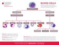
<strong>Blood</strong> Cell & Lymphocyte Development<br />
Multipotential<br />
Hematopoietic Cells<br />
Differentiate & mature into<br />
six types of blood cells<br />
Red Cells<br />
Neutrophils<br />
Eosinophils<br />
Basophils<br />
Monocytes<br />
Platelets<br />
Stem Cells<br />
Stem cells develop into blood cells (hematopoiesis) and lymphocytic cells.<br />
Multipotential<br />
Lymphoid Cells<br />
Differentiate & mature into<br />
three types of lymphocytes<br />
T Lymphocytes<br />
B Lymphocytes<br />
Natural Killer Cells<br />
Bone Marrow. Marrow is a spongy tissue where blood cell development takes<br />
place. It occupies the central cavity of bones. In newborns, all bones have active<br />
marrow. By the time a person reaches young adulthood, the bones of the hands,<br />
feet, arms and legs no longer have functioning marrow. <strong>The</strong> spine (vertebrae), hip<br />
and shoulder bones, ribs, breastbone and skull contain the marrow that makes<br />
blood cells in adults. <strong>The</strong> process of blood cell formation is called “hematopoiesis.”<br />
A small group of cells, the stem cells, develop into all the blood cells in the marrow<br />
by the process of differentiation (see figure above).<br />
In healthy individuals there are enough stem cells to keep producing new blood cells<br />
continuously. <strong>Blood</strong> passes through the marrow and picks up the fully developed<br />
and functional red and white cells and platelets for circulation in the blood.<br />
Some stem cells enter the blood and circulate. <strong>The</strong>y are present in such small<br />
numbers that they cannot be counted or identified by standard blood count tests.
<strong>The</strong>ir presence in the blood is important because they can be collected by a special<br />
technique. <strong>The</strong>re are also methods to induce more stem cells to leave their home<br />
in the marrow and circulate in the blood, allowing a greater stem cell collection<br />
to occur. If enough stem cells are harvested from a compatible donor, they can be<br />
transplanted into a recipient.<br />
Stem cell circulation, from marrow to blood and back, also occurs in the fetus.<br />
After birth, placental and umbilical cord blood can be collected, stored and used as<br />
a source of stem cells for transplantation.<br />
Preparing <strong>Blood</strong> Components<br />
More than 98 percent of the blood supply in the United States comes from<br />
volunteer donors. Most donors give a single unit of whole blood at a site convenient<br />
to their work or home.<br />
<strong>The</strong> availability of plastic bags that can have one or more satellite bags attached in<br />
a completely sterile system allows for flexibility in preparing the donated blood.<br />
<strong>The</strong> use of plastic bags allows the blood center to make a variety of different blood<br />
products. Usually three or four blood components, such as red cells, platelets,<br />
plasma and cryoprecipitate are prepared from each unit of whole blood donated.<br />
“Cryoprecipitate” is the name for the blood component obtained by freezing<br />
plasma and then thawing it at 4°C. It is used to provide certain clotting factors for<br />
people who need them due to a genetic or acquired clotting defect. <strong>The</strong> usefulness<br />
of component therapy is that each patient is given only the specific component<br />
that he or she needs. This allows one donation to benefit up to four patients and<br />
conserves precious blood resources.<br />
Each component has to be prepared within a certain time from collection and<br />
stored at a specific temperature and for a specific length of time to maintain<br />
optimum function. <strong>The</strong> primary blood bag contains an anticoagulant that prevents<br />
the blood from clotting after it has been collected. This unit is spun gently in the<br />
lab using a centrifuge, so that the heavier red cells settle at the bottom of the bag.<br />
<strong>The</strong> lighter plasma, which contains the platelets, can then be siphoned off into<br />
one of the attached satellite bags. A red cell storage solution is then added to the<br />
red cells, the tubing is sealed and the red cells are separated from the other bags. A<br />
red cell unit is about 250 milliliters (about 10 ounces) and is stored at 4°C for 42<br />
days. Ideally, the red cells transfused should be the same ABO and Rh group as the<br />
patient’s. Certain exceptions are made in emergencies.<br />
<strong>The</strong> bag containing the platelet-rich plasma is then centrifuged at a higher speed to<br />
deposit the platelets at the bottom of the bag along with about 50 milliliters (about<br />
two ounces) of plasma. Most of the plasma is siphoned into a third attached bag.<br />
<strong>Blood</strong> <strong>Transfusion</strong> I page 7
<strong>The</strong> unit of platelets is sealed and separated, leaving a bag of plasma. Platelets need<br />
to be stored in an incubator at room temperature and rocked gently. <strong>The</strong>y have a<br />
shelf life of only five days.<br />
Pooled Platelets. About four to five platelet units of the same ABO type as the<br />
patient are pooled together to make a platelet transfusion for an adult. One unit<br />
may be sufficient for an infant. Cryoprecipitate can be made from the plasma, or<br />
the plasma can be stored in a freezer for a year. During this time it may be used for<br />
transfusion or processed further.<br />
Pheresis Platelets. In addition to whole blood donations, some components,<br />
such as platelets, can be collected by apheresis. With apheresis, a healthy donor<br />
comes into the blood center or collection site and the donor’s blood is drawn into a<br />
machine where the blood is separated into its components. <strong>The</strong> cell separator collects<br />
only the part of the blood that is needed by the patient and the rest of the blood is<br />
returned to the donor. This allows a much larger amount of a blood component<br />
to be harvested from a single donor. Also, the donor can be specifically selected for<br />
(matched with) the patient and the donor can donate more frequently, because he or<br />
she does not lose red cells. Pheresis platelets are widely used.<br />
Pheresis platelets have a larger volume of plasma from a single donor, and if the donor<br />
and patient platelets are not ABO identical, the patient has a higher risk for acute<br />
hemolytic transfusion reaction (see page 16). For the same reason, there may be a<br />
higher incidence of transfusion-related acute lung injury (TRALI) (see page 16).<br />
To avoid this, many blood centers are now only collecting pheresis platelets from<br />
donors who do not have HLA antibodies (antibodies that may form as a result of a<br />
challenge to the immune system, pregnancy or organ or tissue transplant).<br />
In most hospitals either pooled platelets or pheresis platelets are available. Most<br />
experts consider pooled platelets and pheresis platelets to be interchangeable with<br />
regard to increasing the patient’s platelet count and controlling bleeding. Both<br />
products can be tested for bacterial contamination.<br />
Safety of <strong>Blood</strong> <strong>Transfusion</strong>s<br />
Autologous and Directed Donations. Autologous donation, in which the<br />
patient donates up to 3 units of his or her own blood to be re-infused later, is<br />
possible for healthy patients who are undergoing a one-time surgery. However, for<br />
patients who are being treated for blood cancers, such donations are not possible<br />
because their own blood lacks adequate numbers of cells.<br />
Some family members ask about “directed donations” in which the family chooses<br />
its own donors for the patient, believing this may be safer. Although this is possible<br />
if a small number of red cells are to be used, e.g., for a surgical procedure, there is<br />
page 8 I 800.955.4572 I www.LLS.org
no evidence that these donations are any safer than the general blood supply. In<br />
fact, under certain circumstances they may be less safe, because related individuals<br />
or friends may not wish to expose a circumstance that makes them unsuitable for<br />
donation. For patients such as those with blood cancers, the need for long-term<br />
blood support, and for specialized components, usually makes this approach<br />
unfeasible.<br />
Donor Screening and Collection. Both patients and doctors are concerned<br />
about the safety of the blood supply. Today, in medically advanced countries, the<br />
benefits of transfusion usually outweigh blood safety concerns for patients with<br />
cancer. <strong>The</strong> risk of transmitting viral diseases such as human immunodeficiency<br />
virus (HIV) and hepatitis by blood transfusion has dropped dramatically in the last<br />
25 years. This is the result of a multilayered approach to safety. First, a voluntary<br />
blood donor pool eliminates individuals who might donate for money and not<br />
be honest about their health history. Public education is important so that people<br />
know that certain diseases can be transmitted by blood, what the risk factors are<br />
for carrying infectious agents and who should refrain from donating because they<br />
are not suitable donors. All potential donors receive written information to urge<br />
them to not donate if they are at risk of transmitting a disease through their blood.<br />
Once a donor comes to a blood donation site, he or she is screened by trained<br />
personnel using a very detailed medical history coupled with a pertinent physical<br />
examination. This ensures that the procedure will be safe both for the donor and<br />
the blood recipient.<br />
<strong>Blood</strong> is collected using a new sterile needle and bag after a meticulous cleaning<br />
of the donor’s arm. Needles are never reused, so there is no risk of infections being<br />
transmitted to the donor. Extra tubes of blood are drawn for the laboratory testing.<br />
All units are checked for their ABO and Rh blood group to ensure there are no red<br />
cell antibodies in the donor’s plasma that might injure the patient’s red cells.<br />
Testing for Carriers of Infectious Disease. Twelve screening tests for seven<br />
infectious diseases are performed on each unit of donated blood. <strong>The</strong>se tests have<br />
become more sensitive over the years. Most of these are indirect tests that detect<br />
antibodies against the infectious disease. <strong>The</strong> tests detect antibodies to<br />
{ { Syphilis<br />
{ { Human immunodeficiency virus (HIV-1 and HIV-2)<br />
{ { Hepatitis B virus core antigen<br />
{ { Hepatitis C virus<br />
{ { Human T lymphocytotropic viruses (HTLV-1 and HTLV-2).<br />
In addition, tests are performed for hepatitis B virus surface antigen, the protein<br />
coat of the hepatitis B virus. Sometimes additional testing is needed for individual<br />
patients, such as for cytomegalovirus (CMV) antibodies.<br />
<strong>Blood</strong> <strong>Transfusion</strong> I page 9
In mid-1999, nucleic acid testing (NAT) for HIV and the hepatitis C virus was<br />
added to the testing. This is a highly sophisticated and sensitive means of detecting<br />
the genetic material of the virus rather than relying on identifying the development<br />
of an antibody in the donor. <strong>The</strong>se tests have further reduced the chance of<br />
transmitting the hepatitis C virus or HIV.<br />
<strong>The</strong> table below shows the current estimates for the residual risk of disease<br />
transmission through blood transfusion.<br />
Infectious Disease Risk Estimates for <strong>Blood</strong> <strong>Transfusion</strong><br />
in the United States<br />
Virus I Test(s) I Risk per Unit<br />
Human Immunodeficiency I Anti-HIV I 1:2 million<br />
Virus (HIV) HIV RNA (MP-NAT) *<br />
Hepatitis C Virus (HCV) I Anti-HCV I 1:1.6 million<br />
HCV RNA (MP-NAT)<br />
Hepatitis B Virus (HBV) I HBs Antigen I 1:400,000<br />
Anti-HBc<br />
HBV DNA<br />
Human T Cell I Anti-HTLV I 1:109,000<br />
Lymphotrophic Virus (HTLV)<br />
Bacteria in Apheresis I Bacterial culture I 1:109,000<br />
Platelets<br />
West Nile Virus I WNV RNA (MP-NAT) (ID-NAT) ** I 1:4.5 million<br />
* MP-NAT- minipool nucleic acid-amplification test<br />
** ID-NAT- individual donor nucleic acid-amplification test<br />
Source: American Red Cross, unpublished data from 2010.<br />
<strong>The</strong> risk of bacterial infection from a unit of red cells that has been routinely stored<br />
at 4°C is thought to be about 1:1 million. Because platelets have to be maintained<br />
at room temperature to preserve their function, the risk of bacterial growth is<br />
higher. Bacterial testing was introduced in 2004 for pheresis platelets and the<br />
residual risk of bacterial infection is now calculated as 1:109,000.<br />
Much research is being focused on methods to inactivate viruses in blood<br />
components. Some coagulation factors, such as factors VIII and IX, are made from<br />
plasma and can be heat-treated to inactivate viruses that might have been present in<br />
plasma. Fresh frozen plasma can also now be processed by a technique called “solvent<br />
detergent treatment,” which eliminates viruses such as HIV and hepatitis B and C<br />
viruses. <strong>The</strong>se viruses have fatty membranes that are destroyed by the detergent.<br />
page 10 I 800.955.4572 I www.LLS.org
Coagulation factors are manufactured from pools of 1,500 donated units. <strong>The</strong>se<br />
products are treated by inactivation techniques and thus are not infectious for<br />
viruses, such as HIV and hepatitis B and C. It is expected that similar technology<br />
may become available for a single unit of plasma, further decreasing the risk of viral<br />
contamination.<br />
<strong>Blood</strong> cells are fragile, and the plasma in which they are suspended cannot be virally<br />
inactivated by harsh procedures such as detergent treatments. Research is under way<br />
to look at gentler techniques for virally inactivating red cells and platelets.<br />
Removing White Cells. White cells contaminate the red cell and platelet<br />
components. <strong>The</strong>se cells are of no use to the patient and are associated with many<br />
reactions during and after transfusion. <strong>The</strong> standard blood filter does not remove<br />
such small cells. However, special filters have been developed that can remove up to<br />
99.99 percent of these cells. <strong>The</strong> technical term for the process of removing white<br />
cells (leukocytes) from blood components is “leukoreduction.” This process used to<br />
be done at the bedside as the blood was being given to patients. Now that removal<br />
of white cells is more common, white cell reduction is often done at blood centers<br />
at the time the components are prepared. This ensures that the filtering is consistent<br />
and components can be tested to ensure that white cell reduction has been<br />
achieved. In many industrialized countries removal of white cells from red cell or<br />
platelet components is now standard practice. In the United States, leukoreduction<br />
is frequently used but it is not a universal practice. Patients requiring transfusion<br />
should ask their doctor about the use of leukoreduced blood components.<br />
<strong>Transfusion</strong>s for Patients with<br />
<strong>Blood</strong> Cancer<br />
This section contains information that applies to leukemia, lymphoma, myeloma,<br />
myelodysplastic syndromes, myeloproliferative neoplasms and other hematological<br />
conditions, such as hereditary anemias and aplastic anemia. In particular, blood or<br />
marrow stem cell transplantation for patients with these diverse diseases invariably<br />
involves frequent blood transfusions. This occurs because the basis of the transplant<br />
treatment is to give very high doses of chemotherapy to the patient to maximize the<br />
chance of a cure. Many drugs used for chemotherapy cause temporarily impaired<br />
blood cell production in the marrow and depressed immune system functions.<br />
<strong>The</strong> disease processes of leukemia, myeloma, and many lymphomas interfere with<br />
the normal production of red cells, white cells and platelets in the marrow. Thus, it<br />
is common for patients with these diseases to develop anemia (a low red cell count),<br />
thrombocytopenia (a low platelet count) and in some cases, leukopenia (a low<br />
white cell count. This can happen before treatment begins, since the cancer cells<br />
inhibit the production of normal blood cells in the marrow. In addition, the drugs<br />
<strong>Blood</strong> <strong>Transfusion</strong> I page 11
used to treat these diseases—which stop disease progression or, in some cases, cure<br />
these diseases—often injure healthy stem cells in the marrow as a side effect. <strong>The</strong>se<br />
injured cells would normally go on to produce red cells, white cells or platelets.<br />
Temporary side effects such as very low red cell or platelet counts can occur for a<br />
few weeks, in most cases, because fewer healthy cells are being made.<br />
<strong>The</strong> need for transfusions varies, depending on the type of blood disease in question<br />
and the type of drugs used in the treatment. For example, almost all patients<br />
with leukemia (a disease primarily affecting the marrow and blood) require some<br />
transfusions during their care. Many patients with Hodgkin or non-Hodgkin<br />
lymphoma (diseases primarily affecting the lymph nodes and spleen) may not<br />
require transfusions unless they require a blood or marrow stem cell transplant or if<br />
the lymphoma involves the marrow.<br />
Individual doctors take different approaches in deciding if transfusion is appropriate<br />
for a given patient, because there is controversy as to how to best balance the<br />
benefits and risks of transfusion in many clinical situations. Studies comparing<br />
various indications for transfusions may help doctors have a more scientific basis for<br />
their decisions, but currently, transfusion policies usually depend on the patient’s<br />
condition and an individual doctor’s training, experience and long-held community<br />
standards of practice. <strong>The</strong> trend in the last few years is to be considerably more<br />
conservative in the use of transfusion products where possible.<br />
Red Cells and Platelets. During and after chemotherapy, it is possible to<br />
replace the red cells and platelets with cells donated by healthy volunteers via<br />
blood transfusions. Severe anemia (a relative term, not well-defined by scientific<br />
studies) or thrombocytopenia can be life-threatening in extreme cases. Most doctors<br />
specializing in the care of patients with blood cancers believe that varying degrees<br />
of replacement by prophylactic red cell transfusion represents a good practice to<br />
prevent complications of anemia, such as fatigue, weakness, shortness of breath<br />
or in extreme cases, heart attack or stroke. Similarly, most doctors advocate giving<br />
prophylactic platelet transfusions to reduce the likelihood of bleeding.<br />
White Cells. Unfortunately, practical methods of safely and effectively transfusing<br />
adequate numbers of granulocytes or other white cells are not yet available to<br />
prevent infection that occurs as a result of a low white cell count. White cell<br />
transfusion is usually reserved for uncommon instances of severe infections with<br />
bacteria or fungi that do not respond to antibiotics or antifungal drugs.<br />
Because the yield of white cells from current collection techniques is insufficient,<br />
some investigative studies and clinical protocols now involve administering<br />
white cell growth factors (e.g., granulocyte-colony stimulating factor [G-CSF])<br />
to volunteer donors, particularly family members, prior to white cell collection<br />
by apheresis. This increases the number of white cells that are in the donor’s<br />
circulation, thus improving the yield of white cells collected. It is hoped that the<br />
page 12 I 800.955.4572 I www.LLS.org
larger number of white cells collected in this manner will be more effective in<br />
fighting infection.<br />
<strong>Transfusion</strong> of Red Cells. Red cell transfusions are used to treat low red cell<br />
counts (anemia), which, if untreated, can cause weakness, lethargy and in extreme<br />
cases, more severe symptoms such as shortness of breath or rapid heartbeat. Most<br />
doctors prescribe red cell transfusions before a patient develops serious symptoms,<br />
particularly when managing older patients or those with a history of heart or blood<br />
vessel disease.<br />
<strong>The</strong>re are few scientific data that guide doctors as to the exact red cell count at<br />
which to prescribe a transfusion. <strong>The</strong> age of the patient, the level of his or her<br />
activity, the presence of other complicating medical conditions and the likelihood<br />
and timeliness of the recovery of red cell production in the marrow each must be<br />
considered along with the red cell count.<br />
All red cell transfusions need to be matched to the patient in the laboratory, and<br />
for patients with blood diseases the donated blood should always have the white<br />
cells removed by filtration.“Leukoreduced” or “leukodepleted” are the medical<br />
terms for white cell removal. Leukoreduction reduces the risks of fever and chills<br />
after transfusion, reduces the risk of not responding to platelet transfusions due<br />
to the development of human leukocyte antigen (HLA) antibodies and reduces<br />
the risk of transmission of some viral infections (e.g., cytomegalovirus, HTLV-1).<br />
Some centers use irradiation of all cell transfusions to patients receiving intensive<br />
chemotherapy or who are considered to have impaired immune systems to<br />
prevent a rare but potentially life-threatening complication of transfusion called<br />
“graft-versus-host disease” (GVHD). Patients undergoing blood or marrow stem<br />
cell transplants generally should receive irradiated blood components during the<br />
transplant period.<br />
Your doctor’s decision to give you red cell transfusions is based on a combination of<br />
factors, including<br />
{ { <strong>The</strong> level of hemoglobin (the protein in red blood cells that carries oxygen)<br />
in your blood<br />
{ { Whether you have symptoms such as fatigue or shortness of breath<br />
{ { Any other health complications you may have, such as heart disease.<br />
Iron Overload. <strong>The</strong> body contains about 2,000 to 3,500 milligrams of iron, most<br />
of which is present in red cells. <strong>The</strong> body has no ability to excrete the excessive<br />
amounts of iron resulting from red cell transfusions.<br />
Each red cell unit contains about 250 milligrams of iron, and patients, who have<br />
regular transfusions, ranging from less than 2 units to 4 or more units of blood<br />
a month, can accumulate too much iron in their bodies as a result. <strong>The</strong> iron is<br />
<strong>Blood</strong> <strong>Transfusion</strong> I page 13
deposited in tissues and major organs such as the liver, heart and pancreas and<br />
can result in serious damage. A patient with iron overload should talk to the<br />
doctor about his or her intake of vitamin C and alcohol, both of which increases<br />
absorption of iron.<br />
If you’re receiving transfusions, your doctor may monitor you for iron overload<br />
with a blood test called a “serum ferritin level,” which measures your body’s iron<br />
store. You may need a drug called an “iron chelator” to remove excess iron from<br />
your body because of transfusion-dependent anemias. Be sure to talk with your<br />
doctor about the potential benefits and risks of using these drugs.<br />
Iron overload is generally not a risk for a patient who has received less than about<br />
20 red cell transfusions over his or her lifetime.<br />
<strong>Transfusion</strong> of Platelets. Platelet transfusions are given to prevent or to treat<br />
bleeding due to severely low platelet counts (thrombocytopenia). <strong>The</strong>re is controversy<br />
as to whether prophylactic platelet transfusions are necessary or beneficial, although<br />
it seems that maintaining a platelet count of greater than 5,000 microliters (μL), and<br />
sometimes higher, reduces the risk of minor bleeding (e.g., nose bleeds, bruises in the<br />
skin called “ecchymoses,” pinpoint bleeding in the skin called “petechiae”). <strong>The</strong> platelet<br />
count at which most hematologists and oncologists believe prophylactic transfusion<br />
(in the absence of bleeding) is indicated has decreased from about 20,000 μL to 10,000<br />
μL at most cancer centers, but there is great individual variation from doctor to doctor<br />
within this range, and from patient to patient. It is uncommon for patients to bleed<br />
when their platelet counts go below 30,000 μL, and most patients can tolerate stable<br />
platelet counts within a range of 5,000 μL to 10,000 μL without bleeding. <strong>The</strong> need<br />
for surgery or other invasive procedures often requires transfusion to maintain a much<br />
higher platelet count during surgery and for a period of healing thereafter.<br />
Platelets can be given as pools made from several units of whole blood from<br />
different donors, or single donor units obtained by apheresis (see page 8). Donated<br />
platelet units should have the white cells removed by filtration prior to transfusion<br />
and, if appropriate, should be irradiated as well.<br />
<strong>Transfusion</strong> of Granulocytes. A patient who has few or no circulating white<br />
cells may develop an infection that does not respond to antibiotics. In some<br />
such instances, use of apheresis to collect donor granulocytes may permit their<br />
transfusion and provide some benefit until the patient’s own white cell counts<br />
recover. As with red cells and platelets, these transfusions should be irradiated prior<br />
to transfusion, but should not be treated with leukoreduction filters, as this would<br />
defeat the purpose of transfusing white cells. <strong>The</strong> white cells are infused through<br />
a standard blood filter that does not filter out white cells, but will filter out any<br />
particles or clotted blood elements. <strong>The</strong>re is uncertainty over whether current<br />
methods of granulocyte collection produce an effective transfusion, which is why<br />
some protocols now include G-CSF stimulation of the granulocyte donor.<br />
page 14 I 800.955.4572 I www.LLS.org
<strong>Transfusion</strong> of Plasma and Cryoprecipitate. Fresh frozen plasma (FFP) and<br />
cryoprecipitate, often called “cryo” for short, are transfused to patients who have<br />
abnormal or low levels of blood-clotting proteins, as in hemophilia. Clotting<br />
protein abnormalities in the plasma may develop in patients with poor clotting<br />
factor production due to liver disease or increased use of clotting factor proteins<br />
due to infection. Fortunately, these conditions are uncommon in patients with<br />
hematologic malignancies, with the exception of promyelocytic leukemia. In this<br />
type of leukemia, abnormal clotting can occur and it may be necessary to transfuse<br />
these liquid fractions of donor blood to prevent or to treat bleeding.<br />
Use of Intravenous Gamma Globulin (IVIG). Gamma globulin prepared from<br />
a pool of donor plasma is sometimes given to patients with hematologic diseases to<br />
supplement their low levels. Very low gamma globulin levels are a frequent feature<br />
of chronic lymphocytic leukemia. Severely low levels of gamma globulin can lead<br />
to an increased risk of some types of bacterial infections. Gamma globulin may<br />
also be of use in reducing the risk of cytomegalovirus disease and other immune<br />
complications of the hematologic disease or its treatment. Gamma globulin is<br />
specially treated by techniques that cannot be used for cell transfusions; it does<br />
not carry the risk of transmission of viruses such as hepatitis C virus or human<br />
immunodeficiency virus (HIV). Most side effects are very modest and can include<br />
mild headache, rash or hives.<br />
<strong>Transfusion</strong> of Albumin. Rarely, transfusion of the most common human blood<br />
protein, albumin, is needed in patients who have severe liver malfunction. Albumin<br />
does not carry a risk of transmission of viruses such as hepatitis C virus or HIV.<br />
Side effects are uncommon with albumin transfusions.<br />
Palliative Care and <strong>Transfusion</strong>s. Palliative care is a form of medical care that<br />
focuses on improving the quality of life for patients facing serious illness. <strong>The</strong> goal<br />
is to prevent and relieve pain and other symptoms and to provide psychological,<br />
spiritual and emotional support. Palliative care is appropriate from the time of<br />
diagnosis and is provided along with curative treatment. <strong>Blood</strong> transfusions can<br />
be used as palliative care. Healthcare coverage for palliative care may differ based<br />
on what treatments are needed. <strong>The</strong>re are doctors and nurses who specialize in<br />
palliative care and who may be part of the patient’s medical care team, or the<br />
patient’s hematologist/oncologist may manage this aspect of care.<br />
Hospice care (care for patients who are thought to have less than six months to live)<br />
can continue to provide palliative care for patients. <strong>The</strong> aim of hospice care is to<br />
provide the best possible quality of life and to relieve pain and symptoms during the<br />
final days of a person’s life at a time when the underlying disease can no longer be<br />
treated or cured. <strong>Blood</strong> transfusions are less frequently used during this time and are<br />
only used when the goal is to alleviate pain and discomfort and enhance the quality<br />
of life, not cure the disease.<br />
<strong>Blood</strong> <strong>Transfusion</strong> I page 15
Complications of <strong>Blood</strong> <strong>Transfusion</strong>s<br />
Most transfusions are not associated with adverse reactions. However, reactions<br />
can occur with any blood component. <strong>The</strong> reaction may occur at the time of the<br />
transfusion, such as abrupt high fever (called a “febrile reaction”) or the destruction<br />
of the transfused red cells (called a “hemolytic reaction”). <strong>Transfusion</strong>-related<br />
acute lung injury (TRALI) is the term for new-onset of acute lung injury (ALI)<br />
that occurs within six hours after the transfusion of a plasma-containing blood<br />
product. <strong>The</strong> cause of TRALI is currently not fully understood. TRALI is treatable<br />
with supportive care, but can be fatal if recognition of TRALI is delayed. Other<br />
deleterious effects, such as the transmission of viruses, are not apparent until weeks<br />
or months later, after the incubation period and the onset of the viral disease.<br />
<strong>The</strong> symptoms of most of the reactions that occur either during or soon after<br />
transfusion are similar. <strong>The</strong>se include the development of a fever, chills, nausea,<br />
pain at the site of the transfusion (an arm vein) or in the back, shortness of<br />
breath, a drop in blood pressure, passing dark or red urine or a rash. Any patient<br />
noticing any change in his or her condition during a transfusion, however slight<br />
it may seem, should alert the nursing staff promptly. Serious complications can be<br />
prevented by early recognition of a reaction, stopping the transfusion and limiting<br />
the amount of blood given.<br />
<strong>The</strong> initial management of all transfusion reactions is the same (except for when<br />
the only reaction is hives. See Reactions that Cause Hives, on page 17), because the<br />
symptoms of different types of reactions may overlap. <strong>The</strong> transfusion is stopped<br />
and the unit is returned to the blood bank for examination to check for factors<br />
that might have caused the transfusion reaction. At the same time the intravenous<br />
line is retained by infusing a glucose solution in case intravenous fluids or drugs are<br />
needed for treatment, and a doctor is called. <strong>Blood</strong> samples may need to be drawn<br />
and treatment started right away. Many transfusion reactions, but not all, can be<br />
prevented or minimized by removing white cells from the component either at the<br />
bedside or in the blood center at the time of collection. Patients with hematologic<br />
diseases usually receive blood component units that are leukoreduced.<br />
Reactions That Damage or Destroy Red Cells. Damage or destruction of<br />
the transfused red cells is rare. However, if this does occur, it represents the most<br />
severe and important acute reaction associated with blood components. Such a<br />
reaction, called an “acute hemolytic transfusion reaction,” can lead to a drop in<br />
blood pressure, bleeding or kidney damage, which may be life threatening. Because<br />
of this, all reactions are considered serious until a hemolytic reaction has been ruled<br />
out. Treatment of a hemolytic reaction includes taking measures to maintain the<br />
blood pressure and prevent kidney damage and bleeding.<br />
Reactions That Cause Fever. Reactions that cause fever, referred to as “febrile<br />
reactions,” are the most common. <strong>The</strong>se account for more than 90 percent of all<br />
page 16 I 800.955.4572 I www.LLS.org
transfusion complications. Fever is sometimes accompanied by chills, and on some<br />
occasions, shortness of breath. <strong>The</strong>se reactions are frightening and uncomfortable<br />
for the patient but are usually not serious. However, they must be distinguished<br />
from the more serious acute hemolytic transfusion reaction mentioned on page 16.<br />
While the reaction is being investigated, the transfusion is delayed. Treatment may<br />
be given to reduce the elevated temperature. Medicines can be given before the<br />
transfusion to prevent such a reaction. A fever reaction is most commonly caused<br />
by antibodies to the small number of white cells mixed with the red cells. <strong>The</strong> use<br />
of red cells from which white cells are removed before storage of the unit is the<br />
most effective means of preventing the high fever and chills.<br />
Unfortunately, during platelet transfusions, reactions causing high fever and chills<br />
are more frequent, because the cause of these reactions is more complex. Filtering<br />
out white cells at the bedside is not as useful in preventing these effects as it is<br />
with red cells. Prestorage leukoreduction is required. Washing of platelets before<br />
transfusion removes certain substances that form immune complexes. In addition<br />
to leukoreduction, washed platelets are occasionally requested for patients with<br />
histories of allergic or anaphylactic reactions.<br />
Reactions That Cause Hives. Hives, which usually itch, are the second most<br />
common side effect of transfusion. <strong>The</strong> medical term for hives is “urticaria.” <strong>The</strong><br />
skin changes are presumably due to soluble substances in the plasma of the donor<br />
that cause an allergic reaction in the patient. <strong>The</strong>se reactions are not dangerous,<br />
but they do cause discomfort and anxiety to the patient. <strong>The</strong>y can be treated<br />
with an antihistamine. For subsequent transfusions to susceptible individuals, the<br />
antihistamines can be given beforehand to prevent a reaction. This is the only reaction<br />
that does not necessarily require discarding the unit. If hives are present without any<br />
other symptoms, the transfusion can be restarted slowly once the hives have resolved.<br />
<strong>The</strong> Patient Makes Antibodies to the Donor’s <strong>Blood</strong>. Some patients may<br />
produce antibodies against certain antigens in transfused blood. Although blood<br />
is typed for the most important antigens on the red cell, ABO and Rh, there are<br />
many other antigens on red cells, white cells, platelets or occasionally in the plasma<br />
that can cause a patient to make antibodies against the donor blood. <strong>The</strong> medical<br />
term for this phenomenon is “alloimmunization.” This effect does not necessarily<br />
cause immediate symptoms but is important if subsequent transfusions are needed.<br />
With red cell transfusions the situation can be managed by selecting donors for<br />
future transfusions with red cells that do not carry the antigens to which the patient<br />
has made an antibody. <strong>The</strong> compatible blood can usually be obtained by testing<br />
the units in the blood bank. However, occasionally a blood unit may need to be<br />
shipped in from another blood center or a rare donor registry. This type of exchange<br />
between blood centers is a common practice and provides a national pool of blood.<br />
With platelet transfusions, the antibodies are formed against white cells. However,<br />
these antibodies may also destroy the transfused platelets. Specifically matched<br />
<strong>Blood</strong> <strong>Transfusion</strong> I page 17
platelets will need to be collected if this occurs. Most blood centers have a pool of<br />
volunteer blood donors who have been human leukocyte antigen (HLA) typed<br />
and are willing to donate by apheresis. <strong>The</strong> platelets will then all come from a few<br />
specifically matched donors who each provide a large dose of platelets. A donor’s<br />
propensity to make antibodies to white cells can be reduced—but not completely<br />
prevented—by the transfusion of red cells and platelets only, with the white cells<br />
removed.<br />
Transmission of Viral Infections. <strong>Blood</strong> is a biological substance and transfusion<br />
may never be risk-free. While the chance of getting a viral disease following blood<br />
transfusion has decreased markedly in the last 20 years, the risk has not been<br />
eliminated. Indirect tests, using detection of antibodies to the viruses, cannot<br />
detect infections that occur between the time of exposure to the virus and the<br />
appearance of the antibody. This period is referred to as the “window period,” and if<br />
a donation is made during this time there remains a very small residual risk of viral<br />
transmission. This is one reason why a careful interview to screen out donors who<br />
are at risk for a transmissible virus infection remains an important aspect of blood<br />
safety procedures. Since 1999, the risk of being infected by HIV and the hepatitis<br />
C virus has been considerably reduced because of the introduction of the more<br />
sensitive nucleic acid testing for these viruses. Now, units that test positive for these<br />
viruses are discarded so infection risk is dramatically lower.<br />
Transmission of Cytomegalovirus (CMV). Cytomegalovirus (CMV) is a<br />
common virus, and about 50 percent of individuals in the United States have<br />
been infected with it by the time they are 50 years old, most without developing<br />
symptoms. However, in premature babies and in patients undergoing blood or<br />
marrow stem cell transplantation, CMV infection can cause serious problems,<br />
such as pneumonia. CMV infection may be due to reactivation of the virus from<br />
a previous exposure or from prior blood transfusion. Patients with leukemia<br />
and those undergoing blood or marrow stem cell transplantation who have<br />
no antibodies to CMV should receive blood components that are negative for<br />
CMV antibodies. Since the virus resides in white cells, it can be transmitted by<br />
blood components that contain white cells. Removal of white cells from blood<br />
components is another approach to preventing CMV. This approach appears to be<br />
as efficient as providing components from CMV antibody-negative donors.<br />
Transmission of Bacterial Infections. Infection with bacteria due to a blood<br />
transfusion is an extremely rare complication with red cell transfusions, on the order<br />
of one per million transfusions. <strong>Blood</strong> is collected and processed in a sterile system.<br />
However, bacteria are very occasionally present in the donor’s blood at the time of<br />
donation or the blood is contaminated at the time of collection. Red cells that are<br />
stored at refrigerator temperatures do not usually provide the right conditions for<br />
organisms to grow, so that infection from red cell transfusions is the least common<br />
complication. However, platelets that are kept at room temperature can allow<br />
bacteria to grow in a contaminated unit. <strong>The</strong>refore, infection following platelet<br />
page 18 I 800.955.4572 I www.LLS.org
transfusions is more common than it is with red cell transfusions. Culturing all<br />
pheresis platelets for bacteria was started in March 2004 in the United States, and<br />
methods of doing the same for platelets made from whole blood have been more<br />
recently introduced.<br />
Graft-Versus-Host Disease (GVHD). Donor white cells (lymphocytes) can<br />
attack the recipient’s skin, liver, bowel and marrow after blood or marrow stem<br />
cell transplantation. <strong>The</strong> result of this attack is called “graft-versus-host disease”<br />
(GVHD). Donor lymphocytes from a blood transfusion have the potential to<br />
produce a similar reaction in the recipient. Although this is very uncommon, it<br />
may happen in patients who have decreased immune system function, referred<br />
to as “immunosuppressed” or “immunocompromised.” Immunosuppression can<br />
result from a disease or intense or prolonged chemotherapy or radiation therapy.<br />
Most centers treat all blood components for transfusion to patients who are severely<br />
immunosuppressed with irradiation. Fortunately, this very severe complication is<br />
rare and almost never occurs after transfusion of irradiated blood. Recipients of<br />
stem cell transplants may develop GVHD, but this complication is usually easier to<br />
manage than GVHD resulting from transfusions.<br />
Effect on a Patient’s Immune System. <strong>The</strong>re is a controversial theory that<br />
transfusions can cause decreases in immune function. <strong>The</strong> medical term for this<br />
effect is “immunomodulation.” It is not clear what the implications of this effect, if<br />
any, are for patients with blood cancer. In other clinical settings (surgery), filtering<br />
out white cells from transfusion components appears to prevent deleterious<br />
immune effects of transfusion to a large degree and this process should be used for<br />
all patients with blood cancer who receive transfusions.<br />
More Information<br />
Free LLS publications include<br />
Acute Lymphoblastic <strong>Leukemia</strong><br />
Acute Myeloid <strong>Leukemia</strong><br />
<strong>Blood</strong> and Marrow Stem Cell Transplantation<br />
Chronic Lymphocytic <strong>Leukemia</strong><br />
Chronic Myelogenous <strong>Leukemia</strong><br />
Hodgkin <strong>Lymphoma</strong><br />
Non-Hodgkin <strong>Lymphoma</strong><br />
Myelodysplastic Syndromes<br />
Myeloma<br />
Understanding Clinical Trials for <strong>Blood</strong> Cancers<br />
Understanding Drug <strong>The</strong>rapy and Managing Side Effects<br />
Understanding Lab and Imaging Tests<br />
<strong>Blood</strong> <strong>Transfusion</strong> I page 19
References<br />
American Red Cross. www.redcrossblood.org<br />
Fang CT, Chambers LA, Kennedy J, Strupp A, Fucci MC, Janas JA, Tang Y,<br />
Hapip CA, Lawrence TB, Dodd RY; American Red Cross Regional <strong>Blood</strong> Centers.<br />
Detection of bacterial contamination in apheresis platelet products: American Red<br />
Cross experience, 2004. <strong>Transfusion</strong>. 2005;45(12):1845-1852.<br />
Spinelli SL, O’Brien JJ, Bancos S, Lehmann GM, Springer DL, Blumberg N,<br />
Francis CW, Taubman MB, Phipps RP. <strong>The</strong> PPAR-platelet connection: modulators<br />
of inflammation and potential cardiovascular effects. PPAR Research. 2008; 2008:<br />
328172. Published online 2007 December 24. doi: 10.1155/2008/328172.<br />
<strong>The</strong> 2007 National <strong>Blood</strong> Collection and Utilization Survey Report. American<br />
Association of <strong>Blood</strong> Banks and the US Department of Health and Human<br />
Services, 2007. www.aabb.org/programs/biovigilance/nbcus/Pages/default.aspx<br />
Acknowledgement<br />
<strong>The</strong> <strong>Leukemia</strong> & <strong>Lymphoma</strong> <strong>Society</strong> gratefully acknowledges, for their critical review and<br />
important contributions to the material presented in this publication,<br />
Joanna Heal, MBBS, MRCP<br />
and<br />
Neil Blumberg, MD<br />
Professor of Pathology and Laboratory Medicine<br />
Director of Clinical Laboratories<br />
Director of the <strong>Transfusion</strong> Medicine Unit, <strong>Blood</strong> Bank, and Stem Cell Storage Facility<br />
University of Rochester Medical Center<br />
Rochester, New York<br />
page 20 I 800.955.4572 I www.LLS.org
REACH OUT TO OUR<br />
INFORMATION SPECIALISTS<br />
<strong>The</strong> <strong>Leukemia</strong> & <strong>Lymphoma</strong><br />
<strong>Society</strong>’s (LLS) Information<br />
Specialists provide patients,<br />
families and healthcare<br />
professionals with the latest<br />
information on leukemia,<br />
lymphoma and myeloma.<br />
Our team consists of master’s<br />
level oncology professionals who<br />
are available by phone Monday<br />
through Friday, 9 am to 6 pm (ET).<br />
Co-Pay Assistance<br />
LLS’s Co-Pay Assistance Program<br />
helps blood cancer patients cover<br />
the costs of private and public<br />
health insurance premiums,<br />
including Medicare and Medicaid,<br />
and co-pay obligations. Support<br />
for this program is based on the<br />
availability of funds by disease.<br />
For more information,<br />
call 877.557.2672 or<br />
visit www.LLS.org/copay.<br />
For a complete directory of our patient services programs, contact us at<br />
800.955.4572 or www.LLS.org<br />
(Callers may request a language interpreter).
For more information, please contact:<br />
or:<br />
National Office<br />
1311 Mamaroneck Avenue, Suite 310, White Plains, NY 10605<br />
Contact our Information Specialists 800.955.4572 (Language interpreters available upon request)<br />
www.LLS.org<br />
Our Mission:<br />
Cure leukemia, lymphoma, Hodgkin’s disease and myeloma, and improve the<br />
quality of life of patients and their families.<br />
LLS is a nonprofit organization that relies on the generosity of individual,<br />
foundation and corporate contributions to advance its mission.<br />
FSC<br />
LOGO<br />
PS44 35M 6/11