Changes in the Phenolic Concentration during Flower Development ...
Changes in the Phenolic Concentration during Flower Development ...
Changes in the Phenolic Concentration during Flower Development ...

Create successful ePaper yourself
Turn your PDF publications into a flip-book with our unique Google optimized e-Paper software.
J. AMER. SOC. HORT. SCI. 134(5):491–496. 2009.<br />
<strong>Changes</strong> <strong>in</strong> <strong>the</strong> <strong>Phenolic</strong> <strong>Concentration</strong> dur<strong>in</strong>g<br />
<strong>Flower</strong> <strong>Development</strong> of Rose ‘KORcrisett’<br />
Valent<strong>in</strong>a Schmitzer 1 , Robert Veberic, Gregor Osterc, and Franci Stampar<br />
Biotechnical Faculty, Agronomy Department, Jamnikarjeva 101, SI-1000 Ljubljana, Slovenia<br />
ADDITIONAL INDEX WORDS. Rosa ·hybrida, stages, color measurements, anthocyan<strong>in</strong>s, phenolics, correlation<br />
ABSTRACT. The concentration of major anthocyan<strong>in</strong>s, quercet<strong>in</strong>s, catech<strong>in</strong>, and phenolic acids dur<strong>in</strong>g flower<br />
development of Rosa ·hybrida L. ‘KORcrisett’ was quantified us<strong>in</strong>g high-performance liquid chromatography/mass<br />
spectrometry. Additionally, <strong>the</strong> changes <strong>in</strong> petal color were monitored colorimetrically at four different stages of<br />
development (bud, partially open flowers, fully open flowers, senescent flowers) and correlation was calculated<br />
between <strong>the</strong> chromaticity parameters and major/total anthocyan<strong>in</strong>s. Color parameters a*, b*, and h8 decreased with<br />
<strong>the</strong> progression of flower development and a*/b* ratio and lightness (L*) <strong>in</strong>creased. In rose petals, a negative trend <strong>in</strong><br />
<strong>the</strong> content of major (pelargonid<strong>in</strong>-3,5-di-O-glucoside, cyanid<strong>in</strong>-3,5-di-O-glucoside) and m<strong>in</strong>or (pelargonid<strong>in</strong>-3-Oglucoside,<br />
cyanid<strong>in</strong>-3-O-glucoside, peonid<strong>in</strong>-3-O-glucoside) anthocyan<strong>in</strong>s was observed dur<strong>in</strong>g flower development.<br />
Buds conta<strong>in</strong>ed almost threefold higher concentrations of pelargonid<strong>in</strong>-3,5-di-O-glucoside and fourfold higher<br />
concentrations of cyanid<strong>in</strong>-3,5-di-O-glucoside than senescent flowers. Buds also conta<strong>in</strong>ed significantly more<br />
quercet<strong>in</strong>s (quercet<strong>in</strong>-3-O-rut<strong>in</strong>oside, quercet<strong>in</strong>-3-O-glucoside, and quercet<strong>in</strong>-3-O-rhamnoside), catech<strong>in</strong>, and<br />
phenolic acids (gallic acid, protocatechulic acid, chlorogenic acid, caffeic acid, p-coumaric acid) than flowers of<br />
subsequent developmental stages. The most significant differences were observed <strong>in</strong> <strong>the</strong> content of gallic acid; buds<br />
conta<strong>in</strong>ed almost sixfold higher values than senescent flowers. Correlation analysis revealed a strong correlation<br />
between chromaticity parameters a*, b*, a*/b* ratio, h8, L*, and major/total anthocyan<strong>in</strong>s with values rang<strong>in</strong>g from<br />
0.60 to –0.84.<br />
The popularity and commercial marketability of rose flowers<br />
primarily depends on <strong>the</strong> size, coloration, longevity, and on <strong>the</strong><br />
scent of rose flowers. A wide spectrum of colors can be attributed<br />
to various pigments such as anthocyan<strong>in</strong>s, quercet<strong>in</strong>s,<br />
and carotenoids (Eugster and Markifischer, 1991) <strong>in</strong> addition<br />
to o<strong>the</strong>r phenolics act<strong>in</strong>g as copigments (Davies and Mazza,<br />
1993).<br />
Dur<strong>in</strong>g flower bud open<strong>in</strong>g and senescence, various events<br />
take place <strong>in</strong> a well-def<strong>in</strong>ed sequence such as cell division,<br />
cellular differentiation, shifts <strong>in</strong> membrane permeability, cell<br />
elongation, and a wide range of gene expression (Kumar et al.,<br />
2008a) <strong>in</strong> association with changes <strong>in</strong> concentration of endogenous<br />
plant growth regulators and secondary metabolites<br />
(Kumar et al., 2008a; Mayak and Halevy, 1972; Sood and<br />
Nagar, 2003). Little <strong>in</strong>formation is available on phenolic<br />
content of develop<strong>in</strong>g rose petals. Sood and Nagar (2003)<br />
observed a sharp <strong>in</strong>crease dur<strong>in</strong>g flower development from <strong>the</strong><br />
flower bud open<strong>in</strong>g to full bloom <strong>in</strong> Rosa bourboniana Desport<br />
and Rosa damascena Mill. In o<strong>the</strong>r plants such as Rosmar<strong>in</strong>us<br />
offic<strong>in</strong>alis L., <strong>the</strong> content of phenolic diterpenes and flavones<br />
<strong>in</strong>creased dur<strong>in</strong>g flower development. However, <strong>the</strong> level of<br />
phenolic acids rema<strong>in</strong>ed constant (Del Bańo et al., 2003).<br />
Anthocyan<strong>in</strong> syn<strong>the</strong>sis is an <strong>in</strong>tegral part of flower development<br />
and generally occurs at later stages of petal development<br />
(Weiss, 2000). The rapid petal growth, result<strong>in</strong>g<br />
from cell expansion, and anthocyan<strong>in</strong> accumulation <strong>in</strong> petals<br />
seem to be regulated by <strong>the</strong> same developmental signals<br />
(Weiss, 2000). The concentration of total anthocyan<strong>in</strong>s <strong>in</strong><br />
petals decl<strong>in</strong>ed dur<strong>in</strong>g flower development <strong>in</strong> Rosa ·hybrida<br />
‘Happ<strong>in</strong>ess’ and ‘P<strong>in</strong>k Coronet’ (Ahuja et al., 1963), Petunia<br />
Received for publication 19 June 2009. Accepted for publication 17 July 2009.<br />
This work is part of program Horticulture No. P4-0013-0481 funded by <strong>the</strong><br />
Slovenian Research Agency (ARRS).<br />
1 Correspond<strong>in</strong>g author. E-mail: valent<strong>in</strong>a.schmitzer@bf.uni-lj.si.<br />
·hybrida Hort. (Ferrante et al., 2006), and Hydrangea macrophylla<br />
Thunb. (Yoshida et al., 2008). This decl<strong>in</strong>e <strong>in</strong> phenolics<br />
and anthocyan<strong>in</strong>s concentration at later stages of flower development<br />
may limit <strong>the</strong> role of <strong>the</strong> peroxidase/phenolics/<br />
ascorbic acid system <strong>in</strong> antioxidant defense and make <strong>the</strong><br />
flower more vulnerable to oxidative stress (Takahama and<br />
Oniki, 1997). Plant tissue color is easily obta<strong>in</strong>ed with a portable<br />
colorimeter and several studies showed that <strong>the</strong>re is a strong<br />
correlation between anthocyan<strong>in</strong>s <strong>in</strong> flowers/leaves and one or<br />
more chromatic parameters (Katori et al., 2002; Schmitzer<br />
et al., 2009). Therefore, changes <strong>in</strong> <strong>the</strong> anthocyan<strong>in</strong> content<br />
could be l<strong>in</strong>ked to visual attributes and described by tristimulus<br />
values.<br />
Potted m<strong>in</strong>iature roses have become <strong>in</strong>creas<strong>in</strong>gly popular<br />
over <strong>the</strong> past few decades mostly as a result of <strong>the</strong> convenient<br />
small size, constant bloom<strong>in</strong>g, and rapid flower development,<br />
which also make it a suitable research material. In <strong>the</strong> present<br />
<strong>in</strong>vestigation, we have monitored <strong>the</strong> changes <strong>in</strong> concentration<br />
of various phenolic compounds and color parameters. To our<br />
knowledge, this work provides <strong>the</strong> first report on abundance of<br />
various anthocyan<strong>in</strong>s, quercet<strong>in</strong>s, catech<strong>in</strong>, and phenolic acids<br />
at four developmental stages of m<strong>in</strong>iature rose ‘KORcrisett’.<br />
The correlations between tristimulus color measurements and<br />
major/total anthocyan<strong>in</strong>s were also evaluated.<br />
Materials and Methods<br />
PLANT MATERIAL AND GROWTH CONDITIONS. ‘KORcrisett’ rose<br />
plants were grown <strong>in</strong> a controlled environment glass greenhouse<br />
at 27/22 °C (day/night) equipped with a cool<strong>in</strong>g system<br />
under natural photoperiod. The greenhouse environmental<br />
control system was set to start cool<strong>in</strong>g at 27 °C. Relative<br />
humidity varied from 75% to 85%. Plants were irrigated daily<br />
us<strong>in</strong>g a flood irrigation system with a 4-m<strong>in</strong> water (18 °C)<br />
J. AMER. SOC. HORT. SCI. 134(5):491–496. 2009. 491
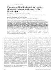
supply. <strong>Flower</strong> petals from <strong>the</strong> outermost whorls were colorimetrically<br />
analyzed and harvested <strong>in</strong> 15 replicates at four<br />
developmental stages (Fig. 1): bud [B (petals closed and fully<br />
pigmented with opened sepals)], partially open flower [PO<br />
(flowers with <strong>the</strong>ir two to three outer whorls unfurled)], fully<br />
open flower [FO (completely unfurled petals)], and senescent<br />
flower [S (petals rolled outward, discoloration detected, flower<br />
easily abscised)]. Petals from <strong>in</strong>dividual flowers were harvested<br />
at each stage and subjected to fur<strong>the</strong>r analysis.<br />
FLOWER COLOR MEASUREMENTS. <strong>Flower</strong> color was measured<br />
by a portable colorimeter (CR-10 Chroma; M<strong>in</strong>olta, Osaka,<br />
Japan) with C illum<strong>in</strong>ant. The colorimeter was calibrated with<br />
a white standard calibration plate before use. In <strong>the</strong> CIE L* a*<br />
b* system of color representation, <strong>the</strong> L* value corresponds to<br />
a dark–bright scale and represents <strong>the</strong> relative lightness of<br />
colors with a range from 0 to 100 (0 = black, 100 = white). The<br />
a* and b* values extend from –60 to 60; a* negative is for green<br />
and a* positive is for red, and b* negative is for blue and<br />
positive for yellow. The a*/b* ratio was calculated for <strong>the</strong><br />
purpose of correlation analysis with anthocyanic pigments. The<br />
hue angle (h°) is expressed <strong>in</strong> degrees from 0° to 360° (0° = red,<br />
90° = yellow, 180° = green and 360° = blue). Color was<br />
measured <strong>in</strong> <strong>the</strong> middle of each petal (three replicates per<br />
flower) to ensure equal measurement conditions.<br />
E XTRACTION AND HIGH- PERFORMANCE LIQUID<br />
CHROMATOGRAPH DETERMINATION OF PHENOLIC COMPOUNDS.<br />
Petals of <strong>in</strong>dividual flowers were ground to a f<strong>in</strong>e powder with<br />
liquid nitrogen and 2 g of powder extracted with 3 mL methanol<br />
conta<strong>in</strong><strong>in</strong>g 3% (v/v) HCOOH and 1% (w/v) 2,6-Di-tert-butyl-4methylphenol<br />
(BHT) <strong>in</strong> an ultrasonic bath for 1 h. Treated<br />
samples were centrifuged for 7 m<strong>in</strong> at 12,000 gn. Supernatant<br />
was filtered through a polyamide filter (Chromafil AO-45/25;<br />
Macherey-Nagel, Düren, Germany) and transferred to a vial<br />
before <strong>in</strong>jection <strong>in</strong> a high-performance liquid chromatograph<br />
(HPLC) system. Samples were analyzed us<strong>in</strong>g a Thermo<br />
F<strong>in</strong>nigan Surveyor HPLC system (Thermo Scientific, San Jose,<br />
CA) with a diode array detector at 280 nm (gallic acid,<br />
protocatechuic acid, catech<strong>in</strong>, chlorogenic acid, caffeic acid,<br />
p-coumaric acid), 350 nm (quercet<strong>in</strong>s) and 530 nm (anthocyan<strong>in</strong>s).<br />
A HPLC column (C18, 150 · 4.6 mm, Gem<strong>in</strong>i 3m;<br />
Phenomenex, Torrance, CA) protected with a Phenomenex<br />
security guard column operated at 25 °C was used. The<br />
<strong>in</strong>jection volume was 20 mL and <strong>the</strong> flow rate ma<strong>in</strong>ta<strong>in</strong>ed at<br />
1mLm<strong>in</strong> –1 . The elution solvents were aqueous 1% formic acid<br />
(A) and 99.8% acetonitrile (B). Samples were eluted accord<strong>in</strong>g<br />
to <strong>the</strong> l<strong>in</strong>ear gradient described by Marks et al. (2007): 0 to 5<br />
m<strong>in</strong>, 3% to 9% B; 5 to 15 m<strong>in</strong>, 9% to 16% B; 15 to 45 m<strong>in</strong>, 16%<br />
to 50% B; 45 to 50 m<strong>in</strong>, 50% isocratic; and f<strong>in</strong>ally wash<strong>in</strong>g and<br />
recondition<strong>in</strong>g of <strong>the</strong> column. The concentrations of phenolic<br />
Fig. 1. Four stages of flower development: 1) bud; 2) partially open flower; 3)<br />
fully open flower; and 4) senescent flower.<br />
compounds were assessed from peak areas and quantified with<br />
<strong>the</strong> use of correspond<strong>in</strong>g external standards and anthocyan<strong>in</strong>s by<br />
<strong>the</strong> use of a calibration curve of cyanid<strong>in</strong>-3,5-di-O-glucoside.<br />
Anthocyan<strong>in</strong>s were fur<strong>the</strong>r identified us<strong>in</strong>g a mass spectrometer<br />
(LCQ Deca XP MAX; Thermo Scientific) with an electroscopy<br />
<strong>in</strong>terface operat<strong>in</strong>g <strong>in</strong> positive ion mode us<strong>in</strong>g MS 2 scann<strong>in</strong>g<br />
mode from m/z 115 to 800. The <strong>in</strong>jection volume was 10 mLand<br />
<strong>the</strong> flow rate ma<strong>in</strong>ta<strong>in</strong>ed at 1 mL m<strong>in</strong> –1 . Capillary temperature<br />
was 250 °C, <strong>the</strong> sheath gas and auxiliary gas were 20 and 8 units,<br />
respectively, <strong>the</strong> capillary voltage was 26 V, and spray voltage<br />
4 V. Multipole Rf amplitude was 550 Vp-p. All compounds were<br />
expressed as mg g –1 fresh weight (FW).<br />
CHEMICALS. The standards used to determ<strong>in</strong>e <strong>the</strong> phenolic<br />
compounds <strong>in</strong> samples were gallic acid, (+)-catech<strong>in</strong>, quercet<strong>in</strong>-<br />
3-O-rut<strong>in</strong>oside, cyan<strong>in</strong> chloride, kuroman<strong>in</strong> chloride, and chlorogenic<br />
acid from Sigma-Aldrich (Ste<strong>in</strong>heim, Germany); catech<strong>in</strong><br />
from Roth (Karlsruhe, Germany); protocatechulic acid from<br />
Merck (Darmstadt, Germany); and caffeic acid, p-coumaric acid,<br />
quercet<strong>in</strong>-3-glucoside, quercet<strong>in</strong>-3-O-rhamnoside, and peonid<strong>in</strong>-<br />
3-O-glucoside from Fluka (Buchs, Switzerland).<br />
The chemicals for <strong>the</strong> sample preparation and mobile phases<br />
were methanol, BHT, and acetonitrile from Sigma-Aldrich and<br />
formic acid from Fluka. The water used <strong>in</strong> <strong>the</strong> mobile phase was<br />
bidistilled and purified with a Milli-Q water purification system<br />
by Millipore (Bedford, MA).<br />
STATISTICAL ANALYSIS. The results were analyzed us<strong>in</strong>g<br />
Statgraphics Plus 4.0 (Manugistics, Rockville, MD) program<br />
us<strong>in</strong>g one-way analysis of variance. Differences <strong>in</strong> phenolic<br />
concentrations among developmental stages were estimated<br />
with Duncan’s multiple range test (P < 0.05). Multiple variable<br />
analysis with Pearson’s product moment correlation coefficient<br />
(r) was calculated between anthocyan<strong>in</strong>s and each of <strong>the</strong> color<br />
variables at P < 0.05.<br />
Results<br />
PETAL COLOR MEASUREMENTS. Color parameters a*, b*, and<br />
h° decl<strong>in</strong>ed at <strong>the</strong> successive stages of flower development.<br />
However, a*/b* ratio and lightness (L*) were <strong>in</strong>creased. The<br />
<strong>in</strong>tense red of <strong>the</strong> buds changed to p<strong>in</strong>k–violet <strong>in</strong> senescent<br />
flowers (Table 1). Analysis of <strong>the</strong> color parameters L* and h°<br />
revealed statistically significant differences among all flower<br />
developmental stages. An <strong>in</strong>crease <strong>in</strong> <strong>the</strong> parameter L* was<br />
observed with each change <strong>in</strong> flower stage; L* <strong>in</strong>creased for<br />
partially opened flowers 9.25%, fully opened flowers 15.60%,<br />
and senescent flowers 23.52% when compared with <strong>the</strong> bud<br />
stage. On <strong>the</strong> o<strong>the</strong>r hand, parameter h° decreased with each<br />
change <strong>in</strong> flower stage; h° was 10.91% lower for partially<br />
opened flowers, 22.43% lower for fully opened flowers, and as<br />
much as 48.11% lower for senescent flowers when compared<br />
with <strong>the</strong> bud stage.<br />
A NTHOCYANIN CONCENTRATION DURING FLOWER<br />
DEVELOPMENT. The HPLC chromatogram at 530 nm revealed different<br />
peaks correspond<strong>in</strong>g to pelargonid<strong>in</strong>-3,5-di-O-glucoside ><br />
cyanid<strong>in</strong>-3,5-di-O-glucoside > pelargonid<strong>in</strong>-3-O-glucoside ><br />
peonid<strong>in</strong>-3-O-glucoside > cyanid<strong>in</strong>-3-O-glucoside. The concentration<br />
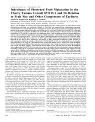
of <strong>the</strong> major anthocyan<strong>in</strong>s (pelargonid<strong>in</strong>-3,5-di-O-glucoside<br />
and cyanid<strong>in</strong>-3,5-di-O-glucoside) <strong>in</strong> rose petals showed<br />
a significant negative trend dur<strong>in</strong>g flower development. In<br />
partially opened flowers, <strong>the</strong> concentration of pelargonid<strong>in</strong>-3,5di-O-glucoside<br />
was 27.32% lower than <strong>in</strong> buds and decl<strong>in</strong>ed to<br />
36.36% at senescent stage (Fig. 2A). Similarly, <strong>the</strong> concentration<br />
492 J. AMER. SOC. HORT. SCI. 134(5):491–496. 2009.
Table 1. Chromatographic parameters (mean ± SE) ofRosa ·hybrida ‘KORcrisett’ petals at four flower stages.<br />
Chromatographic parameter (mean ± SE)<br />
<strong>Flower</strong> stagez a* b* a*/b* ratio L* h°<br />
B 55.49 ± 1.66 by 46.21 ± 3.60 c 1.40 ± 0.16 a 41.08 ± 0.72 a 29.33 ± 0.51 d<br />
PO 55.94 ± 1.50 b 44.42 ± 2.69 c 1.44 ± 0.18 a 44.88 ± 0.69 b 26.13 ± 0.88 c<br />
FO 51.45 ± 1.63 b 27.55 ± 1.39 b 2.06 ± 0.07 b 47.49 ± 1.01 c 22.75 ± 1.01 b<br />
S 40.96 ± 1.88 a 11.30 ± 1.14 a 3.56 ± 0.26 c 50.74 ± 0.73 d 15.22 ± 1.11 a<br />
zB = bud; PO = partially open flower; FO = fully open flower; S = senescent flower.<br />
yDifferent letters (a, b, c, d) <strong>in</strong> rows denote statistically significant differences by Duncan’s multiple range test at P < 0.05.<br />
Fig. 2. <strong>Concentration</strong> of s<strong>in</strong>gle (A–E) and total anthocyan<strong>in</strong>s (F) <strong>in</strong> petals of Rosa ·hybrida ‘KORcrisett’ at four flower stages: bud (B), partially open flower (PO),<br />
fully open flower (FO), and senescent flower (S). Different letters (a–d) denote statistically significant differences by Duncan’s multiple range test atP < 0.05.<br />
of cyanid<strong>in</strong>-3,5-di-O-glucoside was low (26.07%) <strong>in</strong> senescent<br />
flowers <strong>in</strong> comparison with concentrations measured <strong>in</strong> buds<br />
(Fig. 2B). A similar trend was observed <strong>in</strong> <strong>the</strong> concentration of<br />
<strong>the</strong> m<strong>in</strong>or anthocyan<strong>in</strong>, pelargonid<strong>in</strong>-3-O-glucoside (Fig. 2C)<br />
from bud (30.70 ± 1.84 mg g –1 FW) to senescent stage (17.08 ±<br />
1.48 mg g –1 FW). The changes <strong>in</strong> <strong>the</strong> concentration of o<strong>the</strong>r m<strong>in</strong>or<br />
anthocyan<strong>in</strong>s (cyanid<strong>in</strong>-3-O-glucoside and peonid<strong>in</strong>-3-O-glucoside)<br />
dur<strong>in</strong>g flower development were not consistent; however,<br />
statistically significant differences were observed between <strong>the</strong><br />
bud stage and o<strong>the</strong>r flower stages (Fig. 2D–E). The concentration<br />
of total anthocyan<strong>in</strong>s decreased from buds to senescent flowers<br />
and statistically significant differences were observed <strong>in</strong> <strong>the</strong><br />
concentration of total anthocyan<strong>in</strong>s among all developmental<br />
stages (Fig. 2F) with values rang<strong>in</strong>g from 227.70 ± 20.47 mg g –1<br />
FW (senescent flower) to as much 630.68 ± 42.98 mg g –1<br />
FW (buds).<br />
The changes <strong>in</strong> s<strong>in</strong>gle/total anthocyan<strong>in</strong> concentration dur<strong>in</strong>g<br />
flower development were not accompanied by a change <strong>in</strong> <strong>the</strong><br />
ratio among three anthocyan<strong>in</strong>s (pelargonid<strong>in</strong> > cyanid<strong>in</strong> ><br />
peonid<strong>in</strong>) except at <strong>the</strong> senescent stage <strong>in</strong> which a smaller<br />
relative concentration of cyanid<strong>in</strong> was detected as a result of an<br />
<strong>in</strong>crease <strong>in</strong> <strong>the</strong> relative value of both pelargonid<strong>in</strong>s and peonid<strong>in</strong>.<br />
CORRELATION BETWEEN ANTHOCYANINS AND COLOR<br />
MEASUREMENTS. Strong correlations were observed (a <<br />
0.001) among major/total anthocyan<strong>in</strong>s, a*, b*, a*/b* ratio,<br />
h°, and L* <strong>in</strong> rose petals (Table 2). The strongest correlation<br />
was obta<strong>in</strong>ed between L* and cyanid<strong>in</strong>-3,5-di-O-glucoside<br />
(–0.84) followed by pelargonid<strong>in</strong>-3,5-di-O-glucoside (–0.83)<br />
Table 2. Pearson’s correlation coefficients between chromaticity parameters and major/total anthocyan<strong>in</strong>s <strong>in</strong> Rosa ·hybrida ‘KORcrisett’<br />
flowers.<br />
Chromatographic parameter<br />
Anthocyan<strong>in</strong> a* b* a*/b ratio h° L*<br />
Cyanid<strong>in</strong>-3,5-di-O-glucoside 0.82*** z 0.70*** –0.60** 0.80*** –0.84***<br />
Pelargonid<strong>in</strong>-3,5-di-O-glucoside 0.72*** 0.75*** –0.75*** 0.74*** –0.83***<br />
Total anthocyan<strong>in</strong>s 0.75*** 0.74*** –0.65*** 0.74*** –0.82***<br />
zPearson’s correlation coefficients between chromaticity parameters and major/total anthocyan<strong>in</strong>s with statistically significance of <strong>the</strong> estimated<br />
correlation presented at **P < 0.01 and ***P < 0.001.<br />
J. AMER. SOC. HORT. SCI. 134(5):491–496. 2009. 493
and total anthocyan<strong>in</strong>s (–0.82). Similarly strong correlation coefficients<br />
were also observed among cyanid<strong>in</strong>-3,5-di-O-glucoside,<br />
a* (0.82), and h° (0.80). Lowest correlation coefficients were<br />
obta<strong>in</strong>ed between a*/b* ratio, <strong>the</strong> concentration of cyanid<strong>in</strong>-3,5di-O-glucoside<br />
(–0.60), and total anthocyan<strong>in</strong>s (–0.65).<br />
QUERCETINS AND CATECHIN DURING FLOWER MATURATION.<br />
Quercet<strong>in</strong>-3-O-rhamnoside was <strong>the</strong> most abundant quercet<strong>in</strong> <strong>in</strong><br />
rose petals followed by quercet<strong>in</strong>-3-O-glucoside and quercet<strong>in</strong>-<br />
3-O-rut<strong>in</strong>oside (Table 3). The concentration of quercet<strong>in</strong>-3-Orhamnoside<br />
dropped significantly dur<strong>in</strong>g flower development;<br />
<strong>in</strong> senescent flowers, <strong>the</strong> concentration was only 55.44% of <strong>the</strong><br />
one <strong>in</strong> buds. A similar trend was observed for quercet<strong>in</strong>-3-Orut<strong>in</strong>oside<br />
with statistically significant differences observed<br />
between bud stage and o<strong>the</strong>r flower developmental stages.<br />
Catech<strong>in</strong> was <strong>the</strong> dom<strong>in</strong>ant phenolic compound detected <strong>in</strong> rose<br />
petals and its concentration decl<strong>in</strong>ed significantly dur<strong>in</strong>g flower<br />
development. This decl<strong>in</strong>e was twofold from bud (4425.44 ±<br />
271.52 mg g –1 FW) to senescent stage (2040.90 ± 207.74 mg g –1<br />
FW).<br />
PHENOLIC ACIDS DURING FLOWER MATURATION. Caffeic acid<br />
(Fig. 3D) and chlorogenic acid (Fig. 3C) were present <strong>in</strong> highest<br />
concentrations <strong>in</strong> rose petals followed by protocatechulic (Fig.<br />
3B), p-coumaric (Fig. 3E), and gallic acid (Fig. 3A). The most<br />
significant differences were observed <strong>in</strong> <strong>the</strong> concentration of<br />
gallic acid; bud conta<strong>in</strong>ed sixfold higher concentrations of<br />
gallic acid (27.66 ± 1.27 mg g –1 FW) than senescent flowers<br />
(4.76 ± 0.62 mg g –1 FW) and a clear negative trend was also<br />
observed from partially open (13.91 ± 1.15 mg g –1 FW) to fully<br />
open flowers (7.47 ± 0.51 mg g –1 FW). Similarly, <strong>the</strong> concentration<br />
of p-coumaric acid dropped from <strong>the</strong> stage bud to<br />
senescent flower by almost 80%. Significant differences were<br />
observed <strong>in</strong> <strong>the</strong> content of caffeic and chlorogenic acid at<br />
various stages of flower development. Buds conta<strong>in</strong>ed more<br />
than twofold more caffeic acid than senescent flowers and<br />
threefold more chlorogenic acid.<br />
Discussion<br />
<strong>Flower</strong> development <strong>in</strong> R. ·hybrida ‘KORcrisett’ is accompanied<br />
by a substantial change <strong>in</strong> concentration of various<br />
phenolic compounds and petal color. As <strong>the</strong> flower developed,<br />
significant differences were observed <strong>in</strong> color parameters a*,<br />
b*, and L*. The parameter a* is associated with red coloration<br />
<strong>in</strong> rose petals (Biolley and Jay, 1993) and dur<strong>in</strong>g ‘KORcrisett’<br />
flower development, a steady decrease <strong>in</strong> this parameter was<br />
detected. Rose flowers <strong>in</strong> <strong>the</strong> f<strong>in</strong>al stages of flower development<br />
became paler and this characteristic visual change seems to be<br />
associated with a gradual <strong>in</strong>crease of <strong>the</strong> parameter L*. Many<br />
research studies have reported that <strong>the</strong> major anthocyan<strong>in</strong>s <strong>in</strong><br />
plant tissues exhibit a tight correlation with colorimetric <strong>in</strong>dices<br />
such as lightness (L*) and hue angle (h°) (Jia et al., 2008;<br />
Lancaster et al., 1997; Schmitzer et al., 2009). Correlation<br />
Table 3. <strong>Concentration</strong> of quercet<strong>in</strong>s and catech<strong>in</strong> <strong>in</strong> petals of Rosa ·hybrida ‘KORcrisett’ at four flower stages.<br />
Quercet<strong>in</strong>s and catech<strong>in</strong> [mean ± SE (mg g –1 fresh weight)]<br />
<strong>Flower</strong> stagez Quercet<strong>in</strong>-3-O-glucoside Quercet<strong>in</strong>-3-O-rut<strong>in</strong>oside Quercet<strong>in</strong>-3-O-rhamnoside Catech<strong>in</strong><br />
B 33.33 ± 1.97 NS y 11.75 ± 1.29 b 229.35 ± 13.12 c 4425.44 ± 271.52 c<br />
PO 19.82 ± 0.84 NS 6.17 ± 0.47 a 135.75 ± 5.40 b 3673.41 ± 187.88 b<br />
FO 16.13 ± 1.42 NS 6.57 ± 0.74 a 103.10 ± 10.00 a 2574.03 ± 182.59 a<br />
S 24.24 ± 3.11 NS 8.63 ± 1.20 a 102.20 ± 13.19 a 2040.90 ± 207.74 a<br />
zB = bud; PO = partially open flower; FO = fully open flower; S = senescent flower.<br />
yDifferent letters <strong>in</strong> rows (a, b, c, NS = nonsignificant) denote statistically significant differences by Duncan’s multiple range test at P < 0.05.<br />
Fig. 3. <strong>Concentration</strong> of phenolic acids (A–E) <strong>in</strong> petals of Rosa ·hybrida ‘KORcrisett’ at four flower stages: bud (B), partially open flower (PO), fully open flower<br />
(FO), and senescent flower (S). Different letters (a–d, NS = nonsignificant) denote statistically significant differences by Duncan’s multiple range test at P < 0.05.<br />
494 J. AMER. SOC. HORT. SCI. 134(5):491–496. 2009.
analysis revealed a tight correlation between tristimulus values<br />
and major/total anthocyan<strong>in</strong>s <strong>in</strong> ‘KORcrisett’ flower petals with<br />
<strong>the</strong> best correlation obta<strong>in</strong>ed between major/total anthocyan<strong>in</strong>s<br />
and <strong>the</strong> parameter L*. The correlation coefficient was negative<br />
between lightness (L*) and <strong>the</strong> concentration of anthocyan<strong>in</strong>s.<br />
Five anthocyan<strong>in</strong>s detected <strong>in</strong> <strong>the</strong> petals of R. ·hybrida<br />
‘KORcrisett’ were pelargonid<strong>in</strong>-3,5-di-O-glucoside, cyanid<strong>in</strong>-<br />
3,5-di-O-glucoside, pelargonid<strong>in</strong>-3-O-glucoside, peonid<strong>in</strong>-3-<br />
O-glucoside, and cyanid<strong>in</strong>-3-O-glucoside. Occurrence of similar<br />
anthocyan<strong>in</strong>s was also reported <strong>in</strong> o<strong>the</strong>r rose cultivars<br />
(Asen, 1982; Biolley et al., 1994; Mikanagi et al., 1995). The<br />
major anthocyan<strong>in</strong>s <strong>in</strong> m<strong>in</strong>iature rose ‘KORcrisett’ throughout<br />
<strong>the</strong> development were pelargonid<strong>in</strong>-3,5-di-O-glucoside and<br />
cyanid<strong>in</strong>-3,5-di-O-glucoside, which is <strong>in</strong> accordance with <strong>the</strong><br />
results of Biolley et al. (1994) who reported that diglucosides<br />
predom<strong>in</strong>ate over <strong>the</strong> related 3-monoglucosides and <strong>the</strong> cooccurrence<br />
of pelargonid<strong>in</strong> and cyanid<strong>in</strong> is common <strong>in</strong> modern<br />
roses (Mikanagi et al., 1995).<br />
Although research reports on <strong>in</strong>dividual anthocyan<strong>in</strong> turnover<br />
<strong>in</strong> liv<strong>in</strong>g plant tissues are scarce, it is well evident that <strong>the</strong><br />
concentration of s<strong>in</strong>gle and total anthocyan<strong>in</strong>s is not constant<br />
dur<strong>in</strong>g flower development (Dela et al., 2003). In our study, <strong>the</strong><br />
concentration of three major anthocyan<strong>in</strong>s (pelargonid<strong>in</strong>-3,5di-O-glucoside,<br />
cyanid<strong>in</strong>-3,5-di-O-glucoside, pelargonid<strong>in</strong>-3-<br />
O-glucoside) dropped drastically from bud to senescent stage.<br />
However, <strong>the</strong> concentration of peonid<strong>in</strong>-3-O-glucoside and<br />
cyanid<strong>in</strong>-3-O-glucoside rema<strong>in</strong>ed constant. Several factors<br />
might be responsible for this threefold decrease <strong>in</strong> <strong>the</strong> concentration<br />
of major anthocyan<strong>in</strong>s from bud to senescent stage.<br />
First, active degradation of anthocyan<strong>in</strong> was observed <strong>in</strong><br />
Brunfelsia calyc<strong>in</strong>a Benth. (Vakn<strong>in</strong> et al., 2005) and P.<br />
·hybrida (Ferrante et al., 2006). Second, petal expansion<br />
mediated dilution of pigments and <strong>in</strong>crease <strong>in</strong> flower weight<br />
(Vakn<strong>in</strong> et al., 2005). Third, it can be <strong>the</strong> result of a cessation of<br />
petal expansion at later stages of development. Probably<br />
anthocyan<strong>in</strong> concentration and <strong>the</strong> process of senescence are<br />
tightly l<strong>in</strong>ked phenomenon <strong>in</strong> rose flowers. <strong>Flower</strong> development<br />
<strong>in</strong> m<strong>in</strong>iature rose ‘KORcrisett’ also revealed a significant<br />
negative trend <strong>in</strong> analyzed quercet<strong>in</strong>s, catech<strong>in</strong>, and phenolic<br />
acids from bud to senescent stage. Quercet<strong>in</strong>-3-O-glucoside,<br />
quercet<strong>in</strong>-3-O-rhamnoside, and quercet<strong>in</strong>-3-O-rut<strong>in</strong>oside were<br />
<strong>the</strong> major quercet<strong>in</strong>s <strong>in</strong> rose petals (Asen, 1982; Cai et al., 2005;<br />
Mikanagi et al., 1995) and <strong>the</strong>ir concentration decl<strong>in</strong>ed with<br />
flower development. Gallic acid and protocatechuic acid,<br />
catech<strong>in</strong>, chlorogenic, caffeic, and p-coumaric acid (Cai<br />
et al., 2005; Kumar et al., 2008b; Velioglu and Mazza, 1991)<br />
are known components <strong>in</strong> rose petals and with progression of<br />
flower development <strong>in</strong> roses, <strong>the</strong> decrease <strong>in</strong> <strong>the</strong> concentration<br />
of <strong>the</strong>se compounds was detected. Aga<strong>in</strong>, several factors might<br />
expla<strong>in</strong> this gradation. Sequential biosyn<strong>the</strong>sis of flavonols and<br />
anthocyan<strong>in</strong>s <strong>in</strong> rose flowers could be strictly developmentally<br />
regulated like <strong>in</strong> Eustoma grandiflora (Raf.) Sh<strong>in</strong>n. <strong>in</strong> which <strong>the</strong><br />
flavonol concentration decl<strong>in</strong>ed dur<strong>in</strong>g floral development (Noda<br />
et al., 2004). On <strong>the</strong> o<strong>the</strong>r hand, it may be <strong>the</strong> result of a decl<strong>in</strong>e <strong>in</strong><br />
PAL, CHI, DFR, and o<strong>the</strong>r gene transcriptions as was reported <strong>in</strong><br />
Malus ·domestica Borkh. flowers (Dong et al., 1998).<br />
<strong>Flower</strong> development <strong>in</strong>volves a number of <strong>in</strong>terrelated processes<br />
such as growth, senescence, and abscission (Sood and<br />
Nagar, 2003) and our <strong>in</strong>vestigation showed a temporal decl<strong>in</strong>e <strong>in</strong><br />
certa<strong>in</strong> specific anthocyanic pigments and phenolics. This study<br />
provides an <strong>in</strong>sight to explore <strong>the</strong> possible role of phenolics <strong>in</strong><br />
regulation of flower development. The future work will evaluate<br />
<strong>the</strong> relative importance of <strong>the</strong>se phenolics <strong>in</strong> a large number of<br />
rose cultivars for <strong>the</strong>ir antioxidant capacities and responses to<br />
pathogens at various stages of flower development.<br />
Literature cited<br />
Ahuja, K.G., H.L. Mitchell, and W.J. Carpeter. 1963. Quantitative<br />
determ<strong>in</strong>ation of anthocyanid<strong>in</strong>s from petals of rose cultivars ‘P<strong>in</strong>k<br />
Coronet’ and ‘Happ<strong>in</strong>ess’. Proc. Amer. Soc. Hort. Sci. 83:829–832.<br />
Asen, S. 1982. Identification of flavonoid chemical markers <strong>in</strong> roses<br />
and <strong>the</strong>ir high pressure liquid chromatographic resolution and<br />
quantification for cultivar identification. J. Amer. Soc. Hort. Sci.<br />
107:744–750.<br />
Biolley, J.P. and M. Jay. 1993. Anthocyan<strong>in</strong>s <strong>in</strong> modern roses—Chemical<br />
and colorimetric features <strong>in</strong> relation to <strong>the</strong> color range. J. Expt. Bot.<br />
44:1725–1734.<br />
Biolley, J.P., M. Jay, and M.-R. Viricel. 1994. Flavonoid diversity and<br />
metabolism <strong>in</strong> 100 Rosa ·hybrida cultivars. Phytochemistry<br />
35:413–419.<br />
Cai, Y.-Z., J. X<strong>in</strong>g, M. Sun, Z.-Q. Zhan, and H. Corke. 2005. <strong>Phenolic</strong><br />
antioxidants (hydrolysable tann<strong>in</strong>s, flavonols, and anthocyan<strong>in</strong>s)<br />
identified by LC-ESI-MS and MALDI-QIT-TOF MS from Rosa<br />
ch<strong>in</strong>ensis flowers. J. Agr. Food Chem. 53:9940–9948.<br />
Davies, A.J. and G. Mazza. 1993. Copigmentation of simple and<br />
acylated anthocyan<strong>in</strong>s with colorless phenolic compounds. J. Agr.<br />
Food Chem. 41:716–720.<br />
Del Bańo, M.J., J. Lorente, J. Castillo, O. Benavente-García, J.A. Del<br />
Río, A. Ortuńo, K.-W. Quir<strong>in</strong>, and D. Gerard. 2003. <strong>Phenolic</strong><br />
diterpenes, flavones, and rosmar<strong>in</strong>ic acid distribution dur<strong>in</strong>g <strong>the</strong><br />
development of leaves, flowers, stems, and roots of Rosmar<strong>in</strong>us<br />
offic<strong>in</strong>alis. Antioxidant activity. J. Agr. Food Chem. 51:4247–4253.<br />
Dela, G., E. Or, R. Ovadia, A. Nissim-Levi, D. Weiss, and M. Oren-<br />
Shamir. 2003. <strong>Changes</strong> <strong>in</strong> anthocyan<strong>in</strong> concentration and composition<br />
<strong>in</strong> ‘Jaguar’ rose flowers due to transient high-temperature<br />
conditions. Plant Sci. 164:333–340.<br />
Dong, Y.H., L. Beun<strong>in</strong>g, K. Davies, D. Mitra, B. Morris, and A.<br />
Kootstra. 1998. Expression of pigmentation genes and photoregulation<br />
of anthocyan<strong>in</strong> biosyn<strong>the</strong>sis <strong>in</strong> develop<strong>in</strong>g Royal Gala<br />
apple flowers. Aust. J. Plant Physiol. 25:245–252.<br />
Eugster, C.H. and E. Markifischer. 1991. The chemistry of rose<br />
pigments. Angew. Chem. Int. Ed. Engl. 30:654–672.<br />
Ferrante, A., P. Vernieri, F. Tognoni, and G. Serra. 2006. <strong>Changes</strong> <strong>in</strong><br />
abscisic acid and flower pigments dur<strong>in</strong>g flower senescence of<br />
petunia. Biol. Plant. 50:581–585.<br />
Jia, N., Q.-Y. Shu, L.-S. Wang, H. Du, Y.-J. Xu, and Z.-A. Liu. 2008.<br />
Analysis of petal anthocyan<strong>in</strong>s to <strong>in</strong>vestigate color mechanism <strong>in</strong><br />
herbaceous peony cultivars. Scientia Hort. 117:167–173.<br />
Katori, M., K. Watanabe, K. Nomura, and K. Yoneda. 2002. Cultivar<br />
differences <strong>in</strong> anthocyan<strong>in</strong> and carotenoid pigments <strong>in</strong> <strong>the</strong> petals of <strong>the</strong><br />
flower<strong>in</strong>g lotus (Nelumbo spp.). J. Jpn. Soc. Hort. Sci. 71:812–817.<br />
Kumar, N., G.C. Srivastava, and K. Dixit. 2008a. <strong>Flower</strong> bud open<strong>in</strong>g<br />
and senescence <strong>in</strong> roses (Rosa hybrida L.). Plant Growth Regulat.<br />
55:81–99.<br />
Kumar, N., P. Bhandari, B. S<strong>in</strong>gh, A.P. Gupta, and V.K. Kaul. 2008b.<br />
Reversed phase-HPLC for rapid determ<strong>in</strong>ation of polyphenols <strong>in</strong><br />
flowers of rose species. J. Separation Sci. 31:262–267.<br />
Lancaster, J.E., C.E. Lister, P.F. Reay, and C.M. Triggs. 1997.<br />
Influence of pigment composition on sk<strong>in</strong> color <strong>in</strong> a wide range of<br />
fruit and vegetables. J. Amer. Soc. Hort. Sci. 122:594–598.<br />
Marks, S.C., W. Mullen, and A. Crozier. 2007. Flavonoid and<br />
chlorogenic acid profiles of English cider apples. J. Sci. Food Agr.<br />
87:719–728.<br />
Mayak, S. and A.H. Halevy. 1972. Interrelationship of ethylene and<br />
abscisic acid <strong>in</strong> <strong>the</strong> control of rose petal senescence. Plant Physiol.<br />
50:341–346.<br />
Mikanagi, Y., M. Yokoi, Y. Ueda, and N. Saito. 1995. <strong>Flower</strong> flavonol<br />
and anthocyan<strong>in</strong> distribution <strong>in</strong> subgenus Rosa. Biochem. Syst. Ecol.<br />
23:183–200.<br />
J. AMER. SOC. HORT. SCI. 134(5):491–496. 2009. 495
Noda, I., Y. Kanno, N. Kato, K. Kazuma, and M. Suzuki. 2004.<br />
Regulation of gene expression <strong>in</strong>volved <strong>in</strong> flavonol and anthocyan<strong>in</strong><br />
biosyn<strong>the</strong>sis dur<strong>in</strong>g petal development <strong>in</strong> lisianthus (Eustoma grandiflorum).<br />
Physiol. Plant. 122:305–313.<br />
Schmitzer, V., G. Osterc, R. Veberic, and F. Stampar. 2009. Correlation<br />
between chromaticity values and major anthocyan<strong>in</strong>s <strong>in</strong> seven<br />
Acer palmatum Thunb. cultivars. Scientia Hort. 119:442–446.<br />
Sood, S. and P.K. Nagar. 2003. <strong>Changes</strong> <strong>in</strong> abscisic acid and phenols<br />
dur<strong>in</strong>g flower development <strong>in</strong> two diverse species of rose. Acta<br />
Physiol. Plant. 25:411–416.<br />
Takahama, U. and T. Oniki. 1997. A peroxidase/phenolics/ascorbate<br />
system can scavenge hydrogen peroxide <strong>in</strong> plant cells. Physiol. Plant.<br />
101:845–852.<br />
Vakn<strong>in</strong>, H., A. Bar-Akiva, R. Ovadia, A. Nissim-Levi, I. Forer, D.<br />
Weiss, and M. Oren-Shamir. 2005. Active anthocyan<strong>in</strong> degradation<br />
<strong>in</strong> Brunfelsia calyc<strong>in</strong>a (yesterday-today-tomorrow) flowers. Planta<br />
222:19–26.<br />
Velioglu, Y.S. and G. Mazza. 1991. Characterisation of flavonoids <strong>in</strong><br />
petals of Rosa damascena by HPLC and spectral analysis. J. Agr.<br />
Food Chem. 39:463–467.<br />
Weiss, D. 2000. Regulation of flower pigmentation and growth:<br />
Multiple signal<strong>in</strong>g pathways control anthocyan<strong>in</strong> syn<strong>the</strong>sis <strong>in</strong><br />
expand<strong>in</strong>g petals. Physiol. Plant. 110:152–157.<br />
Yoshida, K., I. Daisuke, Y. Sh<strong>in</strong>kai, and T. Kondo. 2008. Change of<br />
color and components <strong>in</strong> sepals of chameleon hydrangea dur<strong>in</strong>g<br />
maturation and senescence. Phytochemistry 69:3159–3165.<br />
496 J. AMER. SOC. HORT. SCI. 134(5):491–496. 2009.