
You also want an ePaper? Increase the reach of your titles
YUMPU automatically turns print PDFs into web optimized ePapers that Google loves.
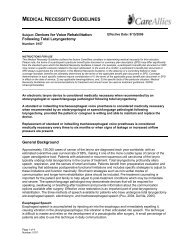
MEDICAL NECESSITY GUIDELINES<br />
Subject: <strong>Benign</strong> <strong>Skin</strong> <strong>Lesion</strong> <strong>Removal</strong><br />
Number: 0302<br />
Page 1 of 7<br />
Number: 0305<br />
Effective Date: 3/15/2006<br />
Revision Date: 3/15/2007<br />
INSTRUCTIONS FOR USE<br />
This Medical Necessity Guideline outlines the factors CareAllies considers in determining medical necessity for this indication.<br />
Please note, the terms of a participant’s particular benefit plan document or summary plan description (SPD) may differ significantly<br />
from the standard upon which this Medical Necessity Guideline is based. For example, a participant’s benefit plan document or SPD<br />
may contain a specific exclusion related to the topic addressed. In the event of a conflict, a participant’s benefit plan document or<br />
SPD always supercedes the information in this Medical Necessity Guideline. In the absence of a controlling federal or state<br />
coverage mandate, benefits are ultimately determined by the terms of the applicable benefit plan document or SPD. Coverage<br />
determinations in each specific instance require consideration of 1) the terms of the applicable group benefit plan document or SPD<br />
in effect on the date of service; 2) any applicable laws/regulations, and; 3) the specific facts of the particular situation. Medical<br />
Necessity Guidelines are not recommendations for treatment and should never be used as treatment guidelines. ©2007<br />
Intracorp/CareAllies<br />
The removal of benign skin lesions (e.g., nevus [mole], sebaceous cyst, wart, seborrheic<br />
keratosis, skin tag or pigmented lesion) is considered medically necessary when ANY of the<br />
following criteria are met:<br />
• There is drainage, inflammation, bleeding, burning, intense itching, or pain associated with the<br />
lesion.<br />
• The lesion is growing in size.<br />
• The lesion obstructs a body orifice, restricts vision or otherwise significantly interferes with normal<br />
function.<br />
• There is clinical suspicion of malignancy (e.g., a change in the ABCDs of skin cancer [asymmetry,<br />
border irregularity, color, and diameter]).<br />
• Due to its anatomical location, the lesion is prone to being recurrently traumatized.<br />
<strong>Benign</strong> skin lesion removal performed solely for the purpose of altering appearance or selfesteem,<br />
or to treat psychological symptomatology or psychosocial complaints related to one’s<br />
appearance is considered cosmetic and not medically necessary.<br />
General Background<br />
The skin is the largest organ of the body and consists of two distinct layers: the dermis and the epidermis.<br />
The epidermis consists of basic keratinocytes that contain keratin, a fibrous protein; melanocytes,<br />
responsible for pigmentation in the lower part; and basal cells, the innermost layer. The dermis is a<br />
connective tissue layer under the epidermis and contains nerve endings, capillaries and sensory<br />
receptors. The skin acts as a primary protective barrier between the internal structures of the body and<br />
the external environment. A skin lesion is an area of the skin in which the appearance has changed; the<br />
change may affect one small spot or an entire area. Typically, skin lesions are classified as noncancerous<br />
(i.e., benign) or cancerous (i.e., malignant). Noncancerous skin lesions may be caused by viruses,<br />
systemic disease, or environmental factors. <strong>Skin</strong> cancer, the most common form of cancer in the United<br />
States, includes basal cell carcinoma, Kaposi’s sarcoma, melanoma, squamous cell carcinoma and<br />
Paget’s disease. The scope of this guideline is limited to removal of noncancerous (i.e., benign) skin<br />
lesions.<br />
While caused by a variety of conditions, benign skin lesions commonly occur in the form of moles (i.e.,<br />
nevi), sebaceous cysts, warts, seborrheic keratoses, skin tags and pigmented lesions. Although many<br />
lesions are removed primarily for cosmetic reasons (i.e., to improve appearance), some may cause
irritation, pain or bleeding and require removal to alleviate symptoms. Surgical removal is also<br />
recommended for any lesion that shows possible signs of malignancy. Clinical signs of suspicion (i.e.,<br />
ABCDs of melanoma) include the following (American Academy of Dermatology [AAD], 2006):<br />
• Asymmetry—One half of a mole is unlike the other half.<br />
• Border irregularity—Border may be irregular, scalloped or poorly circumscribed.<br />
• Color—Color may be varied from one area to another; shades of tan and brown, black;<br />
sometimes white, red or blue.<br />
• Diameter—Melanomas are usually greater than 6 mm in diameter when diagnosed, can also be<br />
smaller.<br />
Types of <strong>Benign</strong> <strong>Skin</strong> <strong>Lesion</strong>s<br />
Nevus (Mole): Moles can appear anywhere on the skin, are usually pigmented with shades of brown and<br />
can be of various sizes and shapes. They are the most common form of benign skin tumor (Bangs, 2003).<br />
Most appear during the first 20 years of life, but some may not appear until later. A "nevus," a term often<br />
used synonymously for a mole, may be either acquired or congenital. Moles may increase in size or<br />
number upon exposure to the sun. Moles that are dysplastic or atypical usually appear larger than<br />
average and irregular in shape, with uneven color patterns. Traditionally, dysplastic nevi were viewed as<br />
precursor lesions to melanoma, and removal was recommended for atypical-appearing moles. More<br />
recently, they have been viewed as markers of increased risk for melanoma (Salopek, 2002).<br />
Prophylactic removal of dysplastic nevi does not eliminate the risk of subsequent melanoma formation<br />
(Swetter, 2003). Changing moles may indicate melanoma, but all moles undergo changes as part of their<br />
natural course, and most changing moles do not represent melanomas (Bangs, 2003). Changes may<br />
occur as a result of involution, irritation or hair follicle rupture. Moles may also enlarge and darken during<br />
pregnancy. <strong>Removal</strong> is indicated for clinical signs of severe atypia, suspicion of malignancy, irritation, or<br />
location in a body area subject to recurrent trauma (AAD, 1993). The treatment of choice for removal is by<br />
shave biopsy or elliptical excision.<br />
Sebaceous Cyst (Epidermal Inclusion Cyst): Some glands in the skin are attached to hair follicles and<br />
produce an oily substance known as sebum. They are found most often on the face, neck, back and<br />
chest. A blocked gland results in the formation of a cyst. Usually these benign cysts do not require<br />
treatment, and they may resolve without treatment. If a cyst becomes infected, it may require removal.<br />
Acne is a common, chronic, inflammatory condition involving the pilosebaceous ducts. The primary lesion<br />
of acne is the comedone. This noninflamed lesion may be open (i.e., blackhead) or closed (i.e.,<br />
whitehead) and may be accompanied by inflammatory lesions, including papules, pustules and cysts.<br />
<strong>Removal</strong> of sebaceous cysts may be performed by cryosurgery, curettage, electrosurgery or excision.<br />
Treatment of acne often employs manual comedone extraction; intralesional injections; incision and<br />
drainage; or electrocauterization.<br />
Wart: Warts are benign skin growths that result from viral infection. The virus that causes warts is<br />
referred to as the human papillomavirus (HPV). Warts may be located on the fingers and hands (i.e.,<br />
common warts) or the soles of the feet (i.e., plantar warts), or they may occur anywhere as small,<br />
smoother warts (i.e., flat warts). Some warts disappear without treatment; others may be become painful<br />
or irritated. They may be removed via topical treatments, cryotherapy, electrosurgery, cutting, or laser<br />
surgery (AAD, 2000).<br />
Seborrheic Keratosis: Noncancerous growths that occur in the outer layer of skin that are usually brown<br />
in color are seborrheic keratoses. These lesions, which vary in size, can also vary in color from brown to<br />
black. Seborrheic keratoses are characterized by a waxy appearance. This type of lesion occurs more<br />
frequently with aging, although it may occur during pregnancy, during estrogen therapy or in association<br />
with other medical problems. <strong>Lesion</strong>s located on the face may affect appearance and are sometimes<br />
removed solely for cosmetic reasons. <strong>Removal</strong> may be warranted if a lesion becomes irritated because of<br />
its location or for suspicion of skin cancer. <strong>Removal</strong> typically is performed by cryosurgery, curettage or<br />
electrosurgery (AAD, 1997).<br />
<strong>Skin</strong> Tag: <strong>Skin</strong> tags, or acrochordons, are benign, soft, fleshy tumors that typically appear in adulthood<br />
(i.e., age 60 and over). They are found in 25% of the population and are more common in women. The<br />
Page 2 of 7<br />
Number: 0305
underlying cause is unknown, but may be hereditary. They often appear in multiple numbers and may<br />
vary in size from one millimeter to one centimeter in diameter. <strong>Skin</strong> tags may be associated with<br />
seborrheic keratosis, a benign hyperkeratotic lesion of the epidermis. <strong>Lesion</strong>s can increase in size and<br />
number with pregnancy or weight gain. It is not unusual for skin tags to return after removal. Due to the<br />
benign nature of skin tags, they rarely require pathologic examination.<br />
<strong>Skin</strong> tags are flesh-colored or hyperpigmented, and are often pedunculated (i.e., attached to the skin by a<br />
thin stalk). They usually occur on the eyelids, neck, axilla or groin. In the majority of cases, skin tags are<br />
asymptomatic and require no intervention. In some limited cases, however, they may be subjected to<br />
repeated trauma or irritation, resulting in chronic inflammation, pain, bleeding or localized infection. If the<br />
lesion is subjected to repeated trauma or irritation, then removal may be medically necessary. Medical<br />
treatment of skin tags includes avoidance of recurrent trauma or irritation (e.g., avoiding irritating jewelry<br />
or tight-fitting clothes) and the application of topical medications such as an antibiotic ointment. Methods<br />
of removal include excision, cautery and cryotherapy. Patients often seek treatment because of the<br />
unsightly appearance of skin tags, requesting removal solely for cosmetic purposes.<br />
Other Pigmented <strong>Lesion</strong>s: Other pigmented lesions of the skin can be either melanocytic or<br />
nonmelanocytic. Melanocytic lesions include common acquired nevi, dysplastic nevi, congenital<br />
pigmented nevi, Spitz nevi, malignant melanomas, blue nevi, lentigines, epheles (freckles) and café-aulait<br />
spots. Nonmelanocytic lesions may include seborrheic keratoses, dermatofibromas, pigmented basal<br />
cell carcinomas, epidermal nevi, lentigines, and vascular lesions (AAD, 2004). Melanocytic lesions can be<br />
identified by appearance in some cases and should be closely followed for changes or abnormalities.<br />
However, some pigmented lesions can be accurately identified only through histological exam. As a<br />
result, removal is indicated for suspicion of melanoma or for lesions that are irritated, infected or painful.<br />
Treatment of <strong>Benign</strong> <strong>Skin</strong> <strong>Lesion</strong>s<br />
In many circumstances, skin lesions do not require any treatment. However, some benign skin lesions<br />
may require medical or surgical treatment to relieve symptoms or prevent complications. Nevertheless, in<br />
some cases, treatment is performed entirely to improve physical appearance. According to various<br />
guidelines published by the AAD, the treatment of benign skin lesions depends on multiple factors,<br />
including lesion type and location, and may include the following methods of treatment:<br />
• medications (e.g., topical, systemic, intralesional)<br />
• radiotherapy<br />
• surgical excision (e.g., scissors, shaving, punch, scalpel, razor, curette)<br />
• electrosurgical devices (e.g., laser)<br />
• destruction (e.g., electrosurgical apparatus, electrocautery, cryosurgery, laser, and chemicals)<br />
• dermabrasion<br />
• incision and drainage<br />
Summary<br />
<strong>Benign</strong> skin lesions occur frequently and in all age groups and may consist of moles, sebaceous cysts,<br />
warts, seborrheic keratoses, skin tags and other pigmented lesions. In some cases, removal may be<br />
medically appropriate for lesions that are symptomatic, obstructive or suspicious. Established methods of<br />
removal vary according to lesion type and location but may include shave biopsy, excision, cryosurgery,<br />
lesional injections, electrosurgery and laser. When removal of the lesion is performed only to improve<br />
physical appearance, the removal is cosmetic and not medically necessary.<br />
Coding/Billing Information<br />
Note: This list of codes may not be all-inclusive.<br />
When medically necessary:<br />
CPT ® *<br />
Codes<br />
Page 3 of 7<br />
Number: 0305<br />
Description
11200 <strong>Removal</strong> of skin tags, multiple fibrocutaneous tags, any area; up to and including<br />
15 lesions<br />
11201 <strong>Removal</strong> of skin tags, multiple fibrocutaneous tags, any area; each additional ten<br />
lesions (List separately in addition to code for primary procedure)<br />
11300 Shaving of epidermal or dermal lesion, single lesion, trunk, arms or legs; lesion<br />
diameter 0.5 cm or less<br />
11301 Shaving of epidermal or dermal lesion, single lesion, trunk, arms or legs; lesion<br />
diameter 0.6 to 1.0 cm<br />
11302 Shaving of epidermal or dermal lesion, single lesion, trunk, arms or legs; lesion<br />
diameter 1.1 to 2.0 cm<br />
11303 Shaving of epidermal or dermal lesion, single lesion, trunk, arms or legs; lesion<br />
diameter over 2.0 cm<br />
11305 Shaving of epidermal or dermal lesion, single lesion, scalp, neck, hands, feet,<br />
genitalia; lesion diameter 0.5 cm or less<br />
11306 Shaving of epidermal or dermal lesion, single lesion, scalp, neck, hands, feet,<br />
genitalia; lesion diameter 0.6 to 1.0 cm<br />
11307 Shaving of epidermal or dermal lesion, single lesion, scalp, neck, hands, feet,<br />
genitalia; lesion diameter 1.1 to 2.0 cm<br />
11308 Shaving of epidermal or dermal lesion, single lesion, scalp, neck, hands, feet,<br />
genitalia; lesion diameter over 2.0 cm<br />
11310 Shaving of epidermal or dermal lesion, single lesion, face, ears, eyelids, nose,<br />
lips, mucous membrane; lesion diameter 0.5 cm or less<br />
11311 Shaving of epidermal or dermal lesion, single lesion, face, ears, eyelids, nose,<br />
lips, mucous membrane; lesion diameter 0.6 to 1.0 cm<br />
11312 Shaving of epidermal or dermal lesion, single lesion, face, ears, eyelids, nose,<br />
lips, mucous membrane; lesion diameter 1.1 to 2.0 cm<br />
11313 Shaving of epidermal or dermal lesion, single lesion, face, ears, eyelids, nose,<br />
lips, mucous membrane; lesion diameter over 2.0 cm<br />
11400 Excision, benign lesion including margins, except skin tag (unless listed<br />
elsewhere), trunk, arms or legs; excised diameter 0.5 cm or less<br />
11401 Excision, benign lesion including margins, except skin tag (unless listed<br />
elsewhere), trunk, arms or legs; excised diameter 0.6 to 1.0 cm<br />
11402 Excision, benign lesion including margins, except skin tag (unless listed<br />
elsewhere), trunk, arms or legs; excised diameter 1.1 to 2.0 cm<br />
11403 Excision, benign lesion including margins, except skin tag (unless listed<br />
elsewhere), trunk, arms or legs; excised diameter 2.1 to 3.0 cm<br />
11404 Excision, benign lesion including margins, except skin tag (unless listed<br />
elsewhere), trunk, arms or legs; excised diameter 3.1 to 4.0 cm<br />
11406 Excision, benign lesion including margins, except skin tag (unless listed<br />
elsewhere), trunk, arms or legs; excised diameter over 4.0 cm<br />
11420 Excision, benign lesion including margins, except skin tag (unless listed<br />
elsewhere), scalp, neck, hands, feet, genitalia; excised diameter 0.5 cm or less<br />
11421 Excision, benign lesion including margins, except skin tag (unless listed<br />
elsewhere), scalp, neck, hands, feet, genitalia; excised diameter 0.6 to 1.0 cm<br />
11422 Excision, benign lesion including margins, except skin tag (unless listed<br />
elsewhere), scalp, neck, hands, feet, genitalia; excised diameter 1.1 to 2.0 cm<br />
11423 Excision, benign lesion including margins, except skin tag (unless listed<br />
elsewhere), scalp, neck, hands, feet, genitalia; excised diameter 2.1 to 3.0 cm<br />
11424 Excision, benign lesion including margins, except skin tag (unless listed<br />
elsewhere), scalp, neck, hands, feet, genitalia; excised diameter 3.1 to 4.0 cm<br />
11426 Excision, benign lesion including margins, except skin tag (unless listed<br />
elsewhere), scalp, neck, hands, feet, genitalia; excised diameter over 4.0 cm<br />
11440 Excision, other benign lesion including margins (unless listed elsewhere), face,<br />
ears, eyelids, nose, lips, mucous membrane; excised diameter 0.5 cm or less<br />
11441 Excision, other benign lesion including margins (unless listed elsewhere), face,<br />
ears, eyelids, nose, lips, mucous membrane; excised diameter 0.6 to 1.0 cm<br />
Page 4 of 7<br />
Number: 0305
11442 Excision, other benign lesion including margins (unless listed elsewhere), face,<br />
ears, eyelids, nose, lips, mucous membrane; excised diameter 1.1 to 2.0 cm<br />
11443 Excision, other benign lesion including margins (unless listed elsewhere), face,<br />
ears, eyelids, nose, lips, mucous membrane; excised diameter 2.1 to 3.0 cm<br />
11444 Excision, other benign lesion including margins (unless listed elsewhere), face,<br />
ears, eyelids, nose, lips, mucous membrane; excised diameter 3.1 to 4.0 cm<br />
11446 Excision, other benign lesion including margins (unless listed elsewhere), face,<br />
ears, eyelids, nose, lips, mucous membrane; excised diameter over 4.0 cm<br />
17110 Destruction (eg, laser surgery, electrosurgery, cryosurgery, chemosurgery,<br />
surgical curettement), of benign lesions other than skin tags or cutaneous<br />
vascular lesions; up to 14 lesions<br />
17111 Destruction (eg, laser surgery, electrosurgery, cryosurgery, chemosurgery,<br />
surgical curettement), of benign lesions other than skin tags or cutaneous<br />
vascular lesions; 15 or more lesions<br />
HCPCS<br />
Codes<br />
ICD-9-CM<br />
Diagnosis<br />
Codes<br />
Page 5 of 7<br />
Number: 0305<br />
Description<br />
No specific codes<br />
Description<br />
078.10 Unspecified viral warts<br />
078.11 Condyloma acuminatum<br />
701.9 Unspecified hypertrophic and atrophic condition of the skin<br />
702.11 Inflamed seborrheic keratosis<br />
702.19 Other seborrheic keratosis<br />
706.2 Sebaceous cyst<br />
709.00 Dyschromia, unspecified<br />
Multiple and varied codes<br />
*Current Procedural Terminology (CPT ® ) © 2006 American Medical Association: Chicago, IL.<br />
References<br />
1. American Academy of Dermatology (AAD). ABCDs of melanoma detection. Copyright © 2006<br />
American Academy of Dermatology. Accessed January 25, 2007. Available at URL address:<br />
http://www.aad.org/public/News/DermInfo/DInfoABCDsMelanoma.htm<br />
2. American Academy of Dermatology (AAD). Moles. Public Resource Center. 1987. Revised: 1987,<br />
1988, 1990, 1991, 1993. Copyright © 2006 American Academy of Dermatology. Accessed<br />
January 25, 2007. Available at URL address:<br />
http://www.aad.org/public/Publications/pamphlets/Moles.htm<br />
3. American Academy of Dermatology (AAD). Practice management. Guidelines of care for nevi I<br />
(nevocelluar nevi and seborrheic keratosis). 2004. Copyright © 2006 American Academy of<br />
Dermatology. Accessed January 25, 2007. Available at URL address:<br />
http://www.aad.org/professionals/guidelines/Nevi1.html<br />
4. American Academy of Dermatology (AAD). Practice management. Guidelines of care for nevi II.<br />
nonmelanocytic nevi, hemartomas, neoplasms, and potentially malignant lesions. Accessed<br />
January 25, 2007. Available at URL address: http://www.aadassociation.org/Guidelines/nevi.html<br />
5. American Academy of Dermatology (AAD). Seborrheic keratosis. Public resource center. 1997.<br />
Revised 1991, 1993. Copyright © 2006 American Academy of Dermatology. Accessed January 24,
Page 6 of 7<br />
Number: 0305<br />
2007. Available at URL address:<br />
http://www.aad.org/public/Publications/pamphlets/SeborrheicKeratoses.htm<br />
6. American Academy of Dermatology (AAD). Warts. Public resource center. 2000. Revised 1991,<br />
1993. Copyright © 2006 American Academy of Dermatology. Accessed January 25, 2007.<br />
Available at URL address: http://www.aad.org/public/Publications/pamphlets/Warts.htm<br />
7. Andrews MD. Cryosurgery for common skin conditions. Am Fam Physician. 2004<br />
May;69(10):2365-72.<br />
8. Bacelieri R, Johnson SM. Cutaneous warts: an evidence-based approach to therapy. Am Fam<br />
Phys. 2005 Aug;72(4):647-52.<br />
9. Bangs SA. The maturing adult: benign tumors of the skin. Clin Fam Pract. 2003 Sep;5(3):733.<br />
10. Centers for Medicare & Medicaid Services (CMS). Medicare coverage database. LMRP for<br />
benign or premalignant skin lesion removal/destruction (L5786). Accessed January 31, 2006.<br />
Available at URL address:<br />
http://www.cms.hhs.gov/mcd/viewlmrp.asp?lmrp_id=5786&lmrp_version=4&show=all<br />
11. Champion RH, Burton JL, Burns DA, Breathnach SM, editors. Rook/Wilkinson/Ebling Textbook of<br />
Dermatology Sixth Edition-Volume 2(of 4). Blackwell Science: 1998. Chapter 36<br />
12. CIGNA HealthCare Medicare Administration. Local Medical Review Policy. LMRP for <strong>Removal</strong> of<br />
<strong>Benign</strong> <strong>Skin</strong> <strong>Lesion</strong>s (L6029). © 2005 CIGNA Corporation. July 1, 2005. Accessed January 24,<br />
2007 Available at URL address: http://www.cignamedicare.com/partb/lmrp_lcd/tn/cms_fu/96-10-<br />
06.html<br />
13. Freeman S. What are these brown spots doc? Am J Med. 2005 Nov;118(11):1218-20.<br />
14. Gay C, Thiese MS, Garner E. Geriatric dermatology. Clin Fam Pract. 2003 Sep;5(3):771.<br />
15. Gibbs S, Harvey I. Topical treatments for cutaneous warts. The Cochrane Database of Systematic<br />
Reviews 2006 Issue 4.Copyright © 2006 The Cochrane Collaboration.<br />
16. Goldman G. The current status of curettage and electrodessication. Dermatol Clin. 2002<br />
Jul;20(3):569-78.<br />
17. Habif T. Warts, herpes simplex and other viral infections. In: Habif T, ed. Clinical Dermatology, 4 th<br />
ed., Ch 12.<br />
18. Habif T. <strong>Benign</strong> skin tumors. In: Habif T, ed. Clinical Dermatology, 4 th ed., Ch 20.<br />
19.<br />
20. Hall JC. Tumors of the <strong>Skin</strong>. In: Hall JC. Sauer's Manual of <strong>Skin</strong> Diseases. Lippincott Williams &<br />
Wilkins; 2000. © 2000 Lippincott Williams & Wilkins. Chapter 32.<br />
21. Hruza GJ. Laser treatment of epidermal and dermal lesions. Dermatol Clin. 2002<br />
Jan;20(1):147-64.<br />
22. Lookingbill DP, Marks JG, editors. Epidermal Growths. In: Principles of Dermatology Third<br />
Edition. Philadelphia: W.B Saunders Company; 2000. © 2000 W.B. Saunders Company. Pp. 74-7.<br />
23. Luba MC, Bangs SA, Mohler AM, Stulberg DL. Common benign skin tumors. Am Fam Physician.<br />
2003 Feb 15;67(4):729-38.<br />
24. Salopek TG. The dilemma of the dysplastic nevus. Dermatol Clin. 2002 Oct;20(4):617-28, viii.
25. Schwartz Robert A, Terlikowska Agnieszka, Patterson Wanda, Farmer Evan, Wells, Michael J,<br />
miller Jeff, et al. Acrochordon. Accessed January 24, 2007. Available at URL address:<br />
http://www.emedicine.com/derm/topic606.htm<br />
26. Swetter SM. Dermatological perspectives of malignant melanoma. Surg Clin North Am. 2003<br />
Feb;83(1):77-95, vi.<br />
27. Warrell DA, Cox TM, Firth JD, editors. Warrell: Oxford textbook of medicine. Chapter 23.1:<br />
Diseases of the skin. Copyright 2003 by Oxford University Press.<br />
28. Young DM, Mathes SJ. <strong>Benign</strong> Tumors. In: Schwartz SI, Shires TG, Spencer FC, Daly JM,<br />
Fischer JE, Galloway AC, editors. Schwartz: Principles of Surgery. New York: McGraw-Hill; 1999.<br />
© 1999 The McGraw-Hill Companies, Inc. Chapter 13.<br />
Page 7 of 7<br />
Number: 0305