Heart Anatomy Lecture - ECC-BOOK - Extracorporeal Circulation ...
Heart Anatomy Lecture - ECC-BOOK - Extracorporeal Circulation ...
Heart Anatomy Lecture - ECC-BOOK - Extracorporeal Circulation ...

Create successful ePaper yourself
Turn your PDF publications into a flip-book with our unique Google optimized e-Paper software.
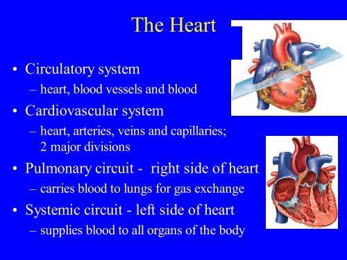
• Circulatory system<br />
The <strong>Heart</strong><br />
– heart, blood vessels and blood<br />
• Cardiovascular system<br />
– heart, arteries, veins and capillaries;<br />
2 major divisions<br />
• Pulmonary circuit - right side of heart<br />
– carries blood to lungs for gas exchange<br />
• Systemic circuit - left side of heart<br />
– supplies blood to all organs of the body
Cardiovascular System Circuit
• muscular pump<br />
• size of a fist<br />
• weighs 10 oz.<br />
Size, Shape and Position<br />
• located in mediastinum,<br />
area between the lungs<br />
• base - broad superior<br />
portion of heart<br />
• apex - inferior end, tilts to<br />
the left, tapers to point
<strong>Heart</strong> Position
United States<br />
National Library of Medicine's Visible Human Project<br />
Joseph Jernigan, a convicted murderer, died by lethal injection in 1993.<br />
He was 39 years old. His body was encased in gelatin, frozen, and carved<br />
into 1,862 transverse (horizontal) slices. Each millimeter-thin section was<br />
photographed, digitized, and electronically reassembled to create the<br />
"Visible Human Male," part of the National Library of Medicine's Visible<br />
Human Project.<br />
In 1995, a 59-year-old Maryland woman became the first "Visible Human<br />
Female."<br />
Doctors use these "Visible Humans" to plan surgeries. Medical students<br />
study their anatomy.<br />
http://www.exploratorium.edu/bodies/vhuman.html<br />
http://www.nlm.nih.gov/research/visible/visible_human.html<br />
http://collab.nlm.nih.gov/webcastsandvideos/visiblehumanvideos/visiblehu<br />
manvideos.html
Section through Visible Human Male<br />
thorax, including heart (with muscular left ventricle),<br />
lungs, spinal column, major vessels, musculature<br />
http://www.nlm.nih.gov/research/visible/photos.html<br />
http://www.nlm.nih.gov/research/visible/image/thorax.jpg
Pericardium<br />
• Allows heart to beat without friction, room to<br />
expand and resists excessive expansion<br />
• Parietal pericardium<br />
– outer, tough, fibrous layer of CT<br />
– inner, thin, smooth, moist serous layer<br />
• Pericardial cavity<br />
– filled with pericardial fluid<br />
• Visceral pericardium (a.k.a. epicardium of heart wall)<br />
– thin, smooth, moist serous layer covers heart surface<br />
• Pericarditis
Pericardium & <strong>Heart</strong> Wall<br />
Parietal pericardium<br />
• Pericardial cavity contains 5-30 ml of pericardial fluid
<strong>Heart</strong> Wall<br />
• Epicardium (a.k.a. visceral pericardium)<br />
– serous membrane covers heart<br />
• Myocardium<br />
– thick muscular layer<br />
– fibrous skeleton - network of collagenous and elastic<br />
fibers<br />
• provides structural support<br />
• attachment for cardiac muscle<br />
• nonconductor important in coordinating contractile activity<br />
• Endocardium<br />
– smooth inner lining
Pericardium & <strong>Heart</strong> Wall<br />
Parietal pericardium<br />
• Pericardial cavity contains 5-30 ml of pericardial fluid
• 4 chambers<br />
• Right and left atria<br />
<strong>Heart</strong> Chambers<br />
– 2 superior, posterior chambers<br />
– receive blood returning to heart<br />
from veins, veins carry blood back<br />
to the heart<br />
• Right and left ventricles<br />
– 2 inferior chambers<br />
– pump blood into arteries, arteries<br />
carry blood away from the heart<br />
• Atrioventricular (coronary) sulcus - separates atria from<br />
ventricles<br />
• Anterior and posterior interventricular sulci - grooves<br />
separate ventricles
External <strong>Anatomy</strong> - Anterior<br />
Atrioventricular<br />
sulcus
Anterior Aspect
External <strong>Anatomy</strong> - Posterior
<strong>Heart</strong> Chambers - Internal<br />
• Interatrial septum<br />
– wall that separates atria<br />
• Interventricular septum<br />
– wall that separates ventricles<br />
• Trabeculae carneae<br />
– internal ridges in both ventricles
Internal <strong>Anatomy</strong> Anterior Aspect
Trace a Drop of Blood<br />
• Deoxygenated blood enters the right side of the<br />
heart via the superior vena cava and the inferior<br />
vena cava<br />
• Right atrium<br />
• Right atrioventricular (AV) valve (tricuspid valve)<br />
• Right ventricle<br />
• Pulmonary semilunar valve<br />
• Pulmonary circuit<br />
– Pulmonary trunk<br />
– Pulmonary arteries to lungs<br />
– Pulmonary veins returning from lungs
Trace a Drop of Blood<br />
• Oxygenated blood enters the left side of the heart<br />
via the pulmonary veins returning from lungs<br />
• Left atrium<br />
• Left AV valve (mitral valve, bicuspid valve)<br />
• Left ventricle<br />
• Aortic semilunar valve<br />
• Systemic circuit<br />
– Aorta<br />
– Other systemic vessels<br />
– Superior vena cava and inferior vena cava<br />
• Back to right atrium
Cardiovascular System Circuit
Blood Flow Through <strong>Heart</strong>
<strong>Heart</strong> Valves<br />
• Ensure one-way blood flow<br />
• Atrioventricular (AV) valves (2)<br />
– right AV valve has 3 cusps (tricuspid valve)<br />
– left AV valve has 2 cusps (mitral, bicuspid valve)<br />
– chordae tendineae - cords connect AV valves to papillary<br />
muscles (on floor of ventricles)<br />
• Semilunar valves - control flow into great arteries<br />
– Pulmonary semilunar valve: from right ventricle into<br />
pulmonary trunk<br />
– Aortic semilunar valve: from left ventricle into aorta
Internal <strong>Anatomy</strong>, Anterior Aspect
<strong>Heart</strong> Valves
<strong>Heart</strong> Internal <strong>Anatomy</strong><br />
• <strong>Heart</strong> bisected in frontal plane, opened like a book
<strong>Heart</strong> Valves
AV Valve Mechanics<br />
• Ventricles relax, pressure drops, semilunar valves<br />
close, AV valves open, blood flows from atria to<br />
ventricles<br />
• Ventricles contract, AV valves close (papillary m.<br />
contract and pull on chordae tendineae to prevent prolapse),<br />
pressure rises, semilunar valves open, blood flows<br />
into great vessels
Operation of Atrioventricular Valves
Operation of Semilunar Valves
• Mitral regurgitation<br />
Valve Diseases<br />
– When left AV valve (bicuspid valve) can’t remain tightly<br />
closed<br />
– Blood leaks back into left atrium<br />
• Mitral stenosis<br />
– Narrow left AV valve (biscupid valve) due to scar tissue<br />
– Hypertension
Coronary <strong>Circulation</strong>blood<br />
supply to the heart<br />
• Blood vessels nourish cardiac muscle<br />
• Coronary arteries<br />
– Distribute oxygenated blood to the heart<br />
– First branch off the base of the aorta<br />
– Deliver blood to the heart when the heart is relaxing<br />
– Right coronary artery (larger than left), 2 branches<br />
– Left coronary artery (smaller than right), 2 branches<br />
– Coronary bypass surgery<br />
• Needed when circulation to the heart is blocked<br />
• reroutes, or "bypasses," blood around clogged arteries to<br />
improve blood flow and oxygen to the heart<br />
http://www.americanheart.org/presenter.jhtml?identifier=4484
Myocardial Infarction<br />
• Myocardial infarction (MI) - sudden death of heart<br />
tissue caused by interruption of blood flow from<br />
vessel narrowing or occlusion<br />
• Anastomoses - anatomical convergence, the<br />
opposite of branch (Example: a point where two<br />
blood vessels merge and combine their<br />
bloodstreams)<br />
– defend against interruption of blood flow to the heart by<br />
providing alternate blood pathways; if one artery becomes<br />
obstructed, blood continues to reach myocardial tissue<br />
through an alternative route
Coronary <strong>Circulation</strong>blood<br />
supply to the heart<br />
• Cardiac veins<br />
– Collect deoxygenated blood from the heart<br />
– Empties into coronary sinus<br />
• Coronary sinus empties into the right atrium
Coronary Vessels - Anterior
Coronary Vessels - Posterior