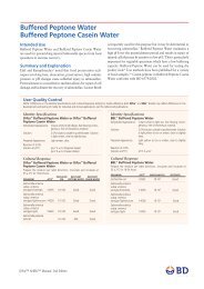
BBL Trypticase Soy Agar with 5% Sheep Blood (TSA II) BBL ... - BD
BBL Trypticase Soy Agar with 5% Sheep Blood (TSA II) BBL ... - BD
BBL Trypticase Soy Agar with 5% Sheep Blood (TSA II) BBL ... - BD

You also want an ePaper? Increase the reach of your titles
YUMPU automatically turns print PDFs into web optimized ePapers that Google loves.
!<br />
<strong>BBL</strong> <strong>Trypticase</strong> <strong>Soy</strong> <strong>Agar</strong> <strong>with</strong> <strong>5%</strong> <strong>Sheep</strong> <strong>Blood</strong> (<strong>TSA</strong> <strong>II</strong>)<br />
<strong>BBL</strong> <strong>Trypticase</strong> <strong>Soy</strong> <strong>Agar</strong> <strong>with</strong> 10% <strong>Sheep</strong> <strong>Blood</strong><br />
L007421 • Rev. 09 • July 2006<br />
QUALITY CONTROL PROCEDURES<br />
I INTRODUCTION<br />
<strong>Trypticase</strong> <strong>Soy</strong> <strong>Agar</strong> supplemented <strong>with</strong> sheep blood is used for the growth of fastidious organisms and for the visualization of<br />
hemolytic reactions.<br />
<strong>II</strong> PERFORMANCE TEST PROCEDURE<br />
1. Inoculate representative samples <strong>with</strong> dilutions of the cultures listed below.<br />
a. Using a volumetric pipettor or equivalent method, deliver 0.1 mL of a dilution yielding 30–300 CFU to each plate and<br />
spread-inoculate using a sterile glass spreader.<br />
b. Incubate the Staphylococcus and Escherichia strains at 35 ± 2°C in an aerobic atmosphere and the Streptococcus strains at<br />
35 ± 2°C in an aerobic atmosphere supplemented <strong>with</strong> 3–<strong>5%</strong> carbon dioxide.<br />
2. Examine plates after 18–24 h for growth, colony size and hemolytic reactions.<br />
3. Expected Results<br />
CLSI Organisms ATCC Recovery<br />
*Streptococcus pyogenes 19615 Growth, beta hemolysis<br />
*Streptococcus pneumoniae 6305 Growth, alpha hemolysis<br />
*Staphylococcus aureus 25923 Growth<br />
*Escherichia coli 25922 Growth<br />
*Recommended organism strain for User Quality Control.<br />
<strong>II</strong>I TEST FOR CAMP REACTION<br />
(Not performed on plates containing 10% blood.)<br />
1. Inoculate representative samples <strong>with</strong> 18-to 24-h <strong>Trypticase</strong> <strong>Soy</strong> <strong>Agar</strong> <strong>with</strong> <strong>5%</strong> <strong>Sheep</strong> <strong>Blood</strong> (<strong>TSA</strong> <strong>II</strong>) cultures of the organisms<br />
listed below.<br />
a. Identify the plates by denoting the Staphylococcus aureus ATCC 25923 streak as a long line across the width of the plate.<br />
Denote streptococcal cultures by the respective numbers perpendicular to the staphylococcal lines.<br />
b. Using an inoculating loop, make a single narrow streak inoculation across the width of each plate <strong>with</strong> the S. aureus<br />
culture and allow the streak to dry. Make a narrow streak <strong>with</strong> each streptococcal culture perpendicular to, but not<br />
touching (<strong>with</strong>in 2–3 mm of) the S. aureus streak. Perpendicular streaks should be at least 5 mm apart.<br />
c. Incubate plates at 35 ± 2°C in an aerobic atmosphere.<br />
2. Examine plates after 18–24 h.<br />
3. Expected Results<br />
Organisms ATCC Reaction<br />
Streptococcus agalactiae 12386 A typical arrowhead or crescent-shaped clearing<br />
(Group B) should occur at the junction of the Streptococcus and<br />
S. aureus streaks <strong>with</strong>in 24 h.<br />
Streptococcus pyogenes 19615 No arrowhead formation. (A bullet-shaped zone of<br />
slight hemolysis may appear at the junction of the<br />
two streaks.)<br />
IV ADDITIONAL QUALITY CONTROL<br />
1. Examine plates as described under “Product Deterioration.”<br />
2. Visually examine representative plates to assure that any existing physical defects will not interfere <strong>with</strong> use.<br />
3. Determine the pH potentiometrically at room temperature for adherence to the specification of 7.4 ± 0.2.<br />
4. Note the firmness of plates during the inoculation procedure.<br />
5. Incubate uninoculated representative plates at 35 ± 2°C for 72 h and examine for microbial contamination.<br />
PRODUCT INFORMATION<br />
V INTENDED USE<br />
<strong>Trypticase</strong> <strong>Soy</strong> <strong>Agar</strong> <strong>with</strong> <strong>5%</strong> or 10% <strong>Sheep</strong> <strong>Blood</strong> is used for cultivating fastidious microorganisms and for the visualization of<br />
hemolytic reactions produced by many bacterial species. Plates labeled “deep fill” contain an additional volume of medium to<br />
reduce the effects of drying during prolonged incubation.<br />
VI SUMMARY AND EXPLANATION<br />
The nutritional composition of <strong>Trypticase</strong> <strong>Soy</strong> <strong>Agar</strong> has made it a popular medium, both unsupplemented and as a base for<br />
media containing blood. <strong>Trypticase</strong> <strong>Soy</strong> <strong>Agar</strong> <strong>with</strong> <strong>5%</strong> or 10% <strong>Sheep</strong> <strong>Blood</strong> is extensively used for the recovery and cultivation<br />
of fastidious microbial species and for the determination of hemolytic reactions which are important differentiating<br />
characteristics for bacteria, especially Streptococcus species.<br />
V<strong>II</strong> PRINCIPLES OF THE PROCEDURE<br />
The combination of casein and soy peptones in the <strong>Trypticase</strong> <strong>Soy</strong> <strong>Agar</strong> base render the medium highly nutritious by supplying<br />
organic nitrogen, particularly amino acids and larger-chained peptides. The sodium chloride maintains osmotic equilibrium.<br />
L007421 1 of 4
Defibrinated sheep blood is the most widely used blood for enriching agar base media. 1 Hemolytic reactions of streptococci are<br />
proper and growth of Haemophilus hemolyticus, a nonpathogen whose hemolytic colonies are indistinguishable from those of<br />
beta-hemolytic streptococci, is inhibited.<br />
<strong>Trypticase</strong> <strong>Soy</strong> <strong>Agar</strong> <strong>with</strong> <strong>5%</strong> <strong>Sheep</strong> <strong>Blood</strong> (<strong>TSA</strong> <strong>II</strong>) provides excellent growth and beta hemolysis by Streptococcus pyogenes<br />
(Lancefield group A) and also provides excellent growth and appropriate hemolytic reactions <strong>with</strong> other fastidious organisms. It is<br />
suitable for performing the CAMP test for the presumptive identification of group B streptococci (S. agalactiae), and for use <strong>with</strong><br />
low concentration (0.04 unit) bacitracin discs (Taxo A) for presumptive identification of group A streptococci (S. pyogenes).<br />
The CAMP test, which only is performed on <strong>TSA</strong> <strong>II</strong>, is based on the formation of a zone of synergistic hemolysis at the junction<br />
of perpendicular streak inocula of Staphylococcus aureus and group B streptococci. The reaction is caused by the sphingomyelinase<br />
(beta-toxin) of S. aureus reacting <strong>with</strong> sphingomyelin in the sheep erythrocyte membrane to produce ceramide. A<br />
non-enzymatic protein (CAMP protein), produced by S. agalactiae, binds to the ceramide and leads to disorganization of the<br />
lipid bilayer of the sheep erythrocyte membrane resulting in complete lysis. 2<br />
<strong>Trypticase</strong> <strong>Soy</strong> <strong>Agar</strong> <strong>with</strong> 10% <strong>Sheep</strong> <strong>Blood</strong> is provided for those laboratories preferring the increased blood content. This<br />
medium is not recommended for performance of the CAMP test. Additionally, the increased blood content can make hemolytic<br />
reactions less distinct and more difficult to read.<br />
I Plate divided Petri dishes containing <strong>Trypticase</strong> <strong>Soy</strong> <strong>Agar</strong> <strong>with</strong> <strong>5%</strong> <strong>Sheep</strong> <strong>Blood</strong> in each half enable two specimens to be<br />
streaked on one plate.<br />
V<strong>II</strong>I REAGENTS<br />
<strong>Trypticase</strong> <strong>Soy</strong> <strong>Agar</strong> <strong>with</strong> <strong>5%</strong> or 10% <strong>Sheep</strong> <strong>Blood</strong><br />
Approximate Formula* Per Liter Purified Water<br />
Pancreatic Digest of Casein....................................................14.5 g<br />
Papaic Digest of <strong>Soy</strong>bean Meal ..............................................5.0 g<br />
Sodium Chloride ......................................................................5.0 g<br />
<strong>Agar</strong> ........................................................................................14.0 g<br />
Growth Factors..........................................................................1.5 g<br />
Defibrinated <strong>Sheep</strong> <strong>Blood</strong> ............................................<strong>5%</strong> or 10%<br />
*Adjusted and/or supplemented as required to meet performance criteria.<br />
Warnings and Precautions: For in vitro Diagnostic Use.<br />
If excessive moisture is observed, invert the bottom over an off-set lid and allow to air dry in order to prevent formation of a<br />
seal between the top and bottom of the plate during incubation.<br />
Pathogenic microorganisms, including hepatitis viruses and Human Immunodeficiency Virus, may be present in clinical<br />
specimens. "Standard Precautions" 3-6 and institutional guidelines should be followed in handling all items contaminated <strong>with</strong><br />
blood and other body fluids. After use, prepared plates, specimen containers and other contaminated materials must be<br />
sterilized by autoclaving before discarding.<br />
Storage Instructions: On receipt, store plates in the dark at 2–8°C. Avoid freezing and overheating. Do not open until ready to<br />
use. Minimize exposure to light. Prepared plates stored in their original sleeve wrapping at 2–8°C until just prior to use may be<br />
inoculated up to the expiration date and incubated for recommended incubation times. Allow the medium to warm to room<br />
temperature before inoculation.<br />
Product Deterioration: Do not use plates if they show evidence of microbial contamination, discoloration, drying, cracking or<br />
other signs of deterioration.<br />
IX SPECIMEN COLLECTION AND HANDLING<br />
Specimens suitable for culture may be handled using various techniques. For detailed information, consult appropriate texts. 7,8<br />
Specimens should be obtained before antimicrobial therapy has been administered. Provision must be made for prompt<br />
delivery to the laboratory.<br />
X PROCEDURE<br />
Material Provided: <strong>TSA</strong> <strong>II</strong> (<strong>Trypticase</strong> <strong>Soy</strong> <strong>Agar</strong> <strong>with</strong> <strong>5%</strong> <strong>Sheep</strong> <strong>Blood</strong>) or <strong>Trypticase</strong> <strong>Soy</strong> <strong>Agar</strong> <strong>with</strong> 10% <strong>Sheep</strong> <strong>Blood</strong> or<br />
<strong>Trypticase</strong> <strong>Soy</strong> <strong>Agar</strong> <strong>with</strong> <strong>5%</strong> <strong>Sheep</strong> <strong>Blood</strong> (<strong>TSA</strong> <strong>II</strong>) - I Plate<br />
Materials Required But Not Provided: Ancillary culture media, reagents, quality control organisms and laboratory equipment as required.<br />
Test Procedure: Observe aseptic techniques.<br />
The agar surface should be smooth and moist, but <strong>with</strong>out excessive moisture.<br />
Streak the specimen as soon as possible after it is received in the laboratory. The streak plate is used primarily to isolate pure<br />
cultures from specimens containing mixed flora.<br />
Alternatively, if material is being cultured directly from a swab, roll the swab over a small area of the surface at the edge; then<br />
streak from this inoculated area.<br />
Since many pathogens require carbon dioxide on primary isolation, plates may be incubated in an atmosphere containing<br />
approximately 3–10% CO2 . 9<br />
Incubate plates at 35 ± 2°C for 18–24 h.<br />
CAMP Test10 Non-hemolytic, bile-esculin negative streptococci or bacitracin-resistant beta-hemolytic streptococci may be tested by the CAMP<br />
test for presumptive identification as S. agalactiae (Lancefield group B). The inoculum may be taken from an overnight broth<br />
culture or from colonies picked from a blood agar plate. Make a single streak of Staphylococcus aureus ATCC 25923 or<br />
ATCC 33862 across the center of a <strong>TSA</strong> <strong>II</strong> plate. If a loop is used, do not use it parallel to the agar surface, since the streak will<br />
be too wide and the results will not be satisfactory. The streptococcal isolates to be tested are inoculated by making a simple<br />
streak perpendicular to the S. aureus line coming as close as possible (2–3 mm), but not touching it. Several streptococcal<br />
isolates may be tested on the same plate. Perpendicular streptococcal streaks should be 5–8 mm apart. Include a known<br />
S. agalactiae for a positive control and S. pyogenes as a negative control. The procedure should be practiced <strong>with</strong> known<br />
cultures before using it to identify unknown isolates.<br />
L007421 2 of 4
NOTE: Studies on the CAMP Test have shown that the reaction is most reliable early in the shelf life of some lots of the<br />
prepared plated medium. It is recommended that S. agalactiae ATCC 12386 be included along <strong>with</strong> patient isolates to verify<br />
satisfactory performance.<br />
Incubate plates in an aerobic atmosphere at 35 ± 2°C for 18–24 h. Do not incubate anaerobically or in a CO2 incubator. Falsepositive<br />
results may occur <strong>with</strong> group A streptococci when incubation is in an anaerobic or CO2-enriched atmosphere. 10,11<br />
User Quality Control: See “Quality Control Procedures.”<br />
Quality Control requirements must be performed in accordance <strong>with</strong> applicable local, state and/or federal regulations or<br />
accreditation requirements and your laboratory’s standard Quality Control procedures. It is recommended that the user refer to<br />
pertinent CLSI (formerly NCCLS) guidance and CLIA regulations for appropriate Quality Control practices.<br />
XI RESULTS<br />
After incubation most plates will show an area of confluent growth. Because the streaking procedure is, in effect, a “dilution”<br />
technique, diminishing numbers of microorganisms are deposited on the streaked areas. Consequently, one or more of these<br />
areas should exhibit isolated colonies of the organisms contained in the specimen. Further, growth of each organism may be<br />
semi-quantitatively scored on the basis of growth in each of the streaked areas.<br />
1. Hemolytic streptococci may appear as translucent or opaque, grayish, small (1 mm), or large matt and mucoid (2–4 mm)<br />
colonies, encircled by a zone of hemolysis. Gram stains should be made and examined to check the macroscopic findings.<br />
(Other organisms which may cause hemolysis include Listeria, various corynebacteria, hemolytic staphylococci, Escherichia<br />
coli and Pseudomonas.)<br />
In reporting, approximate quantitation of the number of colonies of hemolytic streptococci may be helpful to the clinician.<br />
2. CAMP Test - A positive CAMP reaction is indicated by arrowhead or triangular shaped area of increased hemolysis which<br />
forms around the end of the streptococcal streak line closest to the S. aureus growth. The streptococcal growth must be<br />
<strong>with</strong>in the wide zone of partial hemolysis that surrounds the S. aureus growth. A negative reaction may appear as a bulletshaped<br />
zone of slightly increased hemolysis or as no increased hemolysis.<br />
Bacitracin-negative, CAMP-positive, beta-hemolytic streptococci may be reported as presumptive group B streptococci.<br />
CAMP-positive group A species may be differentiated from group B streptococci by hemolysis, bacitracin susceptibility, and<br />
hippurate hydrolysis. Group B streptococci generally have smaller hemolytic zones than group A streptococci. 11<br />
3. Pneumococci usually appear as very flat, smooth, translucent, grayish and sometimes mucoid colonies surrounded by a<br />
narrow zone of “green” (alpha) hemolysis.<br />
4. Staphylococci appear as opaque, white to gold-yellow colonies <strong>with</strong> or <strong>with</strong>out zones of beta hemolysis.<br />
5. Listeria. Small zones of beta hemolysis are produced. They may be distinguished by their rod shape in stains, and by motility<br />
at room temperature.<br />
6. Other organisms representing minimal flora and clinically significant isolates can also be expected to grow on this<br />
nonselective formulation.<br />
X<strong>II</strong> LIMITATIONS OF THE PROCEDURE<br />
For identification, organisms must be in pure culture. Morphological, biochemical, and/or serological tests should be performed<br />
for final identification. Consult appropriate texts for detailed information and recommended procedures. 7,8,12-15<br />
X<strong>II</strong>I PERFORMANCE CHARACTERISTICS<br />
<strong>Trypticase</strong> <strong>Soy</strong> <strong>Agar</strong> <strong>with</strong> <strong>5%</strong> <strong>Sheep</strong> <strong>Blood</strong><br />
<strong>Trypticase</strong> <strong>Soy</strong> <strong>Agar</strong> <strong>with</strong> <strong>5%</strong> <strong>Sheep</strong> <strong>Blood</strong> was used as a control in a study using broth-enhanced culture (Todd Hewitt) and<br />
Optical Immunoassay method for the diagnosis of hemolytic streptococcal infection. Five hundred two (502) specimens were<br />
tested. <strong>TSA</strong> <strong>with</strong> <strong>5%</strong> <strong>Sheep</strong> <strong>Blood</strong> had a sensitivity and specificity of 92.<strong>5%</strong> and 99.4%, respectively. 16 Nguyen et al. used<br />
<strong>Trypticase</strong> <strong>Soy</strong> <strong>Agar</strong> <strong>with</strong> <strong>5%</strong> <strong>Sheep</strong> <strong>Blood</strong> as the ‘gold standard’ for the detection of group B Streptococcus from the lower<br />
genital tract of pregnant women. 17 In another study, Rossmann et al. successfully reisolated Lautropia mirabilis on <strong>Trypticase</strong><br />
<strong>Soy</strong> <strong>Agar</strong> <strong>with</strong> <strong>5%</strong> <strong>Sheep</strong> <strong>Blood</strong> from the oral cavities of human immunodeficiency virus infected children. 18 Of the 85 children<br />
evaluated in the study, 35 (41.4%) were positive for L. mirabilis. Isenberg et al. used <strong>Trypticase</strong> <strong>Soy</strong> <strong>Agar</strong> <strong>with</strong> <strong>5%</strong> <strong>Sheep</strong> <strong>Blood</strong><br />
as a control to evaluate the recovery of Enterococcus from a selective medium under study. 19 Two hundred fifty (250) group D<br />
streptococcal strains isolated from clinical material and 8 strains obtained from the National Communicable Disease Center<br />
(Atlanta, Ga.) were used.<br />
XIV AVAILABILITY<br />
Cat. No. Description<br />
221239 <strong>BBL</strong> <strong>Trypticase</strong> <strong>Soy</strong> <strong>Agar</strong> <strong>with</strong> <strong>5%</strong> <strong>Sheep</strong> <strong>Blood</strong> (<strong>TSA</strong> <strong>II</strong>), Pkg. of 20 plates<br />
221881 <strong>BBL</strong> <strong>Trypticase</strong> <strong>Soy</strong> <strong>Agar</strong> <strong>with</strong> <strong>5%</strong> <strong>Sheep</strong> <strong>Blood</strong> (<strong>TSA</strong> <strong>II</strong>), Pkg. of 30 plates<br />
221261 <strong>BBL</strong> <strong>Trypticase</strong> <strong>Soy</strong> <strong>Agar</strong> <strong>with</strong> <strong>5%</strong> <strong>Sheep</strong> <strong>Blood</strong> (<strong>TSA</strong> <strong>II</strong>), Ctn. of 100 plates<br />
221292 <strong>BBL</strong> <strong>Trypticase</strong> <strong>Soy</strong> <strong>Agar</strong> <strong>with</strong> <strong>5%</strong> <strong>Sheep</strong> <strong>Blood</strong> (<strong>TSA</strong> <strong>II</strong>) - I Plate, Pkg. of 20 plates<br />
221162 <strong>BBL</strong> <strong>Trypticase</strong> <strong>Soy</strong> <strong>Agar</strong> <strong>with</strong> 10% <strong>Sheep</strong> <strong>Blood</strong>, Pkg. of 20 plates<br />
221260 <strong>BBL</strong> <strong>Trypticase</strong> <strong>Soy</strong> <strong>Agar</strong> <strong>with</strong> 10% <strong>Sheep</strong> <strong>Blood</strong>, Ctn. of 100 plates<br />
XV REFERENCES<br />
1. Vera, H.D., and D.A. Power. 1980. Culture media, p. 969. In E.H. Lennette, A. Balows, W.J. Hausler, Jr., and J.P. Truant (ed.), Manual of clinical<br />
microbiology, 3rd ed. American Society for Microbiology, Washington, D.C.<br />
2. Bernheimer, A.W., R. Linder, and L.S. Avigad. 1979. Nature and mechanism of action of the CAMP protein of group B streptococci. Infect.<br />
Immun. 23:838-844.<br />
3. National Committee for Clinical Laboratory Standards. 2001. Approved Guideline M29-A2. Protection of laboratory workers from<br />
occupationally acquired infections, 2nd ed. NCCLS, Wayne, PA.<br />
4. Garner, J.S. 1996. Hospital Infection Control Practices Advisory Committee, U.S. Department of Health and Human Services, Centers for<br />
Disease Control and Prevention. Guideline for isolation precautions in hospitals. Infect. Control Hospital Epidemiol. 17:53-80.<br />
L007421 3 of 4
5. U.S. Department of Health and Human Services. 1999. Biosafety in microbiological and biomedical laboratories, HHS Publication (CDC), 4th<br />
ed. U.S. Government Printing Office, Washington, D.C.<br />
6. Directive 2000/54/EC of the European Parliament and of the Council of 18 September 2000 on the protection of workers from risks related to<br />
exposure to biological agents at work (seventh individual directive <strong>with</strong>in the meaning of Article 16(1) of Directive 89/391/EEC). Official<br />
Journal L262, 17/10/2000, p. 0021-0045.<br />
7. Murray, P.R., E.J. Baron, J.H. Jorgensen, M.A. Pfaller, and R. H. Yolken (ed.). 2003. Manual of clinical microbiology, 8th ed. American Society<br />
for Microbiology, Washington, D.C.<br />
8. Forbes, B.A., D.F. Sahm, and A.S. Weissfeld. 2002. Bailey and Scott's diagnostic microbiology, 11th ed. Mosby, Inc., St. Louis.<br />
9. Ruoff, K.L., R.A. Whiley, and D. Beighton. 2003. Streptococcus, p. 405-421. In P.R. Murray, E.J. Baron, J.H. Jorgensen, M.A. Pfaller, and R.H.<br />
Yolken (ed.), Manual of clinical microbiology, 8th ed. American Society for Microbiology, Washington, D.C.<br />
10. Darling, C.L. 1975. Standardization and evaluation of CAMP reaction for prompt, presumptive identification of Streptococcus agalactiae<br />
(Lancefield group B) in clinical material. J. Clin. Microbiol. 1:171-174.<br />
11. Facklam, R.R., and J.A. Washington <strong>II</strong>. 1991. Streptococci and related catalase-negative gram-positive cocci, p. 238-257. In A. Balows, W.J.<br />
Hausler, Jr., K.L. Herrmann, H.D. Isenberg, and H.J. Shadomy (ed.), Manual of clinical microbiology, 5th ed. American Society for Microbiology,<br />
Washington, D.C.<br />
12. Holt, J.G., N.R. Krieg, P.H.A. Sneath, J.T. Staley, and S.T. Williams (ed.). 1994. Bergey's Manual of determinative bacteriology, 9th ed.<br />
Williams & Wilkins, Baltimore.<br />
13. MacFaddin, J.F. 2000. Biochemical tests for identification of medical bacteria, 3rd ed. Lippincott Williams & Wilkins, Baltimore.<br />
14. Koneman, E.W., S.D. Allen, W.M. Janda, P.C. Schreckenberger, and W.C. Winn, Jr. 1997. Color atlas and textbook of diagnostic microbiology,<br />
5th ed. Lippincott-Raven, Philadelphia.<br />
15. Isenberg, H.D. (ed.). 2004. Clinical microbiology procedures handbook, vol. 1, 2 and 3, 2nd ed. American Society for Microbiology,<br />
Washington, D.C.<br />
16. Fries, S.M. 1995. Diagnosis of group A streptococcal pharyngitis in a private clinic: comparative evaluation of an optical immunoassay method<br />
and culture. J. Pediatr. 126:933-936.<br />
17. Nguyen, T.M., et al. 1998. Detection of group B streptococcus: comparison of an optical immunoassay <strong>with</strong> direct plating and broth-enhanced<br />
culture methods. J. Matern. Fetal. Med. 7:172-176.<br />
18. Rossmann, S.N. et al. 1998. Isolation of Lautropia mirabilis from oral cavities of human immunodeficiency virus-infected children. J. Clin.<br />
Microbiol. 36:1756-1760.<br />
19. Isenberg, H.D., D. Goldberg, and J. Sampson, 1970. Laboratory studies <strong>with</strong> a selective medium. Appl. Microbiol. 20:433-436.<br />
Becton, Dickinson and Company<br />
7 Loveton Circle<br />
Sparks, Maryland 21152 USA<br />
800-638-8663<br />
ATCC is a trademark of the American Type Culture Collection.<br />
<strong>BD</strong>, <strong>BD</strong> Logo, <strong>BBL</strong>, I Plate, Taxo and <strong>Trypticase</strong> are trademarks of Becton, Dickinson and Company. ©2006 <strong>BD</strong>.<br />
L007421 4 of 4