The Biology of Blood-Sucking in Insects, SECOND EDITION
The Biology of Blood-Sucking in Insects, SECOND EDITION
The Biology of Blood-Sucking in Insects, SECOND EDITION

Create successful ePaper yourself
Turn your PDF publications into a flip-book with our unique Google optimized e-Paper software.
This page <strong>in</strong>tentionally left blank
<strong>The</strong> <strong>Biology</strong> <strong>of</strong> <strong>Blood</strong>-<strong>Suck<strong>in</strong>g</strong> <strong>in</strong> <strong>Insects</strong><br />
Second Edition<br />
<strong>Blood</strong>-suck<strong>in</strong>g <strong>in</strong>sects transmit many <strong>of</strong> the most debilitat<strong>in</strong>g diseases<br />
<strong>in</strong> humans, <strong>in</strong>clud<strong>in</strong>g malaria, sleep<strong>in</strong>g sickness, filariasis,<br />
leishmaniasis, dengue, typhus and plague. In addition, these<br />
<strong>in</strong>sects cause major economic losses <strong>in</strong> agriculture both by direct<br />
damage to livestock and as a result <strong>of</strong> the veter<strong>in</strong>ary diseases, such<br />
as the various trypanosomiases, that they transmit. <strong>The</strong> second<br />
edition <strong>of</strong> <strong>The</strong> <strong>Biology</strong> <strong>of</strong> <strong>Blood</strong>-<strong>Suck<strong>in</strong>g</strong> <strong>in</strong> <strong>Insects</strong> is a unique, topicled<br />
commentary on the biological themes that are common <strong>in</strong> the<br />
lives <strong>of</strong> blood-suck<strong>in</strong>g <strong>in</strong>sects. To do this effectively it concentrates<br />
on those aspects <strong>of</strong> the biology <strong>of</strong> these fasc<strong>in</strong>at<strong>in</strong>g <strong>in</strong>sects that<br />
have been clearly modified <strong>in</strong> some way to suit the blood-suck<strong>in</strong>g<br />
habit. <strong>The</strong> book opens with a brief outl<strong>in</strong>e <strong>of</strong> the medical, social and<br />
economic impact <strong>of</strong> blood-suck<strong>in</strong>g <strong>in</strong>sects. Further chapters cover<br />
the evolution <strong>of</strong> the blood-suck<strong>in</strong>g habit, feed<strong>in</strong>g preferences, host<br />
location, the <strong>in</strong>gestion <strong>of</strong> blood and the various physiological adaptations<br />
for deal<strong>in</strong>g with the blood meal. Discussions on host–<strong>in</strong>sect<br />
<strong>in</strong>teractions and the transmission <strong>of</strong> parasites by blood-suck<strong>in</strong>g<br />
<strong>in</strong>sects are followed by the f<strong>in</strong>al chapter, which is designed as a useful<br />
quick-reference section cover<strong>in</strong>g the different groups <strong>of</strong> <strong>in</strong>sects<br />
referred to <strong>in</strong> the text.<br />
For this second edition, <strong>The</strong> <strong>Biology</strong> <strong>of</strong> <strong>Blood</strong>-<strong>Suck<strong>in</strong>g</strong> <strong>in</strong> <strong>Insects</strong> has<br />
been fully updated s<strong>in</strong>ce the first edition was published <strong>in</strong> 1991. It<br />
is written <strong>in</strong> a clear, concise fashion and is well illustrated throughout<br />
with a variety <strong>of</strong> specially prepared l<strong>in</strong>e illustrations and photographs.<br />
<strong>The</strong> text provides a summary <strong>of</strong> knowledge about this<br />
important group <strong>of</strong> <strong>in</strong>sects and will be <strong>of</strong> <strong>in</strong>terest to advanced<br />
undergraduate and to postgraduate students <strong>in</strong> medical and veter<strong>in</strong>ary<br />
parasitology and entomology.<br />
Mike Lehane is Pr<strong>of</strong>essor <strong>of</strong> Molecular Entomology and<br />
Parasitology <strong>in</strong> the Liverpool School <strong>of</strong> Tropical Medic<strong>in</strong>e.
<strong>The</strong> <strong>Biology</strong> <strong>of</strong><br />
<strong>Blood</strong>-<strong>Suck<strong>in</strong>g</strong> <strong>in</strong> <strong>Insects</strong><br />
<strong>SECOND</strong> <strong>EDITION</strong><br />
M. J. Lehane<br />
Liverpool School <strong>of</strong> Tropical Medic<strong>in</strong>e
cambridge university press<br />
Cambridge, New York, Melbourne, Madrid, Cape Town, S<strong>in</strong>gapore, São Paulo<br />
Cambridge University Press<br />
<strong>The</strong> Ed<strong>in</strong>burgh Build<strong>in</strong>g, Cambridge cb2 2ru, UK<br />
Published <strong>in</strong> the United States <strong>of</strong> America by Cambridge University Press, New York<br />
www.cambridge.org<br />
Information on this title: www.cambridge.org/9780521836081<br />
© M. J. Lehane 2005<br />
This book is <strong>in</strong> copyright. Subject to statutory exception and to the provision <strong>of</strong><br />
relevant collective licens<strong>in</strong>g agreements, no reproduction <strong>of</strong> any part may take place<br />
without the written permission <strong>of</strong> Cambridge University Press.<br />
First published <strong>in</strong> pr<strong>in</strong>t format 2005<br />
isbn-13 978-0-511-11553-0 eBook (MyiLibrary)<br />
isbn-10 0-511-11553-9 eBook (MyiLibrary)<br />
isbn-13 978-0-521-83608-1 hardback<br />
isbn-10 0-521-83608-5 hardback<br />
isbn-13 978-0-521-54395-8 paperback<br />
isbn-10 0-521-54395-9 paperback<br />
Cambridge University Press has no responsibility for the persistence or accuracy <strong>of</strong><br />
urls for external or third-party <strong>in</strong>ternet websites referred to <strong>in</strong> this book, and does not<br />
guarantee that any content on such websites is, or will rema<strong>in</strong>, accurate or appropriate.
Contents<br />
List <strong>of</strong> tables page vii<br />
List <strong>of</strong> boxes x<br />
Preface xi<br />
Acknowledgements xiii<br />
1 <strong>The</strong> importance <strong>of</strong> blood-suck<strong>in</strong>g <strong>in</strong>sects 1<br />
2 <strong>The</strong> evolution <strong>of</strong> the blood-suck<strong>in</strong>g habit 7<br />
2.1 Prolonged close association with vertebrates 7<br />
2.2 Morphological pre-adaptation for pierc<strong>in</strong>g 13<br />
3 Feed<strong>in</strong>g preferences <strong>of</strong> blood-suck<strong>in</strong>g <strong>in</strong>sects 15<br />
3.1 Host choice 15<br />
3.2 Host choice and species complexes 24<br />
4 Location <strong>of</strong> the host 27<br />
4.1 A behavioural framework for host location 27<br />
4.2 Appetitive search<strong>in</strong>g 29<br />
4.3 Activation and orientation 32<br />
4.4 Attraction 49<br />
4.5 Movement between hosts 52<br />
5 Ingestion <strong>of</strong> the blood meal 56<br />
5.1 Prob<strong>in</strong>g stimulants 56<br />
5.2 Mouthparts 57<br />
5.3 Vertebrate haemostasis 64<br />
5.4 Host pa<strong>in</strong> 68<br />
5.5 Insect anti-haemostatic and anti-pa<strong>in</strong> factors<br />
<strong>in</strong> saliva 69<br />
5.6 Phagostimulants 76<br />
5.7 <strong>Blood</strong> <strong>in</strong>take 78<br />
6 Manag<strong>in</strong>g the blood meal 84<br />
6.1 Midgut anatomy 84<br />
6.2 <strong>The</strong> blood meal 87<br />
6.3 Gonotrophic concordance 96
vi Contents<br />
6.4 Nutrition 98<br />
6.5 Host hormones <strong>in</strong> the blood meal 103<br />
6.6 Partition<strong>in</strong>g <strong>of</strong> resources from the blood meal 106<br />
6.7 Autogeny 109<br />
7 Host–<strong>in</strong>sect <strong>in</strong>teractions 116<br />
7.1 Insect distribution on the surface <strong>of</strong> the host 117<br />
7.2 Morphological specializations for life on the host 121<br />
7.3 Host immune responses and <strong>in</strong>sect salivary<br />
secretions 126<br />
7.4 Behavioural defences <strong>of</strong> the host 134<br />
7.5 Density-dependent effects on feed<strong>in</strong>g success 142<br />
8 Transmission <strong>of</strong> parasites by blood-suck<strong>in</strong>g <strong>in</strong>sects 150<br />
8.1 Transmission routes 150<br />
8.2 Specificity <strong>in</strong> vector–parasite relationships 163<br />
8.3 Orig<strong>in</strong> <strong>of</strong> vector–parasite relationships 167<br />
8.4 Parasite strategies for contact<strong>in</strong>g a vector 170<br />
8.5 Parasite strategies for contact<strong>in</strong>g a vertebrate host 177<br />
8.6 Vector pathology caused by parasites 179<br />
8.7 Vector immune mechanisms 184<br />
9 <strong>The</strong> blood-suck<strong>in</strong>g <strong>in</strong>sect groups 202<br />
9.1 Insect classification 202<br />
9.2 Phthiraptera 204<br />
9.3 Hemiptera 208<br />
9.4 Siphonaptera 213<br />
9.5 Diptera 219<br />
9.6 Other groups 257<br />
References 259<br />
Index 312
Tables<br />
1.1 An outl<strong>in</strong>e <strong>of</strong> the early <strong>in</strong>vestigations that laid the<br />
foundations <strong>of</strong> medical and veter<strong>in</strong>ary entomology. page 2<br />
1.2 Rounded estimates for the prevalence <strong>of</strong> disease, the<br />
number at risk and the disability adjusted life years<br />
(DALYs) for major vector-borne diseases. 3<br />
1.3 Estimated losses <strong>in</strong> agricultural production caused by<br />
blood-suck<strong>in</strong>g <strong>in</strong>sects. 4<br />
4.1 Generalized opportunities and constra<strong>in</strong>ts on host location<br />
by blood-suck<strong>in</strong>g <strong>in</strong>sects feed<strong>in</strong>g dur<strong>in</strong>g the day or night. 32<br />
4.2 Different blood-suck<strong>in</strong>g <strong>in</strong>sects respond <strong>in</strong> different ways<br />
to spectral <strong>in</strong>formation. 45<br />
5.1 Adaptations <strong>of</strong> mouthpart components for different<br />
purposes <strong>in</strong> various haematophagous <strong>in</strong>sect groups. 58<br />
5.2 <strong>Blood</strong>-suck<strong>in</strong>g <strong>in</strong>sects produce a wide range <strong>of</strong><br />
anti-haemostatic factors <strong>in</strong> their salivary secretions. This<br />
table gives some examples with a range <strong>of</strong> different<br />
activities. 71<br />
5.3 <strong>The</strong> size <strong>of</strong> red corpuscles varies widely <strong>in</strong> different<br />
animals. Given that many blood-suck<strong>in</strong>g <strong>in</strong>sects have<br />
mouthparts with a term<strong>in</strong>al diameter <strong>of</strong> around 10µm, this<br />
may be a factor affect<strong>in</strong>g the feed<strong>in</strong>g efficiency <strong>of</strong><br />
blood-suck<strong>in</strong>g <strong>in</strong>sects on different host species. 80<br />
6.1 <strong>The</strong> size <strong>of</strong> the red blood meal and the time taken <strong>in</strong> its<br />
digestion are affected by a range <strong>of</strong> factors <strong>in</strong>clud<strong>in</strong>g<br />
ambient temperature, age <strong>of</strong> the <strong>in</strong>sect, mat<strong>in</strong>g status,<br />
stage <strong>of</strong> the gonotrophic cycle, previous feed<strong>in</strong>g history,<br />
and source <strong>of</strong> the blood meal. <strong>The</strong> figures given here are a<br />
rough guidel<strong>in</strong>e to the ‘average’ meal size and time for<br />
digestion <strong>in</strong> a variety <strong>of</strong> haematophagous <strong>in</strong>sects. 88<br />
6.2 <strong>The</strong> major constituents <strong>of</strong> the blood are reasonably<br />
uniform <strong>in</strong> most host animals. <strong>The</strong> exception is the high<br />
levels <strong>of</strong> nucleic acids <strong>in</strong> the blood <strong>of</strong> birds and reptiles<br />
because <strong>of</strong> their nucleated red blood cells. Prote<strong>in</strong>s are far<br />
and away the most abundant nutrients <strong>in</strong> blood, and
viii List <strong>of</strong> tables<br />
nutrients are unevenly distributed between whole blood<br />
(B), red blood cells alone (E) and plasma alone (P). 90<br />
6.3 Symbionts are common <strong>in</strong> <strong>in</strong>sects rely<strong>in</strong>g on blood as the<br />
sole food source throughout their lives. An outl<strong>in</strong>e is given<br />
<strong>of</strong> their anatomical locations and the means <strong>of</strong> transmission<br />
from one generation to the next <strong>in</strong> different <strong>in</strong>sect groups. 99<br />
6.4 Three types <strong>of</strong> female Aedes taeniorhynchus have been<br />
identified <strong>in</strong> terms <strong>of</strong> egg development: autogenous<br />
females (1); females that are autogenous if mated (2); and<br />
anautogenous forms (3). This pattern is <strong>in</strong>fluenced by the<br />
feed<strong>in</strong>g success <strong>of</strong> the larval stage, as illustrated <strong>in</strong> this<br />
table. 113<br />
6.5 Some mosquitoes can use sugar meals (10% sucrose <strong>in</strong> this<br />
experiment) to <strong>in</strong>crease the number <strong>of</strong> autogenously<br />
produced eggs. 114<br />
7.1 <strong>The</strong> choice <strong>of</strong> feed<strong>in</strong>g site <strong>of</strong> Aedes triseriatus on eastern<br />
chipmunks and grey squirrels is <strong>in</strong>fluenced by length and<br />
density <strong>of</strong> body hair. <strong>The</strong> different feed<strong>in</strong>g patterns on the<br />
two hosts reflects the differences <strong>in</strong> hair cover between<br />
them. 119<br />
7.2 <strong>The</strong> anti-mosquito behaviour <strong>of</strong> a range <strong>of</strong> ciconiiform<br />
birds, show<strong>in</strong>g that different host species display various<br />
types and degrees <strong>of</strong> defensive behaviour aga<strong>in</strong>st<br />
blood-suck<strong>in</strong>g <strong>in</strong>sects. 137<br />
8.1 Some <strong>of</strong> the most important associations <strong>of</strong><br />
disease-caus<strong>in</strong>g organisms carried to humans and other<br />
animals by blood-suck<strong>in</strong>g <strong>in</strong>sects: (a) viruses, (b) rickettsia<br />
and bacteria, (c) protozoa and (d) nematodes. 151<br />
8.2 <strong>Blood</strong>-suck<strong>in</strong>g <strong>in</strong>sects commonly take meals that are only a<br />
small proportion <strong>of</strong> the total blood present <strong>in</strong> the host<br />
animal (the ratio between total blood <strong>in</strong> the host and size<br />
<strong>of</strong> the <strong>in</strong>sect’s blood meal is given). This m<strong>in</strong>imizes the<br />
chances <strong>of</strong> the <strong>in</strong>sect <strong>in</strong>gest<strong>in</strong>g any <strong>in</strong>dividual parasite<br />
dur<strong>in</strong>g feed<strong>in</strong>g. One strategy adopted by <strong>in</strong>sect-borne<br />
parasites to overcome this problem is to produce large<br />
numbers <strong>of</strong> <strong>in</strong>fective stages which circulate <strong>in</strong> the blood <strong>of</strong><br />
the host. 171<br />
8.3 <strong>The</strong> micr<strong>of</strong>ilariae <strong>of</strong> many filarial worms display a<br />
pronounced periodicity, with micr<strong>of</strong>ilarial numbers <strong>in</strong> the<br />
peripheral blood co<strong>in</strong>cid<strong>in</strong>g with the peak bit<strong>in</strong>g time <strong>of</strong><br />
locally abundant vector species. 173
List <strong>of</strong> tables ix<br />
8.4 Tsetse flies <strong>in</strong>fected with trypanosomes feed more readily<br />
and probe more <strong>of</strong>ten than un<strong>in</strong>fected flies, thereby<br />
<strong>in</strong>creas<strong>in</strong>g the chances <strong>of</strong> parasite transmission. 178<br />
8.5 Comparison <strong>of</strong> the rate <strong>of</strong> formation <strong>of</strong> the peritrophic<br />
matrix among various mosquito species. 189<br />
8.6 <strong>The</strong> melanization response to subsequent challenge <strong>of</strong><br />
<strong>in</strong>fected and un<strong>in</strong>fected Aedes aegypti,asshown by the<br />
<strong>in</strong>trathoracic <strong>in</strong>jection <strong>of</strong> specific micr<strong>of</strong>ilariae (mff) which<br />
normally <strong>in</strong>duce a strong melanization reaction. 200<br />
9.1 <strong>The</strong> groups <strong>of</strong> <strong>in</strong>sect. Those groups conta<strong>in</strong><strong>in</strong>g<br />
blood-suck<strong>in</strong>g <strong>in</strong>sects are shown <strong>in</strong> bold. 203<br />
9.2 <strong>The</strong> geographical distribution <strong>of</strong> triatom<strong>in</strong>e species, which<br />
have become highly adapted to the domestic-peridomestic<br />
environment <strong>of</strong> man and so represent a particular threat as<br />
vectors <strong>of</strong> Chagas’ disease. 211<br />
9.3 <strong>The</strong> divisions <strong>of</strong> the order Diptera and the major families<br />
<strong>in</strong> each division. Families conta<strong>in</strong><strong>in</strong>g blood-suck<strong>in</strong>g species<br />
are <strong>in</strong> bold type. 221
Boxes<br />
3.1 <strong>The</strong> importance <strong>of</strong> rates <strong>of</strong> mosquitoes bit<strong>in</strong>g humans for<br />
the transmission <strong>of</strong> malaria. page 20<br />
3.2 Identification <strong>of</strong> the source <strong>of</strong> a blood meal. 21<br />
7.1 Histopathology <strong>of</strong> the various stages <strong>in</strong> the sequence <strong>of</strong><br />
host response to <strong>in</strong>sect bites. 130<br />
8.1 Four blood cell types characterized <strong>in</strong> Aedes aegypti are<br />
compared to haemocytes described <strong>in</strong> previous studies on<br />
a variety <strong>of</strong> <strong>in</strong>sects. 197
Preface<br />
<strong>Blood</strong>-suck<strong>in</strong>g <strong>in</strong>sects are the vectors <strong>of</strong> many <strong>of</strong> the most debilitat<strong>in</strong>g parasites<br />
<strong>of</strong> humans and their domesticated animals. In addition they are <strong>of</strong><br />
considerable direct cost to the agricultural <strong>in</strong>dustry through losses <strong>in</strong> milk<br />
and meat yields, and through damage to hides, wool and other products.<br />
So, not surpris<strong>in</strong>gly, many books <strong>of</strong> medical and veter<strong>in</strong>ary entomology<br />
have been written. Most <strong>of</strong> these texts are organized taxonomically, giv<strong>in</strong>g<br />
details <strong>of</strong> the life cycles, bionomics, relationships to disease and economic<br />
importance <strong>of</strong> each <strong>of</strong> the <strong>in</strong>sect groups <strong>in</strong> turn. I have taken a different<br />
approach. This book is topic-led and aims to discuss the biological themes<br />
common to the lives <strong>of</strong> blood-suck<strong>in</strong>g <strong>in</strong>sects. To do this I have concentrated<br />
on those aspects <strong>of</strong> the biology <strong>of</strong> these fasc<strong>in</strong>at<strong>in</strong>g <strong>in</strong>sects that have<br />
been clearly modified <strong>in</strong> some way to suit the blood-suck<strong>in</strong>g habit. For<br />
example, I have discussed feed<strong>in</strong>g and digestion <strong>in</strong> some detail because<br />
feed<strong>in</strong>g on blood presents <strong>in</strong>sects with special problems, but I have not<br />
discussed respiration because it is not affected <strong>in</strong> any particular way by<br />
haematophagy. To reflect this better I have made a slight adjustment to<br />
the title <strong>of</strong> the book <strong>in</strong> this second edition. Naturally there is a subjective<br />
element <strong>in</strong> the choice <strong>of</strong> topics for discussion and the weight given to each.<br />
I hope that I have not let my enthusiasm for the particular subjects get the<br />
better <strong>of</strong> me on too many occasions and that the subject material achieves<br />
an overall balance. <strong>The</strong> major changes <strong>in</strong> this second edition most <strong>of</strong>ten<br />
reflect the revolutionary <strong>in</strong>fluence that molecular biology has had on the<br />
subject <strong>in</strong> the past 12 years.<br />
Although the book is not designed as a conventional text <strong>of</strong> medical<br />
and veter<strong>in</strong>ary entomology, <strong>in</strong> Chapter 9 I have given a brief outl<strong>in</strong>e <strong>of</strong><br />
each <strong>of</strong> the blood-suck<strong>in</strong>g <strong>in</strong>sect groups. This chapter is <strong>in</strong>tended as a<br />
quick <strong>in</strong>troduction for those entirely new to the subject, or as a refresher<br />
on particular groups for those already familiar with the divisions <strong>of</strong> bloodsuck<strong>in</strong>g<br />
<strong>in</strong>sects. <strong>The</strong>re are several <strong>in</strong>troductory textbooks <strong>of</strong> medical and<br />
veter<strong>in</strong>ary entomology available to those requir<strong>in</strong>g more <strong>in</strong>formation.<br />
<strong>The</strong> book is primarily <strong>in</strong>tended for advanced undergraduate and for<br />
postgraduate students, but because it looks at topics that cut across the<br />
normal research boundaries <strong>of</strong> physiology and ecology, behaviour and cell<br />
biology, I hope it may also be useful for more established scientists who
xii Preface<br />
want to look outside their own specialism. I have tried to distil this broad<br />
spectrum <strong>of</strong> <strong>in</strong>formation, much <strong>of</strong> which is not readily available to the nonspecialist,<br />
<strong>in</strong>to a brief synthesis. For those who want to look further <strong>in</strong>to a<br />
particular area I have <strong>in</strong>cluded some <strong>of</strong> the references I found most useful <strong>in</strong><br />
writ<strong>in</strong>g the text, and these will provide an entry <strong>in</strong>to the literature. Clearly<br />
the subjects covered by the book encompass a vast number <strong>of</strong> publications<br />
and I am sure to have missed many important and <strong>in</strong>terest<strong>in</strong>g references for<br />
which I apologize <strong>in</strong> advance both to the reader and my fellow scientists.<br />
Many <strong>of</strong> the topics discussed <strong>in</strong> the different chapters are <strong>in</strong>terrelated. To<br />
avoid repetition, and still give the broadest picture possible, I have given<br />
cross-references <strong>in</strong> the text which I hope the reader will f<strong>in</strong>d useful.<br />
From a comparative po<strong>in</strong>t <strong>of</strong> view it is an unfortunate fact that most <strong>of</strong> the<br />
work on blood-suck<strong>in</strong>g <strong>in</strong>sects has been carried out on a few species. Consequently,<br />
tsetse flies and mosquitoes pop up on every other page. In many<br />
<strong>in</strong>stances it rema<strong>in</strong>s to be seen how widely the lessons we have learned<br />
from these well-studied models can be applied. Where possible I have<br />
tried to po<strong>in</strong>t to general patterns that fit whole groups <strong>of</strong> blood-suck<strong>in</strong>g<br />
<strong>in</strong>sects. To help me <strong>in</strong> this I have divided the blood-suck<strong>in</strong>g <strong>in</strong>sects <strong>in</strong>to<br />
three convenient but artificial categories: temporary ectoparasites, permanent<br />
ectoparasites and periodic ectoparasites. <strong>The</strong>se categories are based<br />
solely on the behaviour biology <strong>of</strong> the blood-feed<strong>in</strong>g stadia <strong>in</strong> the lives <strong>of</strong><br />
these <strong>in</strong>sects. Temporary ectoparasites are considered to be those largely<br />
free-liv<strong>in</strong>g <strong>in</strong>sects, such as the tabanids, mosquitoes, blood-feed<strong>in</strong>g bugs<br />
and blackflies, that visit the host only long enough to take a blood meal.<br />
I also <strong>in</strong>clude <strong>in</strong>sects such as the tsetse here, even though the male may<br />
be found <strong>in</strong> swarms closely associated with the host for large parts <strong>of</strong> its<br />
life. Permanent ectoparasites are considered to be those <strong>in</strong>sects that live<br />
almost constantly on the host, such as lice, the sheep ked and tungid fleas.<br />
F<strong>in</strong>ally, periodic ectoparasites are considered to be those <strong>in</strong>sects that spend<br />
considerably longer on the host than is required merely to obta<strong>in</strong> a blood<br />
meal, but that nevertheless spend a significant amount <strong>of</strong> time away from<br />
the host. <strong>Insects</strong> that fall <strong>in</strong>to this category <strong>in</strong>clude many <strong>of</strong> the fleas and<br />
Pupipara. <strong>The</strong>se categories are no more than a useful generalization <strong>in</strong> the<br />
text; I make no claims for their rigour and I realize that it could be argued <strong>in</strong><br />
several <strong>in</strong>stances that an <strong>in</strong>sect will sit as easily <strong>in</strong> one category as another.
Acknowledgements<br />
I gladly and gratefully acknowledge the help I have received from many<br />
people dur<strong>in</strong>g the writ<strong>in</strong>g <strong>of</strong> this book or the previous edition, particularly<br />
P. Bill<strong>in</strong>gsley, A. Blackwell, J. Brady, H. Briegel, I. Burgess, E. Bursell,<br />
R. Dillon, J. D. Edman, R. Galun, A. G. Gatehouse, M. Gaunt, M. Gillies,<br />
R. H. Good<strong>in</strong>g, M. Greaves, C. Green, M. Hafner, J. Hogsette, H. Hurd,<br />
A. M. Jordan, K. C. Kim, J. K<strong>in</strong>gsolver, M. Klowden, A. M. Lackie, B. R.<br />
Laurence, E. Levash<strong>in</strong>a, A. G. Marshall, P. Mellor, D. Molyneux, P. Morrison,<br />
W. A. Nelson, G. O’Meara, G. S. Paulson, G. Port, N. A. Ratcliffe,<br />
J. M. Ribeiro, P. Rossignol, M. Rothschild, W. Rud<strong>in</strong>, C. J. Sch<strong>of</strong>ield, M. W.<br />
Service, J. J. B. Smith, W. Takken, S. Torr, G. A. Vale and J. Waage. I also<br />
thank Paula Hynes, Maria Turton, Paula Dwyer and Dafydd Roberts for<br />
help with the illustrations. F<strong>in</strong>ally, most thanks go to my family, particularly<br />
Stella, without whose encouragement, support and practical help this<br />
book would never have been f<strong>in</strong>ished.
1<br />
<strong>The</strong> importance <strong>of</strong> blood-suck<strong>in</strong>g<br />
<strong>in</strong>sects<br />
<strong>Insects</strong> are the pre-em<strong>in</strong>ent form <strong>of</strong> metazoan life on land. <strong>The</strong> class Insecta<br />
conta<strong>in</strong>s over three-quarters <strong>of</strong> a million described species. Estimates for<br />
the total number <strong>of</strong> extant species vary between 1 and 10 million, and it<br />
has been calculated that as many as 10 19 <strong>in</strong>dividual <strong>in</strong>sects are alive at any<br />
given <strong>in</strong>stant (McGav<strong>in</strong>, 2001). That gives about 200 million for each man,<br />
woman and child on Earth! It is estimated that there are 14 000 species <strong>of</strong><br />
<strong>in</strong>sects from five orders that feed on blood (Adams, 1999) but, thankfully,<br />
only 300 to 400 species regularly attract our attention. <strong>The</strong>se blood-suck<strong>in</strong>g<br />
<strong>in</strong>sects are <strong>of</strong> immense importance to humanity.<br />
Humans evolved <strong>in</strong> a world already stocked with blood-suck<strong>in</strong>g <strong>in</strong>sects.<br />
From their earliest days <strong>in</strong>sects would have annoyed them with their bites<br />
and sickened them with the parasites they transmitted. As humans evolved<br />
from hunters to herders, blood-suck<strong>in</strong>g <strong>in</strong>sects had a further impact on their<br />
wellbe<strong>in</strong>g by lower<strong>in</strong>g the productivity <strong>of</strong> their animals. It is reasonable to<br />
assume that, because <strong>of</strong> their annoyance value, humanity has been <strong>in</strong> battle<br />
with blood-suck<strong>in</strong>g <strong>in</strong>sects from the very beg<strong>in</strong>n<strong>in</strong>g. In recent years this<br />
battle has <strong>in</strong>tensified because <strong>of</strong> an <strong>in</strong>creas<strong>in</strong>g <strong>in</strong>tolerance <strong>of</strong> the discomfort<br />
they cause, our fuller understand<strong>in</strong>g <strong>of</strong> their role <strong>in</strong> disease transmission<br />
and the demand for greater agricultural productivity. But despite considerable<br />
advances <strong>in</strong> our knowledge <strong>of</strong> the <strong>in</strong>sects and improvements <strong>in</strong> the<br />
weapons we have to use aga<strong>in</strong>st them, there is still no sign <strong>of</strong> an eventual<br />
w<strong>in</strong>ner <strong>in</strong> this age-old battle.<br />
Many keen observers <strong>of</strong> nature suspected that <strong>in</strong>sects were <strong>in</strong> some way<br />
<strong>in</strong>volved with many <strong>of</strong> the febrile illnesses <strong>of</strong> humans and their animals<br />
well before confirmatory scientific evidence was available. <strong>The</strong> explorer<br />
Alexander von Humboldt recorded such a belief amongst the tribes <strong>of</strong> the<br />
Or<strong>in</strong>oco region <strong>of</strong> South America. <strong>The</strong> great German bacteriologist Robert<br />
Koch reported the belief <strong>of</strong> the tribes <strong>of</strong> the Usambara Mounta<strong>in</strong>s <strong>of</strong> East<br />
Africa that the mosquitoes they encountered when they descended to the<br />
pla<strong>in</strong>s were the cause <strong>of</strong> malaria (Nuttal, 1899). Sir Richard Burton, <strong>in</strong> his<br />
travels <strong>in</strong> East Africa, recorded the similar belief <strong>of</strong> Somaliland tribes that<br />
mosquitoes were responsible for febrile illnesses (Burton, 1860). Many <strong>of</strong><br />
the peoples liv<strong>in</strong>g near the tsetse fly belts <strong>of</strong> East and West Africa associated<br />
tsetse flies with sleep<strong>in</strong>g sickness <strong>of</strong> humans and nagana <strong>of</strong> animals. In our
2 <strong>The</strong> importance <strong>of</strong> blood-suck<strong>in</strong>g <strong>in</strong>sects<br />
Table 1.1 An outl<strong>in</strong>e <strong>of</strong> the early <strong>in</strong>vestigations that laid the foundations <strong>of</strong><br />
medical and veter<strong>in</strong>ary entomology.<br />
Date Source Subject<br />
1878 Manson Development <strong>of</strong> Wuchereria bancr<strong>of</strong>ti <strong>in</strong> a mosquito<br />
1893 Smith and Kilbourne Babesia bigem<strong>in</strong>a, the causative agent <strong>of</strong> Texas cattle<br />
fever, transmitted by the tick, Boophilus annulatus<br />
1895 Bruce Transmission <strong>of</strong> nagana by tsetse fly<br />
1897 Ross Malaria parasites seen to develop <strong>in</strong> mosquitoes<br />
1898 Ross Transmission <strong>of</strong> avian malaria by mosquitoes<br />
1898 Simond Transmission <strong>of</strong> plague from rat to rat by fleas<br />
1899 Grassi, Bignami and<br />
Bastianelli<br />
Anopheles spp. are the vectors <strong>of</strong> human malaria<br />
1900 Reed et al. Transmission <strong>of</strong> yellow fever by the mosquito Aedes<br />
aegypti<br />
1902 Graham Transmission <strong>of</strong> dengue by mosquitoes<br />
1903 Bruce and Nabarro Sleep<strong>in</strong>g sickness <strong>in</strong> humans transmitted by tsetse fly<br />
1903 Marchoux and<br />
Salimbeni<br />
Transmission <strong>of</strong> fowl spirochaetes, Borrelia conser<strong>in</strong>a,<br />
by the tick Argus persicus<br />
1907 Mackie Spirochaete caus<strong>in</strong>g relaps<strong>in</strong>g fever transmitted by<br />
lice<br />
1909 Chagas Trypansoma cruzi, causative agent <strong>of</strong> Chagas’ disease,<br />
transmitted by reduviid bugs<br />
own western tradition North American stock ranchers held the belief that<br />
Texas cattle fever was transmitted by ticks (<strong>in</strong> the class Arachnida, not<br />
Insecta) well before this was confirmed experimentally.<br />
<strong>The</strong> fact that <strong>in</strong>sects are vectors <strong>of</strong> disease was only confirmed scientifically<br />
at the end <strong>of</strong> the n<strong>in</strong>eteenth century. <strong>The</strong> key discovery was made<br />
<strong>in</strong> 1877 (reported <strong>in</strong> 1878) by a Scottish doctor, Patrick Manson, work<strong>in</strong>g<br />
for the customs and excise service <strong>in</strong> Ch<strong>in</strong>a. He found that larval<br />
stages <strong>of</strong> the filarial worm, Wuchereria bancr<strong>of</strong>ti, developed <strong>in</strong> the body <strong>of</strong> a<br />
mosquito, Culex pipiens qu<strong>in</strong>quefasciatus (Manson, 1878). This was the start<br />
<strong>of</strong> an avalanche <strong>of</strong> <strong>in</strong>vestigations that laid the foundations <strong>of</strong> medical and<br />
veter<strong>in</strong>ary entomology. Some <strong>of</strong> the key discoveries <strong>of</strong> this era are outl<strong>in</strong>ed<br />
<strong>in</strong> Table 1.1. <strong>The</strong> ma<strong>in</strong> <strong>in</strong>sects <strong>in</strong>volved <strong>in</strong> the transmission <strong>of</strong> all the most<br />
important vector-transmitted diseases (Table 1.2) are now well known.<br />
<strong>The</strong> list <strong>of</strong> diseases transmitted is an impressive one and <strong>in</strong>cludes the medical<br />
scourges malaria, sleep<strong>in</strong>g sickness, leishmaniasis, river bl<strong>in</strong>dness, elephantiasis,<br />
yellow fever and dengue, and the veter<strong>in</strong>ary diseases nagana,<br />
surra, souma, bluetongue, African horse sickness and Rift Valley fever
<strong>The</strong> importance <strong>of</strong> blood-suck<strong>in</strong>g <strong>in</strong>sects 3<br />
Table 1.2 Rounded estimates for the prevalence <strong>of</strong> disease, the number at risk<br />
and the disability adjusted life years (DALYs) for major vector-borne diseases.<br />
Figures <strong>in</strong> millions (M). (DALYs were <strong>in</strong>troduced <strong>in</strong> the World Bank<br />
Development report <strong>of</strong> 1990 as an estimate <strong>of</strong> the burden a disease causes to the<br />
health <strong>of</strong> the population. <strong>The</strong>y are <strong>of</strong>ten used for comparative purposes and for<br />
use <strong>in</strong> prioritization.)<br />
Disease Prevalence At risk DALYs<br />
Major<br />
distribution Major vectors<br />
Malaria 273M 2100M 42M Tropics and Anophel<strong>in</strong>e<br />
subtropics mosquitoes<br />
Onchocerciasis 18M 120M 1M Tropical Africa, Blackflies<br />
(river bl<strong>in</strong>dness)<br />
Yemen, Lat<strong>in</strong><br />
America<br />
(Simulium spp.)<br />
Lymphatic<br />
120M 1100M 5.6M Africa, Asia and Various<br />
filariasis<br />
(elephantiasis)<br />
South America mosquitoes<br />
African<br />
0.5M 50M 2M Sub-Saharan Tsetse flies<br />
trypanosomiasis<br />
Africa<br />
Chagas’ disease 16–18M 120M 0.7M Central and<br />
South America<br />
Triatom<strong>in</strong>e bugs<br />
Leishmaniasis 12M 350M 2M Africa, Asia and<br />
Lat<strong>in</strong> America<br />
Sandflies<br />
Dengue 50M 3000M 0.5M Asia, Africa and Various<br />
Americas mosquitoes<br />
Data largely from World Health Organization web pages as <strong>of</strong> 11 December 2002: http://<br />
www.who.<strong>in</strong>t/tdr/media/image.html.<br />
(the piroplasms be<strong>in</strong>g tick-borne). Gaug<strong>in</strong>g the extent <strong>of</strong> these diseases<br />
is much more problematical, even for human disease. One reason is that<br />
health statistics are a mov<strong>in</strong>g target, particularly for those diseases such as<br />
yellow fever that occur as epidemics. But the greatest problem is that the<br />
heartland <strong>of</strong> these vector-borne diseases is <strong>in</strong> the under-developed world<br />
where, for a variety <strong>of</strong> reasons, accurate statistical data are <strong>of</strong>ten difficult or<br />
impossible to gather. For this reason, figures given for the extent <strong>of</strong> a disease<br />
are <strong>of</strong>ten not based entirely on hard data, but are an estimate founded<br />
largely upon the experience <strong>of</strong> an expert. Table 1.2 gives an estimate <strong>of</strong><br />
some <strong>of</strong> the major vector-transmitted diseases <strong>of</strong> humans, but obviously,<br />
as just <strong>in</strong>dicated, care needs to be taken <strong>in</strong> the <strong>in</strong>terpretation <strong>of</strong> the figures.<br />
<strong>Blood</strong>-suck<strong>in</strong>g <strong>in</strong>sects cause very serious losses to agriculture (Table 1.3).<br />
One way this happens is through the transmission <strong>of</strong> parasites. <strong>The</strong>
4 <strong>The</strong> importance <strong>of</strong> blood-suck<strong>in</strong>g <strong>in</strong>sects<br />
Table 1.3 Estimated losses <strong>in</strong> agricultural production caused by blood-suck<strong>in</strong>g<br />
<strong>in</strong>sects.<br />
Animal ma<strong>in</strong>ly Estimated losses Geographical<br />
Insect Year affected (millions US$) region<br />
Haematobia irritans<br />
(horn fly)<br />
Stomoxys calcitrans<br />
(stable fly)<br />
1991 Cattle 800 USA<br />
1965 Cattle 142 USA<br />
Tabanids 1965 Cattle 40 USA<br />
Mosquitoes 1965 Cattle 25 USA<br />
Melophagus ov<strong>in</strong>us<br />
(sheep ked)<br />
Lice 1965 Cattle<br />
1965 Sheep 9.4 USA<br />
Sheep<br />
47<br />
Sw<strong>in</strong>e<br />
3<br />
Goats<br />
0.8<br />
Tsetse fly 1999 Cattle 4500 Sub-Saharan<br />
Africa<br />
<strong>Insects</strong>, ticks, mites 1994 3000 USA<br />
Information from: Budd, 1999; Geden and Hogsette, 1994; Kunz et al., 1991; Steelman,<br />
1976.<br />
most celebrated case is trypanosomiasis, transmitted by tsetse flies across<br />
9 million km 2 <strong>of</strong> Africa (Hursey, 2001), and estimated to cause agricultural<br />
losses <strong>of</strong> about US$4.5 billion a year (Budd, 1999). <strong>The</strong> counter argument<br />
has also been proposed that the tsetse has prevented desertification<br />
<strong>of</strong> large areas <strong>of</strong> land by overgraz<strong>in</strong>g, and has been the saviour <strong>of</strong><br />
Africa’s game animals. <strong>The</strong> debate has been clearly outl<strong>in</strong>ed by Jordan<br />
(1986). Other examples <strong>of</strong> spectacular losses caused by <strong>in</strong>sect-transmitted<br />
disease are the death <strong>in</strong> 1960 <strong>of</strong> 200 000 to 300 000 horses <strong>in</strong> Turkey, Cyprus<br />
and India caused by African horse sickness, transmitted by Culicoides spp.<br />
(Huq, 1961; Shahan and Giltner, 1945); and the estimated deaths <strong>in</strong> the USA,<br />
between 1930 and 1945, <strong>of</strong> up to 300 000 equ<strong>in</strong>es from Western and Eastern<br />
equ<strong>in</strong>e encephalitis transmitted by mosquitoes (Shahan and Giltner,<br />
1945).<br />
In the developed countries it is usually direct losses caused by <strong>in</strong>sects<br />
themselves that are <strong>of</strong> greatest concern. In exceptional circumstances the<br />
<strong>in</strong>sects may be present <strong>in</strong> such numbers that stock are killed; for example,<br />
16 000 animals died <strong>in</strong> Romania <strong>in</strong> 1923 and 13 900 <strong>in</strong> Yugoslavia <strong>in</strong> 1934<br />
because <strong>of</strong> outbreaks <strong>of</strong> the blackfly Simulium colombaschense (Baranov,<br />
47<br />
USA
<strong>The</strong> importance <strong>of</strong> blood-suck<strong>in</strong>g <strong>in</strong>sects 5<br />
1935; Ciurea and D<strong>in</strong>ulescu, 1924). More usually losses are caused not<br />
by death but by distress to the animal. Good examples are the reductions<br />
<strong>in</strong> milk yields, weight ga<strong>in</strong>s or feed efficiencies that are commonly<br />
caused by the pa<strong>in</strong>ful bites <strong>of</strong> the tabanids and bit<strong>in</strong>g flies. Estimated<br />
losses <strong>in</strong> the USA have been calculated (Steelman, 1976). More recent estimates<br />
suggest <strong>in</strong>sects, ticks and mites cost the US livestock producer <strong>in</strong><br />
excess <strong>of</strong> $3 billion annually (Geden and Hogsette, 1994). <strong>The</strong> horn fly<br />
is perhaps the major pest <strong>in</strong> the USA, with an estimated loss <strong>in</strong> excess <strong>of</strong><br />
$800 million annually (Kunz et al., 1991). Losses are caused by reduced feed<br />
conversion efficiency, reduced weight ga<strong>in</strong>s and decreased milk production<br />
and are the result <strong>of</strong> blood loss, annoyance, irritation and behavioural<br />
defensive responses on the part <strong>of</strong> the host.<br />
<strong>The</strong> sheer annoyance that blood-suck<strong>in</strong>g <strong>in</strong>sects cause to us can easily be<br />
overshadowed by their importance <strong>in</strong> medical and veter<strong>in</strong>ary medic<strong>in</strong>e. In<br />
some parts <strong>of</strong> the world, at certa<strong>in</strong> times <strong>of</strong> the year, there may be so many<br />
blood-suck<strong>in</strong>g <strong>in</strong>sects that any activity outside is difficult or impossible<br />
without protective cloth<strong>in</strong>g. For example, the bit<strong>in</strong>g activity <strong>of</strong> the midge<br />
Culicoides impunctatus is thought to cause a 20 per cent loss <strong>in</strong> work<strong>in</strong>g hours<br />
<strong>in</strong> the forestry <strong>in</strong>dustry <strong>in</strong> Scotland dur<strong>in</strong>g the summer months (Hendry<br />
and Godw<strong>in</strong>, 1988). Such disruption is common dur<strong>in</strong>g the summer blooms<br />
<strong>of</strong> <strong>in</strong>sects at many <strong>of</strong> the higher latitudes, and also <strong>in</strong> many <strong>of</strong> the wetter<br />
areas <strong>of</strong> the tropics. <strong>The</strong>se levels <strong>of</strong> annoyance are still rare for most people,<br />
and for this reason the concept <strong>of</strong> nuisance <strong>in</strong>sects is much more difficult<br />
to grasp than that <strong>of</strong> a vector or an agricultural pest caus<strong>in</strong>g economic<br />
damage. Perhaps the best way to view annoyance caused by <strong>in</strong>sects is<br />
as a tolerance threshold. It can then be viewed as a variable with widely<br />
separated upper and lower limits; a handful <strong>of</strong> mosquitoes may be a m<strong>in</strong>or<br />
<strong>in</strong>convenience to the beggar <strong>in</strong> the street but <strong>in</strong>tolerable to the pr<strong>in</strong>ce <strong>in</strong> the<br />
palace.<br />
I suggest that, <strong>in</strong> the developed world at least, we are <strong>in</strong>creas<strong>in</strong>gly<br />
<strong>in</strong>tolerant <strong>of</strong> nuisance <strong>in</strong>sects. <strong>The</strong>re are several underly<strong>in</strong>g reasons: the<br />
<strong>in</strong>creased awareness <strong>in</strong> the general population <strong>of</strong> the importance <strong>of</strong> <strong>in</strong>sects<br />
<strong>in</strong> the spread <strong>of</strong> disease (sometimes over-exaggerated); the grow<strong>in</strong>g stress<br />
placed on hygiene and cleanl<strong>in</strong>ess; and <strong>in</strong>creas<strong>in</strong>g urbanization, so that for<br />
many people blood-suck<strong>in</strong>g <strong>in</strong>sects are not the familiar, everyday th<strong>in</strong>gs<br />
that they were once to our grandparents work<strong>in</strong>g <strong>in</strong> a rural economy.<br />
This reduction <strong>in</strong> our tolerance <strong>of</strong> nuisance <strong>in</strong>sects causes problems. <strong>The</strong><br />
extended leisure time and mobility <strong>of</strong> many people <strong>in</strong> the developed world<br />
means that they spend more time <strong>in</strong> <strong>in</strong>creas<strong>in</strong>gly distant places. <strong>The</strong> countries<br />
<strong>in</strong>volved are <strong>of</strong>ten anxious to promote and develop their tourist <strong>in</strong>dustries,<br />
and this has led to pressure to control nuisance <strong>in</strong>sects. This can<br />
be seen <strong>in</strong> places such as the Camargue <strong>in</strong> southern France, the Scottish
6 <strong>The</strong> importance <strong>of</strong> blood-suck<strong>in</strong>g <strong>in</strong>sects<br />
Highlands (Blackwell, 2000), the Bahamas, New Zealand (Blackwell and<br />
Page, 2003), Florida and many parts <strong>of</strong> the Caribbean (L<strong>in</strong>ley and Davies,<br />
1971). In addition, population growth has put <strong>in</strong>creased pressure on<br />
marg<strong>in</strong>al land which <strong>in</strong> the past may have been left alone because <strong>of</strong> nuisance<br />
<strong>in</strong>sect problems. Development <strong>of</strong> this land for leisure, commerce or<br />
hous<strong>in</strong>g with no <strong>in</strong>sect control <strong>in</strong>put can be disastrous for the developer,<br />
user or purchaser.
2<br />
<strong>The</strong> evolution <strong>of</strong> the bloodsuck<strong>in</strong>g<br />
habit<br />
It is believed that haematophagy arose <strong>in</strong>dependently at least six times<br />
among the arthropods <strong>of</strong> the Jurassic and Cretaceous periods (145–65 million<br />
years ago) (Balashov, 1984; Ribeiro, 1995). <strong>The</strong> very patchy nature <strong>of</strong><br />
the <strong>in</strong>sect fossil record means that discussion <strong>of</strong> the evolution <strong>of</strong> the bloodsuck<strong>in</strong>g<br />
habit has until now relied heavily on detective work, with the<br />
major clues ly<strong>in</strong>g <strong>in</strong> the diversity <strong>of</strong> forms and lifestyles seen <strong>in</strong> modern-day<br />
<strong>in</strong>sects, and <strong>in</strong> some cases <strong>in</strong> the details <strong>of</strong> their relationships with vertebrates.<br />
From careful <strong>in</strong>terpretation <strong>of</strong> this evidence quite credible accounts<br />
<strong>of</strong> the likely evolution <strong>of</strong> the blood-suck<strong>in</strong>g habit can be made. From this<br />
start<strong>in</strong>g po<strong>in</strong>t it has been conv<strong>in</strong>c<strong>in</strong>gly argued that the evolution <strong>of</strong> the<br />
blood-suck<strong>in</strong>g habit <strong>in</strong> <strong>in</strong>sects has occurred on several occasions, <strong>in</strong> each<br />
case along one <strong>of</strong> two ma<strong>in</strong> routes (Waage, 1979), and these are discussed<br />
below. Insect molecular systematics is beg<strong>in</strong>n<strong>in</strong>g to emerge from its ‘Tower<br />
<strong>of</strong> Babel’ stage (Cater<strong>in</strong>o et al., 2000) and it will make a major contribution<br />
<strong>in</strong> def<strong>in</strong><strong>in</strong>g the detail <strong>of</strong> the evolutionary routes taken by haematophagous<br />
<strong>in</strong>sects (Esseghir et al., 1997; Hafner et al., 1994; Lanzaro et al., 1998; Mans<br />
et al., 2002; Sallum et al., 2002). <strong>The</strong> proposed population bottleneck suffered<br />
by phlebotom<strong>in</strong>es <strong>in</strong> the late Pleistocene and the subsequent radiation<br />
<strong>of</strong> the species out from the eastern Mediterranean sub-region is a good<br />
example <strong>of</strong> what we can expect (Esseghir et al., 1997).<br />
2.1 Prolonged close association with vertebrates<br />
In the first route it is suggested that haematophagous forms may have<br />
developed subsequent to a prolonged association between vertebrates and<br />
<strong>in</strong>sects that had no specializations immediately suit<strong>in</strong>g them to the bloodsuck<strong>in</strong>g<br />
way <strong>of</strong> life. <strong>The</strong> most common association <strong>of</strong> this type is likely<br />
to have centred around the attraction <strong>of</strong> <strong>in</strong>sects to the nest or burrow <strong>of</strong><br />
the vertebrate host. <strong>Insects</strong> may have been attracted to the nest for several<br />
reasons. <strong>The</strong> humid, warm environment would have been very favourable<br />
to a great many <strong>in</strong>sects. In some circumstances, such as the location <strong>of</strong> the<br />
nest <strong>in</strong> a semi-arid or arid area, the protected habitat <strong>of</strong>fered by the nest<br />
may have been essential to the <strong>in</strong>sects’ survival. For many <strong>in</strong>sects the nest<br />
would also have proved attractive for the abundant supply <strong>of</strong> food to be
8 <strong>The</strong> evolution <strong>of</strong> the blood-suck<strong>in</strong>g habit<br />
found there. Certa<strong>in</strong>ly many current day <strong>in</strong>sects such as the psocids are<br />
attracted to the high concentrations <strong>of</strong> organic matter to be found <strong>in</strong> nests.<br />
Indeed, psocids may become so <strong>in</strong>timately associated with this habitat that<br />
they develop a phoretic association with birds and mammals, climb<strong>in</strong>g <strong>in</strong>to<br />
fur and feathers, to be translocated from one nest site to another (Mockford,<br />
1967; Mockford, 1971; Pearman, 1960).<br />
Initially feed<strong>in</strong>g on dung, fungus or other organic debris, the <strong>in</strong>sects<br />
attracted to the nest would also have encountered considerable quantities<br />
<strong>of</strong> sloughed sk<strong>in</strong>, hair or feathers. <strong>The</strong> regular, accidental <strong>in</strong>gestion <strong>of</strong><br />
this sloughed body cover<strong>in</strong>g probably led to the selection <strong>of</strong> <strong>in</strong>dividuals<br />
possess<strong>in</strong>g physiological systems capable <strong>of</strong> the efficient use <strong>of</strong> this material.<br />
Behavioural adaptations may then have permitted occasional feed<strong>in</strong>g<br />
direct from the host itself. It is easy to see how this may have gone hand<br />
<strong>in</strong> hand with the adoption <strong>of</strong> a phoretic habit. Morphological and further<br />
behavioural adaptations would have allowed the <strong>in</strong>sect to rema<strong>in</strong> with<br />
the host for longer periods with <strong>in</strong>creas<strong>in</strong>gly efficient feed<strong>in</strong>g on sk<strong>in</strong> and<br />
feathers.<br />
<strong>The</strong> mouthparts developed for this lifestyle, <strong>in</strong> which the <strong>in</strong>sect feeds primarily<br />
on sk<strong>in</strong> and feathers, were almost certa<strong>in</strong>ly <strong>of</strong> the chew<strong>in</strong>g type, such<br />
as those seen <strong>in</strong> the present-day Mallophaga. While these mouthparts are<br />
not primarily designed to pierce sk<strong>in</strong> some mallophagans do feed on blood.<br />
Menacanthus stram<strong>in</strong>eus, apresent-day mallophagan, feeds at the base <strong>of</strong><br />
feathers or on the sk<strong>in</strong> <strong>of</strong> the chicken. <strong>The</strong> <strong>in</strong>sect <strong>of</strong>ten breaks through<br />
to the dermis, giv<strong>in</strong>g it access to blood on which it will feed (Emmerson<br />
et al., 1973). <strong>Blood</strong> has a higher nutritional value than sk<strong>in</strong> and is far easier<br />
to digest. This is reflected <strong>in</strong> the <strong>in</strong>creased fecundity <strong>of</strong> blood-feed<strong>in</strong>g<br />
Anoplura compared to sk<strong>in</strong>-feed<strong>in</strong>g Mallophaga (Marshall, 1981). Once<br />
blood was regularly encountered by <strong>in</strong>sects, it is likely that its high nutritional<br />
value favoured the development <strong>of</strong> a group <strong>of</strong> <strong>in</strong>sects that regularly<br />
exploited blood as a resource. This would have developed progressively,<br />
through physiological, behavioural and morphological adaptations, first<br />
to facultative haematophagy and eventually, <strong>in</strong> some <strong>in</strong>sects, to obligate<br />
haematophagy. One way <strong>in</strong> which the progression from sk<strong>in</strong> feed<strong>in</strong>g to<br />
blood feed<strong>in</strong>g may have occurred is seen <strong>in</strong> members <strong>of</strong> the mallophagan<br />
suborder the Rhynchophthir<strong>in</strong>a, such as the elephant louse, Haematomyzus<br />
elephantis. This <strong>in</strong>sect possesses typical mallophagan bit<strong>in</strong>g-type mouthparts<br />
(Ferris, 1931; Mukerji and Sen-Sarma, 1955) which are not primarily<br />
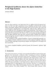
adapted for obta<strong>in</strong><strong>in</strong>g blood. By hold<strong>in</strong>g the mouthparts at the end <strong>of</strong> an<br />
extended rostrum (Fig. 2.1) the <strong>in</strong>sect manages to use them to penetrate the<br />
thick epidermal sk<strong>in</strong> layers <strong>of</strong> the host to get to the blood <strong>in</strong> the dermis.<br />
It is thought that haematophagous lice developed from an orig<strong>in</strong>al nestdwell<strong>in</strong>g,<br />
free-liv<strong>in</strong>g ancestor (Kim, 1985) along the pathway described<br />
above. We do not know when the change occurred from free-liv<strong>in</strong>g nest
2.1 Prolonged close association with vertebrates 9<br />
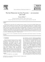
Figure 2.1 Despite hav<strong>in</strong>g the chew<strong>in</strong>g mouthparts typical <strong>of</strong> mallophagans,<br />
Haematomyzus hopk<strong>in</strong>si is unusual <strong>in</strong> feed<strong>in</strong>g on blood. <strong>The</strong> chew<strong>in</strong>g<br />
mouthparts are held on the end <strong>of</strong> an unusual, elongated rostrum, which may<br />
well be an adaptation help<strong>in</strong>g the <strong>in</strong>sect reach the blood-conta<strong>in</strong><strong>in</strong>g dermis<br />
through the thick sk<strong>in</strong> <strong>of</strong> its wart-hog host. (Courtesy <strong>of</strong> V<strong>in</strong>ce Smith)
10 <strong>The</strong> evolution <strong>of</strong> the blood-suck<strong>in</strong>g habit<br />
dweller to parasite, but it may well go right back to the appearance <strong>of</strong> nest<strong>in</strong>g<br />
or communal liv<strong>in</strong>g <strong>in</strong> land-dwell<strong>in</strong>g vertebrates, which is thought to<br />
have happened dur<strong>in</strong>g the Mesozoic (225–65 million years ago). So lice may<br />
have predated the emergence <strong>of</strong> mammals and birds and been parasitic on<br />
their reptilian ancestors (Hopk<strong>in</strong>s, 1949; Rothschild and Clay, 1952). It is<br />
highly likely that ancestral forms were parasitic on primordial mammals<br />
and that from there they radiated along the l<strong>in</strong>es <strong>of</strong> mammalian evolution.<br />
Help<strong>in</strong>g drive this rapid speciation <strong>of</strong> the permanent ectoparasites<br />
was the reproductive isolation they suffered from be<strong>in</strong>g conf<strong>in</strong>ed on specific<br />
vertebrate hosts, which may well have enhanced the effects <strong>of</strong> classical<br />
geographic reproductive isolation. Co-evolution <strong>of</strong> the host and permanent<br />
(and to a lesser extent temporary) ectoparasites probably led to rapid speciation<br />
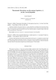
<strong>in</strong> lice and other ectoparasitic forms. <strong>The</strong> evidence for co-speciation<br />
<strong>in</strong> lice is strong. Sequence analysis <strong>of</strong> mitochondrial cytochrome oxidase I<br />
genes suggests co-speciation <strong>in</strong> the pocket gophers Orthogeomys, Geomys<br />
and Thomomys and their chew<strong>in</strong>g lice (Fig. 2.2) (Hafner et al., 1994).<br />
Co-speciation also predicts temporal congruence between chew<strong>in</strong>g lice and<br />
gopher speciation. This is borne out by analysis <strong>of</strong> the molecular data, <strong>in</strong><br />
which the synonymous substitution rate is approximately an order <strong>of</strong> magnitude<br />
greater <strong>in</strong> the lice compared to the gophers. This roughly parallels<br />
the differences <strong>in</strong> generation times <strong>of</strong> the two groups, suggest<strong>in</strong>g equal<br />
rates <strong>of</strong> mutation per generation. While the case for pocket gophers and<br />
their chew<strong>in</strong>g lice is strong, the extent to which co-speciation is generally<br />
the case is unclear. Classical taxonomy, which has tended to group species<br />
with orig<strong>in</strong>s on the same host, may be mislead<strong>in</strong>g. Molecular studies are<br />
show<strong>in</strong>g this is a dangerous practice and that not all species have stuck to<br />
the co-evolutionary model mentioned above (Johnson et al., 2002a; Johnson<br />
et al., 2002b).<br />
Some beetles also appear to be develop<strong>in</strong>g along the evolutionary highway<br />
described above. Several hundred species have been reported from<br />
nests and burrows (Barrera and Machado-Allison, 1965; Medvedev and<br />
Skylar, 1974). Most <strong>of</strong> these are probably free-liv<strong>in</strong>g, feed<strong>in</strong>g on the high<br />
levels <strong>of</strong> organic debris to be found at these sites. Some <strong>of</strong> these beetles have<br />
developed a phoretic association with the mammal which allows them to<br />
transfer efficiently between nest sites. Many <strong>of</strong> these phoretic forms also<br />
feed on the host by scrap<strong>in</strong>g sk<strong>in</strong> and hair, and some have progressed to the<br />
stage when they will occasionally take blood (Barrera, 1966; Wood, 1964).<br />
<strong>The</strong> prolonged association <strong>of</strong> the <strong>in</strong>sect with the vertebrate, which is<br />
the cornerstone <strong>of</strong> this first route for the evolution <strong>of</strong> the blood-feed<strong>in</strong>g<br />
habit, may not always have relied on encounters <strong>in</strong> the nest habitat. Freeliv<strong>in</strong>g<br />
ancestral forms with few, if any, clear adaptations for the bloodsuck<strong>in</strong>g<br />
way <strong>of</strong> life may have also developed prolonged associations with
2.1 Prolonged close association with vertebrates 11<br />
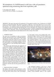
Figure 2.2 Phylogenies <strong>of</strong> pocket gophers and their chew<strong>in</strong>g lice based on<br />
nucleotide sequence data (Hafner et al., 1994). <strong>The</strong> figure shows composite<br />
trees based on multiple methods <strong>of</strong> phylogenetic analysis. Branch lengths are<br />
proportional to <strong>in</strong>ferred amounts <strong>of</strong> genetic change. Pocket gopher genera<br />
are Orthogeomys, Zygogeomys, Pappogeomys, Cratogeomys, Geomys and<br />
Thomomys. Geomys bursarius is represented by two subspecies (a = G. b. halli;<br />
b = G. b. majusculus). Chew<strong>in</strong>g louse genera are Geomydoecus and<br />
Thomomydoecus. <strong>The</strong> program COMPONENT was used to document<br />
significant similarity <strong>in</strong> branch<strong>in</strong>g structure between these trees. Because the<br />
host and parasite trees were based on DNA sequences from the same gene<br />
(cytochrome c oxidase subunit I), rates <strong>of</strong> DNA evolution could be compared<br />
<strong>in</strong> the two groups. Based on these data, Hafner et al. (1994) estimated that<br />
chew<strong>in</strong>g lice were evolv<strong>in</strong>g approximately ten times faster than pocket<br />
gophers <strong>in</strong> this gene region, which is <strong>in</strong> l<strong>in</strong>e with the predictions if<br />
co-evolution is occurr<strong>in</strong>g (see p. 10). But see Page et al. (1996).<br />
the vertebrate at some po<strong>in</strong>t distant from the nest. This type <strong>of</strong> association<br />
may have had several different underly<strong>in</strong>g reasons, such as attraction to<br />
feed on vertebrate secretions, or the use <strong>of</strong> the vertebrate as a bask<strong>in</strong>g or<br />
swarm<strong>in</strong>g site. But probably the most important factor was the use <strong>of</strong> the<br />
host’s dung as a larval habitat.<br />
<strong>The</strong> vertebrate may live <strong>in</strong> a harsh environment where dung rapidly<br />
dries up or it may bury its dung. In either case, closely associat<strong>in</strong>g with
12 <strong>The</strong> evolution <strong>of</strong> the blood-suck<strong>in</strong>g habit<br />
the vertebrate would help the <strong>in</strong>sect <strong>in</strong> locat<strong>in</strong>g usable dung. Non-bloodfeed<strong>in</strong>g<br />
associations aris<strong>in</strong>g for these reasons are seen between some<br />
scarabaeid beetles and vertebrates, but competition to be the first to exploit<br />
dung was probably the commonest reason driv<strong>in</strong>g the <strong>in</strong>sects to an ever<br />
closer association with the vertebrate. Dung is a limited resource and there<br />
is <strong>of</strong>ten <strong>in</strong>tense competition to utilize it as a larval site. Consequently, there<br />
is pressure on the <strong>in</strong>sect to be the first to <strong>in</strong>troduce its eggs <strong>in</strong>to the newly<br />
deposited dung. One way for the <strong>in</strong>sect to achieve this is to form an <strong>in</strong>creas<strong>in</strong>gly<br />
close association with the vertebrate provid<strong>in</strong>g the dung. Such a close<br />
association can be seen with the horn fly, Haematobia irritans, which as an<br />
adult is permanently associated with large vertebrates, only leav<strong>in</strong>g them<br />
to oviposit. It will lay eggs <strong>in</strong> dung with<strong>in</strong> 15 s <strong>of</strong> defecation by the vertebrate<br />
and, with<strong>in</strong> its distribution range, is almost always the first colonizer<br />
<strong>of</strong> freshly deposited dung (Mohr, 1943). If such an <strong>in</strong>sect can feed on the<br />
vertebrate, it m<strong>in</strong>imizes the time it will have to spend away from the host<br />
and therefore maximizes the advantage to be ga<strong>in</strong>ed from the close association,<br />
and this may have led the ancestral adult female to feed on body<br />
secretions, open wounds and sores. As was argued above, because blood<br />
is such a nutritious substance, once it was encountered <strong>in</strong> the diet <strong>of</strong> the<br />
<strong>in</strong>sect, selection is likely to have favoured the progressive development<br />
<strong>of</strong> physical and behavioural adaptations lead<strong>in</strong>g to full haematophagy.<br />
Selection will have led to the progressive development <strong>of</strong> mouthparts,<br />
allow<strong>in</strong>g the <strong>in</strong>sect to dislodge scabs, open up old sores and eventually to<br />
penetrate unbroken sk<strong>in</strong>. <strong>The</strong> evolution <strong>of</strong> organisms capable <strong>of</strong> break<strong>in</strong>g<br />
the sk<strong>in</strong> and obta<strong>in</strong><strong>in</strong>g a blood meal may very well have been an<br />
accelerated process. <strong>The</strong> wounds produced by the first blood-suckers will<br />
have provided regular blood-feed<strong>in</strong>g opportunities for other organisms<br />
that could not break the sk<strong>in</strong> by their own efforts. Once given access to<br />
blood these too may well have followed the evolutionary pathway outl<strong>in</strong>ed<br />
above.<br />
<strong>The</strong> spasmodic appearance <strong>of</strong> blood feed<strong>in</strong>g among male <strong>in</strong>sects is a<br />
difficult issue to expla<strong>in</strong>, but the close or permanent association <strong>of</strong> females<br />
with the vertebrate as discussed above is one factor that may expla<strong>in</strong> it <strong>in</strong><br />
some <strong>in</strong>sects. Under these circumstances it may become advantageous for<br />
the male to become associated with the vertebrate because <strong>of</strong> the greater<br />
likelihood <strong>of</strong> success <strong>in</strong> f<strong>in</strong>d<strong>in</strong>g a mate. This may then lead to blood feed<strong>in</strong>g<br />
<strong>in</strong> the male, as it would m<strong>in</strong>imize the time spent away from its host and<br />
therefore maximize its chances <strong>of</strong> successful mat<strong>in</strong>g. Similarly, if bloodsuck<strong>in</strong>g<br />
females are irregularly and/or widely dispersed <strong>in</strong> a habitat, then<br />
the male may ga<strong>in</strong> a mat<strong>in</strong>g advantage by stay<strong>in</strong>g with the vertebrate and<br />
wait<strong>in</strong>g for the female to arrive to feed. This is seen <strong>in</strong> the ‘follow<strong>in</strong>g swarm’<br />
<strong>of</strong> the tsetse flies but also <strong>in</strong> the mosquito Aedes aegypti, the males <strong>of</strong> which<br />
do not take blood (Teesdale, 1955).
2.2 Morphological pre-adaptation for pierc<strong>in</strong>g 13<br />
2.2 Morphological pre-adaptation for pierc<strong>in</strong>g<br />
<strong>The</strong> second route for the evolution <strong>of</strong> the blood-suck<strong>in</strong>g habit suggests<br />
that blood feed<strong>in</strong>g developed <strong>in</strong> some <strong>in</strong>sect l<strong>in</strong>eages from ancestral <strong>in</strong>sects<br />
that were morphologically pre-adapted for pierc<strong>in</strong>g surfaces (Beklemishev,<br />
1957; Downes, 1970;Waage, 1979). Entomophagous <strong>in</strong>sects are strong candidates<br />
for such a conversion. <strong>The</strong> Rhagionidae are a good example: most<br />
<strong>of</strong> the group are predacious on other <strong>in</strong>sects, but a few species have turned<br />
to blood feed<strong>in</strong>g. How could this changeover have come about? Entomophagous<br />
<strong>in</strong>sects would have been attracted to nests and burrows by<br />
the accumulation <strong>of</strong> <strong>in</strong>sects to be found there and so would have encountered<br />
vertebrates. Away from the nests they would have been attracted to<br />
vertebrates by the accumulation <strong>of</strong> <strong>in</strong>sects around them, or the vertebrates<br />
may have regularly congregated <strong>in</strong> the wet areas that are the breed<strong>in</strong>g<br />
sites for many <strong>of</strong> the ‘lower’ Diptera. <strong>The</strong> vertebrates <strong>in</strong>volved may have<br />
been permanently resident amphibians or reptiles, or larger vertebrates<br />
that regularly visited such sites for dr<strong>in</strong>k<strong>in</strong>g or bath<strong>in</strong>g purposes. In each<br />
<strong>of</strong> these cases it is easy to see how entomophagous <strong>in</strong>sects could have made<br />
repeated and possibly prolonged contact with vertebrates. <strong>The</strong>se predatory<br />
<strong>in</strong>sects would have physiological and morphological adaptations (such as<br />
efficient prote<strong>in</strong>-digest<strong>in</strong>g enzymes and pierc<strong>in</strong>g mouthparts) facilitat<strong>in</strong>g<br />
the switch to haematophagy. Haematophagy <strong>in</strong> these <strong>in</strong>dividuals was at<br />
first probably an occasional, chance event which led to full haematophagy<br />
through cont<strong>in</strong>ued close association with the vertebrate host. It is thought<br />
that haematophagy developed along these l<strong>in</strong>es <strong>in</strong> the ancestors <strong>of</strong> the<br />
blood-feed<strong>in</strong>g bugs and <strong>in</strong> blood-feed<strong>in</strong>g rhagionids and possibly <strong>in</strong> some<br />
blood-feed<strong>in</strong>g Diptera. <strong>The</strong>re is some doubt about which came first, larval<br />
feed<strong>in</strong>g on nest debris or adult feed<strong>in</strong>g on other <strong>in</strong>sects <strong>in</strong> the nest<br />
habitat. Fleas may also have evolved along this pathway from free-liv<strong>in</strong>g<br />
mecopteran stock (H<strong>in</strong>ton, 1958; Tillyard, 1935). <strong>The</strong> Mecoptera, or scorpion<br />
flies, conta<strong>in</strong> a modern-day group, the Boreidae, which are apterous<br />
and are capable <strong>of</strong> jump<strong>in</strong>g. <strong>The</strong>y live <strong>in</strong> moss and feed on <strong>in</strong>sects. Similar<br />
<strong>in</strong>sects may well have been the ancestors <strong>of</strong> the fleas, a view receiv<strong>in</strong>g<br />
support from molecular systematics (Whit<strong>in</strong>g, 2002).<br />
<strong>The</strong> lifestyles <strong>of</strong> several present-day <strong>in</strong>sects support the idea that entomophagous<br />
<strong>in</strong>sects gave rise to some blood-suck<strong>in</strong>g <strong>in</strong>sect groups (Waage,<br />
1979). Many personal experiences <strong>in</strong> Brita<strong>in</strong> (as many as three <strong>in</strong> one day)<br />
with the flower bug Anthocoris nemorum show that this <strong>in</strong>sect is will<strong>in</strong>g<br />
and able to pierce human sk<strong>in</strong>. This <strong>in</strong>sect is entomophagous, liv<strong>in</strong>g on<br />
and around flowers where it pounces on small <strong>in</strong>sects visit<strong>in</strong>g the flowers<br />
to feed. While its prob<strong>in</strong>gs <strong>of</strong> my sk<strong>in</strong> cause a sharp pa<strong>in</strong>, I have yet<br />
to f<strong>in</strong>d one that has obviously <strong>in</strong>gested any blood; however, it still establishes<br />
the fact that entomophagous <strong>in</strong>sects will <strong>of</strong>ten show an <strong>in</strong>terest <strong>in</strong>
14 <strong>The</strong> evolution <strong>of</strong> the blood-suck<strong>in</strong>g habit<br />
vertebrates as potential sources <strong>of</strong> a meal. <strong>The</strong> hemipteran bug Lyctocoris<br />
campestris is also an entomophagous species, but it takes matters further.<br />
It can live <strong>in</strong> birds’ nests, where it feeds on other <strong>in</strong>sects, but it will also<br />
take blood meals from vertebrates (Stys and Daniel, 1957). Evidence from<br />
blood-suck<strong>in</strong>g <strong>in</strong>sects themselves also po<strong>in</strong>ts to the close l<strong>in</strong>ks between<br />
entomophagy and the blood-feed<strong>in</strong>g habit. <strong>The</strong> mosquitoes Aedes aegypti<br />
and Culex tarsalis will take body fluids from <strong>in</strong>sect larvae presented to them<br />
under laboratory conditions (Harris et al., 1969). Indeed, this form <strong>of</strong> feed<strong>in</strong>g<br />
is so successful that these mosquitoes go on to produce viable eggs<br />
as a result, which opens up the <strong>in</strong>trigu<strong>in</strong>g possibility that this may occur<br />
naturally <strong>in</strong> the field.<br />
It has also been argued that haematophagy may have arisen <strong>in</strong> some<br />
<strong>in</strong>sect groups (<strong>in</strong>clud<strong>in</strong>g the mosquitoes; Matt<strong>in</strong>gley (1965)) from plantfeed<strong>in</strong>g<br />
ancestors. This is certa<strong>in</strong>ly a possibility as many plant-feed<strong>in</strong>g<br />
<strong>in</strong>sects possess pierc<strong>in</strong>g and suck<strong>in</strong>g mouthparts that would pre-adapt<br />
them for haematophagy. This is seen <strong>in</strong> the moth Calpe eustrigata, which is<br />
one <strong>of</strong> a group <strong>of</strong> noctuiids that possess an unusually modified, sharp<br />
proboscis used <strong>in</strong> most species for the penetration <strong>of</strong> fruit r<strong>in</strong>ds. But<br />
C. eustrigata uses it to penetrate vertebrate sk<strong>in</strong> for the purposes <strong>of</strong> bloodfeed<strong>in</strong>g.<br />
It is probable that plant-feed<strong>in</strong>g ancestors <strong>of</strong> modern-day blood<br />
feeders would have developed haematophagy only if they were <strong>in</strong> a position<br />
<strong>of</strong> cont<strong>in</strong>ual association with the vertebrate host. This may have<br />
occurred through mechanisms similar to those already outl<strong>in</strong>ed. Attraction<br />
to free-liv<strong>in</strong>g vertebrates may have occurred <strong>in</strong> order to feed on bodily<br />
secretions or to use dung as a larval medium. Or <strong>in</strong>sects may have been<br />
attracted to nests to feed on fruits or seeds stored there by the vertebrate.<br />
In this context it is <strong>in</strong>terest<strong>in</strong>g to note that the hemipteran bugs are exceptional<br />
<strong>in</strong> us<strong>in</strong>g catheps<strong>in</strong>-like digestive prote<strong>in</strong>ases. That is consistent with<br />
aproposed evolutionary path for bugs from sap-suck<strong>in</strong>g (Bill<strong>in</strong>gsley and<br />
Downe, 1988; Houseman et al., 1985; Terra, 1988) or seed-feed<strong>in</strong>g ancestors.<br />
Sap feeders, not need<strong>in</strong>g proteases, may have lost their tryps<strong>in</strong>s. If<br />
they then moved to blood feed<strong>in</strong>g they would need to reacquire proteolytic<br />
activity. Seeds are <strong>of</strong>ten sources <strong>of</strong> powerful anti-ser<strong>in</strong>e protease molecules<br />
produced to protect the seed from <strong>in</strong>sects. Seed feeders may have moved<br />
to catheps<strong>in</strong>s to avoid these <strong>in</strong>hibitors. It is argued that hav<strong>in</strong>g lost their<br />
tryps<strong>in</strong>s they had to make use <strong>of</strong> the catheps<strong>in</strong>s conta<strong>in</strong>ed <strong>in</strong> the lysosomes<br />
<strong>of</strong> all cells, re-rout<strong>in</strong>g them for extracellular digestion. Molecular evidence<br />
suggests Triatom<strong>in</strong>ae from the Americas and Asia are monophyletic with<br />
an orig<strong>in</strong> <strong>in</strong> northern areas <strong>of</strong> South America, <strong>in</strong> Central America, or <strong>in</strong> the<br />
southern region <strong>of</strong> North America about 95 million years ago (Gaunt and<br />
Miles, 2002; Lyman et al., 1999).
3<br />
Feed<strong>in</strong>g preferences <strong>of</strong> bloodsuck<strong>in</strong>g<br />
<strong>in</strong>sects<br />
3.1 Host choice<br />
<strong>Blood</strong>-suck<strong>in</strong>g <strong>in</strong>sects feed from a range <strong>of</strong> different host animals; because<br />
<strong>of</strong> the bites we receive we are acutely aware <strong>of</strong> the fact that many <strong>of</strong> them<br />
feed from humans, but many other animals are also exploited, <strong>in</strong>clud<strong>in</strong>g<br />
other mammals, birds, reptiles, amphibians and fish, and even <strong>in</strong>sects,<br />
arachnids and annelids (Hock<strong>in</strong>g, 1971). Any one <strong>in</strong>sect does not feed<br />
equally well from all <strong>of</strong> these potential resources; it displays host choice. For<br />
some <strong>in</strong>sects, particularly some permanent ectoparasites, the host choice<br />
may be very specific. Occasionally, for example for human lice, just a s<strong>in</strong>gle<br />
species. For other blood-suck<strong>in</strong>g <strong>in</strong>sects host choice is clearly not as<br />
restricted as <strong>in</strong>troduced exotic hosts (e.g. those <strong>in</strong> zoos) quickly become<br />
<strong>in</strong>corporated <strong>in</strong>to the diet <strong>of</strong> local blood-suck<strong>in</strong>g <strong>in</strong>sects.<br />
Let us consider what is meant by host choice. In its ma<strong>in</strong> sense it denotes<br />
the species <strong>of</strong> host animal or animals from which blood-suck<strong>in</strong>g <strong>in</strong>sects<br />
obta<strong>in</strong> their blood meals. But host choice can go beyond particular species<br />
<strong>of</strong> host chosen. <strong>Insects</strong> <strong>of</strong>ten choose to feed on particular <strong>in</strong>dividuals from<br />
among preferred species, which may well have implications for disease<br />
transmission (Burkot, 1988; Kelly, 2001; McCall and Kelly, 2002).<br />
Although most blood-suck<strong>in</strong>g <strong>in</strong>sects <strong>in</strong> their undisturbed, natural surround<strong>in</strong>gs<br />
show a preference for feed<strong>in</strong>g from a particular group or species,<br />
or even a primary cohort <strong>of</strong> their chosen species, the degree <strong>of</strong> host specificity<br />
shown varies greatly from one type <strong>of</strong> <strong>in</strong>sect to the next. Some are<br />
entirely dependent on a s<strong>in</strong>gle species <strong>of</strong> host while others are will<strong>in</strong>g to<br />
feed from a wide range <strong>of</strong> hosts. As a rough rule <strong>of</strong> thumb, it has <strong>of</strong>ten been<br />
said that there is a direct relationship between the locomotory capabilities<br />
<strong>of</strong> an <strong>in</strong>sect and the number <strong>of</strong> different hosts that it utilizes. Thus, the<br />
permanent ectoparasites, with their limited capacity for movement away<br />
from the host, conta<strong>in</strong> most cases <strong>of</strong> precise dependence. For example,<br />
the louse Haematomyzus elephantis is conf<strong>in</strong>ed to elephants, H. hopk<strong>in</strong>si to<br />
the wart-hog (Clay, 1963) and Pediculus capitis to humans. Consider<strong>in</strong>g the<br />
more mobile periodic ectoparasites, we can f<strong>in</strong>d many examples such as<br />
the ‘human’ flea, Pulex irritans, that have a narrow preferred range (badgers<br />
and foxes) on which most <strong>in</strong>sects are found, but that can occasionally
16 Feed<strong>in</strong>g preferences <strong>of</strong> blood-suck<strong>in</strong>g <strong>in</strong>sects<br />
be discovered on a wide range <strong>of</strong> other hosts (<strong>in</strong> this case humans, pigs<br />
and other large mammals) (Marshall, 1981). When consider<strong>in</strong>g the largely<br />
free-liv<strong>in</strong>g, temporary ectoparasites we f<strong>in</strong>d that many have catholic tastes.<br />
<strong>The</strong> mosquito Culex sal<strong>in</strong>arius, for example, has a wide range <strong>of</strong> potential<br />
hosts that <strong>in</strong>cludes birds (45 per cent <strong>of</strong> the blood meals tested), equ<strong>in</strong>es<br />
(17 per cent) and can<strong>in</strong>es (15 per cent) (Cupp and Stokes, 1976). This<br />
mosquito will demonstrate its cosmopolitan tastes further if it is disturbed<br />
dur<strong>in</strong>g the meal by mov<strong>in</strong>g will<strong>in</strong>gly from one host species to another to<br />
complete this s<strong>in</strong>gle, full blood meal. This is a regular and natural occurrence<br />
for this <strong>in</strong>sect. In one <strong>in</strong>vestigation 13 per cent <strong>of</strong> the blood meals<br />
tested were found to be from a mixture <strong>of</strong> hosts (Cupp and Stokes, 1976).<br />
<strong>The</strong> above rule <strong>of</strong> thumb works well <strong>in</strong> a broad sense but <strong>of</strong>ten breaks<br />
down when particular <strong>in</strong>sects are considered, as can be seen with the help<br />
<strong>of</strong> two examples. <strong>The</strong> amblyceran louse, Menacanthus eurysternus, has poor<br />
locomotory abilities <strong>of</strong>f the host and accord<strong>in</strong>g to our rule <strong>of</strong> thumb it<br />
should be limited to a small number <strong>of</strong> hosts. In fact it has been recorded<br />
from 123 different species <strong>of</strong> bird (Price, 1975)! In contrast, each <strong>of</strong> four<br />
closely related species <strong>of</strong> w<strong>in</strong>ged streblid were found to be specific to a different<br />
genus <strong>of</strong> bat and this relationship held true even <strong>in</strong> caves conta<strong>in</strong><strong>in</strong>g<br />
three <strong>of</strong> the bat genera <strong>in</strong> crowded conditions (Maa and Marshall, 1981).<br />
Before look<strong>in</strong>g at some <strong>of</strong> the factors that underp<strong>in</strong> host choice, let us<br />
make some general comments <strong>of</strong> particular relevance to temporary ectoparasites.<br />
<strong>The</strong> commonest hosts are probably large, social herbivores, which<br />
present an abundant and easily visible food source for many ectoparasites.<br />
<strong>The</strong>y are also reliable food sources because normally they move only slowly<br />
from one pasture to another. Carnivores are less abundant than their prey,<br />
<strong>of</strong>ten solitary, less visible and less predictable, occupy<strong>in</strong>g a large home<br />
range. For these reasons carnivores are less likely to be a primary element<br />
<strong>in</strong> the host choice <strong>of</strong> a blood-suck<strong>in</strong>g <strong>in</strong>sect. Also large vertebrates are likely<br />
to be preferred over smaller ones. This is because small vertebrates, which<br />
suffer greater losses from the attention <strong>of</strong> a blood-suck<strong>in</strong>g <strong>in</strong>sect, use their<br />
agility to develop efficient defences.<br />
Mov<strong>in</strong>g from these generalizations to consider the specific choices made<br />
by particular <strong>in</strong>sects is a more complex issue. Sometimes a reasoned case<br />
for the evolution <strong>of</strong> a particular pattern <strong>of</strong> host choice can be made, but<br />
on most occasions we are <strong>in</strong> the dark. <strong>The</strong> choice may be determ<strong>in</strong>ed<br />
by a large number <strong>of</strong> factors (probably act<strong>in</strong>g <strong>in</strong> comb<strong>in</strong>ation), <strong>in</strong>clud<strong>in</strong>g<br />
behavioural, physiological, morphological, ecological, geographical, temporal<br />
and genetic considerations. To give an idea <strong>of</strong> the complexity <strong>of</strong> the<br />
issue, let us look at a small number <strong>of</strong> examples, show<strong>in</strong>g how each <strong>of</strong> these<br />
factors may affect host choice.<br />
Although it is not always the case (Canyon et al., 1998; Charlwood<br />
et al., 1995; Prior and Torr, 2002), host defensive behaviour can have a
3.1 Host choice 17<br />
considerable effect on the number <strong>of</strong> blood-suck<strong>in</strong>g <strong>in</strong>sects successfully<br />
feed<strong>in</strong>g on that host (Torr et al., 2001). <strong>The</strong> anti-mosquito behaviour displayed<br />
by a range <strong>of</strong> ciconiiform birds is a good demonstration <strong>of</strong> the protective<br />
effects host defensive behaviour can have. At roost some <strong>of</strong> these<br />
birds perform up to 3000 defensive movements per hour! But the green<br />
heron and the crowned night heron are far less active, perform<strong>in</strong>g as few<br />
as 650 defensive movements per hour: as a consequence they receive far<br />
more bites than their more active relatives (Webber and Edman, 1972). If no<br />
other factors were <strong>in</strong> operation it is easy to see how natural selection would<br />
bear on such a case and would lead a blood-suck<strong>in</strong>g <strong>in</strong>sect to select and<br />
eventually specialize <strong>in</strong> feed<strong>in</strong>g from the easiest available target. <strong>The</strong> fact<br />
that under natural conditions mosquitoes still feed from a wide range <strong>of</strong><br />
ciconiiform birds, <strong>in</strong>clud<strong>in</strong>g those display<strong>in</strong>g the highest levels <strong>of</strong> defensive<br />
behaviours, is a testament to the complexity <strong>of</strong> the factors that determ<strong>in</strong>e<br />
host choice. None the less, groom<strong>in</strong>g and host defensive behaviour <strong>in</strong> general<br />
are likely to be highly efficient agents <strong>of</strong> natural selection. For humans<br />
the concept <strong>of</strong> defensive behaviour needs to be extended to <strong>in</strong>clude such<br />
practices as sleep<strong>in</strong>g under bednets, screen<strong>in</strong>g houses and the use <strong>of</strong> repellents<br />
(modern preparations conta<strong>in</strong><strong>in</strong>g active <strong>in</strong>gredients such as di-ethyl<br />
toluamide (DEET) or traditional concoctions such as mixtures <strong>of</strong> cow dung,<br />
ur<strong>in</strong>e and ash) (MacCormack, 1984).<br />
<strong>The</strong> <strong>in</strong>tensity <strong>of</strong> defensive behaviour <strong>of</strong>ten correlates with the number<br />
<strong>of</strong> blood-suck<strong>in</strong>g <strong>in</strong>sects attack<strong>in</strong>g the host. Thus is it possible that<br />
the seasonal changes <strong>in</strong> the numbers <strong>of</strong> blood-suck<strong>in</strong>g <strong>in</strong>sects may play<br />
a part <strong>in</strong> the seasonal changes <strong>in</strong> host choice because <strong>of</strong> changes <strong>in</strong> the<br />
amount <strong>of</strong> host defensive behaviour (Edman and Spielman, 1988). Such<br />
seasonal changes <strong>in</strong> host choice are important <strong>in</strong> the transmission <strong>of</strong> some<br />
zoonoses to humans. For example, <strong>in</strong> the USA the arbovirus eastern equ<strong>in</strong>e<br />
encephalitis is enzootic <strong>in</strong> birds and the mosquito Culiseta melanura is its<br />
vector. Dur<strong>in</strong>g the spr<strong>in</strong>g and early summer, when mosquito numbers<br />
are comparatively low, this <strong>in</strong>sect feeds almost exclusively on passer<strong>in</strong>e<br />
birds. Later <strong>in</strong> the season mosquito numbers <strong>in</strong>crease and so does defensive<br />
behaviour by the birds. At this time mosquito host choice becomes<br />
more catholic and <strong>in</strong>cludes other bird groups and mammals. It is also <strong>in</strong><br />
this later part <strong>of</strong> the season that epizootics <strong>of</strong> the virus occur <strong>in</strong> birds and<br />
horses and epidemics occur <strong>in</strong> humans. All this is enabled by the greater<br />
range <strong>of</strong> hosts chosen by the mosquitoes, driven by chang<strong>in</strong>g levels <strong>of</strong> host<br />
defensive behaviour. Mathematical models <strong>of</strong> disease transmission have<br />
been produced that <strong>in</strong>corporate the assumption that different <strong>in</strong>dividual<br />
levels <strong>of</strong> host defensive behaviour lead to a non-homogeneous pattern <strong>of</strong><br />
host choice with<strong>in</strong> a host species (Kelly and Thompson, 2000).<br />
I suggest that physiological factors are ma<strong>in</strong>ly important <strong>in</strong> determ<strong>in</strong><strong>in</strong>g<br />
host choice <strong>in</strong> the sense that once the <strong>in</strong>sect has become associated with a
18 Feed<strong>in</strong>g preferences <strong>of</strong> blood-suck<strong>in</strong>g <strong>in</strong>sects<br />
Figure 3.1 <strong>The</strong> tsetse fly, Gloss<strong>in</strong>a morsitans morsitans, shows greater<br />
longevity when it feeds on rabbits () rather than goats () (Jordan and<br />
Curtis, 1972).<br />
narrow range <strong>of</strong> hosts then specialization <strong>in</strong> its physiology will occur that<br />
may limit the range <strong>of</strong> other hosts it can exploit (Krasnov et al., 2003). This<br />
is because natural selection will ensure that all <strong>of</strong> the <strong>in</strong>sect’s systems will<br />
become tuned to the exploitation <strong>of</strong> the resources <strong>of</strong> its major host, which<br />
may restrict the <strong>in</strong>sect’s ability to deal with unusual situations. In some<br />
cases choice <strong>of</strong> the wrong host can, for physiological reasons, lead to the<br />
death <strong>of</strong> the <strong>in</strong>sect. For example, a blood meal from the gu<strong>in</strong>ea-pig may<br />
form oxyhaemoglob<strong>in</strong> crystals which will rupture the <strong>in</strong>test<strong>in</strong>e <strong>of</strong> several<br />
blood-suck<strong>in</strong>g <strong>in</strong>sects (Krynski et al., 1952).<br />
<strong>The</strong> physiological consequences <strong>of</strong> mov<strong>in</strong>g on to an unusual host can<br />
have a less obvious, but a nevertheless damag<strong>in</strong>g, effect on the success<br />
<strong>of</strong> the <strong>in</strong>sect. Several experimental studies have shown that the fecundity<br />
<strong>of</strong> a blood-suck<strong>in</strong>g <strong>in</strong>sect depends on the host on which the <strong>in</strong>sect<br />
feeds. Reduced fecundity can be generated by a reduced rate <strong>of</strong> development<br />
<strong>of</strong> the <strong>in</strong>sect, reduced longevity (Fig. 3.1), a skewed sex ratio, or<br />
reduced food <strong>in</strong>take or rate <strong>of</strong> digestion (Chang and Judson, 1977; Nelson<br />
et al., 1975; Rothschild, 1975). We can look at the tsetse fly as an example.<br />
Gloss<strong>in</strong>a austeni fed on rabbit blood showed a consistently higher fecundity<br />
than flies <strong>of</strong> the same species fed on goat blood (Jordan and Curtis, 1968).<br />
In a further series <strong>of</strong> experiments it was shown that both G. austeni and
3.1 Host choice 19<br />
G. morsitans morsitans fed on pig blood produced the heaviest puparia,<br />
closely followed by flies fed on goat blood, while the puparia produced<br />
from flies fed on cow blood were considerably lighter (Mews et al., 1976).<br />
Clearly under natural conditions it would be a selective advantage to the<br />
<strong>in</strong>sect to develop mechanisms that avoided such a restriction on its reproductive<br />
success by careful choice <strong>of</strong> hosts. It is also <strong>of</strong> <strong>in</strong>terest to note that<br />
the source <strong>of</strong> the blood meal or its quality may also <strong>in</strong>fluence the course<br />
<strong>of</strong> an <strong>in</strong>fection <strong>in</strong> the <strong>in</strong>sect. If tsetse flies feed on goats or cattle they will<br />
develop a much higher <strong>in</strong>fection rate with Trypanosoma vivax than if they<br />
feed on mice (Maudl<strong>in</strong> et al., 1984). Similar results are also found <strong>in</strong> more<br />
natural vector comb<strong>in</strong>ations (Nguu et al., 1996).<br />
As with the physiological restrictions on host choice just discussed, morphological<br />
factors are also important as a limitation on the range <strong>of</strong> available<br />
hosts once a certa<strong>in</strong> degree <strong>of</strong> specialization for a particular host has<br />
taken place. With permanent and periodic ectoparasites especially, restriction<br />
<strong>in</strong> host range usually <strong>in</strong>volves morphological specialization <strong>of</strong> the<br />
<strong>in</strong>sect’s mouthparts, ovipositor or locomotory attachment apparatus. Specializations<br />
<strong>of</strong> any <strong>of</strong> these can easily restrict the host range <strong>of</strong> the <strong>in</strong>sect<br />
concerned; <strong>in</strong>deed, <strong>in</strong> many cases specialization may even limit the area <strong>of</strong><br />
the body <strong>of</strong> the preferred host that the <strong>in</strong>sect can utilize. This is discussed<br />
<strong>in</strong> more detail <strong>in</strong> Section 7.1.<br />
It has also been suggested that morphological characteristics <strong>of</strong> the host<br />
are important <strong>in</strong> determ<strong>in</strong><strong>in</strong>g host choice. One example is the frequently<br />
reported preference <strong>of</strong> Anopheles gambiae for human adults rather than children.<br />
It is suggested that the choice is a direct consequence <strong>of</strong> the difference<br />
<strong>in</strong> size <strong>of</strong> these two potential hosts, the number <strong>of</strong> bites received by<br />
an <strong>in</strong>dividual be<strong>in</strong>g <strong>in</strong> direct proportion to the surface area that <strong>in</strong>dividual<br />
contributes to the total immediately available to the mosquito (Port et al.,<br />
1980). Interest<strong>in</strong>gly this is not the case for adult cows and calves; here it is<br />
differences <strong>in</strong> levels <strong>of</strong> defensive behaviour that determ<strong>in</strong>e that adults are<br />
bitten more than calves by tsetse flies (Torr et al., 2001).<br />
A clear example <strong>of</strong> ecological <strong>in</strong>fluence on host choice is furnished by the<br />
fleas. <strong>The</strong> immature stages <strong>of</strong> most fleas live on the detritus <strong>in</strong> the homes <strong>of</strong><br />
the host animal. In other words, host choice for fleas is largely determ<strong>in</strong>ed<br />
by the ecology <strong>of</strong> the host, most fleas be<strong>in</strong>g restricted to hosts with longterm<br />
homes, or at least seasonal lairs that are used each year. Because they<br />
so commonly use long-term homes, mammals are the ma<strong>in</strong> hosts for fleas.<br />
Rodents are unrivalled as home builders and as a consequence are the<br />
most afflicted mammals, harbour<strong>in</strong>g 74 per cent <strong>of</strong> all known flea species<br />
(Marshall, 1981). Humans are also habitual home dwellers and because <strong>of</strong><br />
this we are the only primates that are regularly flea-ridden. Interest<strong>in</strong>gly<br />
the 5000-year-old late-Stone-Age Tyrolean ice man was <strong>in</strong>fested with Pulex<br />
irritans (Sp<strong>in</strong>dler, 2001).
20 Feed<strong>in</strong>g preferences <strong>of</strong> blood-suck<strong>in</strong>g <strong>in</strong>sects<br />
A second example <strong>of</strong> ecological <strong>in</strong>fluence on host choice, <strong>of</strong> considerable<br />
importance <strong>in</strong> the health <strong>of</strong> Europeans, can be seen <strong>in</strong> the chang<strong>in</strong>g feed<strong>in</strong>g<br />
patterns <strong>of</strong> the anophel<strong>in</strong>e mosquitoes <strong>of</strong> Europe. Until the middle <strong>of</strong> the<br />
n<strong>in</strong>eteenth century benign tertian malaria (caused by Plasmodium vivax)<br />
was endemic <strong>in</strong> Europe as far north as Scand<strong>in</strong>avia, with P. malariae as<br />
far north as Holland and the UK, and P. falciparum extensive <strong>in</strong> southern<br />
Europe. At this time malaria began to retreat from Europe and transmission<br />
<strong>of</strong> the disease now rarely occurs. Why did this happen? Ronald<br />
Ross described the full cycle <strong>of</strong> malaria <strong>in</strong> 1898 and soon after this, <strong>in</strong><br />
1901, it was realized that there were areas <strong>of</strong> Europe that abounded with<br />
anophel<strong>in</strong>e mosquitoes, and <strong>in</strong>to which malaria carriers rout<strong>in</strong>ely passed,<br />
but <strong>in</strong> which the disease was not transmitted (Falleroni, 1927). Several<br />
theories were put forward to account for this apparent anomaly, but it<br />
was not until after the <strong>in</strong>troduction <strong>of</strong> the precipit<strong>in</strong> test, for the identification<br />
<strong>of</strong> mosquito blood meals (see Pant et al., 1987), that relevant evidence<br />
Box 3.1 <strong>The</strong> importance <strong>of</strong> human-bit<strong>in</strong>g rates <strong>of</strong> mosquitoes for the<br />
transmission <strong>of</strong> malaria<br />
<strong>The</strong> vectorial capacity C <strong>of</strong> a population <strong>of</strong> mosquitoes transmitt<strong>in</strong>g<br />
malaria can be described by the equation<br />
C = m× a 2 n P<br />
×<br />
−ln(P )<br />
where m is the number <strong>of</strong> mosquitoes per person and a is the proportion<br />
<strong>of</strong> these mosquitoes that bite humans (bit<strong>in</strong>g once to acquire the<br />
<strong>in</strong>fection and transmitt<strong>in</strong>g it on the occasion <strong>of</strong> a subsequent meal on a<br />
human, hence a2 )–the quantity a is usually estimated us<strong>in</strong>g techniques<br />
such as those described <strong>in</strong> Box 3.2.<br />
<strong>The</strong> value P n /−ln(P ) describes the expectation <strong>of</strong> the <strong>in</strong>fective life <strong>of</strong><br />
the vector population (Garrett-Jones and Shidrawi, 1969).<br />
If we take the example described <strong>in</strong> the text we can see that <strong>in</strong> those<br />
parts <strong>of</strong> Europe where malaria was be<strong>in</strong>g transmitted a = 1/8, whereas<br />
<strong>in</strong> those areas where mosquitoes were present but no malaria was be<strong>in</strong>g<br />
transmitted a = 1/400.<br />
If we assume that m and P n /−ln(P ) are constants, then the equation<br />
tells us that humans <strong>in</strong> the malarious areas are 2500 times more likely to<br />
contract malaria than those <strong>in</strong> the non-malarious areas. <strong>The</strong> difference<br />
<strong>in</strong> this example is entirely due to the different proportions <strong>of</strong> mosquitoes<br />
bit<strong>in</strong>g humans <strong>in</strong> the two areas. A good account <strong>of</strong> these mathematical<br />
models is given by Dye (1992).<br />
Source: From Burkot (1988)
3.1 Host choice 21<br />
Box 3.2 Identification <strong>of</strong> the source <strong>of</strong> a blood meal<br />
<strong>The</strong> ability to identify the source <strong>of</strong> an <strong>in</strong>sect’s blood meal accurately<br />
is a powerful tool for the <strong>in</strong>vestigation <strong>of</strong> a wide range <strong>of</strong> questions<br />
relat<strong>in</strong>g to the life <strong>of</strong> blood-suck<strong>in</strong>g <strong>in</strong>sects. For example, it allows us<br />
to <strong>in</strong>vestigate the nature <strong>of</strong> host choice and it can help us to understand<br />
whether parasites manipulate the host-seek<strong>in</strong>g behaviour <strong>of</strong> their vectors<br />
(Koella et al., 1998b). It may help <strong>in</strong> monitor<strong>in</strong>g the efficacy <strong>of</strong> various<br />
control or surveillance tools such as repellents, bednets and traps and to<br />
estimate the degree <strong>of</strong> coverage needed for emerg<strong>in</strong>g malaria vacc<strong>in</strong>es<br />
(Mukabana et al., 2002a).<br />
Until recently blood meal identification has been achieved through<br />
immunological means (Pant et al., 1987). Increas<strong>in</strong>gly molecular techniques<br />
are tak<strong>in</strong>g over (Mukabana et al., 2002a). <strong>The</strong>se are usually ‘f<strong>in</strong>gerpr<strong>in</strong>t<strong>in</strong>g’<br />
techniques that look at the number <strong>of</strong> tandem repeats (VNTRs)<br />
and/or short tandem repeats (STRs), both <strong>of</strong> which occur abundantly <strong>in</strong><br />
nuclear DNA <strong>in</strong> most eukaryote genomes. <strong>The</strong> number <strong>of</strong> tandem repeats<br />
at these loci is highly variable with alleles differ<strong>in</strong>g <strong>in</strong> length by an <strong>in</strong>tegral<br />
number <strong>of</strong> repeat units. As well as be<strong>in</strong>g highly polymorphic these loci are<br />
stably <strong>in</strong>herited and so are <strong>in</strong>formative genetic markers. <strong>The</strong> markers can<br />
be easily accessed because they can be amplified <strong>in</strong> vitro by polymerase<br />
cha<strong>in</strong> reaction and the size <strong>of</strong> the products easily determ<strong>in</strong>ed. <strong>The</strong>y have<br />
been used to identify successfully the blood meal source <strong>of</strong> mosquitoes<br />
(Koella et al., 1998a; Mukabana et al., 2002a; Mukabana et al., 2002b),<br />
crab lice (Lord et al., 1998) and tsetse flies (Torr et al., 2001). Not only<br />
can they identify the species that was the source <strong>of</strong> the blood meal, they<br />
can be so specific that they can identify the particular <strong>in</strong>dividual with<strong>in</strong><br />
a species from which the blood meal was taken.<br />
began to accumulate. <strong>The</strong> key factor was that anophel<strong>in</strong>e mosquitoes <strong>in</strong><br />
the malaria-free areas were 400 times as likely to feed on animals other<br />
than humans, while <strong>in</strong> malarious areas 1 <strong>in</strong> 8 feeds were from humans<br />
(Hackett and Missiroli, 1931). <strong>The</strong> importance <strong>of</strong> these rates <strong>of</strong> bit<strong>in</strong>g for<br />
the transmission <strong>of</strong> malaria is expla<strong>in</strong>ed <strong>in</strong> Box 3.1. Immunological tests<br />
are now be<strong>in</strong>g replaced by more sensitive and specific molecular techniques<br />
for the identification <strong>of</strong> vector blood meals (Mukabana et al., 2002a)<br />
(Box 3.2).<br />
Why should the mosquitoes <strong>of</strong> one area bite humans so regularly and<br />
mosquitoes <strong>of</strong> the same species <strong>in</strong> another area bite them so <strong>in</strong>frequently?<br />
Although it was suggested that the mosquitoes were actually chang<strong>in</strong>g<br />
<strong>in</strong> their relative attractions for humans and domesticated animals (particularly<br />
pigs, horses and cattle), no such change could be demonstrated<br />
experimentally. It is now widely accepted that the switch from humans
22 Feed<strong>in</strong>g preferences <strong>of</strong> blood-suck<strong>in</strong>g <strong>in</strong>sects<br />
to animals was a response to the chang<strong>in</strong>g proportions <strong>of</strong> human and<br />
non-human hosts available. At this time animal husbandry practices <strong>in</strong><br />
Europe were chang<strong>in</strong>g so that far more animals were be<strong>in</strong>g kept and were<br />
available to the mosquitoes as food sources. A contributory factor was<br />
the improv<strong>in</strong>g standard <strong>of</strong> liv<strong>in</strong>g for the human population, which led<br />
to larger and brighter hous<strong>in</strong>g that was much less attractive as a rest<strong>in</strong>g<br />
site for the mosquito. <strong>The</strong> mosquitoes were diverted to the darker<br />
animal sheds around the house, considerably reduc<strong>in</strong>g the likelihood <strong>of</strong><br />
their bit<strong>in</strong>g humans (Harrison, 1978; Takken et al., 1999). Also <strong>of</strong> importance<br />
was the decreas<strong>in</strong>g birth rate among the population, reduc<strong>in</strong>g the<br />
numbers <strong>of</strong> human hosts available, a factor accentuated by the <strong>in</strong>creased<br />
mechanization <strong>of</strong> agriculture which also led to a reduction <strong>in</strong> the populations<br />
<strong>in</strong> rural communities. So we can see that host choice is not only<br />
a behavioural, decision-mak<strong>in</strong>g process on the part <strong>of</strong> the <strong>in</strong>sect, but can<br />
also be strongly <strong>in</strong>fluenced by the relative availability <strong>of</strong> different host<br />
species.<br />
Geographical considerations are also important <strong>in</strong> host choice because<br />
if there is no overlap <strong>in</strong> the range <strong>of</strong> <strong>in</strong>sect and potential host then the host<br />
is not available as a food source. Global climate change may be affect<strong>in</strong>g<br />
these overlaps (Sutherst et al., 1998). Geographic overlap <strong>of</strong> host and <strong>in</strong>sect<br />
can work on a variety <strong>of</strong> scales. <strong>The</strong> effect is obvious from a global viewpo<strong>in</strong>t,<br />
but not so obvious when more localized issues are considered. <strong>The</strong><br />
tsetse fly can be used as an example, one that has considerable economic<br />
importance. On the largest geographical scale we can see that tsetse flies,<br />
apart from a small fly belt <strong>in</strong> Saudi Arabia (Elsen et al., 1990), are only found<br />
<strong>in</strong> Africa, where they are widely distributed from the southern borders <strong>of</strong><br />
the Sahara to Mozambique. With<strong>in</strong> this region they transmit trypanosomes<br />
caus<strong>in</strong>g sleep<strong>in</strong>g sickness <strong>in</strong> humans and nagana <strong>in</strong> animals. <strong>The</strong> game animals<br />
<strong>of</strong> Africa have some degree <strong>of</strong> immunity to nagana, but normally it<br />
quickly kills <strong>in</strong>troduced domesticated animals. This disease and its transmission<br />
by the tsetse fly have had a tremendous <strong>in</strong>fluence on the history<br />
<strong>of</strong> tropical Africa. <strong>The</strong> comb<strong>in</strong>ation largely prevented the <strong>in</strong>vasion from<br />
the north <strong>of</strong> armies dependent on horses. It also impeded Africa’s development<br />
by limit<strong>in</strong>g the use <strong>of</strong> draft animals and prevented (and <strong>in</strong>deed<br />
still prevents) the use <strong>of</strong> large tracts <strong>of</strong> land for ranch<strong>in</strong>g. In other words<br />
large herbivores enter<strong>in</strong>g the tsetse’s geographical range, <strong>in</strong>clud<strong>in</strong>g horses<br />
and cattle, become available as hosts and are <strong>of</strong>ten chosen as food sources.<br />
But geographical overlap is also important <strong>in</strong> tsetse fly biology on a much<br />
smaller scale. Long before Bruce (1895) demonstrated that the tsetse fly<br />
transmits the trypanosomes that cause illness and death, the herdsmen <strong>of</strong><br />
southern Africa had learned by bitter experience that the fly was deadly.<br />
This fact became obvious to them because the tsetse flies <strong>of</strong> this region<br />
are not evenly spread throughout the land, but are restricted to certa<strong>in</strong>
3.1 Host choice 23<br />
areas known as fly belts. <strong>The</strong> herdsmen learned that if they avoided these<br />
belts, and the flies that lived there, then their stock did not acquire nagana<br />
(McKelvey, 1973).<br />
Geographical effects on host choice can be seen on an even smaller scale.<br />
<strong>The</strong> tree hole mosquitoes Aedes triseriatus and Ae. hendersoni are sibl<strong>in</strong>g<br />
species liv<strong>in</strong>g sympatrically <strong>in</strong> woodlands <strong>in</strong> the eastern and midwestern<br />
USA. It might be expected that two such closely related species liv<strong>in</strong>g <strong>in</strong><br />
the same woodland would be feed<strong>in</strong>g on similar hosts, but geographical<br />
separation aga<strong>in</strong> affects host choice. Aedes triseriatus feeds ma<strong>in</strong>ly at<br />
ground level and consequently feeds on ground-dwell<strong>in</strong>g animals such as<br />
deer and chipmunk. In contrast, Ae. hendersoni feeds ma<strong>in</strong>ly <strong>in</strong> the canopy<br />
<strong>of</strong> these woodland trees on animals such as tree squirrels (Nasci, 1982).<br />
An important example, because it has consequences for malaria transmission,<br />
occurs with the malaria vectors with<strong>in</strong> the Anopheles gambiae complex<br />
(see Section 3.2). It has <strong>of</strong>ten been reported that An. arabiensis is much more<br />
zoophilic that An. gambiensis s.s. but <strong>in</strong> a carefully designed experiment that<br />
controlled for equal accessibility to hosts, which were presented outside<br />
houses, it was found that both species showed statistically similar levels <strong>of</strong><br />
human- and cattle-bit<strong>in</strong>g activity (Diatta et al., 1998). So an unwill<strong>in</strong>gness<br />
to bite <strong>in</strong>doors may be the basis <strong>of</strong> previous reports <strong>of</strong> zoophily <strong>in</strong> An. arabiensis<br />
rather than a real difference <strong>in</strong> host preference. Clearly geographical<br />
overlap on even the smallest scale is very important <strong>in</strong> determ<strong>in</strong><strong>in</strong>g host<br />
choice; <strong>in</strong>deed, host abundance and proximity may be the ultimate arbiter<br />
<strong>of</strong> host choice <strong>in</strong> many <strong>in</strong>stances.<br />
As we have just seen, availability <strong>of</strong> hosts is <strong>of</strong>ten a prime factor <strong>in</strong><br />
determ<strong>in</strong><strong>in</strong>g host choice. For the host to be available there must not only<br />
be geographic overlap between the <strong>in</strong>sect and host but also a temporal<br />
overlap because most temporary ectoparasites feed only dur<strong>in</strong>g a welldef<strong>in</strong>ed<br />
period <strong>of</strong> the day. Let us look at two contrast<strong>in</strong>g ways <strong>in</strong> which<br />
temporal overlap could occur. Some hosts show such efficient defensive<br />
behaviour that temporary ectoparasites can feed only when the host is at<br />
rest. <strong>The</strong> degree <strong>of</strong> overlap between the activity period <strong>of</strong> the <strong>in</strong>sect and the<br />
rest<strong>in</strong>g period <strong>of</strong> such a host will then be important <strong>in</strong> determ<strong>in</strong><strong>in</strong>g host<br />
choice. In contrast, animals liv<strong>in</strong>g <strong>in</strong> deep burrows are unlikely to be hosts<br />
for exophilic <strong>in</strong>sects when at rest, but may be hosts if their activity period<br />
co<strong>in</strong>cides with that <strong>of</strong> the <strong>in</strong>sect.<br />
Seasonal variation <strong>in</strong> the choice <strong>of</strong> host has been recognized <strong>in</strong> some<br />
blood-suck<strong>in</strong>g <strong>in</strong>sects, and it can have serious consequences for disease<br />
transmission to humans. In North America the arbovirus St Louis<br />
encephalitis is transmitted dur<strong>in</strong>g the summer months by the mosquitoes<br />
Culex nigripalpus and C. tarsalis. Both these mosquitoes show a marked seasonal<br />
change <strong>in</strong> their feed<strong>in</strong>g patterns, switch<strong>in</strong>g from bird feed<strong>in</strong>g <strong>in</strong> the<br />
w<strong>in</strong>ter and spr<strong>in</strong>g to mammal feed<strong>in</strong>g <strong>in</strong> the summer, when arbovirus
24 Feed<strong>in</strong>g preferences <strong>of</strong> blood-suck<strong>in</strong>g <strong>in</strong>sects<br />
transmission occurs (Edman, 1974; Edman and Taylor, 1968; Tempelis<br />
and Wash<strong>in</strong>o, 1967) (see discussion above concern<strong>in</strong>g eastern equ<strong>in</strong>e<br />
encephalitis).<br />
Evidence is beg<strong>in</strong>n<strong>in</strong>g to appear to suggest that memory can play a role<br />
<strong>in</strong> host choice. Thus many blood-fed Anopheles arabiensis that had been<br />
artificially transported returned to the houses where they had previously<br />
obta<strong>in</strong>ed a blood meal. Some probably flew more than 400 m to do so,<br />
which implies a considerable spatial memory (McCall and Kelly, 2002;<br />
McCall et al., 2001). In addition, as well as return<strong>in</strong>g to successful feed<strong>in</strong>g<br />
sites, vectors may choose their next host on the basis <strong>of</strong> previous successful<br />
feed<strong>in</strong>g experiences. Such impr<strong>in</strong>t<strong>in</strong>g was seen <strong>in</strong> Culex spp. <strong>in</strong> field<br />
cages where the tendency was for <strong>in</strong>dividual mosquitoes to choose the<br />
same host on which they had previously fed successfully (Mwandawiro<br />
et al., 2000). Significantly this preference was not reta<strong>in</strong>ed by the <strong>of</strong>fspr<strong>in</strong>g,<br />
which is what would be expected if this is <strong>in</strong>deed learned behaviour.<br />
Such learned behaviour <strong>in</strong> host selection, if it proves to be widespread,<br />
can have a considerable impact on disease transmission (Kelly and<br />
Thompson, 2000; McCall and Kelly, 2002). For example, a major component<br />
<strong>of</strong> host selection with<strong>in</strong> a group <strong>of</strong> hosts may be the level <strong>of</strong> <strong>in</strong>dividual host<br />
defensive behaviour (see above and Chapter 7). Sick hosts tend to display<br />
fewer defensive behaviours and are fed upon most <strong>of</strong>ten (Day and Edman,<br />
1983). If this were absolute then no un<strong>in</strong>fected hosts would ever become<br />
<strong>in</strong>fected. Clearly that is not the case, but nevertheless this aspect <strong>of</strong> host<br />
selection may be a factor determ<strong>in</strong><strong>in</strong>g the rate <strong>of</strong> disease transmission <strong>in</strong> a<br />
population.<br />
It has <strong>of</strong>ten been suggested that there are genetically determ<strong>in</strong>ed<br />
behavioural traits that produce different feed<strong>in</strong>g patterns <strong>in</strong> different populations<br />
<strong>of</strong> a s<strong>in</strong>gle <strong>in</strong>sect species (rather than the well-recognized phenomenon<br />
<strong>of</strong> different feed<strong>in</strong>g patterns seen among members <strong>of</strong> species<br />
complexes – see Section 3.2). Most <strong>of</strong> the support for this is anecdotal but<br />
conv<strong>in</strong>c<strong>in</strong>g evidence has been reported for Aedes simpsoni and Ae. aegypti<br />
(Mukwaya, 1977). <strong>The</strong>se species showed direct evidence <strong>of</strong> both zoophilic<br />
and anthropophilic populations <strong>in</strong> the laboratory and <strong>in</strong> the field. It was<br />
established that s<strong>in</strong>gle species were <strong>in</strong>volved when no cross<strong>in</strong>g or hybrid<br />
sterility appeared dur<strong>in</strong>g cross<strong>in</strong>g and backcross<strong>in</strong>g experiments <strong>of</strong> the<br />
populations studied.<br />
3.2 Host choice and species complexes<br />
For most blood-suck<strong>in</strong>g <strong>in</strong>sects the spectrum <strong>of</strong> host choice differs with<br />
chang<strong>in</strong>g place and season and given different proportions <strong>of</strong> available<br />
hosts. Selective adaptation to such changes <strong>in</strong> local circumstance is the factor<br />
driv<strong>in</strong>g speciation. This process is cont<strong>in</strong>ual and as different species are
3.2 Host choice and species complexes 25<br />
<strong>in</strong> the process <strong>of</strong> emerg<strong>in</strong>g they are not always easily dist<strong>in</strong>guishable by<br />
their morphological characteristics. Indeed, several blood-suck<strong>in</strong>g <strong>in</strong>sect<br />
‘species’, <strong>in</strong>clud<strong>in</strong>g Anopheles gambiae sensu lato and Simulium damnosum<br />
s.l., are known to be species complexes made up <strong>of</strong> reproductively isolated<br />
sibl<strong>in</strong>g species, hav<strong>in</strong>g differ<strong>in</strong>g biological characteristics. <strong>The</strong>se differences<br />
commonly <strong>in</strong>clude host choice. Let us consider <strong>in</strong> more detail the<br />
An. gambiae complex, <strong>in</strong> which the differences and the distribution <strong>of</strong> the<br />
different members <strong>of</strong> the complex can be very important <strong>in</strong> disease control<br />
terms (Besansky, 1999; Coetzee et al., 2000; Coluzzi et al., 2002).<br />
Anopheles gambiae s.l. is an enormously important malaria vector<br />
throughout Africa south <strong>of</strong> the Sahara. A major filariasis vector, it also<br />
transmits the arboviral disease O’nyong-nyong. As such an important vector<br />
it has received considerable attention from entomologists. Before the<br />
realization that An. gambiae s.l. was a complex <strong>of</strong> species, the conflict<strong>in</strong>g<br />
nature <strong>of</strong> the biological data gathered was a source <strong>of</strong> puzzlement and<br />
controversy. It is now known that the An. gambiae complex is made up <strong>of</strong><br />
seven very similar sibl<strong>in</strong>g species and even with<strong>in</strong> those divisions variations<br />
are known to occur (Gentile et al., 2002; Lanzaro et al., 1998; Powell<br />
et al., 1999). Pre-copulatory isolation barriers are believed to m<strong>in</strong>imize sibl<strong>in</strong>g<br />
hybridization (Favia et al., 1997) which would result <strong>in</strong> sterile male<br />
progeny. <strong>The</strong>se barriers are probably the major factors driv<strong>in</strong>g speciation<br />
processes with<strong>in</strong> the complex, but the precise nature <strong>of</strong> the barriers is not<br />
known. Four <strong>of</strong> the seven sibl<strong>in</strong>g species breed <strong>in</strong> fresh water: An. gambiae<br />
sensu stricto, which is distributed throughout sub-Sahelian Africa, particularly<br />
the more humid regions; An. arabiensis, which is also found widely<br />
distributed <strong>in</strong> Africa but shows some preference for drier areas; and An.<br />
quadriannulatus A and B, which show a more restricted range, be<strong>in</strong>g found<br />
only <strong>in</strong> Ethiopia, Zanzibar and parts <strong>of</strong> southern Africa. A fifth species,<br />
An. bwambae, isonly known from m<strong>in</strong>eral spr<strong>in</strong>gs <strong>in</strong> the Semliki forest <strong>of</strong><br />
Uganda; it is a malaria vector, but because <strong>of</strong> its narrow geographical range<br />
is <strong>of</strong> m<strong>in</strong>or importance. F<strong>in</strong>ally there are two species that breed <strong>in</strong> brackish<br />
water: An. melas, which is found along the west African coast, and its east<br />
African equivalent, An. merus.<br />
All four freshwater species and An. bwambae are morphologically <strong>in</strong>dist<strong>in</strong>guishable<br />
and require cytotaxonomic, biochemical or molecular methods<br />
for identification (Coluzzi et al., 2002; Munstermann and Conn, 1997).<br />
<strong>The</strong> saltwater species can be identified from the careful use <strong>of</strong> morphological<br />
criteria. Because <strong>of</strong> their differ<strong>in</strong>g biological characteristics, the seven<br />
sibl<strong>in</strong>g species have very different vectorial capacities. <strong>The</strong> pr<strong>in</strong>cipal vector<br />
<strong>of</strong> human malaria and one <strong>of</strong> the two major vectors <strong>of</strong> filariasis <strong>in</strong> the<br />
complex is An. gambiae s.s., which is primarily an endophilic species bit<strong>in</strong>g<br />
humans. In many parts <strong>of</strong> this mosquito’s range humans are the major<br />
available host and, <strong>in</strong> these circumstances, blood meal identifications <strong>of</strong>ten
26 Feed<strong>in</strong>g preferences <strong>of</strong> blood-suck<strong>in</strong>g <strong>in</strong>sects<br />
reveal that virtually 100 per cent <strong>of</strong> the meals are <strong>of</strong> human orig<strong>in</strong> (Davidson<br />
and Draper, 1953). Interest<strong>in</strong>gly, when cattle or other animals are housed<br />
next to, or <strong>in</strong>, the dwell<strong>in</strong>g place, the proportion <strong>of</strong> feeds from humans can<br />
fall to 50 per cent or less (Diatta et al., 1998; Killeen et al., 2001; White, 1974;<br />
White and Rosen, 1973), which br<strong>in</strong>gs to m<strong>in</strong>d the European malaria story<br />
told <strong>in</strong> Section 3.1. Incomplete contrast, An. quadriannulatus is a strongly<br />
zoophilic species and is not a vector <strong>of</strong> human disease. Over most <strong>of</strong> its<br />
range it is exophilic, but <strong>in</strong> the highlands <strong>of</strong> Ethiopia, probably as an adaptation<br />
to the cold nights, it is endophilic. <strong>The</strong> endophilic form may rarely<br />
feed on humans but does not transmit human disease. <strong>The</strong> fundamental<br />
difference <strong>in</strong> host choice between An. gambiae s.s. and An. quadriannulatus<br />
has an <strong>in</strong>nate olfactory basis (Dekker et al., 2001). Anopheles arabiensis falls<br />
between the two: ecologically and behaviourally it is an extremely plastic<br />
species; across its geographic range, exophilic and endophilic, anthropophilic<br />
and zoophilic forms are to be found. Generally, An. arabiensis is<br />
exophilic and zoophilic, but <strong>in</strong> the absence <strong>of</strong> other hosts it can live quite<br />
happily on humans and may rest <strong>in</strong>side their dwell<strong>in</strong>gs. It is a malaria<br />
vector but is less efficient than An. gambiae s.s. This can be seen <strong>in</strong> the<br />
sporozoite rates (the proportion <strong>of</strong> the mosquito population carry<strong>in</strong>g sporozoites)<br />
<strong>of</strong> An. arabiensis which are commonly about 1/15 <strong>of</strong> those <strong>of</strong> An.<br />
gambiae s.s. (White et al., 1972). <strong>The</strong> higher sporozoite rates <strong>in</strong> An. gambiae<br />
s.s. are not a reflection <strong>of</strong> different susceptibilities to malaria parasites – the<br />
two species are equally susceptible – but arise because An. gambiae s.s. lives<br />
longer and feeds more <strong>of</strong>ten on humans (White, 1974). Despite be<strong>in</strong>g an<br />
important vector <strong>of</strong> malaria, An. arabiensis is not a major filariasis vector as<br />
it tends to occur seasonally which precludes it from ma<strong>in</strong>ta<strong>in</strong><strong>in</strong>g the high<br />
transmission rates required for the establishment <strong>of</strong> endemic foci <strong>of</strong> this<br />
disease (White, 1974). <strong>The</strong> two saltwater species feed primarily on nonhuman<br />
hosts but <strong>in</strong> the absence <strong>of</strong> these they can survive quite happily on<br />
humans. Of the two species, An. melas feeds more readily and regularly<br />
on humans and is therefore the more important vector <strong>of</strong> both malaria and<br />
filariasis, particularly <strong>in</strong> coastal areas where alternative hosts to humans<br />
are scarce. So, even with seven very closely related <strong>in</strong>sects, <strong>in</strong>nate host<br />
preference can vary greatly.
4<br />
Location <strong>of</strong> the host<br />
<strong>The</strong> difficulty that hungry blood-suck<strong>in</strong>g <strong>in</strong>sects have <strong>in</strong> locat<strong>in</strong>g their next<br />
blood meal depends upon the closeness <strong>of</strong> their association with the host.<br />
At one extreme we have the permanent ectoparasites which are <strong>in</strong> the<br />
happy position <strong>of</strong> hav<strong>in</strong>g food cont<strong>in</strong>ually ‘on tap’. Only by accident will<br />
they f<strong>in</strong>d themselves more than a few millimetres from the sk<strong>in</strong> <strong>of</strong> the<br />
host and the blood that it holds. At the other extreme are those temporary<br />
ectoparasites, such as blackflies and tabanids, that do not rema<strong>in</strong> permanently<br />
<strong>in</strong> the vic<strong>in</strong>ity <strong>of</strong> the host. When these <strong>in</strong>sects are hungry their first<br />
problem is to locate the host, <strong>of</strong>ten a difficult and complex behavioural<br />
task. <strong>The</strong>se differences <strong>in</strong> lifestyle are reflected <strong>in</strong> the number <strong>of</strong> antennal<br />
receptors different types <strong>of</strong> blood-suck<strong>in</strong>g <strong>in</strong>sect possess (Chapman, 1982).<br />
Not surpris<strong>in</strong>gly the more <strong>in</strong>dependent, host-seek<strong>in</strong>g <strong>in</strong>sects possess the<br />
most receptors. Thus, lice have only 10 to 20 antennal receptors and fleas<br />
about 50, but the stablefly, which spends most <strong>of</strong> its time at some distance<br />
from the host, has nearly 5000 antennal receptors. Consider<strong>in</strong>g two bugs,<br />
we see that Cimex lectularius has only 56 antennal receptors compared to<br />
2900 on the more adventurous Triatoma <strong>in</strong>festans.<br />
<strong>The</strong> level <strong>of</strong> reliance on blood is also an important factor <strong>in</strong> host location.<br />
So for obligate haematophages such as the tsetse fly and triatom<strong>in</strong>e<br />
bugs regular host location is absolutely essential. In contrast, facultative<br />
haematophages can <strong>of</strong>ten overcome periods when they cannot f<strong>in</strong>d a host<br />
by feed<strong>in</strong>g on other foods such as nectar – mosquitoes and stableflies are<br />
examples.<br />
Most <strong>of</strong> the detailed <strong>in</strong>formation on host f<strong>in</strong>d<strong>in</strong>g is restricted to a small<br />
number <strong>of</strong> temporary ectoparasites and the discussion that follows will<br />
concentrate largely on these.<br />
4.1 A behavioural framework for host location<br />
<strong>The</strong> location <strong>of</strong> the host is an <strong>in</strong>tegrated, but flexible, behavioural package<br />
that gathers momentum as the host is tracked down. <strong>The</strong> behaviour<br />
patterns <strong>in</strong>volved are not arranged <strong>in</strong> a strict hierarchy, that is they do<br />
not occur <strong>in</strong> a strict sequence with behaviour one always be<strong>in</strong>g followed<br />
by behaviour two, followed by three, and so on. This versatility allows a
28 Location <strong>of</strong> the host<br />
flexible response on the part <strong>of</strong> the <strong>in</strong>sect to the differ<strong>in</strong>g circumstances <strong>in</strong><br />
which it will encounter hosts. However, it is probable that <strong>in</strong>sects mostly<br />
encounter host-derived stimuli <strong>in</strong> a particular sequence. <strong>The</strong> <strong>in</strong>sect <strong>of</strong>ten<br />
makes use <strong>of</strong> this predictability by permitt<strong>in</strong>g the current behavioural pattern<br />
<strong>in</strong> the host location sequence to lower the response threshold for subsequent<br />
host-related stimuli. For example, an <strong>in</strong>sect that would not normally<br />
respond to a certa<strong>in</strong> visual stimulus may respond strongly if it has just been<br />
exposed to an <strong>in</strong>crease <strong>in</strong> carbon dioxide levels. In this way a behavioural<br />
momentum is built up dur<strong>in</strong>g host f<strong>in</strong>d<strong>in</strong>g. This behavioural momentum<br />
is further enhanced by the wide range <strong>of</strong> <strong>in</strong>creas<strong>in</strong>gly strong host stimuli<br />
that the <strong>in</strong>sect encounters as host location proceeds (Sutcliffe, 1987).<br />
From observations <strong>of</strong> blood-suck<strong>in</strong>g <strong>in</strong>sects both <strong>in</strong> the laboratory and<br />
the field, and from the clear evidence on the discrim<strong>in</strong>ation and selectivity<br />
they can show, we can predict that a variety <strong>of</strong> host signals are used <strong>in</strong> host<br />
f<strong>in</strong>d<strong>in</strong>g. Information on what signals are used and the processes <strong>in</strong>volved<br />
is still far from complete. In general, visual and olfactory stimuli, aided<br />
by anemotactic and optomotor responses, are the most important signals<br />
when the <strong>in</strong>sect is still at some distance from the host. Nearer to the host<br />
different stimuli become important, particularly humidity and heat.<br />
For the purposes <strong>of</strong> explanation, the various behaviour patterns<br />
<strong>in</strong>volved <strong>in</strong> host location can be conveniently divided <strong>in</strong>to three phases<br />
(<strong>in</strong> reality the whole process is a cont<strong>in</strong>uum with one behaviour pattern<br />
dovetail<strong>in</strong>g <strong>in</strong>to the next) (Sutcliffe, 1987):<br />
(a) Appetitive search<strong>in</strong>g – driven by hunger the <strong>in</strong>sect <strong>in</strong>dulges <strong>in</strong> nonoriented<br />
behaviour likely to br<strong>in</strong>g it <strong>in</strong>to contact with stimuli<br />
derived from a potential host. This usually occurs at specific times<br />
<strong>of</strong> the day regulated by the <strong>in</strong>sect’s <strong>in</strong>ternally programmed activity<br />
cycle.<br />
(b) Activation and orientation – upon receipt <strong>of</strong> host stimuli (activation)<br />
the <strong>in</strong>sect switches from behaviour patterns driven from with<strong>in</strong><br />
(appetitive search<strong>in</strong>g) to oriented host location behaviour driven<br />
by host stimuli. <strong>The</strong> <strong>in</strong>sect uses these host-derived stimuli to track<br />
down the host. <strong>The</strong>se stimuli are <strong>of</strong> <strong>in</strong>creas<strong>in</strong>g variety and strength<br />
as the <strong>in</strong>sect and host come closer together.<br />
(c) Attraction – the f<strong>in</strong>al phase, <strong>in</strong> which host stimuli are used to br<strong>in</strong>g<br />
the <strong>in</strong>sect <strong>in</strong>to the host’s immediate vic<strong>in</strong>ity, and <strong>in</strong> which the decision<br />
<strong>of</strong> whether or not to contact the potential host is made.<br />
Categorization <strong>of</strong> host location <strong>in</strong> this way has another benefit. It clearly<br />
<strong>in</strong>dicates that host location is a series <strong>of</strong> behavioural events, rather than just<br />
one. This may help expla<strong>in</strong> the conflict<strong>in</strong>g reports on <strong>in</strong>sect behavioural<br />
responses to certa<strong>in</strong> host stimuli, and at the same time give us a clear warn<strong>in</strong>g<br />
<strong>of</strong> the need for great care <strong>in</strong> the design <strong>of</strong> behavioural experiments and
4.2 Appetitive search<strong>in</strong>g 29<br />
<strong>in</strong> the <strong>in</strong>terpretation <strong>of</strong> results from them. <strong>The</strong> effect <strong>of</strong> reflected ultraviolet<br />
(UV) light on the tsetse fly can be used to illustrate this po<strong>in</strong>t. Reflected UV<br />
light is a deterrent to the fly dur<strong>in</strong>g the orientation phase <strong>of</strong> host f<strong>in</strong>d<strong>in</strong>g,<br />
but will <strong>in</strong>crease the number <strong>of</strong> flies land<strong>in</strong>g dur<strong>in</strong>g the attraction phase. It<br />
would be easy to confuse the two effects <strong>in</strong> a poorly designed experiment.<br />
Throughout the rest <strong>of</strong> the chapter, to avoid confusion over which phase<br />
<strong>of</strong> host location is be<strong>in</strong>g discussed, I will use the words appetitive search<strong>in</strong>g,<br />
activation, orientation and attraction only to refer to the different phases<br />
<strong>of</strong> host f<strong>in</strong>d<strong>in</strong>g outl<strong>in</strong>ed above.<br />
4.2 Appetitive search<strong>in</strong>g<br />
<strong>Blood</strong>-suck<strong>in</strong>g <strong>in</strong>sects usually have a delay period between their emergence<br />
from the egg, or previous developmental stage, and their first blood<br />
meal. <strong>The</strong> reasons for this are not clear, especially as other activities, such as<br />
mat<strong>in</strong>g and dispersal, commonly occur dur<strong>in</strong>g this period. One reason for<br />
the delay may be that, after adult emergence, the reproductive system <strong>of</strong><br />
many female blood-suck<strong>in</strong>g <strong>in</strong>sects undergoes a maturation period last<strong>in</strong>g<br />
several days. <strong>Blood</strong> meals taken before maturation has passed a certa<strong>in</strong><br />
po<strong>in</strong>t do not add to the reproductive success <strong>of</strong> the <strong>in</strong>sect, but visit<strong>in</strong>g the<br />
host to get them will greatly <strong>in</strong>crease the <strong>in</strong>sect’s chances <strong>of</strong> be<strong>in</strong>g damaged<br />
or killed. For example, <strong>in</strong> the cat flea, Ctenocephalides felis felis, the<br />
reproductive system takes about four days after emergence to mature and<br />
a further two days after the blood meal to produce eggs. It will be selectively<br />
advantageous for the <strong>in</strong>sect to rema<strong>in</strong> <strong>of</strong>f the host (and unfed) for the<br />
first four days after emergence, as this will m<strong>in</strong>imize the danger from the<br />
host’s groom<strong>in</strong>g activity without affect<strong>in</strong>g the <strong>in</strong>sect’s reproductive success<br />
(Osbr<strong>in</strong>k and Rust, 1985). Another reason for the delay <strong>in</strong> blood feed<strong>in</strong>g<br />
may lie <strong>in</strong> the progressive thicken<strong>in</strong>g and harden<strong>in</strong>g <strong>of</strong> the cuticle that takes<br />
place dur<strong>in</strong>g the teneral period. This thicken<strong>in</strong>g may mean the mouthparts<br />
are <strong>in</strong>sufficiently hard to permit efficient sk<strong>in</strong> penetration. This occurs <strong>in</strong><br />
mosquitoes, which for approximately the first 24 hours after emergence<br />
cannot pierce sk<strong>in</strong>. As the post-emergence delay period progresses, or as<br />
the time s<strong>in</strong>ce the last blood meal lengthens, the <strong>in</strong>sect becomes <strong>in</strong>creas<strong>in</strong>gly<br />
hungry and more likely to beg<strong>in</strong> host seek<strong>in</strong>g. Bouts <strong>of</strong> activity will<br />
be ma<strong>in</strong>ly restricted to particular times <strong>of</strong> the day (Gibson and Torr, 1999)<br />
because activity <strong>in</strong> blood-suck<strong>in</strong>g <strong>in</strong>sects, as <strong>in</strong> other animals, occurs <strong>in</strong> set<br />
patterns dur<strong>in</strong>g each 24-hour (circadian) cycle. <strong>The</strong> tim<strong>in</strong>g <strong>of</strong> these activity<br />
bouts is <strong>in</strong>ternally programmed and the time <strong>of</strong> day at which they occur<br />
is characteristic for each species (Fig. 4.1). <strong>The</strong> patterns are not <strong>in</strong>violable;<br />
species commonly show variations <strong>in</strong> periodicity when collected from different<br />
habitats or at different times <strong>of</strong> the year. As hunger <strong>in</strong>creases these<br />
periods <strong>of</strong> activity <strong>in</strong>tensify (Fig. 4.2) and also occupy longer periods <strong>of</strong> time
30 Location <strong>of</strong> the host<br />
Figure 4.1 <strong>Insects</strong> will commonly show just a s<strong>in</strong>gle peak <strong>of</strong> activity <strong>in</strong> a day<br />
(Lewis and Taylor, 1965), but there are plenty <strong>of</strong> exceptions. <strong>The</strong> tsetse fly, for<br />
<strong>in</strong>stance, shows two endogenously controlled peaks <strong>in</strong> activity, one at dawn<br />
and one at dusk (Brady, 1975). <strong>The</strong> key behaviours <strong>in</strong> the life <strong>of</strong> the fly most<br />
frequently take place at these times. Some <strong>of</strong> these are shown <strong>in</strong> the graph,<br />
expressed as percentages <strong>of</strong> mean daily response: A. field bit<strong>in</strong>g activity; B.<br />
spontaneous flight <strong>in</strong> actographs; C. optok<strong>in</strong>etic responsiveness; D. olfactory<br />
responsiveness (Brady, 1975).<br />
(Brady, 1972). Host location dur<strong>in</strong>g the day or night each has its particular<br />
advantages and disadvantages (Table 4.1).<br />
Much <strong>of</strong> the activity seen <strong>in</strong> the hungry <strong>in</strong>sect is appetitive behaviour,<br />
that is behaviour that maximizes the chances <strong>of</strong> the <strong>in</strong>sect contact<strong>in</strong>g a<br />
signal derived from a host animal. <strong>The</strong> simplest appetitive behaviour<br />
pattern is to sit still and wait for a host stimulus to arrive. This may<br />
sound a very chancy bus<strong>in</strong>ess, but provid<strong>in</strong>g the <strong>in</strong>sect chooses the rest<strong>in</strong>g<br />
site carefully, it could be a sound strategy, comb<strong>in</strong><strong>in</strong>g maximum energy
4.2 Appetitive search<strong>in</strong>g 31<br />
Figure 4.2 Hunger will lead to an <strong>in</strong>crease <strong>in</strong> the overall activity <strong>of</strong> the <strong>in</strong>sect.<br />
This can be demonstrated by monitor<strong>in</strong>g various behavioural responses. Here<br />
the chang<strong>in</strong>g, k<strong>in</strong>etic responsiveness <strong>of</strong> male tsetse flies to a mov<strong>in</strong>g ‘target’<br />
(stripe speed) is recorded over a five-day period. As the fly becomes hungrier<br />
it shows <strong>in</strong>creased flight activity <strong>in</strong> response to a given stimulus. It is also<br />
<strong>in</strong>terest<strong>in</strong>g to note that the pattern <strong>of</strong> response stays the same throughout<br />
the period, show<strong>in</strong>g that flies are ‘tuned’ to particular patterns <strong>of</strong> movement<br />
(Brady, 1972).<br />
conservation with a strong chance <strong>of</strong> encounter<strong>in</strong>g a host. This strategy is<br />
likely to be employed to a greater or lesser degree by virtually all bloodsuck<strong>in</strong>g<br />
<strong>in</strong>sects, but it is likely to be used more by forest-dwell<strong>in</strong>g species<br />
than by those liv<strong>in</strong>g <strong>in</strong> more open terra<strong>in</strong>s (Sutcliffe, 1986). <strong>Insects</strong> almost<br />
certa<strong>in</strong>ly also engage <strong>in</strong> active appetitive behaviour. <strong>The</strong> evidence for this<br />
is that non-activat<strong>in</strong>g, non-orientat<strong>in</strong>g, non-attract<strong>in</strong>g sampl<strong>in</strong>g devices,<br />
such as suction traps sunk below ground or electric nets, will <strong>of</strong>ten catch<br />
large numbers <strong>of</strong> hungry female <strong>in</strong>sects that are impregnated but not ready<br />
for egg lay<strong>in</strong>g. As these <strong>in</strong>sects are <strong>of</strong>ten trapped at peak bit<strong>in</strong>g times, it is<br />
quite possible that they are engaged <strong>in</strong> non-oriented appetitive activity.<br />
We know virtually noth<strong>in</strong>g <strong>of</strong> the search patterns used by <strong>in</strong>sects undertak<strong>in</strong>g<br />
active appetitive behaviour. Based on the reasonable assumption<br />
that the <strong>in</strong>sect will attempt to optimize its chances <strong>of</strong> encounter<strong>in</strong>g a host
32 Location <strong>of</strong> the host<br />
Table 4.1 Generalized opportunities and constra<strong>in</strong>ts on host location by<br />
blood-suck<strong>in</strong>g <strong>in</strong>sects feed<strong>in</strong>g dur<strong>in</strong>g the day or night.<br />
Day Night<br />
Disadvantages 1. Greater risk <strong>of</strong> desiccation 1. Poor visual clues (especially<br />
colour)<br />
2. Greater w<strong>in</strong>d turbulence 2. Low w<strong>in</strong>d speed and hence<br />
poor directional clues <strong>in</strong><br />
host-odour plumes<br />
3. Greater risk from predators 3. Greater background levels <strong>of</strong><br />
atmospheric carbon dioxide<br />
4. Host mobile (disadvantage for 4. Host less mobile, so sit-and-<br />
odour-respond<strong>in</strong>g <strong>in</strong>sects?) wait strategies less feasible<br />
5. Greater risk from defensive<br />
behaviour <strong>of</strong> active host<br />
Advantages 1. Good visual clues 1. Less risk <strong>of</strong> desiccation<br />
2. Higher w<strong>in</strong>d speeds provid<strong>in</strong>g 2. Host more likely to be at rest<br />
good directional clues <strong>in</strong> odour so reduced risk from host<br />
plumes<br />
defensive behaviour<br />
3. Reduced background levels <strong>of</strong><br />
atmospheric carbon dioxide<br />
3. Less risk from predators<br />
4. Host mobile, mak<strong>in</strong>g a<br />
4. Less atmospheric turbulence<br />
sit-and-wait strategy feasible and hence more cont<strong>in</strong>uous<br />
odour plumes<br />
Gibson and Torr, 1999.<br />
while m<strong>in</strong>imiz<strong>in</strong>g its energy expenditure, theoretical work has been carried<br />
out to determ<strong>in</strong>e such patterns for fly<strong>in</strong>g <strong>in</strong>sects. In a w<strong>in</strong>d blow<strong>in</strong>g<br />
consistently from one direction the optimum strategy is for the <strong>in</strong>sect to<br />
fly across the w<strong>in</strong>d, allow<strong>in</strong>g the maximum number <strong>of</strong> air streams to be<br />
monitored for a particular energy expenditure (L<strong>in</strong>senmair, 1973). In the<br />
field, w<strong>in</strong>ds <strong>of</strong>ten veer rapidly from one direction to another. If they veer<br />
by more than 30 ◦ from the mean, then downw<strong>in</strong>d flight becomes the most<br />
energy-efficient method <strong>of</strong> sampl<strong>in</strong>g the maximum number <strong>of</strong> airstreams<br />
(Sabelis and Schippers, 1984). Whether these theoretical conclusions relate<br />
to the appetitive behaviours displayed by blood-suck<strong>in</strong>g <strong>in</strong>sects <strong>in</strong> the field<br />
rema<strong>in</strong>s to be seen.<br />
4.3 Activation and orientation<br />
Activation occurs when the <strong>in</strong>sect comes <strong>in</strong>to contact with a suitable signal<br />
from a potential host animal. Such a signal may simply change the
4.3 Activation and orientation 33<br />
behavioural awareness <strong>of</strong> the <strong>in</strong>sect without caus<strong>in</strong>g any observable activity<br />
on the <strong>in</strong>sect’s part. In such a case, orient<strong>in</strong>g behaviour would be<br />
released by a subsequent stimulus, for which the <strong>in</strong>sect has now been<br />
primed. Alternatively, the stimulus may directly cause the <strong>in</strong>sect to switch<br />
over from endogenously driven appetitive search<strong>in</strong>g to oriented host location<br />
behaviour. <strong>The</strong> <strong>in</strong>sect then uses the <strong>in</strong>formation conta<strong>in</strong>ed <strong>in</strong> hostderived<br />
signals to orientate towards the host. A range <strong>of</strong> stimuli are used<br />
by <strong>in</strong>sects <strong>in</strong> activation and orientation.<br />
Olfaction<br />
<strong>The</strong>re seems to be an olfactory component to host f<strong>in</strong>d<strong>in</strong>g <strong>in</strong> virtually all<br />
blood-suck<strong>in</strong>g <strong>in</strong>sects (Takken, 1996). It might be assumed that it is <strong>of</strong><br />
most importance to forest-dwell<strong>in</strong>g <strong>in</strong>sects, because direct visual contact<br />
with the host is most restricted for them. But the evidence from tsetse<br />
flies suggests that the species show<strong>in</strong>g the clearest response to odours are<br />
those (Gloss<strong>in</strong>a morsitans, G. pallidipes and G. longipennis) liv<strong>in</strong>g <strong>in</strong> fairly<br />
open situations. <strong>The</strong> forest-dwell<strong>in</strong>g palpalis group show little response at<br />
a distance. This is probably expla<strong>in</strong>ed by the fate <strong>of</strong> the odour plume <strong>in</strong><br />
the two situations, with a plume at the flight height <strong>of</strong> <strong>in</strong>sects act<strong>in</strong>g as<br />
a much more cont<strong>in</strong>uous, relatively l<strong>in</strong>ear guide for <strong>in</strong>sects <strong>in</strong> an open<br />
situation compared to a forest (David et al., 1982; Elk<strong>in</strong>ton et al., 1987).<br />
Field experiments on plume structure show that an <strong>in</strong>sect follow<strong>in</strong>g an<br />
odour plume could be fly<strong>in</strong>g >90◦ away from the host for up to 25 per cent<br />
<strong>of</strong> the time when follow<strong>in</strong>g an odour plume pass<strong>in</strong>g through vegetation,<br />
even at 5 m from the source (Brady et al., 1989). So odour is probably most<br />
important <strong>in</strong> orientation for night-feed<strong>in</strong>g forms liv<strong>in</strong>g <strong>in</strong> relatively open<br />
situations.<br />
Olfactory stimuli implicated <strong>in</strong> host location to date <strong>in</strong>clude carbon dioxide,<br />
lactic acid, ammonia, acetone, butanone, fatty acids, <strong>in</strong>dole, 6-methyl-<br />
5-hepten-2-one and phenolic components <strong>of</strong> ur<strong>in</strong>e (Geier et al., 1999;<br />
Klowden et al., 1990; Knols et al., 1997; Meijer<strong>in</strong>k et al., 2000). For tsetse<br />
flies the most potent chemicals affect<strong>in</strong>g behaviour are carbon dioxide,<br />
acetone, octenol, butanone and various phenols (Gibson and Torr, 1999),<br />
but when these are dispensed <strong>in</strong> the field at natural dose rates they attract<br />
only about 50 per cent <strong>of</strong> the tsetse a natural host would attract (Hargrove<br />
et al., 1995;Torr et al., 1995). This suggests that other kairomones rema<strong>in</strong> to<br />
be identified.<br />
It is generally accepted that carbon dioxide can be <strong>in</strong>volved <strong>in</strong> both<br />
the activation and orientation <strong>of</strong> virtually all blood-suck<strong>in</strong>g <strong>in</strong>sects. It is<br />
normally present <strong>in</strong> the atmosphere at between about 0.03 per cent and<br />
0.05 per cent, occasionally ris<strong>in</strong>g to 0.1 per cent <strong>in</strong> dense vegetation at night.<br />
It is secreted by the sk<strong>in</strong> <strong>of</strong> hosts, but the major emissions occur <strong>in</strong> exhaled<br />
breath which, <strong>in</strong> humans, conta<strong>in</strong>s about 4.5 per cent carbon dioxide. So
34 Location <strong>of</strong> the host<br />
Figure 4.3 <strong>The</strong> effect <strong>of</strong> vary<strong>in</strong>g the emission rate <strong>of</strong> carbon dioxide on its<br />
draw<strong>in</strong>g power for mosquitoes has been measured by various authors under<br />
field conditions. <strong>The</strong> lower edge <strong>of</strong> the ‘attraction range’ shown <strong>in</strong> the figure<br />
is the furthest trapp<strong>in</strong>g po<strong>in</strong>t at which an effect <strong>of</strong> the carbon dioxide was<br />
noted. <strong>The</strong> upper edge <strong>of</strong> the zone is the nearest trap at which no effect <strong>of</strong><br />
the carbon dioxide was seen (Gillies, 1980).<br />
the carbon dioxide <strong>in</strong> an odour plume produced from a solitary human<br />
would rema<strong>in</strong> above background concentration until the exhaled breath<br />
had been diluted by a factor <strong>of</strong> about 100 (Gillies, 1980). Field record<strong>in</strong>gs<br />
<strong>of</strong> tsetse receptor responses to odours support the idea that odour plumes<br />
break up <strong>in</strong>to filaments and packets under field conditions, with the odour<br />
concentration and frequency <strong>of</strong> packets decreas<strong>in</strong>g with distance from the<br />
host (Voskamp et al., 1998). Predict<strong>in</strong>g how dilution <strong>of</strong> the plume will occur<br />
is no easy task. Packets <strong>of</strong> relatively undiluted odour are likely to travel for<br />
quite some distance downw<strong>in</strong>d, and the distance from the host at which<br />
activation and orientation are still likely to occur will vary accord<strong>in</strong>g to local<br />
meteorological conditions, with w<strong>in</strong>dspeed be<strong>in</strong>g particularly important<br />
(Brady et al., 1995; Griffiths and Brady, 1995). Perhaps the easiest way <strong>of</strong><br />
deal<strong>in</strong>g with the problem is to look directly at the effects <strong>of</strong> odour <strong>in</strong> the field<br />
(Fig. 4.3). <strong>The</strong> range over which a host animal can activate and orientate<br />
an <strong>in</strong>sect, on the basis <strong>of</strong> odour alone, has been calculated for some bloodsuck<strong>in</strong>g<br />
<strong>in</strong>sects. An ox draws the tabanid Philoliche zonata at 80 m, but this
4.3 Activation and orientation 35<br />
is reduced to only 30 m for species <strong>of</strong> Tabanus (Phelps and Vale, 1976).<br />
Calves draw a number <strong>of</strong> mosquito species at distances between 15 and<br />
80 m (Gillies and Wilkes, 1969; Gillies and Wilkes, 1970; Gillies and Wilkes,<br />
1972). For the tsetse flies Gloss<strong>in</strong>a morsitans and G. pallidipes the maximum<br />
odour-based attraction distance for an ox is estimated to be about 90 m<br />
(Vale, 1977; Vale, 1980). <strong>The</strong> number <strong>of</strong> <strong>in</strong>sects attracted is dependent on<br />
the amount <strong>of</strong> odour released. With tsetse aga<strong>in</strong>, an <strong>in</strong>crease <strong>in</strong> the amount<br />
<strong>of</strong> odour released equivalent to an <strong>in</strong>crease <strong>of</strong> 10-fold <strong>in</strong> the body mass <strong>of</strong><br />
hosts resulted <strong>in</strong> a 2.5-fold <strong>in</strong>crease <strong>in</strong> number <strong>of</strong> <strong>in</strong>sects caught (Hargrove<br />
et al., 1995). It seems likely that at least a component <strong>of</strong> this <strong>in</strong>creased catch<br />
rate is that the larger odour dose is draw<strong>in</strong>g flies from further distances<br />
than the 90 m for a s<strong>in</strong>gle ox.<br />
Carbon dioxide on its own has been shown to cause activation <strong>in</strong> several<br />
blood-suck<strong>in</strong>g <strong>in</strong>sects (Bursell, 1984; Bursell, 1987; Omer and Gillies,<br />
1971;Warnes, 1985) and its <strong>in</strong>volvement has been suggested from circumstantial<br />
evidence <strong>in</strong> others (Compton-Knox and Hayes, 1972; Nelson, 1965;<br />
Roberts, 1972). In mosquitoes it is the change <strong>in</strong> concentration <strong>of</strong> carbon<br />
dioxide rather than the level <strong>of</strong> carbon dioxide encountered that is the<br />
important factor elicit<strong>in</strong>g behavioural responses (Kellogg and Wright, 1962;<br />
Wright and Kellogg, 1962). For example, it has been shown <strong>in</strong> mosquitoes<br />
that carbon dioxide causes cont<strong>in</strong>ued upw<strong>in</strong>d flight only when received at<br />
cont<strong>in</strong>ually vary<strong>in</strong>g concentrations (Omer and Gillies, 1971). This is probably<br />
true for all blood-suck<strong>in</strong>g <strong>in</strong>sects. To utilize odour pulses mosquitoes<br />
are sensitive to very small changes <strong>in</strong> carbon dioxide levels. Changes as<br />
small as 0.05 per cent will elicit behavioural responses <strong>in</strong> a w<strong>in</strong>d tunnel<br />
(Mayer and James, 1969) and electrophysiological record<strong>in</strong>gs from the carbon<br />
dioxide receptors on mosquito palps show responses to changes as<br />
small as +0.01 per cent (Grant et al., 1995; Kellogg, 1970). <strong>The</strong> antennal<br />
receptors <strong>of</strong> Stomoxys calcitrans are slightly less sensitive, respond<strong>in</strong>g to an<br />
<strong>in</strong>crease <strong>in</strong> carbon dioxide levels <strong>of</strong> 0.023 per cent, but starvation lowers<br />
the response threshold <strong>of</strong> these receptors (Warnes and F<strong>in</strong>layson, 1986).<br />
It has been shown that this sensitivity to host odours is strongly downregulated<br />
follow<strong>in</strong>g the blood meal and, <strong>in</strong> Aedes aegypti at least, that this<br />
is hormonally controlled. <strong>The</strong> down-regulation <strong>in</strong> sensitivity is at the level<br />
<strong>of</strong> the receptor <strong>of</strong> the peripheral nervous system rather than <strong>in</strong> the central<br />
nervous system (Davis, 1984; Denotter et al., 1991; Fox et al., 2001;<br />
Klowden and Lea, 1979; Klowden et al., 1987; Takken et al., 2001). It has<br />
also been reported that the activation response to carbon dioxide <strong>in</strong> S. calcitrans<br />
rapidly habituates. <strong>The</strong> stimulus had to be <strong>in</strong>creased from 1.04 to<br />
2.04 per cent carbon dioxide to <strong>in</strong>duce the same activation response <strong>in</strong><br />
habituated flies (Warnes, 1985). As mentioned above, odours break <strong>in</strong>to<br />
pulses and filaments the further they move from the source (Murlis et al.,<br />
2000), so the fly<strong>in</strong>g <strong>in</strong>sect will be exposed to pulses <strong>of</strong> odour, <strong>of</strong> vary<strong>in</strong>g
36 Location <strong>of</strong> the host<br />
strength and spac<strong>in</strong>g rather than a cont<strong>in</strong>uous plume <strong>of</strong> odour. Such pulses<br />
will militate aga<strong>in</strong>st habituation. In support <strong>of</strong> this view that habituation is<br />
less likely <strong>in</strong> the field because <strong>of</strong> the pulsed nature <strong>of</strong> the odour, it is worth<br />
not<strong>in</strong>g that <strong>in</strong> moths the responses to pulsed pheromone are considerably<br />
different from the responses obta<strong>in</strong>ed after exposure to an homogeneous<br />
pheromone cloud (Baker, 1986).<br />
As well as act<strong>in</strong>g as an activat<strong>in</strong>g agent, carbon dioxide also acts as<br />
an orientation stimulus guid<strong>in</strong>g blood-suck<strong>in</strong>g <strong>in</strong>sects to hosts (Omer and<br />
Gillies, 1971; Warnes and F<strong>in</strong>layson, 1985). This can be neatly shown by<br />
filter<strong>in</strong>g carbon dioxide from the breath <strong>of</strong> a host (Laarman, 1958) orby<br />
us<strong>in</strong>g two sets <strong>of</strong> <strong>in</strong>animate traps, only one <strong>of</strong> which is baited with carbon<br />
dioxide (Fallis and Raybould, 1975). In both cases fewer <strong>in</strong>sects arrive at<br />
the source without carbon dioxide.<br />
In the natural situation, carbon dioxide is only one <strong>of</strong> several host stimuli<br />
that the <strong>in</strong>sect receives. Carbon dioxide can act <strong>in</strong> concert with other<br />
stimuli giv<strong>in</strong>g a response that is different from that <strong>of</strong> either stimulus given<br />
alone (Gillies, 1980). <strong>The</strong>re is a spectrum <strong>of</strong> responses that can be seen to<br />
these dual stimuli, rang<strong>in</strong>g from synergism (<strong>in</strong> which the two stimuli give<br />
an overall reaction that is greater than the sum <strong>of</strong> the two stimuli given<br />
separately) to an <strong>in</strong>teraction <strong>in</strong> which one stimulus primes the <strong>in</strong>sect to<br />
respond to the second which, if given alone, has no effect (Bar-Zeev et al.,<br />
1977; Bos and Laarman, 1975; Laarman, 1958). For example, lactic acid is an<br />
activat<strong>in</strong>g and orientat<strong>in</strong>g stimulus for some mosquitoes, but only if carbon<br />
dioxide is also present <strong>in</strong> the airstream (Price et al., 1979; Smith et al.,<br />
1970). Other components <strong>of</strong> host breath can also act <strong>in</strong> concert with carbon<br />
dioxide. Octenol, which is a component <strong>of</strong> ox breath, enhances the catches<br />
<strong>of</strong> tsetse flies <strong>in</strong> traps, especially when carbon dioxide is also released (Hall<br />
et al., 1984; Vale and Hall, 1985a). This enhancement is possibly expla<strong>in</strong>ed<br />
by the fact that octenol, which clearly can be used by tsetse for orientation,<br />
does not cause activation <strong>of</strong> tsetse flies. Acetone, another constituent <strong>of</strong> ox<br />
breath, further enhances the effectiveness <strong>of</strong> these traps.<br />
An <strong>in</strong>terest<strong>in</strong>g aspect <strong>of</strong> the <strong>in</strong>teraction <strong>of</strong> host stimuli is that even closely<br />
related species may respond differently to comb<strong>in</strong>ations <strong>of</strong> stimuli. Look<strong>in</strong>g<br />
at the levels <strong>of</strong> efficiency <strong>of</strong> a synthetic odour consist<strong>in</strong>g <strong>of</strong> 1.2 l <strong>of</strong> carbon<br />
dioxide per hour, 5 mg <strong>of</strong> acetone per hour and 0.05 mg <strong>of</strong> octenol per hour<br />
for two tsetse species, we f<strong>in</strong>d that it is almost as effective <strong>in</strong> draw<strong>in</strong>g<br />
Gloss<strong>in</strong>a morsitans morsitans to a field trap as natural ox odour, but is only<br />
half as effective as the ox for G. pallidipes. Similarly, if we look at the draw<strong>in</strong>g<br />
power <strong>of</strong> these bait components for muscoid bit<strong>in</strong>g flies, we see that<br />
they are lured strongly by carbon dioxide alone, or carbon dioxide–acetone<br />
mixtures, but that octenol has little, if any, effect on catches when released at<br />
the levels given above (Vale and Hall, 1985b). Perhaps the clearest example<br />
<strong>of</strong> the species-specific nature <strong>of</strong> the response to odour mixtures is seen <strong>in</strong>
4.3 Activation and orientation 37<br />
the draw<strong>in</strong>g power <strong>of</strong> the bov<strong>in</strong>e ur<strong>in</strong>e components, 4-methylphenol and<br />
3-n-propylphenol, for different tsetse species. Used s<strong>in</strong>gly, 3-n-propylphenol<br />
drew roughly equal numbers <strong>of</strong> G. pallidipes and G. m. morsitans.<br />
When used <strong>in</strong> comb<strong>in</strong>ation, trap catches <strong>in</strong>creased by up to 400 per cent for<br />
G. pallidipes (Vale et al., 1988), <strong>in</strong> contrast to trap catches for G. m. morsitans,<br />
which decreased.<br />
Why have <strong>in</strong>sects developed dist<strong>in</strong>ctive responses to mixtures <strong>of</strong> different<br />
host-derived stimuli? Multiple stimuli are likely to be a far surer guide<br />
to the presence <strong>of</strong> a host than one stimulus received alone. This is because<br />
while one stimulus alone may be <strong>of</strong> non-host orig<strong>in</strong>, this is very unlikely<br />
for comb<strong>in</strong>ations <strong>of</strong> stimuli, especially when they are received <strong>in</strong> particular<br />
proportions. In other words, respond<strong>in</strong>g to stimuli received <strong>in</strong> comb<strong>in</strong>ation<br />
is likely to maximize the chances <strong>of</strong> host encounter while m<strong>in</strong>imiz<strong>in</strong>g<br />
energy consumption. Also, <strong>in</strong>sects show preferences for particular hosts<br />
(see Section 3.1). Respond<strong>in</strong>g to particular comb<strong>in</strong>ations <strong>of</strong> host signals<br />
may permit a degree <strong>of</strong> selection for a particular host that is still some distance<br />
away – different animals have different odours. Carbon dioxide is<br />
released by all potential hosts and, although it may be good for alert<strong>in</strong>g the<br />
<strong>in</strong>sect to the possibility <strong>of</strong> a meal, it does not allow the <strong>in</strong>sect to discrim<strong>in</strong>ate<br />
between hosts. Body odours, however, may be characteristic for particular<br />
groups <strong>of</strong> host animal or even for particular species. An extreme example<br />
<strong>of</strong> selection at a distance is seen <strong>in</strong> the blackfly, Simulium euryadm<strong>in</strong>iculum,<br />
which is drawn to a unique, non-polar product <strong>of</strong> the uropygial gland <strong>of</strong> a<br />
particular water fowl, the loon (Fallis and Smith, 1964; Lowther and Wood,<br />
1964). This is probably an exceptional case, based as it is on a very specific<br />
chemical signal. In most <strong>in</strong>stances, it is more likely that the <strong>in</strong>sect discrim<strong>in</strong>ates<br />
between hosts on the basis <strong>of</strong> the vary<strong>in</strong>g proportions <strong>of</strong> a number<br />
<strong>of</strong> less specific stimuli. <strong>The</strong> Anopheles gambiae complex <strong>of</strong> mosquitoes (see<br />
Chapter 3) provides an example <strong>of</strong> this sort. An. quadriannulatus, which<br />
feeds on a broad range <strong>of</strong> bovids, responds strongly to carbon dioxide<br />
(Dekker and Takken, 1998; Knols et al., 1998). In contrast the highly anthropophilic<br />
An. gambiae s.s. responds only weakly to carbon dioxide but is<br />
strongly attracted by human foot odours (Dekker et al., 1998). <strong>The</strong>se foot<br />
odours are probably generated by the action <strong>of</strong> Coryneform sk<strong>in</strong> bacteria on<br />
human secretions (Braks et al., 2000) and fatty acids may be an important<br />
component (Bosch et al., 2000; Knols et al., 1997; Takken and Knols, 1999).<br />
Respond<strong>in</strong>g to such particular, species-specific odours rather than more<br />
commonly produced odours such as carbon dioxide is a sensible strategy<br />
for such a specialist feeder (Takken and Knols, 1999). It is <strong>in</strong>terest<strong>in</strong>g to<br />
note that people differ <strong>in</strong> their attractiveness to mosquitoes and blackflies<br />
(Ansell et al., 2002; Brady et al., 1997; L<strong>in</strong>dsay et al., 1993; Sch<strong>of</strong>ield and<br />
Sutcliffe, 1997; Sch<strong>of</strong>ield and Sutcliffe, 1996). Work <strong>in</strong> Tanzania showed<br />
that with An. gambiae this differential attraction was entirely odour based
38 Location <strong>of</strong> the host<br />
(Knols et al., 1995). This raises the <strong>in</strong>terest<strong>in</strong>g possibility that differential<br />
attractiveness <strong>of</strong> <strong>in</strong>dividuals for mosquitoes may be due to the status <strong>of</strong><br />
the bacteria they harbour (Takken and Knols, 1999).<br />
It seems that the response to comb<strong>in</strong>ations <strong>of</strong> stimuli can also be used<br />
to detect the presence <strong>of</strong> hosts the <strong>in</strong>sect wishes specifically to avoid. This<br />
is seen <strong>in</strong> the reduced catches <strong>of</strong> tsetse flies us<strong>in</strong>g stationary bait animals if<br />
humans rema<strong>in</strong> <strong>in</strong> the vic<strong>in</strong>ity <strong>of</strong> the bait (Vale, 1974a). In this case humans<br />
can be recognized by tsetse flies not only by their characteristic odour, but<br />
also visually.<br />
How are synergistic responses to host odours generated? Two possibilities<br />
present themselves. First, each odour may be treated separately by the<br />
peripheral nervous system with <strong>in</strong>tegration and amplification <strong>of</strong> the signals<br />
occurr<strong>in</strong>g <strong>in</strong> the central nervous system. Or second, the response <strong>of</strong> peripheral<br />
receptors to a substance may be significantly higher <strong>in</strong> the presence <strong>of</strong><br />
the second chemical. Electrophysiological record<strong>in</strong>gs from mosquito receptors<br />
are consistent with the former theory (Davis and Sokolove, 1975; Mayer<br />
and James, 1970).<br />
Although it is clear that blood-suck<strong>in</strong>g <strong>in</strong>sects use olfactory clues to<br />
trace the host animal, it is still uncerta<strong>in</strong> how they achieve this feat. <strong>The</strong><br />
odour plume is not a smooth, homogeneous cone <strong>of</strong> molecules fann<strong>in</strong>g<br />
out from the source. In field conditions the plume is a series <strong>of</strong> loosely<br />
gathered lamellae and filaments <strong>of</strong> odour that are all cont<strong>in</strong>ually mixed and<br />
dispersed by the vagaries <strong>of</strong> the w<strong>in</strong>d (Wright, 1958). As a consequence, the<br />
plume is a heterogeneous patchwork <strong>of</strong> odours <strong>of</strong> differ<strong>in</strong>g concentrations.<br />
In addition, the w<strong>in</strong>d <strong>of</strong>ten veers from its mean direction, with the effect<br />
that the odour plume meanders as it moves away from the source, possibly<br />
break<strong>in</strong>g <strong>in</strong>to discrete packets. W<strong>in</strong>d direction with<strong>in</strong> each meander or<br />
packet <strong>of</strong> odour rema<strong>in</strong>s remarkably consistent over considerable distances<br />
(David et al., 1982). Trac<strong>in</strong>g the source <strong>of</strong> such a plume is a major task.<br />
Several hypotheses have been proposed to account for the ability <strong>of</strong> <strong>in</strong>sects<br />
accurately to f<strong>in</strong>d the source <strong>of</strong> such an airborne odour trail. Early theories<br />
tended to stress the importance <strong>of</strong> features <strong>of</strong> the odour plume, such as<br />
the <strong>in</strong>creas<strong>in</strong>g concentration <strong>of</strong> odour as the source is approached (Sutton,<br />
1947), the pulse frequency <strong>of</strong> the odour <strong>in</strong> the plume (Wright, 1958), or the<br />
boundaries <strong>of</strong> the plume where odoriferous air meets odour-free air (Farkas<br />
and Shorey, 1972). Currently, the most widely held theories suggest that<br />
the concentration <strong>of</strong> the odour, or other features <strong>of</strong> the plume, are largely<br />
irrelevant and that, above a threshold level, odour presence is merely used<br />
as a switch to release particular behavioural responses (Kennedy, 1983).<br />
<strong>The</strong> best-worked examples <strong>of</strong> fly<strong>in</strong>g <strong>in</strong>sects follow<strong>in</strong>g an odour trail are<br />
male moths hom<strong>in</strong>g <strong>in</strong> on a pheromone source (Baker, 1990). With<strong>in</strong> an<br />
odour plume these <strong>in</strong>sects cont<strong>in</strong>ually fly <strong>in</strong> an <strong>in</strong>ternally programmed,<br />
upw<strong>in</strong>d, zig-zagg<strong>in</strong>g pattern. This ma<strong>in</strong>ly serves to hold them with<strong>in</strong> the<br />
plume and to move them closer to the odour source. To be able to fly
4.3 Activation and orientation 39<br />
Figure 4.4 Fixed objects <strong>in</strong> the field <strong>of</strong> view <strong>of</strong> an <strong>in</strong>sect fly<strong>in</strong>g <strong>in</strong> a straight<br />
l<strong>in</strong>e <strong>in</strong> still air will apparently move backwards and parallel to the <strong>in</strong>sect’s<br />
flight plane. If w<strong>in</strong>d is blow<strong>in</strong>g at an angle to the <strong>in</strong>sect’s flight plane, then<br />
the <strong>in</strong>sect will be blown <strong>of</strong>f course. <strong>The</strong> fixed objects <strong>in</strong> the <strong>in</strong>sect’s field <strong>of</strong><br />
view will now apparently move at an angle to a plane through the long axis<br />
<strong>of</strong> the body (short arrows). Fly<strong>in</strong>g <strong>in</strong>sects can use this <strong>in</strong>formation to<br />
determ<strong>in</strong>e w<strong>in</strong>d direction (Kennedy, 1940).<br />
upw<strong>in</strong>d (positive anemotaxis) the <strong>in</strong>sect must know <strong>in</strong> which direction the<br />
w<strong>in</strong>d is blow<strong>in</strong>g. <strong>The</strong> fly<strong>in</strong>g <strong>in</strong>sect is <strong>of</strong> low <strong>in</strong>ertia and is blown along by<br />
the w<strong>in</strong>d such that, to all <strong>in</strong>tents and purposes, it seems to be <strong>in</strong> still air<br />
except for the movements it generates <strong>in</strong> flight. For this reason the fly<strong>in</strong>g<br />
<strong>in</strong>sect is unable to sense w<strong>in</strong>d direction by us<strong>in</strong>g air movements as a guide.<br />
Instead it determ<strong>in</strong>es w<strong>in</strong>d direction by observ<strong>in</strong>g the apparent movement<br />
<strong>of</strong> fixed objects <strong>in</strong> its environment (this is called optomotor anemotaxis and<br />
is expla<strong>in</strong>ed further <strong>in</strong> Fig. 4.4). When contact with the plume is lost, narrow,<br />
zig-zagg<strong>in</strong>g, upw<strong>in</strong>d flight at a small angle to w<strong>in</strong>d direction gives way
40 Location <strong>of</strong> the host<br />
to a much wider, cast<strong>in</strong>g flight pattern that is at right angles to the w<strong>in</strong>d.<br />
This change <strong>in</strong> the angle <strong>of</strong> flight direction to w<strong>in</strong>d direction, comb<strong>in</strong>ed<br />
with the longer period between turns, optimizes the chances <strong>of</strong> the <strong>in</strong>sect<br />
rega<strong>in</strong><strong>in</strong>g contact with the plume (Kennedy, 1983; L<strong>in</strong>senmair, 1973; Sabelis<br />
and Schippers, 1984). On rega<strong>in</strong><strong>in</strong>g contact with the odour plume, narrow,<br />
zig-zagg<strong>in</strong>g, upw<strong>in</strong>d flight is resumed.<br />
<strong>The</strong>re is still little evidence available concern<strong>in</strong>g blood-suck<strong>in</strong>g <strong>in</strong>sect<br />
odour location, but probably the best-studied example is the tsetse fly. It is<br />
probable that tsetse flies can use upw<strong>in</strong>d, optomotor anemotaxis as an efficient<br />
method for locat<strong>in</strong>g hosts (Brady et al., 1995; Colv<strong>in</strong> et al., 1989; Gibson<br />
and Torr, 1999; Vale, 1974a). Video record<strong>in</strong>gs <strong>of</strong> tsetse flies encounter<strong>in</strong>g<br />
odour plumes <strong>in</strong> the field <strong>in</strong>dicate they turn upw<strong>in</strong>d when encounter<strong>in</strong>g<br />
odour or turn back when an odour plume is lost, suggest<strong>in</strong>g that optomotor<br />
anemotaxis may be <strong>in</strong> operation (Gibson and Brady, 1985; Gibson<br />
and Brady, 1988). However, it has been po<strong>in</strong>ted out that tsetse flies are<br />
rapid fliers – ground speeds <strong>of</strong> 6.5 ms −1 have been recorded (Gibson and<br />
Brady, 1988) –and that they live <strong>in</strong> habitats characterized by very low<br />
w<strong>in</strong>d speeds. It has been argued that under such conditions tsetse may<br />
have difficulty <strong>in</strong> us<strong>in</strong>g optomotor anemotaxis, and an alternative method<br />
for determ<strong>in</strong><strong>in</strong>g w<strong>in</strong>d direction has been proposed. In this hypothesis the<br />
direction <strong>of</strong> the w<strong>in</strong>d (and thus the flight) is not determ<strong>in</strong>ed <strong>in</strong> flight but<br />
is determ<strong>in</strong>ed by the fly’s mechanoreceptors while it is landed (Bursell,<br />
1984; Bursell, 1987). Aga<strong>in</strong>st this it has been po<strong>in</strong>ted out (Carde, 1996) that<br />
w<strong>in</strong>d direction on tree trunks and on the ground is not a good <strong>in</strong>dicator <strong>of</strong><br />
w<strong>in</strong>d direction at the 30–50 cm above ground at which the tsetse typically<br />
approaches a host (Brady et al., 1989). Whichever method is <strong>in</strong> operation,<br />
and it may be both, we know from mark recapture results that tsetse track<br />
odour efficiently. <strong>The</strong>se results show that about half the flies successfully<br />
track<strong>in</strong>g the odour source flew directly upw<strong>in</strong>d to it, probably <strong>in</strong> a s<strong>in</strong>gle<br />
flight (Griffiths et al., 1995) suggest<strong>in</strong>g an ‘aim and shoot’ strategy like that<br />
suggested by Bursell. <strong>The</strong> other half <strong>of</strong> the flies dribbled <strong>in</strong> to the source<br />
over a 20-m<strong>in</strong>ute period, suggest<strong>in</strong>g they lost the odour at some stage and<br />
either landed and waited for another odour clue before cont<strong>in</strong>u<strong>in</strong>g upw<strong>in</strong>d<br />
or that they were engag<strong>in</strong>g <strong>in</strong> flights employ<strong>in</strong>g optomotor anemotaxis.<br />
Modell<strong>in</strong>g various strategies for odour track<strong>in</strong>g by tsetse flies suggests<br />
that upw<strong>in</strong>d anemotaxis, even if it achieves only a modest upw<strong>in</strong>d bias<br />
<strong>of</strong> 20 per cent to 40 per cent, will result <strong>in</strong> virtually all <strong>in</strong>sects track<strong>in</strong>g an<br />
odour source from 100 m away <strong>in</strong> about 300 seconds (Williams, 1994). This<br />
biased random walk approach to host location, whether w<strong>in</strong>d direction is<br />
determ<strong>in</strong>ed when the fly is <strong>in</strong> flight or landed, may be an optimal strategy<br />
for fast-fly<strong>in</strong>g <strong>in</strong>sects like tsetse (Gibson and Torr, 1999).<br />
W<strong>in</strong>d speed <strong>in</strong>fluences the efficiency <strong>of</strong> host location. Thus tsetse flies<br />
are more efficient at track<strong>in</strong>g odour sources as w<strong>in</strong>d speeds <strong>in</strong>crease to
4.3 Activation and orientation 41<br />
0.5 ms −1 , but take longer to track odour sources as w<strong>in</strong>d speed <strong>in</strong>creases<br />
above 1.0 ms −1 . Here the probable reason is the <strong>in</strong>itial relative straighten<strong>in</strong>g<br />
<strong>of</strong> the odour plume followed by its break<strong>in</strong>g <strong>in</strong>to complex lamellae<br />
and plumes as w<strong>in</strong>d speed <strong>in</strong>creases (Brady et al., 1995). For weaker fly<strong>in</strong>g<br />
mosquitoes very low w<strong>in</strong>d speeds may be required to enable them to fly<br />
effectively and therefore to track down hosts. This can be seen from the negative<br />
correlation between suction trap catches <strong>of</strong> mosquitoes <strong>in</strong> Florida and<br />
w<strong>in</strong>d speed. W<strong>in</strong>d speeds <strong>of</strong> 0.5 ms −1 reduced trap catches by 50 per cent<br />
and w<strong>in</strong>d speeds <strong>of</strong> 1.0 ms −1 trap reduced catches by 75 per cent. <strong>The</strong>re<br />
appears to be no w<strong>in</strong>d speed threshold below which this phenomenon<br />
ceases to occur (Bidl<strong>in</strong>gmayer et al., 1995). So for mosquitoes, relatively<br />
low-velocity w<strong>in</strong>ds may be impair<strong>in</strong>g the flight ability <strong>of</strong> the <strong>in</strong>sect that<br />
can only fly at approximately 1.0 ms −1 and so effective host location must<br />
occur at w<strong>in</strong>d speeds below this.<br />
It is <strong>in</strong>terest<strong>in</strong>g to note that many fly<strong>in</strong>g temporary ectoparasites, <strong>in</strong>clud<strong>in</strong>g<br />
tsetse, tabanids, mosquitoes and simuliids, all approach the host while<br />
fly<strong>in</strong>g close to the ground, or slightly above the top <strong>of</strong> the predom<strong>in</strong>ant vegetation.<br />
<strong>The</strong> advantages <strong>of</strong> this are experimentally unproven, but the nearer<br />
you fly to the ground the greater the perceived angular rate <strong>of</strong> change <strong>of</strong><br />
fixed objects on the ground. This will <strong>in</strong>crease the efficiency <strong>of</strong> optomotor<br />
anemotaxis, which would be <strong>of</strong> particular use when w<strong>in</strong>d speeds are low.<br />
A low-fly<strong>in</strong>g <strong>in</strong>sect is also able to resolve f<strong>in</strong>er detail <strong>in</strong> the visual field and<br />
this may also be <strong>of</strong> importance. It has been po<strong>in</strong>ted out that shear forces<br />
generated by the frictional drag <strong>of</strong> the mov<strong>in</strong>g air with the ground <strong>in</strong>crease<br />
rapidly near to the ground. It has been suggested that the low approach to<br />
the host used by many blood-suck<strong>in</strong>g <strong>in</strong>sects may allow the <strong>in</strong>sect to use<br />
these shear forces to determ<strong>in</strong>e w<strong>in</strong>d direction, but no experimental evidence<br />
has been produced to test this hypothesis. While the above is probably<br />
one factor determ<strong>in</strong><strong>in</strong>g this ground-hugg<strong>in</strong>g approach to the host, there<br />
are probably also other factors <strong>in</strong>volved. <strong>The</strong> <strong>in</strong>sect may adopt a compromise<br />
between, on the one hand, the height giv<strong>in</strong>g the most favourable w<strong>in</strong>d<br />
speed for host odour detection, optomotor anemotaxis and upw<strong>in</strong>d flight<br />
and, on the other hand, a height permitt<strong>in</strong>g a stealthy approach to the host<br />
and one giv<strong>in</strong>g maximum protection from any predators.<br />
<strong>The</strong>re have been reports that some blood-feed<strong>in</strong>g <strong>in</strong>sects release odours<br />
while they are feed<strong>in</strong>g that attract other <strong>in</strong>sects to the feed<strong>in</strong>g-permissive<br />
host (Ahmadi and McClelland, 1985; Alekseev et al., 1977; Charlwood et al.,<br />
1995; McCall and Lemoh, 1997; Schle<strong>in</strong> et al., 1984). It is hard to see any selective<br />
advantage to the female <strong>in</strong> expend<strong>in</strong>g energy to help other females f<strong>in</strong>d<br />
a host. In addition, draw<strong>in</strong>g more <strong>in</strong>sects to the host is likely to reduce the<br />
feed<strong>in</strong>g success <strong>of</strong> the orig<strong>in</strong>al feeder through density-dependent effects<br />
(see Section 7.5). Even if she escapes with a full meal, she may well<br />
have impaired her <strong>of</strong>fspr<strong>in</strong>gs’ chances <strong>of</strong> success because <strong>of</strong> the <strong>in</strong>creased
42 Location <strong>of</strong> the host<br />
Proportion respond<strong>in</strong>g<br />
1.00<br />
0.90<br />
0.80<br />
0.70<br />
0.60<br />
0.50<br />
0.40<br />
0.30<br />
0.20<br />
0.10<br />
0.00<br />
Day and night rhythm<br />
Control<br />
<strong>Blood</strong>-fed<br />
0 24 48 72<br />
Hours after blood meal<br />
Figure 4.5 <strong>Blood</strong>-fed mosquitoes are not attracted to hosts, as can be seen<br />
here <strong>in</strong> the response <strong>of</strong> unfed and blood-fed Anopheles gambiae to a human<br />
hand held <strong>in</strong> a w<strong>in</strong>d tunnel-olfactometer at different times after the blood<br />
meal (Takken et al., 2001).<br />
competition for larval resources that will take place. <strong>The</strong>se arguments do<br />
not exclude the possibility that <strong>in</strong>sects are drawn by substances be<strong>in</strong>g<br />
released by successfully feed<strong>in</strong>g <strong>in</strong>sects, they merely suggest that the<br />
release <strong>of</strong> such substances does not have the specific purpose <strong>of</strong> br<strong>in</strong>g<strong>in</strong>g<br />
others to the host. Because it may have implications for the distribution<br />
<strong>of</strong> bites among a host population, this phenomenon may be germane to<br />
patterns <strong>of</strong> disease transmission and deserves more study.<br />
Work on odour reception <strong>in</strong> blood-suck<strong>in</strong>g <strong>in</strong>sects is enter<strong>in</strong>g the molecular<br />
phase, with the description <strong>of</strong> a family <strong>of</strong> odorant receptors <strong>in</strong> Anopheles<br />
gambiae (Biessmann et al., 2002; Fox et al., 2001; Hill et al., 2002; Xu<br />
et al., 2003). Interest<strong>in</strong>gly, one <strong>of</strong> these, a female-specific antennal receptor,<br />
is down-regulated 12 hours after blood feed<strong>in</strong>g (Fox et al., 2001) when<br />
the mosquito shows substantial reduction <strong>in</strong> response to human odours<br />
(Takken et al., 2001) (Fig. 4.5). This evidence adds weight to the possibility<br />
that down-regulation <strong>of</strong> responsiveness <strong>in</strong> the peripheral rather than<br />
the central nervous system may underlie the well-studied phenomenon <strong>of</strong><br />
decreased host-seek<strong>in</strong>g behaviour <strong>in</strong> fed mosquitoes (Davis, 1984).<br />
Vision<br />
Vision is also important <strong>in</strong> the activation and orientation <strong>of</strong> many bloodsuck<strong>in</strong>g<br />
<strong>in</strong>sects. Not surpris<strong>in</strong>gly, it is most widely used by diurnal <strong>in</strong>sects<br />
that live <strong>in</strong> open habitats. This can be seen <strong>in</strong> blackflies: savannah and
4.3 Activation and orientation 43<br />
open-country species, unlike their forest-dwell<strong>in</strong>g relations, are drawn<br />
strongly to silhouette traps even <strong>in</strong> the absence <strong>of</strong> carbon dioxide or<br />
other odour sources (Fredeen, 1961; Peschken and Thorste<strong>in</strong>son, 1965;<br />
Thompson, 1976). It is likely that <strong>in</strong> most <strong>in</strong>stances vision is not used alone<br />
<strong>in</strong> the search for the host, but is <strong>in</strong>tegrated with <strong>in</strong>formation from the other<br />
senses. A common pattern <strong>of</strong> events is that the <strong>in</strong>sect is <strong>in</strong>itially activated<br />
by host odour, and then uses smell to track the host from a distance (to give<br />
an impression <strong>of</strong> scale, let us say from about 100 m – see above). As it closes<br />
on the host, visual contact is made and visual <strong>in</strong>formation is then used <strong>in</strong><br />
the f<strong>in</strong>al stages <strong>of</strong> orientation (Sutcliffe et al., 1995). This expla<strong>in</strong>s results<br />
such as the five- to seven-fold <strong>in</strong>crease <strong>in</strong> efficiency <strong>of</strong> odour-baited traps<br />
for tsetse flies over the same traps rely<strong>in</strong>g on visual appeal alone (Politzar<br />
and Merot, 1984).<br />
At what distance does visual orientation become important? Clearly<br />
this will vary with the visual acuity <strong>of</strong> the <strong>in</strong>sect <strong>in</strong>volved and the size and<br />
visibility <strong>of</strong> the object concerned. <strong>The</strong> more contrast a bait has with the<br />
background, and the larger and more mobile it is, the more visible it will<br />
be over greater distances. Estimates for the range <strong>of</strong> visible effectiveness<br />
are available for some <strong>in</strong>sect–object comb<strong>in</strong>ations. For a range <strong>of</strong> mosquito<br />
species visual orientation to stationary, unpa<strong>in</strong>ted, th<strong>in</strong> plywood traps,<br />
2.4 m long by 1.5 m high, beg<strong>in</strong>s between about 5 and 20 m (Bidl<strong>in</strong>gmayer<br />
and Hem, 1980). For blackflies a cow silhouette trap 1.1 m long becomes<br />
visible at about 8 m (Sutcliffe et al., 1995). Tsetse flies respond to a mov<strong>in</strong>g<br />
screen, 1.3 m long by 1.0 m high, at distances up to about 50 m (Chapman,<br />
1961) and to a herd <strong>of</strong> mov<strong>in</strong>g cattle at up to 183 m (Napier Bax, 1937).<br />
<strong>The</strong> ma<strong>in</strong> visual organs <strong>in</strong> <strong>in</strong>sects are the compound eyes. <strong>The</strong>y provide<br />
the sensory <strong>in</strong>put used <strong>in</strong> the discrim<strong>in</strong>ation <strong>of</strong> pattern and form, movement,<br />
light <strong>in</strong>tensity, contrast and colour. In addition to their compound<br />
eyes, <strong>in</strong>sects possess other light receptors called ocelli. <strong>The</strong>re is still speculation<br />
about their function and it seems probable that this varies among<br />
different <strong>in</strong>sects. For example, ocelli are rapidly respond<strong>in</strong>g horizon detectors<br />
that play an important role <strong>in</strong> ma<strong>in</strong>ta<strong>in</strong><strong>in</strong>g a level attitude <strong>in</strong> flight<br />
<strong>in</strong> some <strong>in</strong>sects (Stange, 1981; Wilson, 1978), but <strong>in</strong> the blood-suck<strong>in</strong>g bug<br />
Triatoma <strong>in</strong>festans they play a significant part <strong>in</strong> the negative response to<br />
light (Lazzari et al., 1998).<br />
<strong>The</strong> <strong>in</strong>itial detection <strong>of</strong> an object depends on differences <strong>in</strong> colour contrast,<br />
relative brightness (<strong>in</strong>tensity contrast) or relative movement between<br />
the object and the background. Once detected, it is possible that the shape<br />
<strong>of</strong> the object may be <strong>of</strong> importance <strong>in</strong> decid<strong>in</strong>g whether it is worth pursu<strong>in</strong>g.<br />
However, shape discrim<strong>in</strong>ation at a distance seems to be poor – for<br />
example, tsetse quite happily pursue vehicles or a variety <strong>of</strong> other <strong>in</strong>animate<br />
objects dragged through the bush. <strong>The</strong> relative importance <strong>of</strong> each<br />
visual capacity is likely to vary with the habits <strong>of</strong> the <strong>in</strong>sect. For example,
44 Location <strong>of</strong> the host<br />
utilization <strong>of</strong> colour <strong>in</strong>formation is unlikely to be well developed <strong>in</strong> <strong>in</strong>sects<br />
active at night (Wen et al., 1997), but these <strong>in</strong>sects may be particularly sensitive<br />
to <strong>in</strong>tensity contrast. For example, the eyes <strong>of</strong> nocturnal mosquitoes<br />
are very much more sensitive to light than those <strong>of</strong> diurnal mosquitoes,<br />
whose eyes have much more resolv<strong>in</strong>g power (Land et al., 1997; Land<br />
et al., 1999), and this may be a general phenomenon <strong>in</strong> blood-feed<strong>in</strong>g <strong>in</strong>sects.<br />
Also, nocturnal mosquitoes have eyes that can function over a wide range<br />
<strong>of</strong> light <strong>in</strong>tensities, <strong>in</strong>clud<strong>in</strong>g very low light <strong>in</strong>tensities such as star-lit conditions,<br />
whereas the eyes <strong>of</strong> diurnal mosquitoes are probably restricted<br />
to effective use <strong>in</strong> daylight only (Land et al., 1999). <strong>The</strong> visual sensitivity<br />
<strong>in</strong> nocturnal mosquitoes is sufficient to permit optomotor anemotaxis (see<br />
above) (Bidl<strong>in</strong>gmayer, 1994). In open-country, day-bit<strong>in</strong>g flies, utilization <strong>of</strong><br />
colour <strong>in</strong>formation and the ability to detect movement may be particularly<br />
highly developed (Gibson and Young, 1991;Green, 1986; Vale, 1974b).<br />
Although <strong>in</strong>sects have eyes and are sensitive to much <strong>of</strong> the light we can<br />
see, it is important to appreciate that they do not see the world as we do.<br />
Behavioural work shows us that blood-suck<strong>in</strong>g <strong>in</strong>sects utilize colour <strong>in</strong>formation<br />
(Table 4.2) (Allan and St<strong>of</strong>folano, 1986b; Bradbury and Bennett,<br />
1974; Green, 1989). It also shows us that <strong>in</strong>sects are sensitive to ultraviolet<br />
light, which is <strong>in</strong>visible to us, and, conversely, that most <strong>in</strong>sects are probably<br />
<strong>in</strong>sensitive to parts <strong>of</strong> the spectrum, particularly the red end above<br />
650 nm, to which we are sensitive. In other words, the coloured pattern<strong>in</strong>g<br />
<strong>of</strong> objects seen by <strong>in</strong>sects is not necessarily that seen by humans. Electroret<strong>in</strong>ogram<br />
responses <strong>in</strong> the compound eyes <strong>of</strong> those blood-suck<strong>in</strong>g<br />
<strong>in</strong>sects studied show peaks <strong>in</strong> two or more areas <strong>of</strong> the spectrum<br />
(Table 4.2). In common with most other <strong>in</strong>sects, blood feeders show peaks <strong>of</strong><br />
sensitivity <strong>in</strong> the near ultraviolet at about 355 nm and <strong>in</strong> the blue–green part<br />
<strong>of</strong> the spectrum between 450 and 550 nm. In addition, <strong>in</strong> the higher Diptera,<br />
another shoulder <strong>of</strong> responsiveness appears <strong>in</strong> the red–orange part <strong>of</strong> the<br />
spectrum around 620 nm. But crude sensitivity measured <strong>in</strong> electroret<strong>in</strong>ograms<br />
is not always a good guide to behavioural responses. Let us look<br />
at one example <strong>of</strong> the complexity <strong>of</strong> behavioural responses to a visual signal.<br />
Tsetse flies are drawn to ultraviolet light sources (Green and Cosens,<br />
1983), but are repelled by ultraviolet-reflect<strong>in</strong>g surfaces as shown by the<br />
reduced efficiency <strong>of</strong> traps covered with them (Green, 1989). So whether<br />
the light is presented as a po<strong>in</strong>t source or a surface can be important.<br />
Th<strong>in</strong>gs are even more complicated because the response to the same stimulus<br />
can vary under different behavioural conditions. So while ultravioletreflect<strong>in</strong>g<br />
surfaces are repellent dur<strong>in</strong>g orientation <strong>of</strong> the tsetse fly<br />
to a trap, the same surfaces promote land<strong>in</strong>g if the fly arrives <strong>in</strong> the<br />
trap’s immediate vic<strong>in</strong>ity (Green, 1989). Underp<strong>in</strong>n<strong>in</strong>g all <strong>of</strong> these differ<strong>in</strong>g<br />
behavioural responses, but giv<strong>in</strong>g no clue to the complexity <strong>of</strong> their nature,<br />
would be a strong electroret<strong>in</strong>ogram reaction to ultraviolet light.
Table 4.2 Different blood-suck<strong>in</strong>g <strong>in</strong>sects respond <strong>in</strong> different ways to spectral <strong>in</strong>formation, but, as we can see <strong>in</strong> this table, a range<br />
<strong>of</strong> flies are generally attracted to blue/black targets while be<strong>in</strong>g repelled by yellow ones.<br />
Least attraction or<br />
repulsion<br />
Spectral<br />
sensitivity Ultraviolet Blue Green Orange–red Most attraction<br />
Mosquitoes + + + Blue, black, red White, yellow<br />
Blackflies + + + Blue, black, red White, yellow, ultraviolet<br />
Tabanids + (350 nm) + (477 nm) + (520 nm) Dark colours, blue, black, Green, silver, yellow<br />
red. Some species only<br />
to white<br />
Stableflies + (360 nm) + (450–550 nm) + + (625 nm) Low-<strong>in</strong>tensity colours, High-<strong>in</strong>tensity colours<br />
blue, ultraviolet<br />
Hornflies + (360 nm) + (460 nm) ultraviolet<br />
Tsetse + (350–365 nm) + (450–550 nm) + + (600–625 nm) Ultraviolet, blue, black, Yellow, green<br />
white
46 Location <strong>of</strong> the host<br />
<strong>The</strong>oretically, there are two different types <strong>of</strong> visual <strong>in</strong>formation available<br />
to blood-suck<strong>in</strong>g <strong>in</strong>sects. <strong>The</strong> first set <strong>of</strong> <strong>in</strong>formation comes from the<br />
<strong>in</strong>tensity contrast between the target and the background. If the <strong>in</strong>sect is<br />
not us<strong>in</strong>g colour <strong>in</strong>formation, the maximum contrast will be between black<br />
and white or between grey and either black or white, depend<strong>in</strong>g on the<br />
shade <strong>of</strong> grey used. <strong>The</strong> ease with which the target background comb<strong>in</strong>ation<br />
is seen by the <strong>in</strong>sect also depends on the <strong>in</strong>tensity (‘brightness’) <strong>of</strong><br />
the objects. <strong>The</strong> second set <strong>of</strong> <strong>in</strong>formation comes from colour. Sensitivity<br />
<strong>of</strong> the <strong>in</strong>sect to colour can be shown <strong>in</strong> experiments demonstrat<strong>in</strong>g either<br />
an <strong>in</strong>creased or decreased response to a colour compared to an achromatic<br />
series (the spectrum <strong>of</strong> greys runn<strong>in</strong>g from black to white) <strong>of</strong> equal <strong>in</strong>tensity<br />
and contrast with the background. In a similar way to the <strong>in</strong>tensity<br />
contrast <strong>in</strong>formation discussed above, the <strong>in</strong>formation to be ga<strong>in</strong>ed from<br />
colour depends on both its <strong>in</strong>tensity and contrast with the background.<br />
<strong>The</strong> colour comb<strong>in</strong>ation giv<strong>in</strong>g maximum contrast will depend on the<br />
visual system <strong>of</strong> the <strong>in</strong>sect, which may be ‘tuned’ to detect red/green divisions<br />
or blue/green divisions, or possibly both, as <strong>in</strong> humans. In the real<br />
world the two sets <strong>of</strong> <strong>in</strong>formation (from <strong>in</strong>tensity contrast and colour) are<br />
effectively <strong>in</strong>term<strong>in</strong>gled. <strong>The</strong> experimental separation <strong>of</strong> the responses to<br />
the two different sets <strong>of</strong> <strong>in</strong>formation is, at best, extremely difficult and<br />
requires careful measurement <strong>of</strong> the visual characteristics <strong>of</strong> the targets<br />
<strong>in</strong>volved.<br />
<strong>The</strong> importance <strong>of</strong> colour and <strong>in</strong>tensity contrast has been illustrated <strong>in</strong><br />
work on the tabanid Tabanus nigrovittatus. Large numbers <strong>of</strong> these flies are<br />
drawn to low-<strong>in</strong>tensity blue panels presented aga<strong>in</strong>st grey backgrounds,<br />
irrespective <strong>of</strong> the <strong>in</strong>tensity <strong>of</strong> those backgrounds. This strongly suggests<br />
that the flies can respond to colour alone, but the most efficient targets<br />
are those show<strong>in</strong>g not only colour contrast but also maximum <strong>in</strong>tensity<br />
contrast with the background. In the case <strong>of</strong> the high-<strong>in</strong>tensity blue panels<br />
used <strong>in</strong> these experiments, this was achieved by present<strong>in</strong>g them aga<strong>in</strong>st<br />
low-<strong>in</strong>tensity, grey background panels (Allan and St<strong>of</strong>folano, 1986b). High<strong>in</strong>tensity<br />
contrast is also an efficient draw for stableflies (La Breque<br />
et al., 1972; Pospisil and Zdarek, 1965), simuliids (Bradbury and Bennett,<br />
1974; Browne and Bennett, 1980) and mosquitoes (Browne and Bennett,<br />
1981). <strong>The</strong> responsiveness <strong>of</strong> blood-suck<strong>in</strong>g <strong>in</strong>sects to targets show<strong>in</strong>g<br />
strong <strong>in</strong>tensity contrast with the background is not surpris<strong>in</strong>g as large<br />
homeotherms are low-<strong>in</strong>tensity objects that will appear dark <strong>in</strong> contrast to<br />
high-<strong>in</strong>tensity vegetation (Allan et al., 1987). Work such as this has important<br />
implications for the design <strong>of</strong> traps that are economically as well as<br />
biologically efficient (Gibson and Torr, 1999).<br />
For humans, the colour giv<strong>in</strong>g the greatest contrast aga<strong>in</strong>st a green<br />
background is blue (Hailman, 1979). We know that blue targets are highly
4.3 Activation and orientation 47<br />
efficient for tsetse flies, tabanids and muscids (Allan and St<strong>of</strong>folano, 1986a;<br />
Allan and St<strong>of</strong>folano, 1986b; Challier et al., 1977; Holloway and Phelps,<br />
1991) but we do not know why. Only when further work has determ<strong>in</strong>ed<br />
the spectral discrim<strong>in</strong>ation functions and colour match<strong>in</strong>g functions <strong>of</strong> the<br />
eyes <strong>of</strong> these higher Diptera will we know if the high efficiency <strong>of</strong> these<br />
traps is because for the <strong>in</strong>sect, as for humans, blue gives maximum colour<br />
contrast with the green background. <strong>The</strong> spectral sensitivity <strong>of</strong> the adult<br />
female mosquito Aedes aegypti has been determ<strong>in</strong>ed and ranges from ultraviolet<br />
(323 nm) to orange-red (621 nm), with sensitivity peaks <strong>in</strong> the ultraviolet<br />
(λ (max) = 323–345 nm) and green (λ (max) = 523 nm) wavelengths,<br />
and this probably accounts for preferences <strong>of</strong> this mosquito for traps <strong>of</strong><br />
a particular colour (Muir et al., 1992). Other studies on mosquitoes show<br />
colour preference is species-specific (Burkett et al., 1998).<br />
Pattern<strong>in</strong>g <strong>of</strong> the target normally decreases its power as an activat<strong>in</strong>g<br />
and orient<strong>in</strong>g agent, as can be seen from the work on Diptera. If the pattern<br />
is a simple contrast<strong>in</strong>g one then considerable numbers <strong>of</strong> tabanids still<br />
arrive, but as the pattern<strong>in</strong>g becomes more complex, progressively fewer<br />
<strong>in</strong>sects are caught (Bracken and Thorste<strong>in</strong>son, 1965; Browne and Bennett,<br />
1980; Hansens et al., 1971). Similarly, the most effective tsetse targets are <strong>of</strong> a<br />
uniform colour; <strong>in</strong>creas<strong>in</strong>g the complexity with chequered patterns, stripes<br />
or complex edges lowers their appeal (Gibson, 1992; Turner and Invest,<br />
1973). It is likely that patterned targets are less effective because <strong>of</strong> reduced<br />
visibility <strong>of</strong> the target from a distance. While consider<strong>in</strong>g pattern<strong>in</strong>g, it<br />
is <strong>in</strong>terest<strong>in</strong>g that not all zebras are striped; only those sympatric with<br />
tsetse flies bear the characteristic stripes, suggest<strong>in</strong>g that strip<strong>in</strong>g may be<br />
a means <strong>of</strong> reduc<strong>in</strong>g the attention zebras receive from tsetse flies and the<br />
trypanosomes they transmit (Gibson, 1992;Waage, 1981).<br />
<strong>The</strong> <strong>in</strong>sect compound eye can be very sensitive to movement. A measure<br />
<strong>of</strong> movement detection is the flicker fusion frequency <strong>of</strong> the eye (the rate <strong>of</strong><br />
flicker that the eye is just unable to dist<strong>in</strong>guish). Flicker fusion frequency<br />
is a function <strong>of</strong> the recovery time <strong>of</strong> the photoreceptors. In humans the<br />
flicker fusion frequency lies somewhere between 20 and 30 flashes per<br />
second (20 to 30 Hz). Fast-fly<strong>in</strong>g, diurnal <strong>in</strong>sects typically have a flicker<br />
fusion frequency <strong>of</strong> 200 to 300 Hz, while <strong>in</strong> slow-fly<strong>in</strong>g or nocturnal <strong>in</strong>sects<br />
this drops to between 10 and 40 flashes per second. <strong>The</strong> ommatidia <strong>of</strong><br />
the tsetse fly Gloss<strong>in</strong>a morsitans morsitans are sensitive to frequencies <strong>of</strong><br />
over 200 Hz (Miall, 1978), enabl<strong>in</strong>g the <strong>in</strong>sect to detect rapid movements<br />
<strong>in</strong> its visual field. Tsetse flies are also sensitive down to 1 flash per 25 s<br />
(0.04 Hz) (Turner and Invest, 1973), suggest<strong>in</strong>g that the <strong>in</strong>sect may also be<br />
responsive to very slow movements across its visual field. Another gauge<br />
<strong>of</strong> movement detection is angular velocity sensitivity, which is a measure<br />
<strong>of</strong> responsiveness to the rate <strong>of</strong> movement <strong>of</strong> an object across the visual
48 Location <strong>of</strong> the host<br />
field. For tsetse flies, activation is optimal at angular velocities <strong>of</strong> about 3<br />
to 7 ◦ per second. This is the equivalent <strong>of</strong> an antelope trott<strong>in</strong>g at 10 km per<br />
hour at 20 to 60 m from the rest<strong>in</strong>g fly, or a vehicle travell<strong>in</strong>g at 30 km per<br />
hour at 60 to 180 m from the fly (Brady, 1972).<br />
How widespread the use <strong>of</strong> movement detection is <strong>in</strong> the f<strong>in</strong>d<strong>in</strong>g <strong>of</strong> hosts<br />
is unclear. A complication, when design<strong>in</strong>g experiments to look at the effect<br />
<strong>of</strong> movement, is that a mov<strong>in</strong>g animal breathes faster and sweats more,<br />
<strong>in</strong>creas<strong>in</strong>g the output <strong>of</strong> odour components which themselves draw bloodsuck<strong>in</strong>g<br />
<strong>in</strong>sects. In consequence targets are commonly substituted for hosts<br />
<strong>in</strong> these experiments. Tsetse flies, males <strong>in</strong> particular, are drawn by large,<br />
dark, mov<strong>in</strong>g objects (Brady, 1972; Gatehouse, 1972; Vale, 1974b), but the<br />
situation here is also complicated. Under field conditions, it seems that most<br />
tsetse drawn to mov<strong>in</strong>g objects mate rather than feed, while those drawn to<br />
stationary objects are almost exclusively <strong>in</strong>terested <strong>in</strong> feed<strong>in</strong>g, not mat<strong>in</strong>g<br />
(Owaga and Challier, 1985; Vale, 1974b). Superficially, this seems to be a<br />
useful strategy act<strong>in</strong>g to m<strong>in</strong>imize wasted effort <strong>in</strong> tsetse flies. Insem<strong>in</strong>ated<br />
female flies mov<strong>in</strong>g to stationary targets do not have to fend <strong>of</strong>f males,<br />
while the males (grouped <strong>in</strong> the follow<strong>in</strong>g swarm around mov<strong>in</strong>g animals)<br />
spend less time chas<strong>in</strong>g previously <strong>in</strong>sem<strong>in</strong>ated females. Movement is also<br />
important for host f<strong>in</strong>d<strong>in</strong>g <strong>in</strong> some mosquito species (Sippel and Brown,<br />
1953), but the evidence for its use <strong>in</strong> host f<strong>in</strong>d<strong>in</strong>g by tabanids (Bracken<br />
et al., 1962; Bracken and Thorste<strong>in</strong>son, 1965; Browne and Bennett, 1980)<br />
and simuliids (Thompson, 1976; Underhill, 1940)isequivocal.<br />
For many species <strong>of</strong> blood-suck<strong>in</strong>g <strong>in</strong>sect, the evidence on shape preference<br />
is also conflict<strong>in</strong>g. One reason for the confusion is that orientation and<br />
attraction are <strong>of</strong>ten confused <strong>in</strong> these studies. Flies may well show a preference<br />
for a particular shape to orientate towards but, on close approach to<br />
the target, they are not attracted to it and sheer <strong>of</strong>f. Unless specific measures<br />
are taken to collect or count these <strong>in</strong>sects, no useful <strong>in</strong>formation on orientation<br />
will come from the experiment (Vale, 1974b). Another reason for<br />
confusion is that much <strong>of</strong> the work on shape preference has been carried<br />
out us<strong>in</strong>g two-dimensional rather than three-dimensional targets. <strong>The</strong>re<br />
are reasons for this. Often the work has had the aim <strong>of</strong> produc<strong>in</strong>g cheap<br />
targets that could be used for the control <strong>of</strong> the <strong>in</strong>sects and the work has<br />
<strong>of</strong>ten shown that <strong>in</strong>sects have preferences. For example, tsetse are drawn to<br />
two-dimensional targets <strong>in</strong> the ranked order: circle > square > horizontal<br />
oblong = vertical oblong (Torr, 1989). For work aimed at <strong>in</strong>vestigat<strong>in</strong>g the<br />
natural host-f<strong>in</strong>d<strong>in</strong>g mechanisms <strong>of</strong> <strong>in</strong>sects, <strong>in</strong>sights are more likely to be<br />
ga<strong>in</strong>ed from the use <strong>of</strong> three-dimensional targets. Tabanids, for example,<br />
show no preference for two-dimensional targets <strong>of</strong> different shape (Browne<br />
and Bennett, 1980; Roberts, 1977), but when three-dimensional targets<br />
are employed, they show considerable preference for spheres over cubes<br />
or vertical cyl<strong>in</strong>ders (Bracken et al., 1962). Clearly, the three-dimensional
4.4 Attraction 49<br />
target comes closest to resembl<strong>in</strong>g the host and it is reasonable to conclude<br />
that shape is important <strong>in</strong> draw<strong>in</strong>g tabanids to hosts. Mosquito species<br />
can also dist<strong>in</strong>guish between three-dimensional targets (a pyramid and a<br />
cube), with different species show<strong>in</strong>g a preference for each (Browne and<br />
Bennett, 1981), but some blood-suck<strong>in</strong>g <strong>in</strong>sects, most simuliids studied for<br />
example, appear to show no preference for visual targets <strong>of</strong> a particular<br />
shape (Bradbury and Bennett, 1974; Browne and Bennett, 1980; Fredeen,<br />
1961).<br />
<strong>The</strong> size <strong>of</strong> traps is important: large traps draw or catch more tabanids<br />
(Bracken and Thorste<strong>in</strong>son, 1965; Thorste<strong>in</strong>son and Bracken, 1965;<br />
Thorste<strong>in</strong>son et al., 1966), simuliids (Anderson and Hoy, 1972) and tsetse<br />
flies (Hargrove, 1980). In addition three-dimensional traps catch more<br />
tabanids than two-dimensional screens (Bracken et al., 1962; Thorste<strong>in</strong>son<br />
et al., 1966). <strong>The</strong> greater draw<strong>in</strong>g power <strong>of</strong> large traps is probably expla<strong>in</strong>ed<br />
<strong>in</strong> part by the wider area over which large and/or three-dimensional traps<br />
are visible. However, alight<strong>in</strong>g responses <strong>of</strong> tsetse flies may also <strong>in</strong>crease<br />
with <strong>in</strong>creas<strong>in</strong>g trap size (Hargrove, 1980).<br />
Odours and visual clues are probably the most important factors <strong>in</strong><br />
the orientation <strong>of</strong> most <strong>in</strong>sects to the host, but there may also be other<br />
mechanisms. It has <strong>of</strong>ten been speculated that <strong>in</strong>visible parts <strong>of</strong> the electromagnetic<br />
spectrum (<strong>in</strong>frared radiation <strong>in</strong> particular) may be <strong>in</strong>volved <strong>in</strong><br />
host location. Sound is a factor guid<strong>in</strong>g the mosquitoes <strong>of</strong> Corethrella spp.<br />
to their tree frog hosts (McKeever, 1977; McKeever and French, 1991).<br />
4.4 Attraction<br />
Vision, as well as be<strong>in</strong>g important <strong>in</strong> activat<strong>in</strong>g and orientat<strong>in</strong>g <strong>in</strong>sects to the<br />
host, is also important <strong>in</strong> their decision as to whether and where to land.<br />
In general, the colour preferences displayed by alight<strong>in</strong>g <strong>in</strong>sects are the<br />
same as those <strong>in</strong>volved <strong>in</strong> their activation and orientation, but not always<br />
(see the discussion <strong>of</strong> tsetse and ultraviolet light above). To generalize,<br />
host-seek<strong>in</strong>g <strong>in</strong>sects prefer to land on dark, low-<strong>in</strong>tensity colours similar<br />
to those <strong>of</strong> many host animals.<br />
Host animals are contoured and shaped, and blood-suck<strong>in</strong>g <strong>in</strong>sects can<br />
recognize different parts <strong>of</strong> the animal. For example, different tabanid<br />
species select markedly different land<strong>in</strong>g sites on cattle (Fig. 7.3) (Mullens<br />
and Gerhardt, 1979). This is probably visual recognition <strong>of</strong> the preferred<br />
site because fly<strong>in</strong>g <strong>in</strong>sects show def<strong>in</strong>ite preferences for particular parts<br />
<strong>of</strong> complex <strong>in</strong>animate targets and also for targets <strong>of</strong> particular shapes. For<br />
example, many simuliids and mosquitoes show a preference for the extremities<br />
<strong>of</strong> a target (Browne and Bennett, 1981; Fallis et al., 1967). Simuliids,<br />
attack<strong>in</strong>g sticky animal silhouettes, are ma<strong>in</strong>ly caught on the projections<br />
(for example ‘ears’) (Wenk and Schlorer, 1963), with the <strong>in</strong>ner (‘body’)
50 Location <strong>of</strong> the host<br />
portions <strong>of</strong> the target catch<strong>in</strong>g few <strong>in</strong>sects (Browne and Bennett, 1980).<br />
In other studies with cow silhouette traps the head proved the important<br />
land<strong>in</strong>g site for simuliids (Sutcliffe et al., 1995). To give another example,<br />
the blackfly, Simulium euryadm<strong>in</strong>iculum, isactivated by the odour <strong>of</strong> the<br />
uropygial gland <strong>of</strong> the loon which it also uses to orientate towards the<br />
host, but it only lands if the target has a prom<strong>in</strong>ent extension mimick<strong>in</strong>g<br />
the neck <strong>of</strong> the bird, which is the normal feed<strong>in</strong>g site <strong>of</strong> the fly (Bennett et al.,<br />
1972; Fallis and Smith, 1964). A neat example <strong>of</strong> the effect <strong>of</strong> shape on the<br />
alight<strong>in</strong>g response is given <strong>in</strong> the five-fold <strong>in</strong>crease <strong>in</strong> tsetse flies land<strong>in</strong>g<br />
on horizontally elongated targets over the same target presented vertically<br />
(Vale, 1974b). Given a target <strong>of</strong> the right shape, blood-suck<strong>in</strong>g <strong>in</strong>sects<br />
will <strong>of</strong>ten choose to alight at a colour or <strong>in</strong>tensity border, or at the edge<br />
caused by the confluence <strong>of</strong> two angled planes (Allan and St<strong>of</strong>folano, 1986b;<br />
Bradbury and Bennett, 1974; Brady and Shereni, 1988;Browne and Bennett,<br />
1980; Fallis et al., 1967;Turner and Invest, 1973). <strong>The</strong> size <strong>of</strong> the target may<br />
also be important <strong>in</strong> determ<strong>in</strong><strong>in</strong>g land<strong>in</strong>g rates for tsetse flies (Hargrove,<br />
1980).<br />
In many cases visual stimuli alone are not sufficient to enable the <strong>in</strong>sect to<br />
alight. Mosquitoes are activated and orientate towards large visible objects,<br />
but on clos<strong>in</strong>g with an <strong>in</strong>animate target (at about 30 cm), and <strong>in</strong> the absence<br />
<strong>of</strong> other host-associated signals, they sheer away from it and do not land<br />
(Bidl<strong>in</strong>gmayer and Hem, 1979). Odours are important, and <strong>in</strong> general those<br />
odours that were <strong>in</strong>volved <strong>in</strong> activation and orientation are also <strong>of</strong> significance<br />
<strong>in</strong> the attraction phase <strong>of</strong> host location (de Jong and Knols, 1995). Similar<br />
f<strong>in</strong>d<strong>in</strong>gs can be seen <strong>in</strong> many tsetse and stableflies react<strong>in</strong>g to unbaited<br />
traps (Hargrove, 1980; Vale, 1982). Tsetse flies tend first to fly around a<br />
target (Vale, 1983). Whether or not they land is strongly <strong>in</strong>fluenced by the<br />
target’s shape, size, colour and pattern and whether carbon dioxide or<br />
other odours are associated with it (Brady and Shereni, 1988; Gibson, 1992;<br />
Green, 1986; Hargrove, 1980; Torr, 1989;Vale, 1974b;Vale and Hall, 1985b;<br />
Warnes, 1995). Clearly a range <strong>of</strong> host-associated stimuli is important to<br />
the <strong>in</strong>sect <strong>in</strong> the attraction phase <strong>of</strong> host location.<br />
In addition to vision and smell, new stimuli are also becom<strong>in</strong>g available<br />
to the <strong>in</strong>sect now it is close to a host. One <strong>of</strong> the most important <strong>of</strong><br />
these is heat. Thus, for some mosquitoes and sandflies, at least, addition<br />
<strong>of</strong> heat significantly <strong>in</strong>creases trap catches (Kl<strong>in</strong>e and Lemire, 1995; Nigam<br />
and Ward, 1991). <strong>Blood</strong>-suck<strong>in</strong>g <strong>in</strong>sects can be very sensitive to heat. <strong>The</strong><br />
mosquito Aedes aegypti shows maximal spike frequency changes <strong>in</strong> the cold<br />
and hot receptors <strong>of</strong> their antenna 1 sensilla coeloconica, <strong>in</strong> response to a<br />
temperature change <strong>of</strong> +0.2 ◦ C (Davis and Sokolove, 1975). Little <strong>in</strong>formation<br />
is available on the distances over which heat is effective. It has been<br />
demonstrated that there are marked convection currents, with local thermal<br />
differences <strong>of</strong> 1 ◦ Cormore, at up to and beyond 40 cm from a human
4.4 Attraction 51<br />
arm (Wright, 1968). Clearly Ae. aegypti could detect such a target thermally<br />
from a considerable distance. <strong>The</strong> more sedentary bedbug appears to be<br />
less sensitive to heat, requir<strong>in</strong>g a temperature differential <strong>of</strong> 1–2 ◦ C (Marx,<br />
1955; Overal and W<strong>in</strong>gate, 1976). <strong>The</strong> bedbug is only drawn to warm (37 ◦ C)<br />
objects when they are presented at a range <strong>of</strong> about 5 cm. Heat may act <strong>in</strong><br />
various ways as a clue to the presence <strong>of</strong> the host. <strong>The</strong> <strong>in</strong>sect may respond<br />
to the radiant heat emitted by the host, to temperature gradients between<br />
the host and <strong>in</strong>sect (convective heat), or directly to the body heat <strong>of</strong> the host<br />
once it has been contacted (conducted heat). Unfortunately it is <strong>of</strong>ten difficult<br />
to establish which heat stimulus is be<strong>in</strong>g utilized by the <strong>in</strong>sect. Both<br />
the hemipteran Rhodnius prolixus (Wigglesworth and Gillett, 1934) and the<br />
yellow fever mosquito Aedes aegypti (Peterson and Brown, 1951) use convective<br />
heat <strong>in</strong> attraction. <strong>The</strong> cat flea, Ctenocephalides felis felis,isdrawn to<br />
warm targets with a maximum response to objects at 40 ◦ C, but only if the<br />
target is mov<strong>in</strong>g or otherwise creates air currents (Osbr<strong>in</strong>k and Rust, 1985).<br />
Whether this is a response to radiant or convective heat is unknown. Heat<br />
is not a short-range orientation factor for all blood-suck<strong>in</strong>g <strong>in</strong>sects. Thus<br />
the draw<strong>in</strong>g power <strong>of</strong> sticky targets for the blackfly Simulium venustum<br />
(Fallis et al., 1967) and horseflies (Bracken and Thorste<strong>in</strong>son, 1965) isnot<br />
enhanced by heat<strong>in</strong>g the target.<br />
Heat, as well as act<strong>in</strong>g as an attract<strong>in</strong>g agent, can also be used to ga<strong>in</strong><br />
<strong>in</strong>formation about the host animal. <strong>The</strong> louse Pediculus humanus rarely<br />
leaves the host, but it will do so if the host dies or suffers from a serious<br />
febrile illness. <strong>The</strong> louse is respond<strong>in</strong>g to temperature changes, leav<strong>in</strong>g<br />
the host if its temperature falls or rises substantially from normal (Buxton,<br />
1947). <strong>The</strong> louse can then move as far as 2 m <strong>in</strong> quest <strong>of</strong> a new host, us<strong>in</strong>g<br />
heat as one <strong>of</strong> the locat<strong>in</strong>g signals (Wigglesworth, 1941). It has occasionally<br />
been suggested that hosts with fever, due perhaps to a parasite transmitted<br />
by a vector <strong>in</strong>sect, may show <strong>in</strong>creased draw<strong>in</strong>g power for a blood-suck<strong>in</strong>g<br />
<strong>in</strong>sect, compared to an un<strong>in</strong>fected host (Gillett and Connor, 1976; Mahon<br />
and Gibbs, 1982;Turell et al., 1984). This suggestion, if correct, would have<br />
considerable epidemiological consequences. It was not found to be the<br />
case for mosquitoes feed<strong>in</strong>g on malaria-<strong>in</strong>fected small mammals, where<br />
hyperthermia had no significant impact on the numbers <strong>of</strong> feed<strong>in</strong>g <strong>in</strong>sects<br />
(Day and Edman, 1984). This was confirmed <strong>in</strong> a study show<strong>in</strong>g mosquitoes<br />
displayed no preference for <strong>in</strong>fected over un<strong>in</strong>fected humans (Burkot et al.,<br />
1989). In addition, direct experiments us<strong>in</strong>g different sk<strong>in</strong> temperatures<br />
with<strong>in</strong> the physiological range showed no effect on mosquito attraction<br />
(Grossman and Pappas, 1991).<br />
As with other host location stimuli (Fig. 4.2), the response <strong>of</strong> the <strong>in</strong>sect<br />
to heat can be <strong>in</strong>fluenced by its physiological state. This is shown well by<br />
the streblid Trichobius major, which lives <strong>in</strong> association with the bat Myotis<br />
velifer. In summer, the bat’s temperature rises to about 33 ◦ C before it takes
52 Location <strong>of</strong> the host<br />
flight. Fed flies are repelled by these temperatures and migrate <strong>of</strong>f the<br />
bat onto the wall <strong>of</strong> the roost. In contrast, hungry flies are drawn by the<br />
heat and migrate onto the bat (Overal, 1980). In w<strong>in</strong>ter, when the bats are<br />
not active, body temperature rema<strong>in</strong>s below 10 ◦ C and the flies are found<br />
permanently on the bat.<br />
Most <strong>of</strong> the studies on water vapour perception by blood-suck<strong>in</strong>g <strong>in</strong>sects<br />
have been physiological or structural (Altner and L<strong>of</strong>tus, 1985) rather than<br />
behavioural, and therefore the degree <strong>of</strong> <strong>in</strong>volvement <strong>of</strong> water vapour <strong>in</strong><br />
short-range orientation-attraction is unclear. When it has been reported as<br />
important it is usually as a synergistic agent with another stimulus. Thus<br />
the upw<strong>in</strong>d movement <strong>of</strong> the mosquito Ae. aegypti <strong>in</strong> a stream <strong>of</strong> warmed<br />
air is enhanced if the relative humidity <strong>of</strong> the air is between 40 and 60 per<br />
cent (Bar-Zeev et al., 1977; Eiras and Jepson, 1994), and similar results are<br />
found for sandflies (Nigam and Ward, 1991).<br />
4.5 Movement between hosts<br />
<strong>The</strong>re is considerable variation <strong>in</strong> the extent <strong>of</strong> the association between<br />
the blood-suck<strong>in</strong>g <strong>in</strong>sect and the host. At one extreme the permanent<br />
ectoparasites are cont<strong>in</strong>uously associated with the host, and at the other<br />
extreme some temporary ectoparasites, <strong>in</strong>clud<strong>in</strong>g many <strong>of</strong> the bloodsuck<strong>in</strong>g<br />
Diptera, visit the host only for long enough to obta<strong>in</strong> a blood<br />
meal. Clearly <strong>in</strong>sects <strong>in</strong> the latter category, which actively seek the host,<br />
must be capable <strong>of</strong> a considerable degree <strong>of</strong> movement, but even permanent<br />
ectoparasites must disperse from one host to another and so must also<br />
have an efficient means <strong>of</strong> locomotion.<br />
For those <strong>in</strong>sects that live with<strong>in</strong> the pelage or feathers <strong>of</strong> the host, hav<strong>in</strong>g<br />
w<strong>in</strong>gs is an encumbrance. For this reason permanent ectoparasites,<br />
and most periodic ectoparasites, have secondarily lost their w<strong>in</strong>gs, rely<strong>in</strong>g<br />
on their legs to enable them to move around on the host and to transfer<br />
between hosts. When hosts are <strong>in</strong> close bodily contact, such as dur<strong>in</strong>g copulation<br />
or suckl<strong>in</strong>g, these ectoparasites walk from the current host onto<br />
a new one. Permanent and periodic ectoparasites commonly have other<br />
adaptations that make movement with<strong>in</strong> the cover<strong>in</strong>g <strong>of</strong> the body easier.<br />
<strong>The</strong>se <strong>in</strong>clude the modification <strong>of</strong> the tarsal claws to enable them to grip<br />
the hairs or feathers <strong>of</strong> the host efficiently. A common modification is<br />
<strong>in</strong> the effective diameter <strong>of</strong> the claw and a loose correlation is found<br />
between the size <strong>of</strong> the claw and the diameter <strong>of</strong> the host’s hairs, etc., when<br />
a range <strong>of</strong> species is considered (Hock<strong>in</strong>g, 1957). As well as tarsal claws,<br />
the legs <strong>of</strong> ectoparasitic <strong>in</strong>sects are <strong>of</strong>ten well endowed with backwardpo<strong>in</strong>t<strong>in</strong>g<br />
setae. <strong>The</strong>se may help the <strong>in</strong>sect to move through and/or ma<strong>in</strong>ta<strong>in</strong><br />
its position <strong>in</strong> the host’s cover<strong>in</strong>g; for example, polyctenid bugs, such
4.5 Movement between hosts 53<br />
as Eoctenes spp., swim rather than walk <strong>in</strong> the fur <strong>of</strong> their hosts, and extensions<br />
<strong>of</strong> the limbs act as flippers to help this type <strong>of</strong> movement (Marshall,<br />
1981). A modification seen <strong>in</strong> the limbs <strong>of</strong> several highly active bat ectoparasites<br />
is the presence <strong>of</strong> pseudojo<strong>in</strong>ts (unsclerotized r<strong>in</strong>gs <strong>of</strong> cuticle), which<br />
<strong>in</strong>crease the flexibility <strong>of</strong> the limb (Marshall, 1981) and presumably aid the<br />
movement <strong>of</strong> the <strong>in</strong>sects on or between host animals.<br />
To return to the question <strong>of</strong> w<strong>in</strong>gs, some periodic ectoparasites, such<br />
as keds <strong>of</strong> Lipoptena spp., have the best <strong>of</strong> both worlds. <strong>The</strong>y emerge with<br />
w<strong>in</strong>gs, which enable them to move rapidly and effectively onto a host,<br />
but once the <strong>in</strong>sect successfully reaches the host the w<strong>in</strong>gs are shed. Presumably<br />
the loss <strong>of</strong> flight capability is more than <strong>of</strong>fset by the advantage<br />
ga<strong>in</strong>ed by be<strong>in</strong>g able to move freely <strong>in</strong> the host’s pelage. Increased mobility<br />
<strong>in</strong> the pelage, enabled by the loss <strong>of</strong> the w<strong>in</strong>gs, may be a crucial factor <strong>in</strong><br />
permitt<strong>in</strong>g the <strong>in</strong>sect to escape from the host’s groom<strong>in</strong>g activities. Interest<strong>in</strong>gly,<br />
keds can still transfer between hosts even after shedd<strong>in</strong>g their w<strong>in</strong>gs,<br />
us<strong>in</strong>g the classic permanent ectoparasite’s method <strong>of</strong> walk<strong>in</strong>g from one to<br />
the other when the hosts are <strong>in</strong> close bodily contact (Samuel and Tra<strong>in</strong>er,<br />
1972). Not all periodic ectoparasites f<strong>in</strong>d w<strong>in</strong>gs a significant encumbrance<br />
<strong>in</strong> their movements around the host animal. For example, many adult hippoboscids<br />
and streblids reta<strong>in</strong> their w<strong>in</strong>gs throughout their life, but <strong>in</strong> these<br />
cases the w<strong>in</strong>gs are considerably modified, be<strong>in</strong>g either toughened or constructed<br />
so that the <strong>in</strong>sect can fold them <strong>in</strong> a way that avoids abrasion by<br />
the host’s cover<strong>in</strong>g (Bequaert, 1953; Marshall, 1981).<br />
Temporary ectoparasites tend to take very large blood meals, thus limit<strong>in</strong>g<br />
the danger from the host by m<strong>in</strong>imiz<strong>in</strong>g the number <strong>of</strong> visits paid<br />
to it, and act<strong>in</strong>g as an <strong>in</strong>surance policy <strong>in</strong> case hosts are difficult to f<strong>in</strong>d<br />
<strong>in</strong> future. Natural selection has ensured that these <strong>in</strong>sects have optimized<br />
their locomotory systems to maximize the size <strong>of</strong> the meal they can take,<br />
while at the same time m<strong>in</strong>imiz<strong>in</strong>g the risks attendant <strong>in</strong> carry<strong>in</strong>g it out. In<br />
tsetse flies the meal is <strong>of</strong>ten two to three times the fly’s unfed body weight<br />
(Langley, 1970). Such a meal seriously impairs the <strong>in</strong>sect’s mobility, and,<br />
after feed<strong>in</strong>g, flight speed falls from 15 to only 3–4 miles per hour (Glasgow,<br />
1961). Clearly this is a dangerous time for the fly. Maximum generation <strong>of</strong><br />
lift by the tsetse fly is atta<strong>in</strong>ed at 32 ◦ C. As tsetse flies normally feed around<br />
dawn and dusk, ambient temperature will usually be below this optimum.<br />
To compensate for this the fed fly generates heat endogenously. Immediately<br />
follow<strong>in</strong>g the blood meal the fly raises its thoracic temperature<br />
towards the optimum <strong>of</strong> 32 ◦ Cby‘buzz<strong>in</strong>g’ (that is, produc<strong>in</strong>g the characteristic<br />
sound after which the tsetse is named). Buzz<strong>in</strong>g is caused by the<br />
rapid contraction <strong>of</strong> the flight musculature while the w<strong>in</strong>gs are uncoupled<br />
from the flight motor mechanism. By rais<strong>in</strong>g the thoracic temperature <strong>in</strong><br />
this way, the tsetse maximizes its lift and flight speed, allow<strong>in</strong>g it to take
54 Location <strong>of</strong> the host<br />
an enormous blood meal while reta<strong>in</strong><strong>in</strong>g the maximum chance <strong>of</strong> avoid<strong>in</strong>g<br />
host defensive responses and escap<strong>in</strong>g predators (Howe and Lehane,<br />
1986).<br />
<strong>The</strong> fleas have lost their ancestral w<strong>in</strong>gs, but many have found a way <strong>of</strong><br />
putt<strong>in</strong>g the thoracic musculature to good use – it helps them to jump. Jump<strong>in</strong>g<br />
fleas represent a halfway house between <strong>in</strong>sects that rely on walk<strong>in</strong>g<br />
to transfer from host to host and those that fly. For their size fleas can jump<br />
amaz<strong>in</strong>g distances. For example, the cat flea, Ctenocephalides felis, which is<br />
only about 3 mm long, can jump a height <strong>of</strong> 330 mm, that is 110 times its own<br />
length (Bossard, 2002; Rothschild et al., 1972). <strong>The</strong>se feats are achieved by<br />
us<strong>in</strong>g its thoracic musculature, which progressively compresses an elastic<br />
prote<strong>in</strong> called resil<strong>in</strong> held <strong>in</strong> the pleural arches. Simultaneously, cuticular<br />
catches are engaged that ensure that the resil<strong>in</strong> rema<strong>in</strong>s compressed when<br />
the thoracic musculature relaxes. Jump<strong>in</strong>g occurs when the flea releases<br />
the catches and the energy stored <strong>in</strong> the compressed resil<strong>in</strong> is released<br />
with explosive force. <strong>The</strong> energy is transmitted to the substrate through<br />
the flea’s h<strong>in</strong>d legs (Bossard, 2002; Rothschild et al., 1972). At room temperature<br />
a flea can jump about every 5 s and can do so repeatedly almost<br />
<strong>in</strong>def<strong>in</strong>itely.<br />
Resil<strong>in</strong> is a remarkable rubber-like substance. It is the most efficient<br />
material known, natural or man-made, for the storage and sudden release<br />
<strong>of</strong> energy. It is little affected by temperature, and therefore the flea can manage<br />
to jump at temperatures that would certa<strong>in</strong>ly ground a fly<strong>in</strong>g <strong>in</strong>sect.<br />
How is this achieved? Although the efficiency <strong>of</strong> the muscles is reduced<br />
at lower temperatures, as long as they can compress the resil<strong>in</strong>, no matter<br />
how long it takes, the fleas can still jump. This is unlike fly<strong>in</strong>g <strong>in</strong>sects, <strong>in</strong><br />
which decreased muscular efficiency at lower temperatures rapidly limits<br />
the ability to fly. Indeed, flight becomes impossible below a certa<strong>in</strong> threshold<br />
temperature.<br />
Fleas use their prodigious jump<strong>in</strong>g feats to contact hosts pass<strong>in</strong>g <strong>in</strong> their<br />
vic<strong>in</strong>ity. A flea <strong>in</strong> the correct physiological state is triggered to jump by hostrelated<br />
stimuli. <strong>The</strong> jump is aimed towards the source <strong>of</strong> the stimuli, and<br />
the legs, especially the middle pair, are held out ready to grasp onto a host<br />
if the jump is successful. <strong>The</strong> success <strong>of</strong> fleas is a testimony to the efficiency<br />
<strong>of</strong> this means <strong>of</strong> mov<strong>in</strong>g onto a host. Jump<strong>in</strong>g seems to comb<strong>in</strong>e the<br />
ability to move rapidly, normally restricted to fly<strong>in</strong>g <strong>in</strong>sects, with the capacity<br />
to do away with w<strong>in</strong>gs, which are such a h<strong>in</strong>drance to movement with<strong>in</strong><br />
the host’s cover<strong>in</strong>g. Jump<strong>in</strong>g is also compatible with the production <strong>of</strong> a<br />
flattened body, which further enhances the <strong>in</strong>sect’s mobility on the host,<br />
but the mechanism is clearly not as efficient as flight <strong>in</strong> host location as we<br />
can see from the distribution <strong>of</strong> fleas. Fleas are ma<strong>in</strong>ly limited to hosts that<br />
rear their young <strong>in</strong> some sort <strong>of</strong> nest, and so fleas are generally not found on<br />
graz<strong>in</strong>g and brows<strong>in</strong>g animals (Traub, 1985). Why should this be so? Fleas,
4.5 Movement between hosts 55<br />
<strong>in</strong> common with the majority <strong>of</strong> temporary ectoparasitic Diptera, have larval<br />
stages that live <strong>in</strong>dependently <strong>of</strong> the host. When the adult flea emerges<br />
it must locate a host and jump<strong>in</strong>g is clearly go<strong>in</strong>g to be efficient over a<br />
much smaller range than flight. So although the newly emerged dipteran<br />
can fly considerable distances to f<strong>in</strong>d a host, the flea must be confident that<br />
it will f<strong>in</strong>d a host <strong>in</strong> its immediate vic<strong>in</strong>ity.
5<br />
Ingestion <strong>of</strong> the blood meal<br />
5.1 Prob<strong>in</strong>g stimulants<br />
Prob<strong>in</strong>g occurs <strong>in</strong> response to the quality and quantity <strong>of</strong> host-related stimuli<br />
(Friend and Smith, 1977). As <strong>in</strong> the other phases <strong>of</strong> host location, the<br />
response is not performed <strong>in</strong> a completely stereotyped way. Anyone who<br />
has slept under a mosquito net is likely to have had firsthand experience<br />
<strong>of</strong> this flexibility <strong>of</strong> responsiveness. If the sk<strong>in</strong> becomes pressed aga<strong>in</strong>st the<br />
net, mosquitoes are quite happy to probe and feed through it. <strong>The</strong> set <strong>of</strong><br />
stimuli received <strong>in</strong> these circumstances must be quite different from the<br />
range <strong>of</strong> host-related stimuli that an <strong>in</strong>sect land<strong>in</strong>g directly on the sk<strong>in</strong><br />
would normally receive. <strong>The</strong> new set <strong>of</strong> stimuli received after land<strong>in</strong>g can<br />
still <strong>in</strong>fluence host choice even at this very late stage, with <strong>in</strong>sects choos<strong>in</strong>g<br />
to leave rather than feed (Gikonyo et al., 2000). Post-land<strong>in</strong>g responses<br />
can also vary with <strong>in</strong>ternal changes <strong>of</strong> circumstance such as the <strong>in</strong>sect’s<br />
degree <strong>of</strong> hunger (Brady, 1972; Brady, 1973; Friend and Smith, 1975) or<br />
water deprivation (Khan and Maibach, 1970; Khan and Maibach, 1971),<br />
feed<strong>in</strong>g experience (Mitchell and Re<strong>in</strong>outs van Haga, 1976) orreproductive<br />
state (Tobe and Davey, 1972). Even if <strong>in</strong>ternal and external factors are<br />
carefully controlled, different <strong>in</strong>dividual <strong>in</strong>sects still show a considerable<br />
degree <strong>of</strong> <strong>in</strong>nate variation <strong>in</strong> their response to a host (Gatehouse, 1970).<br />
This flexibility <strong>of</strong> close-range responsiveness allows the <strong>in</strong>sect to make the<br />
most <strong>of</strong> the differ<strong>in</strong>g circumstances <strong>in</strong> which it contacts hosts.<br />
External factors affect<strong>in</strong>g the read<strong>in</strong>ess <strong>of</strong> <strong>in</strong>sects to probe <strong>in</strong>clude<br />
vibration; surface texture; sk<strong>in</strong>, hair and feather thickness; carbon dioxide<br />
and other odour levels; visual stimuli; contact-chemical stimuli; and<br />
heat and moisture levels. Of these, heat is an important prob<strong>in</strong>g stimulant<br />
<strong>in</strong> many <strong>in</strong>sects (Friend and Smith, 1977). It can be sufficient on its own<br />
to stimulate prob<strong>in</strong>g <strong>in</strong> hungry R. prolixus, which will attempt to probe<br />
the <strong>in</strong>side <strong>of</strong> glass conta<strong>in</strong>ers warmed on the outside by a hand. In these<br />
<strong>in</strong>sects the heat receptors are restricted to the antennae (Flores and Lazzari,<br />
1996; Schmitz et al., 2000). In the tsetse fly the heat receptors are found on<br />
both the antennae and the prothoracic leg tarsi (Dethier, 1954; Re<strong>in</strong>outs van<br />
Haga and Mitchell, 1975). Us<strong>in</strong>g these receptors the tsetse fly can monitor<br />
substrate temperature and, provid<strong>in</strong>g there is a temperature differential <strong>of</strong>
5.2 Mouthparts 57<br />
14 ◦ C between the substrate and the air, prob<strong>in</strong>g may be <strong>in</strong>itiated (Dethier,<br />
1954; Van Naters et al., 1998). Similarly, a rapid <strong>in</strong>crease <strong>in</strong> substrate temperature<br />
<strong>in</strong>duces prob<strong>in</strong>g <strong>in</strong> the stablefly Stomoxys calcitrans (Gatehouse,<br />
1970). In contrast the bedbug Cimex lectularius may be <strong>in</strong>duced to probe<br />
by a temperature differential <strong>of</strong> only 1–2 ◦ C (Aboul-Nasr, 1967). Heat is<br />
also required to stimulate feed<strong>in</strong>g <strong>in</strong> simuliids (Sutcliffe and McIver, 1975)<br />
and tabanids (Lall, 1969). <strong>The</strong> situation is not so clear-cut <strong>in</strong> mosquitoes,<br />
<strong>in</strong> which heat may be an important factor <strong>in</strong>duc<strong>in</strong>g prob<strong>in</strong>g (Davis and<br />
Sokolove, 1975; Eiras and Jepson, 1994; Moskalyk and Friend, 1994), but is<br />
not always a necessary stimulus (Jones and Pillitt, 1973).<br />
5.2 Mouthparts<br />
Vertebrate sk<strong>in</strong> is divided <strong>in</strong>to an outer epidermis and an <strong>in</strong>ner dermis.<br />
Only the dermis is vascularized and any <strong>in</strong>sect <strong>in</strong> search <strong>of</strong> a blood meal<br />
must first f<strong>in</strong>d a means <strong>of</strong> gett<strong>in</strong>g to the blood. <strong>The</strong> mouthparts <strong>of</strong> bloodsuck<strong>in</strong>g<br />
<strong>in</strong>sects are <strong>of</strong>ten highly specialized for this purpose (Bergman,<br />
1996). Some <strong>in</strong>sects are opportunists and will feed on blood when it leaks<br />
to the surface through a wound. This is fairly common <strong>in</strong> the Muscidae;<br />
for example, the non-bit<strong>in</strong>g flies Fannia benjam<strong>in</strong>i and Hydrotaea armipes<br />
take blood from wounds caused by tabanids (Garcia and Radovsky, 1962).<br />
Hydrotaea spp. may even become impatient and crowd around the mouthparts<br />
<strong>of</strong> the bit<strong>in</strong>g fly, feed<strong>in</strong>g concurrently with it (Tashiro and Schwardt,<br />
1953), sometimes <strong>in</strong>terrupt<strong>in</strong>g the feed<strong>in</strong>g <strong>of</strong> the bit<strong>in</strong>g fly and forc<strong>in</strong>g it<br />
away from the wound <strong>in</strong> the process.<br />
Other <strong>in</strong>sects are more ‘pr<strong>of</strong>essional’ <strong>in</strong> their blood-feed<strong>in</strong>g activities and<br />
do not depend on chance for their blood meal. Species such as Philaematomyia<br />
l<strong>in</strong>eata have prestomal teeth which may be sufficiently well developed<br />
to break through the partially dried and clotted surface <strong>of</strong> a wound (Patton<br />
and Craig, 1913). Other species have gone a step further and have mouthparts<br />
that can penetrate <strong>in</strong>tact sk<strong>in</strong>. Mouthparts designed for this purpose<br />
have arisen <strong>in</strong>dependently <strong>in</strong> several different <strong>in</strong>sect groups. <strong>The</strong>y can be<br />
crudely divided <strong>in</strong>to two categories. Pierc<strong>in</strong>g and suck<strong>in</strong>g mouthparts are<br />
seen <strong>in</strong> the bugs, lice, fleas and mosquitoes. In all <strong>of</strong> these the mandibles<br />
and/or maxillae have been modified to form long, th<strong>in</strong>, pierc<strong>in</strong>g stylets<br />
which are also <strong>in</strong>terconnected to form a long tube through which blood<br />
can be sucked. Commonly, <strong>in</strong>sects with these mouthparts take their food<br />
directly from a blood vessel that has been lanced by the mouthparts. <strong>The</strong><br />
second category <strong>in</strong>cludes mouthparts that are used to rip, tear or otherwise<br />
cut the sk<strong>in</strong>, and then to lap or suck blood from the haemorrhagic<br />
pool that forms. <strong>The</strong>se mouthparts are seen <strong>in</strong> the tabanids, blackflies and<br />
bit<strong>in</strong>g flies. A guide to the different mouthpart components <strong>of</strong> the different<br />
<strong>in</strong>sect groups, and an outl<strong>in</strong>e <strong>of</strong> the ways <strong>in</strong> which they have been modified
Table 5.1 Adaptations <strong>of</strong> mouthpart components for different purposes <strong>in</strong> various haematophagous <strong>in</strong>sect groups.<br />
Sheath Salivary canal Food canal Anchorage Puncture Penetration Site <strong>of</strong> feed<strong>in</strong>g<br />
Hemiptera Labium Maxillae Maxillae Mandibular Mandibular Maxillae (and Vessel<br />
teeth<br />
stylets<br />
mandibles <strong>in</strong><br />
Cimex)<br />
Anoplura Investigation Median stylet Hypopharynx Haustellar Haustellum Dorsal, median Vessel<br />
<strong>of</strong> the<br />
(labial?)<br />
teeth<br />
and ventral<br />
labium<br />
(labral?)<br />
stylets<br />
Siphonaptera Labium Maxillae Epipharynx<br />
Maxillae Vessel or pool<br />
?<br />
Diptera, Labium Hypopharynx Labrum (and Maxillary teeth Maxillae Maxillae Vessel or pool<br />
Culicidae<br />
mandibles?)<br />
Simuliidae Labium Hypopharynx Labrum Maxillary teeth Mandibles Mandibles and Pool<br />
maxillae<br />
Tabanidae Labium Hypopharynx Labrum Maxillary teeth Mandibles (and Mandibles and Pool<br />
maxillae?) maxillae<br />
Muscidae Maxillary palps Hypopharynx Labrum – Labellar teeth Labellar teeth Pool<br />
Hippoboscidae Labium Hypopharynx Labrum – Labellar teeth Labellar teeth Pool or vessel<br />
Information largely from Marshall (1981) and Smith (1984).
5.2 Mouthparts 59<br />
Figure 5.1 A longitud<strong>in</strong>al section <strong>of</strong> the head show<strong>in</strong>g the arrangements <strong>of</strong><br />
the mouthparts <strong>of</strong> a blood-suck<strong>in</strong>g louse. (James and Harwood, 1969).<br />
for each feed<strong>in</strong>g task, is given <strong>in</strong> Table 5.1. <strong>The</strong> way that these mouthparts<br />
are used to cut or pierce the sk<strong>in</strong> is still not clear for many species. Some<br />
examples, <strong>of</strong> the better studied and understood cases, are described below.<br />
5.2.1 Lice<br />
<strong>The</strong> labrum forms a short snout at the front <strong>of</strong> the louse. This is everted<br />
dur<strong>in</strong>g feed<strong>in</strong>g to expose a series <strong>of</strong> teeth that grip the host’s sk<strong>in</strong>. With<strong>in</strong><br />
the head is the trophic sac which conta<strong>in</strong>s the three stylets that are the<br />
penetrative elements <strong>of</strong> the mouthparts (together these are known as the<br />
syntrophium or fascicle) (Jobl<strong>in</strong>g 1976). <strong>The</strong> fascicle is formed <strong>of</strong> a dorsal<br />
(hypopharyngeal) stylet, a median stylet (the homologies <strong>of</strong> which are<br />
uncerta<strong>in</strong> but it carries the salivary canal with<strong>in</strong> it), and a ventral (labial)<br />
stylet (Fig. 5.1). <strong>The</strong> ventral stylet is armed with teeth and is used for<br />
pierc<strong>in</strong>g the sk<strong>in</strong> (Smith and Titchener, 1980; Stojanovich, 1945).<br />
5.2.2 Bugs<br />
<strong>The</strong> structure <strong>of</strong> the mouthparts, and the way <strong>in</strong> which they are used <strong>in</strong><br />
feed<strong>in</strong>g, has been extensively studied <strong>in</strong> the reduviid bugs Triatoma and<br />
Rhodnius (Friend and Smith, 1971; Guarneri et al., 2000; Lavoipierre et al.,<br />
1959; Snodgrass, 1944). <strong>The</strong> mouthparts are <strong>in</strong> the form <strong>of</strong> a long, th<strong>in</strong> beak<br />
which, when the <strong>in</strong>sect is not feed<strong>in</strong>g, is folded back under the head. <strong>The</strong><br />
food canal and salivary canal are both formed by the maxillae, which are<br />
jo<strong>in</strong>ed together by hooked structures that have been named coaptations.<br />
<strong>The</strong> maxillae are flanked by the mandibles, which have backward-po<strong>in</strong>t<strong>in</strong>g<br />
teeth on their lateral edge. <strong>The</strong> maxillae and mandibles are <strong>in</strong> the form <strong>of</strong><br />
long, th<strong>in</strong> stylets. <strong>The</strong> labium is the largest structure <strong>in</strong> the mouthparts <strong>of</strong><br />
these bugs. <strong>The</strong> dorsal surface <strong>of</strong> the labium is deeply <strong>in</strong>dented to form a<br />
sunken groove <strong>in</strong> which the stylet-like maxillae and mandibles lie. When<br />
the bug is about to feed the mouthparts are swung forwards <strong>in</strong> front <strong>of</strong>
60 Ingestion <strong>of</strong> the blood meal<br />
the rest <strong>of</strong> the head and are placed <strong>in</strong> contact with the sk<strong>in</strong> <strong>of</strong> the host.<br />
<strong>The</strong> mandibles are then alternately retracted and protracted, when it is<br />
presumed that the mandibular teeth cut the sk<strong>in</strong> <strong>in</strong> a saw-like manner. <strong>The</strong><br />
mandibles do not penetrate far <strong>in</strong>to the sk<strong>in</strong>, but act as an anchorage po<strong>in</strong>t<br />
through which the maxillae are projected. <strong>The</strong> maxillae are flexible and<br />
capable <strong>of</strong> chang<strong>in</strong>g direction with<strong>in</strong> the sk<strong>in</strong> which they penetrate deeply.<br />
Once a blood vessel is penetrated, the left maxilla slides backwards over<br />
the right for some distance, thus disconnect<strong>in</strong>g a catch mechanism that has<br />
been hold<strong>in</strong>g the two together, and permits the tip <strong>of</strong> the left maxilla to fold<br />
outwards from the food canal. Whether this mechanism is to give blood<br />
cells easier access to the food canal or to hold open a punctured capillary<br />
is a matter <strong>of</strong> conjecture.<br />
5.2.3 Blackflies<br />
<strong>The</strong>se are pool-feed<strong>in</strong>g flies and their mouthparts and feed<strong>in</strong>g method<br />
are thought to represent the primitive condition <strong>in</strong> the Nematocera. <strong>The</strong><br />
food canal is a chamber ly<strong>in</strong>g between the labrum and the hypopharynx.<br />
<strong>The</strong> salivary canal is conta<strong>in</strong>ed <strong>in</strong> the hypopharynx. <strong>The</strong> sk<strong>in</strong> is cut by<br />
the mandibles and lac<strong>in</strong>iae <strong>of</strong> the maxillae. Both elements have backwardpo<strong>in</strong>t<strong>in</strong>g<br />
teeth at their distal ends. <strong>The</strong> prom<strong>in</strong>ent snout <strong>of</strong> the blackfly<br />
is formed by the labrum, which has a deep groove on its ventral surface<br />
that holds the other elements <strong>of</strong> the mouthparts when they are not <strong>in</strong> use.<br />
<strong>The</strong> cutt<strong>in</strong>g <strong>of</strong> the sk<strong>in</strong> is a serial process (Wenk, 1962). <strong>The</strong> mouthparts are<br />
opened and applied to the surface <strong>of</strong> the sk<strong>in</strong>. <strong>The</strong> mandibles are repeatedly<br />
protracted and retracted, mak<strong>in</strong>g the <strong>in</strong>itial cut <strong>in</strong> the sk<strong>in</strong> on the retract<strong>in</strong>g<br />
stroke. <strong>The</strong> wound is opened by the <strong>in</strong>sertion <strong>of</strong> the labrum and the lac<strong>in</strong>iae.<br />
<strong>The</strong> lac<strong>in</strong>iae appear to be used as anchorage po<strong>in</strong>ts while all the mouthparts<br />
except the labium are thrust <strong>in</strong>to the wound. This series <strong>of</strong> events is repeated<br />
and the wound is deepened until blood vessels are lacerated and a pool <strong>of</strong><br />
blood forms. <strong>The</strong> labella and ligula <strong>of</strong> the labium form a seal around the<br />
entrance to the wound, which aids the fly <strong>in</strong> efficiently suck<strong>in</strong>g blood from<br />
the form<strong>in</strong>g pool.<br />
5.2.4 Mosquitoes<br />
Mosquitoes have pierc<strong>in</strong>g mouthparts which they use for feed<strong>in</strong>g directly<br />
from blood vessels or from a pool <strong>of</strong> blood <strong>in</strong> the sk<strong>in</strong>. <strong>The</strong>se mouthparts,<br />
and this feed<strong>in</strong>g method, are thought to be the most highly evolved for<br />
blood feed<strong>in</strong>g <strong>in</strong> the Nematocera. Dur<strong>in</strong>g feed<strong>in</strong>g the mouthparts divide<br />
<strong>in</strong>to the fascicle (or stylet bundle) and the labium that encloses it. When<br />
feed<strong>in</strong>g, only the fascicle passes <strong>in</strong>to the sk<strong>in</strong>, while the labium becomes<br />
progressively bent as the mosquito probes <strong>in</strong>creas<strong>in</strong>gly deeper <strong>in</strong>to the<br />
tissues (Fig. 5.2). <strong>The</strong> fascicle is formed <strong>of</strong> the labrum, maxillae, hypopharynx<br />
and mandibles, which are all drawn out <strong>in</strong>to long, th<strong>in</strong> stylets. <strong>The</strong>se
5.2 Mouthparts 61<br />
Figure 5.2 As the mosquito fascicle penetrates the tissues <strong>of</strong> the host it spr<strong>in</strong>gs<br />
out <strong>of</strong> the channel <strong>in</strong> the labium, which is progressively bent as the fascicle<br />
passes deeper <strong>in</strong>to the host.<br />
<strong>in</strong>dividual components are usually held tightly together. It has been suggested<br />
that adhesion <strong>of</strong> the stylets is aided by the surface tension <strong>of</strong> the<br />
saliva (Lee, 1974). While this may be the case <strong>in</strong> the non-feed<strong>in</strong>g mosquito,<br />
it is not true dur<strong>in</strong>g feed<strong>in</strong>g as the mouthparts are immersed <strong>in</strong> liquid. <strong>The</strong><br />
maxillae have a po<strong>in</strong>ted tip and recurved teeth at their distal ends; they<br />
are believed to be the ma<strong>in</strong> penetrative elements <strong>of</strong> the mouthparts. <strong>The</strong><br />
maxillae are thought to thrust alternately and, by us<strong>in</strong>g the teeth to anchor<br />
themselves <strong>in</strong> the tissues, to pull the labrum (food channel) and hypopharynx<br />
(salivary channel) with them as they penetrate the tissues (Rob<strong>in</strong>son,<br />
1939). <strong>The</strong> function <strong>of</strong> the mandibular stylets is unclear, but may <strong>in</strong>volve<br />
protection <strong>of</strong> the bevelled end <strong>of</strong> the labral food canal dur<strong>in</strong>g penetration,<br />
or the separation <strong>of</strong> the salivary canal from the food canal <strong>in</strong> the feed<strong>in</strong>g<br />
process (Lee, 1974; Rob<strong>in</strong>son, 1939; Wahid et al., 2003). Retraction <strong>of</strong> the<br />
mouthparts from the wound is also thought to <strong>in</strong>volve the active participation<br />
<strong>of</strong> the maxillae. When the mouthparts are not <strong>in</strong> use, the fascicle is<br />
enclosed <strong>in</strong> a dorsal or ventral channel <strong>in</strong> the labium.<br />
5.2.5 Tabanids<br />
Close personal contact with some <strong>of</strong> the larger tabanids can be an unforgettable<br />
experience. <strong>The</strong>y are pool feeders, draw<strong>in</strong>g blood from a considerable<br />
wound. <strong>The</strong> maxillae and mandibles bear teeth which are used to<br />
cut the sk<strong>in</strong> <strong>in</strong> a scissor-like fashion on the <strong>in</strong>ward sweep (Dickerson and<br />
Lavoipierre, 1959). <strong>The</strong> toothed maxillae are probably used for anchor<strong>in</strong>g
62 Ingestion <strong>of</strong> the blood meal<br />
the mouthparts, giv<strong>in</strong>g the mandibles the purchase necessary to work on<br />
the sk<strong>in</strong>. <strong>The</strong> labellar lobes, at the tip <strong>of</strong> the labium, are muscoid <strong>in</strong> type and<br />
bear the characteristic pseudotracheal (spong<strong>in</strong>g) channels <strong>of</strong> these flies.<br />
<strong>The</strong> labium, however, does not enter the wound and is not used <strong>in</strong> blood<br />
feed<strong>in</strong>g; <strong>in</strong>stead the food channel is formed by a deep gutter <strong>in</strong> the labrum.<br />
5.2.6 Tsetse flies and stableflies<br />
<strong>The</strong>se pool-feed<strong>in</strong>g flies pierce the sk<strong>in</strong> <strong>in</strong> an essentially different manner<br />
from other blood-suck<strong>in</strong>g Diptera. Instead <strong>of</strong> us<strong>in</strong>g the mandibles<br />
and/or maxillae as cutt<strong>in</strong>g or penetrat<strong>in</strong>g stylets, the bit<strong>in</strong>g flies use the<br />
labium. This forms the major part <strong>of</strong> a semi-rigid set <strong>of</strong> mouthparts normally<br />
called the proboscis, or less <strong>of</strong>ten the haustellum. <strong>The</strong> labium bears<br />
two labellar lobes at its distal end. <strong>The</strong>se lobes are everted dur<strong>in</strong>g feed<strong>in</strong>g,<br />
expos<strong>in</strong>g an array <strong>of</strong> highly sclerotized teeth which are hidden from view<br />
<strong>in</strong> the non-feed<strong>in</strong>g fly (Fig. 5.3). To cut the sk<strong>in</strong> the lobes are repeatedly<br />
and rapidly moved outwards and backwards (the cutt<strong>in</strong>g stroke) (Gordon<br />
et al., 1956). It is unclear how the labium is forced <strong>in</strong>to the sk<strong>in</strong>, but it has<br />
been suggested that the labellar teeth anchor the mouthparts <strong>in</strong> the tissues<br />
so that the backward stroke <strong>of</strong> the labellar lobes forces the tip <strong>of</strong> the<br />
labium further <strong>in</strong>to the tissues. Once the sk<strong>in</strong> is pierced the proboscis is<br />
<strong>of</strong>ten partly withdrawn before be<strong>in</strong>g thrust <strong>in</strong> aga<strong>in</strong> at a slightly different<br />
angle, possibly to locate suitable blood vessels or to <strong>in</strong>crease the size and<br />
rate <strong>of</strong> formation <strong>of</strong> the blood pool. <strong>The</strong> whole tip <strong>of</strong> the proboscis is also<br />
capable <strong>of</strong> considerable rotation about the long axis <strong>of</strong> the proboscis. In<br />
the stablefly this rotation occurs when the proboscis is fully <strong>in</strong>serted <strong>in</strong>to<br />
the sk<strong>in</strong>. This may also be a means <strong>of</strong> <strong>in</strong>creas<strong>in</strong>g the damage to the blood<br />
vessels and the rate at which the blood pool will form. From personal experience,<br />
it also seems to be the most pa<strong>in</strong>ful stage <strong>in</strong> the bite <strong>of</strong> the stablefly.<br />
<strong>The</strong> dorsal surface <strong>of</strong> the labium has a deep gutter which forms the floor <strong>of</strong><br />
the food canal. It also holds the hypopharynx (through which the salivary<br />
canal runs) and the labrum (which forms the ro<strong>of</strong> <strong>of</strong> the food canal).<br />
5.2.7 Fleas<br />
<strong>The</strong> mouthparts <strong>of</strong> the flea are shown <strong>in</strong> Figure 5.4. <strong>The</strong> food canal is formed<br />
by the apposition <strong>of</strong> the epipharyngeal stylet dorsally and laterally, with<br />
the lac<strong>in</strong>iae <strong>of</strong> the maxillae ventrally. Both the epipharyngeal stylet and the<br />
maxillae have an extended stylet-like form, and the sides <strong>of</strong> the maxillae<br />
have backward-po<strong>in</strong>t<strong>in</strong>g teeth at their distal end (Snodgrass, 1944). <strong>The</strong><br />
teeth on the maxillae are used to cut the sk<strong>in</strong> <strong>of</strong> the host. When a blood<br />
vessel has been located and feed<strong>in</strong>g is tak<strong>in</strong>g place, only the epipharyngeal<br />
stylet is with<strong>in</strong> the blood vessel (Lavoipierre and Hamachi, 1961).
5.2 Mouthparts 63<br />
Figure 5.3 Top: the labium <strong>of</strong> the stablefly, Stomoxys calcitrans, isshown after<br />
the labrum and hypopharynx have been removed. <strong>The</strong> labium forms a deep<br />
gutter and the sensory organs that monitor blood flow can be seen. Bottom:<br />
the labellar lobes at the tip <strong>of</strong> the proboscis <strong>of</strong> S. calcitrans are everted dur<strong>in</strong>g<br />
feed<strong>in</strong>g to expose these highly sclerotized teeth.
64 Ingestion <strong>of</strong> the blood meal<br />
Figure 5.4 <strong>The</strong> mouthparts <strong>of</strong> the flea.<br />
5.3 Vertebrate haemostasis<br />
Bleed<strong>in</strong>g occurs at sites <strong>of</strong> tissue damage <strong>in</strong> the vertebrate host. <strong>The</strong> bleed<strong>in</strong>g<br />
is stopped by a series <strong>of</strong> <strong>in</strong>terrelated mechanisms <strong>in</strong>volv<strong>in</strong>g the blood<br />
vessels themselves, coagulation and platelet activity. This process is collectively<br />
known as haemostasis. Haemostasis is clearly <strong>of</strong> great importance<br />
to the blood-feed<strong>in</strong>g <strong>in</strong>sect, which has developed a series <strong>of</strong> mechanisms<br />
to overcome the problems posed by it. Before discuss<strong>in</strong>g these adaptive<br />
features <strong>of</strong> blood-suck<strong>in</strong>g <strong>in</strong>sects, a brief outl<strong>in</strong>e will be given <strong>of</strong> the major<br />
mechanisms <strong>in</strong>volved <strong>in</strong> vertebrate haemostasis. I emphasize that this is<br />
only a brief outl<strong>in</strong>e because haemostasis, with its central importance <strong>in</strong><br />
human health, has been studied <strong>in</strong> considerable depth, and complete books<br />
are produced on this subject alone (Colman, 2001).<br />
5.3.1 Vasoconstriction and platelet plugs<br />
<strong>The</strong> amount <strong>of</strong> blood lost from a damaged vessel is directly proportional to<br />
the quantity <strong>of</strong> blood that flows through it. As a consequence venules and<br />
arterioles contract after <strong>in</strong>jury, reduc<strong>in</strong>g the local flow <strong>of</strong> blood and <strong>in</strong> turn<br />
blood loss. <strong>The</strong> capillaries lack the necessary muscle layer to contract <strong>in</strong><br />
this way, but it is possible that follow<strong>in</strong>g <strong>in</strong>jury blood flow <strong>in</strong> the affected<br />
capillary bed is reduced by contraction <strong>of</strong> the precapillary sph<strong>in</strong>cter.<br />
<strong>The</strong> blood vascular system is l<strong>in</strong>ed by endothelium. <strong>The</strong> cells <strong>of</strong> the<br />
endothelium have a regulatory role, <strong>in</strong>hibit<strong>in</strong>g <strong>in</strong>appropriate episodes <strong>of</strong>
5.3 Vertebrate haemostasis 65<br />
coagulation or platelet aggregation, which could lead to thrombus formation<br />
<strong>in</strong> the closed vascular system. <strong>The</strong> rupture <strong>of</strong> the wall <strong>of</strong> a blood<br />
vessel leads to the exposure <strong>of</strong> blood to non-endothelial tissues. Many <strong>of</strong><br />
the components <strong>of</strong> these tissues, collagen <strong>in</strong> particular, carry negatively<br />
charged groups on their surface. Platelets adhere strongly to these negatively<br />
charged surfaces and are <strong>in</strong>duced to synthesize and secrete a number<br />
<strong>of</strong> factors. One <strong>of</strong> the most powerful <strong>of</strong> these substances is thromboxane<br />
A2 which, along with seroton<strong>in</strong> (5HT) (which is also secreted by platelets),<br />
causes local vasoconstriction. <strong>The</strong> platelets also secrete adenos<strong>in</strong>e diphosphate<br />
(ADP), which supplements the ADP released dur<strong>in</strong>g cell damage<br />
at the site <strong>of</strong> <strong>in</strong>jury. <strong>The</strong> ADP, <strong>in</strong> comb<strong>in</strong>ation with thromboxane A2 and<br />
thromb<strong>in</strong>, <strong>in</strong>duces aggregation <strong>of</strong> platelets at the site <strong>of</strong> <strong>in</strong>jury, and the form<strong>in</strong>g<br />
platelet plug beg<strong>in</strong>s to block the hole <strong>in</strong> the vascular system (Fig. 5.5).<br />
<strong>The</strong> platelet plug is sufficient on its own rapidly (a few seconds) to block<br />
small <strong>in</strong>juries to capillaries. To block more extensive <strong>in</strong>juries the platelet<br />
plug needs to be stabilized and strengthened. This is achieved by the <strong>in</strong>troduction<br />
<strong>of</strong> a network <strong>of</strong> fibr<strong>in</strong> <strong>in</strong>to the plug dur<strong>in</strong>g blood coagulation.<br />
5.3.2 Coagulation<br />
Damage to the blood vessels, and the subsequent exposure <strong>of</strong> the subendothelial<br />
tissues, not only <strong>in</strong>duces platelet aggregation at the site <strong>of</strong><br />
<strong>in</strong>jury, but also blood coagulation. <strong>The</strong> coagulation system is organized<br />
as a biological amplifier. A small <strong>in</strong>itial stimulus is turned <strong>in</strong>to a major<br />
response by a cha<strong>in</strong> <strong>of</strong> enzyme reactions (the coagulation cascade) <strong>in</strong>terl<strong>in</strong>ked<br />
by a series <strong>of</strong> positive feedback loops. <strong>The</strong> coagulation cascade can<br />
be <strong>in</strong>itiated <strong>in</strong> two ways. First, damage to the tissues leads to the release <strong>of</strong><br />
tissue thromboplast<strong>in</strong> (tissue factor); this beg<strong>in</strong>s coagulation by the extr<strong>in</strong>sic<br />
pathway. Alternatively, the exposure <strong>of</strong> factor XII (which is present <strong>in</strong><br />
blood plasma) to negatively charged surfaces such as collagen (as happens<br />
when the endothelial l<strong>in</strong><strong>in</strong>g <strong>of</strong> the vascular system is broken) causes<br />
its conversion <strong>in</strong>to activated factor XII (i.e. XIIa). This beg<strong>in</strong>s coagulation<br />
by the second route, the <strong>in</strong>tr<strong>in</strong>sic pathway. It is now clear that the extr<strong>in</strong>sic<br />
pathway, dependent upon tissue factor, is the predom<strong>in</strong>ant pathway<br />
physiologically. Tissue factor is not present on normal endothelium and is<br />
absent from red cells. All other tissues express tissue factor and it is crucial<br />
<strong>in</strong> the haemostatic response to <strong>in</strong>jury. <strong>The</strong> extr<strong>in</strong>sic and <strong>in</strong>tr<strong>in</strong>sic pathways<br />
are connected by feedback loops and both lead <strong>in</strong>to a common f<strong>in</strong>al portion<br />
<strong>of</strong> the coagulation cascade (Fig. 5.5).<br />
Activated factor XII activates prekallikre<strong>in</strong> to kallikre<strong>in</strong> which, <strong>in</strong> the<br />
presence <strong>of</strong> high molecular weight (HMW) k<strong>in</strong><strong>in</strong>ogen, causes the conversion<br />
<strong>of</strong> more XII to XIIa <strong>in</strong> a positive feedback loop. Factor XIIa <strong>in</strong> the<br />
presence <strong>of</strong> HMW k<strong>in</strong><strong>in</strong>ogen causes the conversion <strong>of</strong> factor XI to XIa.<br />
Factor XIa <strong>in</strong> the presence <strong>of</strong> calcium ions <strong>in</strong> turn converts factor IX to IXa.
66 Ingestion <strong>of</strong> the blood meal<br />
Figure 5.5 An outl<strong>in</strong>e <strong>of</strong> haemostasis. Mechanisms lead<strong>in</strong>g to vasoconstriction<br />
the diagram. <strong>The</strong> ma<strong>in</strong> elements <strong>of</strong> the coagulation system are shown on the
5.3 Vertebrate haemostasis 67<br />
and plugg<strong>in</strong>g <strong>of</strong> the puncture by platelet activity are shown on the left side <strong>of</strong><br />
right-hand page.
68 Ingestion <strong>of</strong> the blood meal<br />
This step is the po<strong>in</strong>t <strong>of</strong> convergence <strong>of</strong> the extr<strong>in</strong>sic and <strong>in</strong>tr<strong>in</strong>sic pathways.<br />
Conversion <strong>of</strong> IX to IXa can also be caused by tissue thromboplast<strong>in</strong><br />
<strong>in</strong> the presence <strong>of</strong> activated factor VII. Factor IXa can then cleave factor X<br />
to Xa <strong>in</strong> the presence <strong>of</strong> calcium ions, phospholipid (which may be supplied<br />
by the platelet membranes) and thromb<strong>in</strong>-altered factor VIII. This<br />
conversion (X to Xa) is probably achieved on the surface <strong>of</strong> the form<strong>in</strong>g<br />
platelet plug (phospholipid co-factor), whereas achiev<strong>in</strong>g the correct symmetry<br />
for the conversion is probably the function <strong>of</strong> the other co-factors.<br />
Factor VIIa can also stimulate the conversion <strong>of</strong> X to Xa, and factor Xa<br />
stimulates the conversion <strong>of</strong> IX to IXa <strong>in</strong> a positive feedback loop that acts<br />
to amplify the system. Both factor IXa and Xa can stimulate the further<br />
conversion <strong>of</strong> VII to VIIa <strong>in</strong> two positive feedback loops, but the ma<strong>in</strong><br />
function <strong>of</strong> Xa (<strong>in</strong> the presence <strong>of</strong> thromb<strong>in</strong>-altered factor V, phospholipid<br />
and calcium ions) is the conversion <strong>of</strong> prothromb<strong>in</strong> to thromb<strong>in</strong>. Thromb<strong>in</strong><br />
stimulates the further aggregation <strong>of</strong> platelets and their secretion <strong>of</strong><br />
ADP. It cleaves fibr<strong>in</strong>ogen to fibr<strong>in</strong>, which polymerizes <strong>in</strong>to an <strong>in</strong>soluble<br />
fibr<strong>in</strong> network. Thromb<strong>in</strong> also stimulates the conversion <strong>of</strong> factor XIII to<br />
XIIIa which, <strong>in</strong> the presence <strong>of</strong> calcium ions, acts to stabilize the form<strong>in</strong>g<br />
fibr<strong>in</strong> network. <strong>The</strong> network <strong>of</strong> fibr<strong>in</strong> strengthens the platelet plug, which<br />
blocks the hole <strong>in</strong> the blood vascular system and stops further bleed<strong>in</strong>g<br />
(Fig. 5.5).<br />
5.4 Host pa<strong>in</strong><br />
Most blood-suck<strong>in</strong>g <strong>in</strong>sects use their mouthparts <strong>in</strong> such a delicate way that<br />
they avoid caus<strong>in</strong>g immediate pa<strong>in</strong> to the host dur<strong>in</strong>g sk<strong>in</strong> penetration, but<br />
avoid<strong>in</strong>g trigger<strong>in</strong>g the pa<strong>in</strong> associated with <strong>in</strong>flammation at the bite site<br />
is more difficult. Tissue <strong>in</strong>jury <strong>in</strong> the host will lead to <strong>in</strong>flammation, which<br />
<strong>in</strong> simple terms is a triple response <strong>of</strong> pa<strong>in</strong>, redness and heat. <strong>The</strong> redness<br />
and heat are associated with vasodilation, which is an advantage to the<br />
<strong>in</strong>sect. <strong>The</strong> pa<strong>in</strong> potentially presents a serious problem. Most commonly it<br />
is the irritability promoted by pa<strong>in</strong> and itch<strong>in</strong>ess at the bite site that alerts<br />
the host to the feed<strong>in</strong>g <strong>in</strong>sect. For the first <strong>in</strong>sect feed<strong>in</strong>g at least, there<br />
is a delay period between the <strong>in</strong>itiation <strong>of</strong> feed<strong>in</strong>g and the onset <strong>of</strong> this<br />
irritability, generally referred to as the safe feed<strong>in</strong>g period (see Chapter 7).<br />
<strong>The</strong> <strong>in</strong>sect tries to complete its meal <strong>in</strong>side the safe feed<strong>in</strong>g period or to<br />
extend the safe feed<strong>in</strong>g period by <strong>in</strong>terrupt<strong>in</strong>g host processes lead<strong>in</strong>g to<br />
pa<strong>in</strong> and irritation. <strong>The</strong> major host factors lead<strong>in</strong>g to pa<strong>in</strong> are the release<br />
<strong>of</strong> adenos<strong>in</strong>e triphosphate (ATP) from damaged cells, release <strong>of</strong> seroton<strong>in</strong><br />
and histam<strong>in</strong>e from platelets and mast cells, and bradyk<strong>in</strong><strong>in</strong> production<br />
follow<strong>in</strong>g the activation <strong>of</strong> factor XII by tissue-exposed collagen (Cook and<br />
McCleskey, 2002; Julius and Basbaum, 2001).
5.5 Anti-haemostatic and anti-pa<strong>in</strong> factors <strong>in</strong> saliva 69<br />
5.5 Insect anti-haemostatic and anti-pa<strong>in</strong> factors <strong>in</strong> saliva<br />
<strong>The</strong> <strong>in</strong>troduction <strong>of</strong> saliva <strong>in</strong>to the host stimulates immune and irritant<br />
responses that alert the host to the presence <strong>of</strong> the <strong>in</strong>sect. This creates<br />
an immediate danger for the <strong>in</strong>sect. In addition, <strong>in</strong> the longer term,<br />
immune responses to saliva may <strong>in</strong>crease mortality <strong>in</strong> the host (Ghosh and<br />
Mukhopadhyay, 1998) and may deny the <strong>in</strong>sect access to a meal altogether<br />
(see Chapter 7). Clearly, <strong>in</strong> an evolutionary sense, these are all th<strong>in</strong>gs the<br />
<strong>in</strong>sect would wish to avoid. So it is clear, given these negative aspects<br />
<strong>of</strong> saliva <strong>in</strong>jection, that there must also be considerable benefits. Saliva is<br />
produced by most, possibly all, terrestrial animals. Its orig<strong>in</strong>al function<br />
was probably as a lubricant for the mouthparts as they worked aga<strong>in</strong>st one<br />
another. It is proposed that the specific components <strong>of</strong> the saliva, which<br />
vary accord<strong>in</strong>g to the diet <strong>of</strong> the animal, appeared later <strong>in</strong> evolution to provide<br />
secondary functions to the lubricatory role. Perhaps the most obvious<br />
advantage that saliva can br<strong>in</strong>g is the presence <strong>of</strong> enzymes which are used<br />
<strong>in</strong> the digestion <strong>of</strong> the meal, but <strong>in</strong> blood-suck<strong>in</strong>g <strong>in</strong>sects this is not the<br />
case. Few digestive enzymes have been found <strong>in</strong> these <strong>in</strong>sects, and those<br />
that are present are at concentrations too small to make it likely that they<br />
are important factors <strong>in</strong> blood meal digestion (Good<strong>in</strong>g, 1972). Clearly the<br />
<strong>in</strong>troduction <strong>in</strong>to the host <strong>of</strong> macromolecules for the purpose <strong>of</strong> digestion is<br />
likely to stimulate a rapid and pronounced response alert<strong>in</strong>g the host to the<br />
<strong>in</strong>sect’s presence and plac<strong>in</strong>g the <strong>in</strong>sect <strong>in</strong> great danger. So their almost<br />
complete absence <strong>in</strong> blood-suck<strong>in</strong>g <strong>in</strong>sects is probably an adaptation<br />
designed to elim<strong>in</strong>ate this response. Exceptional examples such as the salivary<br />
hyaluronidase <strong>in</strong> sandflies and blackflies are unlikely to be digestive.<br />
<strong>The</strong> probable function <strong>of</strong> this hyaluronidase, for example, is <strong>in</strong> <strong>in</strong>creas<strong>in</strong>g<br />
the size <strong>of</strong> the lesion for these pool feeders and spread<strong>in</strong>g the pharmacologically<br />
active salivary agents beyond the wound (Ribeiro et al., 2000).<br />
So what do blood-suck<strong>in</strong>g <strong>in</strong>sects <strong>in</strong>troduce <strong>in</strong>to the wound <strong>in</strong> their<br />
saliva? Well virtually all <strong>of</strong> the relatively few species studied produce<br />
an anti-coagulant, an anti-platelet compound and a vasodilator (sometimes<br />
more than one <strong>of</strong> each), and these <strong>in</strong> comb<strong>in</strong>ation allow the <strong>in</strong>sect<br />
to overcome the haemostatic defences <strong>of</strong> the host and to feed successfully.<br />
(<strong>The</strong> only exception to this found to date is the face fly Haematobia irritans<br />
<strong>in</strong> which only an anti-clott<strong>in</strong>g agent is present (Cupp et al., 1998a),<br />
and the suggestion is that this fly is at a very early stage <strong>of</strong> adaptation<br />
to blood-feed<strong>in</strong>g (Ribeiro and Francischetti, 2003).) <strong>The</strong>se compounds also<br />
<strong>of</strong>ten ameliorate the pa<strong>in</strong> caused by the feed<strong>in</strong>g process, thus ensur<strong>in</strong>g<br />
feed<strong>in</strong>g is a much safer process for the <strong>in</strong>sect. It is clear that <strong>in</strong> evolutionary<br />
terms these compounds have been appropriated for use by bloodsuck<strong>in</strong>g<br />
<strong>in</strong>sects on many different occasions because closely related species<br />
<strong>of</strong>ten use quite separate molecular mechanisms. For example, the sandflies
70 Ingestion <strong>of</strong> the blood meal<br />
Lutzomyia and Phlebotomus have completely different molecular species<br />
lead<strong>in</strong>g to vasodilation, as do the mosquitoes Aedes and Anopheles (Ribeiro<br />
and Francischetti, 2003). Another example is that <strong>of</strong> the anti-clott<strong>in</strong>g mechanisms<br />
used by mosquitoes. Anopheles has a small, unique anti-thromb<strong>in</strong><br />
molecule (Francischetti et al., 1999;Valenzuela et al., 1999) while Aedes uses<br />
a serp<strong>in</strong> <strong>in</strong>hibitor <strong>of</strong> factor Xa (Stark and James, 1995). Mass-sequenc<strong>in</strong>g<br />
approaches are go<strong>in</strong>g to lead to further rapid progress <strong>in</strong> our understand<strong>in</strong>g<br />
<strong>of</strong> the pharmacology <strong>of</strong> <strong>in</strong>sect salivary gland secretions (Valenzuela<br />
et al., 2002). Let us look <strong>in</strong> more detail at the range <strong>of</strong> compounds we already<br />
know are found <strong>in</strong> blood-suck<strong>in</strong>g <strong>in</strong>sect saliva.<br />
<strong>Blood</strong>-suck<strong>in</strong>g <strong>in</strong>sects release blood from the circulatory system <strong>of</strong> their<br />
hosts by use <strong>of</strong> their mouthparts. <strong>The</strong>y then take their blood meal either<br />
from a pool that forms on the surface <strong>of</strong> the sk<strong>in</strong>, from a haematoma formed<br />
beneath the surface <strong>of</strong> the sk<strong>in</strong>, or sometimes directly from the blood vessel.<br />
It is vitally important for the <strong>in</strong>sect that the blood rema<strong>in</strong>s <strong>in</strong> a liquid<br />
form until feed<strong>in</strong>g is complete. Should the blood coagulate, not only will<br />
the <strong>in</strong>sect be unable to complete the blood meal, but its mouthparts will<br />
be blocked by the form<strong>in</strong>g clot. Given the possibility <strong>of</strong> this unpleasant<br />
and probably fatal event, it is not surpris<strong>in</strong>g that the saliva <strong>of</strong> most bloodsuck<strong>in</strong>g<br />
<strong>in</strong>sects conta<strong>in</strong>s anti-coagulants (Table 5.2). Consistent with the<br />
polyphyletic orig<strong>in</strong>s <strong>of</strong> blood-feed<strong>in</strong>g <strong>in</strong> <strong>in</strong>sects, it has been shown that<br />
different <strong>in</strong>sects produce different anti-coagul<strong>in</strong>s that act at various po<strong>in</strong>ts<br />
<strong>in</strong> the coagulation cascade. For example, the saliva <strong>of</strong> the tsetse fly, Gloss<strong>in</strong>a<br />
morsitans, conta<strong>in</strong>s an anti-thromb<strong>in</strong> (Cappello et al., 1998). <strong>The</strong> salivary<br />
anti-coagul<strong>in</strong> <strong>of</strong> Rhodnius prolixus, which has been named Prolix<strong>in</strong> S, disrupts<br />
the coagulation cascade by prevent<strong>in</strong>g factor VIII sitt<strong>in</strong>g <strong>in</strong> the Xase<br />
complex (Isawa et al., 2000; Ribeiro et al., 1995; Zhang et al., 1998). Anopheles<br />
stephensi produces hamadar<strong>in</strong>, which acts aga<strong>in</strong>st factor XII (Isawa et al.,<br />
2002). From the widespread occurrence <strong>of</strong> anti-coagul<strong>in</strong>s <strong>in</strong> the saliva it<br />
seems clear that they play an important role <strong>in</strong> feed<strong>in</strong>g by many bloodsuck<strong>in</strong>g<br />
<strong>in</strong>sects, but it is <strong>in</strong>terest<strong>in</strong>g to note that <strong>in</strong>sects can feed without<br />
them. Experiments <strong>in</strong> which the salivary glands are surgically removed<br />
from <strong>in</strong>sects known to possess anti-coagul<strong>in</strong>s have shown that the operated<br />
<strong>in</strong>sects are still capable <strong>of</strong> tak<strong>in</strong>g blood meals for a limited time. Operated<br />
R. prolixus can successfully feed from a live rabbit (Ribeiro and Garcia,<br />
1981a), and tsetse flies with their salivary glands removed can still successfully<br />
complete a number <strong>of</strong> feeds on a host (although eventually lethal<br />
clots did form <strong>in</strong> the mouthparts <strong>of</strong> these flies) (Lester and Lloyd, 1929).<br />
Given this, it is not surpris<strong>in</strong>g to f<strong>in</strong>d that saliva performs other functions<br />
<strong>in</strong> addition to prevent<strong>in</strong>g blood coagulation.<br />
For temporary and periodic ectoparasites, visit<strong>in</strong>g the host to obta<strong>in</strong><br />
blood is a very hazardous part <strong>of</strong> their lives. For most there will be strong<br />
evolutionary pressures to m<strong>in</strong>imize host contact time. Mechanical <strong>in</strong>juries
5.5 Anti-haemostatic and anti-pa<strong>in</strong> factors <strong>in</strong> saliva 71<br />
Table 5.2 <strong>Blood</strong>-suck<strong>in</strong>g <strong>in</strong>sects produce a wide range <strong>of</strong> anti-haemostatic<br />
factors <strong>in</strong> their salivary secretions. This table gives some examples with a range<br />
<strong>of</strong> different activities<br />
Insect<br />
Anti-haemostatic<br />
factor Function Reference<br />
Haematobia irritans Thrombostat<strong>in</strong> Anti-thromb<strong>in</strong> (Zhang et al., 2002)<br />
Anopheles stephensi Hamadar<strong>in</strong> Anti-factor XII (Isawa et al., 2002)<br />
Rhodnius prolixus Prolix<strong>in</strong> S Anti-factor IXa / Xa (Isawa et al., 2000)<br />
Phlebotomus<br />
Apyrase Hydrolysis <strong>of</strong> ADP (Valenzuela et al.,<br />
papatasi<br />
and ATP<br />
2001)<br />
Rhodnius prolixus RPAI Anti-platelet (Francischetti<br />
aggregation et al., 2000)<br />
Chrysops Chrysopt<strong>in</strong> Fibr<strong>in</strong>ogen receptor<br />
antagonist<br />
(Reddy et al., 2000)<br />
Rhodnius prolixus Nitrophor<strong>in</strong> Anti-histam<strong>in</strong>ic (Ribeiro, 1998)<br />
Triatoma <strong>in</strong>festans Unnamed Local anaesthetic:<br />
Sodium channel<br />
blockage<br />
(Dan et al., 1999)<br />
Lutzomyia<br />
Maxadilan Peptide vasodilator (Moro and Lerner,<br />
longipalpis<br />
1997)<br />
Anopheles albimanus Peroxidase Destroy<strong>in</strong>g<br />
(Ribeiro and<br />
vasoconstrictors Valenzuela,<br />
1999)<br />
Aedes Sialok<strong>in</strong><strong>in</strong> Peptide vasodilator (Champagne and<br />
Ribeiro, 1994)<br />
Cimex lectularius Nitrophor<strong>in</strong> Nitrovasodilator (Valenzuela and<br />
Ribeiro, 1998)<br />
to small blood vessels, such as those that occur dur<strong>in</strong>g the prob<strong>in</strong>g <strong>of</strong><br />
<strong>in</strong>sect mouthparts, are plugged with<strong>in</strong> seconds by aggregat<strong>in</strong>g platelets<br />
(Vargaftig et al., 1981) (the process is outl<strong>in</strong>ed above and <strong>in</strong> Fig. 5.5). This<br />
method <strong>of</strong> controll<strong>in</strong>g blood loss from small blood vessels is very important<br />
for the animal. If platelet activity is <strong>in</strong>hibited, even m<strong>in</strong>or damage to<br />
small blood vessels can lead to severe bleed<strong>in</strong>g. <strong>The</strong> more efficiently bloodsuck<strong>in</strong>g<br />
<strong>in</strong>sects can overcome this platelet block<strong>in</strong>g, the more rapidly they<br />
can feed and get away from the host. <strong>The</strong> saliva <strong>of</strong> at least 23 species <strong>in</strong> 8<br />
families (Champagne et al., 1995a) conta<strong>in</strong>s an enzyme, apyrase, that acts<br />
on the platelet-aggregat<strong>in</strong>g factor ADP, convert<strong>in</strong>g it to non-active adenos<strong>in</strong>e<br />
monophosphate (AMP) and orthophosphate (Ribeiro et al., 1985).<br />
This <strong>in</strong>hibits platelet aggregation (Fig. 5.6). <strong>The</strong>re are at least two dist<strong>in</strong>ct<br />
molecular families <strong>of</strong> apyrases <strong>in</strong> blood-suck<strong>in</strong>g <strong>in</strong>sects, the mosquitoes
72 Ingestion <strong>of</strong> the blood meal<br />
Figure 5.6 Platelet aggregation is <strong>in</strong>hibited by apyrase from the salivary<br />
glands <strong>of</strong> anophel<strong>in</strong>e mosquitoes: Anopheles sp. nr. salbaii (•); An. stephensi<br />
(); An. freeborni () (Ribeiro et al., 1985).<br />
us<strong>in</strong>g apyrases from the 5 ′ nucleotidase family (Champagne et al., 1995b)<br />
while sandflies and bedbugs have apyrases belong<strong>in</strong>g to a novel prote<strong>in</strong><br />
family (Valenzuela et al., 2001). <strong>The</strong> time taken by a mosquito to feed is<br />
directly dependent upon the amount <strong>of</strong> apyrase it can <strong>in</strong>ject <strong>in</strong>to the wound<br />
(Fig. 5.7). An apyrase that <strong>in</strong>hibits platelet aggregation is also conta<strong>in</strong>ed<br />
<strong>in</strong> the saliva <strong>of</strong> Rhodnius prolixus (Sarkis et al., 1986), together with RPAI-1,<br />
which also <strong>in</strong>hibits platelet aggregation but by scaveng<strong>in</strong>g low concentrations<br />
<strong>of</strong> ADP (Francischetti et al., 2000). Deerflies use another method <strong>of</strong><br />
<strong>in</strong>hibit<strong>in</strong>g platelet aggregation. <strong>The</strong>y use a prote<strong>in</strong> called chrysopt<strong>in</strong> which<br />
<strong>in</strong>hibits the b<strong>in</strong>d<strong>in</strong>g <strong>of</strong> fibr<strong>in</strong>ogen to the fibr<strong>in</strong>ogen/glycoprote<strong>in</strong> IIb/IIIa<br />
receptor on platelets (Reddy et al., 2000).<br />
Damage to host blood vessels usually results <strong>in</strong> vasoconstriction, which,<br />
if it is not countered, will <strong>in</strong>crease feed<strong>in</strong>g time for blood-suck<strong>in</strong>g <strong>in</strong>sects<br />
because blood flow to their mouthparts is restricted. <strong>Blood</strong>-suck<strong>in</strong>g <strong>in</strong>sect<br />
saliva usually conta<strong>in</strong>s components promot<strong>in</strong>g vasodilation. Cimex lectularius<br />
and Rhodnius prolixus conta<strong>in</strong> nitric oxide (NO) which promotes<br />
vasodilation (and <strong>in</strong>hibits platelet aggregation). Nitric oxide is unstable<br />
and <strong>in</strong> <strong>in</strong>sects prote<strong>in</strong>s have developed that carry it to its site <strong>of</strong> action. In<br />
bugs these are called nitrophor<strong>in</strong>s (Champagne et al., 1995a; Montfort et al.,<br />
2000; Valenzuela and Ribeiro, 1998). In some <strong>in</strong>sects peptide vasodilators
5.5 Anti-haemostatic and anti-pa<strong>in</strong> factors <strong>in</strong> saliva 73<br />
Figure 5.7 Feed<strong>in</strong>g time <strong>in</strong> mosquitoes is directly related to the quantity <strong>of</strong><br />
apyrase <strong>in</strong> their salivary glands. This is illustrated here, where median prob<strong>in</strong>g<br />
time for Anopheles freeborni (lowest prob<strong>in</strong>g time), An. stephensi and An.<br />
sp. nr. salbaii (highest prob<strong>in</strong>g time) are plotted aga<strong>in</strong>st the mean quantity<br />
<strong>of</strong> salivary apyrase present <strong>in</strong> the pair <strong>of</strong> salivary glands <strong>of</strong> each species.<br />
Bars = standard errors. (Ribeiro et al., 1985).<br />
are present, for example maxadilan (the most potent vasodilator known) <strong>in</strong><br />
the sandfly Lutzomyia longipalpis (Champagne and Ribeiro, 1994; Cupp et al.,<br />
1998b; Moro and Lerner, 1997). Another system operates <strong>in</strong> the saliva <strong>of</strong> the<br />
mosquito Anopheles albimanus, which conta<strong>in</strong>s a peroxidase/catechol oxidase<br />
that promotes vasodilation presumably by destroy<strong>in</strong>g catecholam<strong>in</strong>es<br />
<strong>in</strong> the host sk<strong>in</strong> (Ribeiro and Valenzuela, 1999). Adenos<strong>in</strong>e is produced by<br />
the sandfly Phlebotomus papatasi, and this is its ma<strong>in</strong> vasodilatory substance<br />
(Katz et al., 2000; Ribeiro et al., 1999).<br />
It seems probable that this m<strong>in</strong>imization <strong>of</strong> contact time between the<br />
host and the feed<strong>in</strong>g <strong>in</strong>sect is the key function <strong>of</strong> the saliva <strong>of</strong> bloodsuck<strong>in</strong>g<br />
<strong>in</strong>sects. As stated above, this m<strong>in</strong>imization <strong>of</strong> contact time has<br />
a strong selective advantage, particularly for the temporary ectoparasite,<br />
<strong>in</strong>creas<strong>in</strong>g its chances <strong>of</strong> surviv<strong>in</strong>g these most dangerous episodes <strong>in</strong> its life.<br />
Strong evidence that salivary agents permit the m<strong>in</strong>imization <strong>of</strong> prob<strong>in</strong>g<br />
and feed<strong>in</strong>g times was obta<strong>in</strong>ed from experiments <strong>in</strong> which R. prolixus had<br />
their salivary glands surgically removed. While these <strong>in</strong>sects could feed<br />
equally rapidly from blood presented <strong>in</strong> an artificial feed<strong>in</strong>g apparatus,
74 Ingestion <strong>of</strong> the blood meal<br />
the modified <strong>in</strong>sects fed more slowly from a host than sham operated<br />
controls (Ribeiro and Garcia, 1981b). Similarly, operated Ae. aegypti fed<br />
more slowly from a host animal than did controls with <strong>in</strong>tact salivary<br />
ducts (Mell<strong>in</strong>k and Van Den Bovenkamp, 1981). <strong>The</strong> <strong>in</strong>verse relationship<br />
between the quantity <strong>of</strong> anti-haemostatic factors <strong>in</strong> the saliva and the time<br />
it takes the <strong>in</strong>sect to feed from a host has now been clearly established<br />
(Fig. 5.7). <strong>The</strong> mechanisms that allow rapid feed<strong>in</strong>g to occur are obvious for<br />
pool-feed<strong>in</strong>g <strong>in</strong>sects, because anti-haemostatic factors allow a haematoma<br />
to form more quickly once a blood vessel has been located <strong>in</strong> the probed<br />
sk<strong>in</strong>. Anti-haemostatic factors also enable rapid feed<strong>in</strong>g to occur <strong>in</strong> <strong>in</strong>sects<br />
believed to be vessel feeders, but here the mechanisms <strong>in</strong>volved are not so<br />
clear. It has been suggested that the presence <strong>of</strong> these factors enables rapid<br />
location <strong>of</strong> the blood vessels. How this occurs has not been experimentally<br />
determ<strong>in</strong>ed. One possibility suggested is that vessel feeders <strong>in</strong>itially feed<br />
from the haematoma they cause <strong>in</strong> the probed sk<strong>in</strong> and, as this is sucked dry<br />
<strong>of</strong> blood, their mouthparts are drawn towards the lacerated vessel (Ribeiro,<br />
1987). Another suggestion is that the vasodilatory substances <strong>in</strong> the saliva<br />
may <strong>in</strong>crease the blood flow <strong>in</strong> the area <strong>of</strong> sk<strong>in</strong> be<strong>in</strong>g probed, <strong>in</strong>creas<strong>in</strong>g<br />
the chances <strong>of</strong> encounter<strong>in</strong>g a vessel with a good supply <strong>of</strong> blood, which <strong>in</strong><br />
turn would decrease feed<strong>in</strong>g time (Pappas et al., 1986). Yet another possibility<br />
is that the apyrase system <strong>of</strong> saliva may prevent clump<strong>in</strong>g <strong>of</strong> platelets at<br />
the tip or <strong>in</strong>side the feed<strong>in</strong>g canal <strong>of</strong> the feed<strong>in</strong>g <strong>in</strong>sect, which would also<br />
expedite feed<strong>in</strong>g. None <strong>of</strong> the above hypotheses are mutually exclusive<br />
and they could all play a role <strong>in</strong> the feed<strong>in</strong>g process.<br />
<strong>The</strong> saliva <strong>of</strong> some blood-suck<strong>in</strong>g <strong>in</strong>sects (R. prolixus, for example) is<br />
known to conta<strong>in</strong> anti-histam<strong>in</strong>e (Ribeiro, 1982), which <strong>in</strong> addition to act<strong>in</strong>g<br />
as an antagonist to vasoconstriction also acts as an anti-<strong>in</strong>flammatory<br />
agent. Such an agent may extend the ‘safe feed<strong>in</strong>g period’, that is, the<br />
period before the <strong>in</strong>flammatory reaction and the itch<strong>in</strong>ess associated with<br />
it draw the attention <strong>of</strong> the host to the bit<strong>in</strong>g site. Even relatively modest<br />
<strong>in</strong>creases <strong>in</strong> this period could have considerable advantages for the<br />
bit<strong>in</strong>g <strong>in</strong>sect and particularly for temporary ectoparasites (see Chapter 7).<br />
Other agents act<strong>in</strong>g on pa<strong>in</strong> agonists, <strong>in</strong> addition to anti-histam<strong>in</strong>ics, are<br />
also present <strong>in</strong> saliva. Bradyk<strong>in</strong><strong>in</strong> is an <strong>in</strong>ducer <strong>of</strong> pa<strong>in</strong> and so hamadar<strong>in</strong>,<br />
which can prevent bradyk<strong>in</strong><strong>in</strong> formation, would decrease pa<strong>in</strong> at the site <strong>of</strong><br />
the bite (Isawa et al., 2002). Seroton<strong>in</strong>, which also <strong>in</strong>duces pa<strong>in</strong>, is removed<br />
from the site <strong>of</strong> the bite by some <strong>in</strong>sects (Ribeiro, 1982), and ATP is also<br />
a pa<strong>in</strong>-<strong>in</strong>duc<strong>in</strong>g substance that is removed from the site <strong>of</strong> the bite by<br />
salivary apyrases <strong>in</strong> many <strong>in</strong>sects. Some <strong>in</strong>sects, such as the yellow fever<br />
mosquito Aedes aegypti and the sandfly Lutzomyia longipalpis, secrete adenos<strong>in</strong>e<br />
deam<strong>in</strong>ase <strong>in</strong> their saliva (Charlab et al., 2000; Ribeiro et al., 2001). This<br />
enzyme will remove from the site <strong>of</strong> the bite adenos<strong>in</strong>e, a molecule associated<br />
with both the <strong>in</strong>itiation <strong>of</strong> pa<strong>in</strong> perception and the <strong>in</strong>duction <strong>of</strong> mast<br />
cell degranulation. In the process it will produce <strong>in</strong>os<strong>in</strong>e, a molecule that
5.5 Anti-haemostatic and anti-pa<strong>in</strong> factors <strong>in</strong> saliva 75<br />
potently <strong>in</strong>hibits the production <strong>of</strong> <strong>in</strong>flammatory cytok<strong>in</strong>es (Ribeiro et al.,<br />
2001). However, the complex and varied nature <strong>of</strong> the evolution <strong>of</strong> salivary<br />
pharmacological agents is well illustrated here because other blood-feed<strong>in</strong>g<br />
<strong>in</strong>sects adopt the opposite strategy and actually secrete pharmacologically<br />
significant quantities <strong>of</strong> adenos<strong>in</strong>e <strong>in</strong> their saliva. In these <strong>in</strong>sects it is suggested<br />
the adenos<strong>in</strong>e plays a vital role as a vasodilator, a role that has been<br />
successfully filled by appropriat<strong>in</strong>g other molecules <strong>in</strong> <strong>in</strong>sects destroy<strong>in</strong>g<br />
adenos<strong>in</strong>e (Ribeiro and Francischetti, 2003; Ribeiro et al., 1999). It might also<br />
be speculated that these other <strong>in</strong>sects also have as yet undiscovered means<br />
<strong>of</strong> deal<strong>in</strong>g with the mast cell degranulation that these copious amounts <strong>of</strong><br />
adenos<strong>in</strong>e would <strong>in</strong>duce.<br />
As we saw above, pa<strong>in</strong> can be reduced by the destruction <strong>of</strong> host agonists<br />
that stimulate nerves. An anaesthetic, a substance act<strong>in</strong>g directly on nerve<br />
conduction, would also help <strong>in</strong>crease the safe feed<strong>in</strong>g period. It has been<br />
suggested that the saliva <strong>of</strong> Ae. aegypti conta<strong>in</strong>s such an anaesthetic, serv<strong>in</strong>g<br />
to deaden the response <strong>of</strong> local nerve end<strong>in</strong>gs when the <strong>in</strong>sect is prob<strong>in</strong>g.<br />
<strong>The</strong> benefits <strong>of</strong> such a component <strong>in</strong> the saliva are clear, but its presence has<br />
not been substantiated (Mell<strong>in</strong>k and Van Den Bovenkamp, 1981). <strong>The</strong>re is<br />
more compell<strong>in</strong>g evidence for an as yet unidentified anaesthetic compound<br />
<strong>in</strong> the saliva <strong>of</strong> Triatoma <strong>in</strong>festans (Dan et al., 1999). This compound works<br />
through the block<strong>in</strong>g <strong>of</strong> sodium channels <strong>in</strong> the nerves. <strong>The</strong> presence <strong>of</strong><br />
such a compound <strong>in</strong> Triatoma <strong>in</strong>festans might be expected as these large<br />
<strong>in</strong>sects take a particularly long time to feed, up to 15 m<strong>in</strong>utes for an adult<br />
<strong>in</strong>sect.<br />
5.5.1 Other salivary functions<br />
Secondary roles for saliva have been suggested, <strong>in</strong>clud<strong>in</strong>g play<strong>in</strong>g a part <strong>in</strong><br />
sk<strong>in</strong> penetration. It is common for small parasites (the <strong>in</strong>fective third stage<br />
larva <strong>of</strong> hookworms and the cercaria <strong>of</strong> schistosomes, for example) to use<br />
s<strong>of</strong>ten<strong>in</strong>g agents and enzymes to facilitate their passage across the sk<strong>in</strong>. But<br />
blood-suck<strong>in</strong>g <strong>in</strong>sects seem to rely almost entirely on mechanical means<br />
<strong>of</strong> penetration. This would seem sensible for temporary ectoparasites at<br />
least because <strong>of</strong> the selective advantage to be ga<strong>in</strong>ed from feed<strong>in</strong>g quickly.<br />
Chemical-based systems to s<strong>of</strong>ten the sk<strong>in</strong> are likely to slow down feed<strong>in</strong>g<br />
because they are relatively slow-act<strong>in</strong>g. But <strong>in</strong> some <strong>in</strong>stances chemicals<br />
may be useful. Thus pool-feed<strong>in</strong>g sandflies and blackflies have a salivary<br />
hyaluronidase, and it is suggested this may help the spread <strong>of</strong> salivary<br />
pharmacologically active agents <strong>in</strong> the vic<strong>in</strong>ity <strong>of</strong> the feed<strong>in</strong>g lesion, perhaps<br />
to <strong>in</strong>crease the size <strong>of</strong> the feed<strong>in</strong>g lesion itself (Ribeiro et al., 2000). Also<br />
it has been reported that the saliva <strong>of</strong> the cat flea Ctenocephalides felis has a<br />
sk<strong>in</strong>-s<strong>of</strong>ten<strong>in</strong>g agent that makes it easier for the mouthparts to penetrate<br />
(Fe<strong>in</strong>gold and Benjam<strong>in</strong>i, 1961).<br />
Another <strong>in</strong>terest<strong>in</strong>g role suggested for saliva <strong>in</strong> mosquitoes is <strong>in</strong> hold<strong>in</strong>g<br />
the stylet bundle together by surface tension (Lee, 1974).
76 Ingestion <strong>of</strong> the blood meal<br />
5.6 Phagostimulants<br />
Hav<strong>in</strong>g successfully cut the sk<strong>in</strong> and dealt with haemostasis, the <strong>in</strong>sect<br />
is now <strong>in</strong> contact with the blood meal. <strong>The</strong> <strong>in</strong>sect still makes no assumptions,<br />
but carefully looks for a set <strong>of</strong> blood-associated cues before it beg<strong>in</strong>s<br />
<strong>in</strong>gestion. In l<strong>in</strong>e with their polyphyletic orig<strong>in</strong>, there are several cues that<br />
different species <strong>of</strong> blood-suck<strong>in</strong>g <strong>in</strong>sect may look for, but each particular<br />
species tends to use a very small range <strong>of</strong> clues. <strong>The</strong> fact that relatively<br />
few signals are acceptable to a particular <strong>in</strong>sect is reflected by the relatively<br />
small numbers <strong>of</strong> receptors found on the mouthparts <strong>of</strong> most blood-suck<strong>in</strong>g<br />
<strong>in</strong>sects (Chapman, 1982).<br />
<strong>Blood</strong>-suck<strong>in</strong>g <strong>in</strong>sects can be classified <strong>in</strong>to three groups dependent on<br />
the blood-associated clues they use as phagostimulants (Galun, 1986). This<br />
classification (outl<strong>in</strong>ed below) is <strong>in</strong>terest<strong>in</strong>g <strong>in</strong> that it shows that, even<br />
with<strong>in</strong> a closely related evolutionary group, different clues have emerged<br />
for the recognition <strong>of</strong> blood. <strong>The</strong> mosquitoes are an example: culic<strong>in</strong>es<br />
respond maximally to ADP while aed<strong>in</strong>es prefer ATP (Galun, 1987; Galun<br />
et al., 1988; Galun et al., 1993) and anophel<strong>in</strong>es will feed <strong>in</strong> the absence <strong>of</strong><br />
nucleotides.<br />
(a) <strong>Insects</strong> us<strong>in</strong>g clues associated with the cellular fraction <strong>of</strong> the blood<br />
meal. Extrapolat<strong>in</strong>g from the few species studied to date, it seems<br />
probable that most blood-suck<strong>in</strong>g <strong>in</strong>sect species fall <strong>in</strong>to this group.<br />
Tsetse flies (Galun and Kabayo, 1988), tabanids (Friend and St<strong>of</strong>folano,<br />
1984), blackflies (Smith and Friend, 1982) and culic<strong>in</strong>e<br />
mosquitoes (Friend, 1978; Galun et al., 1963; Hosoi, 1958; Hosoi,<br />
1959) all feed readily on whole blood, but are very reluctant to feed<br />
on plasma presented alone. It is clear that the convergent evolution<br />
that has occurred among the different members <strong>of</strong> this group has<br />
produced a range <strong>of</strong> slightly different mechanisms allow<strong>in</strong>g them<br />
to discrim<strong>in</strong>ate between plasma and whole blood. Most rely on<br />
nucleotides derived from platelets (Galun et al., 1993) modulated<br />
by other factors <strong>in</strong> the blood (Werner-Reiss et al., 1999). <strong>The</strong> major<br />
stimulant for most <strong>of</strong> the <strong>in</strong>sects <strong>in</strong> this group is ATP, but some,<br />
like Simulium venustum, are maximally responsive to ADP (Smith<br />
and Friend, 1982).<br />
Work with ATP analogues suggests that the range <strong>of</strong> responsiveness<br />
seen reflects differences <strong>in</strong> the b<strong>in</strong>d<strong>in</strong>g <strong>of</strong> the stimulants to the<br />
membrane <strong>of</strong> receptors <strong>of</strong> the gustatory sensillae <strong>in</strong> the <strong>in</strong>sect’s<br />
foregut. Stimulation is not dependent on ATP act<strong>in</strong>g as an energy<br />
source for the receptor. Rather, it has been suggested that stimulation<br />
occurs <strong>in</strong> response to conformational changes <strong>in</strong> the membrane<br />
brought about by nucleotide b<strong>in</strong>d<strong>in</strong>g, which <strong>in</strong> turn opens<br />
membrane channels permitt<strong>in</strong>g an <strong>in</strong>flux <strong>of</strong> sodium ions to the<br />
receptor (Galun, 1986). Like so many <strong>of</strong> the responses occurr<strong>in</strong>g
5.6 Phagostimulants 77<br />
dur<strong>in</strong>g host location, it is notable that sensitivity to ATP is not a<br />
fixed phenomenon (see Chapter 4). As the <strong>in</strong>sect becomes hungrier<br />
so it becomes more sensitive to ATP (Friend and Smith, 1975).<br />
(b) <strong>Insects</strong> us<strong>in</strong>g clues present <strong>in</strong> the blood plasma. <strong>The</strong> sandfly Lutzomyia<br />
longipalpis and the anophel<strong>in</strong>e mosquitoes Anopheles freeborni, An.<br />
stephensi, An. gambiae and An. dirus all feed happily on cell-free<br />
plasma (Galun et al., 1985; Ready, 1978). In fact the sandfly will<br />
even feed readily on isotonic sal<strong>in</strong>e while An. freeborni, An. stephensi<br />
and An. gambiae will feed on isotonic sal<strong>in</strong>e when 10 −3 M sodium<br />
hydrogen carbonate is added. Anopheles dirus feeds on this solution<br />
if album<strong>in</strong> is added. It seems that the major consideration <strong>in</strong> this<br />
group is the tonicity <strong>of</strong> the solution, and that ATP has no impact<br />
on feed<strong>in</strong>g.<br />
(c) <strong>Insects</strong> <strong>in</strong>termediate between (a) and (b) above. <strong>The</strong>se <strong>in</strong>sects rely on<br />
more than one stimulus for full engorgement. <strong>The</strong> flea Xenopsylla<br />
cheopis is a straightforward example. It responds to the tonicity <strong>of</strong><br />
the meal and feeds on solutions isotonic with plasma; a full feed<strong>in</strong>g<br />
response can be elicited to such solutions if they are supplemented<br />
with ATP (Galun, 1966). Rhodnius prolixus also responds<br />
to a range <strong>of</strong> other nucleotides and phosphate derivatives from<br />
the cellular fraction <strong>of</strong> blood, but it will, despite be<strong>in</strong>g an obligate<br />
haematophage, take isotonic sal<strong>in</strong>e <strong>in</strong> the absence <strong>of</strong> nucleotides<br />
(Guerenste<strong>in</strong> and Nunez, 1994).<br />
Whether they use nucleotides as a stimulant or not, all blood-feed<strong>in</strong>g<br />
<strong>in</strong>sects require isotonic sal<strong>in</strong>e before they will display optimal feed<strong>in</strong>g <strong>in</strong><br />
an artificial system. Tonicity and the presence <strong>of</strong> aden<strong>in</strong>e nucleotides are<br />
not the only engorgement stimuli used by blood-suck<strong>in</strong>g <strong>in</strong>sects. This can<br />
be seen <strong>in</strong> the body lice which will feed, but not fully engorge, on plasma<br />
and will not feed on isotonic sal<strong>in</strong>e. Nor can they be stimulated to take<br />
a full meal by the addition <strong>of</strong> ATP to plasma. It seems that other, small<br />
molecular weight components <strong>of</strong> the cellular fraction are important here<br />
(Mumcuoglu and Galun, 1987).<br />
Thickness <strong>of</strong> the substrate be<strong>in</strong>g probed can also be an important part <strong>of</strong><br />
the series <strong>of</strong> events lead<strong>in</strong>g up to blood feed<strong>in</strong>g. Tsetse flies feed far more<br />
readily when presented with blood under thicker, agar membranes than<br />
under th<strong>in</strong>ner, parafilm membranes (Burg et al., 1993; Langley and Maly,<br />
1969; Margalit et al., 1972). It has been suggested that the thicker membrane<br />
may be more effective because <strong>of</strong> the stimulation <strong>of</strong> <strong>in</strong>creased numbers<br />
<strong>of</strong> external receptors on the <strong>in</strong>serted mouthparts (Rice et al., 1973). <strong>The</strong><br />
<strong>in</strong>clusion <strong>of</strong> ATP <strong>in</strong> the thicker membranes further <strong>in</strong>creases the prob<strong>in</strong>g<br />
response.
78 Ingestion <strong>of</strong> the blood meal<br />
Figure 5.8 <strong>The</strong> cibarial pump <strong>of</strong> Rhodnius prolixus, show<strong>in</strong>g the relaxed pump<br />
with a closed lumen (left) and the cavitated lumen (right), caused by the<br />
contraction <strong>of</strong> the pump’s well-developed musculature (Bennet-Clark, 1963).<br />
5.7 <strong>Blood</strong> <strong>in</strong>take<br />
Hav<strong>in</strong>g located the blood, the <strong>in</strong>sect has to transfer it from the host to its<br />
gut. To do this it uses pumps located <strong>in</strong> the head capsule. <strong>The</strong>re may be only<br />
one, the cibarial pump, as <strong>in</strong> the bugs and the muscoid flies, or this may<br />
be supplemented by a second, pharyngeal pump, as <strong>in</strong> the mosquitoes,<br />
blackflies and tabanids. Essentially the pumps work by creat<strong>in</strong>g a negative<br />
pressure difference between the tip <strong>of</strong> the mouthparts and the pump<br />
by muscular cavitation <strong>of</strong> the pump’s lumen. <strong>The</strong> action <strong>of</strong> the cibarial<br />
pump <strong>of</strong> Rhodnius prolixus has been studied <strong>in</strong> some detail (Bennet-<br />
Clark, 1963; Smith, 1979; Smith and Friend, 1970) and I will use it as an<br />
example.<br />
<strong>The</strong> head <strong>of</strong> a fifth <strong>in</strong>star nymph <strong>of</strong> R. prolixus is about 5 mm long and<br />
0.8 mm <strong>in</strong> diameter. <strong>The</strong> pump and its associated musculature nearly fill<br />
the head capsule. <strong>The</strong> pump is formed from a rigid, ventrally positioned<br />
V-shaped girder 3.5 mm long and 0.28 mm wide. Attached to this, by ‘rubbery’<br />
ligaments on either side, is a more flexible overly<strong>in</strong>g element which<br />
acts as a piston. <strong>The</strong> piston is attached to the well-developed muscles <strong>of</strong><br />
the pump (Fig. 5.8). <strong>The</strong> fifth <strong>in</strong>star nymph is capable <strong>of</strong> <strong>in</strong>gest<strong>in</strong>g a meal<br />
<strong>of</strong> 300 mg <strong>in</strong> 15 m<strong>in</strong>utes. <strong>The</strong> term<strong>in</strong>al diameter <strong>of</strong> the mouthparts is about<br />
8 µm (or 10 µm ifthe left maxilla is fully retracted). This gives an astonish<strong>in</strong>g<br />
flow rate at the open<strong>in</strong>g <strong>of</strong> the mouthparts <strong>of</strong> between approximately<br />
4.4 and 6.6 m s −l . <strong>The</strong> cibarial pump supplies the force to move<br />
the blood. Given that the viscosity <strong>of</strong> blood at 37 o Cisprobably about<br />
3 centipoises (cP) (Altman and Dittmer, 1971), the pressure differential the<br />
cibarial pump must generate to move the blood can be calculated from<br />
the Hagen–Poiseuille equation to be about 576 kPa (5.76 atm) at a term<strong>in</strong>al<br />
diameter <strong>of</strong> 10 µm, and a massive 1405 kPa (14.05 atm) at a term<strong>in</strong>al diameter<br />
<strong>of</strong> 8 µm. How the blood avoids cavitation at these sorts <strong>of</strong> pressures<br />
is hard to understand and it is possible that we are misunderstand<strong>in</strong>g the<br />
mechanics <strong>of</strong> this system.
5.7 <strong>Blood</strong> <strong>in</strong>take 79<br />
Many <strong>of</strong> the calculations carried out on the movement <strong>of</strong> blood <strong>in</strong> <strong>in</strong>sects<br />
have used the Hagen–Poiseuille equation, which describes the mechanics<br />
<strong>of</strong> Newtonian fluids. <strong>The</strong>re is some uncerta<strong>in</strong>ty as to the validity <strong>of</strong> these<br />
calculations because blood is a non-Newtonian fluid, that is, its apparent<br />
viscosity will depend upon the shear rate to which it is subjected. However,<br />
at the tube radii and feed<strong>in</strong>g pressures seen <strong>in</strong> pierc<strong>in</strong>g and suck<strong>in</strong>g <strong>in</strong>sects<br />
it is believed that blood behaves essentially as a Newtonian fluid and so<br />
the Hagen–Poiseuille equation is still used <strong>in</strong> calculations on blood movements<br />
(Loudon and McCulloh, 1999). Another problem encountered when<br />
perform<strong>in</strong>g calculations on blood flow <strong>in</strong> <strong>in</strong>sect mouthparts is that there<br />
is still considerable doubt about the viscosity <strong>of</strong> blood at the tube diameters<br />
and pressures seen <strong>in</strong> <strong>in</strong>sect mouthparts. Estimates for the viscosity<br />
<strong>of</strong> blood <strong>in</strong> vivo range from about 1.5 cP, which is the measured value for<br />
plasma alone, to about 3 cP, which has been measured for whole blood (but<br />
<strong>in</strong> tubes with much larger diameters than <strong>in</strong>sect mouthparts).<br />
Despite these uncerta<strong>in</strong>ties, models describ<strong>in</strong>g the flow <strong>of</strong> blood <strong>in</strong> narrow<br />
tubes have been developed and used to describe the mechanics <strong>of</strong> feed<strong>in</strong>g<br />
<strong>in</strong> blood-suck<strong>in</strong>g <strong>in</strong>sects (Daniel and K<strong>in</strong>gsolver, 1983; Tawfik, 1968).<br />
Analysis <strong>of</strong> these models suggests that the biomechanics <strong>of</strong> feed<strong>in</strong>g are<br />
reasonably accurately described by the Hagen–Poiseuille equation:<br />
Q = πr 4 pt<br />
8ηl<br />
where Q is the meal size, p is the pressure difference between the tip <strong>of</strong><br />
the mouthparts and the lumen <strong>of</strong> the pump, r is the radius <strong>of</strong> the feed<strong>in</strong>g<br />
tube, l is the length <strong>of</strong> the feed<strong>in</strong>g tube, t is the feed<strong>in</strong>g time and η is<br />
the dynamic viscosity <strong>of</strong> the blood. <strong>The</strong> feed<strong>in</strong>g models developed have<br />
demonstrated several <strong>in</strong>terest<strong>in</strong>g relationships. First, the time required to<br />
complete a blood meal is most sensitive to the radius <strong>of</strong> the food canal. As<br />
arule <strong>of</strong> thumb, this means that for a food canal <strong>of</strong> fixed length, and for a<br />
fixed pressure drop applied by the cibarial pump, feed<strong>in</strong>g time will <strong>in</strong>crease<br />
16-fold for a halv<strong>in</strong>g <strong>in</strong> the radius <strong>of</strong> the canal. To a lesser extent feed<strong>in</strong>g<br />
time is also directly proportional to the length <strong>of</strong> the food canal, and is <strong>in</strong><br />
<strong>in</strong>verse proportion to the pressure difference between the cibarial pump<br />
and the blood source. <strong>The</strong>se relationships are illustrated <strong>in</strong> Figure 5.9.<br />
Because <strong>of</strong> the dangers <strong>in</strong>herent <strong>in</strong> obta<strong>in</strong><strong>in</strong>g a blood meal, short feed<strong>in</strong>g<br />
time is likely to be a character that is strongly selected for <strong>in</strong> temporary<br />
ectoparasites. As we have seen, small <strong>in</strong>creases <strong>in</strong> the radius <strong>of</strong> the feed<strong>in</strong>g<br />
canal can br<strong>in</strong>g large decreases <strong>in</strong> the time taken for the <strong>in</strong>sect to achieve a<br />
full blood meal. <strong>The</strong>refore we might expect that natural selection is act<strong>in</strong>g<br />
to maximize the diameter <strong>of</strong> the feed<strong>in</strong>g canal. <strong>The</strong> fact that <strong>in</strong>sects such as<br />
the triatom<strong>in</strong>e bugs reta<strong>in</strong> a food canal with a term<strong>in</strong>al diameter (8–10 µm)<br />
little larger than that <strong>of</strong> blood cells is strong evidence that other equally<br />
important selection factors must be <strong>in</strong> operation to prevent a larger term<strong>in</strong>al
Table 5.3 <strong>The</strong> size <strong>of</strong> red corpuscles varies widely <strong>in</strong> different animals. Given<br />
that many blood-suck<strong>in</strong>g <strong>in</strong>sects have mouthparts with a term<strong>in</strong>al diameter <strong>of</strong><br />
around 10 µm this may be a factor affect<strong>in</strong>g the feed<strong>in</strong>g efficiency <strong>of</strong><br />
blood-suck<strong>in</strong>g <strong>in</strong>sects feed<strong>in</strong>g on different host species.<br />
Host<br />
Red blood corpuscle<br />
dimensions (µm)<br />
estimated from dry<br />
films Host<br />
Mammals Reptiles<br />
Man 7.5 Alligator 23.2<br />
Horse 5.5 Tortoise 18.0<br />
Cow 5.9<br />
Sheep 4.8 Amphibians<br />
Congo ‘snake’ 62.5<br />
Birds (Amphiuma means)<br />
Turkey 15.5 Frog 24.8<br />
Chicken 11.2 (Rana catesbeiana)<br />
Data from Altman and Dittmer (1971).<br />
Red blood corpuscle<br />
dimensions (µm)<br />
estimated from dry<br />
films<br />
Figure 5.9 <strong>The</strong> time needed to complete a blood meal <strong>of</strong> 4.2 mm 3 is plotted<br />
aga<strong>in</strong>st the radius <strong>of</strong> the food canal for two lengths <strong>of</strong> the feed<strong>in</strong>g canal and<br />
for two pressure differences, 100 kPa (. ....)and10kPa(—)(Daniel and<br />
K<strong>in</strong>gsolver, 1983).
5.7 <strong>Blood</strong> <strong>in</strong>take 81<br />
Figure 5.10 <strong>The</strong> feed<strong>in</strong>g time (expressed as log tmax s) for Aedes decreases<br />
with decreas<strong>in</strong>g haematocrit, irrespective <strong>of</strong> the pressure differential exerted<br />
by the <strong>in</strong>sect. Because many parasitic <strong>in</strong>fections cause a fall <strong>in</strong> the haematocrit<br />
<strong>of</strong> the host, this would favour those <strong>in</strong>sects feed<strong>in</strong>g on <strong>in</strong>fected hosts (but<br />
other factors must also be taken <strong>in</strong>to consideration) (Daniel and K<strong>in</strong>gsolver,<br />
1983).<br />
aperture from emerg<strong>in</strong>g. <strong>The</strong> most obvious <strong>of</strong> these selection pressures<br />
is that a small term<strong>in</strong>al diameter is required <strong>in</strong> order to efficiently (and<br />
pa<strong>in</strong>lessly?) penetrate the tissues <strong>of</strong> the host. If we look at the term<strong>in</strong>al<br />
diameter <strong>of</strong> the food canal <strong>of</strong> other species <strong>of</strong> pierc<strong>in</strong>g <strong>in</strong>sects we see a convergence<br />
<strong>in</strong> size that supports this argument. For example, the term<strong>in</strong>al<br />
diameters <strong>of</strong> the food canal <strong>of</strong> Cimex, Aedes and Pediculus are 8 µm, 11 µm<br />
and 10 µm, respectively (Tawfik, 1968). However, the factors def<strong>in</strong><strong>in</strong>g the<br />
size <strong>of</strong> the term<strong>in</strong>al aperture <strong>in</strong> Rhodnius prolixus must be <strong>in</strong> very f<strong>in</strong>e balance<br />
because even an <strong>in</strong>crease <strong>in</strong> term<strong>in</strong>al diameter from 8 to 11 µm (which<br />
is the diameter successfully used by Aedes) has not arisen, although this<br />
would theoretically decrease the feed<strong>in</strong>g time by a factor <strong>of</strong> approximately<br />
3.6, that is from about 15 m<strong>in</strong>utes to under 5 m<strong>in</strong>utes! This would seem<br />
to be an enormous potential advantage to the <strong>in</strong>sect, yet it has not been<br />
adopted. It is possible that the morphological adaptation seen <strong>in</strong> the triatom<strong>in</strong>e<br />
bugs, <strong>in</strong> which the left maxilla is redrawn on the right to <strong>in</strong>crease
82 Ingestion <strong>of</strong> the blood meal<br />
Figure 5.11 <strong>The</strong> measured feed<strong>in</strong>g rate for Rhodnius prolixus is plotted on<br />
the reciprocal <strong>of</strong> the viscosity <strong>of</strong> the fluid <strong>in</strong>gested (•, solid l<strong>in</strong>e). <strong>The</strong> dashed<br />
l<strong>in</strong>e shows the feed<strong>in</strong>g time predicted from the Hagen–Poiseuille equation.<br />
<strong>Blood</strong> viscosity at 37 ◦ Cisestimated to be about 3 cP. Increas<strong>in</strong>g viscosity will<br />
lead to a decrease <strong>in</strong> feed<strong>in</strong>g rate which can be predicted by the<br />
Hagen–Poiseuille equation. Decreas<strong>in</strong>g viscosity due to, for example,<br />
decreas<strong>in</strong>g haematocrit, will cause an <strong>in</strong>crease <strong>in</strong> feed<strong>in</strong>g rate, but this will<br />
not necessarily be predicted by the Hagen–Poiseuille equation (Smith, 1979).<br />
the effective open<strong>in</strong>g <strong>of</strong> the term<strong>in</strong>al aperture from 8 to 10 µm, is an evolutionary<br />
attempt to have the best <strong>of</strong> both worlds, a f<strong>in</strong>e diameter stylet dur<strong>in</strong>g<br />
prob<strong>in</strong>g and a wider food canal dur<strong>in</strong>g feed<strong>in</strong>g. In many host species<br />
the diameter <strong>of</strong> red blood corpuscles is considerably greater than 10 µm<br />
(Table 5.3) and this may have <strong>in</strong>terest<strong>in</strong>g consequences <strong>in</strong> terms <strong>of</strong> feed<strong>in</strong>g<br />
time and/or host choice.<br />
Another notable relationship aris<strong>in</strong>g from these models is that between<br />
the haematocrit <strong>of</strong> the blood (that is, the proportion <strong>of</strong> red cells present)<br />
and its viscosity. A decrease <strong>in</strong> the haematocrit leads to a decrease <strong>in</strong> the<br />
apparent viscosity <strong>of</strong> blood. In the orig<strong>in</strong>al <strong>in</strong>terpretation <strong>of</strong> the model it<br />
was suggested that a decrease <strong>of</strong> 30 per cent <strong>in</strong> the haematocrit would<br />
lead to about a 30 per cent decrease <strong>in</strong> the time needed to complete the
5.7 <strong>Blood</strong> <strong>in</strong>take 83<br />
meal (Fig. 5.10) (Daniel and K<strong>in</strong>gsolver, 1983). This is predicted from the<br />
Hagen–Poiseuille equation, but experimental work on R. prolixus suggests<br />
this is an overestimate and that a 15 per cent decrease <strong>in</strong> <strong>in</strong>gestion time<br />
would be expected (Fig. 5.11) (Smith, 1979)–still a significant decrease. <strong>The</strong><br />
<strong>in</strong>sect is quite likely to encounter lowered haematocrits <strong>in</strong> the field as it is<br />
known that blood-feed<strong>in</strong>g <strong>in</strong>sects may directly cause anaemia <strong>in</strong> their hosts<br />
(Sch<strong>of</strong>ield, 1981) and the parasites that they transmit may also decrease the<br />
host’s haematocrit (Taylor and Hurd, 2001). It would be advantageous for<br />
the <strong>in</strong>sects to feed on these hosts, <strong>in</strong> the sense that this would decrease the<br />
time necessary to complete feed<strong>in</strong>g.<br />
One fact that seems to have been ignored <strong>in</strong> the exploration <strong>of</strong> these models<br />
is that blood viscosity is <strong>in</strong>versely related to temperature. <strong>The</strong> measured<br />
absolute viscosity <strong>of</strong> blood from female humans rises from about 3 cP at<br />
37 ◦ Ctoabout 4.46 cP at 20 ◦ C (Altman and Dittmer, 1971). <strong>The</strong> models<br />
have been <strong>in</strong>terpreted with the assumption that the blood be<strong>in</strong>g <strong>in</strong>gested<br />
is always at 37 ◦ C. This is not necessarily the case. <strong>The</strong> temperature <strong>of</strong> the<br />
sk<strong>in</strong> at many po<strong>in</strong>ts on the host’s body will be significantly lower than core<br />
body temperature. <strong>The</strong> Hagen–Poiseuille equation predicts that because <strong>of</strong><br />
the higher viscosity <strong>of</strong> blood, at 20 ◦ Citwill take about 1.49 times longer to<br />
complete a full blood meal. <strong>The</strong> effect <strong>of</strong> viscosity <strong>of</strong> the meal on feed<strong>in</strong>g<br />
time has been verified experimentally (Fig. 5.11) (Smith, 1979). Because <strong>of</strong><br />
this effect on the rate <strong>of</strong> <strong>in</strong>gestion, <strong>in</strong>creased blood viscosity at lower temperatures<br />
may well be a factor that <strong>in</strong>fluences the choice <strong>of</strong> <strong>in</strong>sect feed<strong>in</strong>g<br />
sites on the host.
6<br />
Manag<strong>in</strong>g the blood meal<br />
6.1 Midgut anatomy<br />
<strong>Blood</strong>-suck<strong>in</strong>g <strong>in</strong>sects can be divided <strong>in</strong>to two groups depend<strong>in</strong>g on the<br />
design <strong>of</strong> the alimentary canal for the storage <strong>of</strong> the blood meal. In<br />
one group, typified by Hemiptera and fleas, the alimentary canal is a<br />
simple tube with no diverticulae and the blood is stored <strong>in</strong> the midgut.<br />
In the second group, typified by Diptera, the gut has between one and<br />
three diverticulae which may be used, <strong>in</strong> addition to the midgut, for the<br />
storage <strong>of</strong> the blood meal (Fig. 6.1).<br />
<strong>The</strong> midgut is the site <strong>of</strong> blood meal digestion and absorption. Two basic<br />
patterns <strong>of</strong> digestion are seen <strong>in</strong> blood-suck<strong>in</strong>g <strong>in</strong>sects: a batch system and<br />
a cont<strong>in</strong>uous system (Fig. 6.1). In the batch system, which is well illustrated<br />
by mosquitoes, sandflies and fleas, digestion proceeds almost simultaneously<br />
over the entire surface <strong>of</strong> the food bolus. <strong>The</strong> cont<strong>in</strong>uous system is<br />
typical <strong>of</strong> higher Diptera and Hemiptera, the blood meal be<strong>in</strong>g held <strong>in</strong> a<br />
specialized portion <strong>of</strong> the anterior midgut where no digestion takes place.<br />
Portions <strong>of</strong> the blood meal are then gradually passed down through the<br />
digestive and absorptive mid and posterior regions <strong>of</strong> the midgut. In this<br />
cont<strong>in</strong>uous system much <strong>of</strong> the meal will have been completely processed<br />
and defecated before some has even entered the digestive section <strong>of</strong> the<br />
midgut.<br />
<strong>The</strong> blood meal is normally separated from the midgut epithelium<br />
by an extracellular layer known as the peritrophic matrix (previously<br />
known as the peritrophic membrane). <strong>The</strong> major constituents <strong>of</strong> this layer<br />
appear to be glycosam<strong>in</strong>oglycans overlay<strong>in</strong>g a chit<strong>in</strong> scattold. Two types<br />
<strong>of</strong> peritrophic matrix are recognized based on their method <strong>of</strong> production<br />
(Lehane, 1997; Waterhouse, 1953). <strong>The</strong> most common production method<br />
<strong>in</strong> <strong>in</strong>sects is by secretion from cells along the complete length <strong>of</strong> the midgut.<br />
This type <strong>of</strong> peritrophic matrix (type I) is absent <strong>in</strong> the unfed <strong>in</strong>sect, and<br />
is only produced when the blood meal has been taken. Type I peritrophic<br />
matrix is found <strong>in</strong> adult mosquitoes (Fig. 6.2), blackflies, sandflies and<br />
tabanids.<br />
Type II peritrophic matrix is found <strong>in</strong> adult muscids, tsetse flies and the<br />
hippoboscids. Type II peritrophic matrix is produced by a special organ,
6.1 Midgut anatomy 85<br />
Figure 6.1 Divided on the basis <strong>of</strong> structure, there are two basic designs <strong>of</strong><br />
alimentary canal, either the straight tube (A and C) or an alimentary canal<br />
with diverticula (B and D). <strong>Blood</strong>-suck<strong>in</strong>g <strong>in</strong>sects can also be divided <strong>in</strong>to two<br />
categories based on their method <strong>of</strong> digest<strong>in</strong>g and absorb<strong>in</strong>g the blood meal.<br />
Batch processors (A and B) commence digestion over the entire surface <strong>of</strong> the<br />
food bolus; cont<strong>in</strong>uous processors (C and D) segregate the <strong>in</strong>test<strong>in</strong>e <strong>in</strong>to a<br />
‘production l<strong>in</strong>e’, gradually pass<strong>in</strong>g a stream <strong>of</strong> food down the ‘l<strong>in</strong>e’ from the<br />
storage region to the digestive and absorptive regions.<br />
the proventriculus (= cardia), which is sited at the junction <strong>of</strong> the fore- and<br />
midgut (Lehane, 1976b; Lehane et al., 1996a). Type II peritrophic matrix<br />
is secreted cont<strong>in</strong>uously and forms a complete cyl<strong>in</strong>der l<strong>in</strong><strong>in</strong>g the midgut<br />
<strong>in</strong> both fed and unfed flies. Consequently it is always present to separate<br />
the blood meal from the midgut epithelium. Contrast that to type I peritrophic<br />
matrix, which is only produced after the blood meal is taken thus<br />
ensur<strong>in</strong>g there is a period when the blood meal is <strong>in</strong> direct contact with the<br />
midgut epithelial cells. This may be a key factor <strong>in</strong> parasite and pathogen<br />
<strong>in</strong>teractions with blood-suck<strong>in</strong>g <strong>in</strong>sects (see Section 8.7).<br />
<strong>The</strong> peritrophic matrix is a semipermeable filter with a pore size <strong>of</strong> about<br />
9nm<strong>in</strong>tsetse flies (Miller and Lehane, 1990). This pore size permits the<br />
passage <strong>of</strong> globular prote<strong>in</strong>s <strong>of</strong> about 140 kDa and so presents no barrier to<br />
either tryps<strong>in</strong>s (about 25 kDa) or to the passage <strong>of</strong> the peptides produced<br />
dur<strong>in</strong>g digestion. A peritrophic matrix is not produced <strong>in</strong> hemipteran bugs,<br />
but they do produce an extracellular coat<strong>in</strong>g <strong>in</strong> response to the blood meal
Figure 6.2 Top: an example <strong>of</strong> a type I peritrophic matrix from the posterior<br />
midgut <strong>of</strong> Anopheles stephensi at about 60 hours after the blood meal. <strong>The</strong><br />
peritrophic matrix (P) can be seen ly<strong>in</strong>g between the midgut epithelium (E)<br />
and the food bolus (F). (Micrograph k<strong>in</strong>dly provided by W. Rud<strong>in</strong>.) Bottom:<br />
<strong>The</strong> five-layered, type II peritrophic matrix <strong>of</strong> Stomoxys calcitrans.
6.2 <strong>The</strong> blood meal 87<br />
(Bill<strong>in</strong>gsley and Downe, 1983; Bill<strong>in</strong>gsley and Downe, 1986; Lane and<br />
Harrison, 1979; Silva et al., 1995). This surface coat<strong>in</strong>g is known as the<br />
extracellular membrane layer (ECML), and possibly fulfils the same function<br />
as a peritrophic matrix. It is <strong>in</strong>terest<strong>in</strong>g to note that structures similar<br />
to the ECML are beg<strong>in</strong>n<strong>in</strong>g to be described <strong>in</strong> mosquitoes (Zieler et al.,<br />
2000).<br />
<strong>The</strong> function <strong>of</strong> the peritrophic matrix is still an enigma, although several<br />
suggestions have been made. One function that seems clear is that the<br />
peritrophic matrix is the first l<strong>in</strong>e <strong>of</strong> anti-microbial protection for the cells <strong>of</strong><br />
the midgut, and the presence <strong>of</strong> lect<strong>in</strong>s capable <strong>of</strong> b<strong>in</strong>d<strong>in</strong>g bacteria has been<br />
shown <strong>in</strong> blowflies and tsetse flies (Grubh<strong>of</strong>fer et al., 1997; Lehane, 1997;<br />
Peters et al., 1983). Other suggested functions are based on the division <strong>of</strong><br />
the midgut lumen <strong>in</strong>to an endoperitrophic and ectoperitrophic space by the<br />
peritrophic matrix. It is suggested that this division <strong>in</strong>creases the efficiency<br />
<strong>of</strong> digestion by spatially separat<strong>in</strong>g the various digestive processes, or that<br />
it permits the conservation <strong>of</strong> digestive enzymes which can be recirculated<br />
back up the midgut <strong>in</strong> the ectoperitrophic space (Terra, 1988a;Terra, 2001),<br />
or that the production <strong>of</strong> an unstirred layer next to the midgut cells may<br />
<strong>in</strong>crease absorption efficiency. <strong>The</strong>se various suggestions are at least partly<br />
true, but there is still a nagg<strong>in</strong>g feel<strong>in</strong>g that there is some fundamental and<br />
still unrecognized role for the peritrophic matrix <strong>in</strong> digestive physiology<br />
and some speculations have been made (Lehane, 1997). (See Section 8.7 for<br />
further discussion <strong>of</strong> the peritrophic matrix.)<br />
6.2 <strong>The</strong> blood meal<br />
<strong>The</strong> size <strong>of</strong> the blood meal is affected by a range <strong>of</strong> factors <strong>in</strong>clud<strong>in</strong>g ambient<br />
temperature, <strong>in</strong>sect age, mat<strong>in</strong>g status, stage <strong>of</strong> the gonotrophic cycle,<br />
previous feed<strong>in</strong>g history, and source <strong>of</strong> the blood meal. Nevertheless, it is<br />
fair to say that temporary ectoparasites <strong>of</strong>ten take very large blood meals.<br />
Adult <strong>in</strong>sects will commonly take meals that are twice their unfed body<br />
weight, while nymphal stages <strong>of</strong> the blood-suck<strong>in</strong>g Hemiptera may take<br />
meals <strong>of</strong> ten times their unfed weight! <strong>The</strong>se large blood meals certa<strong>in</strong>ly<br />
impair the mobility <strong>of</strong> the <strong>in</strong>sect, <strong>in</strong>creas<strong>in</strong>g the short-term chances <strong>of</strong> its<br />
be<strong>in</strong>g swatted by the host or eaten by a predator. For example, the flight<br />
speed <strong>of</strong> Gloss<strong>in</strong>a swynnertoni is reduced from 15 to 3–4 miles per hour after<br />
feed<strong>in</strong>g (Glasgow, 1961), with the fly <strong>of</strong>ten only capable <strong>of</strong> a downward<br />
‘glide’ away from the host. However, as can be seen by its widespread<br />
adoption by so many different groups <strong>of</strong> blood-suck<strong>in</strong>g <strong>in</strong>sect, the disadvantages<br />
<strong>of</strong> tak<strong>in</strong>g such large blood meals must clearly be outweighed by<br />
the advantages. A cost–benefit analysis <strong>of</strong> feed<strong>in</strong>g frequency, which clearly<br />
affects blood meal size, has been made for tsetse (Hargrove and Williams,<br />
1995).
88 Manag<strong>in</strong>g the blood meal<br />
Table 6.1 <strong>The</strong> size <strong>of</strong> the red blood meal and the time taken <strong>in</strong> its digestion are<br />
affected by a range <strong>of</strong> factors <strong>in</strong>clud<strong>in</strong>g ambient temperature, age <strong>of</strong> the <strong>in</strong>sect,<br />
mat<strong>in</strong>g status, stage <strong>of</strong> the gonotrophic cycle, previous feed<strong>in</strong>g history, source <strong>of</strong><br />
the blood meal, etc. <strong>The</strong> figures given here are a rough guidel<strong>in</strong>e to the ‘average’<br />
meal size and time for digestion <strong>in</strong> a variety <strong>of</strong> haematophagous <strong>in</strong>sects.<br />
Species<br />
‘Average’ time for the<br />
digestion <strong>of</strong> a blood meal<br />
by the adult female <strong>in</strong>sect<br />
(hours)<br />
Pediculus humanus 4 30<br />
Cimex lectularius 168 130<br />
Triatoma <strong>in</strong>festans 336 210<br />
Aedes aegypti 60 109<br />
Anopheles<br />
quadrimaculatus<br />
60 122<br />
Culex qu<strong>in</strong>quefasciatus 70 140<br />
Culicoides impunctatus 130 –<br />
Stomoxys calcitrans 48 110<br />
Gloss<strong>in</strong>a morsitans<br />
morsitans<br />
48 170<br />
Pulex irritans 36 –<br />
‘Average’ meal size for an<br />
adult female <strong>in</strong>sect (<strong>in</strong> an<br />
early phase <strong>of</strong> the<br />
reproductive cycle)<br />
(% unfed body weight)<br />
<strong>The</strong> ma<strong>in</strong> benefit <strong>of</strong> tak<strong>in</strong>g very large blood meals is probably the m<strong>in</strong>imization<br />
<strong>of</strong> the number <strong>of</strong> visits that the temporary ectoparasite must pay<br />
to the host; this could be advantageous <strong>in</strong> several ways. If hosts are difficult<br />
to f<strong>in</strong>d, then the <strong>in</strong>sect must make the most <strong>of</strong> each encounter. It would also<br />
be selectively advantageous to take the largest meal possible if the extra<br />
time and energy required for more frequent host visits could be better spent<br />
<strong>in</strong> reproductive activity. But the ma<strong>in</strong> advantage to the <strong>in</strong>sect is probably<br />
the reduced risk <strong>of</strong> be<strong>in</strong>g swatted which goes with visit<strong>in</strong>g the host as few<br />
times as possible. Tak<strong>in</strong>g such large and discomfort<strong>in</strong>g meals is likely to be<br />
a disadvantage to permanent ectoparasites because it would restrict their<br />
mobility <strong>in</strong> the host’s cover<strong>in</strong>g and none <strong>of</strong> the benefits outl<strong>in</strong>ed above<br />
would be ga<strong>in</strong>ed. In consequence many <strong>of</strong> the permanent ectoparasites<br />
take smaller, more frequent meals. For example, the anopluran lice feed<br />
every few hours and take meals that are only 20 per cent to 30 per cent <strong>of</strong><br />
their unfed weights (Buxton, 1947; Murray and Nicholls, 1965).<br />
Temporary ectoparasites have adapted their morphology and physiology<br />
to m<strong>in</strong>imize the risks <strong>in</strong>volved <strong>in</strong> tak<strong>in</strong>g such large meals. When a<br />
blood meal is taken it imposes considerable mechanical stresses on the
6.2 <strong>The</strong> blood meal 89<br />
Ganglion number<br />
Figure 6.3 Overdistension <strong>of</strong> the abdomen dur<strong>in</strong>g blood feed<strong>in</strong>g is prevented<br />
by abdom<strong>in</strong>al stretch receptors. This can be illustrated by the overdistension<br />
<strong>of</strong> the abdomen that occurs when the abdom<strong>in</strong>al cord <strong>of</strong> adult female Aedes<br />
aegypti is cut at various po<strong>in</strong>ts along its length. (Redrawn from Gwadz (1969).)<br />
storage zone <strong>of</strong> the gut and the abdom<strong>in</strong>al wall. <strong>The</strong> crop <strong>of</strong> tsetse flies and<br />
the midgut storage regions <strong>of</strong> other blood feeders are capable <strong>of</strong> considerable<br />
stretch<strong>in</strong>g to accommodate the blood meal. Undoubtedly the nature<br />
<strong>of</strong> the <strong>in</strong>tercellular junctions (Bill<strong>in</strong>gsley, 1990) and underly<strong>in</strong>g muscular<br />
coat <strong>of</strong> the midgut cells helps blood feeders to withstand these mechanical<br />
stresses. <strong>The</strong> abdom<strong>in</strong>al wall is also very elastic or has folds permitt<strong>in</strong>g the<br />
considerable distension dur<strong>in</strong>g feed<strong>in</strong>g which is so characteristic <strong>of</strong> these<br />
blood-suck<strong>in</strong>g <strong>in</strong>sects. <strong>The</strong>re is some evidence that the abdom<strong>in</strong>al wall <strong>of</strong><br />
Rhodnius prolixus may be plasticized <strong>in</strong> response to feed<strong>in</strong>g (Bennet-Clark,<br />
1963), the elasticity <strong>of</strong> the abdom<strong>in</strong>al wall be<strong>in</strong>g switched on and <strong>of</strong>f <strong>in</strong><br />
response to the blood meal.<br />
<strong>The</strong> abdom<strong>in</strong>al wall is provided with stretch receptors to prevent<br />
overdistension (Fig. 6.3) (Gwadz, 1969; Maddrell, 1963). This is neatly<br />
shown <strong>in</strong> female tsetse flies, which reta<strong>in</strong> the develop<strong>in</strong>g larva <strong>in</strong>side the<br />
abdomen until the larva is fully mature. As the larva grows, the size <strong>of</strong><br />
each blood meal dim<strong>in</strong>ishes so that the abdomen never exceeds a certa<strong>in</strong><br />
volume (Tobe and Davey, 1972). <strong>The</strong> disadvantages <strong>of</strong> tak<strong>in</strong>g very
90 Manag<strong>in</strong>g the blood meal<br />
Table 6.2 <strong>The</strong> major constituents <strong>of</strong> the blood are reasonably uniform <strong>in</strong> most<br />
host animals. <strong>The</strong> exception is <strong>in</strong> the high levels <strong>of</strong> nucleic acids <strong>in</strong> birds and<br />
reptiles because <strong>of</strong> their nucleated red blood cells. Prote<strong>in</strong>s are far and away the<br />
most abundant nutrients <strong>in</strong> blood, and nutrients are unevenly distributed<br />
between whole blood (B), red blood cells alone (E), or plasma alone (P).<br />
Water<br />
(g/100 ml) Lipid (g/100 ml)<br />
Carbohydrate<br />
(g/100 ml)<br />
Prote<strong>in</strong><br />
(g/100 ml)<br />
Nucleic acid<br />
(g/100 ml)<br />
Man B 80 0.652 0.088 20.5 –<br />
E72 0.596 0.074 36.8 0.136<br />
P94 0.60 0.097 7.41 0.057<br />
Cow B 81 – 0.046 – –<br />
E62 – – 29 –<br />
P91 0.348 – 8.32 –<br />
Chicken B 87 – 0.170 – –<br />
E72 – – 29 4.216<br />
P94 0.520 – 3.6 –<br />
Data drawn from Albritton, 1952, and Altman and Dittmer, 1971.<br />
large blood meals are also overcome by mak<strong>in</strong>g use <strong>of</strong> the fact that about<br />
80 per cent <strong>of</strong> the blood meal is water (Table 6.2). Most <strong>of</strong> this water is<br />
not required by the <strong>in</strong>sects, and they possess very efficient physiological<br />
systems for its rapid excretion, thereby reduc<strong>in</strong>g their weight and restor<strong>in</strong>g<br />
their mobility. To achieve this the meal is held <strong>in</strong> a dist<strong>in</strong>ct region<br />
<strong>of</strong> the midgut where the epithelium is adapted for rapid water transfer<br />
(Fig. 6.4). In tsetse flies, stableflies and triatom<strong>in</strong>e bugs, water movement<br />
across this epithelium is l<strong>in</strong>ked to a ouaba<strong>in</strong>-sensitive, Na + –K + -ATPase<br />
located <strong>in</strong> the basal membranes <strong>of</strong> the epithelium with chloride as the<br />
counter-ion (Farmer et al., 1981; Good<strong>in</strong>g, 1975; Macvicker et al., 1994;<br />
Peacock, 1981; Peacock, 1982). This very efficient pump works by generat<strong>in</strong>g<br />
an osmotic gradient across the epithelium which drags water passively<br />
after it. It is possible that the pump <strong>in</strong> Rhodnius is switched on by the same<br />
diuretic hormone, released from the mesothoracic ganglion <strong>in</strong> response to<br />
the blood meal, which stimulates a 1000-fold <strong>in</strong>crease <strong>in</strong> fluid secretion<br />
from the Malpighian tubules. <strong>The</strong>se systems are so efficient that tsetse flies<br />
can shed about 40 per cent <strong>of</strong> the weight <strong>of</strong> the meal <strong>in</strong> the first 30 m<strong>in</strong>utes<br />
follow<strong>in</strong>g feed<strong>in</strong>g (Gee, 1975; Moloo and Kutuza, 1970), and most <strong>of</strong><br />
the fluid <strong>in</strong> the very large blood meals <strong>of</strong> triatom<strong>in</strong>e bugs is discarded<br />
with<strong>in</strong> four hours <strong>of</strong> <strong>in</strong>gestion (Pereira et al., 1998). A possible danger <strong>in</strong><br />
this massive flux <strong>of</strong> water through the haemolymph <strong>of</strong> the <strong>in</strong>sect is that<br />
the haemolymph balance will be disturbed. This does not occur, probably
6.2 <strong>The</strong> blood meal 91<br />
Figure 6.4 Left: an electron micrograph <strong>of</strong> the epithelium <strong>of</strong> the reservoir<br />
region <strong>of</strong> the anterior midgut <strong>of</strong> Stomoxys calcitrans. <strong>The</strong> blood meal is stored<br />
<strong>in</strong> this region. Right: the extensive basal <strong>in</strong>fold<strong>in</strong>gs <strong>of</strong> the reservoir region and<br />
the accumulations <strong>of</strong> mitochondria around them, which are typical <strong>of</strong> tissues<br />
designed for the rapid transport <strong>of</strong> water.<br />
because any flux <strong>in</strong> the haemolymph volume alters the concentration <strong>of</strong><br />
the diuretic hormone. This <strong>in</strong> turn causes a feedback response <strong>in</strong> the rate<br />
<strong>of</strong> absorption by the midgut and/or secretion by the Malpighian tubule<br />
epithelium to correct the situation (Maddrell, 1980).<br />
Most <strong>of</strong> the osmotic pressure <strong>in</strong> the blood meal is exerted by the salts it<br />
conta<strong>in</strong>s. However, the rapid and energy-efficient removal <strong>of</strong> water from<br />
the blood meal would be assisted slightly by the precipitation <strong>of</strong> the soluble<br />
prote<strong>in</strong>s <strong>in</strong> the plasma, because this would reduce the osmotic pressure<br />
<strong>of</strong> the gut contents to some extent. Given the rubbery consistency<br />
<strong>of</strong> the blood meal soon after its <strong>in</strong>gestion <strong>in</strong> many <strong>in</strong>sects, it is possible<br />
that they have mechanisms to do precisely this, but the removal <strong>of</strong><br />
water from the meal is also likely to lead to this rubbery consistency.<br />
<strong>The</strong> ‘coagul<strong>in</strong>s’ secreted <strong>in</strong>to the <strong>in</strong>gested meal by many blood-suck<strong>in</strong>g<br />
Diptera (Good<strong>in</strong>g, 1972) precipitate at least a part <strong>of</strong> the soluble components<br />
<strong>of</strong> the plasma, thereby reduc<strong>in</strong>g its osmotic pressure. However, coagulation<br />
cannot expla<strong>in</strong> the consistency <strong>of</strong> the blood meal <strong>in</strong> all blood feeders<br />
because blood-suck<strong>in</strong>g Hemiptera actually produce anti-coagulants<br />
<strong>in</strong> the midgut (Good<strong>in</strong>g, 1972). Whether the jelly-like consistency <strong>of</strong><br />
the blood is brought about by water removal, haemagglut<strong>in</strong>ation, the
92 Manag<strong>in</strong>g the blood meal<br />
<strong>in</strong>troduction <strong>of</strong> precipitat<strong>in</strong>g agents, or a comb<strong>in</strong>ation <strong>of</strong> factors requires<br />
further <strong>in</strong>vestigation.<br />
Type II peritrophic matrix, by virtue <strong>of</strong> its filtration properties, may make<br />
water removal from blood meals more energy efficient. <strong>The</strong> large meal<br />
size <strong>in</strong> tsetse flies produces hydrostatic pressure with<strong>in</strong> the peritrophic<br />
matrix, lead<strong>in</strong>g to bulk filtration <strong>of</strong> the blood meal such that plasma, and<br />
those blood solutes hav<strong>in</strong>g a diameter <strong>of</strong> less than 9 nm, are exuded <strong>in</strong>to<br />
the ectoperitrophic space (Miller and Lehane, 1990). Once <strong>in</strong> the ectoperitrophic<br />
space this filtrate can be efficiently absorbed by active transport<br />
through cells <strong>in</strong> the anterior part <strong>of</strong> the midgut. Bulk filtration saves the fly<br />
energy by reduc<strong>in</strong>g the osmotic pressure <strong>of</strong> the fluid it is absorb<strong>in</strong>g.<br />
<strong>The</strong>re are other ways <strong>in</strong> which <strong>in</strong>sects can m<strong>in</strong>imize the dangers <strong>of</strong> tak<strong>in</strong>g<br />
such large blood meals. Lift <strong>in</strong> tsetse flies is proportional to the square<br />
<strong>of</strong> w<strong>in</strong>g beat frequency, and w<strong>in</strong>g beat frequency <strong>in</strong>creases with temperature<br />
up to32 ◦ C (Hargrove, 1980). Fed tsetse flies maximize their mobility<br />
by generat<strong>in</strong>g heat <strong>in</strong> the thorax (Howe and Lehane, 1986). <strong>The</strong>y do this<br />
by rapidly vibrat<strong>in</strong>g the thoracic flight-box after uncoupl<strong>in</strong>g the w<strong>in</strong>gs,<br />
produc<strong>in</strong>g not only heat but also the characteristic buzz<strong>in</strong>g sound after<br />
which the tsetse flies are named. This endogenously generated heat can<br />
have an important impact on the fly. A fly at an ambient temperature <strong>of</strong><br />
20 ◦ C can <strong>in</strong>crease its lift potential by about 17 per cent by rais<strong>in</strong>g thoracic<br />
temperature to the optimum <strong>of</strong> 32 ◦ C. Buzz<strong>in</strong>g also <strong>in</strong>creases abdom<strong>in</strong>al<br />
temperature, which allows more rapid excretion <strong>of</strong> water from the meal,<br />
further improv<strong>in</strong>g the mobility <strong>of</strong> the fed fly.<br />
<strong>The</strong> time taken for digestion <strong>of</strong> the blood meal varies widely both <strong>in</strong>traand<br />
<strong>in</strong>terspecifically, and is strongly <strong>in</strong>fluenced by ambient temperature,<br />
blood source, meal size, and several other factors (approximate times are<br />
given <strong>in</strong> Table 6.1). This can be <strong>of</strong> importance when us<strong>in</strong>g serology or DNAbased<br />
techniques <strong>in</strong> epidemiological studies to determ<strong>in</strong>e host choice rates<br />
(see Section 3.1) because it will determ<strong>in</strong>e the time period over which the<br />
blood meal is still identifiable. Determ<strong>in</strong><strong>in</strong>g digestion time can be complicated<br />
<strong>in</strong> some <strong>in</strong>sects by their habit <strong>of</strong> jettison<strong>in</strong>g the semi-digested rema<strong>in</strong>s<br />
<strong>of</strong> one meal if another is <strong>of</strong>fered. This potentially wasted resource is put to<br />
good use <strong>in</strong> some fleas: the larval forms feed on this supply <strong>of</strong> semi-digested<br />
blood from the adults.<br />
One <strong>of</strong> the most important events <strong>in</strong> blood digestion is the lysis <strong>of</strong> the red<br />
blood cells because these conta<strong>in</strong> much <strong>of</strong> the prote<strong>in</strong> <strong>in</strong> the meal (Table 6.2).<br />
Some mechanical haemolysis occurs <strong>in</strong> a few blood-suck<strong>in</strong>g <strong>in</strong>sects. In<br />
fleas this is achieved by repeatedly push<strong>in</strong>g the food bolus forwards<br />
aga<strong>in</strong>st a series <strong>of</strong> backwardly project<strong>in</strong>g proventricular sp<strong>in</strong>es, and some<br />
mosquitoes possess a cibarial armature which ruptures some (15 per cent to<br />
50 per cent has been reported) <strong>of</strong> the blood cells as they flow past (Chadee<br />
et al., 1996; Coluzzi et al., 1982). Cibarial armatures are found <strong>in</strong> other
6.2 <strong>The</strong> blood meal 93<br />
blood-suck<strong>in</strong>g Diptera as well as mosquitoes, although it is not known<br />
if they cause haemolysis <strong>of</strong> their blood meals. It is known that cibarial<br />
armatures damage <strong>in</strong>gested filarial worms, and it has been suggested that<br />
they have developed as a defence aga<strong>in</strong>st these parasites. It seems unlikely<br />
that the primary role <strong>of</strong> a cibarial armature is <strong>in</strong> haemolysis given their relative<br />
<strong>in</strong>efficiency. In most blood-suck<strong>in</strong>g <strong>in</strong>sects it is clear that blood meal<br />
haemolysis is achieved by chemical means.<br />
Chemical haemolys<strong>in</strong>s may be produced <strong>in</strong> the salivary gland, as <strong>in</strong><br />
Cimex lectularius (Sangiorgi and Fros<strong>in</strong>i, 1940), but are more normally produced<br />
<strong>in</strong> the midgut. In Rhodnius prolixus haemolys<strong>in</strong> is produced <strong>in</strong> the<br />
anterior, storage region <strong>of</strong> the midgut where no proteolytic digestion <strong>of</strong><br />
the blood takes place (De Azambuja et al., 1983); the molecule <strong>in</strong>volved is<br />
a small basic peptide. In the tsetse fly and stablefly the haemolytic agent<br />
is secreted <strong>in</strong> the posterior, digestive region <strong>of</strong> the midgut, <strong>in</strong> response to<br />
some component <strong>of</strong> the cellular fraction <strong>of</strong> the blood meal (Good<strong>in</strong>g, 1977;<br />
Kirch et al., 1991a; Kirch et al., 1991b; Spates, 1981; Spates et al., 1982). In the<br />
tsetse fly the haemolys<strong>in</strong> is prote<strong>in</strong>aceous, but <strong>in</strong> the stablefly particular<br />
species <strong>of</strong> free fatty acid may br<strong>in</strong>g about haemolysis.<br />
<strong>The</strong> salivary glands <strong>of</strong> blood-suck<strong>in</strong>g <strong>in</strong>sects are unusual <strong>in</strong> produc<strong>in</strong>g<br />
virtually no digestive enzymes (Good<strong>in</strong>g, 1972). <strong>The</strong> loss <strong>of</strong> hydrolytic<br />
enzymes <strong>in</strong> the saliva is probably related to the need for the <strong>in</strong>sect to<br />
cause the least possible disturbance to the host dur<strong>in</strong>g feed<strong>in</strong>g. Most<br />
digestive enzymes <strong>in</strong> blood-suck<strong>in</strong>g <strong>in</strong>sects arise from the midgut cells.<br />
Because the blood meal is predom<strong>in</strong>antly prote<strong>in</strong> (about 95 per cent),<br />
it is not surpris<strong>in</strong>g that the major digestive enzymes are prote<strong>in</strong>ases,<br />
and molecular <strong>in</strong>formation is beg<strong>in</strong>n<strong>in</strong>g to be gathered on these (Lehane<br />
et al., 1998; Muller et al., 1995; Muller et al., 1993). Tryps<strong>in</strong>s (prote<strong>in</strong>ases<br />
active at alkal<strong>in</strong>e pH) are predom<strong>in</strong>ant, with carboxypeptidases and chymotryps<strong>in</strong>s<br />
play<strong>in</strong>g a subsidiary role <strong>in</strong> blood meal digestion <strong>in</strong> most bloodsuck<strong>in</strong>g<br />
<strong>in</strong>sects. Hemipteran bugs are exceptional <strong>in</strong> us<strong>in</strong>g catheps<strong>in</strong>-like<br />
prote<strong>in</strong>ases (active at acid pH). This is consistent with a proposed evolutionary<br />
path for blood-suck<strong>in</strong>g hemipteran bugs from sap-suck<strong>in</strong>g ancestors<br />
or from bugs feed<strong>in</strong>g on seeds conta<strong>in</strong><strong>in</strong>g ser<strong>in</strong>e prote<strong>in</strong>ase <strong>in</strong>hibitors.<br />
Hemiptera with such an ancestry either did not need tryps<strong>in</strong>s (sap feeders)<br />
or were forced to use other proteolytic means because <strong>of</strong> the plants’<br />
defences (seed feeders). When they became blood feeders they required<br />
digestive prote<strong>in</strong>ases once more. All cells produce catheps<strong>in</strong>s for use <strong>in</strong><br />
lysosomes, and hemipterans may possibly have re-routed these enzymes<br />
for extracellular digestion (Bill<strong>in</strong>gsley and Downe, 1988; Houseman et al.,<br />
1985b; Terra et al., 1988b).<br />
Some blood-suck<strong>in</strong>g <strong>in</strong>sects, such as Stomoxys calcitrans, secrete digestive<br />
enzymes by the regulated route; that is, digestive enzymes are stored <strong>in</strong><br />
secretory granules <strong>in</strong> midgut cells and are secreted <strong>in</strong> response to the meal.
94 Manag<strong>in</strong>g the blood meal<br />
Figure 6.5 <strong>The</strong> level <strong>of</strong> tryps<strong>in</strong> secreted <strong>in</strong>to the <strong>in</strong>test<strong>in</strong>e is directly controlled<br />
by the quantity <strong>of</strong> prote<strong>in</strong> present <strong>in</strong> the digestive region <strong>of</strong> the midgut.<br />
(Redrawn from Houseman et al. (1985a).)<br />
In these <strong>in</strong>sects digestion starts immediately the meal is taken, with more<br />
enzymes be<strong>in</strong>g rapidly synthesized and secreted (Lehane, 1976a; Lehane,<br />
1987; Lehane, 1988; Lehane, 1989; M<strong>of</strong>fatt et al., 1995). In other <strong>in</strong>sects, such<br />
as the mosquitoes, no significant store <strong>of</strong> enzymes is held <strong>in</strong> the unfed<br />
midgut; enzymes are only produced <strong>in</strong> significant quantities some hours<br />
after the meal is taken (Graf et al., 1986; Hecker and Rud<strong>in</strong>, 1981; Noriega<br />
and Wells, 1999). In all blood-suck<strong>in</strong>g <strong>in</strong>sects, however, the levels <strong>of</strong> digestive<br />
enzymes <strong>in</strong>crease after the blood meal, reach<strong>in</strong>g a peak before decl<strong>in</strong><strong>in</strong>g<br />
to rest<strong>in</strong>g levels as digestion is completed. Secretion <strong>of</strong> digestive enzymes<br />
is regulated by the quantity <strong>of</strong> prote<strong>in</strong> found <strong>in</strong> the midgut (Fig. 6.5)<br />
(Houseman et al., 1985a; Lehane, 1977a; Lehane et al., 1996b; Noriega<br />
et al., 2002; Noriega and Wells, 1999). It is not known if control is exercised<br />
directly on the enzyme-secret<strong>in</strong>g cells or if it <strong>in</strong>volves hormones released<br />
from the large number <strong>of</strong> endocr<strong>in</strong>e cells that are <strong>in</strong>terspersed among the<br />
digestive cells <strong>of</strong> the midgut. Some <strong>of</strong> these cells have been shown to<br />
conta<strong>in</strong> FMRFamide and pancreatic polypeptide-like immunoreactivity,<br />
<strong>in</strong>dicat<strong>in</strong>g a role for these hormones <strong>in</strong> the regulation <strong>of</strong> digestive events<br />
(Bill<strong>in</strong>gsley, 1990; Lehane and Bill<strong>in</strong>gsley, 1996). It seems that different
6.2 <strong>The</strong> blood meal 95<br />
ratios <strong>of</strong> the various digestive enzymes can be <strong>in</strong>duced by blood meals<br />
from different hosts, suggest<strong>in</strong>g a f<strong>in</strong>e level <strong>of</strong> control. For example, blood<br />
from bird hosts, which conta<strong>in</strong>s nucleated red cells, may <strong>in</strong>duce high levels<br />
<strong>of</strong> digestive DNAase. Variation <strong>in</strong> the digestive response to blood <strong>of</strong> different<br />
orig<strong>in</strong>s may have an important bear<strong>in</strong>g on the susceptibility <strong>of</strong> the<br />
<strong>in</strong>sect to parasites. Thus sandfly susceptibility to Leishmania is dependent<br />
upon the sources <strong>of</strong> its blood meals (Nieves and Pimenta, 2002; Schle<strong>in</strong> and<br />
Jacobson, 1998; Schle<strong>in</strong> et al., 1983) and success <strong>of</strong> trypanosome <strong>in</strong>fections<br />
<strong>in</strong> tsetse flies depends on which hosts the flies feed on after the <strong>in</strong>fectious<br />
blood meal (Masan<strong>in</strong>ga and Mihok, 1999; Olubayo et al., 1994).<br />
Vertebrate serum conta<strong>in</strong>s prote<strong>in</strong>ase <strong>in</strong>hibitors. <strong>The</strong>se are designed to<br />
control tissue destruction by endogenously produced ser<strong>in</strong>e prote<strong>in</strong>ases,<br />
such as those produced by granulocytes. <strong>Insects</strong> must overcome the effect<br />
<strong>of</strong> these <strong>in</strong>hibitors if they are to digest the blood meal. <strong>The</strong>se <strong>in</strong>hibitors slow<br />
the rate <strong>of</strong> blood meal proteolysis, especially <strong>in</strong> the early phases <strong>of</strong> digestion<br />
when relatively small amounts <strong>of</strong> <strong>in</strong>sect tryps<strong>in</strong> are present (Huang, 1971).<br />
<strong>Blood</strong> from different vertebrates displays different levels <strong>of</strong> <strong>in</strong>hibition <strong>of</strong><br />
<strong>in</strong>sect tryps<strong>in</strong>s. Pig and human serum possess only about a third <strong>of</strong> the<br />
<strong>in</strong>hibitory effect <strong>of</strong> cow plasma on tsetse fly tryps<strong>in</strong>s (Good<strong>in</strong>g, 1974). It is<br />
possible that these <strong>in</strong>hibitors may play a part <strong>in</strong> caus<strong>in</strong>g the different rates<br />
<strong>of</strong> digestion seen <strong>in</strong> an <strong>in</strong>sect deal<strong>in</strong>g with blood from different sources,<br />
but this is certa<strong>in</strong>ly not always the case, as we can see if we look at the<br />
tsetse aga<strong>in</strong>. Despite a 300 per cent difference <strong>in</strong> tryps<strong>in</strong> <strong>in</strong>hibition levels <strong>in</strong><br />
the sera <strong>of</strong> the two blood meals, tsetse flies can digest a cow meal and a pig<br />
meal at the same rate. <strong>The</strong>re is some evidence that <strong>in</strong>sects compensate for<br />
the effects <strong>of</strong> these <strong>in</strong>hibitors. For example, blood possess<strong>in</strong>g high levels <strong>of</strong><br />
prote<strong>in</strong>ase <strong>in</strong>hibitor may stimulate tsetse flies to secrete <strong>in</strong>creased levels <strong>of</strong><br />
tryps<strong>in</strong> (Good<strong>in</strong>g, 1977).<br />
Follow<strong>in</strong>g extracellular digestion, further digestion <strong>of</strong> the meal takes<br />
place on and <strong>in</strong> the midgut epithelial cells. We know that am<strong>in</strong>opeptidase<br />
is present (Lemos et al., 1996; Noriega et al., 2002; Rosenfeld and Vanderberg,<br />
1998), probably bound to the microvillar membranes <strong>of</strong> midgut epithelial<br />
cells, and it is possible that <strong>in</strong>tracellular digestion <strong>of</strong> absorbed peptides<br />
occurs <strong>in</strong> lysosomes (Bill<strong>in</strong>gsley and Downe, 1985;Terra and Ferreira, 1994).<br />
Other digestive enzymes such as <strong>in</strong>vertase, amylase, and esterases can also<br />
be found <strong>in</strong> the gut (Good<strong>in</strong>g, 1972); these probably play a subsidiary role <strong>in</strong><br />
blood digestion while some may be necessary for deal<strong>in</strong>g with the <strong>in</strong>sect’s<br />
non-blood meals.<br />
Absorption <strong>of</strong> the products <strong>of</strong> digestion also occurs <strong>in</strong> the midgut, but<br />
relatively little is known <strong>of</strong> the processes <strong>in</strong>volved. In those <strong>in</strong>sects show<strong>in</strong>g<br />
cont<strong>in</strong>uous digestion, absorption <strong>of</strong> the digestive products occurs <strong>in</strong><br />
a specialized region <strong>of</strong> the posterior midgut. <strong>The</strong> region <strong>in</strong>volved is <strong>of</strong>ten<br />
characterized by the gradual appearance and accumulation <strong>of</strong> large lipid
96 Manag<strong>in</strong>g the blood meal<br />
globules <strong>in</strong> the apices <strong>of</strong> its cells, followed by their gradual disappearance<br />
as the <strong>in</strong>sect is starved. This reflects the metabolic fate <strong>of</strong> the blood meal<br />
<strong>in</strong> adult <strong>in</strong>sects <strong>in</strong> which the absorptive products are largely converted to<br />
and stored as fats, even though the blood meal consists <strong>of</strong> about 95 per cent<br />
prote<strong>in</strong> and only about 4 per cent lipid. <strong>The</strong>se absorptive cells are probably<br />
<strong>in</strong>volved <strong>in</strong> the conversion <strong>of</strong> the prote<strong>in</strong> products <strong>of</strong> digestion to lipids<br />
(Lehane, 1977b). Most <strong>of</strong> these lipids will eventually be transferred to other<br />
organs such as the fat body or ovaries (Arrese et al., 2001).<br />
It is also possible, but less likely, that all conversions occur <strong>in</strong> the fat<br />
body and that the midgut wall is merely a storage organ, as seems to be the<br />
case <strong>in</strong> one part <strong>of</strong> the midgut <strong>of</strong> Rhodnius prolixus (Bill<strong>in</strong>gsley and Downe,<br />
1989). In <strong>in</strong>sects where the immature stages are also blood feeders, there<br />
may be less emphasis on conversion <strong>of</strong> digestive products to lipid because<br />
a considerable amount <strong>of</strong> prote<strong>in</strong> is used <strong>in</strong> growth and development.<br />
In <strong>in</strong>sects <strong>in</strong> which digestion is a batch process there is less evidence for<br />
cell specialization <strong>in</strong> the midgut; most cells <strong>in</strong> these forms are probably<br />
responsible for secretion as well as absorption (Rud<strong>in</strong> and Hecker, 1979;<br />
Rud<strong>in</strong> and Hecker, 1982). Lipid accumulations still occur <strong>in</strong> the midgut<br />
cells and the blood is still largely converted to lipid for storage.<br />
6.3 Gonotrophic concordance<br />
In adult <strong>in</strong>sects the blood meal is largely used to provide resources for the<br />
reproductive effort. Thus <strong>in</strong> females digestion and ovarian development<br />
are physiologically <strong>in</strong>tegrated. <strong>The</strong> coord<strong>in</strong>ation <strong>of</strong> these events has been<br />
well studied <strong>in</strong> anautogenous mosquitoes <strong>in</strong> which the blood meal triggers<br />
egg development. In these mosquitoes there is a previtellogenic period<br />
before the blood meal is taken when the fat body becomes capable <strong>of</strong> <strong>in</strong>tense<br />
synthesis <strong>of</strong> yolk prote<strong>in</strong> precursors. This process is thought to be under<br />
the control <strong>of</strong> juvenile hormone III. <strong>The</strong>re is then a period <strong>of</strong> arrest until<br />
a blood meal is taken. Follow<strong>in</strong>g the blood meal the mosquito enters an<br />
<strong>in</strong>tense phase <strong>of</strong> production <strong>of</strong> yolk prote<strong>in</strong>s (the vitellogenic period). <strong>The</strong>se<br />
are produced by the fat body and transferred to and accumulate <strong>in</strong> the yolk<br />
bodies <strong>of</strong> the oocytes. Hormonal controls over this process, which centre<br />
on 20-hydroxyecdysone, are complex and are reviewed by Raikhel et al.<br />
(2002).<br />
Many female blood-suck<strong>in</strong>g <strong>in</strong>sects will develop and lay a batch <strong>of</strong> eggs<br />
each time a blood meal is taken, provid<strong>in</strong>g the quantity <strong>of</strong> blood <strong>in</strong>gested<br />
exceeds a m<strong>in</strong>imum threshold level. This process is called gonotrophic<br />
concordance (Swellengrebel, 1929) and is exhibited by many mosquitoes,<br />
blackflies, sandflies, tabanids and, <strong>in</strong> a rather poorly def<strong>in</strong>ed way, triatom<strong>in</strong>e<br />
bugs. <strong>The</strong> number <strong>of</strong> eggs produced by these <strong>in</strong>sects is directly<br />
proportional to the size <strong>of</strong> the blood meal (Goodchild, 1955; Roy, 1936)<br />
up to a maximum that is determ<strong>in</strong>ed by the number <strong>of</strong> ovarioles <strong>in</strong> the
6.3 Gonotrophic concordance 97<br />
Figure 6.6 <strong>The</strong> graph shows the regular accumulation <strong>of</strong> fluorescent pigments<br />
<strong>in</strong> the head <strong>of</strong> an adult female tsetse fly (Gloss<strong>in</strong>a morsitans morsitans) asit<br />
grows older. This can be used to estimate the age <strong>of</strong> <strong>in</strong>sects caught <strong>in</strong> the field<br />
(Lehane and Mail, 1985).<br />
ovary. This <strong>in</strong> turn can be <strong>in</strong>fluenced by the nutritional history <strong>of</strong> the <strong>in</strong>sect<br />
<strong>in</strong> its immature stages (Colless and Chellapah, 1960). In <strong>in</strong>sects show<strong>in</strong>g<br />
gonotrophic concordance it is sometimes possible, by the very careful dissection<br />
and observation <strong>of</strong> the ovaries, to estimate how many egg-lay<strong>in</strong>g<br />
(gonotrophic) cycles the <strong>in</strong>sect has undergone. This <strong>in</strong> turn <strong>in</strong>dicates how<br />
many blood meals have been taken by the <strong>in</strong>sect and also allows us to estimate<br />
the <strong>in</strong>sect’s physiological age and possibly its calendar age (Det<strong>in</strong>ova,<br />
1962; Hoc and Schaub, 1996). Although this technique requires great skill<br />
to perform and considerable judgement <strong>in</strong> its use, the <strong>in</strong>formation ga<strong>in</strong>ed<br />
can be <strong>of</strong> great value to the epidemiologist.<br />
Not all blood-suck<strong>in</strong>g <strong>in</strong>sects show gonotrophic concordance. Many,<br />
<strong>in</strong>clud<strong>in</strong>g bit<strong>in</strong>g flies, tsetse flies, hippoboscids and lice, require several<br />
meals for the production <strong>of</strong> each batch <strong>of</strong> eggs. Even some mosquitoes<br />
show a degree <strong>of</strong> gonotrophic dissociation, and some may need more than<br />
one meal to produce a s<strong>in</strong>gle batch <strong>of</strong> eggs. For these <strong>in</strong>sects ovarian dissection<br />
techniques are generally less successful <strong>in</strong> determ<strong>in</strong><strong>in</strong>g <strong>in</strong>sect age and<br />
another technique, dependent on the regular accumulation <strong>of</strong> fluorescent<br />
pterid<strong>in</strong>es <strong>in</strong> the <strong>in</strong>sect, can give good results (Lehane, 1985; Lehane and<br />
Mail, 1985; Msangi et al., 1998) (Fig. 6.6).
98 Manag<strong>in</strong>g the blood meal<br />
6.4 Nutrition<br />
<strong>Blood</strong>-suck<strong>in</strong>g <strong>in</strong>sects can be divided <strong>in</strong>to three groups based on their feed<strong>in</strong>g<br />
strategies. <strong>The</strong> first group is a m<strong>in</strong>or one conta<strong>in</strong><strong>in</strong>g forms such as the<br />
human-feed<strong>in</strong>g Congo floor maggot, Auchmeromyia luteola, <strong>in</strong> which only<br />
the larval stage is blood-feed<strong>in</strong>g. <strong>The</strong> second group conta<strong>in</strong>s <strong>in</strong>sects that<br />
feed exclusively on blood throughout their entire life cycle and <strong>in</strong>cludes<br />
tsetse flies, streblids, hippoboscids and nycteribiids, triatom<strong>in</strong>e and cimicid<br />
bugs, and lice. <strong>The</strong> third group comprises <strong>in</strong>sects <strong>in</strong> which the adults<br />
are blood feeders but the larval stages are not, and <strong>in</strong>cludes mosquitoes,<br />
blackflies, ceratopogonids, sandflies, bit<strong>in</strong>g muscoids, horseflies and fleas<br />
(although the young stages <strong>of</strong> fleas may feed upon semi-digested faeces<br />
derived from blood-feed<strong>in</strong>g adult fleas). <strong>The</strong> third group can be further<br />
subdivided <strong>in</strong>to three categories: (i) species with adults who are obligatory<br />
haematophages, such as fleas; (ii) forms such as stableflies <strong>in</strong> which adults<br />
are optional blood feeders with both sexes also tak<strong>in</strong>g non-blood meals;<br />
and (iii) forms <strong>in</strong> which only the adult female is an optional blood feeder<br />
but the adult male never feeds on blood, such as mosquitoes.<br />
<strong>Insects</strong> rely<strong>in</strong>g solely on blood as a food source throughout their life harbour<br />
specialized symbiotic micro-organisms. <strong>The</strong>se are usually not found<br />
<strong>in</strong> <strong>in</strong>sects that use food sources other than blood at some stage <strong>in</strong> the life<br />
cycle. This strongly suggests that blood is not a complete food source and<br />
that the symbionts supplement the <strong>in</strong>sect’s nutrition <strong>in</strong> some way. Various<br />
methods have been developed for the removal <strong>of</strong> symbionts from<br />
liv<strong>in</strong>g <strong>in</strong>sects so that their role <strong>in</strong> nutrition can be determ<strong>in</strong>ed. Some <strong>of</strong><br />
these methods depend on the physical location <strong>of</strong> the symbionts or their<br />
means <strong>of</strong> transfer from one <strong>in</strong>sect to the next, both <strong>of</strong> which may differ<br />
among <strong>in</strong>sect groups (Table 6.3). Symbiont-free reduviid bugs have been<br />
produced by physically prevent<strong>in</strong>g passage <strong>of</strong> the symbiont to the next<br />
generation (Brecher and Wigglesworth, 1944) orbyantibiotic treatment<br />
(Beard et al., 1992). In the louse Pediculus humanus, the organ conta<strong>in</strong><strong>in</strong>g<br />
the symbionts (the mycetome) has been physically removed (Aschner, 1932;<br />
Aschner, 1934; Baudisch, 1958). In tsetse flies, antibiotics have been used<br />
aga<strong>in</strong>st the symbionts (Hill et al., 1973; Pell and Southern, 1976; Schle<strong>in</strong>,<br />
1977; Southwood et al., 1975)orthey have been attacked with lysozyme or<br />
with specific antibodies (Nogge, 1978).<br />
Symbionts are <strong>of</strong>ten absent <strong>in</strong> adult males. For example, <strong>in</strong> the lice and<br />
the nycteribiid Eucampsipoda aegyptica, despite be<strong>in</strong>g present <strong>in</strong> the juvenile<br />
stages, symbionts are not present <strong>in</strong> the adult males (Aschner, 1946). Suppression<br />
<strong>of</strong> the symbionts <strong>in</strong> the juvenile stages impairs subsequent <strong>in</strong>sect<br />
development. Loss <strong>of</strong> symbionts from the adult does not affect the general<br />
health <strong>of</strong> the <strong>in</strong>dividual measured, for example <strong>in</strong> terms <strong>of</strong> longevity.<br />
However, loss <strong>of</strong> symbionts from adults does affect the reproductive
6.4 Nutrition 99<br />
Table 6.3 Symbionts are common <strong>in</strong> <strong>in</strong>sects rely<strong>in</strong>g on blood as the sole food<br />
source throughout their lives. An outl<strong>in</strong>e is given <strong>of</strong> their anatomical locations<br />
and the means <strong>of</strong> transmission from one generation to the next <strong>in</strong> different <strong>in</strong>sect<br />
groups.<br />
Insect group Location <strong>of</strong> symbionts<br />
Anoplura Intracellular <strong>in</strong> midgut<br />
epithelium, between the<br />
epithelial cells <strong>of</strong> the midgut or<br />
<strong>in</strong> a mycetome beneath the<br />
midgut epithelium<br />
<strong>in</strong> mycetomes <strong>in</strong> the haemocoel<br />
Means <strong>of</strong> transmission between<br />
generations<br />
Infection <strong>of</strong> the egg dur<strong>in</strong>g its<br />
<strong>in</strong>tra-uter<strong>in</strong>e development <strong>in</strong><br />
the female<br />
Cimicidae Mycetomes <strong>in</strong> the haemocoel <strong>The</strong> oocyte is <strong>in</strong>fected and so<br />
the new <strong>in</strong>dividual conta<strong>in</strong>s<br />
symbionts at conception<br />
Reduviidae Partly <strong>in</strong>tracellular <strong>in</strong> the<br />
midgut epithelial cells and<br />
partly <strong>in</strong> the gut lumen<br />
Hippoboscidae Intracellular <strong>in</strong> a specialized<br />
zone <strong>of</strong> cells <strong>in</strong> the gut<br />
epithelium or <strong>in</strong> a mycetome<br />
adjacent to the gut epithelium<br />
Streblidae Small numbers <strong>of</strong> symbionts<br />
are found <strong>in</strong> a variety <strong>of</strong><br />
different cells <strong>of</strong> the gut,<br />
Malpighian tubules, fat body<br />
and milk glands<br />
Nycteribiidae In mycetomes which may lie at<br />
various sites <strong>in</strong> the haemocoel<br />
symbiont-conta<strong>in</strong><strong>in</strong>g cells may<br />
also occur <strong>in</strong> the fat body and<br />
<strong>in</strong> the female, <strong>in</strong>tertw<strong>in</strong>ed with<br />
the tubules <strong>of</strong> the milk glands,<br />
or only cells <strong>of</strong> the milk gland<br />
may be <strong>in</strong>fected<br />
Gloss<strong>in</strong>idae Intracellular <strong>in</strong> a specialized<br />
zone <strong>of</strong> cells <strong>in</strong> the gut<br />
epithelium<br />
Information from Buchner, 1965.<br />
<strong>The</strong> first-stage nymph acquires<br />
the <strong>in</strong>fection from egg shells or<br />
the faeces <strong>of</strong> <strong>in</strong>fected bugs<br />
Transmitted to the<br />
<strong>in</strong>tra-uter<strong>in</strong>e larva <strong>in</strong> the<br />
secretion <strong>of</strong> the mother’s milk<br />
gland<br />
Transmitted <strong>in</strong> the secretion <strong>of</strong><br />
the mother’s milk gland<br />
Transmission is via the secretion<br />
<strong>of</strong> the mother’s milk gland<br />
Transmitted to the <strong>in</strong>trauter<strong>in</strong>e<br />
larva <strong>in</strong> the secretion <strong>of</strong> the<br />
mother’s milk gland
100 Manag<strong>in</strong>g the blood meal<br />
Figure 6.7 <strong>The</strong> diet <strong>of</strong> symbiont-free female tsetse flies was supplemented<br />
with a range <strong>of</strong> vitam<strong>in</strong>s (no omission). In a series <strong>of</strong> experiments, the named<br />
vitam<strong>in</strong> was then omitted from the full diet. <strong>The</strong> effect on fecundity<br />
(expressed as puparial output per surviv<strong>in</strong>g female per n<strong>in</strong>e-day period) is<br />
shown (Nogge, 1981).<br />
performance <strong>of</strong> the adult female. <strong>The</strong> general situation is well illustrated<br />
<strong>in</strong> tsetse flies, <strong>in</strong> which the number <strong>of</strong> symbionts halves <strong>in</strong> the emerg<strong>in</strong>g<br />
adult males while it doubles <strong>in</strong> emerg<strong>in</strong>g adult females to give a complement<br />
<strong>of</strong> four times that seen <strong>in</strong> the mature male (Nogge and Ritz, 1982).<br />
This <strong>in</strong>crease <strong>in</strong> symbiont numbers is necessary for female tsetse flies to<br />
reproduce normally as symbiont-suppressed females are sterile (Nogge,<br />
1978). Sterility can be partially reversed by feed<strong>in</strong>g the symbiont-free flies<br />
on blood supplemented with various vitam<strong>in</strong>s (Nogge, 1981). <strong>The</strong> essential<br />
supplements are the B group vitam<strong>in</strong>s thiam<strong>in</strong>e and pyridox<strong>in</strong>e, along<br />
with biot<strong>in</strong> (vitam<strong>in</strong> H), folic acid and pantothenic acid (Fig. 6.7).<br />
<strong>The</strong> anti-bacterial enzyme lysozyme is abundant <strong>in</strong> <strong>in</strong>sect body tissues<br />
and it may be used to regulate the body areas that can be colonized by<br />
symbionts because, <strong>in</strong> tsetse flies, only the mycetome region <strong>of</strong> the midgut<br />
lacks this enzyme (Nogge and Ritz, 1982). Because there are such clear-cut<br />
differences between the numbers <strong>of</strong> symbionts <strong>in</strong> male and female tsetse<br />
flies, there obviously must be other factors at play with<strong>in</strong> the mycetome<br />
cells themselves that regulate symbiont numbers (Nogge and Ritz, 1982).<br />
<strong>The</strong> genome <strong>of</strong> one <strong>of</strong> the three symbionts <strong>of</strong> tsetse flies – Gloss<strong>in</strong>idia wigglesworthia<br />
(Aksoy, 2000) –has been fully sequenced (Akman et al., 2002)
6.4 Nutrition 101<br />
and full sequenc<strong>in</strong>g programs are under way for the other two symbionts.<br />
This will facilitate detailed molecular experimentation which will lead to<br />
rapid progress <strong>in</strong> understand<strong>in</strong>g the <strong>in</strong>teractions <strong>of</strong> symbiont, host <strong>in</strong>sect<br />
and parasites transmitted. For example, molecular approaches have shown<br />
that the tsetse endosymbiont Sodalis gloss<strong>in</strong>idius utilizes a type III secretion<br />
system for cell <strong>in</strong>vasion (Dale et al., 2001) suggest<strong>in</strong>g that Sodalis may have<br />
evolved from an ancestor such as Salmonella with a parasitic <strong>in</strong>tracellular<br />
lifestyle. That lends credence to the idea that vertically transmitted mutualistic<br />
endosymbionts may have evolved from horizontally transmitted<br />
parasites through a parasitism–mutualism cont<strong>in</strong>uum (Dale et al., 2001).<br />
<strong>The</strong>se symbionts may prove a useful means <strong>of</strong> express<strong>in</strong>g transgenes<br />
<strong>in</strong> <strong>in</strong>sects, such as the tsetse fly, which cannot themselves be transformed<br />
us<strong>in</strong>g technologies <strong>in</strong>volv<strong>in</strong>g <strong>in</strong>jection <strong>of</strong> embryos. It is possible that such<br />
transgenes might kill the parasites the <strong>in</strong>sect transmits, thus chang<strong>in</strong>g<br />
the vectorial capacity <strong>of</strong> whole populations <strong>of</strong> <strong>in</strong>sects (Aksoy, 2000; Beard<br />
et al., 1992; Durvasula et al., 1997).<br />
<strong>The</strong> health <strong>of</strong> the host can <strong>in</strong>fluence the nutritional quality <strong>of</strong> its blood.<br />
<strong>The</strong>oretically, <strong>in</strong>sects achieve maximum prote<strong>in</strong> <strong>in</strong>take <strong>in</strong> a blood meal with<br />
a high haematocrit (Fig. 6.8), although slight anaemia has been experimentally<br />
shown to <strong>in</strong>crease erythrocyte <strong>in</strong>take (Taylor and Hurd, 2001). <strong>The</strong><br />
normal human blood haematocrit is about 40 per cent, but many blood<br />
parasites cause a reduction <strong>in</strong> this value, either by direct destruction <strong>of</strong><br />
red blood cells or <strong>in</strong>directly by <strong>in</strong>terfer<strong>in</strong>g with the physiology <strong>of</strong> the host.<br />
So, <strong>in</strong> theory at least, blood-suck<strong>in</strong>g <strong>in</strong>sects might improve their nutritional<br />
status by selectively seek<strong>in</strong>g out and feed<strong>in</strong>g on un<strong>in</strong>fected hosts, but other<br />
factors also need to be considered (see Chapter 3).<br />
Particular species <strong>of</strong> blood-suck<strong>in</strong>g <strong>in</strong>sect f<strong>in</strong>d some hosts are ‘better’<br />
food sources than others. For example, some <strong>in</strong>sects when they feed on a<br />
different host species may show reduced fecundity generated by a reduced<br />
rate <strong>of</strong> development, a skewed sex ratio, reduced food <strong>in</strong>take or rate <strong>of</strong><br />
digestion, or reduced longevity (Nelson et al., 1975; Rothschild, 1975). This<br />
can be seen <strong>in</strong> the mosquito Culex pipiens, which produced 82 eggs mg −l <strong>of</strong><br />
<strong>in</strong>gested canary blood compared to 40 eggs mg −l <strong>of</strong> human blood (Woke,<br />
1937). Similarly, the tsetse fly, Gloss<strong>in</strong>a austeni, despite show<strong>in</strong>g similar survivorship<br />
when fed on rabbit or goat blood, showed a consistently higher<br />
fecundity when fed on rabbit blood (Fig. 6.9) (Jordan and Curtis, 1968).<br />
Both the quantity and the quality <strong>of</strong> the <strong>of</strong>fspr<strong>in</strong>g can be affected. Gloss<strong>in</strong>a<br />
austeni and G. m. morsitans produce the heaviest puparia when fed on pig<br />
blood, followed closely by puparia produced by flies fed on goat blood –<br />
with the puparia produced from flies fed on cow blood be<strong>in</strong>g considerably<br />
lighter. <strong>The</strong> differences <strong>in</strong> the suitability <strong>of</strong> hosts as food sources are not<br />
just <strong>in</strong>terspecific. For example, Xenopsylla cheopis fed on baby mice produce<br />
far fewer eggs than fleas fed on adult mice (Fig. 6.10). From the studies
102 Manag<strong>in</strong>g the blood meal<br />
s<br />
Figure 6.8 <strong>The</strong> rate <strong>of</strong> prote<strong>in</strong> <strong>in</strong>take from a blood meal depends on the<br />
proportion <strong>of</strong> cells <strong>in</strong> the blood. <strong>The</strong> normal haematocrit for human blood is<br />
about 40 per cent, but this is lowered by many parasitic <strong>in</strong>fections. If no other<br />
factors are considered, lower<strong>in</strong>g <strong>of</strong> the haematocrit will lower the rate <strong>of</strong><br />
prote<strong>in</strong> <strong>in</strong>take, a factor that will favour blood-suck<strong>in</strong>g <strong>in</strong>sects feed<strong>in</strong>g from<br />
un<strong>in</strong>fected hosts. Natural selection has ensured the evolution <strong>of</strong> mouthparts<br />
show<strong>in</strong>g efficient mouthpart design. We can see this if we look at pharyngeal<br />
height <strong>in</strong> Aedes aegypti. <strong>The</strong> broken l<strong>in</strong>e shows prote<strong>in</strong> <strong>in</strong>take calculated for<br />
this mosquito. If pharyngeal height were either <strong>in</strong>creased or decreased then<br />
prote<strong>in</strong> <strong>in</strong>take rate would fall. This is illustrated here: prote<strong>in</strong> <strong>in</strong>take rates are<br />
calculated for theoretical pharyngeal heights <strong>of</strong> 0.2 times and 2.0 times that<br />
<strong>in</strong> the mosquito. (Information k<strong>in</strong>dly supplied by T. L. Daniel and J. G.<br />
K<strong>in</strong>gsolver.)<br />
performed to date, it is <strong>of</strong>ten not clear what the particular differences<br />
between various blood sources are. <strong>The</strong> different effects observed may be a<br />
function <strong>of</strong> reduced food <strong>in</strong>take, due to immune or other defence responses<br />
<strong>of</strong> the different hosts, or to the ratio <strong>of</strong> specific elements <strong>in</strong> the meal such<br />
as the am<strong>in</strong>o acids. It has also been suggested that the levels <strong>of</strong> host hormones<br />
<strong>in</strong> the blood meal can have a pr<strong>of</strong>ound impact on the biology <strong>of</strong><br />
some blood-suck<strong>in</strong>g <strong>in</strong>sects, and we will look at that next.
6.5 Host hormones <strong>in</strong> the blood meal 103<br />
Figure 6.9 Gloss<strong>in</strong>a austeni shows <strong>in</strong>creased fecundity when fed on rabbit<br />
blood () rather than goat blood () (Jordan and Curtis, 1968).<br />
6.5 Host hormones <strong>in</strong> the blood meal<br />
Work done on the rabbit flea, Spilopsyllus cuniculi, feed<strong>in</strong>g on the European<br />
rabbit Oryctolagus cuniculus provides evidence for a strong relationship<br />
between the reproductive cycles <strong>of</strong> the <strong>in</strong>sect and host hormonal levels<br />
(Mead-Briggs, 1964; Rothschild and Ford, 1973). Throughout the rabbit’s<br />
non-breed<strong>in</strong>g season the distribution <strong>of</strong> fleas on its surface is related to<br />
sk<strong>in</strong> temperature. <strong>The</strong> fleas are usually grouped on the rabbit’s ears, where<br />
they are attached by their mouthparts. In very cold weather the fleas move<br />
onto the body <strong>of</strong> the animal and <strong>in</strong> hot weather the fleas leave the rabbit<br />
completely if they cannot hold their temperature below about 27 ◦ C. This<br />
description <strong>of</strong> flea distribution on the body <strong>of</strong> the host is true only <strong>in</strong> a<br />
snapshot sense. If the association is cont<strong>in</strong>uously observed, fleas are seen<br />
constantly mov<strong>in</strong>g between rabbit and nest or directly from one rabbit to<br />
another.<br />
<strong>The</strong> distribution described above, modulated by a range <strong>of</strong> environmental<br />
and physiological factors, persists throughout the rabbit’s non-breed<strong>in</strong>g<br />
season. With the onset <strong>of</strong> mat<strong>in</strong>g, <strong>in</strong> March and April, changes beg<strong>in</strong> to<br />
occur. Dur<strong>in</strong>g mat<strong>in</strong>g, the rabbits’ ear temperature rises by up to 7 ◦ C<br />
and this stimulates the movement <strong>of</strong> fleas from one host to another. It
104 Manag<strong>in</strong>g the blood meal<br />
Figure 6.10 Fleas fed on baby mice produce far fewer <strong>of</strong>fspr<strong>in</strong>g than those<br />
fed on adult mice. , Adult mouse, fleas held at 30 ◦ C; – -, adult mouse,<br />
fleas held at 22 ◦ C; , young mouse, 30 ◦ C; - -, young mouse 22 ◦ C (Buxton,<br />
1948).<br />
is suggested that the chang<strong>in</strong>g blood levels <strong>of</strong> various hormones such as<br />
progest<strong>in</strong> and oestrogen that occur <strong>in</strong> the female rabbit (doe) when she<br />
copulates <strong>in</strong>duce the fleas to attach firmly to the doe. <strong>The</strong> effect <strong>of</strong> the<br />
<strong>in</strong>creased <strong>in</strong>terchange <strong>of</strong> fleas between hosts, comb<strong>in</strong>ed with the <strong>in</strong>creased<br />
attachment to female rabbits, is that fleas tend to accumulate on the does<br />
dur<strong>in</strong>g the mat<strong>in</strong>g season. About 10 days before the doe gives birth there is<br />
a large rise <strong>in</strong> the corticosteroid hormone levels <strong>in</strong> her blood. It is suggested<br />
that the raised levels <strong>of</strong> these hormones <strong>in</strong> the flea’s blood meal, comb<strong>in</strong>ed<br />
with other hormonal <strong>in</strong>fluences from the rabbit, cause a series <strong>of</strong> dramatic<br />
changes <strong>in</strong> the fleas. Ovarian and testicular maturation take place, accompanied<br />
by hypertrophy <strong>of</strong> the midgut, salivary glands and fat body. <strong>The</strong><br />
fleas also <strong>in</strong>crease their feed<strong>in</strong>g rates from about once every 15–30 m<strong>in</strong>utes<br />
to once every 4 m<strong>in</strong>utes <strong>in</strong> males and to once every m<strong>in</strong>ute or so <strong>in</strong> females.<br />
More changes occur <strong>in</strong> the hormonal levels <strong>in</strong> the doe’s blood when she<br />
gives birth, and it has been suggested that the fleas move from the ears to<br />
the face <strong>of</strong> the doe <strong>in</strong> response to these. From there about 80 per cent <strong>of</strong> the<br />
doe’s fleas will eventually move onto the new litter <strong>of</strong> rabbits.
6.5 Host hormones <strong>in</strong> the blood meal 105<br />
As the fleas feed on the young rabbits they encounter a new range <strong>of</strong><br />
hormones <strong>in</strong> their food. <strong>The</strong>y are also exposed to kairomones, which are<br />
probably associated with the ur<strong>in</strong>e <strong>of</strong> the young rabbits. It is suggested that<br />
the comb<strong>in</strong>ed effect <strong>of</strong> these factors is to stimulate the fleas <strong>in</strong>to an orgy <strong>of</strong><br />
mat<strong>in</strong>g and egg-lay<strong>in</strong>g over the eight-day period follow<strong>in</strong>g the birth <strong>of</strong> the<br />
litter. By this time the hormonal levels <strong>in</strong> the litter have changed, and it is<br />
suggested that this <strong>in</strong>duces the fleas to move back to the doe over the next<br />
14 days. It is then suggested that under the <strong>in</strong>fluence <strong>of</strong> the new levels <strong>of</strong><br />
lute<strong>in</strong>iz<strong>in</strong>g hormone and progest<strong>in</strong>s <strong>in</strong> the doe’s blood, the somatic changes<br />
the flea underwent on the pregnant doe are reversed, with the flea revert<strong>in</strong>g<br />
to its non-reproductive phase until it encounters another pregnant<br />
rabbit.<br />
While the events outl<strong>in</strong>ed above <strong>in</strong>dicate the typical response <strong>of</strong> most<br />
fleas <strong>in</strong> a given population, not every flea responds to the chang<strong>in</strong>g physiological<br />
status <strong>of</strong> the rabbit <strong>in</strong> exactly the same way. This variation is to be<br />
expected as it will have survival value for the flea population, allow<strong>in</strong>g it<br />
to withstand potential disasters such as the death <strong>of</strong> a litter or the abandonment<br />
<strong>of</strong> a burrow. Another such adaptation is seen <strong>in</strong> the development <strong>of</strong><br />
the flea larvae <strong>in</strong> the burrow. One cohort develops rapidly and leaves the<br />
burrow with the does and the young rabbits. <strong>The</strong> second cohort conta<strong>in</strong>s<br />
<strong>in</strong>dividuals that are larger, better stocked with reserves, than those <strong>in</strong> the<br />
first, and these larger larvae develop more slowly, rema<strong>in</strong><strong>in</strong>g <strong>in</strong> the burrow<br />
to await the reoccupation <strong>of</strong> the nest. In this way a disaster befall<strong>in</strong>g one<br />
group <strong>of</strong> rabbits will not necessarily mean a catastrophe for that particular<br />
population <strong>of</strong> fleas.<br />
L<strong>in</strong>k<strong>in</strong>g reproductive effort to that <strong>of</strong> the host is clearly advantageous to<br />
the <strong>in</strong>sect as its <strong>of</strong>fspr<strong>in</strong>g have the maximum chance <strong>of</strong> successfully f<strong>in</strong>d<strong>in</strong>g<br />
a host that is not already saturated with parasites. This is a common<br />
strategy <strong>in</strong> many blood-suck<strong>in</strong>g <strong>in</strong>sects and also <strong>in</strong> other parasites. What is<br />
unusual here (and <strong>in</strong> a few other ectoparasitic forms) is the very complex<br />
physiological l<strong>in</strong>k between host and parasite ensur<strong>in</strong>g the precise tim<strong>in</strong>g<br />
<strong>of</strong> reproduction <strong>in</strong> the <strong>in</strong>vertebrate partner. Complex reproductive control<br />
mechanisms may have evolved <strong>in</strong> fleas because <strong>of</strong> the special feed<strong>in</strong>g<br />
requirements <strong>of</strong> the larvae, which need the supply <strong>of</strong> dried blood found <strong>in</strong><br />
adult flea faeces. Clearly this food resource is likely to be generally available<br />
only when rabbits habitually rest <strong>in</strong> a s<strong>in</strong>gle place over a considerable<br />
period <strong>of</strong> time, the most reliable time be<strong>in</strong>g when a new litter is produced<br />
<strong>in</strong> the nest.<br />
Attractive as the above story may be, it should be po<strong>in</strong>ted out that studies<br />
<strong>in</strong> other flea–host associations have not shown these close l<strong>in</strong>ks between<br />
host hormone levels and flea reproduction (L<strong>in</strong>dsay and Galloway, 1998;<br />
Reichardt and Galloway, 1994). This is a fasc<strong>in</strong>at<strong>in</strong>g experimental system<br />
deserv<strong>in</strong>g more study.
106 Manag<strong>in</strong>g the blood meal<br />
6.6 Partition<strong>in</strong>g <strong>of</strong> resources from the blood meal<br />
How are the resources ga<strong>in</strong>ed from the blood meal partitioned <strong>in</strong>to the<br />
various activities that the blood-suck<strong>in</strong>g <strong>in</strong>sect has to perform? Tsetse flies<br />
provide a well-studied example. <strong>The</strong> newly emerged adults use much <strong>of</strong><br />
the resources <strong>of</strong> the first blood meals to build up their thoracic musculature<br />
(Bursell, 1961). Subsequently resources are partitioned between basal<br />
metabolism, flight and reproductive effort. An energy budget is shown <strong>in</strong><br />
Figure 6.11 (Adl<strong>in</strong>gton et al., 1996; Bursell and Taylor, 1980; Gaston and<br />
Randolph, 1993; Loder et al., 1998; Loke and Randolph, 1995). Clearly,<br />
under each <strong>of</strong> the different conditions shown <strong>in</strong> Figure 6.11, the reproductive<br />
effort <strong>in</strong> the female consumes a considerable proportion <strong>of</strong> the available<br />
energy, while it appears to be <strong>of</strong> negligible importance <strong>in</strong> the male.<br />
Aga<strong>in</strong>, regardless <strong>of</strong> the different conditions, nearly half the resources <strong>of</strong> the<br />
blood meal are devoted to excretion and the digestion <strong>of</strong> the meal. As basal<br />
metabolism is directly related to temperature, at lower temperatures more<br />
energy is left over each day for flight. <strong>The</strong> length <strong>of</strong> the <strong>in</strong>terlarval period<br />
is <strong>in</strong>versely related to temperature. In consequence at lower temperatures<br />
fewer resources are put <strong>in</strong>to the female reproductive effort each day and<br />
so aga<strong>in</strong> more energy is available for flight. In reality, it is likely that these<br />
theoretical effects are <strong>of</strong>fset by the <strong>in</strong>creas<strong>in</strong>g <strong>in</strong>termeal period as the temperature<br />
falls, which will reduce the total energy available each day.<br />
<strong>The</strong> females have considerably less energy available for flight than the<br />
males because <strong>of</strong> the major <strong>in</strong>vestment <strong>of</strong> their resources <strong>in</strong> reproduction<br />
(Fig. 6.11). Both sexes probably expend similar amounts <strong>of</strong> energy <strong>in</strong> host<br />
location and seek<strong>in</strong>g out refuges <strong>in</strong> the <strong>in</strong>termeal period. As additional<br />
flights <strong>in</strong> the males are sexually appetitive ones (active seek<strong>in</strong>g <strong>of</strong> receptive<br />
virg<strong>in</strong> females), they also <strong>in</strong>vest any spare energy <strong>in</strong> reproductive effort<br />
(Adl<strong>in</strong>gton et al., 1996; Bursell and Taylor, 1980; Loke and Randolph, 1995).<br />
Diptera and Hymenoptera typically use carbohydrate, and Lepidoptera<br />
and Orthoptera carbohydrate and lipid as fuel for flight (Beenakkers et al.,<br />
1984). Tsetse flies are unusual <strong>in</strong> us<strong>in</strong>g the am<strong>in</strong>o acid prol<strong>in</strong>e as a substrate<br />
for flight (Bursell, 1975; Bursell, 1981). Female Aedes aegypti can also utilize<br />
prol<strong>in</strong>e as an addition to their usual carbohydrate sources <strong>of</strong> energy<br />
(Scaraffia and Wells, 2003). Use <strong>of</strong> prol<strong>in</strong>e is possibly a biochemical adaptation<br />
to the high-prote<strong>in</strong> diet associated with the blood-suck<strong>in</strong>g habit, a<br />
suggestion supported by the fact that non-blood-feed<strong>in</strong>g male Aedes aegypti<br />
do not use prol<strong>in</strong>e as a fuel for flight (Scaraffia and Wells, 2003). However,<br />
blood feed<strong>in</strong>g cannot be the only cause because phytophagous beetles also<br />
utilize prol<strong>in</strong>e <strong>in</strong> flight (Gade and Auerswald, 2002). A range <strong>of</strong> Diptera<br />
has been classified on the ability <strong>of</strong> the <strong>in</strong>sects’ flight muscle mitochondria<br />
to oxidize either the am<strong>in</strong>o acid prol<strong>in</strong>e or pyruvate and glycerophosphate<br />
(both <strong>of</strong> which are markers <strong>of</strong> carbohydrate as a fuel source) (Bursell, 1975).
6.6 Partition<strong>in</strong>g <strong>of</strong> resources from the blood meal 107<br />
Figure 6.11 Tsetse flies partition the energy derived from the blood meal <strong>in</strong>to<br />
the various activities the <strong>in</strong>sects must perform. <strong>The</strong> daily <strong>in</strong>put <strong>of</strong> energy is<br />
represented by the area <strong>of</strong> the circle. Partition<strong>in</strong>g is affected by the sex <strong>of</strong> the<br />
fly and the conditions under which it is held (the temperature and feed<strong>in</strong>g<br />
<strong>in</strong>tervals are given). <strong>The</strong> numbers <strong>in</strong> the flight sectors <strong>of</strong> the pie charts are the<br />
daily duration <strong>of</strong> flight <strong>in</strong> m<strong>in</strong>utes that these reserves would permit (Bursell<br />
and Taylor, 1980).<br />
<strong>The</strong> <strong>in</strong>sects were found to fall <strong>in</strong>to three categories (Fig. 6.12). In the first<br />
category are flies that utilize carbohydrate as an energy source for flight.<br />
Flies <strong>in</strong> this category either do not feed on blood, or only the female feeds on<br />
blood. In the second category are facultative haematophages, both sexes <strong>of</strong><br />
which feed on blood as well as other foods; these <strong>in</strong>sects can efficiently<br />
use both prol<strong>in</strong>e and carbohydrate for flight. In the third category are<br />
the tsetse flies, both sexes <strong>of</strong> which are obligate haematophages and use<br />
only prol<strong>in</strong>e as the energy source for flight. This analysis shows that as<br />
blood <strong>in</strong>creases <strong>in</strong> prom<strong>in</strong>ence <strong>in</strong> the diet, there is a greater tendency to<br />
use prol<strong>in</strong>e as an energy source, but prol<strong>in</strong>e is also used as an energy<br />
source <strong>in</strong> non-blood-suck<strong>in</strong>g <strong>in</strong>sects such as the colorado beetle. Clearly<br />
the biochemical capability for the use <strong>of</strong> prol<strong>in</strong>e is widespread <strong>in</strong> <strong>in</strong>sects<br />
and the blood-suck<strong>in</strong>g habit is only one lifestyle promot<strong>in</strong>g its use over<br />
other energy sources. Why at least some blood-suck<strong>in</strong>g <strong>in</strong>sects choose to<br />
utilize am<strong>in</strong>o acids as an energy source <strong>in</strong>stead <strong>of</strong> conventional carbohydrates<br />
or lipids has been considered by Bursell (1981), whose arguments<br />
are outl<strong>in</strong>ed below.
108 Manag<strong>in</strong>g the blood meal<br />
Figure 6.12 When compared on the ability <strong>of</strong> their flight muscle mitochondria<br />
to utilize either the am<strong>in</strong>o acid prol<strong>in</strong>e or pyruvate (a marker for the use <strong>of</strong><br />
carbohydrates as a fuel source), blood-suck<strong>in</strong>g <strong>in</strong>sects fall <strong>in</strong>to three groups,<br />
as shown here. (Redrawn from <strong>in</strong>formation <strong>in</strong> Bursell 1975, 1981.)<br />
About 90 per cent <strong>of</strong> the dry weight <strong>of</strong> the blood meal is prote<strong>in</strong> and<br />
so the most abundantly available resources are am<strong>in</strong>o acids (Table 6.2).<br />
<strong>The</strong>se could be converted to carbohydrate through the gluconeogenic pathway<br />
to provide a good energy source, one that is far more easily stored<br />
than am<strong>in</strong>o acids and, because it is highly soluble, could be easily translocated<br />
to the flight muscles when required. Gluconeogenesis would be best
6.7 Autogeny 109<br />
performed on the am<strong>in</strong>o acids glyc<strong>in</strong>e, alan<strong>in</strong>e and ser<strong>in</strong>e, to yield three<br />
carbon fragments, but these am<strong>in</strong>o acids are dra<strong>in</strong>ed from the available<br />
pool by the need to dispose <strong>of</strong> nitrogen <strong>in</strong> uric acid (McCabe and Bursell,<br />
1975a; McCabe and Bursell, 1975b). <strong>The</strong> other am<strong>in</strong>o acids produce two or<br />
four carbon fragments, which it would be energetically uneconomical to<br />
convert to carbohydrates.<br />
Am<strong>in</strong>o acids could also be converted to lipids to provide an excellent<br />
energy source that could be readily stored <strong>in</strong> the body. Mobilization <strong>of</strong><br />
lipids <strong>in</strong> the haemolymph is difficult, but this difficulty has been overcome<br />
<strong>in</strong> all <strong>in</strong>sects. In some <strong>in</strong>sects mobilization is so efficient that lipids form a<br />
fuel for flight, but this is not the case <strong>in</strong> Diptera.<br />
So the direct use <strong>of</strong> an am<strong>in</strong>o acid as a fuel source may be an energetically<br />
useful alternative <strong>in</strong> <strong>in</strong>sects with a high proportion <strong>of</strong> prote<strong>in</strong> <strong>in</strong> their diets.<br />
This would be particularly true if an am<strong>in</strong>o acid is available that is a good<br />
energy source, is low <strong>in</strong> nitrogen atoms to m<strong>in</strong>imize the energy used for<br />
its excretion, and that is highly soluble to permit easy translocation to the<br />
flight muscles dur<strong>in</strong>g times <strong>of</strong> high demand. Figure 6.13 shows that prol<strong>in</strong>e<br />
is not only highly soluble, but also has a high net energy yield, carry<strong>in</strong>g<br />
only a s<strong>in</strong>gle nitrogen atom per molecule. It has been firmly established that<br />
tsetse flies do <strong>in</strong>deed use prol<strong>in</strong>e as the prime energy source for flight (see<br />
Bursell, 1981). <strong>The</strong> energy is supplied by the partial oxidation <strong>of</strong> prol<strong>in</strong>e to<br />
alan<strong>in</strong>e <strong>in</strong> the flight muscle, with the subsequent reconversion <strong>of</strong> alan<strong>in</strong>e<br />
to prol<strong>in</strong>e <strong>in</strong> the fat body at the expense <strong>of</strong> stored triglyceride (Fig. 6.14).<br />
<strong>The</strong> synthetic pathway <strong>in</strong> the fat body cannot keep pace with the rate <strong>of</strong><br />
prol<strong>in</strong>e oxidation occurr<strong>in</strong>g dur<strong>in</strong>g flight, and so tsetse flies are restricted<br />
to fairly short flight periods. <strong>The</strong> ‘prol<strong>in</strong>e battery’ is then recharged dur<strong>in</strong>g<br />
the enforced rests between flights.<br />
Partition<strong>in</strong>g <strong>of</strong> am<strong>in</strong>o acids from the blood meal has also been well studied<br />
<strong>in</strong> mosquitoes. Partition<strong>in</strong>g between energy storage and reproduction is<br />
<strong>in</strong>fluenced by the size <strong>of</strong> the female mosquito, which <strong>in</strong> turn depends on the<br />
success <strong>of</strong> larval feed<strong>in</strong>g (Briegel et al., 2001; Takken et al., 1998), on adult<br />
nutrition <strong>in</strong>clud<strong>in</strong>g sugar feed<strong>in</strong>g (Naksathit and Scott, 1998; Naksathit<br />
et al., 1999), on the age <strong>of</strong> the mosquito (Naksathit et al., 1999), on the<br />
species <strong>of</strong> host from which the blood is taken (Harr<strong>in</strong>gton et al., 2001), on<br />
whether or not the mosquito is mated (Klowden, 1993) and on the stage <strong>of</strong><br />
the gonotrophic cycle the female is at (Briegel et al., 2002).<br />
6.7 Autogeny<br />
Autogeny, the capacity to produce at least one egg batch without the<br />
need for a blood meal, is found <strong>in</strong> several blood-feed<strong>in</strong>g <strong>in</strong>sects, <strong>in</strong>clud<strong>in</strong>g<br />
some mosquitoes, ceratopogonids, sandflies, blackflies and tabanids.<br />
Autogenous <strong>in</strong>sects carry over the nutritional requirements to produce the
110 Manag<strong>in</strong>g the blood meal<br />
Figure 6.13 <strong>The</strong> energy yield (ATP yield from total oxidation <strong>of</strong> the am<strong>in</strong>o<br />
acid m<strong>in</strong>us the energy required to detoxify its nitrogen) is expressed aga<strong>in</strong>st<br />
water solubility for a range <strong>of</strong> am<strong>in</strong>o acids that might potentially be flight<br />
energy substrates. (Drawn from data <strong>in</strong> Bursell, 1981.)<br />
egg batch either largely or wholly from the immature feed<strong>in</strong>g stages. <strong>The</strong><br />
adult female may supplement these reserves from several non-blood food<br />
sources (see below). Both facultative and obligate autogeny are known. Facultatively<br />
autogenous <strong>in</strong>sects have the choice <strong>of</strong> produc<strong>in</strong>g an egg batch<br />
autogenously or <strong>of</strong> tak<strong>in</strong>g a blood meal and produc<strong>in</strong>g a larger egg batch.<br />
This flexibility allows the <strong>in</strong>sect to determ<strong>in</strong>e whether autogeny is the best<br />
strategy consider<strong>in</strong>g both the quality <strong>of</strong> its larval feed<strong>in</strong>g and the availability<br />
<strong>of</strong> hosts for the adult. If no hosts are available to the adult, some eggs<br />
can still be produced. If hosts are available, the adult female can choose to<br />
blood feed and many more eggs can be produced.<br />
Facultative autogeny is clearly a useful strategy. <strong>Insects</strong> for which autogeny<br />
is obligatory do not have this choice. Even if feed<strong>in</strong>g is poor for the<br />
immature stages <strong>of</strong> obligate forms, they are nevertheless obliged to produce<br />
an autogenous egg batch before blood feed<strong>in</strong>g. This usually means<br />
fewer eggs <strong>in</strong> the first batch. Some <strong>in</strong>sects appear to have gone further<br />
still and are unable to take blood at all, with egg production be<strong>in</strong>g entirely<br />
reliant on larval feed<strong>in</strong>g. So autogeny, <strong>in</strong> one <strong>of</strong> its various forms, may be
Figure 6.14 Prol<strong>in</strong>e is used as an energy source for flight <strong>in</strong> the tsetse fly and by adult female Aedes aegypti. <strong>The</strong><br />
metabolic pathways <strong>in</strong>volved <strong>in</strong> both the derivation <strong>of</strong> energy from the am<strong>in</strong>o acid and the subsequent recharg<strong>in</strong>g <strong>of</strong><br />
the ‘prol<strong>in</strong>e battery’ from triglycerides <strong>in</strong> the fat body are shown. Key enzymes are: A, Alan<strong>in</strong>e am<strong>in</strong>otransferase; B,<br />
malate dehydrogenase; C, NAD-l<strong>in</strong>ked ‘malic’ enzyme; D, pyruvate carboxylase. (Modified from Bursell, 1977; Scaraffia<br />
and Wells, 2003.)
112 Manag<strong>in</strong>g the blood meal<br />
a useful adaptation by <strong>in</strong>sects to suit the quality <strong>of</strong> larval and/or adult<br />
feed<strong>in</strong>g encountered by them.<br />
<strong>The</strong> selection pressures that lead to the development <strong>of</strong> autogeny can<br />
operate at the level <strong>of</strong> either the adult or immature stage <strong>of</strong> the <strong>in</strong>sect. Let<br />
us look at the larval stage first. Density-dependent effects on the larvae<br />
<strong>of</strong> the pitcher plant mosquito, Wyeomyia smithii, determ<strong>in</strong>e the degree <strong>of</strong><br />
autogeny displayed by the adult female. In the northern part <strong>of</strong> its range<br />
the larvae <strong>of</strong> this mosquito are never overcrowded <strong>in</strong> their very specialized<br />
habitat, the leaf pitchers <strong>of</strong> the purple pitcher plant (Bradshaw, 1980),<br />
but <strong>in</strong> the southern part <strong>of</strong> their range crowd<strong>in</strong>g does occur (Bradshaw<br />
and Holtzapfel, 1983). Because they can acquire sufficient reproductive<br />
resources dur<strong>in</strong>g larval feed<strong>in</strong>g, the northern females produce several egg<br />
clutches autogenously (Hudson, 1970). In their southern range, where larval<br />
feed<strong>in</strong>g is poorer because <strong>of</strong> crowd<strong>in</strong>g, the females require blood to<br />
produce the second and subsequent egg batches (Bradshaw, 1980).<br />
An example <strong>of</strong> selective pressures operat<strong>in</strong>g on the adult stage is given<br />
by two populations <strong>of</strong> Aedes taeniorhynchus studied on the Florida Keys<br />
(O’Meara and Edman, 1975). One population, on Big P<strong>in</strong>e Key, had easy<br />
access to hosts (deer), while the second population on Flam<strong>in</strong>go Key had<br />
few potential hosts available. Not surpris<strong>in</strong>gly, the engorgement rates <strong>of</strong><br />
mosquitoes sampled on Big P<strong>in</strong>e Key (20.9 per cent) were far higher than<br />
those on Flam<strong>in</strong>go Key (8.1 per cent). When the populations were <strong>in</strong>vestigated<br />
for the presence <strong>of</strong> autogenous <strong>in</strong>dividuals, it was found that the<br />
Flam<strong>in</strong>go Key population carried a significantly higher percentage <strong>of</strong> autogenous<br />
forms and that these showed higher fecundity than the autogenous<br />
forms from Big P<strong>in</strong>e Key. From this and other similar studies, it seems that<br />
availability <strong>of</strong> blood sources for the adult female can be a factor <strong>in</strong>fluenc<strong>in</strong>g<br />
the levels <strong>of</strong> autogeny <strong>in</strong> a population (O’Meara, 1985; O’Meara,<br />
1987), but <strong>in</strong> many localities closely related autogenous and anautogenous<br />
forms are sympatric. <strong>The</strong>refore, while the availability <strong>of</strong> nutrients may be<br />
a major factor <strong>in</strong> the expression <strong>of</strong> autogeny, as shown <strong>in</strong> the two examples<br />
given above, it cannot entirely expla<strong>in</strong> the occurrence <strong>of</strong> autogeny.<br />
Autogeny <strong>in</strong> mosquitoes can be triggered by mat<strong>in</strong>g; a component <strong>of</strong> male<br />
accessory gland secretion is the trigger<strong>in</strong>g substance (O’Meara and Evans,<br />
1976). This phenomenon can be seen <strong>in</strong> Aedes taeniorhynchus, <strong>in</strong>which the<br />
situation is a complex <strong>in</strong>teraction between the genotype <strong>of</strong> the mosquito,<br />
mat<strong>in</strong>g and larval diet (Table 6.4) (O’Meara, 1979; O’Meara, 1985). Male<strong>in</strong>duced<br />
autogeny usually leads to the production <strong>of</strong> relatively few eggs<br />
(about 25) compared to populations display<strong>in</strong>g non-male-<strong>in</strong>duced autogeny<br />
(about 65) or conta<strong>in</strong><strong>in</strong>g blood-fed females (about 150) (O’Meara and<br />
Evans, 1973). Females that can be stimulated <strong>in</strong>to autogeny by mat<strong>in</strong>g<br />
actively seek a blood meal, which substantially <strong>in</strong>creases their reproductive<br />
success.
6.7 Autogeny 113<br />
Table 6.4 Three types <strong>of</strong> female Aedes taeniorhynchus have been identified <strong>in</strong><br />
terms <strong>of</strong> egg development: autogenous females (1), females that are autogenous if<br />
mated (2), and anautogenous forms (3). This pattern is <strong>in</strong>fluenced by the feed<strong>in</strong>g<br />
success <strong>of</strong> the larval stage, as illustrated <strong>in</strong> this table.<br />
Larval diet Type 1 female Type 2 female Type 3 female<br />
High autogenous autogenous if mated anautogenous<br />
Intermediate autogenous if mated autogenous if mated anautogenous<br />
Low autogenous anautogenous anautogenous<br />
O’Meara, 1985.<br />
Autogeny is, then, a back-up strategy that permits a few eggs to be produced<br />
even when the search for a host is unsuccessful. In Hemiptera (but<br />
not <strong>in</strong> the mosquitoes described above) the mated male may provide other<br />
factors (possibly nutrients) that assist <strong>in</strong> the development and <strong>in</strong>crease the<br />
size <strong>of</strong> the egg batch. This is shown <strong>in</strong> both Cimex and Rhodnius females,<br />
which will produce up to 25 per cent fewer eggs if mated with a previously<br />
unfed male compared to a fed one (Buxton, 1930).<br />
Interest<strong>in</strong>gly, the male <strong>in</strong>sect is rarely considered <strong>in</strong> discussions <strong>of</strong> autogeny,<br />
possibly because much <strong>of</strong> the work on autogeny has used species <strong>in</strong><br />
which only the female feeds on blood. Like females, the male <strong>in</strong>sect’s capacity<br />
for successful reproductive activity can depend on its feed<strong>in</strong>g history.<br />
Unfed male lice (Good<strong>in</strong>g, 1968) orunfed male Gloss<strong>in</strong>a morsitans, both<br />
<strong>of</strong> which will attempt to mate but are <strong>in</strong>capable <strong>of</strong> <strong>in</strong>sem<strong>in</strong>at<strong>in</strong>g females<br />
(Foster, 1976), can be compared with both Cimex and Rhodnius males, which<br />
can successfully mate before they are fed (Buxton, 1930). <strong>The</strong> factors <strong>in</strong>fluenc<strong>in</strong>g<br />
these different evolutionary choices rema<strong>in</strong> unexplored.<br />
Some autogenous <strong>in</strong>sects can <strong>in</strong>crease the number <strong>of</strong> eggs they lay by<br />
feed<strong>in</strong>g on sugar. Mosquitoes, for example, can efficiently convert sugar<br />
to fat (Briegel et al., 2002) and feed<strong>in</strong>g on sugar meals alone leads to the<br />
accumulation <strong>of</strong> fat <strong>in</strong> the body (Van Handel, 1984), especially <strong>in</strong> facultatively<br />
autogenous mosquitoes (O’Meara, 1985)<strong>in</strong>which sugar feed<strong>in</strong>g may<br />
be used to boost the number <strong>of</strong> autogenously produced eggs (Table 6.5).<br />
<strong>The</strong> sugar is obta<strong>in</strong>ed from a variety <strong>of</strong> sources, <strong>in</strong>clud<strong>in</strong>g flower nectaries,<br />
overripe fruits and aphid honeydew. Not all <strong>in</strong>sects are capable <strong>of</strong> efficient<br />
conversion <strong>of</strong> sugars to fats. Stomoxys calcitrans does not show a net<br />
<strong>in</strong>crease <strong>in</strong> the fat reserves <strong>of</strong> the body after sugar meals (Venkatesh and<br />
Morrison, 1982;Venkatesh et al., 1981), and so sugar meals will make little<br />
direct contribution to female fecundity. However, sugar may still make an<br />
<strong>in</strong>direct contribution to fecundity by <strong>in</strong>creas<strong>in</strong>g the energy available to the<br />
<strong>in</strong>sect. For example, the energy available from the sugar meal <strong>in</strong>creases the
114 Manag<strong>in</strong>g the blood meal<br />
Table 6.5 Some mosquitoes can use sugar meals (10 per cent sucrose <strong>in</strong> this<br />
experiment) to <strong>in</strong>crease the number <strong>of</strong> autogenously produced eggs.<br />
Female<br />
Species % Eggs per female<br />
(stra<strong>in</strong>) Treatment n Autogenous (mean ± S.E.)<br />
Aedes taeniorhynchus unfed 20 90.0 40.8 ± 4.6<br />
(flam<strong>in</strong>go) sugar 20 85.0 63.7 ± 7.4<br />
Aedes bahamensis unfed 20 100.0 53.5 ± 2.1<br />
(Grand Bahama) sugar 20 100.0 61.0 ± 1.7<br />
Adapted from O’Meara, 1985.<br />
<strong>in</strong>sect’s flight capacity and its chances <strong>of</strong> f<strong>in</strong>d<strong>in</strong>g a host. It will also lower<br />
the demand on the female to use part <strong>of</strong> the blood meal as an energy source,<br />
thereby free<strong>in</strong>g more resources for egg development.<br />
Feed<strong>in</strong>g on more ‘watery’ foods (for example nectar) <strong>in</strong> addition to blood<br />
is common <strong>in</strong> several blood-suck<strong>in</strong>g <strong>in</strong>sect groups, <strong>in</strong>clud<strong>in</strong>g mosquitoes,<br />
blackflies, tabanids and blood-feed<strong>in</strong>g muscoids. However, a blood meal<br />
is essential for the successful development <strong>of</strong> large egg batches <strong>in</strong> most<br />
<strong>of</strong> these <strong>in</strong>sects. It is therefore important to these <strong>in</strong>sects that they are<br />
not prevented from tak<strong>in</strong>g a blood meal because they have just fed on<br />
sugar solution. To avoid this, the <strong>in</strong>sects display a dual sense <strong>of</strong> hunger. In<br />
other words, despite feed<strong>in</strong>g to repletion on sugar-water, these optionally<br />
blood-suck<strong>in</strong>g <strong>in</strong>sects will still take a blood meal if it becomes available.<br />
<strong>The</strong> reverse is also true. Recently blood-fed Aedes aegypti, which had previously<br />
been water-starved, will still probe hot-water-soaked pads (Khan<br />
and Maibach, 1971). This dual sense <strong>of</strong> hunger is common to those Diptera,<br />
blood feeders and non-blood feeders alike, that will feed on sugar solutions<br />
but that require a prote<strong>in</strong> meal before they can produce eggs. Regulation<br />
<strong>of</strong> this dual sense <strong>of</strong> hunger has been studied <strong>in</strong> the non-blood-feed<strong>in</strong>g<br />
blowfly Phormia reg<strong>in</strong>a (Belzer, 1978a; Belzer, 1978b; Belzer, 1978c; Belzer,<br />
1979). It has been shown that sugar and prote<strong>in</strong> <strong>in</strong>take are regulated <strong>in</strong><br />
two separate ways. Negative feedback from the recurrent nerve on the<br />
foregut regulates sugar <strong>in</strong>take, while feedback from stretch receptors <strong>in</strong><br />
the abdomen controls the <strong>in</strong>take <strong>of</strong> prote<strong>in</strong>. This bicameral control system<br />
enables the dual sense <strong>of</strong> hunger because the <strong>in</strong>sect’s hunger for sugar<br />
is satiated well before complete distension <strong>of</strong> the abdomen. This leaves<br />
room for a prote<strong>in</strong> meal, which is stopped follow<strong>in</strong>g full stretch<strong>in</strong>g <strong>of</strong> the<br />
abdomen.<br />
<strong>The</strong> efficient use <strong>of</strong> this dual sense <strong>of</strong> hunger is also aided by the structure<br />
<strong>of</strong> the gut <strong>in</strong> Diptera that are optional blood feeders (Fig. 6.1). Under most
6.7 Autogeny 115<br />
circumstances sugar meals are sent to the crop and only regurgitated <strong>in</strong>to<br />
the midgut as required. This leaves the midgut free to accept an immediate<br />
blood meal should one become available. So the <strong>in</strong>sect can bypass the sugar<br />
meal and immediately digest the more important prote<strong>in</strong> meal. Diversion<br />
<strong>of</strong> sugar to the crop may possibly have other functions. Nectar can conta<strong>in</strong><br />
<strong>in</strong>hibitors <strong>of</strong> <strong>in</strong>sect prote<strong>in</strong>ases (Bailey, 1952), so the sudden arrival <strong>of</strong> a full<br />
nectar meal <strong>in</strong> the midgut might impair enzyme activity. Divert<strong>in</strong>g sugar<br />
to the crop and gradually regurgitat<strong>in</strong>g it <strong>in</strong> small packets may thus protect<br />
the enzymes needed for digestion <strong>of</strong> the blood meal. Sugar storage <strong>in</strong> the<br />
crop may also be important for water conservation <strong>in</strong> the <strong>in</strong>sect because<br />
the crop is cuticle-l<strong>in</strong>ed and no absorption takes place from it.<br />
Us<strong>in</strong>g this dual sense <strong>of</strong> hunger is clearly an advantage to the <strong>in</strong>sect<br />
when the availability <strong>of</strong> blood meals is unpredictable. <strong>The</strong> sugar meals<br />
tide the <strong>in</strong>sect over between blood meals, but do not seriously impair the<br />
<strong>in</strong>sect’s capacity to take a blood meal should one become available.
7<br />
Host–<strong>in</strong>sect <strong>in</strong>teractions<br />
Every animal needs to ma<strong>in</strong>ta<strong>in</strong> a steady <strong>in</strong>ternal environment <strong>in</strong> order<br />
to carry out the various physiological processes that together allow life<br />
to cont<strong>in</strong>ue. This ma<strong>in</strong>tenance is known as homeostasis. One <strong>of</strong> the key<br />
organs <strong>in</strong> homeostasis is the animal’s surface cover<strong>in</strong>g, the sk<strong>in</strong>, cuticle or<br />
other tegumentary substance. In mammals and birds the surface cover<strong>in</strong>g<br />
is extended to <strong>in</strong>corporate an outer <strong>in</strong>sulat<strong>in</strong>g layer <strong>of</strong> hair or feathers.<br />
In many such animals this layer has proved to be an excellent home for<br />
permanent and periodic ectoparasitic <strong>in</strong>sects, provid<strong>in</strong>g many different<br />
species with a relatively constant environment <strong>in</strong> which to live.<br />
Vertebrate sk<strong>in</strong> is formed <strong>of</strong> an <strong>in</strong>ner dermis and an outer epidermis<br />
(Fig. 7.1). <strong>The</strong> thickness <strong>of</strong> these two layers, and the ratio between them,<br />
varies considerably between different parts <strong>of</strong> the body. <strong>The</strong> dermis is a connective<br />
tissue layer conta<strong>in</strong><strong>in</strong>g blood and lymphatic vessels, and nerves.<br />
Embedded <strong>in</strong> the dermis are the ac<strong>in</strong>i <strong>of</strong> the gland systems that open onto<br />
the sk<strong>in</strong>’s surface. <strong>The</strong>re are two basic types <strong>of</strong> sk<strong>in</strong>-associated gland <strong>in</strong><br />
mammals. <strong>The</strong> sebaceous glands produce an oily secretion called sebum,<br />
which helps prevent the sk<strong>in</strong> and hair from dry<strong>in</strong>g out and possesses<br />
anti-microbial components. <strong>The</strong> sweat glands (which are not present <strong>in</strong><br />
carnivores or rodents) produce a watery secretion used <strong>in</strong> temperature<br />
regulation and <strong>in</strong> ma<strong>in</strong>ta<strong>in</strong><strong>in</strong>g water and salt balance <strong>in</strong> the body. Birds<br />
have only the uropygial (preen or oil) gland, which is located <strong>in</strong> front <strong>of</strong><br />
the tail and is used to provide oil for preen<strong>in</strong>g. <strong>The</strong> pr<strong>in</strong>cipal function<br />
<strong>of</strong> preen<strong>in</strong>g is to ma<strong>in</strong>ta<strong>in</strong> the condition <strong>of</strong> the feathers, particularly their<br />
water-repellent properties.<br />
<strong>The</strong> epidermis is formed <strong>of</strong> epithelial cells and does not conta<strong>in</strong> nerves,<br />
blood or lymph vessels. <strong>The</strong> epidermal cells ly<strong>in</strong>g at the dermis–epidermis<br />
junction divide and give rise to a stream <strong>of</strong> new cells that gradually pass<br />
through the various levels <strong>of</strong> the epidermis, eventually arriv<strong>in</strong>g at the<br />
sk<strong>in</strong>’s surface. Dur<strong>in</strong>g this migration they synthesize and store <strong>in</strong>creas<strong>in</strong>g<br />
amounts <strong>of</strong> kerat<strong>in</strong>, before eventually dy<strong>in</strong>g and form<strong>in</strong>g the outer kerat<strong>in</strong>ized<br />
sk<strong>in</strong> layer. <strong>The</strong>y are ultimately lost from the surface <strong>of</strong> the sk<strong>in</strong> as<br />
dry, scaly flakes. Kerat<strong>in</strong>ized cells also make up hair, feathers and nails. Hair<br />
and feathers serve a variety <strong>of</strong> useful functions such as extra mechanical
7.1 Insect distribution on the surface <strong>of</strong> the host 117<br />
Figure 7.1 A section through mammalian sk<strong>in</strong>.<br />
protection, <strong>in</strong>sulation, colouration (for camouflage or communication<br />
purposes), locomotion or an aid to buoyancy.<br />
7.1 Insect distribution on the surface <strong>of</strong> the host<br />
Permanent and periodic ectoparasites are distributed among the hair and<br />
feathers <strong>in</strong> a non-random fashion. One <strong>of</strong> the major <strong>in</strong>fluences on their distribution<br />
is the variation <strong>in</strong> the microclimate <strong>in</strong> different parts <strong>of</strong> the body<br />
cover<strong>in</strong>g. <strong>The</strong> most important microclimatic factor is the temperature gradient<br />
found <strong>in</strong> the cover<strong>in</strong>g; this has both a vertical and lateral component.<br />
<strong>The</strong> degree <strong>of</strong> shade and the humidity level <strong>in</strong> different parts <strong>of</strong> the hair or<br />
feather cover<strong>in</strong>g also appear to be important <strong>in</strong> some <strong>in</strong>stances.<br />
<strong>The</strong> temperature <strong>in</strong> the hair or feather cover<strong>in</strong>g is determ<strong>in</strong>ed by ambient<br />
and core body temperature, the thickness <strong>of</strong> the cover<strong>in</strong>g and the degree<br />
<strong>of</strong> exposure to the Sun’s rays. Insulation and ambient temperature are<br />
not the only variables here; core body temperature varies with the health<br />
<strong>of</strong> the animal, its activity patterns, age and species (birds commonly have<br />
body temperatures over 40 ◦ C, while marsupials fall <strong>in</strong> the range 30–35 ◦ C).<br />
By alter<strong>in</strong>g local blood flow, the animal can also alter the temperature <strong>of</strong><br />
selected parts <strong>of</strong> its body surface to suit particular circumstances. This<br />
means that the temperature <strong>of</strong> the body’s extremities (tail, feet, nose, etc.)<br />
is <strong>of</strong>ten considerably cooler than core body temperature. Over the trunk<br />
<strong>of</strong> the body, sk<strong>in</strong> temperature is usually ma<strong>in</strong>ta<strong>in</strong>ed nearer core body temperature.<br />
Here the density <strong>of</strong> hairs or feathers, their length and orientation<br />
to the sk<strong>in</strong> (which the animal can alter) are normally the factors determ<strong>in</strong><strong>in</strong>g<br />
the temperature, rather than alterations <strong>in</strong> the local blood supply. Let<br />
us look at some examples <strong>of</strong> the effect <strong>of</strong> temperature gradients on <strong>in</strong>sect<br />
distribution <strong>in</strong> the body cover<strong>in</strong>g.
118 Host–<strong>in</strong>sect <strong>in</strong>teractions<br />
Figure 7.2 Fleece thickness <strong>in</strong> sheep affects microclimate <strong>in</strong> the fleece, which<br />
<strong>in</strong> turn controls the distance from the sk<strong>in</strong> at which pupae <strong>of</strong> Melophagus<br />
ov<strong>in</strong>us are attached. (Redrawn from Evans, 1950.)<br />
<strong>The</strong> sheep ked, Melophagus ov<strong>in</strong>us, migrates vertically with<strong>in</strong> the sheep’s<br />
fleece accord<strong>in</strong>g to fluctuations <strong>in</strong> temperature (Evans, 1950). This has been<br />
shown by look<strong>in</strong>g at the larviposition behaviour <strong>of</strong> the ked. As the thickness<br />
<strong>of</strong> the fleece varies, the fly moves nearer to or further from the sk<strong>in</strong><br />
until it f<strong>in</strong>ds the right temperature for puparial deposition (Fig. 7.2). <strong>The</strong><br />
distribution <strong>of</strong> lice on sheep has also been partly attributed to temperature<br />
variation. L<strong>in</strong>ognathus ovillus and L. pedalis both prefer hairs to fleece and<br />
both require similar, comparatively low temperatures for successful reproduction.<br />
L<strong>in</strong>ognathus ovillus is most commonly found on the face <strong>of</strong> sheep,<br />
while the foot louse, L. pedalis,isrestricted ma<strong>in</strong>ly to the legs (Murray, 1963).<br />
An extreme example <strong>of</strong> temperature-dependent site selection is seen <strong>in</strong> the<br />
anopluran louse, Lepidophthirus macrorh<strong>in</strong>i, which is found on the southern<br />
elephant seal, Mirounga leon<strong>in</strong>a. <strong>The</strong>se lice are <strong>in</strong>active below 5 ◦ C and are<br />
most active at 20–30 ◦ C. This essentially limits their distribution to the h<strong>in</strong>d<br />
flippers because on shore, with an air temperature <strong>of</strong> 1.8 ◦ C, the temperature<br />
<strong>of</strong> the back <strong>of</strong> the seal is 6 ◦ C and that <strong>of</strong> the flippers is about 30 ◦ C.<br />
<strong>The</strong> seal comes ashore only for two brief periods (three to five weeks) each<br />
year; at sea the flippers are still the chosen site for the lice because they
7.1 Insect distribution on the surface <strong>of</strong> the host 119<br />
Table 7.1 <strong>The</strong> choice <strong>of</strong> feed<strong>in</strong>g site <strong>of</strong> Aedes triseriatus on eastern chipmunks<br />
and grey squirrels is <strong>in</strong>fluenced by body hair length and density. <strong>The</strong> different<br />
feed<strong>in</strong>g patterns on the two hosts reflects the differences <strong>in</strong> hair cover between<br />
them. N.S.= non-significant<br />
Host Back Ear Eyelid Foot Nose Chi 2 test<br />
Eastern chipmunk 0 27 13 2 8 P < 0.01<br />
Mean hair density per 9 mm2 (1583) (153) (254) (49) (58)<br />
Mean hair length (mm) (10.5) (1.6) (2.9) (2.1) (0.9)<br />
Grey squirrel 0 19 11 16 4 P < 0.01<br />
Mean hair density per 9 mm2 (454) (120) (59) (81) (119)<br />
Mean hair length (mm) (13.6) (2.2) (2.0) (3.8) (1.4)<br />
Chi 2 test N.S. N.S. P < 0.01 N.S.<br />
(Edman et al., 1985).<br />
rema<strong>in</strong>, periodically at least, the warmest sk<strong>in</strong> areas as they are used to<br />
dissipate heat dur<strong>in</strong>g bouts <strong>of</strong> strenuous activity and the seal may warm<br />
its flippers at other times by hold<strong>in</strong>g them out <strong>of</strong> the water. It is presumably<br />
dur<strong>in</strong>g these warmer periods that the louse feeds while at sea (Murray and<br />
Nicholls, 1965).<br />
Anyone who keeps a bird or a dog, or who has looked carefully at<br />
themselves naked <strong>in</strong> the mirror, knows that the texture, size, density and<br />
colours <strong>of</strong> feathers or hairs show considerable variation from one body<br />
area to another. Such variations <strong>in</strong> the physical nature <strong>of</strong> the habitat are<br />
a second factor lead<strong>in</strong>g to the concentration <strong>of</strong> permanent and periodic<br />
ectoparasites <strong>in</strong> specific areas. For example, the louse Haematop<strong>in</strong>us as<strong>in</strong>i<br />
deposits its eggs on the coarse hairs <strong>of</strong> the tail, legs and mane <strong>of</strong> the horse<br />
but not on the f<strong>in</strong>er body hair. <strong>The</strong> non-blood-feed<strong>in</strong>g louse Damal<strong>in</strong>ia equi<br />
is smaller than H. as<strong>in</strong>i so it cannot use these large-diameter hairs and is<br />
conf<strong>in</strong>ed to the horse’s f<strong>in</strong>er body hair (Murray, 1957). A similar differential<br />
distribution is seen <strong>in</strong> the cattle lice Haematop<strong>in</strong>us eurysternus and Damal<strong>in</strong>ia<br />
bovis (Matthysse, 1946).<br />
<strong>The</strong> feed<strong>in</strong>g sites <strong>of</strong> temporary ectoparasites are also affected by the<br />
vary<strong>in</strong>g degree <strong>of</strong> cover and the different sk<strong>in</strong> thicknesses on different parts<br />
<strong>of</strong> the host’s body (Table 7.1)(aquick rule <strong>of</strong> thumb suggests that the thicker<br />
the overly<strong>in</strong>g coat, the th<strong>in</strong>ner the underly<strong>in</strong>g sk<strong>in</strong>). This is illustrated by<br />
different species <strong>of</strong> tabanid, which select markedly different land<strong>in</strong>g sites<br />
on cattle (Fig. 7.3). A positive correlation has been shown between the hair<br />
depth at the land<strong>in</strong>g site and the length <strong>of</strong> the mouthparts <strong>of</strong> the tabanid<br />
choos<strong>in</strong>g to land there (Mullens and Gerhardt, 1979). Another example is<br />
furnished by the distribution <strong>of</strong> Lipoptena cervi on the host animal. <strong>The</strong>
120 Host–<strong>in</strong>sect <strong>in</strong>teractions<br />
Figure 7.3 Different species <strong>of</strong> tabanid show a marked preference for<br />
different land<strong>in</strong>g sites on the host. Some <strong>of</strong> these sites are illustrated here.<br />
<strong>The</strong>re is a correlation between the length <strong>of</strong> the mouthparts <strong>of</strong> the various<br />
species and the thickness <strong>of</strong> the various land<strong>in</strong>g sites (r = 0.5513). (Redrawn<br />
from Mullens and Gerhardt, 1979.)<br />
total span <strong>of</strong> the tarsi <strong>of</strong> this hippoboscid is about 0.22 mm, limit<strong>in</strong>g the fly<br />
to host regions with hair <strong>of</strong> this or a smaller diameter, such as the deer’s<br />
gro<strong>in</strong>, which is one <strong>of</strong> the fly’s favoured sites (Haarlov, 1964). A second<br />
factor determ<strong>in</strong><strong>in</strong>g site selection by L. cervi appears to be sk<strong>in</strong> thickness.<br />
<strong>The</strong> mouthparts are only 0.9 mm long and the fly needs to feed where the<br />
vascularization <strong>in</strong> the dermis is nearest to the sk<strong>in</strong>’s surface. Clearly the sk<strong>in</strong><br />
areas that have the greatest degree <strong>of</strong> vascularization near to the surface
7.2 Morphological specializations for life on the host 121<br />
are probably also the warmest; thus the gro<strong>in</strong>, which is the favoured site<br />
for L. cervi,isthe warmest sk<strong>in</strong> area on the deer. Heat may be an important<br />
factor guid<strong>in</strong>g such <strong>in</strong>sects to their preferred site.<br />
<strong>The</strong> degree <strong>of</strong> protection <strong>of</strong>fered by a particular site is another important<br />
physical characteristic affect<strong>in</strong>g ectoparasite distribution. Thus, no lice are<br />
found on the heads <strong>of</strong> British grebes or divers as the head feathers <strong>of</strong>fer little<br />
or no protection for the <strong>in</strong>sects when the birds are underwater (Rothschild<br />
and Clay, 1952). Some aquatic mammals, such as the coypu, trap air <strong>in</strong><br />
certa<strong>in</strong> parts <strong>of</strong> the pelage when they are div<strong>in</strong>g. <strong>The</strong> ectoparasites <strong>of</strong> such<br />
animals are <strong>of</strong>ten restricted to those parts <strong>of</strong> the body that rema<strong>in</strong> dry<br />
throughout the dive (Newson and Holmes, 1968). But some ectoparasites<br />
can adapt to a more or less complete loss <strong>of</strong> sk<strong>in</strong> cover<strong>in</strong>g. For example,<br />
elephants (Mukerji and Sen-Sarma, 1955) and pigs (Henry and Conley,<br />
1970) have only sparse body hair; the lice found on these animals have<br />
adapted to liv<strong>in</strong>g <strong>in</strong> folds <strong>in</strong> the sk<strong>in</strong>.<br />
7.2 Morphological specializations for life on the host<br />
As with all liv<strong>in</strong>g organisms, blood-suck<strong>in</strong>g <strong>in</strong>sects are cont<strong>in</strong>uously evolv<strong>in</strong>g<br />
to better fit their ever-chang<strong>in</strong>g niche. Many <strong>of</strong> the characters adopted<br />
are subtle and difficult to ascribe to the blood-suck<strong>in</strong>g way <strong>of</strong> life, while<br />
others are more clear-cut, particularly those <strong>of</strong> the permanent ectoparasites<br />
with their highly specialized lifestyle. <strong>The</strong> most obvious specializations<br />
<strong>of</strong> blood-suck<strong>in</strong>g <strong>in</strong>sects, their mouthparts, are dealt with separately <strong>in</strong><br />
Section 5.2.<br />
<strong>Blood</strong>-suck<strong>in</strong>g <strong>in</strong>sects are far smaller than their hosts. <strong>The</strong>re is a rough<br />
correlation between the size <strong>of</strong> permanent ectoparasites and that <strong>of</strong> their<br />
hosts which suggests there may be an optimum size ratio, but the adaptive<br />
significance <strong>of</strong> the relationship has not been expla<strong>in</strong>ed (Kim, 1985). In the<br />
case <strong>of</strong> the permanent and periodic ectoparasites it is obvious that small size<br />
is a great advantage to the <strong>in</strong>sect <strong>in</strong> help<strong>in</strong>g it escape the host’s groom<strong>in</strong>g<br />
activities. In the temporary ectoparasites small size helps them to approach<br />
and escape with their blood meal unnoticed by the host. However, not all<br />
blood-suck<strong>in</strong>g <strong>in</strong>sects are ‘small’. When viewed from a human perspective<br />
some <strong>of</strong> the larger tabanids and triatom<strong>in</strong>e bugs are, <strong>in</strong> anyone’s eyes,<br />
substantial <strong>in</strong>sects. But the potential problems caused by their unusual<br />
bulk are acceptable only because they are <strong>of</strong>fset by other factors <strong>in</strong> their<br />
relationship with the host. For example, an adult Triatoma <strong>in</strong>festans is about<br />
an <strong>in</strong>ch <strong>in</strong> length, but because <strong>of</strong> its exceptionally stealthy approach to the<br />
host, commonly a sleep<strong>in</strong>g human, and its use <strong>of</strong> a salivary anaesthetic<br />
(Dan et al., 1999), it normally escapes with a full blood meal despite the<br />
fact that it <strong>of</strong>ten feeds around the host’s face. Large tabanids prosper either
122 Host–<strong>in</strong>sect <strong>in</strong>teractions<br />
because the host, commonly a large herbivore, is relatively <strong>in</strong>sensitive to<br />
the attack or because it is unable to defend itself effectively.<br />
Many blood-suck<strong>in</strong>g <strong>in</strong>sects have shapes clearly modified to suit a particular<br />
lifestyle. <strong>The</strong> lateral flatten<strong>in</strong>g <strong>of</strong> fleas and some streblids and the<br />
dorsoventral flatten<strong>in</strong>g <strong>of</strong> lice, polyctenid bugs and hippoboscid flies is a<br />
morphological adaptation to life <strong>in</strong> the cover<strong>in</strong>g layer <strong>of</strong> their hosts. Flatten<strong>in</strong>g<br />
<strong>of</strong> these <strong>in</strong>sects allows them considerable freedom <strong>of</strong> movement among<br />
hair or feathers, and also allows adpression aga<strong>in</strong>st the host or its cover<strong>in</strong>g.<br />
Both <strong>of</strong> these adaptations help the <strong>in</strong>sect to evade the host’s groom<strong>in</strong>g<br />
activities. <strong>The</strong> flatten<strong>in</strong>g may even allow the <strong>in</strong>sect to avoid the t<strong>in</strong>es <strong>of</strong><br />
comb-like devices used <strong>in</strong> groom<strong>in</strong>g. <strong>The</strong> flatten<strong>in</strong>g <strong>of</strong> the triatom<strong>in</strong>e and<br />
cimicid bugs allows them to retreat dur<strong>in</strong>g the <strong>in</strong>termeal period to the<br />
safety <strong>of</strong> cracks and crevices <strong>in</strong> the home <strong>of</strong> the host.<br />
W<strong>in</strong>gs may also be a barrier to rapid movement with<strong>in</strong> the cover<strong>in</strong>g <strong>of</strong><br />
the host. Ectoparasites have dealt with this problem <strong>in</strong> several ways. Some,<br />
such as the hippoboscid Melophagus ov<strong>in</strong>us, have lost their w<strong>in</strong>gs entirely<br />
and rely on other means <strong>of</strong> transfer between hosts. Other forms lose their<br />
w<strong>in</strong>gs once the host is found. This may happen by the progressive abrasion<br />
<strong>of</strong> the w<strong>in</strong>gs, as occurs <strong>in</strong> some moths, or by the deliberate shedd<strong>in</strong>g <strong>of</strong><br />
part or all <strong>of</strong> the w<strong>in</strong>g, as occurs <strong>in</strong> females <strong>of</strong> the streblid Ascodipteron and<br />
<strong>in</strong> hippoboscids such as Lipoptena. Aswell as permitt<strong>in</strong>g easier passage<br />
through the cover<strong>in</strong>g <strong>of</strong> the host, loss <strong>of</strong> the w<strong>in</strong>gs also prevents the <strong>in</strong>sect<br />
undertak<strong>in</strong>g flights that may cause it to lose contact with the host. <strong>The</strong><br />
female streblids <strong>of</strong> the genus Ascodipteron go even further. On contact<strong>in</strong>g a<br />
host they shed not only their w<strong>in</strong>gs but also their legs. <strong>The</strong>y then burrow<br />
deeply <strong>in</strong>to the host’s sk<strong>in</strong> (dur<strong>in</strong>g which they may lose the halteres as well)<br />
so that only the rearmost abdom<strong>in</strong>al segments are exposed (Marshall, 1981).<br />
Flatten<strong>in</strong>g is a general adaptation to life <strong>in</strong> the host’s cover<strong>in</strong>g, but<br />
more subtle adaptations are <strong>of</strong>ten seen that suit the permanent ectoparasite<br />
to particular parts <strong>of</strong> its host’s body. Consequently, different species<br />
<strong>in</strong>habit<strong>in</strong>g similar sites on an animal <strong>of</strong>ten adopt a similar shape and size.<br />
Conversely, large morphological differences may be seen between related<br />
species liv<strong>in</strong>g on different parts <strong>of</strong> the same animal. Such subtle adaptations<br />
can <strong>of</strong>ten be seen <strong>in</strong> the surface cover<strong>in</strong>g <strong>of</strong> ectoparasites. Commonly<br />
the cuticle <strong>of</strong> permanent ectoparasites is covered with sp<strong>in</strong>es and bristles,<br />
which may be aggregated together <strong>in</strong> the form <strong>of</strong> combs. <strong>The</strong>se cuticular<br />
extensions are seen <strong>in</strong> the streblids, but are particularly well developed<br />
<strong>in</strong> the polyctenids, nycteribiids and fleas. Other ectoparasites from<br />
the Anoplura, Staphyl<strong>in</strong>idae, Pyralidae and Hippoboscidae possess analogous<br />
cuticular structures (Marshall, 1981). <strong>The</strong>se cuticular extensions are<br />
usually associated with delicate appendages such as antennae, or weaker<br />
areas <strong>in</strong> the <strong>in</strong>sect’s cuticle such as the <strong>in</strong>tersegmental membranes. Clearly<br />
the type and degree <strong>of</strong> development <strong>of</strong> the cuticular extension adopted by
7.2 Morphological specializations for life on the host 123<br />
the ectoparasite are partly dependent on the type <strong>of</strong> host it is associated<br />
with. This can be seen <strong>in</strong> the remarkable degree <strong>of</strong> convergent evolution<br />
that has occurred among distantly related ectoparasites on different host<br />
types. At a macro level this is shown <strong>in</strong> bird-<strong>in</strong>fest<strong>in</strong>g forms, which tend<br />
to have much longer, more slender bristles than mammal-<strong>in</strong>fest<strong>in</strong>g close<br />
relatives. <strong>The</strong> nature <strong>of</strong> the association is also important. Nest-dwell<strong>in</strong>g<br />
fleas that visit the host only briefly to feed tend to have a reduced cuticular<br />
embellishment compared to fleas that live for long periods <strong>in</strong> the host’s<br />
fur. But the association between host type and the form <strong>of</strong> the cuticular<br />
cover<strong>in</strong>g <strong>of</strong> the <strong>in</strong>sects ectoparasitic upon it can be traced down to a much<br />
more detailed level. Traub (1985) states:<br />
<strong>The</strong> chaetotactic modifications may be so diagnostic that <strong>in</strong>festation<br />
<strong>of</strong> a shrew can be recognised merely by exam<strong>in</strong><strong>in</strong>g the sp<strong>in</strong>es <strong>of</strong> the<br />
flea, irrespective <strong>of</strong> the taxonomic stand<strong>in</strong>g or geographic location <strong>of</strong><br />
the flea.<br />
<strong>The</strong> function <strong>of</strong> the tremendous array <strong>of</strong> combs, bristles and sp<strong>in</strong>es<br />
present on ectoparasites has been the subject <strong>of</strong> some debate. It has been<br />
proposed that the combs are used for attachment or to help prevent dislodgement<br />
(Am<strong>in</strong> and Wagner, 1983; Traub, 1985); this could be achieved<br />
by <strong>in</strong>terlock<strong>in</strong>g with host hair, particularly when there is backward movement<br />
<strong>of</strong> the <strong>in</strong>sect. It has also been argued that the combs protect delicate<br />
body regions (Marshall, 1981; Smit, 1972). It seems improbable that the<br />
remarkable degree <strong>of</strong> convergent evolution <strong>in</strong> the shape, size and spac<strong>in</strong>gs<br />
<strong>of</strong> combs that is shown by poorly related fleas on the same host, or<br />
the correlation <strong>of</strong> the spac<strong>in</strong>g <strong>of</strong> the cuticular extensions with the size <strong>of</strong><br />
the host hair (Fig. 7.4) (Humphries, 1967) would be seen if the cuticular<br />
embellishments had only a protective function. In addition, there is a good<br />
correlation between the degree <strong>of</strong> development <strong>of</strong> these cuticular embellishments<br />
and the comparative risk to the flea if it lost contact with the host.<br />
Thus, the bristles, sp<strong>in</strong>es and combs are developed to the highest degree <strong>in</strong><br />
ectoparasites <strong>of</strong> nocturnally active, fly<strong>in</strong>g or tree-dwell<strong>in</strong>g hosts, while they<br />
are least developed <strong>in</strong> ectoparasites <strong>of</strong> diurnally active, surface-dwell<strong>in</strong>g<br />
communal forms (Traub, 1985). Clearly the danger to the flea <strong>in</strong> los<strong>in</strong>g host<br />
contact <strong>in</strong> the former case is far greater than <strong>in</strong> the latter, aga<strong>in</strong> <strong>in</strong>dicat<strong>in</strong>g an<br />
anchorage function for the combs. An attachment function for the combs<br />
therefore seems hard to dispute. On the other hand, these embellishments<br />
are commonly associated with delicate parts <strong>of</strong> the <strong>in</strong>sect’s body, such as<br />
antennae, mouthparts or <strong>in</strong>tersegmental membranes, po<strong>in</strong>t<strong>in</strong>g to a protective<br />
function (Marshall, 1981). In my op<strong>in</strong>ion there is no biological reason<br />
why the embellishments could not perform both functions. <strong>The</strong> association<br />
with the weaker po<strong>in</strong>ts <strong>of</strong> the cuticle suggests a possible protective<br />
orig<strong>in</strong> with a later adaptive evolution <strong>of</strong> the size, shape and spac<strong>in</strong>g <strong>of</strong>
124 Host–<strong>in</strong>sect <strong>in</strong>teractions<br />
Figure 7.4 <strong>The</strong>re is a significant correlation between the spac<strong>in</strong>gs <strong>of</strong> the<br />
sp<strong>in</strong>es on ectoparasites and the width <strong>of</strong> the host’s hair. This suggests that the<br />
sp<strong>in</strong>es can <strong>in</strong>terlock with the hair, help<strong>in</strong>g prevent the dislodgement <strong>of</strong> the<br />
parasite , 15spp. <strong>of</strong> flea; , Platypsyllus castoris; , Nycteribia biarticulata.<br />
(Redrawn from Humphries, 1967.)<br />
the cuticular extensions to meet the anchorage requirements demanded<br />
by the host’s cover<strong>in</strong>g and lifestyle. <strong>The</strong> evidence suggests that the combs,<br />
bristles and sp<strong>in</strong>es seen today serve the dual purpose <strong>of</strong> attachment and<br />
protection.<br />
An urgent problem that must be faced by all <strong>in</strong>sects is water loss. <strong>The</strong><br />
lipids <strong>of</strong> the cuticle play a vital role as a waterpro<strong>of</strong> cover<strong>in</strong>g. Abrasion <strong>of</strong><br />
the surface layers <strong>of</strong> the cuticle may cause a dramatic and fatal <strong>in</strong>crease <strong>in</strong><br />
water losses. Ectoparasites, particularly those that move rapidly through<br />
closely packed hair, are at risk because <strong>of</strong> the abrasive character <strong>of</strong> their<br />
surround<strong>in</strong>gs. Waterpro<strong>of</strong><strong>in</strong>g <strong>of</strong> the cuticle depends not only on the outer<br />
wax layer but also on lipid <strong>in</strong> the other constituent layers <strong>of</strong> the cuticle.<br />
For this reason, and because <strong>of</strong> the th<strong>in</strong>ner separat<strong>in</strong>g layer between the<br />
haemolymph and the air, water loss from an abraded th<strong>in</strong> area <strong>of</strong> cuticle<br />
such as an <strong>in</strong>tersegmental membrane is probably considerably greater than<br />
losses from abraded areas <strong>of</strong> thicker cuticle. Combs, sp<strong>in</strong>es and bristles<br />
that guide hair and feathers away from these membranes may well have<br />
an important role <strong>in</strong> protect<strong>in</strong>g the <strong>in</strong>sect from desiccation.
7.2 Morphological specializations for life on the host 125<br />
<strong>Blood</strong>-suck<strong>in</strong>g <strong>in</strong>sects, particularly the permanent and periodic ectoparasites,<br />
have developed specific means <strong>of</strong> attachment to the host. Tarsal<br />
claws are almost universal and are used for gripp<strong>in</strong>g the hair or feathers<br />
<strong>of</strong> the host animal. Hippoboscids possess paired tarsal claws, each <strong>of</strong><br />
which operates aga<strong>in</strong>st a basal thumb. Ornithophilic species tend to have<br />
lighter, more deeply cleft claws than mammophilic forms (Kim and Adler,<br />
1985). <strong>The</strong>re are similar arrangements <strong>in</strong> nycteribiids (<strong>The</strong>odor, 1967) and<br />
the legs <strong>of</strong> anopluran lice also possess very well-developed claws. Some<br />
permanent ectoparasites effect an even stronger attachment to the host.<br />
<strong>The</strong> sticktight fleas, such as Echidnophaga spp., have anchor<strong>in</strong>g mouthparts<br />
that allow them to attach firmly to the host for long periods. Burrow<strong>in</strong>g<br />
fleas such as the chigoe flea <strong>of</strong> man, Tunga penetrans, actually tunnel <strong>in</strong>to<br />
the sk<strong>in</strong> (it is possible that the host immune response to the attached flea is<br />
<strong>of</strong> more importance <strong>in</strong> the embedd<strong>in</strong>g <strong>of</strong> the flea than the burrow<strong>in</strong>g activities<br />
<strong>of</strong> the flea itself (Traub, 1985)). <strong>The</strong>se burrow<strong>in</strong>g forms are commonly<br />
neosomic (Audy et al., 1972), show<strong>in</strong>g a tremendous enlargement <strong>of</strong> the<br />
abdomen compared to the rest <strong>of</strong> the body after attachment <strong>of</strong> the adult to<br />
the host. Streblids <strong>of</strong> the genus Ascodipteron also attach firmly to the host<br />
and are neosomic. <strong>The</strong> adult females shed their w<strong>in</strong>gs and their legs once<br />
the bat host has been contacted, then burrow deeply <strong>in</strong>to its sk<strong>in</strong>.<br />
To ensure that their <strong>of</strong>fspr<strong>in</strong>g rema<strong>in</strong> <strong>in</strong> contact with the host, lice possess<br />
special cement-produc<strong>in</strong>g glands and the cement is prote<strong>in</strong>aceous<br />
(Burkhart et al., 1999). This cement is used by the female to glue her eggs<br />
to the host’s hairs or feathers or, <strong>in</strong> the case <strong>of</strong> the human body louse,<br />
Pediculus humanus, tothe host’s clothes. One flea, Uropsylla tasmanica, is<br />
also known to glue its eggs to the fur <strong>of</strong> the host (Audy et al., 1972; Dunnet,<br />
1970), and females <strong>of</strong> the w<strong>in</strong>gless hippoboscid, Melophagus ov<strong>in</strong>us, glue<br />
their viviparously produced <strong>of</strong>fspr<strong>in</strong>g to the fleece <strong>of</strong> sheep.<br />
As mentioned above, hair and feathers form an abrasive and <strong>in</strong>trusive<br />
environment. Ectoparasites, particularly the permanent ectoparasites, have<br />
an adapted body shape to m<strong>in</strong>imize the damage that the cover<strong>in</strong>g causes.<br />
For this reason, a reduction <strong>in</strong> the size <strong>of</strong> antennae and/or their protection<br />
<strong>in</strong> refuges is common <strong>in</strong> permanent ectoparasites. For example, the<br />
more highly specialized forms <strong>of</strong> anopluran mammal-<strong>in</strong>fest<strong>in</strong>g lice, such<br />
as the L<strong>in</strong>ognathidae, have even fewer than the five antennal segments<br />
seen <strong>in</strong> other Anoplura. Robert Hooke (1664) first described the antennal<br />
groove <strong>in</strong>to which the flea’s antennae can be lowered and raised aga<strong>in</strong> as<br />
required. Nycteribiids and streblids also hide their antennae <strong>in</strong> grooves.<br />
In hippoboscids the delicate arista <strong>of</strong> the flattened antennae can be hidden<br />
<strong>in</strong> a groove <strong>of</strong> the larger second antennal segment. Like the antennae,<br />
the potentially vulnerable mouthparts are also usually protected <strong>in</strong> some<br />
fashion. This may be achieved by fold<strong>in</strong>g them back under the head, as<br />
occurs <strong>in</strong> Polyctenidae; by fold<strong>in</strong>g them under and partly retract<strong>in</strong>g them
126 Host–<strong>in</strong>sect <strong>in</strong>teractions<br />
<strong>in</strong>to the head, as occurs <strong>in</strong> Siphonaptera; by retract<strong>in</strong>g the delicate components<br />
fully <strong>in</strong>to the protection <strong>of</strong> the head and its snout-like extension, as<br />
occurs <strong>in</strong> Anoplura; or by heavy cuticularization <strong>of</strong> the complete head and<br />
thorax, as occurs <strong>in</strong> hippoboscids. In nycteribiids the head is bent back and<br />
lies protected <strong>in</strong> a groove on the dorsal surface <strong>of</strong> the thorax.<br />
Adaptation <strong>of</strong> the sensory organs occurs <strong>in</strong> response to the bloodsuck<strong>in</strong>g<br />
habit. Reduction <strong>in</strong> the size <strong>of</strong> the antennae and their protection<br />
has been discussed above. <strong>The</strong> eyes are commonly absent <strong>in</strong> permanent<br />
ectoparasites, particularly the smaller forms: eyes are m<strong>in</strong>imal or absent <strong>in</strong><br />
polyctenids, apterous hippoboscids, nycteribiids, streblids and anopluran<br />
lice. This may be another adaptive response to the abrasive surround<strong>in</strong>gs<br />
<strong>of</strong> these <strong>in</strong>sects, permitt<strong>in</strong>g the thicken<strong>in</strong>g <strong>of</strong> the cuticle <strong>of</strong> the head capsule<br />
and so m<strong>in</strong>imiz<strong>in</strong>g abrasion damage. <strong>The</strong> absence or poor quality <strong>of</strong> receptors<br />
is compensated for by the cont<strong>in</strong>ued close association <strong>of</strong> the <strong>in</strong>sect<br />
with the host.<br />
7.3 Host immune responses and <strong>in</strong>sect salivary secretions<br />
After a few feed<strong>in</strong>gs by a particular blood-suck<strong>in</strong>g <strong>in</strong>sect species on a host<br />
animal, a pruritic, red weal will start to appear at bit<strong>in</strong>g sites. This is the<br />
basis <strong>of</strong> most people’s earliest awareness <strong>of</strong> blood-suck<strong>in</strong>g <strong>in</strong>sects – their<br />
bites itch ...butwhat causes the itch?<br />
Three possibilities immediately present themselves: (a) the response is a<br />
localized traumatic reaction to the <strong>in</strong>jury caused by the <strong>in</strong>sect’s mouthparts;<br />
(b) it is a response to a tox<strong>in</strong> <strong>in</strong>troduced <strong>in</strong>to the wound <strong>in</strong> the <strong>in</strong>sect’s saliva;<br />
or, (c) it is an immune response to an antigen <strong>in</strong> the saliva. It was shown not<br />
to be a response to mechanical <strong>in</strong>jury <strong>in</strong> a series <strong>of</strong> experiments <strong>in</strong> which the<br />
salivary glands <strong>of</strong> mosquitoes were surgically severed. <strong>The</strong>se mosquitoes<br />
were then unable to <strong>in</strong>troduce saliva <strong>in</strong>to the wound dur<strong>in</strong>g feed<strong>in</strong>g and<br />
no host reaction occurred when they subsequently fed on a sensitized host<br />
(Hudson et al., 1960). So the saliva causes the problem. Accumulated weight<br />
<strong>of</strong> evidence, largely based around the passive transfer <strong>of</strong> reactivity from<br />
sensitized to naive hosts, has subsequently shown that the response is<br />
immunologically based and is due to an antigen rather than a tox<strong>in</strong> <strong>in</strong> the<br />
saliva. Identification <strong>of</strong> these allergens has proved difficult, and it is likely<br />
that each <strong>in</strong>sect possesses several molecules <strong>in</strong> its saliva that are potential<br />
allergens, and that <strong>in</strong>dividual hosts respond differently to these molecules<br />
depend<strong>in</strong>g on host species, genetic makeup and physiological history<br />
(Arlian, 2002; Belkaid et al., 2000; McDermott et al., 2000). For example,<br />
<strong>in</strong> cat fleas a major allergen for dogs is an 18-kDa prote<strong>in</strong> named Cte f 1<br />
(McDermott et al., 2000), but for humans the major cat flea allergen has<br />
potentially been identified as a different prote<strong>in</strong> <strong>of</strong> 34-kDa (Trudeau et al.,<br />
1993).
Figure 7.5 A summary <strong>of</strong> type I and type IV immune responses to <strong>in</strong>sect bites.
Figure 7.5 (cont.)
7.3 Host immune responses and <strong>in</strong>sect salivary secretions 129<br />
Figure 7.6 Type I and IV immune responses may follow an <strong>in</strong>sect bite and<br />
both are associated with an <strong>in</strong>tense itch<strong>in</strong>ess (pruritus). <strong>The</strong> mechanisms<br />
lead<strong>in</strong>g to the itch are summarized.<br />
Some <strong>of</strong> the details <strong>of</strong> the vertebrate response to <strong>in</strong>sect saliva are known,<br />
and it seems that the vertebrate reaction <strong>of</strong>ten follows a five-stage career<br />
(Nelson, 1987; Reunala et al., 1990) consist<strong>in</strong>g <strong>of</strong>:<br />
(1) No response.<br />
(2) A delayed (type IV) immune response with associated pruritus<br />
(itch<strong>in</strong>ess).<br />
(3) An immediate (type I) immune response followed, 24–48 hours<br />
later, by a delayed (type IV) immune response, with both stages<br />
hav<strong>in</strong>g an associated pruritus.<br />
(4) Only the immediate (type I) immune response with an associated<br />
pruritus.<br />
(5) No response.<br />
A detailed discussion <strong>of</strong> immunological mechanisms is outside the scope<br />
<strong>of</strong> this book, but the basic details <strong>of</strong> type I and type IV reactions are given<br />
<strong>in</strong> Figure 7.5 (Jones, 1996; Sandeman, 1996) and the ma<strong>in</strong> factors lead<strong>in</strong>g<br />
to pruritus are outl<strong>in</strong>ed <strong>in</strong> Figure 7.6.<br />
<strong>The</strong> various stages <strong>in</strong> the sequence <strong>of</strong> response to <strong>in</strong>sect bites given<br />
above were first described <strong>in</strong> some depth for the <strong>in</strong>teraction <strong>of</strong> gu<strong>in</strong>ea pigs
130 Host–<strong>in</strong>sect <strong>in</strong>teractions<br />
Box 7.1 Histopathology <strong>of</strong> the various stages <strong>in</strong> the sequence <strong>of</strong> host<br />
response to <strong>in</strong>sect bites.<br />
<strong>The</strong> various stages <strong>in</strong> the sequence <strong>of</strong> response to <strong>in</strong>sect bites were first<br />
described <strong>in</strong> some depth for the <strong>in</strong>teraction between gu<strong>in</strong>ea pigs and<br />
fleas (Larrivee et al., 1964); an outl<strong>in</strong>e <strong>of</strong> the details <strong>of</strong> the histopathology<br />
are given here.<br />
Stage 1 lasts about 5 days and the animal shows no response to the<br />
flea bites dur<strong>in</strong>g this period.<br />
Stage 2 beg<strong>in</strong>s about 6 days after the flea’s first feed on the gu<strong>in</strong>ea<br />
pig. Follow<strong>in</strong>g feed<strong>in</strong>g there is no immediate immune response, but<br />
24 hours after the blood meal a clear response is visible; the bite site<br />
has been <strong>in</strong>filtrated with monocytes and lymphocytes and, to a lesser<br />
extent, with eos<strong>in</strong>ophils.<br />
Stage 3 beg<strong>in</strong>s about 9 days after the flea’s first feed on the gu<strong>in</strong>ea<br />
pig. <strong>The</strong> immediate immune response to <strong>in</strong>sect feed<strong>in</strong>g now becomes<br />
apparent, and 20 m<strong>in</strong>utes after the blood meal has been taken the bite<br />
site is <strong>in</strong>vaded by large numbers <strong>of</strong> eos<strong>in</strong>ophils. <strong>The</strong> delayed response is<br />
still occurr<strong>in</strong>g and by 24 hours after the bite the eos<strong>in</strong>ophils have been<br />
largely replaced by monocytes and lymphocytes.<br />
Stage 4 beg<strong>in</strong>s about 50 days after the flea’s first feed on the gu<strong>in</strong>ea<br />
pig. <strong>The</strong> immediate response is <strong>in</strong> operation and the bite site is rapidly<br />
<strong>in</strong>filtrated by eos<strong>in</strong>ophils. <strong>The</strong> delayed response has now largely disappeared<br />
and very few monocytes or lymphocytes appear <strong>in</strong> the wound.<br />
Stage 5 beg<strong>in</strong>s about 80 days after the flea’s first feed on the gu<strong>in</strong>ea<br />
pig. Desensitization to the bites occurs and almost no cellular response<br />
to the bite can be seen.<br />
and fleas (Larrivee et al., 1964). <strong>The</strong> details <strong>of</strong> the histopathology seen <strong>in</strong><br />
this <strong>in</strong>teraction, which are outl<strong>in</strong>ed <strong>in</strong> Box 7.1, are not standard for every<br />
<strong>in</strong>sect bite, but are a good general guide to the type <strong>of</strong> response that has<br />
been reported <strong>in</strong> the few thorough <strong>in</strong>vestigations carried out to date (Jones,<br />
1996; Reunala et al., 1990; Sandeman, 1996).<br />
<strong>The</strong> number <strong>of</strong> bites required before the onset <strong>of</strong> each <strong>of</strong> the five stages<br />
<strong>of</strong> the response is known to vary considerably among different <strong>in</strong>dividuals,<br />
with different <strong>in</strong>sect–vertebrate comb<strong>in</strong>ations and with the conditions<br />
under which the study is made (Nelson, 1987). <strong>The</strong> tim<strong>in</strong>g seen <strong>in</strong> the<br />
highly controlled gu<strong>in</strong>ea pig and flea study cannot be used as a def<strong>in</strong>itive<br />
guide to reaction times <strong>in</strong> the real world where widely vary<strong>in</strong>g responses<br />
occur. As a rough guide, regular exposure to bites commonly means stage 5<br />
(desensitization) is reached with<strong>in</strong> two years. With irregular exposure each<br />
stage can be very protracted and stage 5 may never be reached. Even when<br />
it does occur, the lack <strong>of</strong> response seen <strong>in</strong> stage 5 may be restricted to
7.3 Host immune responses and <strong>in</strong>sect salivary secretions 131<br />
particular zones <strong>of</strong> the body that are receiv<strong>in</strong>g regular bites. If the bit<strong>in</strong>g<br />
<strong>in</strong>sect moves just a few <strong>in</strong>ches from this zone it may still <strong>in</strong>duce a full<br />
reaction at the new bit<strong>in</strong>g site.<br />
<strong>The</strong>re are other factors def<strong>in</strong><strong>in</strong>g the nature <strong>of</strong> the immune response and<br />
its tim<strong>in</strong>g <strong>in</strong> addition to the host’s degree <strong>of</strong> prior exposure to antigen<br />
and the regularity <strong>of</strong> that exposure. <strong>The</strong>se factors <strong>in</strong>clude the age and<br />
nutritional status <strong>of</strong> the host, the nature <strong>of</strong> the antigens <strong>in</strong>troduced by the<br />
<strong>in</strong>sect, and the route by which they are <strong>in</strong>troduced (i.e. pool versus vessel<br />
feeders). <strong>The</strong>re is also a genetic component <strong>in</strong> host responsiveness. In any<br />
given host population there is usually a spectrum <strong>of</strong> reactivity to bites. At<br />
one extreme, a few <strong>of</strong> the <strong>in</strong>dividuals be<strong>in</strong>g bitten may die from anaphylactic<br />
shock, others may develop massive oedema or an <strong>in</strong>tense pruritis,<br />
while at the other extreme some <strong>in</strong>dividuals may fail to respond to bites<br />
at all; most hosts will fall somewhere <strong>in</strong> between (Arlian, 2002). Work on<br />
mice <strong>in</strong>fested with the louse Polyplax serrata has illustrated the variation<br />
<strong>in</strong> resistance that can occur (Clifford et al., 1967); this work is outl<strong>in</strong>ed <strong>in</strong><br />
Figure 7.7.<br />
In research <strong>in</strong> which mosquitoes were allowed to feed on the <strong>in</strong>vestigator,<br />
the time <strong>of</strong> onset <strong>of</strong> the pruritis associated with the immediate response<br />
(stages 3 and 4) was established to be about three m<strong>in</strong>utes (Gillett, 1967).<br />
Because <strong>of</strong> the alert<strong>in</strong>g effect <strong>of</strong> the pruritis on the host, the mosquito would<br />
be well advised to have completed the meal <strong>in</strong>side these three m<strong>in</strong>utes and<br />
to have left the scene <strong>of</strong> the crime. Those still feed<strong>in</strong>g after three m<strong>in</strong>utes<br />
have a greater chance <strong>of</strong> be<strong>in</strong>g swatted (a most efficient agent <strong>of</strong> natural<br />
selection) or <strong>of</strong> be<strong>in</strong>g disturbed before a full meal has been taken (which<br />
may affect reproductive success). In this way the immediate response, and<br />
the irritability result<strong>in</strong>g from it, are likely to be strong factors select<strong>in</strong>g<br />
haematophagous <strong>in</strong>sects for rapid feed<strong>in</strong>g, especially if the host animal is<br />
an efficient groomer. This view is supported by comparative work on the<br />
rapidity <strong>of</strong> feed<strong>in</strong>g <strong>of</strong> wild and colonized mosquitoes whose regular hosts<br />
<strong>in</strong>clude primates. <strong>The</strong> colonized mosquitoes had been fed on restra<strong>in</strong>ed<br />
hosts over a period <strong>of</strong> three years prior to the experiments, and this had<br />
effectively removed the selective pressure for rapid feed<strong>in</strong>g. It was found<br />
dur<strong>in</strong>g the experiments that these mosquitoes would <strong>of</strong>ten fail to feed<br />
with<strong>in</strong> the ‘safe period’ (i.e. <strong>in</strong> the three m<strong>in</strong>utes before the onset <strong>of</strong> the<br />
immediate response). In contrast, wild mosquitoes rarely failed to complete<br />
feed<strong>in</strong>g with<strong>in</strong> the ‘safe period’ (Gillett, 1967). <strong>The</strong>se conclusions are supported<br />
by separate, more recent work on wild and colonized mosquitoes<br />
(Chadee and Beier, 1997).<br />
Not all blood-suck<strong>in</strong>g <strong>in</strong>sects complete a blood meal <strong>in</strong> under three m<strong>in</strong>utes.<br />
For example, adult triatom<strong>in</strong>e bugs may take 20 m<strong>in</strong>utes to complete<br />
a meal. Such <strong>in</strong>sects are likely to require special measures to deal with the<br />
immediate hypersensitivity response and host irritability, so perhaps it is
132 Host–<strong>in</strong>sect <strong>in</strong>teractions<br />
Figure 7.7 Mice prevented from groom<strong>in</strong>g can control <strong>in</strong>festations <strong>of</strong> lice<br />
(Polyplax serrata) only by immunological means. By <strong>in</strong>fest<strong>in</strong>g different stra<strong>in</strong>s<br />
<strong>of</strong> mice (see key) the genetically determ<strong>in</strong>ed variability <strong>in</strong> the immunological<br />
responses <strong>of</strong> the host becomes clear, as some stra<strong>in</strong>s develop greater<br />
<strong>in</strong>festations than others. Mortality (per cent) is greatest <strong>in</strong> those stra<strong>in</strong>s<br />
develop<strong>in</strong>g the highest <strong>in</strong>festations. Louse <strong>in</strong>festations are quantified as an<br />
average score. This is an arbitrary scale from 0 to 40 where 0 = no lice;<br />
10 = rare; 20 = few to moderate; 30 = many; 40 = very many. (Redrawn from<br />
Clifford et al. (1967.)<br />
not surpris<strong>in</strong>g that these longest feed<strong>in</strong>g <strong>of</strong> blood-suck<strong>in</strong>g <strong>in</strong>sects are the<br />
only ones for which a local anaesthetic appears to be produced (Dan et al.,<br />
1999). <strong>The</strong> consequences <strong>of</strong> host irritability are considered further below.<br />
It is <strong>in</strong>terest<strong>in</strong>g to consider the concept <strong>of</strong> a safe period when, as happens<br />
under field conditions, a series <strong>of</strong> temporary ectoparasites attack a<br />
host over a period <strong>of</strong> time. In these circumstances only the first <strong>in</strong>sect to<br />
feed has a safe period. <strong>Insects</strong> attack<strong>in</strong>g subsequently are likely to be met<br />
by an irritable host. As argued above, an irritable host is likely to be dangerous,<br />
possibly kill<strong>in</strong>g the <strong>in</strong>sect or impair<strong>in</strong>g its reproductive success<br />
by <strong>in</strong>terrupt<strong>in</strong>g its feed<strong>in</strong>g. This series <strong>of</strong> events is to the reproductive<br />
advantage <strong>of</strong> the first <strong>in</strong>sect that managed to feed with<strong>in</strong> the safe period,<br />
because its <strong>of</strong>fspr<strong>in</strong>g will have fewer competitors. In this sense stimulat<strong>in</strong>g<br />
an immune response <strong>in</strong> the host could be seen, not as an unfortunate
7.3 Host immune responses and <strong>in</strong>sect salivary secretions 133<br />
consequence <strong>of</strong> the <strong>in</strong>jection <strong>of</strong> anti-haemostatic factors <strong>in</strong> saliva, but as a<br />
positive advantage to particularly stealthy <strong>in</strong>sects or to an <strong>in</strong>sect that can<br />
arrive early enough and feed quickly enough.<br />
It is clear that the safe period is not the only th<strong>in</strong>g to consider when<br />
look<strong>in</strong>g at host immune mechanisms and <strong>in</strong>sect saliva, because <strong>in</strong>sect<br />
saliva has a wide variety <strong>of</strong> molecules capable <strong>of</strong> modulat<strong>in</strong>g the host<br />
immune response on a wide scale (Gillespie et al., 2000; Schoeler and Wikel,<br />
2001). Some <strong>in</strong>sects live for long periods with the same host. For example,<br />
58 per cent <strong>of</strong> female Ctenocephalides felis felis live on the same host for up<br />
to 113 days (Dryden, 1989). In these cases the <strong>in</strong>sect may benefit greatly from<br />
modulat<strong>in</strong>g the immune response <strong>of</strong> its long-term host. Thus Phlebotomus<br />
papatasi, which lives <strong>in</strong> the nests <strong>of</strong> rodents, causes a strong delayed-type<br />
hypersensitivity response. If <strong>in</strong>sects subsequently feed at a delayed-type<br />
hypersensitivity site, they can <strong>in</strong>gest a meal much faster than at normal<br />
sk<strong>in</strong> sites because <strong>of</strong> the <strong>in</strong>creased blood flow at these hypersensitivity<br />
sites. So an argument can be made for P. papatasi and other arthropods<br />
that feed regularly on the same host (e.g. fleas, bed bugs) that the strong,<br />
saliva-<strong>in</strong>duced immune response may reflect an adaptation <strong>of</strong> the <strong>in</strong>sect to<br />
manipulate host immunity for the <strong>in</strong>sect’s own advantage (Belkaid et al.,<br />
2000). Interest<strong>in</strong>gly, and seem<strong>in</strong>gly <strong>in</strong> contrast, another sandfly, Lutzomya<br />
longipalpis, may <strong>in</strong>hibit delayed-type hypersensitivity responses. Maxadilan<br />
produced <strong>in</strong> saliva <strong>of</strong> this <strong>in</strong>sect <strong>in</strong>hibits nitric oxide production by<br />
macrophages (Gillespie et al., 2000), secretion <strong>of</strong> TNFα and augments production<br />
<strong>of</strong> prostagland<strong>in</strong> E2, IL-6 and IL-10 (Bozza et al., 1998; Soares et al.,<br />
1998). This may lead, for example, to the <strong>in</strong>hibition <strong>of</strong> activation <strong>of</strong> delayedtype<br />
hypersensitivity responses. Aga<strong>in</strong>, for this <strong>in</strong>sect feed<strong>in</strong>g regularly<br />
upon the same host, a benefit may be ga<strong>in</strong>ed from m<strong>in</strong>imiz<strong>in</strong>g the host’s<br />
irritability by modulat<strong>in</strong>g the allergic response to previous bites. This is a<br />
complex field <strong>of</strong> <strong>in</strong>vestigation where much rema<strong>in</strong>s to be learned, and it is<br />
possible that each <strong>in</strong>sect–host salivary / immune system relationship may<br />
function <strong>in</strong> subtly different ways.<br />
While arguments such as those given above can be made for <strong>in</strong>sects<br />
regularly <strong>in</strong> contact with the same host, the selective advantage <strong>of</strong> longterm<br />
modulation <strong>of</strong> the host immune system to <strong>in</strong>sects not feed<strong>in</strong>g regularly<br />
on the same host is not obvious. For example, mosquitoes and blackflies<br />
produce salivary molecules that downregulate the production <strong>of</strong> pro<strong>in</strong>flammatory<br />
cytok<strong>in</strong>es (Bissonnette et al., 1993; Cross et al., 1994). <strong>The</strong><br />
downregulated immune response will appear <strong>in</strong> the host long after the<br />
<strong>in</strong>sect has paid probably its only visit to that host. How this could be selectively<br />
advantageous to the <strong>in</strong>sect is unclear. It seems possible that these<br />
phenomena may merely represent side effects <strong>of</strong> biologically very active<br />
molecules whose role is <strong>in</strong> the short-term modulation <strong>of</strong> haemostasis (e.g.<br />
anticoagulation, vasodilation) and that these side effects may <strong>in</strong> many cases
134 Host–<strong>in</strong>sect <strong>in</strong>teractions<br />
have no selective advantage to the <strong>in</strong>sect. Whether they are side effects or<br />
not, these phenomena are <strong>of</strong> major importance <strong>in</strong> disease transmission (see<br />
Section 8.7).<br />
7.4 Behavioural defences <strong>of</strong> the host<br />
In New York State, at the height <strong>of</strong> the fly season, a horse may receive up to<br />
4000 bites per day from tabanids, with blood loss <strong>of</strong> up to half a litre (Tashiro<br />
and Schwardt, 1953). Triatom<strong>in</strong>e bugs feed<strong>in</strong>g on humans <strong>in</strong> houses <strong>in</strong> Lat<strong>in</strong><br />
America may cause blood loss <strong>of</strong> 2–3 ml per day, contribut<strong>in</strong>g to chronic<br />
blood loss and iron-deficiency anaemia (Sch<strong>of</strong>ield, 1981). While these might<br />
be exceptional cases, they still clearly expla<strong>in</strong> why vertebrates make efforts<br />
to defend themselves from blood-suck<strong>in</strong>g <strong>in</strong>sects. Apart from direct loss<br />
<strong>of</strong> blood, there are also costs to the host from immunological responses to<br />
the <strong>in</strong>sect bite (Lochmiller and Deerenberg, 2000) and, obviously, from any<br />
parasites transmitted to the host. On the other hand, behavioural defences<br />
aga<strong>in</strong>st <strong>in</strong>sect attack will also be a cost and, <strong>in</strong> an evolutionary sense, the<br />
host must balance one cost aga<strong>in</strong>st the other. Our understand<strong>in</strong>g <strong>of</strong> the<br />
<strong>in</strong>terplay between the two is still rather poor.<br />
Given the correct microclimatic and physical conditions, <strong>in</strong>sects usually<br />
choose to feed (and reside <strong>in</strong> the case <strong>of</strong> permanent and periodic ectoparasites)<br />
at host body sites where they will be least disturbed. Because the<br />
host grooms or preens and shows a variety <strong>of</strong> other defensive behaviours<br />
towards ectoparasites, this means, <strong>in</strong> practice, that ectoparasites are usually<br />
found <strong>in</strong> only a restricted part <strong>of</strong> their potential range. This is most<br />
obviously seen <strong>in</strong> permanent and periodic ectoparasites. <strong>The</strong> distribution<br />
<strong>of</strong> the louse Polyplax serrata on the mouse is a well-described example. Normally,<br />
an <strong>in</strong>fested mouse bears about 100 lice, which are congregated on its<br />
head and neck. <strong>The</strong> mouse grooms its head and neck with its toes, whereas<br />
it grooms the rest <strong>of</strong> its body by us<strong>in</strong>g its two lower <strong>in</strong>cisors <strong>in</strong> a comb-like<br />
manner. If the mouse is prevented from groom<strong>in</strong>g with its teeth, lice move<br />
onto the body and <strong>in</strong>crease <strong>in</strong> numbers to over 2000 <strong>in</strong> three weeks. Clearly,<br />
groom<strong>in</strong>g with the teeth is a more efficient method <strong>of</strong> remov<strong>in</strong>g lice than<br />
groom<strong>in</strong>g with the toes. Groom<strong>in</strong>g with the toes does nevertheless kill lice,<br />
as demonstrated by the <strong>in</strong>creased louse populations on the head and neck<br />
when the mouse is prevented from us<strong>in</strong>g its feet for groom<strong>in</strong>g (Murray,<br />
1987).<br />
Mutual groom<strong>in</strong>g is another important factor limit<strong>in</strong>g ectoparasite numbers.<br />
This can be seen <strong>in</strong> stable hierarchies <strong>of</strong> mice, <strong>in</strong> which, if they were<br />
prevented from groom<strong>in</strong>g with their feet, louse burdens on the head and<br />
neck areas were still kept low because mice groomed each other us<strong>in</strong>g their<br />
teeth (Lodmell et al., 1970). Support<strong>in</strong>g data for wild populations <strong>of</strong> rodents<br />
is available (Stanko et al., 2002). Interest<strong>in</strong>gly, <strong>in</strong> the laboratory study if the
7.4 Behavioural defences <strong>of</strong> the host 135<br />
hierarchy <strong>of</strong> the group was not stable, groom<strong>in</strong>g did not control louse<br />
numbers and mice died from the excessive burdens <strong>of</strong> lice that developed<br />
(Lodmell et al., 1970). Mutual groom<strong>in</strong>g is also seen <strong>in</strong> social birds such<br />
as pengu<strong>in</strong>s (Brook, 1985) and, <strong>of</strong> course, <strong>in</strong> the primates, <strong>in</strong>clud<strong>in</strong>g man.<br />
Mutual groom<strong>in</strong>g to regulate ectoparasite numbers may be an important<br />
factor determ<strong>in</strong><strong>in</strong>g social <strong>in</strong>teractions (Bize et al., 2003).<br />
Groom<strong>in</strong>g, as a means <strong>of</strong> permanent ectoparasite control, decreases <strong>in</strong><br />
efficiency with <strong>in</strong>creas<strong>in</strong>g host size. A healthy mouse can restrict lice to the<br />
head and neck. On the rat Rattus norveigicus lice are to be found widely<br />
distributed on the trunk. On the vole Microtus arvalis, which is <strong>in</strong>termediate<br />
<strong>in</strong> size between the mouse and the rat, lice are aga<strong>in</strong> restricted to the<br />
neck and head, with another colony at the base <strong>of</strong> the tail (Murray, 1987).<br />
But, although larger animals may be unsuccessful <strong>in</strong> restrict<strong>in</strong>g parasites<br />
to particular parts <strong>of</strong> the body, groom<strong>in</strong>g by the host is still important<br />
<strong>in</strong> restrict<strong>in</strong>g the total number <strong>of</strong> ectoparasites on the body as a whole.<br />
For example, the ox can use the coarse surface <strong>of</strong> its tongue as a comb <strong>in</strong><br />
groom<strong>in</strong>g and can significantly reduce the numbers <strong>of</strong> lice on its body as a<br />
consequence (Lewis et al., 1967).<br />
Conventional wisdom suggests that when the host moults the number<br />
<strong>of</strong> ectoparasites falls significantly. Moult<strong>in</strong>g takes a variety <strong>of</strong> forms <strong>in</strong><br />
different animals, but <strong>in</strong> most birds and mammals it is usually a gradual<br />
process. However, it probably causes most damage to ectoparasite populations<br />
when it is sudden, is widespread on the body surface and <strong>in</strong>volves<br />
the loss <strong>of</strong> a considerable proportion <strong>of</strong> the body’s protective cover<strong>in</strong>g.<br />
Observation suggests that there is some direct parasite loss with the cover<strong>in</strong>g<br />
itself, while changes <strong>in</strong> the microclimatic and physical characteristics<br />
<strong>of</strong> the <strong>in</strong>sect’s environment also probably reduce parasite numbers. Other<br />
<strong>in</strong>sects die due to the <strong>in</strong>creased efficiency <strong>of</strong> groom<strong>in</strong>g <strong>in</strong> the th<strong>in</strong>ner outer<br />
cover<strong>in</strong>g <strong>of</strong> hair or feathers. Moult<strong>in</strong>g may also occur artificially, as it does<br />
each season <strong>in</strong> the shear<strong>in</strong>g <strong>of</strong> sheep, when it causes a dramatic fall <strong>in</strong> the<br />
population <strong>of</strong> the sheep ked Melophagus ov<strong>in</strong>us (Evans, 1950). Although<br />
humans do not moult, it has been argued that hairlessness <strong>in</strong> humans is<br />
virtually a state <strong>of</strong> permanent moult that has evolved to reduce parasite<br />
loads (Pagel and Bodmer, 2003).<br />
<strong>The</strong> conventional wisdom concern<strong>in</strong>g moult<strong>in</strong>g and ectoparasite numbers<br />
outl<strong>in</strong>ed above is based on observational evidence, but recent experimental<br />
evidence on the effect <strong>of</strong> feather moult on ectoparasites calls this<br />
conventional wisdom <strong>in</strong>to question (Moyer et al., 2002). In this work feral<br />
pigeons were <strong>in</strong>duced to moult by alter<strong>in</strong>g day length. <strong>The</strong> visual data <strong>in</strong>dicated<br />
a significant effect <strong>of</strong> the moult on lice numbers. However, us<strong>in</strong>g a<br />
more robust body-wash<strong>in</strong>g method for count<strong>in</strong>g ectoparasites, the authors<br />
showed that the moult <strong>in</strong> fact had no effect on louse abundance. <strong>The</strong> lice<br />
were actively seek<strong>in</strong>g refuge <strong>in</strong>side the sheath that encases develop<strong>in</strong>g
136 Host–<strong>in</strong>sect <strong>in</strong>teractions<br />
feathers, where the lice cannot be seen. <strong>The</strong>se data suggest that more rigorous<br />
experimental work is needed <strong>in</strong> this area.<br />
Some lifestyles mean that the negative effects <strong>of</strong> moult<strong>in</strong>g may be<br />
avoided by some <strong>in</strong>sects. <strong>The</strong> lice Haematop<strong>in</strong>us as<strong>in</strong>i and H. eurysternus<br />
both deposit their eggs on the coarse hairs <strong>of</strong> the tail, legs and mane but<br />
not the f<strong>in</strong>er body hair <strong>of</strong> their respective hosts, the horse and cow. <strong>The</strong><br />
non-blood-feed<strong>in</strong>g lice Damal<strong>in</strong>ia equi and D. bovis are smaller and egg<br />
attachment is conf<strong>in</strong>ed to the f<strong>in</strong>er hair cover<strong>in</strong>g <strong>of</strong> the horse and cow,<br />
respectively (Matthysse, 1946; Murray, 1957). Observational evidence suggests<br />
that both Damal<strong>in</strong>ia spp. are seriously affected by moult<strong>in</strong>g, as large<br />
numbers <strong>of</strong> eggs will be shed along with the body hair <strong>of</strong> the animals, but<br />
the two Haematop<strong>in</strong>us spp. are hardly affected because, unlike the body<br />
hairs, the long hairs <strong>of</strong> the mane and tail are not shed wholesale <strong>in</strong> the<br />
moult.<br />
Even though permanent ectoparasites are normally best adapted for<br />
life on one particular part <strong>of</strong> a host animal, and are restricted further by<br />
the groom<strong>in</strong>g activities <strong>of</strong> that animal, given the right circumstances they<br />
can <strong>of</strong>ten spread widely over the animal’s surface. This is <strong>of</strong>ten seen <strong>in</strong><br />
sick or <strong>in</strong>jured hosts. In such animals ectoparasites are dispersed over an<br />
unusually large host body area, <strong>of</strong>ten <strong>in</strong> abnormally high numbers. <strong>The</strong><br />
pr<strong>in</strong>cipal reason for this is probably the <strong>in</strong>ability <strong>of</strong> the host to groom itself<br />
efficiently, but delay <strong>in</strong> moult<strong>in</strong>g and the possibility that an unhealthy host<br />
is more attractive or available to the <strong>in</strong>sect (see Section 8.4) may also be<br />
contributory factors.<br />
<strong>The</strong> number <strong>of</strong> temporary ectoparasites that successfully feed on a host<br />
is also affected by host defensive activity. One detailed study has been conducted<br />
on the feed<strong>in</strong>g success <strong>of</strong> mosquitoes on a selection <strong>of</strong> ciconiiform<br />
birds (herons and egrets). <strong>The</strong> birds were held overnight <strong>in</strong> test cages conta<strong>in</strong><strong>in</strong>g<br />
mosquitoes and the feed<strong>in</strong>g success <strong>of</strong> the <strong>in</strong>sects was determ<strong>in</strong>ed<br />
the follow<strong>in</strong>g morn<strong>in</strong>g. It was clear from the results that the black-crowned<br />
night heron and green heron were bitten far more frequently (by three<br />
to eight times) than the other five species used. This variability <strong>of</strong> bit<strong>in</strong>g<br />
frequency was not related to size, colour, weight or smell <strong>of</strong> the birds.<br />
Experiments <strong>in</strong> which restra<strong>in</strong>ed birds were exposed to mosquitoes made<br />
it clear that different levels <strong>of</strong> anti-mosquito behaviour among the different<br />
birds determ<strong>in</strong>ed which species were bitten most <strong>of</strong>ten. <strong>The</strong> types <strong>of</strong><br />
anti-mosquito behaviour seen <strong>in</strong> these birds is summarized <strong>in</strong> Table 7.2.<br />
Foot-peck<strong>in</strong>g and foot-slapp<strong>in</strong>g appeared to be most effective <strong>in</strong> species<br />
where mosquitoes attack the exposed leg (Webber and Edman, 1972). <strong>The</strong><br />
five species that achieved a degree <strong>of</strong> success <strong>in</strong> protect<strong>in</strong>g themselves from<br />
mosquito attack showed an average <strong>of</strong> about 3000 movements per hour,<br />
so they were virtually <strong>in</strong> perpetual motion! <strong>The</strong> most frequently bitten<br />
species, the green heron and black-crowned night heron, also displayed
7.4 Behavioural defences <strong>of</strong> the host 137<br />
Table 7.2 <strong>The</strong> anti-mosquito behaviour <strong>of</strong> a range <strong>of</strong> ciconiiform birds, show<strong>in</strong>g<br />
that different host species display various types and degrees <strong>of</strong> defensive<br />
behaviour aga<strong>in</strong>st blood-suck<strong>in</strong>g <strong>in</strong>sects.<br />
Anti-mosquito<br />
behaviour<br />
Night<br />
heron<br />
Green<br />
heron<br />
Little<br />
blue<br />
heron<br />
White<br />
ibis<br />
Louisiana<br />
heron<br />
Cattle<br />
egret<br />
Us<strong>in</strong>g head and bill<br />
Head shake + + + + + + +<br />
Head rub (body) + + + + +<br />
Bill snap or jab + + + + + + +<br />
Bill rub (body) + + + + +<br />
Bill rub (legs) + + + +<br />
Bill rub (perch) +<br />
Bill peck (body) + + + + +<br />
Bill peck (legs) + + + + +<br />
Bill peck (perch)<br />
Us<strong>in</strong>g legs and feet<br />
+ +<br />
Foot shake +<br />
Foot stamp (perch) + + + +<br />
Foot slap (other foot) + + + +<br />
Head scratch<br />
Us<strong>in</strong>g body<br />
+ + +<br />
W<strong>in</strong>g flip or flap + + + +<br />
Body fluff + + +<br />
From Edman and Kale (1971).<br />
Snowy<br />
egret<br />
anti-mosquito activity, but at the lower level <strong>of</strong> about 650 movements per<br />
hour (Webber and Edman, 1972).<br />
<strong>The</strong>se studies also showed that host anti-mosquito behaviour could have<br />
a significant impact on the amount <strong>of</strong> blood a feed<strong>in</strong>g mosquito obta<strong>in</strong>ed.<br />
Less than 2 per cent <strong>of</strong> the mosquitoes feed<strong>in</strong>g on the black-crowned night<br />
heron or the green heron failed to get a full blood meal, but between<br />
15 per cent and 31 per cent <strong>of</strong> those feed<strong>in</strong>g on bird species that showed<br />
more efficient anti-mosquito behaviour obta<strong>in</strong>ed less than half <strong>of</strong> a complete<br />
blood meal.<br />
<strong>The</strong> degree <strong>of</strong> host defensive behaviour seen is directly related to the<br />
number <strong>of</strong> <strong>in</strong>sects attack<strong>in</strong>g the host (Edman et al., 1972;Waage and Nondo,<br />
1982), is variable among different <strong>in</strong>dividual hosts (Anderson and Brust,<br />
1996). It may be related to host size, smaller animals groom<strong>in</strong>g more than<br />
large animals to make up for the <strong>in</strong>creased costs <strong>of</strong> parasitism due to the<br />
larger surface to volume ratios <strong>in</strong> small animals (Moor<strong>in</strong>g et al., 2000).
138 Host–<strong>in</strong>sect <strong>in</strong>teractions<br />
Figure 7.8 Calves display<strong>in</strong>g the highest levels <strong>of</strong> behavioural defences have<br />
the fewest numbers <strong>of</strong> stableflies settl<strong>in</strong>g on them. Different calves are<br />
represented by different symbols. (Redrawn from Warnes and F<strong>in</strong>layson,<br />
1987.)<br />
In consequence, the number <strong>of</strong> mosquitoes successfully obta<strong>in</strong><strong>in</strong>g a blood<br />
meal, and the amount <strong>of</strong> blood obta<strong>in</strong>ed by a population <strong>of</strong> mosquitoes,<br />
is regulated by the number <strong>of</strong> mosquitoes attack<strong>in</strong>g the host and the efficiency<br />
<strong>of</strong> the host’s anti-mosquito mechanisms. Interactions such as this<br />
may constitute an efficient density-dependent means <strong>of</strong> limit<strong>in</strong>g the size<br />
<strong>of</strong> blood-suck<strong>in</strong>g <strong>in</strong>sect populations, particularly those closely associated<br />
with a s<strong>in</strong>gle host species (Sch<strong>of</strong>ield, 1982) (see Section 7.5).<br />
Mammals also engage <strong>in</strong> defensive behaviour aga<strong>in</strong>st temporary<br />
ectoparasites. Most <strong>of</strong> us have seen cows flick<strong>in</strong>g their ears or swish<strong>in</strong>g<br />
their tails <strong>in</strong> response to the attentions <strong>of</strong> the large numbers <strong>of</strong> <strong>in</strong>sects that<br />
are attracted to them. <strong>The</strong> numbers <strong>of</strong> ear flicks and tail swishes are directly<br />
related to the number <strong>of</strong> flies that are present on the cattle (Harris et al.,<br />
1987), and these mechanisms, together with head sw<strong>in</strong>gs, leg kicks and<br />
shudder<strong>in</strong>g <strong>of</strong> the sk<strong>in</strong>, are an effective means <strong>of</strong> reduc<strong>in</strong>g annoyance from<br />
these <strong>in</strong>sects (Fig. 7.8)(Warnes and F<strong>in</strong>layson, 1987). <strong>The</strong> efficiency <strong>of</strong> these<br />
mechanisms <strong>in</strong> reduc<strong>in</strong>g fly attack can be considerable, as can be seen from<br />
field work <strong>in</strong> Zimbabwe, <strong>in</strong> which 15 times more tsetse flies fed on sedated<br />
goats that were unable to show defensive behaviours than on unsedated<br />
animals. Responses <strong>of</strong> game animals to tsetse and other bit<strong>in</strong>g flies are
7.4 Behavioural defences <strong>of</strong> the host 139<br />
also directly correlated with the number <strong>of</strong> flies attack<strong>in</strong>g the animal. <strong>The</strong><br />
responses seen <strong>in</strong>clude neck shudder<strong>in</strong>g, tail lash<strong>in</strong>g and scrap<strong>in</strong>g <strong>of</strong> the<br />
body with the hooves (<strong>in</strong> giraffe), and distressed lions roll<strong>in</strong>g on their backs,<br />
hid<strong>in</strong>g <strong>in</strong> hyena holes or climb<strong>in</strong>g trees to avoid attacks from Stomoxys spp.<br />
(Kangwangye, 1977). Mice will bury themselves (Kavaliers et al., 2001), and<br />
elephants even use tools to protect themselves (Hart and Hart, 1994).<br />
As well as these physical attempts to ward <strong>of</strong>f the <strong>in</strong>sects, large mammals<br />
show aggregation behaviour as a defence aga<strong>in</strong>st temporary ectoparasites.<br />
This operates through selfish herd and encounter-dilution effects much as<br />
these behaviours do <strong>in</strong> the protection <strong>of</strong> animals from predators (Moor<strong>in</strong>g<br />
and Hart, 1992). Group<strong>in</strong>g behaviour is clearly seen <strong>in</strong> caribou and re<strong>in</strong>deer<br />
populations, which are gregarious throughout the year, but <strong>in</strong> the postcalv<strong>in</strong>g<br />
season the formation <strong>of</strong> particularly large herds occurs. <strong>The</strong>se large<br />
aggregations <strong>of</strong> animals co<strong>in</strong>cide with the seasonal peaks <strong>of</strong> blood-suck<strong>in</strong>g<br />
<strong>in</strong>sects on these northern ranges. <strong>The</strong> attack rate from blood-suck<strong>in</strong>g <strong>in</strong>sects<br />
on these herded rangifers is about ten times higher for animals on the<br />
periphery <strong>of</strong> the herd compared to those at the centre, demonstrat<strong>in</strong>g the<br />
selfish herd effect (Breev, 1950). When carbon dioxide-baited silhouette<br />
traps were substituted for real animals and placed <strong>in</strong> a herded pattern,<br />
the same was true. Even though the ‘herd’ size was only 24, the traps<br />
on the periphery received more attention from blood-suck<strong>in</strong>g <strong>in</strong>sects than<br />
did traps at the centre <strong>of</strong> the ‘herd’ (Helle and Aspi, 1983).<br />
Encounter-dilution effects occur when blood-suck<strong>in</strong>g <strong>in</strong>sect encounters<br />
with a host group are fewer than the sum <strong>of</strong> encounters <strong>of</strong> the <strong>in</strong>sects with<br />
distributed hosts. <strong>The</strong>se effects will occur if groups are detected proportionately<br />
less <strong>of</strong>ten than distributed hosts and provided that blood-suck<strong>in</strong>g<br />
<strong>in</strong>sects do not <strong>in</strong>crease their rate <strong>of</strong> attack on the group compared to distributed<br />
hosts. Such an effect is suggested by several studies on horses<br />
that have shown that the number <strong>of</strong> flies on a horse depends upon how<br />
many horses are grouped together (Duncan and Vigne, 1979; Rubenste<strong>in</strong><br />
and Hohmann, 1989; Rutberg, 1987). In one <strong>of</strong> these studies, when horses<br />
were <strong>in</strong> groups <strong>of</strong> 8–32 <strong>in</strong>dividuals they had fewer than a third <strong>of</strong> the flies<br />
per capita than horses <strong>in</strong> groups <strong>of</strong> 3. Horses mov<strong>in</strong>g from small groups<br />
to large groups showed the expected fall <strong>in</strong> fly numbers, discount<strong>in</strong>g the<br />
possibility that they were <strong>in</strong>herently more attractive for flies (Duncan and<br />
Vigne, 1979). A particularly conv<strong>in</strong>c<strong>in</strong>g demonstration <strong>of</strong> this effect was<br />
achieved us<strong>in</strong>g carbon dioxide-baited silhouette traps: when these were<br />
placed either <strong>in</strong> a herded pattern or to represent <strong>in</strong>dividual animals, <strong>in</strong>sects<br />
were found to be more attracted to <strong>in</strong>dividual traps (Helle and Aspi, 1983).<br />
Temporary ectoparasites appear to be attracted to s<strong>in</strong>gle animals with<br />
a good area <strong>of</strong> clear space around them, a situation <strong>in</strong> which they are less<br />
likely to be crushed or swatted and which provides easier access to the<br />
lower halves <strong>of</strong> the animals, the preferred feed<strong>in</strong>g sites <strong>of</strong> many temporary
140 Host–<strong>in</strong>sect <strong>in</strong>teractions<br />
ectoparasites. Indeed, if <strong>in</strong>sect attack is particularly high, cattle crowd<br />
closely together and eventually lie down, expos<strong>in</strong>g only their backs to<br />
the attackers. As a f<strong>in</strong>al response to <strong>in</strong>tense <strong>in</strong>sect activity, cattle will stampede.<br />
<strong>The</strong> efficiency <strong>of</strong> herd<strong>in</strong>g as a mechanism for reduc<strong>in</strong>g the attentions<br />
received from blood-suck<strong>in</strong>g <strong>in</strong>sects may be <strong>in</strong>creased by the movement <strong>of</strong><br />
the herd onto selected sites (Downs et al., 1986; Keiper and Berger, 1982). At<br />
times <strong>of</strong> peak fly activity Camargue horses aggregate at sites known locally<br />
as chomadous. <strong>The</strong>se are sites exposed to maximum w<strong>in</strong>d velocity, which<br />
acts to m<strong>in</strong>imize <strong>in</strong>sect activity. Similar sites are chosen by cattle subjected<br />
to severe attack by blackflies. Re<strong>in</strong>deer also congregate at specific areas<br />
known as tanders, and it is suspected that local climatic or biotic factors at<br />
these sites m<strong>in</strong>imize <strong>in</strong>sect activity.<br />
<strong>The</strong> accounts <strong>of</strong> selfish herd and encounter-dilution effects given above<br />
refer to the degree <strong>of</strong> exposure <strong>of</strong> hosts to temporary ectoparasites <strong>in</strong> cases<br />
when herd<strong>in</strong>g can significantly decrease the number <strong>of</strong> bites an <strong>in</strong>dividual<br />
host receives. But epidemiological models suggest that herd<strong>in</strong>g may lead<br />
to <strong>in</strong>creased <strong>in</strong>festation with periodic and permanent ectoparasites such as<br />
fleas and lice that rely on contact for transmission (Anderson and May, 1978;<br />
Morand and Poul<strong>in</strong>, 1998). Field data support this conclusion for some<br />
ectoparasite–host <strong>in</strong>teractions (Krasnov et al., 2003) but not for all (Sorci<br />
et al., 1997; Stanko et al., 2002). <strong>The</strong> models mentioned above do not account<br />
for variations <strong>in</strong> host behaviour, and so a possible explanation for the latter<br />
f<strong>in</strong>d<strong>in</strong>gs might be <strong>in</strong>creased mutual groom<strong>in</strong>g <strong>in</strong> larger groups. Clearly<br />
more experimental data are needed before we have a clear understand<strong>in</strong>g<br />
<strong>of</strong> host group size and ectoparasite exposure rates.<br />
Laboratory studies have also shown that <strong>in</strong>fection <strong>of</strong> the host can have<br />
an impact on the number <strong>of</strong> successful attacks by temporary ectoparasites.<br />
Many rodent species show highly efficient anti-mosquito behaviour and<br />
for this reason are only rarely fed on by mosquitoes. It is also known that<br />
rodent populations may be enzootic for various mosquito-borne diseases<br />
such as the arboviral disease Venezuelan equ<strong>in</strong>e encephalitis. How can<br />
these seem<strong>in</strong>gly conflict<strong>in</strong>g pieces <strong>of</strong> <strong>in</strong>formation be reconciled? Under<br />
experimental conditions the mouse Mus musculus, when fit and healthy,<br />
displays a series <strong>of</strong> very efficient behavioural mechanisms that prevent<br />
feed<strong>in</strong>g by mosquitoes. Malaria-<strong>in</strong>fected mice show the periodic peaks<br />
<strong>of</strong> parasitaemia that are typical <strong>of</strong> this disease. Dur<strong>in</strong>g, and particularly<br />
just after, these peaks the sick mouse is less able to defend itself aga<strong>in</strong>st<br />
mosquitoes, which feed readily from it (Fig. 7.9). <strong>The</strong>se feed<strong>in</strong>g opportunities<br />
for the mosquito, which occur a day or two after peak parasitaemia <strong>in</strong><br />
the mouse, co<strong>in</strong>cide with a peak <strong>in</strong> the number <strong>of</strong> gametocytes (the stage<br />
<strong>of</strong> the malaria parasite that is <strong>in</strong>fectious for the mosquito) <strong>in</strong> the mouse’s<br />
blood (Day and Edman, 1983). In other words, the malaria parasite appears<br />
to have modulated the behavioural activity <strong>of</strong> the host <strong>in</strong> such a way
Figure 7.9 Feed<strong>in</strong>g <strong>of</strong> mosquitoes on restra<strong>in</strong>ed mice <strong>in</strong>fected with malaria<br />
is cont<strong>in</strong>uous and shows no peaks (B, upper plot; D, upper plot). Mosquito<br />
feed<strong>in</strong>g on unrestra<strong>in</strong>ed mice <strong>in</strong>fected with malaria shows clear peaks<br />
(B, lower plot, Aedes aegypti; D,lower plot, Culex qu<strong>in</strong>quefasciatus) which<br />
occur just after the occurrence <strong>of</strong> maximum parasitaemia (A). Virtually no<br />
feed<strong>in</strong>g occurs from un<strong>in</strong>fected, unrestra<strong>in</strong>ed mice (C, Aedes aegypti; E,<br />
Culex qu<strong>in</strong>quefasciatus) which show efficient anti-mosquito behaviour.<br />
(Redrawn from Day and Edman, 1983.)
142 Host–<strong>in</strong>sect <strong>in</strong>teractions<br />
as to maximize its chances <strong>of</strong> transmission by the vector. But how does<br />
the un<strong>in</strong>fected mouse become <strong>in</strong>fected <strong>in</strong> the first place if it is only rarely<br />
fed upon by mosquitoes? Mice <strong>of</strong>ten eat attack<strong>in</strong>g mosquitoes, and it has<br />
been shown that they can become <strong>in</strong>fected with both malaria (Edman et al.,<br />
1985) and La Crosse virus (Yuill, 1983) <strong>in</strong>this way. It is possible that this<br />
is a normal means <strong>of</strong> transmission. Clearly if these sorts <strong>of</strong> relationships<br />
occur <strong>in</strong> the field, they will be important not only <strong>in</strong> the epidemiology <strong>of</strong><br />
the particular diseases concerned, but also <strong>in</strong> mak<strong>in</strong>g us th<strong>in</strong>k more deeply<br />
about the ways <strong>in</strong> which disease transmission occurs.<br />
Other behavioural activities <strong>of</strong> the host may also affect ectoparasitic<br />
<strong>in</strong>sects. Many birds take dust baths and some ano<strong>in</strong>t themselves with<br />
the defensive secretions <strong>of</strong> certa<strong>in</strong> ants (Formicidae), a practice known<br />
as ant<strong>in</strong>g. Both <strong>of</strong> these behaviours have been reported to kill ectoparasites,<br />
but whether this is their prime purpose is unknown. Mud-bath<strong>in</strong>g,<br />
which many large mammals enjoy, partially protects them from temporary<br />
ectoparasites as the <strong>in</strong>sects are probably unable to bite through the<br />
dried crusts <strong>of</strong> mud seen on such animals. Immersion <strong>in</strong> water, as practised<br />
by hippopotami, is also a very efficient means <strong>of</strong> avoid<strong>in</strong>g blood-suck<strong>in</strong>g<br />
<strong>in</strong>sects.<br />
Before leav<strong>in</strong>g behavioural defences, it might be <strong>in</strong>terest<strong>in</strong>g to consider<br />
why <strong>in</strong>sect bites itch so irritat<strong>in</strong>gly. It has been po<strong>in</strong>ted out that most temporarily<br />
ectoparasitic Diptera, <strong>in</strong>clud<strong>in</strong>g mosquitoes, blackflies, tsetse flies<br />
and Culicoides, all cause pronounced hypersensitivity reactions that do not<br />
provide any significant protection aga<strong>in</strong>st the bit<strong>in</strong>g <strong>in</strong>sect (Sandeman,<br />
1996). Do such responses have any selective advantage at all to the host?<br />
I th<strong>in</strong>k they may have for the follow<strong>in</strong>g reason. Most animals, <strong>in</strong>clud<strong>in</strong>g<br />
humans, have a strong aversion to be<strong>in</strong>g bitten and will take considerable<br />
measures to avoid it – this can be seen <strong>in</strong> the energetically costly behavioural<br />
defences described above and <strong>in</strong> the high sales <strong>of</strong> DEET and other repellents<br />
<strong>in</strong> our societies. Tak<strong>in</strong>g such measures and consequently be<strong>in</strong>g bitten<br />
less means a reduction <strong>in</strong> the chance <strong>of</strong> acquir<strong>in</strong>g parasitic disease from<br />
vector <strong>in</strong>sects and this will have a very strong selective advantage – one<br />
that may have selected for our strong allergic responses to the bites <strong>of</strong><br />
<strong>in</strong>sects.<br />
7.5 Density-dependent effects on feed<strong>in</strong>g success<br />
Population limitation may be achieved by s<strong>in</strong>gle, but more normally by<br />
multiple, factors and the limit<strong>in</strong>g mechanisms that operate may change<br />
<strong>in</strong> time and space. <strong>The</strong>se factors are commonly subdivided <strong>in</strong>to densitydependent<br />
and density-<strong>in</strong>dependent categories. Separat<strong>in</strong>g the effects <strong>of</strong><br />
one from the other, under natural conditions, is <strong>of</strong>ten a complicated process,<br />
not least because density-dependent factors will always <strong>in</strong>fluence the
7.5 Density-dependent effects on feed<strong>in</strong>g success 143<br />
fitness <strong>of</strong> <strong>in</strong>dividuals to withstand density-<strong>in</strong>dependent pressures. But to<br />
generalize, <strong>in</strong> rapidly fluctuat<strong>in</strong>g environments or environments that the<br />
<strong>in</strong>sect f<strong>in</strong>ds harsh, density-<strong>in</strong>dependent factors such as temperature and<br />
humidity are <strong>of</strong>ten <strong>of</strong> greatest importance <strong>in</strong> limit<strong>in</strong>g population numbers.<br />
In more congenial circumstances, where populations will tend to expand<br />
more smoothly and cont<strong>in</strong>uously, <strong>in</strong>sect populations are more likely to be<br />
limited by density-dependent factors such as competition for food or space,<br />
or by <strong>in</strong>creased exploitation <strong>of</strong> the population by parasites or predators.<br />
<strong>Blood</strong>-suck<strong>in</strong>g <strong>in</strong>sects show a range <strong>of</strong> lifestyle strategies. At one end <strong>of</strong><br />
this spectrum are <strong>in</strong>sects such as the mosquitoes that are r-selected, hav<strong>in</strong>g<br />
high reproductive rates adapted to maximize the <strong>in</strong>stantaneous rate <strong>of</strong> population<br />
<strong>in</strong>crease <strong>in</strong> unstable habitats and show<strong>in</strong>g strong dispersal capacity.<br />
At the other end <strong>of</strong> the spectrum are those <strong>in</strong>sects, like reduviid bugs<br />
and tsetse flies, that are K-selected, have low reproductive rates, and are<br />
adapted to succeed under highly competitive conditions <strong>in</strong> stable habitats.<br />
To generalize once aga<strong>in</strong>, density-dependent effects are likely to be more<br />
important <strong>in</strong> the limitation <strong>of</strong> population size <strong>in</strong> K rather than r strategists.<br />
In l<strong>in</strong>e with the approach taken <strong>in</strong> this book, I <strong>in</strong>tend to discuss only densitydependent<br />
effects on feed<strong>in</strong>g success as these present some circumstances<br />
peculiar to the blood-suck<strong>in</strong>g habit.<br />
Food availability can be a density-dependent factor limit<strong>in</strong>g population<br />
size <strong>in</strong> blood-suck<strong>in</strong>g <strong>in</strong>sects, as it can <strong>in</strong> all other animals. In many, if not<br />
most, circumstances blood-suck<strong>in</strong>g <strong>in</strong>sects will differ from most other animals<br />
<strong>in</strong> the nature <strong>of</strong> their density-dependent control by food availability.<br />
This is because it is not normally a shortfall <strong>in</strong> the sheer physical quantity<br />
<strong>of</strong> food (i.e. blood) that limits their population growth, but the <strong>in</strong>creas<strong>in</strong>g<br />
difficulty <strong>of</strong> obta<strong>in</strong><strong>in</strong>g this food as the density <strong>of</strong> the <strong>in</strong>sect population<br />
<strong>in</strong>creases. <strong>The</strong>re are two clear ways <strong>in</strong> which this may happen, both <strong>of</strong><br />
which depend on the <strong>in</strong>creased stimulation <strong>of</strong> the host’s immune system<br />
as the number <strong>of</strong> attack<strong>in</strong>g <strong>in</strong>sects rises.<br />
<strong>The</strong> first way the host’s immune system can <strong>in</strong>fluence the feed<strong>in</strong>g success<br />
<strong>of</strong> <strong>in</strong>sects is through acquired resistance (Nelson, 1987; Ratzlaff and<br />
Wikel, 1990), the most conv<strong>in</strong>c<strong>in</strong>g examples <strong>of</strong> which occur <strong>in</strong> permanent<br />
ectoparasites. When acquired resistance is seen <strong>in</strong> a host immune response<br />
appears to br<strong>in</strong>g about a change <strong>in</strong> the physical nature <strong>of</strong> the feed<strong>in</strong>g site<br />
such that the <strong>in</strong>sect f<strong>in</strong>ds it more difficult to feed. <strong>The</strong> circulat<strong>in</strong>g antibodies<br />
produced aga<strong>in</strong>st the salivary antigens <strong>of</strong> the <strong>in</strong>sect appear to have little<br />
impact (Fig. 7.10). Acquired resistance rarely, if ever, reaches the status<br />
<strong>of</strong> full immunity. It normally results <strong>in</strong> just a lower<strong>in</strong>g <strong>of</strong> the numbers <strong>of</strong><br />
<strong>in</strong>sects that can ma<strong>in</strong>ta<strong>in</strong> themselves on the host rather than their complete<br />
elim<strong>in</strong>ation.<br />
A characteristic pattern <strong>of</strong> population growth and decl<strong>in</strong>e is seen when<br />
permanent ectoparasites move onto a naive host which then develops an
144 Host–<strong>in</strong>sect <strong>in</strong>teractions<br />
Figure 7.10 When tsetse flies cont<strong>in</strong>ually feed at high densities on the<br />
previously exposed ear <strong>of</strong> a rabbit (), their percentage survival decreases<br />
compared to flies that feed on the naive ear <strong>of</strong> the same rabbit (). This<br />
suggests that localized immune responses and not circulat<strong>in</strong>g antibodies are<br />
implicated <strong>in</strong> acquired resistance. (Redrawn from Parker and Good<strong>in</strong>g, 1979.)<br />
acquired resistance (Fig. 7.11). For the first four weeks follow<strong>in</strong>g <strong>in</strong>festation<br />
<strong>of</strong> a h<strong>in</strong>dfoot-amputated mouse with Polyplax serrata, the numbers <strong>of</strong> lice<br />
on the mouse <strong>in</strong>crease. Dur<strong>in</strong>g this time the sk<strong>in</strong> becomes <strong>in</strong>filtrated with<br />
large numbers <strong>of</strong> lymphocytes, eos<strong>in</strong>ophils and neutrophils which peak <strong>in</strong><br />
the second week and decl<strong>in</strong>e thereafter. <strong>The</strong>se changes are accompanied by<br />
hyperplasia <strong>of</strong> the sk<strong>in</strong> and vasoconstriction. This immune response to the<br />
presence <strong>of</strong> the lice then leads to the development <strong>of</strong> acquired resistance,<br />
which appears over the next eight weeks. Acquired resistance is characterized<br />
by an <strong>in</strong>crease <strong>in</strong> the numbers <strong>of</strong> lymphocytes and monocytes <strong>in</strong><br />
the sk<strong>in</strong> as well as <strong>in</strong>creas<strong>in</strong>g numbers <strong>of</strong> fibroblasts and mast cells. As<br />
these changes occur so the numbers <strong>of</strong> lice on the h<strong>in</strong>dfoot-amputated<br />
mouse decrease until eventually very few, or even none, rema<strong>in</strong>. This<br />
second phase <strong>of</strong> response, which co<strong>in</strong>cides with the decl<strong>in</strong>e <strong>in</strong> the numbers<br />
<strong>of</strong> lice on the host, is not considered part <strong>of</strong> the immune response<br />
because polymorphonuclear granulocytes are low throughout. It is a<br />
chronic response resembl<strong>in</strong>g that to topically applied chemical irritants.<br />
Further evidence that this is an acquired host response, <strong>in</strong>duced by the<br />
feed<strong>in</strong>g activity <strong>of</strong> the lice, is shown by the fact that the degree <strong>of</strong> resistance
7.5 Density-dependent effects on feed<strong>in</strong>g success 145<br />
Figure 7.11 A h<strong>in</strong>dfoot-amputated mouse <strong>in</strong>fected with the louse Polyplax<br />
serrata will show localized acquired resistance and the numbers <strong>of</strong> lice will<br />
fall with time. This type <strong>of</strong> cycle can only be produced on an <strong>in</strong>fested area <strong>of</strong> a<br />
mouse once <strong>in</strong> its lifetime. <strong>The</strong> decrease <strong>in</strong> the number <strong>of</strong> lice is caused by<br />
starvation. (Redrawn from Nelson et al., 1977.)<br />
expressed is directly correlated to the <strong>in</strong>tensity and duration <strong>of</strong> the <strong>in</strong>festation<br />
on the h<strong>in</strong>dfoot-amputated mouse. <strong>The</strong> response <strong>of</strong> the mouse to the<br />
lice also appears to <strong>in</strong>clude some progressive, physiological adaptation to<br />
the <strong>in</strong>creas<strong>in</strong>g burden, because the sudden transfer <strong>of</strong> large numbers <strong>of</strong> lice<br />
from an <strong>in</strong>fested mouse to a healthy, naive, h<strong>in</strong>dfoot-amputated mouse can<br />
lead to the death <strong>of</strong> the new host with<strong>in</strong> 24 hours, possibly from toxic or<br />
anaphylactic shock.<br />
Further work on the louse/h<strong>in</strong>dfoot-amputated mouse comb<strong>in</strong>ation<br />
showed that when resistant sk<strong>in</strong> from sensitized mice was grafted onto<br />
naive, athymic mice it cont<strong>in</strong>ued to show resistance to louse feed<strong>in</strong>g. Lice<br />
fed happily on adjacent sk<strong>in</strong>, or sk<strong>in</strong> grafted from non-resistant mice, but<br />
not on the grafted resistant sk<strong>in</strong> (Bell et al., 1982; Nelson and Kozub, 1980).<br />
So, acquired resistance is a local phenomenon restricted to the area <strong>of</strong> sk<strong>in</strong><br />
exposed to feed<strong>in</strong>g lice. <strong>The</strong> operat<strong>in</strong>g factor seems to be the impairment<br />
<strong>of</strong> louse feed<strong>in</strong>g caused by the restriction <strong>in</strong> blood flow to that sk<strong>in</strong> region.<br />
What is the importance <strong>of</strong> this localized response to the host? Permanent<br />
ectoparasites tend to accumulate <strong>in</strong> the places that the host f<strong>in</strong>ds the most<br />
difficult to groom. <strong>The</strong> <strong>in</strong>creas<strong>in</strong>g development <strong>of</strong> acquired resistance <strong>in</strong>
146 Host–<strong>in</strong>sect <strong>in</strong>teractions<br />
that area gradually reduces louse numbers because the least fit lice show<br />
reduced fecundity and <strong>in</strong>creased generation time, or die <strong>of</strong> starvation or<br />
are driven out onto groomable areas <strong>of</strong> the body. In this way numbers <strong>in</strong><br />
the ungroomable, favoured sites oscillate around a critical density at which<br />
the localized immune response beg<strong>in</strong>s to appear. So, acquired resistance is<br />
a means <strong>of</strong> limit<strong>in</strong>g ectoparasite numbers <strong>in</strong> ungroomable regions <strong>of</strong> the<br />
body.<br />
<strong>The</strong> effect <strong>of</strong> acquired resistance on temporary ectoparasites is less clear.<br />
<strong>The</strong>re certa<strong>in</strong>ly can be effects on the feed<strong>in</strong>g <strong>in</strong>sect but these have only<br />
been demonstrated <strong>in</strong> the laboratory under conditions <strong>of</strong> <strong>in</strong>tense, cont<strong>in</strong>uous<br />
challenge. For example, when colonized tsetse flies are rout<strong>in</strong>ely fed<br />
on rabbits’ ears, a stage is commonly reached when the blood supply to the<br />
ears is significantly reduced (Parker and Good<strong>in</strong>g, 1979). A similar effect<br />
has been seen with mosquitoes feed<strong>in</strong>g on the ears <strong>of</strong> mice (Mell<strong>in</strong>k, 1981).<br />
It is known that the response is localized <strong>in</strong> the exposed ear (Fig. 7.10) and<br />
it is not correlated with the levels <strong>of</strong> circulat<strong>in</strong>g antibody. <strong>The</strong> response<br />
<strong>in</strong> the ear is associated with <strong>in</strong>creased mortality <strong>of</strong> feed<strong>in</strong>g tsetse flies<br />
(Fig. 7.10) and with lowered pupal weights (Parker and Good<strong>in</strong>g, 1979).<br />
<strong>The</strong> obvious explanation for the observed effects on these <strong>in</strong>sects is that<br />
they are tak<strong>in</strong>g smaller blood meals, but this is not the case and the underly<strong>in</strong>g<br />
causes are not clear. It is also unclear whether this evidence can<br />
be used to argue for naturally occurr<strong>in</strong>g acquired resistance to temporary<br />
ectoparasites, because the artificially high feed<strong>in</strong>g levels seen <strong>in</strong> these<br />
experiments are unlikely to occur <strong>in</strong> field situations. No record <strong>of</strong> a naturally<br />
occurr<strong>in</strong>g acquired resistance to temporary ectoparasites has yet been<br />
recorded.<br />
<strong>The</strong> second way that the immune response can dim<strong>in</strong>ish the feed<strong>in</strong>g success<br />
<strong>of</strong> blood-suck<strong>in</strong>g <strong>in</strong>sects is through its stimulation <strong>of</strong> <strong>in</strong>creased defensive<br />
behaviour by the host (see above). <strong>The</strong> level <strong>of</strong> defensive behaviour<br />
<strong>in</strong> the irritable host is dependent on the density <strong>of</strong> attack<strong>in</strong>g <strong>in</strong>sects and is<br />
provoked by the pruritis <strong>in</strong>duced by <strong>in</strong>sect bites. In this way the immune<br />
response limits the feed<strong>in</strong>g success <strong>of</strong> the <strong>in</strong>sect population as a whole, even<br />
though early assailants may obta<strong>in</strong> a full blood meal before pruritis and<br />
defensive behaviour are stimulated. Reduced feed<strong>in</strong>g success <strong>in</strong> the <strong>in</strong>sect<br />
population probably leads to <strong>in</strong>creased generation times, reduced fecundity<br />
and <strong>in</strong>creased mortality. An effect <strong>of</strong> this k<strong>in</strong>d has been recorded under<br />
experimental conditions <strong>in</strong> the reduviid bug Triatoma <strong>in</strong>festans (Sch<strong>of</strong>ield,<br />
1982). As the number <strong>of</strong> bugs <strong>in</strong> a population <strong>in</strong>creases, ‘scramble competition’<br />
comes <strong>in</strong>to operation – the number <strong>of</strong> bugs successfully feed<strong>in</strong>g<br />
and the amount <strong>of</strong> blood they are <strong>in</strong>gest<strong>in</strong>g decl<strong>in</strong>es (Fig. 7.12). <strong>The</strong>re is<br />
also evidence for density-dependent effects on feed<strong>in</strong>g success <strong>in</strong> tsetse flies<br />
(Sch<strong>of</strong>ield and Torr, 2002;Torr and Mangwiro, 2000;Vale, 1977), mosquitoes<br />
(Waage and Nondo, 1982;Webber and Edman, 1972), horseflies (Waage and
7.5 Density-dependent effects on feed<strong>in</strong>g success 147<br />
Figure 7.12 Increas<strong>in</strong>g the density <strong>of</strong> fifth-<strong>in</strong>star nymphs <strong>of</strong> Triatoma<br />
<strong>in</strong>festans leads to a decrease <strong>in</strong> both the <strong>in</strong>take <strong>of</strong> blood () and the number<br />
<strong>of</strong> bugs successfully feed<strong>in</strong>g (). This is a factor <strong>in</strong> the density-dependent<br />
limitation <strong>of</strong> bug populations. (Redrawn from Sch<strong>of</strong>ield, 1982.)<br />
Davies, 1986) and sandflies (Kelly et al., 1996). In Triatoma <strong>in</strong>festans reduced<br />
feed<strong>in</strong>g success leads to an <strong>in</strong>crease <strong>in</strong> the development times <strong>of</strong> the immature<br />
stages, a decrease <strong>in</strong> fecundity and an <strong>in</strong>crease <strong>in</strong> the likelihood <strong>of</strong> adult<br />
flight (Fig. 7.13) (Lehane and Sch<strong>of</strong>ield, 1981; Lehane and Sch<strong>of</strong>ield, 1982).<br />
Each <strong>of</strong> these effects serves to adjust the population to a lower stable density,<br />
which occurs, <strong>in</strong> this <strong>in</strong>stance, without an <strong>in</strong>crease <strong>in</strong> mortality. <strong>The</strong>re<br />
is evidence from South America that the size <strong>of</strong> reduviid bug populations<br />
<strong>in</strong> dwell<strong>in</strong>gs shows a strong positive correlation with the number <strong>of</strong> occupants<br />
(see Sch<strong>of</strong>ield, 1985), so these limit<strong>in</strong>g mechanisms may be operat<strong>in</strong>g<br />
<strong>in</strong> the field. <strong>The</strong> potential effects <strong>of</strong> density-dependent feed<strong>in</strong>g success on<br />
bug populations have been modelled (Castanera et al., 2003).<br />
Under field conditions the relationship between feed<strong>in</strong>g success and the<br />
number <strong>of</strong> attack<strong>in</strong>g <strong>in</strong>sects could be very complex. We know that there<br />
are <strong>in</strong>terspecific differences <strong>in</strong> host tolerance to <strong>in</strong>sect bit<strong>in</strong>g. <strong>The</strong>re are also<br />
<strong>in</strong>traspecific differences which can depend on genetic makeup, host health,<br />
age and other factors. Tolerances also probably change <strong>in</strong> a complex manner<br />
with chang<strong>in</strong>g <strong>in</strong>sect density. <strong>The</strong>re are likely to be <strong>in</strong>terspecific effects on<br />
feed<strong>in</strong>g <strong>in</strong>sects when mixed <strong>in</strong>sect species are feed<strong>in</strong>g on the same animal
148 Host–<strong>in</strong>sect <strong>in</strong>teractions<br />
Figure 7.13 <strong>The</strong> probability <strong>of</strong> flight for female Triatoma <strong>in</strong>festans <strong>in</strong>creases<br />
markedly as the bug gets hungrier and its weight/length ratio decreases. <strong>The</strong><br />
frequency <strong>of</strong> weight/length ratios <strong>of</strong> a population <strong>of</strong> bugs taken from rural<br />
houses <strong>in</strong> Brazil shows that flights would be expected under natural<br />
conditions. Such flights may serve to reduce population density <strong>in</strong> the house,<br />
<strong>in</strong>creas<strong>in</strong>g the feed<strong>in</strong>g success <strong>of</strong> the rema<strong>in</strong><strong>in</strong>g <strong>in</strong>dividuals and regulat<strong>in</strong>g<br />
population density. (Redrawn from Lehane and Sch<strong>of</strong>ield, 1982.)<br />
(Sch<strong>of</strong>ield and Torr, 2002). For example, large numbers <strong>of</strong> <strong>in</strong>sects present<br />
on a host may disturb each other when feed<strong>in</strong>g. Extensive field work is<br />
required before we will beg<strong>in</strong> to clarify how important density-dependent<br />
effects are on feed<strong>in</strong>g success as a means <strong>of</strong> limit<strong>in</strong>g blood-suck<strong>in</strong>g <strong>in</strong>sect<br />
populations. Density-dependent feed<strong>in</strong>g success is most likely to act as<br />
a factor limit<strong>in</strong>g population size when the <strong>in</strong>sects have access to only a<br />
limited number <strong>of</strong> host animals and/or a limited number <strong>of</strong> host species.<br />
Such circumstances regularly arise for arctic <strong>in</strong>sects, and for nest-dwell<strong>in</strong>g<br />
forms (<strong>in</strong>clud<strong>in</strong>g the domestic bug populations discussed above). Studies<br />
on these forms are most likely to improve our knowledge <strong>in</strong> this area.<br />
<strong>The</strong> life histories <strong>of</strong> many blood-suck<strong>in</strong>g <strong>in</strong>sects (for example mosquitoes<br />
and blackflies) are such that the <strong>in</strong>traspecific competition for blood is m<strong>in</strong>imized<br />
and as a consequence the reproductive potential <strong>of</strong> the available
7.5 Density-dependent effects on feed<strong>in</strong>g success 149<br />
females is maximized. This is achieved by hav<strong>in</strong>g larval forms that feed<br />
on non-blood food sources, and this is the case for most holometabolous<br />
blood-suck<strong>in</strong>g <strong>in</strong>sects. Many blood-suck<strong>in</strong>g forms take this further and<br />
reserve blood feed<strong>in</strong>g for the adult females, males normally feed<strong>in</strong>g from<br />
a variety <strong>of</strong> sugar sources. Both strategies limit the attention paid to the<br />
host, the dividend for the <strong>in</strong>sect be<strong>in</strong>g the reduced response it promotes<br />
from the host and the improved chance <strong>of</strong> successful feed<strong>in</strong>g by the mature<br />
females. But not all blood-suck<strong>in</strong>g <strong>in</strong>sects adopt this dual feed<strong>in</strong>g strategy.<br />
In hemimetabolous <strong>in</strong>sects, such as lice and the reduviid bugs, both adult<br />
and juvenile stages compete for the same blood food sources. Although this<br />
is clearly a successful life history strategy for these species, it must limit the<br />
theoretical numbers <strong>of</strong> successful adult females compared to a situation <strong>in</strong><br />
which juvenile stages and males are nourished by an alternative food.<br />
As mentioned above, <strong>in</strong> most circumstances the total volume <strong>of</strong> available<br />
blood is not a limit<strong>in</strong>g factor on the population size <strong>of</strong> blood-suck<strong>in</strong>g<br />
<strong>in</strong>sects, but blood is not a resource that is evenly spread throughout an<br />
ecosystem; it is collected together <strong>in</strong> ‘packets’ (hosts). In some situations,<br />
where hosts are scarce, the difficulty <strong>of</strong> f<strong>in</strong>d<strong>in</strong>g such a ‘packet’ may be a<br />
factor limit<strong>in</strong>g population size. In circumstances where the availability <strong>of</strong><br />
a blood meal is unpredictable, many blood-suck<strong>in</strong>g <strong>in</strong>sects cease to rely<br />
entirely on the adult obta<strong>in</strong><strong>in</strong>g a blood meal <strong>in</strong> order to lay eggs. Instead<br />
they concentrate on maximiz<strong>in</strong>g larval nutrition and carry over reserves to<br />
the adult stage, permitt<strong>in</strong>g them to produce a batch <strong>of</strong> eggs autogenously<br />
(see Section 6.7).<br />
Humanity’s manipulation <strong>of</strong> the planet has had a pr<strong>of</strong>ound impact on<br />
the availability <strong>of</strong> food for blood-suck<strong>in</strong>g <strong>in</strong>sects. This <strong>in</strong> turn has had a<br />
tremendous <strong>in</strong>fluence on the distribution and abundance <strong>of</strong> blood-suck<strong>in</strong>g<br />
<strong>in</strong>sects and on the epidemiology <strong>of</strong> the diseases they transmit. A good<br />
example is furnished by the chang<strong>in</strong>g feed<strong>in</strong>g patterns <strong>of</strong> the anophel<strong>in</strong>e<br />
mosquitoes <strong>of</strong> Europe discussed <strong>in</strong> Section 3.1. Another example is the<br />
growth <strong>of</strong> unplanned tropical urbanization, which has greatly <strong>in</strong>creased the<br />
available food resource for some blood-suck<strong>in</strong>g <strong>in</strong>sects. Partly as a result<br />
<strong>of</strong> this, populations <strong>of</strong> <strong>in</strong>sects such as the yellow fever mosquito, Aedes<br />
aegypti, have boomed. Orig<strong>in</strong>ally thought to be a rot hole-breed<strong>in</strong>g species<br />
limited to the forests <strong>of</strong> east Africa, this mosquito has followed humans<br />
<strong>in</strong>to unplanned urban developments throughout the tropics where, along<br />
with other exploiters <strong>of</strong> these conditions such as Culex qu<strong>in</strong>quefasciatus, it<br />
is responsible for a considerable degree <strong>of</strong> disease transmission.
8<br />
Transmission <strong>of</strong> parasites by<br />
blood-suck<strong>in</strong>g <strong>in</strong>sects<br />
Like all other organisms, blood-suck<strong>in</strong>g <strong>in</strong>sects have their own array <strong>of</strong><br />
parasites. Many <strong>of</strong> these parasites are common to a range <strong>of</strong> different<br />
<strong>in</strong>sects, blood-suck<strong>in</strong>g and non-blood-suck<strong>in</strong>g alike. Others are transmitted<br />
between the vertebrate host and the <strong>in</strong>sect and so are peculiar to bloodsuck<strong>in</strong>g<br />
<strong>in</strong>sects, and the parasites normally depend on the blood-suck<strong>in</strong>g<br />
habit for their existence. In l<strong>in</strong>e with the approach taken <strong>in</strong> this book, it<br />
is the relationships between blood-suck<strong>in</strong>g <strong>in</strong>sects and this latter group <strong>of</strong><br />
parasites on which I will concentrate <strong>in</strong> this chapter.<br />
8.1 Transmission routes<br />
Table 8.1 shows that blood-suck<strong>in</strong>g <strong>in</strong>sects are responsible for the transmission<br />
<strong>of</strong> many important disease-caus<strong>in</strong>g organisms. At its simplest,<br />
transmission may <strong>in</strong>volve the <strong>in</strong>sect as a mechanical bridge between two<br />
vertebrate host species. At its most complex, transmission <strong>in</strong>volves an<br />
obligatory period <strong>of</strong> replication and/or development by the parasite <strong>in</strong><br />
the vector <strong>in</strong>sect. A division is <strong>of</strong>ten drawn between ‘mechanical’ and<br />
cyclical or ‘biological’ transmission.<br />
Mechanical transmission is said to occur when the blood-suck<strong>in</strong>g <strong>in</strong>sect<br />
is no more than a fly<strong>in</strong>g p<strong>in</strong>, transferr<strong>in</strong>g pathogens from one vertebrate<br />
host to another on contam<strong>in</strong>ated mouthparts. Relatively little work has<br />
been carried out on the relationships grouped together as mechanical transmission<br />
and the possibility <strong>of</strong> more complex <strong>in</strong>teractions should not be<br />
ignored. For example, it has been suggested, on the basis <strong>of</strong> epidemiological<br />
evidence, that some mechanism for the concentration <strong>of</strong> parasites <strong>in</strong><br />
the vector’s mouthparts may occur <strong>in</strong> the mechanical transmission <strong>of</strong> trypanosomes<br />
such as Trypanosoma vivax viennei and T. brucei evansi (Wells,<br />
1982). As our understand<strong>in</strong>g <strong>of</strong> the subtleties and complexities <strong>of</strong> the <strong>in</strong>teractions<br />
between parasite and vector <strong>in</strong>creases, the collection <strong>of</strong> parasites<br />
placed <strong>in</strong> the ‘mechanical’ group will probably decrease.<br />
Clearly any blood-suck<strong>in</strong>g <strong>in</strong>sect is a potential mechanical transmitter,<br />
but, because the pathogens cannot survive for long outside the host’s body,<br />
<strong>in</strong>sects that habitually take a succession <strong>of</strong> partial meals from several vertebrate<br />
hosts are probably the most efficient mechanical transmitters. Larger
Table 8.1 Some <strong>of</strong> the most important associations <strong>of</strong> disease-caus<strong>in</strong>g organisms carried to humans and other animals by<br />
blood-suck<strong>in</strong>g <strong>in</strong>sects: (a) viruses, (b) rickettsia and bacteria, (c) protozoa and (d) nematodes.<br />
Major vectors Major hosts Geographical distribution<br />
(a) VIRUSES<br />
Semliki Forest virus mosquitoes humans N. Africa, S. Africa, Amazon, Philipp<strong>in</strong>es,<br />
Madagascar<br />
chikungunya mosquitoes humans, monkeys India, Africa, E. Asia<br />
Venezuelan equ<strong>in</strong>e encephalitis mosquitoes humans, equ<strong>in</strong>es, rodents, birds S. America, southern USA, Europe<br />
Western equ<strong>in</strong>e encephalitis Culex tarsalis humans, equ<strong>in</strong>es, birds, reptiles, America, Czechc Republic, Italy<br />
amphibians<br />
Yellow fever Aedes aegypti, several other humans, monkeys Africa, tropical America<br />
mosquito species<br />
Dengue Aedes aegypti<br />
humans S.E. Asia, Caribbean<br />
Aedes spp.<br />
St Louis encephalitis Culex tarsalis<br />
humans, birds, rodents, bats America<br />
Culex spp.<br />
Japanese encephalitis Culex tritaeniorrhynchus humans, equ<strong>in</strong>es, pigs, birds S.E. Asia<br />
Culex spp.<br />
Murray Valley encephalitis Culex annulirostris humans, birds E. Australia, Philipp<strong>in</strong>es, New Gu<strong>in</strong>ea<br />
wesselsbron mosquitoes humans, sheep Central and S. Africa, Thailand<br />
Ilheus forest mosquitoes humans, monkeys, birds Central and S. America<br />
West Nile virus Culex spp. humans, birds Central and N. Africa, India,<br />
Mediterranean, former Soviet Union<br />
Sandfly fever Phlebotomus papatasii humans Mediterranean, Near East, India, Sri<br />
Lanka, southern Ch<strong>in</strong>a, Central Asia<br />
African horse sickness Culicoides spp. equ<strong>in</strong>es S., Central and E. Africa, India<br />
Bluetongue Culicoides spp. sheep, rodents worldwide<br />
(cont.)
Table 8.1 (cont.)<br />
Major vectors Major hosts Geographical distribution<br />
Rift Valley fever Aedes spp. humans, domestic and feral Central, E. Africa and southern Africa<br />
Eretmopodites spp.<br />
animals<br />
Ochlerotatus spp.<br />
California encephalitis Aedes spp.<br />
humans N. America<br />
Culex spp.<br />
Myxomatosis mosquitoes<br />
rabbits America, Europe, Australia<br />
Spilopsyllus cuniculi<br />
Culicoides spp.<br />
(b) RICKETTSIA AND BACTERIA<br />
Rickettsia prowazekii Pediculus humanus humanus humans, possibly cycles <strong>in</strong> worldwide<br />
Pediculus humanus capitis domesticated animals<br />
Rickettsia typhi Leptopsylla segnis<br />
humans, rats, mice worldwide<br />
Xenopsylla cheopis<br />
Nosopsyllus fasciatus<br />
Rochalimaea qu<strong>in</strong>tana Pediculus humanus humanus humans Europe, Mexico, Ch<strong>in</strong>a, Ethiopia, Algeria<br />
Bartonella bacilliformis Phlebotomus verrucarum humans S. America<br />
Anaplasma marg<strong>in</strong>ale bit<strong>in</strong>g flies cattle, zebra, water buffalo, tropics and subtropics<br />
bison, antelope, deer, elk and<br />
camels<br />
Eperythrozoon suis Haematop<strong>in</strong>us suis pigs worldwide<br />
Borrelia recurrentis Pediculus humanus humanus humans Ethiopia, Eritrea<br />
Francisella (Pasteurella)<br />
Chrysops spp.<br />
humans, rodents, rabbits, birds northern hemisphere<br />
tularensis<br />
Tabanus spp.<br />
Yers<strong>in</strong>ia (Pasteurella) pestis Xenopsylla cheopis<br />
humans, rodents Manchuria, S.E. Ch<strong>in</strong>a, Thailand, Java,<br />
Synosternus pallidus<br />
Burma, E. Africa, India, Iran,<br />
Nosopsyllus fasciatus<br />
Madagascar<br />
Treponema pertenue Aedes aegypti humans northern South America
(c) PROTOZOA<br />
Plasmodiidae<br />
Plasmodium falciparum Anopheles spp. humans tropics, subtropics and some temperate<br />
countries<br />
Plasmodium ovale Anopheles spp. humans tropical Africa<br />
Plasmodium vivax Anopheles spp. humans some temperate and tropical areas<br />
worldwide<br />
Plasmodium malariae Anopheles spp. humans, chimpanzees tropical Africa, India and S.E. Asia<br />
Plasmodium gall<strong>in</strong>aceum Aedes spp.<br />
domestic fowl India<br />
Armigeres spp.<br />
Plasmodium relictum Culex spp.<br />
pigeons, anatidae, passer<strong>in</strong>es worldwide<br />
Anopheles spp.<br />
Aedes spp.<br />
Plasmodium cynomolgi Anopheles spp. monkeys, macaques, occasionally Asia<br />
humans<br />
Plasmodium knowlesi Anopheles hackeri monkeys, macaques, occasionally S.E. Asia<br />
humans<br />
Plasmodium berghei Anopheles spp. tree rats Central Africa<br />
Haemoproteus columbae hippoboscids various birds, particularly<br />
worldwide<br />
pigeons and doves<br />
Leucocytozoon simondi Simulium spp. ducks and geese N. America, Europe, Asia<br />
Leucocytozoon smithi Simulium spp. turkeys N. America, Europe<br />
Trypanosomatidae<br />
Leishmania tropica Phlebotomus spp. humans, dogs, rodents Asia, Middle East, Mediterranean<br />
Leishmania donovani Phlebotomus spp. humans, rodents, serval, genet S. America, tropical and N. Africa, Asia<br />
cats, dogs, foxes, jackals<br />
Mediterranean<br />
Leishmania brasiliensis Lutzomyia spp. humans, rodents, monkeys S. America, Iran<br />
(cont.)
Table 8.1 (cont.)<br />
Major vectors Major hosts Geographical distribution<br />
Trypanosoma lewisi Gloss<strong>in</strong>a spp. equ<strong>in</strong>es, cattle, sheep, dogs, cats, Africa from latitudes <strong>of</strong> 15<br />
many wild game animals are<br />
reservoirs<br />
◦ Nto25◦S Trypanosoma gambiense Gloss<strong>in</strong>a spp. humans, cattle, sheep, goats, W. and Central Africa from latitudes <strong>of</strong><br />
horses, dogs, cats, pigs<br />
15◦ Nto18◦S Trypanosoma rhodesiense Gloss<strong>in</strong>a spp. humans, wild game animals are tropical E. Africa<br />
reservoirs<br />
Trypanosoma congolense Gloss<strong>in</strong>a spp. a very wide range <strong>of</strong> domestic tropical Africa<br />
and wild game animals<br />
Trypanosoma vivax Gloss<strong>in</strong>a spp. bit<strong>in</strong>g flies cattle, water buffalo, sheep, tropical Africa, Caribbean, Central and<br />
camels, goats, horses,<br />
S. America<br />
antelope, deer<br />
Trypanosoma uniforme Gloss<strong>in</strong>a spp. sheep, goats, cattle, antelope tropical Central Africa<br />
Trypanosoma simiae Gloss<strong>in</strong>a spp. bit<strong>in</strong>g flies wart-hog, pigs, camels tropical E. and Central Africa<br />
Trypanosoma suis Gloss<strong>in</strong>a spp. pigs Zaire<br />
Trypanosoma evansi bit<strong>in</strong>g flies camels, horses and dogs, a wide India, Far East, N. Africa, Near East,<br />
range <strong>of</strong> domestic and feral Central and S. America<br />
animals are reservoirs<br />
Trypanosoma equ<strong>in</strong>um bit<strong>in</strong>g flies equ<strong>in</strong>es, dogs, cattle, sheep, Central and S. America<br />
goats<br />
Trypanosoma theileri Tabanus spp.<br />
cattle worldwide<br />
Haematopota spp.<br />
Trypanosoma melophagium Melophagus ov<strong>in</strong>us sheep worldwide<br />
Trypanosoma lewisi Ceratophyllus fasciatus rats worldwide<br />
Trypanosoma rangeli reduviid bugs humans, dogs, cats, opossums, S. America<br />
monkeys
S. America<br />
Trypanosoma cruzi reduviid bugs humans, opossums, armadillos,<br />
wide range <strong>of</strong> feral and<br />
domestic reservoir hosts<br />
(d) NEMATODES<br />
Onchocercidae<br />
Onchocerca volvulus Simulium spp. humans tropical Africa, Central America<br />
Onchocerca gutturosa Simulium spp.<br />
cattle, buffalo worldwide<br />
Odagmia spp.<br />
Friesia spp.<br />
Onchocerca gibsoni Culicoides pungens cattle, zebu Asia, Australasia, southern Africa<br />
Onchocerca lienalis Simulium spp. cattle Australia, N. America<br />
Onchocerca cervicalis Anopheles spp.<br />
equ<strong>in</strong>es worldwide<br />
Culicoides spp.<br />
humans tropics, subtropics and some temperate<br />
countries<br />
humans S. America, Caribbean<br />
India, S.E. Asia<br />
humans, leaf monkeys, cats,<br />
dogs, civet cats, pangol<strong>in</strong>s<br />
Malaysia<br />
Filariidae<br />
Wuchereria bancr<strong>of</strong>ti Culex spp.<br />
Aedes spp.<br />
Mansonia spp.<br />
Anopheles spp.<br />
Mansonella ozzardi Culicoides furens<br />
Simulium amazonicum<br />
Brugia malayi Aedes spp.<br />
Mansoni spp.<br />
Anopheles spp.<br />
Brugia pahangi Mansonia spp.<br />
Armigera spp.<br />
Africa<br />
cats, dogs, civet cats, leaf<br />
monkeys, tigers, slow loris<br />
cats, dogs, genet cats, bush<br />
babies<br />
Brugia patei Mansonia spp.<br />
Aedes spp.<br />
(cont.)
Table 8.1 (cont.)<br />
Brugia timori Anopheles spp. humans Indonesia<br />
Loa loa Chrysops silacea<br />
humans, baboons, monkeys W. and Central Africa<br />
Chrysops dimidiata<br />
Dir<strong>of</strong>ilaria immitis mosquito spp. dogs, foxes, wolves, cats and tropics, subtropics, some temperate<br />
occasionally humans<br />
countries<br />
Dir<strong>of</strong>ilaria repens<br />
mosquito spp.<br />
dogs and occasionally humans, Europe, Asia, S. America E. Europe<br />
Parafilaria multipapillosa Haematobia atripalpis<br />
equ<strong>in</strong>es, anatid birds, sheep, N. America N. America<br />
Ornith<strong>of</strong>ilaria fallisensis Simulium spp.<br />
deer, elk<br />
Elaeophora schnedieri Hybomitra spp.<br />
Tabanus spp.<br />
Setariidae<br />
Setaria equ<strong>in</strong>a Aedes spp.<br />
equ<strong>in</strong>es worldwide<br />
Culex spp.<br />
Setaria labiatopapillosa Anopheles spp. cattle, deer, giraffe, antelope worldwide<br />
Dipetalonema reconditum Ctenocephalides spp.<br />
dogs N. America, Africa, S. Europe<br />
Pulex spp.<br />
Dipetalonema evansi Aedes detritus camels Egypt, Far East, E. former Soviet Union<br />
Dipetalonema streptocerca Culicoides spp. humans Central and W. Africa<br />
Dipetalonema perstans Culicoides spp. human, anthropoid apes Africa, S. America<br />
Stephan<strong>of</strong>ilaria stilesi Stomoxys calcitrans cattle former Soviet Union, N. America<br />
Spiruridae<br />
Habronema majus Stomoxys calcitrans equ<strong>in</strong>es worldwide
8.1 Transmission routes 157<br />
<strong>in</strong>sects that give pa<strong>in</strong>ful bites, such as bit<strong>in</strong>g muscids and tabanids, are<br />
<strong>of</strong>ten disturbed by the vertebrate host before complet<strong>in</strong>g their meal. As<br />
they commonly feed on large sociable herbivores and are highly mobile<br />
<strong>in</strong>sects, the unf<strong>in</strong>ished meal is <strong>of</strong>ten quickly completed on a second vertebrate<br />
host. Another important factor <strong>in</strong> mechanical transmission is the<br />
amount <strong>of</strong> blood that can be transferred between animals by the <strong>in</strong>sect.<br />
This is because, for any given level <strong>of</strong> parasitaemia, the more blood transmitted,<br />
the greater the chance <strong>of</strong> disease transmission. Large tabanids, such<br />
as Tabanus fuscicostatus, may transmit as much as a nanolitre <strong>of</strong> blood<br />
on their spong<strong>in</strong>g mouthparts. Probably for these reasons, tabanids are<br />
the mechanical transmitters <strong>of</strong> trypanosomes such as Trypanosoma evansi<br />
(= T. equ<strong>in</strong>um, T. hippicum and T. venezuelense) and on occasion T. equiperdum<br />
(Soulsby, 1982) which cause serious and <strong>of</strong>ten fatal diseases <strong>in</strong> equ<strong>in</strong>es<br />
<strong>in</strong> many parts <strong>of</strong> the world. <strong>The</strong>re is strong experimental evidence for<br />
the mechanical transmission <strong>of</strong> Trypanosoma vivax by the tabanid Atylotus<br />
agrestis (Desquesnes and Dia, 2003). Smaller <strong>in</strong>sects that have less pa<strong>in</strong>ful<br />
bites and that are capable <strong>of</strong> mov<strong>in</strong>g smaller quantities <strong>of</strong> blood between<br />
vertebrates are still important mechanical vectors. Thus myxoma virus,<br />
which causes myxomatosis <strong>in</strong> rabbits, is mechanically transmitted by the<br />
rabbit flea, Spilopsyllus cuniculi,<strong>in</strong>Brita<strong>in</strong> and by other fleas and mosquitoes<br />
<strong>in</strong> Australia.<br />
Cyclical or biological transmission is said to occur when there is a biological<br />
dependency between the vector and parasite, with the pathogen<br />
undergo<strong>in</strong>g a period <strong>of</strong> growth and development and, <strong>in</strong> some <strong>in</strong>stances,<br />
multiplication <strong>in</strong> the <strong>in</strong>sect. Biological transmission is the most common<br />
and important means <strong>of</strong> pathogen transmission by <strong>in</strong>sects.<br />
Each parasite has its own peculiarities <strong>of</strong> lifestyle <strong>in</strong> the <strong>in</strong>sect. Some <strong>of</strong><br />
these characteristics can be used to subdivide biological transmission <strong>in</strong>to<br />
the follow<strong>in</strong>g categories (Huff, 1931):<br />
(a) Propagative transmission<br />
Propagative transmission occurs when the pathogen multiplies<br />
<strong>in</strong> the <strong>in</strong>sect host but undergoes no development. This occurs<br />
with bacterial pathogens such as Yers<strong>in</strong>ia pestis; the large numbers<br />
<strong>of</strong> pathogens leav<strong>in</strong>g the flea are essentially the same as<br />
those <strong>in</strong>gested. Arboviruses are usually <strong>in</strong>cluded <strong>in</strong> this category,<br />
although this is not strictly correct as these pathogens acquire a<br />
portion <strong>of</strong> the plasma membrane from the cell <strong>in</strong> which they were<br />
formed. This coat is species-specific and <strong>in</strong> this respect the virus<br />
that leaves the <strong>in</strong>sect is different from the one that entered it. In<br />
essential details, however, such as the nature <strong>of</strong> the envelope and<br />
coat prote<strong>in</strong>s and the genetic <strong>in</strong>formation conta<strong>in</strong>ed <strong>in</strong> each virus<br />
particle, they are members <strong>of</strong> this category.
158 Transmission <strong>of</strong> parasites by blood-suck<strong>in</strong>g <strong>in</strong>sects<br />
(b) Cyclo-propagative transmission<br />
Cyclo-propagative transmission occurs when the pathogen not<br />
only multiplies <strong>in</strong> the <strong>in</strong>sect but also changes its form <strong>in</strong> some<br />
manner. A large number <strong>of</strong> parasites are <strong>in</strong>cluded <strong>in</strong> this group,<br />
such as many <strong>of</strong> the trypanosomes, leishmaniases and the malaria<br />
parasites.<br />
(c) Cyclo-developmental transmission<br />
Cyclo-developmental transmission occurs when the pathogen<br />
undergoes a developmental transformation <strong>in</strong> the vector but does<br />
not multiply, which is true <strong>of</strong> helm<strong>in</strong>ths such as the filarial worms.<br />
<strong>The</strong> details <strong>of</strong> the sojourn <strong>in</strong> the <strong>in</strong>sect tend to vary not only from one<br />
group <strong>of</strong> pathogens to another but also from species to species. As space<br />
does not allow a detailed discussion <strong>of</strong> the passage <strong>of</strong> each important parasite,<br />
some generalizations based on major parasite groups are given below.<br />
In addition, an outl<strong>in</strong>e <strong>of</strong> the ma<strong>in</strong> routes taken by parasites transmitted<br />
by <strong>in</strong>sect vectors is given <strong>in</strong> Figure 8.1.<br />
Leishmania can be subdivided <strong>in</strong>to three groups (suprapylarian, peripylarian<br />
and hypopylarian) accord<strong>in</strong>g to their distribution <strong>in</strong> their sandfly<br />
hosts (La<strong>in</strong>son and Shaw, 1987). <strong>The</strong> suprapylaria are limited to the portion<br />
<strong>of</strong> the gut anterior to the pylorus <strong>of</strong> their sandfly vectors. <strong>The</strong> peripylaria<br />
occur <strong>in</strong> the posterior area <strong>of</strong> the gut, <strong>in</strong> the abdom<strong>in</strong>al midgut and <strong>in</strong> the<br />
pylorus. <strong>The</strong> hypopylaria are restricted to the h<strong>in</strong>dgut. In the subgenus<br />
Leishmania, amastigotes are released <strong>in</strong> the midgut <strong>of</strong> the sandfly from<br />
the vertebrate macrophages <strong>in</strong>gested <strong>in</strong> the blood meal. Division <strong>of</strong> these<br />
amastigotes occurs <strong>in</strong> the blood meal <strong>in</strong> the first two to three days follow<strong>in</strong>g<br />
feed<strong>in</strong>g. <strong>The</strong> free-swimm<strong>in</strong>g promastigotes produced rema<strong>in</strong> constra<strong>in</strong>ed<br />
with<strong>in</strong> the peritrophic matrix. <strong>The</strong> surface molecules <strong>of</strong> <strong>in</strong>sect forms <strong>of</strong> the<br />
parasite <strong>in</strong>clude lipophosphoglycan (LPG) which appears to be <strong>in</strong>volved<br />
both <strong>in</strong> defence aga<strong>in</strong>st <strong>in</strong>sect proteases and <strong>in</strong> attachment to the gut surfaces<br />
<strong>of</strong> the sandfly (Handman, 2000). As the peritrophic matrix breaks<br />
down at the completion <strong>of</strong> blood-meal digestion, parasites escape and<br />
shortened haptomonad-like promastigotes attach to the stomadaeal valve.<br />
Colonization <strong>of</strong> the oesophagus and pharynx by small, rounded, sessile,<br />
flagellated paramastigotes follows. A similar picture is seen <strong>in</strong> the subgenus<br />
Viannia, except that the ileum and pylorus regions <strong>of</strong> the h<strong>in</strong>dgut become<br />
colonized with small, rounded, haptomonad-like promastigotes and paramastigotes<br />
(Molyneux and Killick-Kendrick, 1987). Infective metacyclic<br />
stage parasites f<strong>in</strong>ally form and are transferred to the vertebrate from the<br />
sandfly’s mouthparts when it feeds (Sacks, 1989; Saraiva et al., 1995). Intracellular<br />
forms have been reported <strong>in</strong> <strong>in</strong>sect guts, but their importance is<br />
unknown (Molyneux et al., 1975). Leishmania can undergo sexual recomb<strong>in</strong>ation<br />
at a very low frequency (Dujard<strong>in</strong> et al., 1995; Panton et al., 1991),
Figure 8.1 An outl<strong>in</strong>e <strong>of</strong> various routes parasites take <strong>in</strong> their vector hosts. <strong>The</strong> box represents the <strong>in</strong>sect.
160 Transmission <strong>of</strong> parasites by blood-suck<strong>in</strong>g <strong>in</strong>sects<br />
but the importance <strong>of</strong> sex for this and some other parasites is debatable<br />
(Lythgoe, 2000; Victoir and Dujard<strong>in</strong>, 2002).<br />
Trypanosomes transmitted by <strong>in</strong>sects can be divided <strong>in</strong>to two groups<br />
based on the route taken out <strong>of</strong> the vector. Those transmitted via the proboscis<br />
are known as the salivaria and those transmitted <strong>in</strong> the faeces, the<br />
stercoraria. <strong>The</strong> stercoraria consist <strong>of</strong> parasites such as Trypanosoma cruzi,<br />
transmitted by reduviid bugs and caus<strong>in</strong>g Chagas’ disease or South American<br />
sleep<strong>in</strong>g sickness <strong>in</strong> humans; T. lewisi <strong>of</strong> rats, transmitted by rat fleas;<br />
and T. theileri, transmitted to cattle by tabanids. In T. lewisi circulat<strong>in</strong>g<br />
trypomastigotes are <strong>in</strong>gested <strong>in</strong> the blood meal <strong>of</strong> the flea. Rarely, trypomastigotes<br />
<strong>in</strong>vade midgut epithelial cells and undergo reproduction<br />
(it is uncerta<strong>in</strong> whether this is sexual or asexual), produc<strong>in</strong>g further trypomastigotes.<br />
<strong>The</strong>se emerge and <strong>in</strong>vade new cells to repeat the cycle.<br />
Eventually free trypomastigotes transform <strong>in</strong>to epimastigotes. <strong>The</strong>se pass<br />
down the gut, divide, and transform <strong>in</strong>to the <strong>in</strong>fective trypomastigotestage.<br />
<strong>The</strong>se become established <strong>in</strong> the h<strong>in</strong>dgut and are passed <strong>in</strong> the<br />
faeces. <strong>The</strong> situation <strong>in</strong> T. cruzi is different, with development occurr<strong>in</strong>g<br />
entirely <strong>in</strong> the lumen <strong>of</strong> the gut. With<strong>in</strong> hours <strong>of</strong> <strong>in</strong>gestion <strong>of</strong> the <strong>in</strong>fective<br />
meal, stumpy trypomastigotes are formed, and 14–20 hours after the meal<br />
amastigotes appear, these are jo<strong>in</strong>ed by sphaeromastigotes, which divide<br />
asexually (there is also evidence for sexual reproduction but the details are<br />
poorly understood (Gaunt et al., 2003)). <strong>The</strong>se then form promastigotes and<br />
later small epimastigotes, which divide to give rise to long epimastigotes<br />
that attach to the cuticle <strong>of</strong> the bug’s rectum. <strong>The</strong>se progressively transform<br />
<strong>in</strong>to the <strong>in</strong>fective-stage metatrypanosomes about 8 (nymph) to 15<br />
(adult bug) days after the <strong>in</strong>fective blood meal.<br />
<strong>The</strong> salivaria can be subdivided on the basis <strong>of</strong> their <strong>in</strong>fectivity to the<br />
vector and their site <strong>of</strong> development. <strong>The</strong> (Trypanosoma) vivax (subgenus<br />
Duttonella) group are highly <strong>in</strong>fective to the <strong>in</strong>sect and development is<br />
entirely limited to the <strong>in</strong>sect’s proboscis and cibarium. Those trypanosomes<br />
that are able to anchor themselves to the wall <strong>of</strong> the proboscis before be<strong>in</strong>g<br />
swept <strong>in</strong>to the midgut with the blood meal are the founders <strong>of</strong> the <strong>in</strong>fection.<br />
Epimastigotes are formed, which divide rapidly to produce colonies<br />
attached to the mouthpart walls. Some <strong>of</strong> these parasites detach and pass<br />
forward to enter the hypopharynx, where they transform to trypomastigote<br />
forms. <strong>The</strong>se <strong>in</strong> turn develop <strong>in</strong>to the <strong>in</strong>fective metatrypanosomes (Aksoy<br />
et al., 2003).<br />
<strong>The</strong> (Trypanosoma) brucei (subgenus Trypanozoon)group have the lowest<br />
<strong>in</strong>sect <strong>in</strong>fectivity <strong>of</strong> the four salivarian subgenera. <strong>The</strong> trypanosomes first<br />
establish themselves <strong>in</strong> the gut and the f<strong>in</strong>al site <strong>of</strong> development is the<br />
salivary glands. <strong>The</strong>y rema<strong>in</strong> <strong>in</strong> the endoperitrophic space for two to four<br />
days after the blood meal, then they beg<strong>in</strong> to appear <strong>in</strong> the ectoperitrophic<br />
space. It is uncerta<strong>in</strong> if the trypanosomes achieve this by penetrat<strong>in</strong>g the
8.1 Transmission routes 161<br />
peritrophic matrix (Ellis and Evans, 1977) orbypass<strong>in</strong>g down the gut to<br />
the rectum, where the peritrophic matrix is torn up by the rectal sp<strong>in</strong>es,<br />
and pass<strong>in</strong>g around the open ends <strong>of</strong> the matrix back up the gut <strong>in</strong> the<br />
ectoperitrophic space. Trypanosomes are believed to subsequently leave<br />
the ectoperitrophic space by penetrat<strong>in</strong>g the peritrophic matrix where it<br />
emerges from the proventriculus, an area <strong>of</strong> the membrane thought to be<br />
‘s<strong>of</strong>t’. Once back <strong>in</strong> the endoperitrophic space, the trypanosomes travel<br />
up the foregut to the end <strong>of</strong> the food canal. <strong>The</strong>y then emerge from the<br />
food canal and enter the hypopharynx and travel to the salivary glands via<br />
the salivary ducts, where they develop <strong>in</strong>to vertebrate-<strong>in</strong>fective metacyclic<br />
forms (Aksoy et al., 2003).<br />
<strong>The</strong> (Trypanosoma) congolense (subgenus Nannomonas) group occupies<br />
an <strong>in</strong>termediate position between the vivax and brucei groups. It displays<br />
an <strong>in</strong>termediate degree <strong>of</strong> <strong>in</strong>fectivity for Gloss<strong>in</strong>a. <strong>The</strong> life cycle is similar<br />
to the brucei group, but f<strong>in</strong>al development is <strong>in</strong> the mouthparts not the<br />
salivary glands. In the mouthparts the trypanosomes attach to the walls <strong>of</strong><br />
the labrum and some enter the hypopharynx, which is where the <strong>in</strong>fective<br />
metacyclics are found (Aksoy et al., 2003).<br />
<strong>The</strong> fourth group <strong>of</strong> the salivaria (subgenus Pycnomonas) has only a s<strong>in</strong>gle<br />
representative, Trypanosoma suis, which has a very limited distribution;<br />
accounts <strong>of</strong> its life cycle are <strong>in</strong>complete (Aksoy et al., 2003).<br />
In the Haemosporidia, male (micro-) and female (macro-) gametocytes<br />
are taken up <strong>in</strong> the blood meal by the vector, commonly a mosquito.<br />
Almost immediately, the male gametocyte produces numbers <strong>of</strong> flagellate<br />
microgametes by a process termed exflagellation. <strong>The</strong>se fertilize the<br />
female gametocyte and the result<strong>in</strong>g ook<strong>in</strong>ete penetrates the midgut wall.<br />
Most Haemosporidia, <strong>in</strong>clud<strong>in</strong>g the human malaria parasites, then produce<br />
a th<strong>in</strong>-walled oocyst that becomes established on the outer wall <strong>of</strong><br />
the gut epithelium beneath the basement membrane and muscle layers.<br />
Exceptionally, oocysts <strong>of</strong> Hepatocystis break free <strong>of</strong> the gut, and travel to<br />
the head capsule <strong>of</strong> their Culicoides hosts before encyst<strong>in</strong>g. Rapid division<br />
occurs <strong>in</strong> the oocyst, with the production <strong>of</strong> fusiform sporozoites some<br />
time between the 4th and 15th day follow<strong>in</strong>g the <strong>in</strong>fected blood meal.<br />
<strong>The</strong> number <strong>of</strong> sporozoites produced varies with species: Plasmodium falciparum<br />
produces about 10 000 and some haemoproteids as few as 30 or 40.<br />
<strong>The</strong> oocyst ruptures, releas<strong>in</strong>g the sporozoites, which become distributed<br />
throughout the <strong>in</strong>sect’s body. Some penetrate to the salivary gland lumen<br />
and, dur<strong>in</strong>g <strong>in</strong>sect feed<strong>in</strong>g, are passed to the next vertebrate host <strong>in</strong> the<br />
saliva. Hepatocystis are believed to take a slightly different route, with the<br />
sporozoites directly enter<strong>in</strong>g the mouthparts.<br />
Adult filarial worms produce live young called micr<strong>of</strong>ilariae. <strong>The</strong>se firststage<br />
larvae are <strong>in</strong>gested along with the blood meal <strong>of</strong> the vector <strong>in</strong>sect,<br />
which may be a mosquito, muscid fly, flea, ceratopogonid midge, tabanid
162 Transmission <strong>of</strong> parasites by blood-suck<strong>in</strong>g <strong>in</strong>sects<br />
or blackfly, depend<strong>in</strong>g on the species <strong>of</strong> worm. Micr<strong>of</strong>ilariae <strong>of</strong> some filarial<br />
worm species possess a sheath consist<strong>in</strong>g <strong>of</strong> the elongated remnant <strong>of</strong><br />
the eggshell. This sheath is normally shed once the worm is <strong>in</strong> the vector’s<br />
<strong>in</strong>test<strong>in</strong>e. Successful micr<strong>of</strong>ilariae usually pass through the <strong>in</strong>test<strong>in</strong>al<br />
wall with<strong>in</strong> an hour <strong>of</strong> blood meal <strong>in</strong>gestion. Once <strong>in</strong> the haemolymph,<br />
they pass to one <strong>of</strong> the <strong>in</strong>sect’s <strong>in</strong>ternal organs, <strong>in</strong>side which they develop<br />
through the second larval stage to the <strong>in</strong>fective third stage. <strong>The</strong> choice <strong>of</strong><br />
<strong>in</strong>ternal organ for development varies among species <strong>of</strong> filarial worm. <strong>The</strong><br />
filarial worms <strong>of</strong> humans, Onchocerca volvulus, Brugia malayi and Wuchereria<br />
bancr<strong>of</strong>ti, all take about 8–12 days to develop <strong>in</strong> the thoracic flight muscles<br />
<strong>of</strong> the vector. Another filarial worm <strong>of</strong> humans, Loa loa, develops <strong>in</strong> the fat<br />
body <strong>of</strong> Chrysops spp., while the filarial worms <strong>of</strong> dogs, Dir<strong>of</strong>ilaria immitis<br />
and D. repens, both develop <strong>in</strong> the Malpighian tubules <strong>of</strong> their mosquito<br />
vectors. <strong>The</strong> <strong>in</strong>fective, third-stage larva moves via the haemolymph to the<br />
<strong>in</strong>sect’s mouthparts. It escapes from the <strong>in</strong>sect’s haemolymph by break<strong>in</strong>g<br />
through the cuticle at the time <strong>of</strong> the next blood meal to become deposited<br />
on the sk<strong>in</strong> <strong>of</strong> the vertebrate host, which the worm enters through the<br />
puncture hole left by the <strong>in</strong>sect’s mouthparts.<br />
Arboviruses are <strong>in</strong>gested by the vector <strong>in</strong> the blood meal. Infection <strong>of</strong><br />
the <strong>in</strong>sect occurs only if the virus is <strong>in</strong>gested <strong>in</strong> sufficient concentration<br />
to overcome the <strong>in</strong>sect’s gut barrier. <strong>The</strong> <strong>in</strong>fection threshold level varies<br />
among different arbovirus <strong>in</strong>sect <strong>in</strong>teractions. <strong>The</strong> nature <strong>of</strong> the gut barrier<br />
is not understood but may centre around the number <strong>of</strong> available<br />
attachment sites for the virus on the surface <strong>of</strong> the midgut cells. <strong>The</strong> gut<br />
barrier may be <strong>in</strong>timately associated with virus–vector specificity as it is<br />
<strong>of</strong>ten possible to <strong>in</strong>fect otherwise refractory <strong>in</strong>sect species by the direct<br />
<strong>in</strong>jection <strong>of</strong> the virus <strong>in</strong>to the <strong>in</strong>sect’s haemocoel. In relation to the gut barrier,<br />
it is notable that mosquitoes are more easily <strong>in</strong>fected with arboviruses<br />
when they are concurrently <strong>in</strong>fected with micr<strong>of</strong>ilariae, which probably<br />
assist the passage <strong>of</strong> the virus through the <strong>in</strong>test<strong>in</strong>al barriers (Mellor and<br />
Boorman, 1980; Turell et al., 1984a). Even after successful <strong>in</strong>fection <strong>of</strong> the<br />
gut epithelium, the virus may be prevented from <strong>in</strong>fect<strong>in</strong>g the haemocoel<br />
by the ‘mesenteronal escape barrier’, and the salivary glands by the ‘salivary<br />
gland <strong>in</strong>fection barrier’ (Kramer et al., 1981). <strong>The</strong> virus can escape the<br />
salivary gland barrier by concurrent <strong>in</strong>fections with Plasmodium parasites<br />
(Vaughan and Turell, 1996). <strong>The</strong>se barriers are also dose-dependent, but the<br />
mechanisms <strong>in</strong>volved are not understood. Even if the arbovirus achieves<br />
the <strong>in</strong>fection <strong>of</strong> the salivary glands, transmission will only occur if the virus<br />
overcomes the ‘salivary gland escape barrier’ (Fu et al., 1999; Grimstad<br />
et al., 1985).<br />
Once <strong>in</strong> the haemolymph, arboviruses <strong>in</strong>fect other tissues <strong>in</strong> addition<br />
to the salivary glands. Infection <strong>of</strong> the ovaries is particularly important<br />
<strong>in</strong> some virus–vector associations because transovarial transmission can<br />
occur to the next generation <strong>of</strong> vectors (DeFoliart et al., 1987; Watts et al.,
8.2 Specificity <strong>in</strong> vector–parasite relationships 163<br />
1973). Not only can the female progeny <strong>in</strong>fect new vertebrate hosts, but the<br />
<strong>in</strong>fected (non-blood-feed<strong>in</strong>g) male progeny may <strong>in</strong>crease the population<br />
<strong>of</strong> <strong>in</strong>fected vectors by venereally transmitt<strong>in</strong>g the arbovirus to previously<br />
un<strong>in</strong>fected females (Thompson, 1977).<br />
8.2 Specificity <strong>in</strong> vector–parasite relationships<br />
Each parasite transmitted by blood-suck<strong>in</strong>g <strong>in</strong>sects is normally associated<br />
with a restricted number <strong>of</strong> vector species. Even mechanically transmitted<br />
organisms such as the myxoma virus show a degree <strong>of</strong> specificity <strong>in</strong><br />
their vector associations. When myxomatosis escaped from an experimental<br />
area <strong>of</strong> the Murray Valley <strong>in</strong> Australia <strong>in</strong> 1950, it first moved along major<br />
watercourses and was transmitted by the locally abundant, river-haunt<strong>in</strong>g<br />
mosquito, Culex annulirostris. In subsequent seasons it managed to move<br />
out across vast areas <strong>of</strong> semi-arid country by us<strong>in</strong>g another mosquito,<br />
Anopheles annulipes, which survived the harsh local conditions by us<strong>in</strong>g<br />
rabbit burrows for shelter. In Europe the myxoma virus uses yet another<br />
carrier, the European rabbit flea, Spilopsyllus cuniculi. Specificity <strong>in</strong> this<br />
<strong>in</strong>stance is largely determ<strong>in</strong>ed by details <strong>of</strong> local ecology, the myxoma virus<br />
mak<strong>in</strong>g use <strong>of</strong> any appropriate ‘fly<strong>in</strong>g p<strong>in</strong>’ for its mechanical transmission<br />
from one vertebrate host to another. Relationships <strong>in</strong> which the parasite<br />
has a biological dependence on the host <strong>in</strong>sect are more specific. Thus,<br />
the four malaria parasites <strong>of</strong> humans are only transmitted by anophel<strong>in</strong>e<br />
mosquitoes, Culicoides spp. are the vectors <strong>of</strong> bluetongue virus and Chagas’<br />
disease is transmitted by reduviid bugs.<br />
<strong>The</strong>re are several ways <strong>in</strong> which a specific relationship between a<br />
pathogen and its vector <strong>in</strong>sect can be mediated. Of prime importance is<br />
vector–pathogen co<strong>in</strong>cidence. Except for transovarial transmission, if the<br />
<strong>in</strong>sect does not feed on a host conta<strong>in</strong><strong>in</strong>g the parasite it cannot become<br />
a vector. So a vital factor determ<strong>in</strong><strong>in</strong>g vector–parasite relationships is the<br />
choice <strong>of</strong> host the potential vector feeds on. Host choice is discussed <strong>in</strong><br />
detail <strong>in</strong> Chapter 3.<br />
<strong>The</strong> importance <strong>of</strong> physiological factors <strong>in</strong> determ<strong>in</strong><strong>in</strong>g vector–parasite<br />
specificity can be shown under experimental conditions when abnormal<br />
comb<strong>in</strong>ations <strong>of</strong> pathogen and vector are contrived. Under these circumstances<br />
we f<strong>in</strong>d that, like the human malaria parasites, which die if <strong>in</strong>gested<br />
by any <strong>in</strong>sect other than an anophel<strong>in</strong>e mosquito host, most parasites will<br />
only develop successfully <strong>in</strong> a very narrow range <strong>of</strong> <strong>in</strong>sect species. Physiologically<br />
based susceptibility to particular parasites, as well as vary<strong>in</strong>g<br />
among different <strong>in</strong>sect species (<strong>in</strong>terspecifically), also differs among <strong>in</strong>dividuals<br />
<strong>of</strong> the same species (<strong>in</strong>traspecifically). This has been clearly shown<br />
<strong>in</strong> the relationship <strong>of</strong> mosquitoes with both malaria parasites (Coll<strong>in</strong>s<br />
et al., 1986; Huff, 1929; Huff, 1931; Kilama and Craig, 1969; Ward, 1963)<br />
and filarial worms (Kartman, 1953; Macdonald, 1962a; Macdonald, 1962b;
164 Transmission <strong>of</strong> parasites by blood-suck<strong>in</strong>g <strong>in</strong>sects<br />
Figure 8.2 <strong>The</strong> susceptibility <strong>of</strong> blood-suck<strong>in</strong>g <strong>in</strong>sects for the organisms they<br />
transmit can be genetically determ<strong>in</strong>ed. Here a west African stra<strong>in</strong> <strong>of</strong> Aedes<br />
aegypti was selected for susceptibility to the filarial worm Brugia malayi over<br />
five generations. <strong>The</strong> selected mosquitoes were then ma<strong>in</strong>ta<strong>in</strong>ed as a colony<br />
for several generations without an appreciable loss <strong>in</strong> susceptibility. (Drawn<br />
from data <strong>in</strong> Macdonald, 1962b.)<br />
Macdonald, 1963). In both associations there is considerable variation <strong>in</strong><br />
the ability <strong>of</strong> <strong>in</strong>dividual mosquitoes <strong>of</strong> a given species to permit the development<br />
<strong>of</strong> the parasites. This can range from complete refractor<strong>in</strong>ess to<br />
complete susceptibility. Selection experiments have shown that vector susceptibility<br />
is usually genetically based (Beerntsen et al., 2000) (Fig. 8.2), but<br />
extrachromosomal <strong>in</strong>heritance has been reported (Maudl<strong>in</strong> and Dukes,<br />
1985; Trpis et al., 1981). It should also be borne <strong>in</strong> m<strong>in</strong>d that parasite populations<br />
display a spectrum <strong>of</strong> <strong>in</strong>fectivities for available vectors (Laurence<br />
and Pester, 1967), which further complicates the situation found <strong>in</strong> the field.<br />
Our understand<strong>in</strong>g <strong>of</strong> the mechanisms <strong>in</strong>volved <strong>in</strong> determ<strong>in</strong><strong>in</strong>g the success<br />
or failure <strong>of</strong> vector parasite <strong>in</strong>teractions is still far from complete, particularly<br />
at the molecular level. However, some <strong>in</strong>formation, particularly at<br />
the genetic level, is available and is described below.<br />
Anopheles gambiae is the major vector <strong>of</strong> human malaria <strong>in</strong> Africa. Experimentally<br />
it can also transmit a range <strong>of</strong> other malaria parasites <strong>of</strong> simians,<br />
rodents and birds. A stra<strong>in</strong> <strong>of</strong> the mosquito selected for refractor<strong>in</strong>ess to the
8.2 Specificity <strong>in</strong> vector–parasite relationships 165<br />
simian parasite Plasmodium cynomolgi Bisalso refractory to many, but not<br />
all, other malaria species (Coll<strong>in</strong>s et al., 1986; Severson et al., 2001) and<br />
also reacts to <strong>in</strong>animate objects (Gorman et al., 1996), suggest<strong>in</strong>g a general<br />
refractory mechanism. Refractory mosquitoes <strong>of</strong> this stra<strong>in</strong> defend themselves<br />
aga<strong>in</strong>st the parasites by encapsulat<strong>in</strong>g and melaniz<strong>in</strong>g ook<strong>in</strong>etes (see<br />
Section 8.6). Investigations have revealed one major (Pen1) and two m<strong>in</strong>or<br />
(Pen2 and Pen3) quantitative trait loci (QTL) for the refractory encapsulation<br />
reaction (Severson et al., 2001; Zheng et al., 1997), and it now appears<br />
that different QTL may be <strong>in</strong>volved <strong>in</strong> responses to different stra<strong>in</strong>s <strong>of</strong><br />
Plasmodium (Zheng et al., 2003).<br />
Other refractory mechanisms also operate <strong>in</strong> the Anopheles–Plasmodium<br />
relationship. Plasmodium gall<strong>in</strong>aceum may be killed by a non-melaniz<strong>in</strong>g<br />
response, caus<strong>in</strong>g the lysis <strong>of</strong> ook<strong>in</strong>etes <strong>in</strong> the midgut wall <strong>of</strong> Anopheles<br />
gambiae (Vernick et al., 1995). A similar mechanism may operate <strong>in</strong> Aedes<br />
aegypti, controlled largely by a s<strong>in</strong>gle gene, pls, onchromosome 2 (Kilama<br />
and Craig, 1969; Severson et al., 1995; Thathy et al., 1994). <strong>The</strong> mechanism<br />
<strong>in</strong> Anopheles seems to be controlled through a s<strong>in</strong>gle dom<strong>in</strong>ant locus on<br />
chromosome 3L (Severson et al., 2001). Field studies have identified two<br />
more resistance markers Pf<strong>in</strong>1 and Pf<strong>in</strong>2 and more are probably wait<strong>in</strong>g to<br />
be discovered (Niare et al., 2002).<br />
Work on the refractor<strong>in</strong>ess <strong>of</strong> mosquitoes to filarial worms also shows<br />
a wide range <strong>of</strong> genes controll<strong>in</strong>g susceptibility. Susceptibility <strong>of</strong> Aedes<br />
aegypti to Brugia malayi (and some other filarial worms develop<strong>in</strong>g <strong>in</strong> thoracic<br />
musculature) is controlled by two QTL, a major one at fsb[1, LF178]<br />
(probably the same as the f m locus orig<strong>in</strong>ally identified <strong>in</strong> the 1960s) and a<br />
m<strong>in</strong>or one, fsb[2, LF98] (Macdonald, 1962a; Macdonald, 1963; Macdonald<br />
and Ramachandran, 1965; Severson et al., 1994). <strong>The</strong> modulat<strong>in</strong>g impact <strong>of</strong><br />
the m<strong>in</strong>or genes is suggested by laboratory studies. For example, look<strong>in</strong>g<br />
aga<strong>in</strong> at Aedes aegypti that are fully susceptible to Brugia pahangi (i.e. f m /f m ),<br />
we f<strong>in</strong>d that about a quarter <strong>of</strong> the larvae that enter the thoracic flight<br />
muscles still die <strong>in</strong> the first few days (Beckett and Macdonald, 1971). Susceptibility<br />
<strong>of</strong> the same mosquito to another filarial worm, Dir<strong>of</strong>ilaria immitis<br />
(develop<strong>in</strong>g <strong>in</strong> Malpighian tubules) is controlled by a different sex-l<strong>in</strong>ked<br />
recessive gene designated f t (McGreevy et al., 1974). Another QTL, idb[2,<br />
LF181] appears to <strong>in</strong>fluence the number <strong>of</strong> micr<strong>of</strong>ilaria <strong>in</strong>gested by the<br />
mosquito and the number penetrat<strong>in</strong>g the midgut epithelium (Beerntsen<br />
et al., 1995). Susceptibility <strong>of</strong> Aedes aegypti to Plasmodium gall<strong>in</strong>aceum and<br />
yellow fever virus also appears to be associated with the region <strong>of</strong> chromosome<br />
2 conta<strong>in</strong><strong>in</strong>g idb[2, LF181] and fsb[2, LF98]. From the data above it<br />
might be thought that each gene is act<strong>in</strong>g on a particular organ system and<br />
conferr<strong>in</strong>g refractor<strong>in</strong>ess to anyth<strong>in</strong>g attempt<strong>in</strong>g to develop there. This is<br />
not the case <strong>in</strong> the follow<strong>in</strong>g example <strong>in</strong> Culex pipiens. In this mosquito<br />
a sex-l<strong>in</strong>ked recessive gene sb controls susceptibility to Brugia pahangi,
166 Transmission <strong>of</strong> parasites by blood-suck<strong>in</strong>g <strong>in</strong>sects<br />
but does not control susceptibility for another filarial worm, Wuchereria<br />
bancr<strong>of</strong>ti, even though both follow the same route <strong>in</strong>to the mosquito and<br />
both develop <strong>in</strong> the <strong>in</strong>sect’s flight musculature (Obiamiwe and Macdonald,<br />
1973).<br />
Susceptibility <strong>of</strong> mosquitoes to arboviruses is also genetically determ<strong>in</strong>ed.<br />
Refractor<strong>in</strong>ess operates as a series <strong>of</strong> barriers at various levels <strong>in</strong> the<br />
passage <strong>of</strong> the virus through the mosquito. Thus, dengue-2 virus development<br />
<strong>in</strong> Aedes aegypti can be determ<strong>in</strong>ed by a midgut barrier controlled by<br />
two QTL on chromosomes 2 and 3. Another QTL on chromosome 3 then<br />
regulates escape <strong>of</strong> the virus from the midgut and dissem<strong>in</strong>ation around<br />
the body <strong>of</strong> the mosquito (Bosio et al., 1998; Bosio et al., 2000).<br />
<strong>The</strong> molecular nature <strong>of</strong> the genetic factors described above will be fasc<strong>in</strong>at<strong>in</strong>g<br />
to discover. It has been speculated that <strong>in</strong> the Plasmodium–mosquito<br />
relations at least, many may represent pattern recognition molecules (Niare<br />
et al., 2002; Zheng, 1999).<br />
Non-Mendelian <strong>in</strong>heritance <strong>of</strong> susceptibility to Brugia malayi and<br />
B. pahangi has been reported <strong>in</strong> mosquitoes <strong>of</strong> the Aedes scutellaris complex<br />
(Trpis et al., 1981). It has also been reported to govern the susceptibility<br />
<strong>of</strong> tsetse flies for trypanosomes (Maudl<strong>in</strong> and Dukes, 1985). In tsetse flies<br />
susceptibility appears to depend upon the numbers <strong>of</strong> rickettsia-like organisms<br />
(RLOs) that are present <strong>in</strong> the female parental fly (Maudl<strong>in</strong> and Ellis,<br />
1985). (Subsequent work has shown that the term RLO was used to refer to<br />
both Sodalis and the Wolbachia-related symbionts <strong>of</strong> tsetse flies (Aksoy et al.,<br />
2003).) Under natural conditions only about 10 per cent or fewer <strong>of</strong> tsetse<br />
fly populations are susceptible to trypanosome <strong>in</strong>fection (Jordan 1974). It<br />
has been suggested that refractory tsetse defend themselves aga<strong>in</strong>st <strong>in</strong>vad<strong>in</strong>g<br />
trypanosomes by us<strong>in</strong>g gut-based lect<strong>in</strong>s (see Section 8.7). Lect<strong>in</strong>s b<strong>in</strong>d<br />
to specific sugar residues on the surface <strong>of</strong> more than one trypanosome,<br />
thereby l<strong>in</strong>k<strong>in</strong>g them together (agglut<strong>in</strong>ation) or directly <strong>in</strong>duc<strong>in</strong>g cell<br />
death (Pearson et al., 2000; Welburn, 1987; Welburn and Murphy, 1998).<br />
<strong>The</strong> suggestion is that <strong>in</strong> susceptible flies RLOs <strong>in</strong>terfere with these lect<strong>in</strong>s<br />
by produc<strong>in</strong>g an enzyme called chit<strong>in</strong>ase. This enzyme attacks chit<strong>in</strong> <strong>in</strong><br />
the midgut, releas<strong>in</strong>g glucosam<strong>in</strong>e, which b<strong>in</strong>ds to specific lect<strong>in</strong>s, neutraliz<strong>in</strong>g<br />
them and thus <strong>in</strong>hibit<strong>in</strong>g their action on trypanosomes (Welburn<br />
et al., 1993).<br />
An <strong>in</strong>terest<strong>in</strong>g example <strong>of</strong> how a very <strong>in</strong>timate relationship can be built<br />
up between a particular parasite and a particular vector can be seen <strong>in</strong> the<br />
relationships <strong>of</strong> Leishmania and sandflies (Mbow et al., 1998; <strong>The</strong>odos et al.,<br />
1991; <strong>The</strong>odos and Titus, 1993;Titus and Ribeiro, 1990). When small numbers<br />
<strong>of</strong> Leishmania major promastigotes are artificially <strong>in</strong>oculated <strong>in</strong>to a vertebrate<br />
host they fail to establish an <strong>in</strong>fection. In contrast, the same number<br />
<strong>of</strong> parasites <strong>in</strong>jected by needle along with salivary extracts from sandflies<br />
successfully establish an <strong>in</strong>fection and undergo an <strong>in</strong>crease <strong>in</strong> numbers
8.3 Orig<strong>in</strong> <strong>of</strong> vector–parasite relationships 167<br />
<strong>of</strong> two to three orders <strong>of</strong> magnitude. In the vertebrate host Leishmania are<br />
obligate <strong>in</strong>tracellular parasites <strong>of</strong> macrophages. <strong>The</strong> macrophage can kill<br />
Leishmania by the production <strong>of</strong> nitric oxide (NO) and other oxygen-derived<br />
metabolites. <strong>The</strong> macrophage is stimulated to do this by IFN-γ (gamma<br />
<strong>in</strong>terferon) produced by activated parasite-specific T cells. <strong>The</strong> Leishmania<br />
enhanc<strong>in</strong>g factor (LEF) <strong>of</strong> the salivary glands <strong>of</strong> sandflies <strong>in</strong>hibits this IFNγ<br />
-stimulated production <strong>of</strong> superoxide (Hall and Titus, 1995). In addition<br />
LEF <strong>in</strong>hibits the ability <strong>of</strong> the macrophages to present leishmanial antigens<br />
to parasite-specific T cells (<strong>The</strong>odos and Titus, 1993). Immunomodulation<br />
also appears to be <strong>in</strong>timately l<strong>in</strong>ked with <strong>in</strong>duction <strong>of</strong> the cytok<strong>in</strong>e IL-4<br />
(Mbow et al., 1998). <strong>The</strong> immunomodulatory properties <strong>of</strong> sandfly saliva<br />
are not specific to Leishmania because they <strong>in</strong>fluence responses to antigens<br />
not related to the parasite (Titus, 1998).<br />
What are the molecular components <strong>of</strong> LEF? Maxadilan, a potent<br />
vasodilatory substance (see Section 5.1), has immunomodulatory effects<br />
on human cells and is at least a component <strong>of</strong> LEF (Morris et al., 2001;<br />
Rogers and Titus, 2003). Maxadilan is present only <strong>in</strong> New World sandflies<br />
(Lutzomyia). <strong>The</strong> components <strong>of</strong> LEF <strong>in</strong> Old World sandflies (Phlebotomus)<br />
are less clear but may <strong>in</strong>volve adenos<strong>in</strong>e, adenos<strong>in</strong>e deam<strong>in</strong>ase and<br />
hyaluronidase (Kamhawi, 2000; Sacks and Kamhawi, 2001).<br />
<strong>The</strong> associations described above are elegant but are based on experimental<br />
co-<strong>in</strong>jection <strong>of</strong> saliva extracts and parasites by needle. Natural<br />
transmission by sandfly bite gives a different or at least modified picture,<br />
and further work will be necessary before the differences are expla<strong>in</strong>ed<br />
(Kamhawi et al., 2000). Also, the picture above refers to vertebrate hosts<br />
that did not have pre-exposure to sandfly saliva. Pre-exposure, by be<strong>in</strong>g bitten<br />
by un<strong>in</strong>fected sandflies, abrogates many <strong>of</strong> the phenomena described<br />
above and <strong>in</strong>deed gives some protection aga<strong>in</strong>st <strong>in</strong>fection with Leishmania<br />
(Kamhawi et al., 2000). Nevertheless, LEF has not been found <strong>in</strong> bloodsuck<strong>in</strong>g<br />
<strong>in</strong>sects other than sandflies and it may prove to be an important<br />
factor <strong>in</strong> the co-evolution <strong>of</strong> Leishmania and their sandfly vectors.<br />
8.3 Orig<strong>in</strong> <strong>of</strong> vector–parasite relationships<br />
<strong>The</strong>re has been considerable debate concern<strong>in</strong>g the orig<strong>in</strong> <strong>of</strong> vector–<br />
parasite associations. Because parasites leave few, if any, fossils, attempts<br />
to describe the evolutionary development <strong>of</strong> the associations has, <strong>in</strong> the<br />
past at least, been largely speculation. <strong>The</strong> arrival <strong>of</strong> phylogenetics based<br />
on nucleic acid or prote<strong>in</strong>-cod<strong>in</strong>g gene sequence comparisons has provided<br />
more solid data that enable these arguments to be moved forward.<br />
Two conflict<strong>in</strong>g evolutionary pathways have been proposed for many <strong>of</strong><br />
the associations. In one route, the parasites are seen as be<strong>in</strong>g orig<strong>in</strong>ally<br />
associated with the <strong>in</strong>vertebrate, the vertebrate becom<strong>in</strong>g <strong>in</strong>volved when
168 Transmission <strong>of</strong> parasites by blood-suck<strong>in</strong>g <strong>in</strong>sects<br />
a last<strong>in</strong>g <strong>in</strong>teraction between vertebrate and <strong>in</strong>vertebrate has developed.<br />
<strong>The</strong> second route proposes an orig<strong>in</strong> <strong>of</strong> the parasite <strong>in</strong> the vertebrate, with<br />
transmission to the <strong>in</strong>vertebrate occurr<strong>in</strong>g when the vector developed the<br />
blood-suck<strong>in</strong>g habit. Two such possible orig<strong>in</strong>s have been suggested for<br />
the family Trypanosomatidae, which conta<strong>in</strong>s the important genera Leishmania<br />
and Trypanosoma (Hughes and Piontkivska, 2003; La<strong>in</strong>son and Shaw,<br />
1987; Molyneux, 1984; Stevens et al., 2001).<br />
Monogenetic flagellates are common <strong>in</strong>test<strong>in</strong>al parasites <strong>of</strong> <strong>in</strong>vertebrates.<br />
Majority op<strong>in</strong>ion suggests that present-day parasitic forms had<br />
their orig<strong>in</strong>s <strong>in</strong> similar <strong>in</strong>test<strong>in</strong>al forms. Regular transmission can only have<br />
been established some time after the evolution <strong>of</strong> a recurr<strong>in</strong>g, close association<br />
between the <strong>in</strong>sect and vertebrate. Initially transmission was probably<br />
by contam<strong>in</strong>ation <strong>in</strong> the <strong>in</strong>sect faeces. Stercorarian (posterior station) trypanosomes,<br />
such as the causative agent <strong>of</strong> Chagas’ disease, Trypanosoma<br />
cruzi, are still found today. Transmission via the mouthparts is seen as a<br />
later development as the salivarian trypanosomes adapted to life <strong>in</strong> the<br />
anterior parts <strong>of</strong> the <strong>in</strong>sect’s <strong>in</strong>test<strong>in</strong>e. Molecular evidence for evolution <strong>of</strong><br />
the digenetic (two-host) lifestyle from an <strong>in</strong>itial <strong>in</strong>vertebrate-only <strong>in</strong>fection<br />
is seen <strong>in</strong> an extensive phylogenetic analysis show<strong>in</strong>g a clade formed by<br />
the genera Leishmania and Endotrypanum, which are digenetic, along with<br />
<strong>in</strong>sect-only parasites <strong>in</strong> the genera Leptomonas, Crithidia, Blastocrithidia and<br />
Wallace<strong>in</strong>a (Hughes and Piontkivska, 2003).<br />
<strong>The</strong>re is also a m<strong>in</strong>ority view that vector-transmitted trypanosomes <strong>of</strong><br />
vertebrates did not develop along this route, but <strong>in</strong>stead developed from<br />
vertebrate gut-dwell<strong>in</strong>g forms that <strong>in</strong>vaded the bloodstream. Certa<strong>in</strong>ly if<br />
this occurred, and the levels <strong>of</strong> parasitaemia achieved <strong>in</strong> the blood were<br />
sufficiently great, then the chances <strong>of</strong> immediate transmission by bloodsuck<strong>in</strong>g<br />
<strong>in</strong>sects would be high (present-day forms can be mechanically<br />
transmitted by <strong>in</strong>sects). In support <strong>of</strong> a vertebrate orig<strong>in</strong>, it is po<strong>in</strong>ted out<br />
that when flagellates parasitic <strong>in</strong> <strong>in</strong>vertebrates are experimentally transferred<br />
to vertebrates they do not survive well. Conversely, vertebrate parasites<br />
successfully survive <strong>in</strong> a wide variety <strong>of</strong> <strong>in</strong>sects, <strong>in</strong>clud<strong>in</strong>g species<br />
that are not vectors.<br />
Phylogenies based on rRNA sequence data suggest that the Trypanosomatidae<br />
is polyphyletic (Hughes and Piontkivska, 2003) and that parasitism<br />
may have arisen <strong>in</strong>dependently on several occasions with<strong>in</strong> the<br />
group. This presents the possibility that both hypotheses outl<strong>in</strong>ed above<br />
may be true, with some digenetic Trypanosomatids be<strong>in</strong>g first parasitic <strong>in</strong><br />
<strong>in</strong>sects while others were first parasitic <strong>in</strong> vertebrates.<br />
It has <strong>of</strong>ten been said that there is an <strong>in</strong>verse relationship between the<br />
pathology caused to the host, and the length <strong>of</strong> association <strong>of</strong> that parasite<br />
and host, and this has been <strong>in</strong>voked to claim an <strong>in</strong>vertebrate orig<strong>in</strong> for most<br />
<strong>in</strong>sect-transmitted parasites, <strong>in</strong>clud<strong>in</strong>g the Trypanosomatidae. Although
8.3 Orig<strong>in</strong> <strong>of</strong> vector–parasite relationships 169<br />
this pr<strong>in</strong>ciple is regularly repeated, it is not supported by the available<br />
data (Ball, 1943; Schall, 2002).<br />
<strong>The</strong> occurrence <strong>of</strong> sexual reproduction <strong>in</strong> the <strong>in</strong>sect host has occasionally<br />
been suggested as powerful evidence for an <strong>in</strong>vertebrate orig<strong>in</strong> (La<strong>in</strong>son<br />
and Shaw, 1987), but it is certa<strong>in</strong>ly not universally true. As we shall now see,<br />
the majority op<strong>in</strong>ion is that malaria parasites orig<strong>in</strong>ated <strong>in</strong> vertebrates even<br />
though sexual reproduction occurs <strong>in</strong> the <strong>in</strong>sect. It is generally accepted<br />
that malaria parasites evolved from gut-dwell<strong>in</strong>g parasitic apicomplexans<br />
(Baker, 1965). <strong>The</strong>re are no gut-dwell<strong>in</strong>g monogenetic, monoxenous<br />
parasitic apicomplexans <strong>in</strong> present-day Diptera (although they are represented,<br />
if rather poorly, <strong>in</strong> other <strong>in</strong>sect orders). <strong>The</strong>re are many such<br />
forms <strong>in</strong> vertebrates, some with a tendency to leave the gut and to become<br />
tissue-dwell<strong>in</strong>g, po<strong>in</strong>t<strong>in</strong>g to a vertebrate orig<strong>in</strong> for malaria parasites. <strong>The</strong><br />
strongest argument <strong>in</strong> favour <strong>of</strong> a vertebrate beg<strong>in</strong>n<strong>in</strong>g is the observation<br />
that if malaria parasites were derived from <strong>in</strong>vertebrate species, then it<br />
would be expected that the life cycle would be the <strong>in</strong>verse <strong>of</strong> that actually<br />
found. So Bray (1963) argues, by reference to coccidian life cycles,<br />
that exocoelomic schizogony would be expected to occur <strong>in</strong> the <strong>in</strong>sect gut,<br />
and schizogony and gametogony <strong>in</strong> the haemocoel, with sporogony occurr<strong>in</strong>g<br />
<strong>in</strong> the vertebrate host. Arguments have also been put <strong>in</strong> favour <strong>of</strong> an<br />
<strong>in</strong>vertebrate orig<strong>in</strong>: greater apparent pathogenicity <strong>of</strong> malaria parasites <strong>in</strong><br />
their vertebrate hosts and sexual reproduction occurr<strong>in</strong>g <strong>in</strong> the <strong>in</strong>sect have<br />
both been cited <strong>in</strong> this cause. As suggested above, neither <strong>of</strong> these is a<br />
strong basis for argument. More conv<strong>in</strong>c<strong>in</strong>gly, it has been po<strong>in</strong>ted out that<br />
malaria parasites are found only <strong>in</strong> one order <strong>of</strong> <strong>in</strong>sects, the Diptera, while<br />
they are found <strong>in</strong> three classes <strong>of</strong> vertebrates. This might suggest an orig<strong>in</strong><br />
<strong>in</strong> <strong>in</strong>vertebrates, but equally may reflect a later radiation <strong>in</strong>to various<br />
vertebrate groups once the highly efficient vectorial l<strong>in</strong>k had been forged.<br />
<strong>The</strong> molecular phylogenetic data does not help to dist<strong>in</strong>guish between an<br />
<strong>in</strong>sect or vertebrate orig<strong>in</strong>. <strong>The</strong> data suggests apicomplexa are extremely<br />
ancient and that apicomplexans may have parasitized ancestors <strong>of</strong> the<br />
chordates and co-evolved along with the vertebrates <strong>in</strong>to the Plasmodium<br />
species we see <strong>in</strong> modern-day birds and mammals (Escalante and Ayala,<br />
1995).<br />
It is suggested that the filarial worms may have evolved from gutdwell<strong>in</strong>g<br />
vertebrate parasites (Anderson, 1957). <strong>The</strong>se parasites could have<br />
reached the orbit <strong>of</strong> the eye by migration via the oesophagus, nasal cavities<br />
and nasolacrimal duct. Once <strong>in</strong> the eye, the larval stages could be<br />
picked up by eye-feed<strong>in</strong>g <strong>in</strong>sects. Present-day, orbit-dwell<strong>in</strong>g spirurids,<br />
such as the rum<strong>in</strong>ant parasite <strong>The</strong>lazii gulosa, are transmitted <strong>in</strong> this way<br />
by non-blood-suck<strong>in</strong>g flies such as Fannia and Musca. <strong>The</strong> next stage could<br />
have been the <strong>in</strong>vasion <strong>of</strong> other tissues, either because the <strong>in</strong>fective-stage<br />
larva were returned to a wound away from the eye or, more likely, because
170 Transmission <strong>of</strong> parasites by blood-suck<strong>in</strong>g <strong>in</strong>sects<br />
<strong>of</strong> migration by the adult nematode. <strong>The</strong>se early tissue-dwell<strong>in</strong>g forms<br />
are likely to have caused wounds <strong>in</strong> the sk<strong>in</strong> that attracted <strong>in</strong>sects, which<br />
then became the vectors <strong>of</strong> their larvae; modern examples <strong>of</strong> this sort <strong>of</strong><br />
life cycle exist. <strong>The</strong> bov<strong>in</strong>e-<strong>in</strong>fest<strong>in</strong>g filarial worm Parafilaria bovicola produces<br />
subcutaneous nodules that burst and bleed, attract<strong>in</strong>g vectors such<br />
as Musca lusoria and M. xanthomelas, <strong>in</strong>sects that are not normally bloodfeed<strong>in</strong>g.<br />
Both Parafilaria multipapillosa and Stephan<strong>of</strong>ilaria stilesi have a similar<br />
life cycle, but this time the vector may be a blood-suck<strong>in</strong>g <strong>in</strong>sect such<br />
as Haematobia atripalpis or Stomoxys calcitrans. <strong>The</strong> next step on the evolutionary<br />
path is the abandonment <strong>of</strong> lesion formation as the larvae come<br />
to depend on <strong>in</strong>sects that can readily break the sk<strong>in</strong> to obta<strong>in</strong> their blood<br />
meal. Initially the micr<strong>of</strong>ilariae are likely to have stayed <strong>in</strong> the dermal tissues<br />
and to have relied on pool feeders for their transmission. This is still<br />
seen <strong>in</strong> current-day Onchocerca spp. with their reliance on simuliid and<br />
ceratopogonid flies as vectors. F<strong>in</strong>ally, it is suggested, micr<strong>of</strong>ilariae moved<br />
to the peripheral bloodstream and became dependent on capillary-feed<strong>in</strong>g<br />
<strong>in</strong>sects such as the mosquitoes, a situation seen <strong>in</strong> the important parasites<br />
<strong>of</strong> humans Wuchereria bancr<strong>of</strong>ti and Brugia malayi. Molecular phylogenetic<br />
studies have yet to cast light on the ancient orig<strong>in</strong>s <strong>of</strong> this group (Xie et al.,<br />
1994).<br />
8.4 Parasite strategies for contact<strong>in</strong>g a vector<br />
Vector-borne parasites adopt strategies that enhance their chances <strong>of</strong><br />
encounter<strong>in</strong>g a vector. <strong>The</strong>re are several examples. <strong>Blood</strong>-suck<strong>in</strong>g <strong>in</strong>sects<br />
take small blood meals compared to the total volume <strong>of</strong> blood <strong>in</strong> the host<br />
animal (Table 8.2). This means that the chances <strong>of</strong> any s<strong>in</strong>gle <strong>in</strong>fective stage<br />
encounter<strong>in</strong>g a vector <strong>in</strong>sect are slim – as high as two million to one aga<strong>in</strong>st<br />
for one example given <strong>in</strong> Table 8.2.Toovercome this problem, vector-borne<br />
parasites produce very large numbers <strong>of</strong> <strong>of</strong>fspr<strong>in</strong>g; malaria gametocytes,<br />
micr<strong>of</strong>ilariae, trypanosomes and arboviruses are all good examples. Failure<br />
<strong>of</strong> some parasites to produce sufficiently high parasitaemias may partly<br />
help expla<strong>in</strong> why more <strong>of</strong> them are not transmitted by <strong>in</strong>sects. For example,<br />
human immunodeficiency virus (HIV), the causative agent <strong>of</strong> acquired<br />
immune deficiency syndrome (AIDS), produces a level <strong>of</strong> viraemia <strong>in</strong><br />
patients that is estimated to be about six orders <strong>of</strong> magnitude too low for it<br />
to be a candidate for regular mechanical transmission by arthropods (Piot<br />
and Sch<strong>of</strong>ield, 1986;Webb et al., 1989). In addition to be<strong>in</strong>g present <strong>in</strong> large<br />
numbers, the <strong>of</strong>fspr<strong>in</strong>g <strong>of</strong> many vector-borne parasites concentrate at sites<br />
that give them the optimum chance <strong>of</strong> encounter<strong>in</strong>g a suitable <strong>in</strong>sect. For<br />
example, the gametocytes <strong>of</strong> both Plasmodium <strong>in</strong>ui and P. yoelii are not all<br />
equally <strong>in</strong>fective to the vector. <strong>The</strong> younger, slightly larger gametocytes are<br />
more <strong>in</strong>fective than the older, smaller forms. It is the younger gametocytes<br />
that are preferentially <strong>in</strong>gested by blood-feed<strong>in</strong>g mosquitoes because they
8.4 Parasite strategies for contact<strong>in</strong>g a vector 171<br />
Table 8.2 <strong>Blood</strong>-suck<strong>in</strong>g <strong>in</strong>sects commonly take meals that are only a small<br />
proportion <strong>of</strong> the total blood present <strong>in</strong> the host animal (the ratio between total<br />
blood <strong>in</strong> the host and size <strong>of</strong> the <strong>in</strong>sect’s blood meal is given). This m<strong>in</strong>imizes the<br />
chances <strong>of</strong> the <strong>in</strong>sect <strong>in</strong>gest<strong>in</strong>g any <strong>in</strong>dividual parasite dur<strong>in</strong>g feed<strong>in</strong>g. One<br />
strategy adopted by <strong>in</strong>sect-borne parasites to overcome this problem is to produce<br />
large numbers <strong>of</strong> <strong>in</strong>fective stages that circulate <strong>in</strong> the blood <strong>of</strong> the host.<br />
Host<br />
Approximate total blood<br />
volume <strong>in</strong> host adult (ml) Ratio Vector<br />
Human 4000 2 × 10⁶ (47.9 ml kg<br />
Anopheles 0.002<br />
−1 )<br />
Cow 34 200 1.14 × 10⁶ (57 ml kg<br />
Gloss<strong>in</strong>a 0.03<br />
−1 )<br />
Chicken 140 4.6 × 10⁴ (95.5 ml kg<br />
Culex 0.003<br />
−1 )<br />
Approximate blood<br />
meal volume (ml)<br />
are concentrated <strong>in</strong> the peripheral capillary beds <strong>of</strong> the sk<strong>in</strong> from which<br />
the mosquitoes feed (Dei Cas et al., 1980). <strong>The</strong>re is also evidence for concentration<br />
<strong>of</strong> Plasmodium falciparum gametocytes <strong>in</strong> human blood (Pichon<br />
et al., 2000).<br />
Many micr<strong>of</strong>ilariae are also found concentrated <strong>in</strong> precise locations <strong>in</strong><br />
the vertebrate host <strong>in</strong> order to <strong>in</strong>crease their chances <strong>of</strong> encounter<strong>in</strong>g a<br />
suitable vector. Onchocerca volvulus micr<strong>of</strong>ilariae are limited to the dermis,<br />
and consequently they are not <strong>in</strong>gested by vessel-feed<strong>in</strong>g <strong>in</strong>sects such<br />
as mosquitoes, <strong>in</strong> which they cannot develop; <strong>in</strong>stead, they are <strong>in</strong>gested<br />
by pool-feed<strong>in</strong>g <strong>in</strong>sects such as their blackfly vectors. In another filarial<br />
worm, Parafilaria multipapillosa, the adults produce nodules beneath the<br />
sk<strong>in</strong> <strong>in</strong> which micr<strong>of</strong>ilariae congregate. <strong>The</strong>se nodules burst to produce a<br />
bloody wound that is attractive to blood-suck<strong>in</strong>g flies such as the vector<br />
Haematobia atripalpis. Other micr<strong>of</strong>ilariae are found <strong>in</strong> the blood circulation,<br />
from where they are picked up by vessel-feed<strong>in</strong>g vectors. In addition<br />
to this general localization at the feed<strong>in</strong>g site <strong>of</strong> their vector, mosquitoes<br />
pick up more micr<strong>of</strong>ilariae than is to be expected from the micr<strong>of</strong>ilarial<br />
density measured <strong>in</strong> host blood and the size <strong>of</strong> the blood meal <strong>in</strong>gested.<br />
This suggests a concentration effect is occurr<strong>in</strong>g at some level <strong>in</strong> the system.<br />
For example, Culex qu<strong>in</strong>quefasciatus feed<strong>in</strong>g on hosts show<strong>in</strong>g the very<br />
low micr<strong>of</strong>ilarial density <strong>of</strong> three micr<strong>of</strong>ilaria per millilitre <strong>of</strong> blood <strong>in</strong>gest<br />
30 times the expected number <strong>of</strong> parasites. <strong>The</strong> probable explanation for<br />
the apparent anomaly is that micr<strong>of</strong>ilaria concentrate <strong>in</strong> those parts <strong>of</strong> the<br />
peripheral circulation most likely to be lanced by the vector.<br />
Filarial worms also display micr<strong>of</strong>ilarial periodicity, which results <strong>in</strong> the<br />
periodic concentration <strong>of</strong> parasites <strong>in</strong> the peripheral circulation <strong>in</strong> order to
172 Transmission <strong>of</strong> parasites by blood-suck<strong>in</strong>g <strong>in</strong>sects<br />
Figure 8.3 Micr<strong>of</strong>ilariae <strong>of</strong> Wuchereria bancr<strong>of</strong>ti appear <strong>in</strong> their largest<br />
numbers at the peak bit<strong>in</strong>g times <strong>of</strong> their vectors. <strong>The</strong> periodic stra<strong>in</strong> (A) is<br />
carried by night-bit<strong>in</strong>g vectors such as Culex qu<strong>in</strong>quefasciatus, and the<br />
subperiodic stra<strong>in</strong> (B) by day-bit<strong>in</strong>g mosquitoes such as Aedes polynesiensis<br />
(Hawk<strong>in</strong>g, 1962).
8.4 Parasite strategies for contact<strong>in</strong>g a vector 173<br />
Table 8.3 <strong>The</strong> micr<strong>of</strong>ilariae <strong>of</strong> many filarial worms display a pronounced<br />
periodicity, with micr<strong>of</strong>ilarial numbers <strong>in</strong> the peripheral blood co<strong>in</strong>cid<strong>in</strong>g with<br />
the peak bit<strong>in</strong>g time <strong>of</strong> locally abundant vector species.<br />
Periodicity Parasite Major host Major vector<br />
Nocturnal Wuchereria bancr<strong>of</strong>ti<br />
(periodic form)<br />
humans Culex qu<strong>in</strong>quefasciatus<br />
Brugia malayi humans Mansonia spp.<br />
Brugia pahangi cats Mansonia spp.,<br />
Armigeres spp<br />
Brugia patei cats Mansonia spp.,<br />
Aedes spp.<br />
Dir<strong>of</strong>ilaria corynodes monkeys Aedes spp.<br />
Dir<strong>of</strong>ilaria repens dogs Mansonia spp.,<br />
Aedes spp.<br />
Loa loa monkeys Chrysops spp. (night-bit<strong>in</strong>g)<br />
Diurnal Loa loa humans Chrysops spp. (day-bit<strong>in</strong>g)<br />
Wuchereria bancr<strong>of</strong>ti<br />
(subperiodic form)<br />
humans Aedes polynesiensis<br />
Dipetalonema<br />
reconditum<br />
dogs Ctenocephalides canis<br />
Ornith<strong>of</strong>ilaria<br />
fallisensis<br />
ducks Simulium spp.<br />
optimize their chances <strong>of</strong> encounter<strong>in</strong>g a suitable vector <strong>in</strong>sect. <strong>The</strong>re are<br />
two dist<strong>in</strong>ct stra<strong>in</strong>s <strong>of</strong> the filarial parasite <strong>of</strong> humans Wuchereria bancr<strong>of</strong>ti.<br />
In the periodic stra<strong>in</strong>, the micr<strong>of</strong>ilariae are found <strong>in</strong> the lung dur<strong>in</strong>g the<br />
day and only at night do they appear <strong>in</strong> numbers <strong>in</strong> the peripheral circulation<br />
(Fig. 8.3). <strong>The</strong> appearance <strong>of</strong> these micr<strong>of</strong>ilariae <strong>in</strong> the peripheral<br />
blood co<strong>in</strong>cides with the peak bit<strong>in</strong>g time <strong>of</strong> the major vector (which is<br />
Culex qu<strong>in</strong>quefasciatus over a large part <strong>of</strong> the parasite’s range). Over a<br />
limited part <strong>of</strong> the parasite’s range, particularly <strong>in</strong> the South Pacific, it is<br />
transmitted by day-bit<strong>in</strong>g mosquitoes such as Aedes polynesiensis. In these<br />
areas the parasite is diurnally subperiodic, with micr<strong>of</strong>ilariae present <strong>in</strong><br />
the blood throughout the 24-hour period but show<strong>in</strong>g a clear peak <strong>in</strong> the<br />
afternoon. Similar synchronization between the appearance <strong>of</strong> micr<strong>of</strong>ilariae<br />
<strong>in</strong> the peripheral blood and the peak bit<strong>in</strong>g time <strong>of</strong> the major vector is<br />
seen <strong>in</strong> many other filarial worm–vector associations (Table 8.3). Clearly the<br />
appearance <strong>of</strong> the micr<strong>of</strong>ilariae <strong>in</strong> the blood at particular times <strong>of</strong> the day is<br />
a means <strong>of</strong> maximiz<strong>in</strong>g the chances that the micr<strong>of</strong>ilariae will be <strong>in</strong>gested<br />
by a vector <strong>in</strong>sect. But why not stay <strong>in</strong> the peripheral blood throughout the<br />
day? <strong>The</strong>re must be a selective advantage <strong>in</strong> mov<strong>in</strong>g from the peripheral<br />
blood. <strong>The</strong> most likely explanation is that a micr<strong>of</strong>ilaria show<strong>in</strong>g periodicity<br />
greatly reduces its chances <strong>of</strong> be<strong>in</strong>g <strong>in</strong>gested by a vector <strong>in</strong> which it
174 Transmission <strong>of</strong> parasites by blood-suck<strong>in</strong>g <strong>in</strong>sects<br />
Figure 8.4 <strong>The</strong> number <strong>of</strong> trypanosomes found <strong>in</strong> the peripheral circulation<br />
<strong>of</strong> the frog Rana clamitans shows a circadian rhythm. <strong>The</strong> fluctuations for five<br />
separate frogs are shown (Southworth et al., 1968).<br />
cannot develop. It is also possible that outside the peak bit<strong>in</strong>g times <strong>of</strong> their<br />
vectors, micr<strong>of</strong>ilariae move <strong>in</strong>to a physiologically more favourable part <strong>of</strong><br />
the body, a move that will <strong>in</strong>crease their longevity and, consequently, their<br />
chances <strong>of</strong> transmission.<br />
Although the rhythmicity <strong>of</strong> trypanosomes has not been extensively<br />
<strong>in</strong>vestigated, 24-hour periodicities are also known from this group. Indeed,<br />
it has been suggested that periodicity may be widespread among these<br />
protozoans (Worms, 1972). In the frog Rana clamitans, <strong>in</strong>dividuals <strong>of</strong> the<br />
Trypanosoma rotatorium complex congregate <strong>in</strong> the kidney and appear as a<br />
flush <strong>in</strong> the peripheral blood around midday (Fig. 8.4) (Southworth et al.,<br />
1968). Trypanosoma m<strong>in</strong>asense shows a mid-afternoon peak <strong>in</strong> the peripheral<br />
blood <strong>of</strong> the Brazilian marmoset. Trypanosoma lewisi and T. duttoni also show<br />
circadian periodicity <strong>in</strong> rats and mice, respectively (Cornford et al., 1976),<br />
with peak parasitaemia occurr<strong>in</strong>g soon after dusk. Trypanosoma congolense<br />
will also display periodicity under laboratory conditions (Bungener and<br />
Muller, 1976; Hawk<strong>in</strong>g, 1976), although other workers have been unable<br />
to show such clear rhythmicity <strong>in</strong> cattle under field conditions. It would<br />
be <strong>in</strong>terest<strong>in</strong>g to know if these peaks enhance the chances <strong>of</strong> trypanosome<br />
transmission to their vectors.
8.4 Parasite strategies for contact<strong>in</strong>g a vector 175<br />
Circadian variation <strong>in</strong> the <strong>in</strong>fectivity <strong>of</strong> gametocytes for mosquitoes<br />
has been reported for several malaria parasites <strong>of</strong> birds and mammals<br />
(Coatney et al., 1971; Hawk<strong>in</strong>g et al., 1972; Hawk<strong>in</strong>g et al., 1971; Hawk<strong>in</strong>g<br />
et al., 1968; Hawk<strong>in</strong>g et al., 1966). Periodicity also occurs <strong>in</strong> Plasmodium falciparum,<br />
but its functional significance <strong>in</strong> the field is still not clear (Bray<br />
et al., 1976; Gautret, 2001; Gautret and Motard, 1999; Magesa et al., 2000).<br />
<strong>The</strong> follow<strong>in</strong>g hypothesis has been proposed to account for the periodicity<br />
observed <strong>in</strong> the laboratory. <strong>The</strong> period <strong>of</strong> ripeness <strong>of</strong> the gametocytes is<br />
short (much shorter than 24 hours). <strong>The</strong> gametocytes arise at schizogony<br />
from the same merozoites as the asexual forms, with the tim<strong>in</strong>g <strong>of</strong> peak<br />
gametocyte maturity regulated by the tim<strong>in</strong>g <strong>of</strong> schizogony. <strong>The</strong> tim<strong>in</strong>g<br />
<strong>of</strong> schizogony <strong>in</strong> turn is almost constant for particular species or stra<strong>in</strong>s<br />
<strong>of</strong> parasite. In most Plasmodiidae it occurs either at 24-hour <strong>in</strong>tervals or<br />
at multiples <strong>of</strong> 24 hours. This would enable the parasite to produce maximum<br />
numbers <strong>of</strong> mature gametocytes at precise times <strong>of</strong> the day. Another<br />
possible explanation <strong>of</strong> the circadian variation <strong>in</strong> <strong>in</strong>fectivity may be the<br />
differential availability <strong>of</strong> gametocytes at various times <strong>of</strong> the day. This<br />
has been reported <strong>in</strong> a related species, Leucocytozoon smithi, <strong>in</strong>the turkey<br />
(Gore and Pittman-Noblet, 1978); this parasite changes the peripheral distribution<br />
<strong>of</strong> its gametocytes <strong>in</strong> parallel with diurnal changes <strong>in</strong> host body<br />
temperature (under the <strong>in</strong>fluence <strong>of</strong> hormones and the day–night cycle).<br />
Parasites are also capable <strong>of</strong> manipulat<strong>in</strong>g the behaviour <strong>of</strong> the vertebrate<br />
host to facilitate their transmission to vector <strong>in</strong>sects. <strong>The</strong> mouse Mus<br />
musculus,<strong>in</strong>common with many other rodent species, shows very efficient<br />
anti-<strong>in</strong>sect behavioural defences. In consequence, it is only rarely fed upon<br />
by temporary ectoparasites. However, if the mouse is <strong>in</strong>fected with malaria<br />
there is a period dur<strong>in</strong>g and just after peak parasitaemia when the mouse<br />
is disabled and unable to defend itself, dur<strong>in</strong>g which time mosquitoes<br />
can feed readily from it (Day and Edman, 1983). Under experimental conditions<br />
most mosquitoes feed on these mice about two days after peak<br />
parasitaemia, and this co<strong>in</strong>cides with a peak <strong>in</strong> gametocyte numbers (see<br />
Fig. 7.9). So the parasite has modulated the host’s behaviour <strong>in</strong> such a way,<br />
and at a precise time, that will maximize the chances <strong>of</strong> parasite transfer<br />
to a vector <strong>in</strong>sect.<br />
Parasites can also affect haemostatic mechanisms <strong>in</strong> the vertebrate host<br />
<strong>in</strong> ways that enhance their chances <strong>of</strong> transmission. <strong>The</strong> longer an <strong>in</strong>sect<br />
takes to obta<strong>in</strong> its meal, the more likely it is to be swatted or disturbed<br />
by the host. To m<strong>in</strong>imize host contact time, <strong>in</strong>sects produce various antihaemostatic<br />
factors <strong>in</strong> the saliva (see Section 5.2). Parasites with<strong>in</strong> the vertebrate<br />
host animal can also <strong>in</strong>terfere with host haemostasis. Mosquitoes<br />
feed<strong>in</strong>g on mice or hamsters <strong>in</strong>fected with Plasmodium chabaudi or Rift Valley<br />
fever virus, respectively, had their prob<strong>in</strong>g times reduced by at least one<br />
m<strong>in</strong>ute (Fig 8.5) (Rossignol et al., 1985). It is also known that tsetse flies can
176 Transmission <strong>of</strong> parasites by blood-suck<strong>in</strong>g <strong>in</strong>sects<br />
Figure 8.5 Mosquitoes feed more quickly on <strong>in</strong>fected hosts. This can be seen<br />
<strong>in</strong> the trends <strong>in</strong> prob<strong>in</strong>g times observed <strong>in</strong> groups <strong>of</strong> mosquitoes variously fed<br />
on un<strong>in</strong>fected hosts (A and B ), mice <strong>in</strong>fected with Plasmodium chabaudi<br />
(A ), or hamsters <strong>in</strong>fected with Rift Valley fever virus (B ) (Rossignol et al.,<br />
1985).
8.5 Parasite strategies for contact<strong>in</strong>g a host 177<br />
feed more rapidly on trypanosome-<strong>in</strong>fected oxen than un<strong>in</strong>fected beasts<br />
(Moloo et al., 2000). In both cases this would <strong>in</strong>crease the chances <strong>of</strong> the<br />
<strong>in</strong>sect successfully escap<strong>in</strong>g with a blood meal (and the parasite). <strong>The</strong>se and<br />
other changes <strong>in</strong> the vertebrate host blood system caused by vector-borne<br />
parasites may well be a parasite strategy to <strong>in</strong>crease its chances <strong>of</strong> be<strong>in</strong>g<br />
successfully transmitted by a vector <strong>in</strong>sect (Aikawa et al., 1980; Halstead,<br />
1990; Wilson et al., 1982). <strong>The</strong> selective advantage to the <strong>in</strong>sect, from the<br />
reduced dangers <strong>in</strong> feed<strong>in</strong>g rapidly, might lead to vectors choos<strong>in</strong>g to feed<br />
on <strong>in</strong>fected hosts. Whether this short-term advantage would outweigh the<br />
longer-term disadvantage <strong>of</strong> becom<strong>in</strong>g parasitized is not clear (see below).<br />
Another <strong>in</strong>tellectually attractive proposition is that the fevers so typical<br />
<strong>of</strong> many vector-borne illnesses are caused by the parasite as a means <strong>of</strong><br />
attract<strong>in</strong>g vectors to the <strong>in</strong>fected host. This possibility has been proposed<br />
on many occasions (Gillett and Connor, 1976), but experimental work on<br />
malaria-<strong>in</strong>fected mice has shown no significant <strong>in</strong>crease <strong>in</strong> numbers <strong>of</strong><br />
feed<strong>in</strong>g mosquitoes when the host is hyperthermic (Day and Edman, 1984).<br />
Work on S<strong>in</strong>bis virus-<strong>in</strong>fected chickens has shown that there are attractive<br />
factors other than heat associated with <strong>in</strong>fected hosts that have not yet<br />
been identified. In these experiments, <strong>in</strong>fected chickens held <strong>in</strong>side cages<br />
to which mosquitoes have no access attract more mosquitoes than un<strong>in</strong>fected<br />
controls, despite there be<strong>in</strong>g no consistent differences <strong>in</strong> temperature<br />
or carbon dioxide output from the traps (Mahon and Gibbs, 1982).<br />
It is possible that these attractive factors are <strong>in</strong>directly generated by the<br />
parasite to enhance its chances <strong>of</strong> transmission. Similarly, it is known that<br />
trypanosome-<strong>in</strong>fected oxen are more attractive to tsetse flies than un<strong>in</strong>fected<br />
oxen, although the reasons are not known (Baylis and Mbwabi,<br />
1995). A similar situation occurs <strong>in</strong> Leishmania-<strong>in</strong>fected hosts, and it has<br />
been shown that there are odours particular to the <strong>in</strong>fected host. If these<br />
odours attract more sandflies, clearly that is likely to enhance chances <strong>of</strong><br />
the parasites’ transmission (O’Shea et al., 2002).<br />
8.5 Parasite strategies for contact<strong>in</strong>g a vertebrate host<br />
Parasites may alter the behaviour, particularly the feed<strong>in</strong>g behaviour, <strong>of</strong> the<br />
vector <strong>in</strong>sect to enhance their chances <strong>of</strong> enter<strong>in</strong>g the vertebrate host (Hurd,<br />
2003). In terms <strong>of</strong> feed<strong>in</strong>g behaviour, development <strong>of</strong> a mature <strong>in</strong>fection <strong>in</strong><br />
the vector <strong>of</strong>ten leads to <strong>in</strong>creased feed<strong>in</strong>g activity by the vector, enhanc<strong>in</strong>g<br />
the chances <strong>of</strong> parasite transmission – examples are given below. In many<br />
<strong>in</strong>stances, it will be equally important to the parasite that the vector is not<br />
manipulated to undergo <strong>in</strong>creased feed<strong>in</strong>g activity before the parasite has<br />
reached maturity and is ready for transmission because that will <strong>in</strong>crease<br />
the risk <strong>of</strong> the vector’s death without <strong>in</strong>creas<strong>in</strong>g the chances <strong>of</strong> parasite<br />
transmission.
178 Transmission <strong>of</strong> parasites by blood-suck<strong>in</strong>g <strong>in</strong>sects<br />
Table 8.4 Tsetse flies <strong>in</strong>fected with trypanosomes feed more readily and probe<br />
more <strong>of</strong>ten than un<strong>in</strong>fected flies’ thereby <strong>in</strong>creas<strong>in</strong>g the chances <strong>of</strong> parasite<br />
transmission (Jenni et al., 1980).<br />
40 <strong>in</strong>fected<br />
flies<br />
40 un<strong>in</strong>fected<br />
flies<br />
Number<br />
feed<strong>in</strong>g<br />
Starv<strong>in</strong>g for 24 hours Starv<strong>in</strong>g for 48 hours<br />
Number<br />
feed<strong>in</strong>g<br />
on 1st<br />
probe<br />
Mean no.<br />
probes ±<br />
S.E. before<br />
feed<strong>in</strong>g<br />
Number<br />
feed<strong>in</strong>g<br />
Number<br />
feed<strong>in</strong>g<br />
on 1st<br />
probe<br />
Mean no.<br />
probes ±<br />
S.E. before<br />
feed<strong>in</strong>g<br />
40 3 5.08 ± 0.40 40 4 4.68 ± 0.37<br />
28 13 1.80 ± 0.21 38 24 1.53 ± 0.13<br />
Infected sandflies have difficulty <strong>in</strong> feed<strong>in</strong>g, and will <strong>of</strong>ten bite a host<br />
repeatedly before a blood meal is taken (Beach et al., 1985; Rogers et al.,<br />
2002). <strong>The</strong> phenomenon has been known for many years and is <strong>of</strong>ten<br />
referred to as the ‘blocked fly hypothesis’. <strong>The</strong> problem occurs because <strong>of</strong><br />
the blockage <strong>of</strong> the stomodeal valve by a promastigote-derived secretory<br />
gel (PSG) plug. This is a filamentous gel matrix that is partly formed <strong>of</strong> promastigote<br />
proteophosphoglycan (fPPG) (Stierh<strong>of</strong> et al., 1999). Embedded<br />
<strong>in</strong> it are the majority <strong>of</strong> the metacyclic promastigote Leishmania population<br />
<strong>in</strong> the sandflies, along with leptomonad promastigotes. Parasites are<br />
regurgitated <strong>in</strong>to the wound dur<strong>in</strong>g repeated feed<strong>in</strong>g attempts. This a clear<br />
example <strong>of</strong> a parasite-derived product (fPPG) alter<strong>in</strong>g the behaviour <strong>of</strong> the<br />
vector and thus enhanc<strong>in</strong>g the chances <strong>of</strong> transmission <strong>of</strong> the parasite from<br />
the vector <strong>in</strong>sect to the vertebrate host.<br />
In a similar story, the plague bacillus Yers<strong>in</strong>ia pestis also <strong>in</strong>terferes with<br />
the feed<strong>in</strong>g <strong>of</strong> its host the plague flea, Xenopsylla cheopis. Blockage <strong>of</strong> the alimentary<br />
canal by the parasite leads to the regurgitation <strong>of</strong> the bacillus <strong>in</strong>to<br />
the wound and its consequent transmission (Bacot and Mart<strong>in</strong>, 1914). Early<br />
reports suggested that blockage was due to blood coagulation <strong>in</strong>duced by a<br />
bacterial plasm<strong>in</strong>ogen activator (Cavanaugh, 1971), but this is now known<br />
not to be the case (H<strong>in</strong>nebusch et al., 1998). <strong>The</strong> success <strong>of</strong> midgut blockage<br />
decreases with <strong>in</strong>creas<strong>in</strong>g temperature and this expla<strong>in</strong>s the pattern <strong>of</strong><br />
bubonic plague epidemics, which are always abruptly term<strong>in</strong>ated by the<br />
arrival <strong>of</strong> the hot season (mean monthly ambient temperature > 27 ◦ C).<br />
It has been reported that the tsetse flies Gloss<strong>in</strong>a morsitans morsitans and<br />
G. austeni will probe the host more frequently and feed more voraciously<br />
when they are <strong>in</strong>fected with the salivarian trypanosome Trypanosoma brucei<br />
(Table 8.4) (Jenni et al., 1980). Similar results have been reported <strong>in</strong> tsetse<br />
<strong>in</strong>fected with Trypanosoma congolense (Roberts, 1981). <strong>The</strong> colonization <strong>of</strong>
8.6 Vector pathology caused by parasites 179<br />
the foregut by the trypanosomes may possibly <strong>in</strong>terfere with the feed<strong>in</strong>g<br />
process, <strong>in</strong> particular with the function <strong>of</strong> those labral mechanoreceptors<br />
that are responsible for detect<strong>in</strong>g blood flow rate <strong>in</strong> the food canal. Also,<br />
colonies <strong>of</strong> trypanosomes <strong>in</strong> the gut reduce the cross-sectional area <strong>of</strong> the<br />
food canal, which will significantly <strong>in</strong>terfere with the feed<strong>in</strong>g process (see<br />
Section 5.6). To <strong>in</strong>gest the same size <strong>of</strong> meal the fly probably has to feed<br />
longer, or to take more, smaller meals, both <strong>of</strong> which may help <strong>in</strong> parasite<br />
transmission. F<strong>in</strong>ally, colonies <strong>of</strong> trypanosomes occurr<strong>in</strong>g <strong>in</strong> the hypopharynx<br />
mean that considerably higher pressures have to be applied by the<br />
<strong>in</strong>sect to secrete its saliva. This aga<strong>in</strong> is likely to <strong>in</strong>crease the chances <strong>of</strong><br />
trypanosome <strong>in</strong>fection <strong>of</strong> the vertebrate host. However, the situation is not<br />
clear-cut because other workers have been unable to repeat these results<br />
(Jefferies, 1984; Moloo, 1983; Moloo and Dar, 1985).<br />
Malaria parasites have also been reported to <strong>in</strong>terfere with the feed<strong>in</strong>g<br />
process, <strong>in</strong>creas<strong>in</strong>g the number <strong>of</strong> feed<strong>in</strong>g attempts made by some<br />
(Rossignol et al., 1984; Wekesa et al., 1992) but not all mosquitoes (Li<br />
et al., 1992). Infected mosquitoes are more persistent <strong>in</strong> their bit<strong>in</strong>g activities<br />
(Anderson et al., 1999) and bite more hosts than un<strong>in</strong>fected mosquitoes,<br />
both <strong>of</strong> which are to the advantage <strong>of</strong> the parasite <strong>in</strong> its attempts to reach<br />
a vertebrate host (Koella et al., 1998). Interest<strong>in</strong>gly, <strong>in</strong>creased persistence<br />
was seen only when sporozoites were <strong>in</strong> the salivary glands and ready to<br />
be transferred to the vertebrate host. At earlier stages <strong>of</strong> <strong>in</strong>fection, when<br />
<strong>in</strong>creased vector–host contact would raise the chances <strong>of</strong> death <strong>of</strong> the vector<br />
(and parasites), <strong>in</strong>fected mosquitoes actually showed reduced feed<strong>in</strong>g<br />
persistence (Anderson et al., 1999). Disruption <strong>of</strong> feed<strong>in</strong>g activity occurs<br />
this time not because <strong>of</strong> blockage <strong>of</strong> the gut, but probably as a result <strong>of</strong><br />
the sporozoite-<strong>in</strong>duced pathology <strong>in</strong> the salivary gland and the attendant<br />
four- to five-fold decrease <strong>in</strong> the levels <strong>of</strong> apyrase produced <strong>in</strong> the saliva.<br />
Without sufficient apyrase the <strong>in</strong>sect has trouble <strong>in</strong> feed<strong>in</strong>g rapidly (see<br />
Section 5.5). This <strong>in</strong>crease <strong>in</strong> feed<strong>in</strong>g time and/or number <strong>of</strong> hosts bitten<br />
is likely to result <strong>in</strong> an <strong>in</strong>crease <strong>in</strong> the number <strong>of</strong> parasites transmitted. It is<br />
quite possible that other parasites that <strong>in</strong>vade the salivary glands, such as<br />
Trypanosoma brucei or the arboviruses, might also impair the function <strong>of</strong> the<br />
glands, caus<strong>in</strong>g a similar <strong>in</strong>crease <strong>in</strong> the duration <strong>of</strong> vector–host contact.<br />
Reduced feed<strong>in</strong>g success and <strong>in</strong>creased prob<strong>in</strong>g times have also been seen<br />
<strong>in</strong> some, but not all, female Aedes triseriatus mosquitoes <strong>in</strong>fected with La<br />
Crosse virus and <strong>in</strong> Aedes aegypti <strong>in</strong>fected with dengue virus (Grimstad<br />
et al., 1980; Platt et al., 1997).<br />
8.6 Vector pathology caused by parasites<br />
Given the brief association <strong>of</strong> parasite and vector dur<strong>in</strong>g mechanical transmission,<br />
it is not surpris<strong>in</strong>g that little, if any, pathological effect is seen.<br />
However, considerable damage can be caused dur<strong>in</strong>g the more extensive
180 Transmission <strong>of</strong> parasites by blood-suck<strong>in</strong>g <strong>in</strong>sects<br />
associations seen dur<strong>in</strong>g biological transmission. In the most extreme cases,<br />
the <strong>in</strong>gested parasite can lead to the death <strong>of</strong> the vector <strong>in</strong>sect. For example,<br />
Rickettsia prowazekii and R. typhi can both cause the death <strong>of</strong> the louse<br />
Pediculus humanus (Jenk<strong>in</strong>s, 1964; Weyer, 1960). Normally, <strong>in</strong>creased mortality<br />
occurs when the <strong>in</strong>sect has <strong>in</strong>gested either an unusually large number<br />
<strong>of</strong> parasites, or an uncommonly large number <strong>of</strong> parasites have survived<br />
entry and are develop<strong>in</strong>g successfully <strong>in</strong> the <strong>in</strong>sect. This is well illustrated<br />
<strong>in</strong> the <strong>in</strong>fection <strong>of</strong> Aedes trivittatus with the filarial worm Dir<strong>of</strong>ilaria immitis.<br />
<strong>The</strong>se mosquitoes can tolerate low numbers <strong>of</strong> micr<strong>of</strong>ilariae, but when they<br />
feed on dog blood show<strong>in</strong>g micr<strong>of</strong>ilaraemia as high as 347 micr<strong>of</strong>ilariae per<br />
20 ml, a significantly <strong>in</strong>creased rate <strong>of</strong> mortality occurs <strong>in</strong> the vector population<br />
(Fig. 8.6) (Christensen, 1978). Reduced longevity <strong>of</strong> the vector can<br />
also be seen <strong>in</strong> some malaria <strong>in</strong>fections <strong>of</strong> mosquitoes (Gad et al., 1979; Kle<strong>in</strong><br />
et al., 1982), although whether this is true for natural vector–parasite comb<strong>in</strong>ations<br />
is still far from clear (Ferguson and Read, 2002b). Trypanosome<br />
<strong>in</strong>fections <strong>of</strong> salivary glands <strong>in</strong> tsetse fly, but not <strong>in</strong>fections <strong>of</strong> the midgut,<br />
also reduce vector longevity (Maudl<strong>in</strong> et al., 1998). Increased mortality <strong>in</strong><br />
the vector population, particularly if it occurs early, before the <strong>in</strong>fection is<br />
mature, is not only harmful for the <strong>in</strong>sect but also to the parasite, reduc<strong>in</strong>g<br />
overall levels <strong>of</strong> transmission. Also, if this <strong>in</strong>creased mortality significantly<br />
reduces the reproductive success <strong>of</strong> the vector population, then it may lead<br />
to the selection <strong>of</strong> more resistant stra<strong>in</strong>s <strong>of</strong> the vector species. For these reasons<br />
it seems probable that the parasite, <strong>in</strong> an evolutionary sense, will try<br />
to avoid reduc<strong>in</strong>g vector longevity.<br />
Parasites also have sublethal effects on vector <strong>in</strong>sects. In particular, they<br />
may impair the reproductive potential <strong>of</strong> the vector (Hurd et al., 1995). This<br />
has been shown <strong>in</strong> laboratory experiments <strong>in</strong> which <strong>in</strong>fected mosquitoes<br />
produce reduced numbers <strong>of</strong> eggs (Freier and Friedman, 1976; Hacker and<br />
Kilama, 1974; Hogg and Hurd, 1995; Maier and Omer, 1973). While this<br />
may happen by decreas<strong>in</strong>g the longevity <strong>of</strong> the vector, as mentioned above,<br />
it is likely that more subtle mechanisms are also operat<strong>in</strong>g (Hurd, 2003).<br />
Invasion <strong>of</strong> the midgut by ook<strong>in</strong>etes co<strong>in</strong>cides with resorption <strong>of</strong> eggs<br />
<strong>in</strong> the ovary caused by apoptosis <strong>of</strong> the follicle cell (Ahmed et al., 2001;<br />
Carward<strong>in</strong>e and Hurd, 1997; Hopwood et al., 2001). Plasmodium <strong>in</strong>fection <strong>of</strong><br />
the mosquito also leads to reduced fat body production <strong>of</strong> vitellogen<strong>in</strong>, the<br />
major yolk prote<strong>in</strong> (Ahmed et al., 2001), and subsequent egg batch size and<br />
egg hatch rates are significantly reduced (Ahmed et al., 1999). <strong>The</strong>se changes<br />
could be caused by the stress and damage associated with <strong>in</strong>fection, but<br />
there are a number <strong>of</strong> other possible explanations. Firstly, it is possible that<br />
reproductive success may be impaired because <strong>of</strong> competition with the<br />
parasites for available nutrients. A second possibility is that the vector is<br />
deliberately conserv<strong>in</strong>g resources for use <strong>in</strong> the immune response to the<br />
parasite (Ahmed et al., 2002; Moret and Schmid-Hempel, 2000). A third
8.6 Vector pathology caused by parasites 181<br />
Figure 8.6 Mosquitoes <strong>in</strong>gest<strong>in</strong>g low numbers <strong>of</strong> micr<strong>of</strong>ilariae (A, broken<br />
l<strong>in</strong>e) show little <strong>in</strong>creased mortality over un<strong>in</strong>fected controls (A, solid l<strong>in</strong>e),<br />
but the <strong>in</strong>gestion <strong>of</strong> abnormally high numbers <strong>of</strong> micr<strong>of</strong>ilariae (B, broken<br />
l<strong>in</strong>e) leads to significant changes (Christensen, 1978).
182 Transmission <strong>of</strong> parasites by blood-suck<strong>in</strong>g <strong>in</strong>sects<br />
suggestion is that limit<strong>in</strong>g short-term reproductive success may limit the<br />
effects <strong>of</strong> the parasite on the vector so that it lives longer and has <strong>in</strong>creased<br />
reproductive success <strong>in</strong> the long term (Perr<strong>in</strong> et al., 1996). Alternatively, and<br />
fourthly, the parasite may be orchestrat<strong>in</strong>g the changes with the <strong>in</strong>tention <strong>of</strong><br />
<strong>in</strong>creas<strong>in</strong>g vector survival until the parasite has matured and transmission<br />
can occur (Hurd, 2003). As yet, parasite-derived manipulator molecules<br />
have not been found <strong>in</strong> blood-suck<strong>in</strong>g <strong>in</strong>sect–parasite comb<strong>in</strong>ations, but<br />
evidence for their existence is gather<strong>in</strong>g <strong>in</strong> Tenebrio molitor <strong>in</strong>fected with<br />
the rat tapeworm Hymenolepis dim<strong>in</strong>uta (Hurd, 1998).<br />
Once the parasite <strong>in</strong>fection is mature and transmission can occur, the<br />
evolutionary pressures on the parasite are different, with the parasite’s<br />
ma<strong>in</strong> concern be<strong>in</strong>g to maximize transmission to the next host even if this<br />
is at the expense <strong>of</strong> the vector (Hurd, 2003). Invasion <strong>of</strong> the salivary glands<br />
<strong>of</strong> Aedes aegypti by sporozoites <strong>of</strong> Plasmodium gall<strong>in</strong>aceum reduces salivary<br />
apyrase by as much as two thirds (Rossignol et al., 1984). Apyrase is a<br />
major component <strong>of</strong> the salivary anti-clott<strong>in</strong>g mechanisms (see Section 5.5).<br />
This salivary pathology <strong>in</strong>creases prob<strong>in</strong>g times <strong>in</strong> mosquitoes, thereby<br />
<strong>in</strong>creas<strong>in</strong>g the chances <strong>of</strong> the <strong>in</strong>sect be<strong>in</strong>g disturbed (or killed) while feed<strong>in</strong>g<br />
(Gillett, 1967). <strong>The</strong>se mosquitoes are also more ready to desist from feed<strong>in</strong>g,<br />
which is likely to reduce feed<strong>in</strong>g success, but <strong>in</strong>crease the chances <strong>of</strong> the<br />
vector feed<strong>in</strong>g on another host. Both phenotypes will <strong>in</strong>crease the chances<br />
<strong>of</strong> parasite transmission; both effects will also militate towards decreased<br />
egg production (Rossignol et al., 1986). Impaired reproductive success, <strong>in</strong><br />
the absence <strong>of</strong> any balanc<strong>in</strong>g effects <strong>in</strong> the system, probably favours the<br />
selection <strong>of</strong> more refractory stra<strong>in</strong>s <strong>of</strong> the vector population. However,<br />
by this time <strong>in</strong> the <strong>in</strong>fection cycle the mosquito will already be relatively<br />
old and probably will have produced enough eggs to counterbalance this<br />
effect.<br />
<strong>The</strong> flight capabilities <strong>of</strong> <strong>in</strong>fected tsetse flies are impaired because trypanosomes<br />
have used a considerable proportion <strong>of</strong> the reserves normally<br />
used for flight (Bursell, 1981). In males, this reduces flight potential by<br />
about 15 per cent and there is a significantly greater effect <strong>in</strong> females which,<br />
because <strong>of</strong> the reproductive effort, have fewer reserves to spare for other<br />
purposes. In Anopheles stephensi <strong>in</strong>fected with Plasmodium yoelii, flight activity<br />
falls by about one third as the oocysts mature and rupture (Rowland<br />
and Boersma, 1988). It has also been shown that Plasmodium cynomolgi B<br />
<strong>in</strong>fections <strong>in</strong> the same mosquito will reduce both speed and duration <strong>of</strong><br />
flight (Schiefer et al. 1977). Similar effects are found <strong>in</strong> filarial <strong>in</strong>fections<br />
<strong>of</strong> mosquitoes, when <strong>in</strong>vasion <strong>of</strong> the flight muscles by filarial worms is<br />
followed by the loss <strong>of</strong> glycogen from the affected fibres (Lehane and Laurence,<br />
1977). Aedes aegypti parasitized by Brugia pahangi fly for a significantly<br />
shorter time and will not show the characteristic <strong>in</strong>crease <strong>in</strong> flight<br />
time with <strong>in</strong>creas<strong>in</strong>g hunger typical <strong>of</strong> un<strong>in</strong>fected mosquitoes (Fig. 8.7)
8.6 Vector pathology caused by parasites 183<br />
Figure 8.7 Infection <strong>of</strong> Aedes aegypti with the filarial worm Brugia pahangi<br />
reduces the <strong>in</strong>sect’s flight capacity. It also <strong>in</strong>hibits the gradual build-up <strong>in</strong><br />
flight time after the blood meal that is characteristic <strong>of</strong> un<strong>in</strong>fected flies<br />
(Hockmeyer et al., 1975).<br />
(Hockmeyer et al., 1975). Filarial worms <strong>in</strong>terfere with flight at two stages<br />
<strong>of</strong> development (Rowland and L<strong>in</strong>dsay, 1986). Flight is temporarily dim<strong>in</strong>ished<br />
dur<strong>in</strong>g the first two days after feed<strong>in</strong>g but then recovers to normal<br />
levels. After this, <strong>in</strong>sects harbour<strong>in</strong>g small numbers <strong>of</strong> worms show normal<br />
flight activity. In mosquitoes harbour<strong>in</strong>g 13 or more filarial worms, flight<br />
activity shows a second and dramatic fall 7 to 8 days after the <strong>in</strong>fective blood<br />
meal, to about 10 per cent <strong>of</strong> that seen <strong>in</strong> control <strong>in</strong>sects. This co<strong>in</strong>cides with<br />
the emergence <strong>of</strong> the <strong>in</strong>fective-stage larvae from the flight muscles and their<br />
migration to the mouthparts. Arbovirus <strong>in</strong>fection <strong>of</strong> mosquitoes can also<br />
impair flight activity (Lee et al., 2000). Results on the effects <strong>of</strong> parasites<br />
on vector flight are not all <strong>in</strong> one direction. In some studies parasitism has<br />
been seen to have no effect on flight activity (Wee and Anderson, 1995); <strong>in</strong><br />
others an <strong>in</strong>crease <strong>in</strong> the flight range <strong>of</strong> a parasitized vector has even been<br />
reported (Alekseyev et al., 1984).<br />
Common sense suggests that vectors with decreased flight capabilities<br />
will be less successful <strong>in</strong> reach<strong>in</strong>g a host and transmitt<strong>in</strong>g the disease. If
184 Transmission <strong>of</strong> parasites by blood-suck<strong>in</strong>g <strong>in</strong>sects<br />
this is true, it is another good reason for the parasite to try to m<strong>in</strong>imize the<br />
damage that it causes to its vector, but it is still too early to come to such<br />
hard and fast conclusions about the <strong>in</strong>teractions <strong>of</strong> parasites and vectors.<br />
Under some circumstances a limited amount <strong>of</strong> vector pathology might<br />
be to the parasite’s advantage. Tak<strong>in</strong>g lymphatic filariasis as a speculative<br />
example, it is known that the number <strong>of</strong> worms <strong>in</strong> the vertebrate host can be<br />
modulated by the host’s immune response. Hosts with elephantiasis show<br />
the greatest immune reaction and display the lowest number <strong>of</strong> parasites.<br />
<strong>The</strong> greatest number <strong>of</strong> parasites is seen <strong>in</strong> vertebrate hosts display<strong>in</strong>g little<br />
or no pathology. Children <strong>of</strong> <strong>in</strong>fected mothers are exposed <strong>in</strong> the womb<br />
to filarial antigens and may come to accept these as ‘self’. Such children<br />
react little to filarial worms when they themselves become <strong>in</strong>fected and<br />
so are excellent hosts for the worms. So lymphatic filariasis is a familial<br />
<strong>in</strong>fection (Haque and Capron, 1982; Malhotra et al., 2003) <strong>in</strong>which filarial<br />
worms prosper by be<strong>in</strong>g transferred to the <strong>of</strong>fspr<strong>in</strong>g <strong>of</strong> <strong>in</strong>fected mothers.<br />
In such circumstances there might be a selective advantage <strong>in</strong> caus<strong>in</strong>g<br />
limited damage to the flight capacity <strong>of</strong> the vector because this may localize<br />
transmission with the result that transfer is more likely to occur to a tolerant<br />
host (P. A. Rossignol, personal communication, 2003).<br />
8.7 Vector immune mechanisms<br />
I have outl<strong>in</strong>ed above some <strong>of</strong> the ways <strong>in</strong> which parasites can damage<br />
the <strong>in</strong>sect, and I have also po<strong>in</strong>ted out selective pressures that may favour<br />
the emergence <strong>of</strong> refractory vectors. At first glance, as with other parasitic<br />
diseases, it is hard to see why completely refractory populations <strong>of</strong><br />
vector do not appear. <strong>The</strong> fact is they do not, and parasites cont<strong>in</strong>ue to<br />
exist; there must be reasons for this. <strong>The</strong> most obvious reason may be that<br />
<strong>in</strong>sects cannot produce refractory stra<strong>in</strong>s aga<strong>in</strong>st their parasites because<br />
as quickly as they adopt new defence mechanisms, the parasites evolve<br />
stra<strong>in</strong>s capable <strong>of</strong> avoid<strong>in</strong>g them. Another suggestion is that, under some<br />
circumstances, susceptible stra<strong>in</strong>s are more fit than refractory ones. For<br />
example, any physiologically based defence mechanism will entail energy<br />
costs to the vector that will presumably affect other fitness-relevant traits<br />
such as growth and reproduction (Ferdig et al., 1993; Koella and Boete,<br />
2002; Lochmiller and Deerenberg, 2000; Schmid-Hempel, 2003; Webster<br />
and Woolhouse, 1999). <strong>The</strong> costs may be associated with actually mount<strong>in</strong>g<br />
an immune response to the parasite or merely reta<strong>in</strong><strong>in</strong>g the capacity to<br />
produce the immune response. In most vector populations the rate <strong>of</strong> <strong>in</strong>fection<br />
is normally less than 10 per cent. Hence the low probability <strong>of</strong> be<strong>in</strong>g<br />
<strong>in</strong>fected <strong>in</strong> such populations may mean that adoption <strong>of</strong> all-out refractor<strong>in</strong>ess<br />
is not the best selective strategy and a less radical solution may be<br />
more advantageous. For example, there is some evidence that <strong>in</strong>sects show
8.7 Vector immune mechanisms 185<br />
specific immune responses to particular pathogens (Ferrari et al., 2001;<br />
Mallon et al., 2003), and such a narrow response to a regularly encountered<br />
parasite may be highly efficient. In other <strong>in</strong>stances the value <strong>of</strong> the<br />
immune response to the <strong>in</strong>sect (<strong>in</strong> terms <strong>of</strong> reproductive success) may vary<br />
accord<strong>in</strong>g to the <strong>in</strong>sect’s age. Here a sensible option may be to alter life history<br />
strategies, and this has been demonstrated <strong>in</strong> the <strong>in</strong>fection <strong>of</strong> the snail<br />
<strong>in</strong>termediate hosts <strong>of</strong> Schistosoma mansoni. Infection <strong>of</strong> snails will eventually<br />
lead to a reduction <strong>in</strong>, or possibly complete <strong>in</strong>hibition <strong>of</strong>, egg-lay<strong>in</strong>g.<br />
Snails cont<strong>in</strong>uously exposed to <strong>in</strong>fection partly overcome this by a significant<br />
<strong>in</strong>crease <strong>in</strong> the level <strong>of</strong> egg-lay<strong>in</strong>g <strong>in</strong> the prepatent period (M<strong>in</strong>chella,<br />
1985; M<strong>in</strong>chella and Loverde, 1983). This <strong>in</strong>surance aga<strong>in</strong>st the possibility<br />
<strong>of</strong> be<strong>in</strong>g parasitized has the advantage that if the snail does not become<br />
<strong>in</strong>fected it has m<strong>in</strong>imized wastage <strong>of</strong> resources that could have been used<br />
<strong>in</strong> the reproductive effort. We might make a similar argument for the association<br />
between vector and malaria parasites discussed above. <strong>The</strong> parasites<br />
do not deleteriously affect the vector <strong>in</strong>sect until the <strong>in</strong>fective stages are<br />
produced. This takes between 8 and 14 days from the <strong>in</strong>fective blood meal.<br />
Dur<strong>in</strong>g this time the mosquitoes are likely to have produced two or more<br />
egg batches. Given the heavy bias towards young <strong>in</strong>sects <strong>in</strong> mosquito populations,<br />
the production <strong>of</strong> two to three egg batches may well represent<br />
a good reproductive effort; resources put towards refractory mechanisms<br />
might be selectively disadvantageous by reduc<strong>in</strong>g this early reproductive<br />
effort. Questions such as these are beg<strong>in</strong>n<strong>in</strong>g to attract the attention <strong>of</strong> evolutionary<br />
ecologists (Schmid-Hempel, 2003), and a much clearer understand<strong>in</strong>g<br />
<strong>of</strong> these relationships can be expected.<br />
A second way <strong>in</strong> which susceptible stra<strong>in</strong>s may be more fit than refractory<br />
ones is if a degree <strong>of</strong> mutualism has evolved between vector and<br />
‘parasite’. For example, if be<strong>in</strong>g parasitized causes only small losses to the<br />
vector then they may be outweighed by any <strong>of</strong> the follow<strong>in</strong>g advantages: an<br />
<strong>in</strong>capacitated host is unlikely to damage or kill the vector (see Chapter 7);<br />
a parasitized host’s haemostatic mechanisms may be impaired by the<br />
<strong>in</strong>fection, so decreas<strong>in</strong>g the time necessary for feed<strong>in</strong>g and <strong>in</strong>creas<strong>in</strong>g the<br />
chances <strong>of</strong> successful feed<strong>in</strong>g (see Section 8.5); the viscosity <strong>of</strong> the host’s<br />
blood may be lowered by the parasite, this aga<strong>in</strong> may reduce the time<br />
required to complete feed<strong>in</strong>g (see Section 5.6).<br />
All <strong>of</strong> the factors above would also have to be viewed <strong>in</strong> the light <strong>of</strong><br />
different virulence levels seen <strong>in</strong> different parasite populations (Ferguson<br />
and Read, 2002a; Williams and Day, 2001). Undoubtedly, natural populations<br />
<strong>of</strong> vector also vary <strong>in</strong> their susceptibility and other physiological<br />
responses to parasites. How all these <strong>in</strong>teract<strong>in</strong>g factors comb<strong>in</strong>e under<br />
field conditions to <strong>in</strong>fluence vector and parasite success is a considerable<br />
challenge to evaluate. <strong>The</strong>se factors have been discussed (Hurd, 2003)<br />
and some attempts have been made to produce mathematical models <strong>of</strong>
186 Transmission <strong>of</strong> parasites by blood-suck<strong>in</strong>g <strong>in</strong>sects<br />
these <strong>in</strong>terrelated and counterbalanc<strong>in</strong>g effects that deal with the impact<br />
either on the vector population (Dobson 1988) orondisease transmission<br />
(K<strong>in</strong>gsolver, 1987; Rossignol and Rossignol, 1988). More basic data and<br />
more comprehensive models are required before we will have a clearer<br />
picture <strong>of</strong> these complex <strong>in</strong>teractions.<br />
An area where rapid progress has been made is <strong>in</strong> understand<strong>in</strong>g the<br />
<strong>in</strong>sect immune system itself. Not surpris<strong>in</strong>gly, the <strong>in</strong>sect immune system<br />
plays a key role <strong>in</strong> the relationship <strong>of</strong> the vector and the parasites it transmits.<br />
While some work has been done on the immune system <strong>in</strong> bloodsuck<strong>in</strong>g<br />
<strong>in</strong>sects, most <strong>of</strong> the key work has been performed on model <strong>in</strong>sects,<br />
particularly Drosophila. Because <strong>of</strong> the undoubted importance <strong>of</strong> the <strong>in</strong>sect<br />
immune system <strong>in</strong> vector–parasite <strong>in</strong>teractions, I will comb<strong>in</strong>e <strong>in</strong>formation<br />
from model <strong>in</strong>sects and blood-suck<strong>in</strong>g <strong>in</strong>sects <strong>in</strong> this section <strong>in</strong> order<br />
to try to give a fuller picture than would be possible by concentrat<strong>in</strong>g on<br />
blood-suck<strong>in</strong>g <strong>in</strong>sects alone.<br />
<strong>Insects</strong> possess an immune system that protects them from the potentially<br />
damag<strong>in</strong>g effects <strong>of</strong> biological <strong>in</strong>vaders. To give a broad view <strong>of</strong> the<br />
function <strong>of</strong> <strong>in</strong>sect defence mechanisms, let us first compare them with the<br />
vertebrate immune system, which has been studied <strong>in</strong> immense detail.<br />
Perhaps the overrid<strong>in</strong>g feature <strong>of</strong> the vertebrate system is its ability to step<br />
up both the speed and <strong>in</strong>tensity <strong>of</strong> the immune response on a second and<br />
subsequent exposure to a pathogen, a capacity termed acquired immunity.<br />
Vertebrate immune mechanisms display<strong>in</strong>g acquired immunity are characterized<br />
by their memory, the specificity <strong>of</strong> the response and its widespread<br />
dissem<strong>in</strong>ation and amplification as a result <strong>of</strong> challenge (Janeway et al.,<br />
2001). Like vertebrates, <strong>in</strong>sects clearly have the ability to dist<strong>in</strong>guish self<br />
from non-self. <strong>The</strong>y are also capable <strong>of</strong> amplification <strong>of</strong> both cellular and<br />
humoral responses to <strong>in</strong>fection, and are able to dissem<strong>in</strong>ate these responses<br />
throughout their bodies. But <strong>in</strong>sects appear to lack the memory and specificity<br />
so characteristic <strong>of</strong> the vertebrate system. (Some evidence for memory<br />
<strong>in</strong> <strong>in</strong>vertebrate immunity is beg<strong>in</strong>n<strong>in</strong>g to appear (Kurtz and Franz, 2003).)<br />
Although <strong>in</strong>sect defence mechanisms may lack some <strong>of</strong> the more sophisticated<br />
components seen <strong>in</strong> vertebrates, the tremendous success <strong>of</strong> <strong>in</strong>sects as<br />
agroup is pro<strong>of</strong> that their immune mechanisms meet their survival needs.<br />
<strong>The</strong> difference <strong>in</strong> the degree <strong>of</strong> sophistication required <strong>in</strong> the two immune<br />
systems is probably expla<strong>in</strong>ed by the short generation time <strong>of</strong> most <strong>in</strong>sects<br />
where a memory component to immunity would add little to the <strong>in</strong>sect’s<br />
reproductive success (Anderson, 1986). So what is the <strong>in</strong>sect immune<br />
system like?<br />
As well as acquired immunity, vertebrates also have an <strong>in</strong>nate (nonadaptive)<br />
immune system that forms the first l<strong>in</strong>e <strong>of</strong> host defence, brought<br />
<strong>in</strong>to action immediately follow<strong>in</strong>g <strong>in</strong>fection (Janeway et al., 2001). Unlike<br />
the acquired system described above, which is modified and moulded
8.7 Vector immune mechanisms 187<br />
by the immunological experience <strong>of</strong> the animal, the <strong>in</strong>nate system relies<br />
on hardwired, germl<strong>in</strong>e-encoded systems <strong>of</strong> pathogen recognition and<br />
destruction. <strong>Insects</strong> possess an immune system that has remarkable parallels<br />
to the <strong>in</strong>nate immune system seen <strong>in</strong> vertebrates and similar systems <strong>in</strong><br />
plants, suggest<strong>in</strong>g <strong>in</strong>nate-type defence systems arose very early <strong>in</strong> evolutionary<br />
history (H<strong>of</strong>fmann and Reichhart, 2002; Menezes and Jared, 2002).<br />
Insect <strong>in</strong>nate immunity is a complex, <strong>in</strong>teract<strong>in</strong>g system <strong>of</strong> several components.<br />
It comprises physical barriers, <strong>in</strong>clud<strong>in</strong>g the cuticle and peritrophic<br />
matrix, and cellular components <strong>in</strong>volved <strong>in</strong> phagocytosis and<br />
cellular encapsulation responses. It also has humoral components such as<br />
anti-microbial peptides and cytotoxic free radicals; enzyme cascades lead<strong>in</strong>g<br />
to coagulation or melanization for wound heal<strong>in</strong>g and non-cellular<br />
encapsulation <strong>of</strong> <strong>in</strong>vaders; and lect<strong>in</strong>s, which may be important because <strong>of</strong><br />
their agglut<strong>in</strong>at<strong>in</strong>g activity or may have more subtle recognition and regulatory<br />
roles <strong>in</strong> the immune response. It is certa<strong>in</strong> that the <strong>in</strong>sect immune<br />
system is tightly coord<strong>in</strong>ated, and it is probable that it has considerable<br />
redundancy built <strong>in</strong>to it. For example, Drosophila mutants such as dom<strong>in</strong>o<br />
that do not produce normal haemocytes, Black cell that do not perform<br />
melanization responses, or imd that do not produce normal anti-microbial<br />
peptide responses all survive as well as wild-type flies when bacterially<br />
challenged <strong>in</strong> the laboratory. But if another mutation is <strong>in</strong>troduced to the<br />
<strong>in</strong>sects so that, for example, they lack both haemocytes and anti-microbial<br />
peptides (dom<strong>in</strong>o imd double mutants) or haemocytes and melanization<br />
ability (dom<strong>in</strong>o Black cell double mutants), then they become highly susceptible<br />
to bacterial <strong>in</strong>fection (Braun et al., 1998). Similarly, mutants <strong>in</strong>capable<br />
<strong>of</strong> produc<strong>in</strong>g anti-microbial peptides survive well if the haemocyte<br />
response is <strong>in</strong>tact, but if haemocyte function is saturated by overload<strong>in</strong>g<br />
the system then the mutant larvae become highly susceptible to bacterial<br />
challenge (Elrod-Erickson et al., 2000). <strong>The</strong>se experiments are strong evidence<br />
for redundancy and synergy <strong>in</strong> the <strong>in</strong>sect immune system. We will<br />
look at each <strong>of</strong> the component parts <strong>of</strong> the immune system us<strong>in</strong>g, where<br />
possible, examples from blood-suck<strong>in</strong>g <strong>in</strong>sects.<br />
To get <strong>in</strong>side the body <strong>of</strong> the <strong>in</strong>sect, <strong>in</strong>vad<strong>in</strong>g organisms must cross<br />
an epithelial barrier; there are several possible routes. <strong>The</strong> <strong>in</strong>vader may<br />
directly attack the cuticle, perhaps simplify<strong>in</strong>g the task by select<strong>in</strong>g the<br />
th<strong>in</strong>nest areas such as the <strong>in</strong>tersegmental membranes or the trachea, but for<br />
<strong>in</strong>sects, as for other highly organized Metazoa, the <strong>in</strong>test<strong>in</strong>e is the ma<strong>in</strong> site<br />
<strong>of</strong> attack. <strong>The</strong>re are two reasons for this. First, the <strong>in</strong>test<strong>in</strong>e’s role <strong>in</strong> digestive<br />
physiology requires that physically it is a relatively unprotected epithelial<br />
layer. Second, most <strong>in</strong>sects <strong>in</strong>gest many potentially hostile organisms<br />
dur<strong>in</strong>g the course <strong>of</strong> their feed<strong>in</strong>g activities. Importantly, blood-suck<strong>in</strong>g<br />
<strong>in</strong>sects are <strong>in</strong> an unusual and privileged position here because blood taken<br />
directly from the host’s circulatory system is largely a sterile food. For
188 Transmission <strong>of</strong> parasites by blood-suck<strong>in</strong>g <strong>in</strong>sects<br />
Figure 8.8 Several adult Diptera possess a series <strong>of</strong> sclerotized sp<strong>in</strong>es and<br />
teeth <strong>in</strong> the foregut that are capable <strong>of</strong> fatally damag<strong>in</strong>g <strong>in</strong>vad<strong>in</strong>g<br />
micr<strong>of</strong>ilariae <strong>in</strong> the blood meal. <strong>The</strong> percentage <strong>of</strong> Wuchereria bancr<strong>of</strong>ti<br />
micr<strong>of</strong>ilariae that successfully migrate from the gut <strong>of</strong> Aedes aegypti<br />
(, which lacks an armature) are compared with Anopheles gambiae species A<br />
(, which possesses a well-developed armature) (McGreevy et al., 1978).<br />
<strong>in</strong>sects that feed exclusively on blood (e.g. tsetse flies, many lice and<br />
triatom<strong>in</strong>e bugs) this means that the meal is only rarely a source <strong>of</strong> potential<br />
<strong>in</strong>fection. This will presumably act to lessen the selective pressure ensur<strong>in</strong>g<br />
efficient <strong>in</strong>test<strong>in</strong>al defences <strong>in</strong> blood-suck<strong>in</strong>g <strong>in</strong>sects. This may well<br />
be an important factor <strong>in</strong> permitt<strong>in</strong>g <strong>in</strong>vasion <strong>of</strong> the vector by parasitic<br />
organisms.<br />
<strong>The</strong> <strong>in</strong>sect fore- and h<strong>in</strong>dgut are <strong>of</strong> ectodermal orig<strong>in</strong> and are l<strong>in</strong>ed by<br />
cuticle. Occasionally this cuticle may be arranged <strong>in</strong>to armatures that help<br />
protect the <strong>in</strong>sect aga<strong>in</strong>st <strong>in</strong>vad<strong>in</strong>g organisms (Fig. 8.8). <strong>The</strong> midgut epithelium<br />
is <strong>of</strong> endodermal orig<strong>in</strong> and does not have a cuticular l<strong>in</strong><strong>in</strong>g. In most<br />
<strong>in</strong>sects the food (and any organisms <strong>in</strong>gested with it) is still separated from<br />
the midgut cells by an extracellular layer known as the peritrophic matrix<br />
(Lehane, 1997). <strong>The</strong> peritrophic matrix can function as a barrier prevent<strong>in</strong>g<br />
<strong>in</strong>vad<strong>in</strong>g organisms <strong>in</strong>gested with the food com<strong>in</strong>g <strong>in</strong>to contact with the<br />
midgut epithelium, although this is not always the case. To appreciate why,<br />
we need to look at how the peritrophic matrix is formed.
8.7 Vector immune mechanisms 189<br />
Table 8.5 Comparison <strong>of</strong> the rate <strong>of</strong> formation <strong>of</strong> the<br />
peritrophic matrix among various mosquito species.<br />
Formation (hours)<br />
Species First signs Fully formed<br />
Culex tarsalis 8–12 24<br />
Culex nigripalpus 6 24<br />
Culex pipiens pipiens 12 –<br />
Aedes aegypti – 4–6<br />
4–8 12<br />
Aedes triseriatus 0.8 4<br />
Anopheles stephensi 15–20 30<br />
12 48<br />
Anopheles atroparvus – 24<br />
Two types <strong>of</strong> peritrophic matrix are recognized, based on their method<br />
<strong>of</strong> production. Type I peritrophic matrix is formed from secretions <strong>of</strong> cells<br />
along the complete length <strong>of</strong> the midgut. This is the most common method<br />
for the production <strong>of</strong> peritrophic matrix and is widespread among <strong>in</strong>sects<br />
and many other animal groups (Peters, 1968). Type I peritrophic matrix<br />
is found <strong>in</strong> many haematophagous <strong>in</strong>sects, <strong>in</strong>clud<strong>in</strong>g adult mosquitoes<br />
(Fig. 6.2), blackflies, sandflies and tabanids. Type I peritrophic matrix is<br />
not present <strong>in</strong> the hungry <strong>in</strong>sect but is produced <strong>in</strong> response to the blood<br />
meal. Even though relatively few studies have been performed, it is clear<br />
that the thickness and rate <strong>of</strong> development <strong>of</strong> type I peritrophic matrix is<br />
species-specific (Table 8.5). For example, if we compare peritrophic matrix<br />
formation <strong>in</strong> the three temperate simuliids Simulium equ<strong>in</strong>um, S. ornatum<br />
and S. l<strong>in</strong>eatum,wesee that at 24 hours after the blood meal the mean peritrophic<br />
matrix thicknesses are 9.03 mm, 11.7 mm and 18.95 mm, respectively.<br />
In S. ornatum a third <strong>of</strong> the f<strong>in</strong>al thickness (3.91 mm) is achieved<br />
<strong>in</strong> the first two m<strong>in</strong>utes follow<strong>in</strong>g the blood meal and by one hour it is<br />
80 per cent (9.25 mm) <strong>of</strong> its f<strong>in</strong>al thickness. By comparison, <strong>in</strong> S. equ<strong>in</strong>um<br />
the peritrophic matrix is only about 4.25 mm thick one hour after the meal<br />
and <strong>in</strong> S. l<strong>in</strong>eatum only about 40 per cent <strong>of</strong> the total thickness (8.07 mm) is<br />
achieved <strong>in</strong> one hour after feed<strong>in</strong>g (Reid and Lehane, 1984). Clearly such<br />
differences will be important <strong>in</strong> terms <strong>of</strong> the defensive barrier effect <strong>of</strong> type I<br />
peritrophic matrix aga<strong>in</strong>st <strong>in</strong>vaders <strong>in</strong> the blood meal.<br />
Type II peritrophic matrix is found <strong>in</strong> all larval Diptera and <strong>in</strong> adult<br />
tsetse flies, hippoboscids and bit<strong>in</strong>g muscids. It is produced by an organ<br />
known commonly as the proventriculus, but more correctly as the cardium,<br />
which is situated at the junction <strong>of</strong> the fore- and midgut. <strong>The</strong> type II
190 Transmission <strong>of</strong> parasites by blood-suck<strong>in</strong>g <strong>in</strong>sects<br />
peritrophic matrix passes backwards down the length <strong>of</strong> the midgut, form<strong>in</strong>g<br />
an unbroken cyl<strong>in</strong>der that conta<strong>in</strong>s and separates the food from the<br />
midgut epithelium. Type II peritrophic matrix is cont<strong>in</strong>uously secreted<br />
and is fully formed on leav<strong>in</strong>g the proventriculus (Fig. 6.2). Consequently,<br />
unlike the situation with type I peritrophic matrix, there is usually no<br />
time dur<strong>in</strong>g the blood meal when the peritrophic matrix is absent or only<br />
partially formed. <strong>The</strong>se fundamental differences <strong>in</strong> type I and type II peritrophic<br />
matrix, ensur<strong>in</strong>g <strong>in</strong>gested parasites never have direct access to<br />
the midgut epithelium <strong>in</strong> <strong>in</strong>sects with type II peritrophic matrix but are<br />
given access to the epithelium <strong>in</strong> <strong>in</strong>sects with type I peritrophic matrix<br />
<strong>in</strong> the lag phase before the peritrophic matrix is produced follow<strong>in</strong>g the<br />
blood meal, may be crucial <strong>in</strong> determ<strong>in</strong><strong>in</strong>g which <strong>in</strong>sects are vectors and<br />
which are not. For example, biological vectors <strong>of</strong> arbovirus possess a type I<br />
peritrophic matrix that allows the virus to access midgut epithelial cells<br />
directly; they are not transmitted biologically by <strong>in</strong>sects with a type II peritrophic<br />
matrix <strong>in</strong> which virus particles cannot directly contact the midgut<br />
epithelium.<br />
Clearly, to permit digestion and subsequent absorption <strong>of</strong> the meal,<br />
while reta<strong>in</strong><strong>in</strong>g the bulk <strong>of</strong> the meal with<strong>in</strong> the endoperitrophic space, the<br />
peritrophic matrix must be a semi-permeable filter. On the basis <strong>of</strong> experimental<br />
results on isolated peritrophic matrix preparations, we estimate the<br />
pore size <strong>in</strong> the type II peritrophic matrix <strong>of</strong> the adult tsetse Gloss<strong>in</strong>a morsitans<br />
morsitans, to be about 9 nm, mak<strong>in</strong>g the peritrophic matrix permeable<br />
to globular molecules <strong>of</strong> up to a molecular weight <strong>of</strong> about 150 kDa (Miller<br />
and Lehane, 1990). It has been suggested that ‘pore’ size <strong>in</strong> some types <strong>of</strong><br />
type I peritrophic matrix may be much greater (200 nm <strong>in</strong> Locusta (Peters<br />
et al., 1973)).<br />
So, there are two ways <strong>in</strong> which the peritrophic matrix may fail to be<br />
an effective physical barrier prevent<strong>in</strong>g <strong>in</strong>vad<strong>in</strong>g organisms contact<strong>in</strong>g<br />
midgut cells. <strong>The</strong> first occurs when the pores <strong>of</strong> the peritrophic matrix are<br />
larger than the <strong>in</strong>vad<strong>in</strong>g organism. Clearly the 9 nm pores seen <strong>in</strong> the peritrophic<br />
matrix <strong>of</strong> the tsetse fly are too small to allow the free passage <strong>of</strong> any<br />
potential <strong>in</strong>vaders, but the 200 nm pores reported <strong>in</strong> some types <strong>of</strong> type I<br />
peritrophic matrix would allow the free passage <strong>of</strong> arboviruses while deny<strong>in</strong>g<br />
entry to bacteria and eukaryote organisms. Second, as stated above, the<br />
type I peritrophic matrix may fail to be an effective physical barrier when<br />
it is not <strong>in</strong> position when the blood meal is taken. <strong>The</strong> more rapidly the peritrophic<br />
matrix is produced, the shorter this w<strong>in</strong>dow <strong>of</strong> contact between<br />
the blood meal and the midgut epithelium will be. As noted above, the<br />
speed at which the peritrophic matrix is produced is species-specific and<br />
highly variable (Table 8.5).<br />
Even when it is not an absolute barrier to <strong>in</strong>fection, the peritrophic<br />
matrix can still be a factor limit<strong>in</strong>g the <strong>in</strong>tensity <strong>of</strong> <strong>in</strong>fection. For example,
8.7 Vector immune mechanisms 191<br />
micr<strong>of</strong>ilariae <strong>in</strong>gested along with the blood meal must penetrate the midgut<br />
<strong>of</strong> blackflies with<strong>in</strong> about the first four hours, or they become trapped<br />
(Eichler, 1973; Laurence, 1966; Lewis, 1953). Less than 50 per cent <strong>of</strong> the<br />
micr<strong>of</strong>ilariae are normally successful <strong>in</strong> mak<strong>in</strong>g this migration <strong>in</strong> simuliids<br />
and the rest die <strong>in</strong> the blood meal. Micr<strong>of</strong>ilariae <strong>in</strong>gested by mosquitoes are<br />
usually more successful and up to 90 per cent escape from the gut (Ewert,<br />
1965; Zahedi, 1994). To achieve this they also leave the midgut quickly,<br />
beg<strong>in</strong>n<strong>in</strong>g their migration with<strong>in</strong> m<strong>in</strong>utes <strong>of</strong> the blood meal be<strong>in</strong>g taken.<br />
Usually, over 50 per cent have left with<strong>in</strong> two hours (Laurence and Pester,<br />
1961; Wharton, 1957). It is widely speculated, but not yet clearly proven,<br />
that it is the develop<strong>in</strong>g type I peritrophic matrix that is the most significant<br />
barrier, although other factors such as the progressive gell<strong>in</strong>g or clott<strong>in</strong>g <strong>of</strong><br />
the blood may also be important (Kartman, 1953; Sutherland et al., 1986).<br />
<strong>The</strong> develop<strong>in</strong>g type I peritrophic matrix <strong>of</strong> susceptible mosquitoes may<br />
also be the factor limit<strong>in</strong>g the numbers <strong>of</strong> successfully migrat<strong>in</strong>g malaria<br />
ook<strong>in</strong>etes, even though these ook<strong>in</strong>etes can secrete chit<strong>in</strong>ase to help them to<br />
break down the peritrophic matrix (Shahabudd<strong>in</strong>, 1998; Sieber et al., 1991).<br />
Malaria gametocytes, <strong>in</strong>gested with the blood meal, have to undergo a<br />
maturation period before they develop <strong>in</strong>to ook<strong>in</strong>etes capable <strong>of</strong> migrat<strong>in</strong>g<br />
through the midgut wall. Dur<strong>in</strong>g this maturation period, the mosquito’s<br />
digestive cycle beg<strong>in</strong>s, <strong>in</strong>clud<strong>in</strong>g the development and thicken<strong>in</strong>g <strong>of</strong> the<br />
type I peritrophic matrix. If the gametocyte maturation period is sidestepped<br />
by the feed<strong>in</strong>g <strong>of</strong> already matured Plasmodium berghei ook<strong>in</strong>etes<br />
to Anopheles atroparvus, then the number <strong>of</strong> parasites successfully <strong>in</strong>vad<strong>in</strong>g<br />
the midgut <strong>in</strong>creases greatly (Janse et al., 1985). <strong>The</strong> simplest explanation <strong>of</strong><br />
these results is that more <strong>of</strong> these ook<strong>in</strong>etes penetrate the midgut because,<br />
at this earlier time, the peritrophic matrix is either absent or th<strong>in</strong>ner. <strong>The</strong><br />
view that the mosquito peritrophic matrix is a barrier to Plasmodium is supported<br />
by other experiments. Type I peritrophic matrix <strong>of</strong> Aedes aegypti can<br />
be artificially thickened, and this significantly reduces the number <strong>of</strong> Plasmodium<br />
gall<strong>in</strong>aceum ook<strong>in</strong>etes successfully penetrat<strong>in</strong>g the gut (Bill<strong>in</strong>gsley<br />
and Rud<strong>in</strong>, 1992; Ponnudurrai et al., 1988). Also support<strong>in</strong>g the idea that<br />
the peritrophic matrix is a barrier are the observations that Plasmodium<br />
berghei can <strong>in</strong>fect Anopheles atroparvus but Plasmodium falciparum cannot.<br />
One explanation for this is found <strong>in</strong> the fact that P. berghei ook<strong>in</strong>etes develop<br />
more rapidly than those <strong>of</strong> P. falciparum and can enter the midgut before the<br />
peritrophic matrix <strong>of</strong> the mosquito forms. In contrast, both parasites can<br />
<strong>in</strong>fect Anopheles stephensi because the peritrophic matrix <strong>of</strong> this mosquito<br />
takes much longer to be produced, by which time ook<strong>in</strong>etes <strong>of</strong> both species<br />
will have matured (Ponnudurai et al., 1988).<br />
Organisms that are successful <strong>in</strong> cross<strong>in</strong>g the cuticular/peritrophic<br />
matrix barrier and the outer epithelial barrier <strong>of</strong> an <strong>in</strong>sect are then presented<br />
with the defensive mechanisms <strong>of</strong> the <strong>in</strong>sect’s blood system. Insect blood is
192 Transmission <strong>of</strong> parasites by blood-suck<strong>in</strong>g <strong>in</strong>sects<br />
conta<strong>in</strong>ed <strong>in</strong> an open haemocoelic space that is not l<strong>in</strong>ed by endothelium, so<br />
the organs <strong>of</strong> the body are separated from the blood only by the basement<br />
membranes <strong>of</strong> their own cells. <strong>The</strong> key event <strong>in</strong> immunity is the ability<br />
to recognize foreignness; because <strong>of</strong> its position the basement membrane<br />
l<strong>in</strong><strong>in</strong>g the haemocoel probably plays a key role <strong>in</strong> self / non-self recognition<br />
<strong>in</strong> <strong>in</strong>sects. In <strong>in</strong>nate immunity, foreignness is recognized through pattern<br />
recognition receptors (PRRs). <strong>The</strong>se will b<strong>in</strong>d to pathogen-associated<br />
molecular pattern (PAMP) molecules. <strong>The</strong> PAMPs clearly recognized to<br />
date are, not surpris<strong>in</strong>gly, the exposed wall components <strong>of</strong> pathogens.<br />
PAMPs <strong>in</strong>clude lipopolysaccharide (LPS) and peptidoglycans from bacteria<br />
and β-1.3 glucans from fungi. Two types <strong>of</strong> PRR are believed to occur<br />
<strong>in</strong> <strong>in</strong>sects, those that are soluble <strong>in</strong> the haemolymph and those that are associated<br />
with cell membranes; some PRR may exist <strong>in</strong> both forms. It is already<br />
known that the family <strong>of</strong> peptidoglycan recognition prote<strong>in</strong>s (PGRPs) are<br />
<strong>in</strong>sect PRR (Choe et al., 2002; Gottar et al., 2002; Kurata, 2004; Michel et al.,<br />
2001; Ramet et al., 2002b). PGRP–LC, PGRP-LA and PGRP-LE appear to<br />
be humoral receptors act<strong>in</strong>g upstream <strong>of</strong> signall<strong>in</strong>g pathways controll<strong>in</strong>g<br />
expression <strong>of</strong> anti-microbial peptides (see below and Fig. 8.9). PGRP-LC is<br />
also <strong>in</strong>volved <strong>in</strong> phagocytosis. PGRP-LE is particularly <strong>in</strong>volved <strong>in</strong> epithelial<br />
immune responses and also <strong>in</strong> the upregulation <strong>of</strong> the prophenol oxidase<br />
cascade (see below) (Kurata, 2004). <strong>The</strong>re are 12 and 7 PGRP genes <strong>in</strong><br />
the Drosophila and Anopheles genomes, respectively. If all are PRR and have<br />
different selectivities, then these PRR alone could provide a broad spectrum<br />
<strong>of</strong> recognition capabilities. As might be predicted from their PRR<br />
function PGRP genes are expressed <strong>in</strong> the most appropriate sites from an<br />
immunity po<strong>in</strong>t <strong>of</strong> view, the haemocytes, fat body, midgut and cuticular<br />
epithelium (Werner et al., 2000). In addition to PRR the <strong>in</strong>sect also conta<strong>in</strong>s<br />
opson<strong>in</strong>s, which are molecules capable <strong>of</strong> mark<strong>in</strong>g an object as foreign.<br />
Many molecules have been suggested to be potential opson<strong>in</strong>s, <strong>in</strong>clud<strong>in</strong>g<br />
lect<strong>in</strong>s, hemol<strong>in</strong> and LPS-b<strong>in</strong>d<strong>in</strong>g prote<strong>in</strong> (Lav<strong>in</strong>e and Strand, 2002; Lehane<br />
et al., 2004). <strong>The</strong> best evidence is available for the complement-like prote<strong>in</strong><br />
αTEP1, which b<strong>in</strong>ds to the surface <strong>of</strong> bacteria and is required for optimal<br />
phagocytosis <strong>of</strong> bacteria <strong>in</strong> vitro and <strong>in</strong> vivo (Levash<strong>in</strong>a et al., 2001)<br />
(Levash<strong>in</strong>a, personal communication, 2001). In addition, b<strong>in</strong>d<strong>in</strong>g <strong>of</strong> the<br />
same molecule to Plasmodium mediates parasite kill<strong>in</strong>g <strong>in</strong> Anopheles<br />
mosquitoes (Bland<strong>in</strong> et al., submitted).<br />
Recognition <strong>of</strong> foreignness will trigger a humoral and/or cellular<br />
immune response <strong>in</strong> the <strong>in</strong>sect. Humoral responses <strong>in</strong> the <strong>in</strong>sect <strong>in</strong>clude the<br />
production <strong>of</strong> a range <strong>of</strong> anti-microbial peptides (AMP). <strong>The</strong>se are largely<br />
made by the fat body, but are also produced by epithelial surfaces such as<br />
the <strong>in</strong>sect gut (Lehane et al., 1997; Tzou et al., 2000). In Drosophila there are<br />
seven families <strong>of</strong> anti-microbial peptide that can be grouped <strong>in</strong>to three<br />
functional groups: (1) drosomyc<strong>in</strong> and metchnikow<strong>in</strong> show antifungal
Figure 8.9 <strong>The</strong> molecular components <strong>in</strong>volved <strong>in</strong> Toll and Imd, the two major<br />
signall<strong>in</strong>g pathways <strong>in</strong> the Drosophila immune response (De Gregorio et al.,<br />
2002), modified from Tzou et al. (2002) and H<strong>of</strong>fmann (2003). As presented,<br />
the figure suggests an exclusive gram-negative and gram-positive response<br />
pathway; <strong>in</strong> reality the eventual picture is likely to be more complex<br />
(H<strong>of</strong>fmann, 2003; Leulier et al., 2003) with considerable crosstalk. In the Toll<br />
pathway fungal challenge leads to the activation <strong>of</strong> the ser<strong>in</strong>e protease<br />
persephone, which cleaves Spätzle (Ligoxygakis et al., 2002). <strong>The</strong> pattern<br />
recognition receptor for fungi is unknown. <strong>The</strong> pattern recognition receptor<br />
for gram-positive bacteria is PGRP-SA (Michel et al., 2001) and GNBP1<br />
(H<strong>of</strong>fmann, 2003), but the protease-cleav<strong>in</strong>g Spätzle is unknown. Spätzlecleav<strong>in</strong>g<br />
proteases can be <strong>in</strong>hibited by the serp<strong>in</strong> Necrotic (Levash<strong>in</strong>a et al.,<br />
1999). Cleaved Spätzle b<strong>in</strong>ds Toll via a leuc<strong>in</strong>e-rich doma<strong>in</strong>. Intracellularly a<br />
receptor–adaptor complex is formed compris<strong>in</strong>g Toll, the k<strong>in</strong>ase, Pelle and the<br />
death doma<strong>in</strong> conta<strong>in</strong><strong>in</strong>g prote<strong>in</strong>s MyD88 and Tube. Via an unknown k<strong>in</strong>ase,<br />
this complex leads to the disassociation <strong>of</strong> the ankyr<strong>in</strong> doma<strong>in</strong> prote<strong>in</strong> Cactus<br />
from the NFκB class prote<strong>in</strong>s Dorsal and Dif, which then enter the nucleus.<br />
<strong>The</strong> atypical k<strong>in</strong>ase DaKPC works with<strong>in</strong> the nucleus ref<strong>in</strong><strong>in</strong>g the<br />
transcriptional activity <strong>of</strong> these NFκB prote<strong>in</strong>s (Avila et al., 2002) that regulate<br />
transcription <strong>of</strong> a large number <strong>of</strong> genes (Irv<strong>in</strong>g et al., 2001). In the Imd<br />
pathway the pattern recognition receptor can be PGRP-LC (Choe et al., 2002;<br />
Gottar et al., 2002; Ramet et al., 2002b) orPGRP-LE (Takehana et al., 2002).<br />
<strong>The</strong>re is evidence that PGRP-LC is membrane-spann<strong>in</strong>g. <strong>The</strong> response <strong>of</strong> the<br />
Imd pathway is <strong>in</strong>fluenced by nitric oxide (Foley and O’Farrell, 2003) and the<br />
phospholipase A(2)-generated fatty acid cascade (Yajima et al., 2003).<br />
Unusually, Relish is endoproteolytically cleaved, probably by DREDD (Stoven<br />
et al., 2003) under the <strong>in</strong>fluence <strong>of</strong> Ird5 and Kenny (Silverman et al., 2000), to<br />
achieve its activation. JNK can be activated downstream <strong>of</strong> Imd, possibly<br />
regulat<strong>in</strong>g genes <strong>in</strong>volved <strong>in</strong> tissue repair (Boutros et al., 2002). Detailed<br />
comparison <strong>of</strong> Drosophila and Anopheles immunity genes has already been<br />
performed (Christophides et al., 2002), and a comparison <strong>of</strong> the Drosophila<br />
genes and Gloss<strong>in</strong>a morsitans morsitans midgut EST has been published<br />
(Lehane et al., 2004).
Figure 8.10 Estimated <strong>in</strong>duction levels <strong>of</strong> immune marker genes <strong>in</strong> salivary<br />
glands (SG), midguts (MG) and abdom<strong>in</strong>al wall tissues (conta<strong>in</strong><strong>in</strong>g ovaries and<br />
Malpighian tubules) (AB) <strong>of</strong> malaria-<strong>in</strong>fected Anopheles gambiae 24 hours,<br />
10, 15, 20 and 25 days after feed<strong>in</strong>g on an <strong>in</strong>fected mouse, as compared with<br />
mosquitoes fed on naive mice. It is clear that <strong>in</strong>duction <strong>of</strong> immunity genes<br />
occurs as the particular tissues become <strong>in</strong>fected with malaria parasites<br />
(Dimopoulos et al., 1998).
8.7 Vector immune mechanisms 195<br />
activity; (2) dipterc<strong>in</strong>, attac<strong>in</strong>, cecrop<strong>in</strong> and drosoc<strong>in</strong> are ma<strong>in</strong>ly active<br />
aga<strong>in</strong>st gram-negative bacteria; (3) defens<strong>in</strong>, which is ma<strong>in</strong>ly active aga<strong>in</strong>st<br />
gram-positive bacteria (Bulet et al., 1999). In Anopheles gambiae there are<br />
three families <strong>of</strong> AMP: defens<strong>in</strong>s, cecrop<strong>in</strong>s and gambic<strong>in</strong> (Dimopoulos,<br />
2003). <strong>The</strong> potential broad spectrum <strong>of</strong> activity <strong>of</strong> <strong>in</strong>sect AMP is illustrated<br />
by the fact that one mosquito, An. gambiae, like Drosophila, uses a defens<strong>in</strong><br />
family molecule aga<strong>in</strong>st gram-positive bacteria, while another mosquito,<br />
Aedes aegypti, uses a defens<strong>in</strong> family molecule aga<strong>in</strong>st gram-negative<br />
bacteria (Sh<strong>in</strong> et al., 2003).<br />
<strong>The</strong> recognition system <strong>in</strong> <strong>in</strong>sects is sophisticated enough to ensure that<br />
upregulation <strong>of</strong> particular AMP can be specifically tailored to the particular<br />
pathogen challenge received. So a gram-negative bacterium <strong>in</strong>fection<br />
<strong>in</strong> Drosophila results <strong>in</strong> upregulation <strong>of</strong> dipteric<strong>in</strong>, attac<strong>in</strong>, cecrop<strong>in</strong><br />
and drosoc<strong>in</strong> genes, while coat<strong>in</strong>g the fly with fungal spores specifically<br />
upregulates anti-fungal AMP (Lemaitre et al., 1997). Remarkably,<br />
the two signall<strong>in</strong>g pathways, termed Toll and Imd (Fig. 8.9), which lead<br />
to upregulation <strong>of</strong> these AMP genes <strong>in</strong> Drosophila, bear a strik<strong>in</strong>g resemblance<br />
to pathways regulat<strong>in</strong>g <strong>in</strong>nate immune responses <strong>in</strong> mammals and<br />
plants, <strong>in</strong>dicat<strong>in</strong>g a very ancient evolutionary orig<strong>in</strong> for these responses<br />
(H<strong>of</strong>fmann and Reichhart, 2002).<br />
AMP may play a role <strong>in</strong> anti-parasite activities <strong>in</strong> <strong>in</strong>sects. AMP genes are<br />
upregulated <strong>in</strong> vector <strong>in</strong>sects <strong>in</strong> response to parasite <strong>in</strong>fection (Dimopoulos<br />
et al., 2002; Hao et al., 2001; Lowenberger et al., 1996; Richman et al., 1997).<br />
However, it is not always clear this is entirely a response to the parasite<br />
as many parasites, <strong>in</strong> enter<strong>in</strong>g the body <strong>of</strong> the <strong>in</strong>sect, will cause physical<br />
<strong>in</strong>jury and allow <strong>in</strong>gress <strong>of</strong> other pathogens (e.g. micr<strong>of</strong>ilaria cross<strong>in</strong>g the<br />
<strong>in</strong>sect midgut permit <strong>in</strong>vasion <strong>of</strong> arboviruses (Turell et al., 1984b)), which<br />
may also upregulate immune responses. Some evidence suggests AMP<br />
may <strong>in</strong>fluence the outcome <strong>of</strong> the parasitic <strong>in</strong>fection (Boulanger et al., 2002;<br />
Hao et al., 2001; Lowenberger et al., 1996; Shahabudd<strong>in</strong> et al., 1998), but not<br />
all AMP are necessarily <strong>in</strong>volved, as gene knockout studies suggest that<br />
defens<strong>in</strong> 1 does not play a major anti-Plasmodium role <strong>in</strong> Anopheles gambiae<br />
(Bland<strong>in</strong> et al., 2002).<br />
Other humoral factors are also important <strong>in</strong> the <strong>in</strong>sect immune system.<br />
Lect<strong>in</strong>s are prote<strong>in</strong>s or glycoprote<strong>in</strong>s that recognize and b<strong>in</strong>d specific carbohydrate<br />
moieties. Because <strong>of</strong> this capability, they are able to agglut<strong>in</strong>ate<br />
cells or precipitate complex carbohydrates. Lect<strong>in</strong>s, with a range <strong>of</strong> carbohydrate<br />
specificities, are widespread <strong>in</strong> <strong>in</strong>sects and are immune-<strong>in</strong>ducible<br />
(Komano et al., 1980). It is unlikely, under most circumstances, that the<br />
number <strong>of</strong> <strong>in</strong>vaders will be sufficiently high for lect<strong>in</strong>s to exert defensive<br />
control by agglut<strong>in</strong>at<strong>in</strong>g them. Instead, it seems more probable that lect<strong>in</strong>s<br />
act as opson<strong>in</strong>s, b<strong>in</strong>d<strong>in</strong>g <strong>in</strong>vader and receptors on the blood cell as a prelude<br />
to phagocytosis or encapsulation (Wilson et al., 1999;Yuand Kanost, 2000),
196 Transmission <strong>of</strong> parasites by blood-suck<strong>in</strong>g <strong>in</strong>sects<br />
or as <strong>in</strong>ducers <strong>of</strong> apoptosis <strong>in</strong> the pathogen (Pearson et al., 2000). Lect<strong>in</strong>s<br />
are also found <strong>in</strong> the <strong>in</strong>sect <strong>in</strong>test<strong>in</strong>e <strong>in</strong> triatom<strong>in</strong>e bugs (Pereira et al.,<br />
1981) and tsetse flies (Ibrahim et al., 1984). It has been suggested these<br />
gut-based lect<strong>in</strong>s may also have a defensive function (Maudl<strong>in</strong> and Welburn,<br />
1987; Peters et al., 1983). Other humoral defence mechanisms are<br />
known to occur <strong>in</strong> the haemolymph, although their biochemical basis is<br />
not understood. For example, the cell-free tsetse fly haemolymph conta<strong>in</strong>s<br />
a non-<strong>in</strong>ducible, temperature-sensitive factor that is capable <strong>of</strong> <strong>in</strong>activat<strong>in</strong>g<br />
some trypanosomes (Cr<strong>of</strong>t et al., 1982; East et al., 1983). This factor shows<br />
species-specificity as it is active aga<strong>in</strong>st trypanosomes that tsetse flies commonly<br />
encounter, such as Trypanosoma brucei, T. vivax and T. congolense,<br />
but it is not active aga<strong>in</strong>st trypanosomes ‘unusual’ to tsetse flies, such as<br />
T. dionisii from bats. As such, it provides evidence for the evolutionary ‘f<strong>in</strong>e<br />
tun<strong>in</strong>g’ <strong>of</strong> the immune system to meet species-specific needs.<br />
<strong>The</strong> <strong>in</strong>sect haemolymph also conta<strong>in</strong>s a variable number <strong>of</strong> cells known<br />
collectively as haemocytes. As well as circulat<strong>in</strong>g haemocytes, <strong>in</strong>sects<br />
may also have accumulations <strong>of</strong> fixed cells that take part <strong>in</strong> the defence<br />
responses (Hillyer and Christensen, 2002; Kaaya and Ratcliffe, 1982). No<br />
clear-cut classification <strong>of</strong> <strong>in</strong>sect haemocytes is yet available, although<br />
attempts at generalization have been made (Brehel<strong>in</strong>, 1986). <strong>The</strong> blood<br />
cells <strong>of</strong> Aedes aegypti have been classified on the basis <strong>of</strong> morphology, lect<strong>in</strong><br />
b<strong>in</strong>d<strong>in</strong>g, enzyme activity and histochemistry <strong>in</strong>to four types (Hillyer and<br />
Christensen, 2002). To illustrate the problems <strong>in</strong> the classification <strong>of</strong> blood<br />
cell types, these four cell types are compared <strong>in</strong> Box 8.1 to other <strong>in</strong>sect blood<br />
cell types previously described. Attempts are now be<strong>in</strong>g made to classify<br />
blood cells on the basis <strong>of</strong> antibody b<strong>in</strong>d<strong>in</strong>g and genetic markers (Gard<strong>in</strong>er<br />
and Strand, 1999; Lebestky et al., 2000). <strong>The</strong> latter approach <strong>in</strong> particular<br />
holds much promise for sort<strong>in</strong>g out the relationships <strong>of</strong> haemocytes <strong>in</strong><br />
different taxa (Lav<strong>in</strong>e and Strand, 2002).<br />
<strong>The</strong> cells <strong>of</strong> the <strong>in</strong>sect blood system are capable <strong>of</strong> a range <strong>of</strong> defensive<br />
responses. Phagocytosis <strong>of</strong> bacteria, yeasts, fungi, viruses, protozoa<br />
and apoptotic bodies occur <strong>in</strong> <strong>in</strong>sects. In Drosophila the major cell type<br />
<strong>in</strong>volved <strong>in</strong> phagocytosis is the plasmatocyte (Elrod-Erickson et al., 2000;<br />
Meister and Lagueux, 2003). Phagocytosis occurs after the pathogen is<br />
bound either directly, or via an opson<strong>in</strong>, to a receptor on the phagocyte surface.<br />
<strong>The</strong> complement-like prote<strong>in</strong> αTEP1 <strong>of</strong> Anopheles gambiae is just such<br />
an opson<strong>in</strong>, capable <strong>of</strong> enhanc<strong>in</strong>g phagocytosis <strong>of</strong> some gram-negative<br />
bacteria (Levash<strong>in</strong>a et al., 2001). After the pathogen-conta<strong>in</strong><strong>in</strong>g vacuole,<br />
the phagosome, forms it fuses with other vesicles that are believed to conta<strong>in</strong><br />
a variety <strong>of</strong> pathogen-kill<strong>in</strong>g molecules and a mature phagolysosome<br />
is formed. <strong>The</strong> signall<strong>in</strong>g pathways <strong>in</strong>volved <strong>in</strong> this process may be highly<br />
conserved <strong>in</strong> the animal k<strong>in</strong>gdom (Soldatos et al., 2003). In mammalian<br />
systems the kill<strong>in</strong>g factors <strong>in</strong> the phagolysosome are commonly reactive
8.7 Vector immune mechanisms 197<br />
Box 8.1 Four blood cell types characterized <strong>in</strong> Aedes aegypti are<br />
compared to haemocytes described <strong>in</strong> previous studies on a variety <strong>of</strong><br />
<strong>in</strong>sects (Hillyer and Christensen, 2002).<br />
AEDES<br />
AEGYPTI<br />
haemocyte<br />
types granulocyte oenocytoid thrombocytoid<br />
Previous<br />
classifications<br />
granulocyte (Butt and<br />
Shields, 1996)<br />
(Ochiai et al.,<br />
1992) (Akai and<br />
Sato, 1973)<br />
plasmatocyte (Ashida et al.,<br />
1988)<br />
(Ksiazkiewicz-<br />
Ilijewa and<br />
Rosciszewska,<br />
1979)<br />
(Wigglesworth,<br />
1979) (Brehel<strong>in</strong>,<br />
1982)<br />
oenocytoid (Brehel<strong>in</strong> et al.,<br />
1978) (Ashida<br />
et al., 1988)<br />
(Butt and<br />
Shields, 1996)<br />
(Wigglesworth,<br />
1979)<br />
thrombocytoid (Ratcliffe and (Brehel<strong>in</strong> et al.,<br />
Rowley, 1979) 1978)<br />
coagulocyte (Brehel<strong>in</strong> et al.,<br />
1978)<br />
nephrocyte (Kaaya and<br />
Ratcliffe, 1982)<br />
proleukocyte (Akai and Sato,<br />
1973)<br />
adipohaemocyte
198 Transmission <strong>of</strong> parasites by blood-suck<strong>in</strong>g <strong>in</strong>sects<br />
oxygen <strong>in</strong>termediates (ROI) and reactive nitrogen <strong>in</strong>termediates (RNI).<br />
<strong>The</strong>re is evidence for ROI and RNI use <strong>in</strong> <strong>in</strong>sect cells (Nappi et al., 2000;<br />
Whitten and Ratcliffe, 1999) and <strong>in</strong> haemolymph (but not specifically the<br />
blood cells) <strong>of</strong> mosquitoes (Luckhart and Rosenberg, 1999; Luckhart et al.,<br />
1998).<br />
Encapsulation is another possible immune response to pathogen entry.<br />
<strong>The</strong> capsules enclos<strong>in</strong>g the pathogen are <strong>of</strong>ten formed by haemocytes, and<br />
these cellular capsules may eventually become melanized. Non-cellular<br />
melanized capsules also occur. A primary difference between cellular and<br />
non-cellular encapsulation is the speed at which they can occur. While cellular<br />
encapsulation may take anyth<strong>in</strong>g from a few hours to several days to<br />
complete, humoral encapsulation may be achieved <strong>in</strong> 10–30 m<strong>in</strong>utes (Gotz,<br />
1986). In Drosophila the haemocytes <strong>in</strong>volved <strong>in</strong> cellular encapsulation<br />
are lamellocytes, which are produced <strong>in</strong> large numbers from haemopoietic<br />
tissues follow<strong>in</strong>g an appropriate challenge such as parasite <strong>in</strong>vasion<br />
<strong>of</strong> the body (Meister and Lagueux, 2003). Cellular encapsulation occurs<br />
when haemocytes adhere to a pathogen <strong>in</strong> sufficient numbers to completely<br />
surround it. This normally <strong>in</strong>volves a change <strong>in</strong> the ‘stick<strong>in</strong>ess’ and<br />
‘spread<strong>in</strong>g ability’ <strong>of</strong> circulat<strong>in</strong>g haemocytes that can be brought about by<br />
cytok<strong>in</strong>es such as the 25 am<strong>in</strong>o acid peptide GBP and the 23 am<strong>in</strong>o acid<br />
PSP (Lav<strong>in</strong>e and Strand, 2002; Matsumoto et al., 2003). <strong>The</strong>se cytok<strong>in</strong>es<br />
stimulate the export to the surface <strong>of</strong> the haemocyte <strong>of</strong> adhesive molecules<br />
previously held <strong>in</strong>tracellularly <strong>in</strong> granules (Strand and Clark, 1999). <strong>The</strong>re<br />
is circumstantial evidence for the <strong>in</strong>volvement <strong>of</strong> <strong>in</strong>tegr<strong>in</strong>s, well known <strong>in</strong><br />
mammalian systems, <strong>in</strong> such a role <strong>in</strong> the adhesion process. <strong>The</strong> capsule<br />
stops <strong>in</strong>creas<strong>in</strong>g <strong>in</strong> size when a basement membrane-like cover<strong>in</strong>g appears<br />
over its surface and other haemocytes no longer attach to the surface <strong>of</strong><br />
the capsule (Liu et al., 1998). Presumably the basement membrane makes<br />
the surface <strong>of</strong> the capsule look like all the other surfaces bathed by the<br />
haemolymph; <strong>in</strong> other words, the basement membrane-covered capsule<br />
looks like ‘self’. Inside the capsule the pathogen dies – possibly by asphyxiation,<br />
poison<strong>in</strong>g by its own waste products, through the actions <strong>of</strong> ROI,<br />
RNI or AMP, or through starvation (Chen and Chen, 1995). If the capsule<br />
becomes melanized, which is <strong>of</strong>ten the case, toxic qu<strong>in</strong>ones may also help<br />
kill the <strong>in</strong>vader (Lav<strong>in</strong>e and Strand, 2002). Filarial worms are commonly<br />
encapsulated <strong>in</strong> <strong>in</strong>sects, either <strong>in</strong> cellular capsules (Liu et al., 1998)ornoncellular<br />
capsules (Chen and Laurence, 1985; Chikilian et al., 1994), both <strong>of</strong><br />
which may be melanized. Similarly, Plasmodium oocysts can be enveloped<br />
<strong>in</strong> a non-cellular melanized capsule when they form on the outer surface<br />
<strong>of</strong> the midgut <strong>of</strong> a refractory stra<strong>in</strong> <strong>of</strong> Anopheles gambiae (Paskewitz et al.,<br />
1988).<br />
Encapsulation is not limited to parasites <strong>in</strong> the haemocoel; it can also be<br />
used as a defence aga<strong>in</strong>st <strong>in</strong>tracellular parasites. <strong>The</strong> mosquito Anopheles
8.7 Vector immune mechanisms 199<br />
labranchiae atroparvus is resistant to <strong>in</strong>fection with the filarial nematode<br />
Brugia pahangi. Although the first-stage larvae can become established <strong>in</strong><br />
the flight muscles <strong>of</strong> the mosquito, these <strong>in</strong>tracellular parasites are soon<br />
enveloped <strong>in</strong> a melaniz<strong>in</strong>g capsule without the apparent <strong>in</strong>volvement <strong>of</strong><br />
haemocytes (Chikilian et al., 1995; Lehane and Laurence, 1977). Dir<strong>of</strong>ilaria<br />
immitis <strong>in</strong> Malpighian tubules can suffer a similar fate (Mahmood, 2000).<br />
Melanization is a key component <strong>of</strong> many encapsulation reactions and is<br />
the end result <strong>of</strong> a multi-enzyme pathway known as the prophenol oxidase<br />
(proPO) cascade that results <strong>in</strong> the conversion <strong>of</strong> tyros<strong>in</strong>e to melan<strong>in</strong>. <strong>The</strong><br />
central enzyme <strong>in</strong> this system is phenoloxidase, which oxidizes phenols<br />
to qu<strong>in</strong>ones, which then polymerize to melan<strong>in</strong> (Soderhall and Cerenius,<br />
1998). <strong>The</strong> phenoloxidases are usually present <strong>in</strong> an <strong>in</strong>active pro-form <strong>in</strong><br />
the <strong>in</strong>sect and are probably activated by a cascade <strong>of</strong> ser<strong>in</strong>e proteases follow<strong>in</strong>g<br />
pathogen recognition by PRR or other appropriate challenges to<br />
the <strong>in</strong>sect. This system is very tightly regulated because uncontrolled activation<br />
<strong>of</strong> the pathway would seriously damage or kill the <strong>in</strong>sect, as a result<br />
<strong>of</strong> the toxic nature <strong>of</strong> some <strong>of</strong> the products <strong>of</strong> the cascade or <strong>of</strong> the danger<br />
<strong>of</strong> the wholesale melanization <strong>of</strong> the haemolymph space. Regulation is<br />
aided by the fact that components <strong>of</strong> the activated proPO cascade seem to<br />
form aggregates <strong>in</strong> discrete locations such as at the surface <strong>of</strong> a pathogen<br />
or wound site. For example, they seem to form on and adhere to parasite<br />
surfaces, hence the sequence <strong>of</strong> melanization seen <strong>in</strong> cellular encapsulation<br />
that proceeds outwards from the <strong>in</strong>vader’s surface. This may be a reflection<br />
<strong>of</strong> the activation <strong>of</strong> the cascade by components attached to PAMPs<br />
on the pathogen’s surface. Another element <strong>of</strong> the tight control is seen<br />
<strong>in</strong> Drosophila, <strong>in</strong>which components <strong>of</strong> the proPO system are conta<strong>in</strong>ed<br />
<strong>in</strong> crystal cells and only released from them on receipt <strong>of</strong> an appropriate<br />
stimulus (Meister and Lagueux, 2003).<br />
<strong>The</strong>re is a coagulation system <strong>in</strong> <strong>in</strong>sects that depends upon humoral<br />
and haemocyte factors, and <strong>in</strong> addition haemocytes can help form a clot<br />
at wound<strong>in</strong>g sites. Melanization is part <strong>of</strong> this wound-heal<strong>in</strong>g system and<br />
loss <strong>of</strong> crystal cells <strong>in</strong> mutant Drosophila impairs wound seal<strong>in</strong>g and repair<br />
(Ramet et al., 2002a; Royet et al., 2003). Relatively little is known <strong>of</strong> these<br />
processes <strong>in</strong> <strong>in</strong>sects, although similar systems have been very extensively<br />
studied <strong>in</strong> crustaceans and chelicerates (Iwanaga, 2002).<br />
If a parasite is to develop successfully <strong>in</strong> a vector <strong>in</strong>sect, it must either<br />
avoid trigger<strong>in</strong>g the <strong>in</strong>sect’s immune response (i.e. escape recognition) or<br />
be able to suppress or withstand its effects. It may escape recognition by<br />
hav<strong>in</strong>g <strong>in</strong>herent surface properties that mimic those <strong>of</strong> the host, by acquir<strong>in</strong>g<br />
material from the host that enables it to masquerade as host tissue<br />
(‘self’) or by becom<strong>in</strong>g <strong>in</strong>tracellular. Aedes trivittatus is refractory to <strong>in</strong>fection<br />
with the filarial nematode Brugia pahangi, but, if the micr<strong>of</strong>ilariae are<br />
allowed to penetrate the midgut <strong>of</strong> a susceptible stra<strong>in</strong> <strong>of</strong> Aedes aegypti
200 Transmission <strong>of</strong> parasites by blood-suck<strong>in</strong>g <strong>in</strong>sects<br />
Table 8.6 <strong>The</strong> melanization response to subsequent challenge <strong>of</strong> <strong>in</strong>fected and<br />
un<strong>in</strong>fected Aedes aegypti,asshown by the <strong>in</strong>trathoracic <strong>in</strong>jection <strong>of</strong> specific<br />
micr<strong>of</strong>ilariae (mff) that normally <strong>in</strong>duce a strong melanization reaction.<br />
No. Challenge<br />
Infected with Brugia 63 Brugia pahangi mff. 38.2 ± 6.3<br />
pahangi 17 Dir<strong>of</strong>ilaria immitis mff. 51.2 ± 6.6<br />
Un<strong>in</strong>fected 68 Brugia pahangi mff. 64.5 ± 6.5<br />
20 Dir<strong>of</strong>ilaria immitis mff. 71.3 ± 3.9<br />
Christensen and LaFond, 1986.<br />
Percentage mff melanized<br />
± S.E.<br />
before be<strong>in</strong>g <strong>in</strong>trathoracically <strong>in</strong>jected <strong>in</strong>to Ae. trivittatus, between 31 per<br />
cent to 43 per cent <strong>of</strong> them will avoid encapsulation and melanization <strong>in</strong> the<br />
haemocoel (Lafond et al., 1985). Clearly someth<strong>in</strong>g happens to change the<br />
properties <strong>of</strong> the parasite <strong>in</strong> its migration across the midgut <strong>of</strong> the susceptible<br />
mosquito. <strong>The</strong> first suggestion was that the parasite absorbs mosquitoderived<br />
material onto its surface and hides beh<strong>in</strong>d this coat, but attempts<br />
to demonstrate such adsorption, us<strong>in</strong>g <strong>in</strong>direct fluorescent antibody techniques,<br />
have failed. A second suggestion centres on the loss <strong>of</strong> the parasite’s<br />
high electronegative surface charge as it migrates through the midgut <strong>of</strong><br />
Ae. aegypti (Christensen et al., 1987). This change <strong>in</strong> charge may account<br />
for the reduction <strong>in</strong> immune response observed, but conclusive evidence<br />
rema<strong>in</strong>s to be gathered. A parasitic strepsipteran <strong>in</strong>sect is able to avoid the<br />
<strong>in</strong>sect immune response by caus<strong>in</strong>g itself to be covered <strong>in</strong> a host-derived<br />
epithelial layer with the basement membrane <strong>of</strong> the epithelial layer fac<strong>in</strong>g<br />
the haemolymph <strong>of</strong> the host, which effectively makes the epitheliumenveloped<br />
parasite ‘self’ as far as the <strong>in</strong>sect host’s immune system is<br />
concerned (Kathirithamby et al., 2003).<br />
An alternative strategy is not to pose as self but to suppress the <strong>in</strong>sect’s<br />
immune response. Parasitoid wasps are known to be able to suppress the<br />
<strong>in</strong>sect immune system (Strand and Pech, 1995). Suppression may be a sensible<br />
strategy for parasitoids, which do not require a fit host for their transmission.<br />
It would seem a more dangerous strategy for long-lived parasites<br />
that require a fit vector for their successful transmission because <strong>in</strong>fections<br />
attendant on immunosuppression will reduce fitness (Lackie, 1986). Nevertheless,<br />
some parasites do depress the immune function <strong>of</strong> their vector<br />
<strong>in</strong>sects. For example, suppression <strong>of</strong> the <strong>in</strong>sect immune system is suggested<br />
by the decreased xenograft rejection seen <strong>in</strong> Triatoma <strong>in</strong>festans <strong>in</strong>fected with<br />
Trypanosoma cruzi (Bitkowska et al., 1982). Also, develop<strong>in</strong>g micr<strong>of</strong>ilariae<br />
and Plasmodium both actively suppress the melaniz<strong>in</strong>g immune response
8.7 Vector immune mechanisms 201<br />
<strong>of</strong> the mosquito (Table 8.6) (Boete et al., 2002; Christensen and LaFond,<br />
1986). <strong>The</strong> effects <strong>of</strong> the immunosuppression are m<strong>in</strong>imized <strong>in</strong> both these<br />
latter cases, be<strong>in</strong>g spatially limited <strong>in</strong> the case <strong>of</strong> the filaria and limited<br />
to the early stages <strong>of</strong> ook<strong>in</strong>ete formation <strong>in</strong> the Plasmodium example. This<br />
compromise may avoid the dangers <strong>of</strong> more general immunosuppression<br />
mentioned above. From these data, it seems possible that immune suppression<br />
<strong>of</strong> the <strong>in</strong>sect, over the short time period that parasites need for<br />
transmission, may be an acceptable risk for the parasite.<br />
<strong>The</strong> genome-sequenc<strong>in</strong>g projects will greatly accelerate discoveries <strong>in</strong><br />
<strong>in</strong>sect <strong>in</strong>nate immunity and <strong>in</strong> <strong>in</strong>sect parasite <strong>in</strong>teractions. For example,<br />
annotation <strong>of</strong> the Anopheles gambiae genome characterized 242 immune<br />
genes, <strong>in</strong>clud<strong>in</strong>g 18 gene families (Christophides et al., 2002). In addition the<br />
use <strong>of</strong> microarrays identified 200-plus genes that are immune-responsive<br />
(Dimopoulos et al., 2002). Immune systems <strong>in</strong> tsetse flies are also be<strong>in</strong>g<br />
<strong>in</strong>vestigated on a similar broad scale (Lehane et al., 2003). Understand<strong>in</strong>g<br />
the <strong>in</strong>terrelated roles <strong>of</strong> these gene products <strong>in</strong> immune reactions <strong>in</strong><br />
Anopheles and other vector <strong>in</strong>sects is a major challenge, but one on which<br />
very rapid progress can be expected.
9<br />
<strong>The</strong> blood-suck<strong>in</strong>g <strong>in</strong>sect groups<br />
This section <strong>of</strong> the book gives an outl<strong>in</strong>e <strong>of</strong> the major groups <strong>of</strong> <strong>in</strong>sect<br />
that feed on blood, concentrat<strong>in</strong>g on those groups that are habitual blood<br />
feeders. Detailed coverage <strong>of</strong> each blood-suck<strong>in</strong>g group is not attempted;<br />
that would far exceed the space available <strong>in</strong> a book such as this. Instead,<br />
this chapter has been written as a quick reference section for those new to<br />
medical and veter<strong>in</strong>ary entomology and for those who need a quick outl<strong>in</strong>e<br />
<strong>of</strong> a particular group <strong>in</strong> order to make most use <strong>of</strong> other parts <strong>of</strong> the book. A<br />
basic outl<strong>in</strong>e <strong>of</strong> each assemblage is given, cover<strong>in</strong>g the essential details <strong>of</strong><br />
the group’s medical and veter<strong>in</strong>ary importance, morphology, life history<br />
and bionomics.<br />
<strong>Insects</strong> are <strong>in</strong>dividuals, and even with<strong>in</strong> a s<strong>in</strong>gle species <strong>in</strong>sects <strong>of</strong>ten do<br />
th<strong>in</strong>gs <strong>in</strong> subtly different ways from each other; this is, after all, a prerequisite<br />
for evolution. Consequently, generaliz<strong>in</strong>g about a genus, family and<br />
whole class <strong>of</strong> <strong>in</strong>sects is an imprecise bus<strong>in</strong>ess, but bear<strong>in</strong>g <strong>in</strong> m<strong>in</strong>d that<br />
there are many exceptions to these generalizations, this approach can still<br />
be a good way <strong>of</strong> <strong>in</strong>troduc<strong>in</strong>g typical group characters. In discuss<strong>in</strong>g each<br />
group, quantitative values are <strong>of</strong>ten given for fecundity, duration <strong>of</strong> each<br />
stage <strong>in</strong> the life cycle, longevity, etc. <strong>The</strong>se figures are good guidel<strong>in</strong>es to<br />
expected values under optimum conditions, but <strong>in</strong> the field one can expect<br />
a good deal <strong>of</strong> variation.<br />
Before look<strong>in</strong>g at each group separately, we need to briefly look at how<br />
they fit <strong>in</strong>to a general classification scheme and to expla<strong>in</strong> some <strong>of</strong> the rules<br />
and conventions by which <strong>in</strong>sects are named.<br />
9.1 Insect classification<br />
<strong>The</strong> Insecta are the dom<strong>in</strong>ant terrestrial class <strong>in</strong> the extremely successful<br />
phylum Arthropoda. <strong>The</strong> taxonomy <strong>of</strong> the group is the subject <strong>of</strong> much<br />
debate. A very traditional view <strong>of</strong> the group would divide the class Insecta<br />
<strong>in</strong>to two subclasses: Apterygota (w<strong>in</strong>gless) and Pterygota (w<strong>in</strong>ged). <strong>The</strong>re<br />
are 29 orders <strong>of</strong> <strong>in</strong>sects (McGav<strong>in</strong>, 2001) and most <strong>of</strong> these (<strong>in</strong>clud<strong>in</strong>g all<br />
blood-suck<strong>in</strong>g species) fall <strong>in</strong>to the Pterygota (Table 9.1). This subclass is<br />
further subdivided <strong>in</strong>to two divisions: Exopterygota (Hemimetabola), <strong>in</strong><br />
which the young (<strong>of</strong>ten called nymphs) resemble the parents except that
9.1 Insect classification 203<br />
Table 9.1 <strong>The</strong> groups <strong>of</strong> <strong>in</strong>sect. Those groups conta<strong>in</strong><strong>in</strong>g blood-suck<strong>in</strong>g <strong>in</strong>sects<br />
are shown <strong>in</strong> bold.<br />
Phylum Class Order Common name<br />
Arthropoda Insecta Hymenoptera Sawflies, wasps,<br />
bees, ants<br />
Trichoptera Caddisflies<br />
Lepidoptera Butterflies and<br />
moths<br />
Mecoptera Scorpionflies<br />
Siphonaptera Fleas<br />
Diptera Flies, mosquitoes<br />
Coleoptera Beetles<br />
Strepsiptera Strepsipterans<br />
Neuroptera Lacew<strong>in</strong>gs and<br />
antlions<br />
Megaloptera Alderflies<br />
Raphidioptera Snakeflies<br />
Thysanoptera Thrips<br />
Hemiptera Bugs, aphids<br />
Psocoptera Booklice and<br />
barklice<br />
Phthiraptera Lice<br />
Embioptera Web sp<strong>in</strong>ners<br />
Plecoptera Stoneflies<br />
Zoraptera Angel <strong>in</strong>sects<br />
Mantodea Mantids<br />
Blattodea Cockroaches<br />
Isoptera Termites<br />
Dermaptera Earwigs<br />
Grylloblattodea Rock crawlers or ice<br />
crawlers<br />
Phasmatodea Stick and leaf<br />
<strong>in</strong>sects<br />
Orthoptera Grasshoppers,<br />
crickets, locusts<br />
Odonata Dragonflies and<br />
damselflies<br />
Ephemeroptera Mayflies<br />
Thysanura Siverfish<br />
Archaeognatha Bristletails
204 <strong>The</strong> blood-suck<strong>in</strong>g <strong>in</strong>sect groups<br />
they lack w<strong>in</strong>gs and are smaller; and Endopterygota (Holometabola), <strong>in</strong><br />
which the young (usually called larvae or pupae) do not resemble the<br />
adults and <strong>in</strong> which the transformation from the larval configuration to<br />
the adult occurs <strong>in</strong> a quiescent stage, called the pupa.<br />
Orders <strong>of</strong> <strong>in</strong>sects may be subdivided <strong>in</strong>to suborder, division, superfamily,<br />
family, subfamily, tribe, genus, subgenus, species and subspecies.<br />
In most everyday work, order, family, genus and species are the most<br />
common and useful group<strong>in</strong>gs. To help with the complexities <strong>of</strong> taxonomy,<br />
the fourth edition <strong>of</strong> the International Code <strong>of</strong> Zoological Nomenclature<br />
(ICZN, 1999) lays down certa<strong>in</strong> articles and op<strong>in</strong>ions about the nam<strong>in</strong>g<br />
<strong>of</strong> these taxonomic compartments. Most orders <strong>of</strong> <strong>in</strong>sects end <strong>in</strong> the term<br />
‘-ptera’, all superfamilies <strong>in</strong> the term ’-oidea’, all families <strong>in</strong> the term ‘-idae’,<br />
all subfamilies <strong>in</strong> the term ‘-<strong>in</strong>ae’ and tribes <strong>in</strong> ‘-<strong>in</strong>i’. Species are <strong>in</strong>dicated by<br />
the generic followed by the specific name, e.g. Rhodnius pallescens (usually<br />
abbreviated to the form R. pallescens after one full citation). In the scientific<br />
literature, especially when nam<strong>in</strong>g a little-known species, it is good practice<br />
on the first citation <strong>of</strong> a species to go further and to <strong>in</strong>clude the name<br />
<strong>of</strong> the author <strong>of</strong> the orig<strong>in</strong>al taxonomic description, e.g. Rhodnius pallescens<br />
Barber. Rarely, the date <strong>of</strong> the orig<strong>in</strong>al description is also <strong>in</strong>cluded, so we<br />
get Anopheles labranchiae Falleroni, 1927. <strong>The</strong> appearance <strong>of</strong> the author <strong>in</strong><br />
brackets after the specific name, such as Triatoma <strong>in</strong>festans (Klug), <strong>in</strong>dicates<br />
the generic name has been changed s<strong>in</strong>ce the nam<strong>in</strong>g and orig<strong>in</strong>al description<br />
<strong>of</strong> the species by that author. Subspecies are identified by a tr<strong>in</strong>omial,<br />
such as Gloss<strong>in</strong>a morsitans centralis. Inthe case <strong>of</strong> species complexes (see<br />
Section 3.2) such as the Simulium damnosum complex, another convention<br />
has been adopted. Because Simulium damnosum refers to a species as well<br />
as the complex as a whole, it is <strong>of</strong>ten necessary to dist<strong>in</strong>guish between the<br />
use <strong>of</strong> the term <strong>in</strong> the two <strong>in</strong>stances. This is done by append<strong>in</strong>g the terms<br />
sensu lato, usually abbreviated to s.l., when the name refers to the complex<br />
and sensu stricto, usually abbreviated to s.s. ors.str., when the name refers<br />
to the species.<br />
9.2 Phthiraptera<br />
<strong>The</strong> order Phthiraptera conta<strong>in</strong>s the lice, <strong>of</strong> which about 4000 species have<br />
been described. <strong>The</strong> traditional classification divides Phthiraptera <strong>in</strong>to<br />
three suborders, Mallophaga, Anoplura and Rhynchophthir<strong>in</strong>a, and I will<br />
use this convenient classification here. Many authorities now raise Mallophaga<br />
and Anoplura to the status <strong>of</strong> order and then separate Mallophaga<br />
<strong>in</strong>to three suborders, Amblycera, Ischnocera and Rhynchophthir<strong>in</strong>a. Others<br />
suggest that Anoplura, Amblycera, Ischnocera and Rhynchophthir<strong>in</strong>a<br />
be considered as four suborders <strong>of</strong> the Phthiraptera.
9.2 Phthiraptera 205<br />
<strong>The</strong> Anoplura are the smallest suborder and they conta<strong>in</strong> the suck<strong>in</strong>g<br />
lice, all <strong>of</strong> which are blood-feed<strong>in</strong>g parasites <strong>of</strong> eutherian mammals;<br />
most (67 per cent) are found on rodents. Although anoplurans are<br />
haematophagous, only a few Mallophaga regularly feed on blood. To be<br />
more specific, the Ischnocera do not as a rule feed on blood, although it<br />
may be taken if readily available on the sk<strong>in</strong>’s surface, the Amblycera may<br />
take blood as a regular component <strong>of</strong> the diet; and the two species <strong>in</strong> the<br />
third suborder, the Rhynchophthir<strong>in</strong>a, are both blood feeders.<br />
Three species <strong>of</strong> lice are regularly found on people: Pediculus humanus<br />
(= P. humanus humanus = P. corporis), the body louse; P. capitis (= P. humanus<br />
capitis), the head louse; and Pthirus pubis, the crab or pubic louse. <strong>The</strong>re is<br />
some confusion concern<strong>in</strong>g the status <strong>of</strong> the first two because they <strong>in</strong>terbreed<br />
under laboratory conditions, but there is little evidence <strong>of</strong> this occurr<strong>in</strong>g<br />
naturally.<br />
As well as the psychological damage humans may suffer as a result <strong>of</strong><br />
the stigma associated with louse <strong>in</strong>festation, they also run the risk <strong>of</strong> physical<br />
damage. Individuals heavily <strong>in</strong>fested with lice commonly show allergic<br />
responses to saliva <strong>in</strong>jection or to the <strong>in</strong>halation <strong>of</strong> louse faeces. Such <strong>in</strong>dividuals<br />
literally feel ‘lousy’ as a result <strong>of</strong> their lice, possibly because <strong>of</strong> sleep<br />
disturbance or long-term exposure to debilitat<strong>in</strong>g salivary antigens. <strong>The</strong>re<br />
is evidence that such <strong>in</strong>dividuals can be educationally impaired (hence the<br />
term ‘nitwit’, with ‘nits’ be<strong>in</strong>g a colloquial term for louse eggs). <strong>The</strong> allergic<br />
responses to louse <strong>in</strong>festation may cause the sk<strong>in</strong> <strong>of</strong> heavily <strong>in</strong>fested <strong>in</strong>dividuals<br />
to become thickened and heavily pigmented. Itch<strong>in</strong>g and scratch<strong>in</strong>g<br />
may lead to secondary <strong>in</strong>fection <strong>of</strong> scarified areas.<br />
In livestock, such itch<strong>in</strong>g and scratch<strong>in</strong>g may lead to economically<br />
noticeable damage to the wool or hides, and the value <strong>of</strong> wool may be<br />
further impaired by sta<strong>in</strong><strong>in</strong>g from louse faeces. Cattle, particularly calves,<br />
also respond to lice by <strong>in</strong>creased lick<strong>in</strong>g and this may lead to the formation<br />
<strong>of</strong> hair balls <strong>in</strong> the gut. Lice populations reach peak numbers dur<strong>in</strong>g w<strong>in</strong>ter<br />
when the stock have longer and thicker coats. <strong>The</strong> irritation caused by the<br />
lice may disturb eat<strong>in</strong>g or sleep<strong>in</strong>g habits to the extent that egg or milk<br />
yields may be impaired. Heavy <strong>in</strong>festations <strong>of</strong> Haematop<strong>in</strong>us eurysternus,<br />
particularly on adult beef cattle, may cause anaemia, lower<strong>in</strong>g <strong>of</strong> food conversion<br />
efficiency, abortion, sterility and sometimes even death. <strong>The</strong> foot<br />
louse <strong>of</strong> sheep, L<strong>in</strong>ognathus pedalis, may cause lameness.<br />
<strong>The</strong> human body lice Pediculus humanus are the vectors <strong>of</strong> Rickettsia<br />
prowazekii, the causative agent <strong>of</strong> epidemic typhus; Borrelia recurrentis, the<br />
causative agent <strong>of</strong> epidemic relaps<strong>in</strong>g fever; and, less commonly, Bartonella<br />
qu<strong>in</strong>tana, the causative agent <strong>of</strong> qu<strong>in</strong>tana (trench) fever. Louse-borne epidemic<br />
typhus has been a scourge <strong>of</strong> impoverished, overcrowded, undernourished<br />
communities throughout history, particularly <strong>in</strong> the temperate
206 <strong>The</strong> blood-suck<strong>in</strong>g <strong>in</strong>sect groups<br />
regions <strong>of</strong> the world. To take just one example, around the end <strong>of</strong> the First<br />
World War up to 30 million people are believed to have been afflicted by the<br />
disease <strong>in</strong> Europe, with a fatality rate <strong>of</strong> more than 10 per cent. Although<br />
the disease can be experimentally transmitted by all three human lice, it<br />
appears that <strong>in</strong> epidemics the disease is transmitted by the body louse.<br />
Typhus is not transmitted by the bite <strong>of</strong> the louse but rather <strong>in</strong> the louse’s<br />
faeces or when the <strong>in</strong>fected louse is crushed. <strong>The</strong> <strong>in</strong>fective material may be<br />
scratched through the sk<strong>in</strong>, or it can ga<strong>in</strong> access to the body across mucous<br />
membranes. <strong>The</strong> rickettsiae are fatal to the louse, whose gut is damaged or<br />
ruptured by the <strong>in</strong>vad<strong>in</strong>g and replicat<strong>in</strong>g organisms. <strong>The</strong>re are two def<strong>in</strong>ed<br />
ways <strong>in</strong> which the organism persists between epidemics. In the short term<br />
the rickettsiae rema<strong>in</strong> viable <strong>in</strong> dry faeces for over two months. In the longer<br />
term <strong>in</strong>fected, but relatively unaffected, humans are the reservoir. As many<br />
as 16 per cent <strong>of</strong> these asymptomatic carriers, who have recovered from the<br />
primary attack <strong>of</strong> the disease, may subsequently fall ill with Brill Z<strong>in</strong>sser’s<br />
disease and, if louse-<strong>in</strong>fested, form the focus <strong>of</strong> another epidemic. <strong>The</strong>re is<br />
some evidence that Rickettsia prowazekii is also found <strong>in</strong> domesticated and<br />
other animals and that transmission among these may occur through ticks,<br />
lice or fleas, but the relationship and importance <strong>of</strong> this to human disease<br />
is unknown.<br />
Epidemic relaps<strong>in</strong>g fever, caused by the spirochaete Borrelia recurrentis,<br />
occurs throughout the world. Mortalities as high as 50 per cent were associated<br />
with this much-feared disease before the advent <strong>of</strong> modern antibacterial<br />
agents. Transmission does not occur by the bite <strong>of</strong> the louse or <strong>in</strong> the<br />
faeces, <strong>in</strong>stead the <strong>in</strong>fected louse must be crushed to release the <strong>in</strong>fective<br />
organisms, which enter the body through sk<strong>in</strong> abrasions or across mucous<br />
membranes. Lice, unlike ticks, do not transovarially transmit the organisms<br />
to the next generation <strong>of</strong> potential vectors.<br />
Adult lice vary <strong>in</strong> size from about 0.5 mm to about 8 mm depend<strong>in</strong>g on<br />
species. <strong>The</strong>y are dorsoventrally flattened, w<strong>in</strong>gless, brownish-grey <strong>in</strong>sects<br />
with three to five segmented antennae, ocelli are absent and eyes are slight<br />
or absent (Fig. 9.1). In Amblycera the antennae are partly protected <strong>in</strong><br />
grooves on the head. <strong>The</strong> Anoplura, which are blood feeders, have mouthparts<br />
that are highly modified for suck<strong>in</strong>g. <strong>The</strong> mouthparts are housed <strong>in</strong><br />
the stylet sac <strong>in</strong>side the head with only the open<strong>in</strong>g <strong>of</strong> the sac visible at the<br />
anterior tip <strong>of</strong> the head. <strong>The</strong> Mallophaga, only some species <strong>of</strong> which are<br />
blood feeders, have chew<strong>in</strong>g mouthparts that are used to cut their various<br />
foods. Incidentally <strong>in</strong> some cases, certa<strong>in</strong>ly by design <strong>in</strong> others (such as the<br />
Rhynchophthir<strong>in</strong>a), this may lead to bleed<strong>in</strong>g <strong>of</strong> the host and the mallophagan<br />
may then feed on the released blood. In fact, the Rhynchophthir<strong>in</strong>a<br />
hold their mouthparts on an extended rostrum that is almost certa<strong>in</strong>ly an<br />
adaptation to facilitate blood feed<strong>in</strong>g through the thick sk<strong>in</strong> <strong>of</strong> their hosts<br />
(see Fig. 2.1). Lice have a tough, leathery cuticle capable <strong>of</strong> considerable
9.2 Phthiraptera 207<br />
Figure 9.1 An adult crab louse, Pthirus pubis, and an egg <strong>of</strong> the same species<br />
firmly glued to a host hair. (Redrawn from Smith, 1973.)<br />
expansion after a blood meal because <strong>of</strong> the provision <strong>of</strong> concert<strong>in</strong>a-like<br />
folds.<br />
In the Anoplura the three thoracic segments are fused. In the Mallophaga<br />
the prothorax is free, although the meso- and metathorax may be fused. Lice<br />
tend to have short, stout legs each bear<strong>in</strong>g one (most mammal-<strong>in</strong>fest<strong>in</strong>g<br />
forms) or two (bird-<strong>in</strong>fest<strong>in</strong>g forms) pairs <strong>of</strong> term<strong>in</strong>al claws that are used<br />
for hold<strong>in</strong>g onto the host cover<strong>in</strong>g. Lice can move quite rapidly <strong>in</strong> the<br />
pelage. <strong>The</strong> crab louse, Pthirus pubis, can move a distance equivalent to the<br />
length <strong>of</strong> the torso <strong>in</strong> about 30 m<strong>in</strong>utes (Burgess et al., 1983) but mostly<br />
rema<strong>in</strong>s attached to the sk<strong>in</strong> by its mouthparts. In contrast, the body louse,<br />
Pediculus humanus, can move much more rapidly, at rates <strong>of</strong> 0.5 cm s −1 .In<br />
P. humanus the foreleg is modified for hold<strong>in</strong>g the h<strong>in</strong>dleg <strong>of</strong> the female<br />
dur<strong>in</strong>g copulation.<br />
Lice are ectoparasitic at all stages <strong>of</strong> their lives, tak<strong>in</strong>g blood meals <strong>of</strong><br />
up to a third <strong>of</strong> their own body weight every few hours. Lice will die<br />
with<strong>in</strong> one or two days if deprived <strong>of</strong> blood. <strong>The</strong> newly emerged female<br />
usually feeds before copulation. <strong>The</strong> pregnant female louse seeks out a<br />
particular temperature zone with<strong>in</strong> the host’s cover<strong>in</strong>g <strong>in</strong> which to lay<br />
her eggs, glu<strong>in</strong>g them to the host’s cover<strong>in</strong>g us<strong>in</strong>g material from special<br />
cement-produc<strong>in</strong>g glands. Pediculus capitis glues eggs to the base <strong>of</strong> human<br />
hair and the sheep louse, Damal<strong>in</strong>ia ovis, lays most <strong>of</strong> the eggs with<strong>in</strong> 6 mm<br />
<strong>of</strong> the host’s sk<strong>in</strong>. Egg-lay<strong>in</strong>g sites are also determ<strong>in</strong>ed by other factors,<br />
particularly the diameter <strong>of</strong> the host’s hair. For example, Haematop<strong>in</strong>us as<strong>in</strong>i<br />
lays its eggs on the coarser hairs <strong>of</strong> the horse’s tail, mane, forelock and that<br />
zone above the hooves <strong>of</strong> some horses known as the feathers. Lice produce<br />
relatively few eggs <strong>in</strong> a batch: human lice produce up to six eggs per day;<br />
the cattle louse Haematop<strong>in</strong>us eurysternus produces about two eggs per day.<br />
Lice produce eggs every day because they feed several times a day. Adult<br />
female Pediculus humanus live for a month or so and <strong>in</strong> that time produce
208 <strong>The</strong> blood-suck<strong>in</strong>g <strong>in</strong>sect groups<br />
about 300 eggs, although when the female is older than two weeks many <strong>of</strong><br />
the eggs are not viable. Pediculus humanus, which has adapted to life <strong>in</strong> the<br />
artificial body cover<strong>in</strong>g presented by human clothes, attaches most <strong>of</strong> its<br />
eggs to the clothes. Hatch<strong>in</strong>g <strong>of</strong> these eggs is dependent on temperature,<br />
but eggs will not hatch beyond about a month after lay<strong>in</strong>g and so clothes<br />
not worn for this period are unlikely to be a source <strong>of</strong> <strong>in</strong>festation. <strong>The</strong><br />
nymphs feed on blood and pass through their three larval <strong>in</strong>stars to the<br />
adult stage <strong>in</strong> about two weeks.<br />
In common with other obligate haematophages, lice have symbiotic<br />
micro-organisms that provide nutritional supplements. In the Anoplura<br />
the mycetome hous<strong>in</strong>g the symbionts is situated on the ventral surface <strong>of</strong><br />
the ventricular region <strong>of</strong> the midgut, and <strong>in</strong> adult females symbionts are<br />
also associated with the ovary, from which they are transovarially transmitted<br />
to the <strong>of</strong>fspr<strong>in</strong>g. In the Mallophaga the mycetocytes are distributed<br />
among the cells <strong>of</strong> the fat body and, <strong>in</strong> females, symbionts are also associated<br />
with the ovary.<br />
9.3 Hemiptera<br />
<strong>The</strong> order Hemiptera conta<strong>in</strong>s those <strong>in</strong>sects usually known as true bugs.<br />
Most hemipterans are either entomophagous or plant feeders, but three<br />
families conta<strong>in</strong> several species <strong>of</strong> blood-suck<strong>in</strong>g <strong>in</strong>sects, which range from<br />
the small to the very large. <strong>The</strong> first <strong>of</strong> these families is the Cimicidae, all<br />
members <strong>of</strong> which are blood-feed<strong>in</strong>g. Cimicids are now found throughout<br />
the world but are particularly well represented <strong>in</strong> the northern hemisphere.<br />
<strong>The</strong> majority are parasites on bats and/or birds, although a m<strong>in</strong>ority <strong>of</strong><br />
species feed on larger mammals. <strong>The</strong>se <strong>in</strong>clude the two species normally<br />
known as bedbugs, Cimex lectularius and C. hemipterus, which normally feed<br />
on humans. In addition the primarily bat-feed<strong>in</strong>g form, Leptocimex boueti,<br />
also regularly feeds on humans. Cimex lectularius has been carried to all<br />
corners <strong>of</strong> the world <strong>in</strong> people’s belong<strong>in</strong>gs but is encountered most <strong>of</strong>ten<br />
<strong>in</strong> temperate regions. Cimex hemipterus is largely a tropical species, while<br />
L. boueti is found only <strong>in</strong> tropical west Africa. Bedbugs bite at night,<br />
and heavy <strong>in</strong>festations can disturb the sleep<strong>in</strong>g human. Some <strong>in</strong>dividuals<br />
develop marked responses to bedbug bites that may <strong>in</strong>clude oedema,<br />
<strong>in</strong>flammation, and an <strong>in</strong>durated r<strong>in</strong>g at the site <strong>of</strong> the bite. Heavy <strong>in</strong>festation<br />
has been associated with chronic iron-deficiency anaemia. A role for<br />
cimicids <strong>in</strong> the transmission <strong>of</strong> disease has yet to be proven, although <strong>in</strong> the<br />
laboratory bedbugs have been shown to excrete hepatitis-B surface antigen<br />
for up to six weeks after an <strong>in</strong>fected meal (Ogston and London, 1980).<br />
Cimicids are also important economic pests <strong>of</strong> poultry: prom<strong>in</strong>ent species<br />
<strong>in</strong>clude Haematosiphon <strong>in</strong>odorus <strong>in</strong> Central America and Ornithocoris toledo,<br />
formerly important <strong>in</strong> Brazil.
9.3 Hemiptera 209<br />
Figure 9.2 An adult bedbug. (Redrawn from Smith, 1973.)<br />
Adult bedbugs are typical cimicids, be<strong>in</strong>g w<strong>in</strong>gless, dorsoventrally flattened,<br />
brownish <strong>in</strong>sects about 4–7 mm <strong>in</strong> length. Viewed from above they<br />
have a rather oval shape (Fig. 9.2); the short rudimentary w<strong>in</strong>gs, or hemelytra,<br />
are clearly seen and the broad head and well-developed eyes are<br />
clearly visible. When these <strong>in</strong>sects are not feed<strong>in</strong>g, the mouthparts are<br />
folded beneath the head and thorax. Dur<strong>in</strong>g feed<strong>in</strong>g they are swung forwards<br />
<strong>in</strong> front <strong>of</strong> the head. <strong>The</strong> adult male carries a curved aedeagus at the<br />
tip <strong>of</strong> the 11-segmented abdomen. On the ventral surface <strong>of</strong> the female’s<br />
fourth abdom<strong>in</strong>al segment is the open<strong>in</strong>g <strong>of</strong> the organ <strong>of</strong> Berlese (or Ribaga)<br />
used to store sperm. Mat<strong>in</strong>g <strong>in</strong> bedbugs is very unusual. <strong>The</strong> male<br />
penetrates the cuticle <strong>of</strong> the female and deposits his sperm <strong>in</strong>to the organ<br />
<strong>of</strong> Berlese. <strong>The</strong> sperm eventually reach the ovaries by migrat<strong>in</strong>g through<br />
the haemolymph to the base <strong>of</strong> the oviducts, which they ascend to reach<br />
the unfertilized egg. <strong>The</strong> female lays about eight eggs a week and may<br />
produce 100 or more by the end <strong>of</strong> her long life. <strong>The</strong> eggs are cemented<br />
<strong>in</strong>to cracks and crevices <strong>in</strong> which the adults congregate and hide between<br />
feed<strong>in</strong>g forays, and they hatch <strong>in</strong> about seven to ten days. <strong>The</strong>re are five<br />
nymphal <strong>in</strong>stars, each <strong>of</strong> which requires one or two blood meals before<br />
moult<strong>in</strong>g to the next stage. Under good conditions the egg-to-adult period<br />
may be as short as five weeks, but bedbugs are characterized by a marked<br />
ability to withstand starvation – <strong>in</strong> the laboratory adult bugs may go as<br />
long as 18 months between meals! Consequently adult lifespans and the<br />
length <strong>of</strong> time spent <strong>in</strong> each nymphal <strong>in</strong>star are particularly variable <strong>in</strong><br />
these <strong>in</strong>sects.<br />
Nymphs and adults <strong>of</strong> both sexes feed on blood. As is normal <strong>in</strong> <strong>in</strong>sects<br />
liv<strong>in</strong>g entirely on blood, cimicids have symbiotic micro-organisms that provide<br />
supplementary nutrition. In bedbugs these are housed <strong>in</strong> a mycetome<br />
sited <strong>in</strong> the abdomen. Bedbugs are unusual <strong>in</strong> hav<strong>in</strong>g two different symbiotic<br />
organisms <strong>in</strong> their mycetome, one pleiomorphic and one rod-shaped.
210 <strong>The</strong> blood-suck<strong>in</strong>g <strong>in</strong>sect groups<br />
Bedbugs feed ma<strong>in</strong>ly at night, with a feed<strong>in</strong>g peak just before dawn. Dur<strong>in</strong>g<br />
the ten-m<strong>in</strong>ute-or-so feed<strong>in</strong>g period, they take two to five times their own<br />
body weight <strong>in</strong> blood from their hosts, who are usually asleep. Once they<br />
have fed, bugs return to the cracks and crevices <strong>in</strong> which they hide. Bug<br />
congregation <strong>in</strong> such places is <strong>in</strong>fluenced by an aggregation pheromone<br />
and then by thigmotaxis. <strong>The</strong> bugs also produce an alarm pheromone that<br />
can cause the rapid dispersal <strong>of</strong> these congregations. Bedbugs are unable<br />
to fly, so they are heavily reliant on passive dispersal from one home to<br />
another; movement is <strong>of</strong>ten <strong>in</strong> luggage or secondhand furniture – sometimes<br />
valuable antiques.<br />
<strong>The</strong> second hemipteran family conta<strong>in</strong><strong>in</strong>g blood-feed<strong>in</strong>g bugs is the<br />
Reduviidae, which <strong>in</strong>cludes the subfamily Triatom<strong>in</strong>ae made up <strong>of</strong> 138<br />
species, <strong>in</strong> the ma<strong>in</strong> blood feeders. <strong>The</strong>se bugs feed on a variety <strong>of</strong> vertebrates,<br />
<strong>in</strong>clud<strong>in</strong>g humans, and many are <strong>in</strong>timately associated with the<br />
habitual rest<strong>in</strong>g sites or nests <strong>of</strong> birds, mammals and other animals. Most<br />
triatom<strong>in</strong>es (125 spp.) are conf<strong>in</strong>ed to the Americas, but there are also one<br />
pantropical species (Triatoma rubr<strong>of</strong>asciata), six species <strong>in</strong> the Indian subcont<strong>in</strong>ent<br />
and seven species <strong>in</strong> South-East Asia. Triatom<strong>in</strong>es that are important<br />
from a human perspective are those found <strong>in</strong> the Americas, from 40 ◦ N<br />
to 46 ◦ S. <strong>The</strong>se bugs have many local names, <strong>in</strong>clud<strong>in</strong>g cone nose bugs,<br />
kiss<strong>in</strong>g bugs or v<strong>in</strong>chucas. Some species, Triatoma <strong>in</strong>festans <strong>in</strong> particular<br />
and to a lesser extent Rhodnius prolixus, have become closely associated<br />
with humans’ domestic and peridomestic environment. Sylvatic populations<br />
<strong>of</strong> Triatoma <strong>in</strong>festans are limited to the Cochabamba Valley <strong>of</strong> Bolivia,<br />
where they live among rocks and feed largely on wild gu<strong>in</strong>ea pig colonies.<br />
Throughout the rest <strong>of</strong> its range, which extends through parts <strong>of</strong> Argent<strong>in</strong>a,<br />
Bolivia, Brazil, Chile, Paraguay, Peru and Uruguay, the <strong>in</strong>sect is an entirely<br />
domestic and peridomestic species. <strong>The</strong> transition from a sylvatic to a<br />
domestic lifestyle is believed to have occurred <strong>in</strong> pre-Columbian times<br />
(Sch<strong>of</strong>ield, 1988). <strong>The</strong> bugs readily adapted to the unplastered, thatchedro<strong>of</strong><br />
dwell<strong>in</strong>gs that are still so common <strong>in</strong> rural and unplanned urban<br />
areas <strong>of</strong> South America. Until the <strong>in</strong>ception <strong>of</strong> the Southern Cone Project,<br />
an <strong>in</strong>secticide-based control programme aimed at reduc<strong>in</strong>g <strong>in</strong>cidence <strong>of</strong><br />
Chagas’ disease (Dias et al., 2002), Triatoma <strong>in</strong>festans was still extend<strong>in</strong>g its<br />
geographical range. <strong>The</strong>se bugs will feed on humans and other available<br />
vertebrates, such as chickens, dogs, goats or gu<strong>in</strong>ea pigs. Houses become<br />
<strong>in</strong>fested only if they have unplastered or cracked walls, thatched ro<strong>of</strong>s or<br />
other places where bugs can easily hide. This species and others (Table 9.2)<br />
are the vectors <strong>of</strong> Trypanosoma cruzi, the causative agent <strong>of</strong> South American<br />
trypanosomiasis, or Chagas’ disease. This is a zoonosis which, <strong>in</strong> its<br />
sylvatic cycle, is transmitted between a variety <strong>of</strong> animals, ma<strong>in</strong>ly rodents<br />
and marsupials, by sylvatic Triatom<strong>in</strong>ae. This is perhaps the classic parasitic<br />
disease <strong>of</strong> poverty because <strong>of</strong> the sharp economically based dist<strong>in</strong>ction
9.3 Hemiptera 211<br />
Table 9.2 <strong>The</strong> geographical distribution <strong>of</strong> triatom<strong>in</strong>e species, which have<br />
become highly adapted to the domestic-peridomestic environment <strong>of</strong> humans and<br />
so represent a particular threat as vectors <strong>of</strong> Chagas’ disease.<br />
Vector species Geographical distribution<br />
Triatoma <strong>in</strong>festans Widespread from Southern Argent<strong>in</strong>a to north-east Brazil<br />
Triatoma brasiliensis Arid regions <strong>of</strong> north-eastern Brazil<br />
Triatoma dimidiata Humid coastal areas from Central America to Brazil<br />
Panstrongylus megistus Humid coastal regions <strong>of</strong> eastern and south-eastern Brazil<br />
Rhodnius prolixus Drier savannah areas from southern Mexico to northern<br />
South America<br />
Information largely from Sch<strong>of</strong>ield (1988).<br />
<strong>in</strong> vector <strong>in</strong>sect distribution. Poor-quality houses <strong>in</strong> which <strong>in</strong>fected people<br />
live can <strong>of</strong>ten be found next door to un<strong>in</strong>fested, higher-quality houses <strong>in</strong><br />
which no one is <strong>in</strong>fected. <strong>The</strong> high standard <strong>of</strong> liv<strong>in</strong>g <strong>in</strong> California means<br />
that although 25 per cent <strong>of</strong> Triatoma protract <strong>in</strong> that state are <strong>in</strong>fected with<br />
Trypanosoma cruzi (Wood, 1942) and will bite humans, cases <strong>of</strong> Chagas’ disease<br />
are thankfully very rare <strong>in</strong>deed, with only n<strong>in</strong>e autochthonous cases<br />
reported <strong>in</strong> the USA.<br />
Adults <strong>of</strong> the smallest triatom<strong>in</strong>e species, Alberprosenia goyovargasi, are<br />
only about 0.5 cm long, but those <strong>of</strong> the largest species, such as Dipetalogaster<br />
maxima, may be over 4.5 cm long. Triatom<strong>in</strong>es are <strong>of</strong>ten dist<strong>in</strong>ctively<br />
marked with yellow or orange patches on the fr<strong>in</strong>ges <strong>of</strong> the abdomen,<br />
pronotum and bases <strong>of</strong> the forew<strong>in</strong>gs, which contrast aga<strong>in</strong>st brown to<br />
black bodies. <strong>The</strong>y all possess a characteristically elongated head bear<strong>in</strong>g<br />
two prom<strong>in</strong>ent eyes and four-segmented antennae laterally <strong>in</strong>serted on the<br />
head (Fig. 9.3). As <strong>in</strong> the bedbugs, the non-feed<strong>in</strong>g <strong>in</strong>sect folds the straight,<br />
three-segmented elongated rostrum (proboscis) under the head. <strong>The</strong>se<br />
mouthparts are swung forwards <strong>in</strong> front <strong>of</strong> the head for feed<strong>in</strong>g. When<br />
viewed from above, the characteristic triangular shape <strong>of</strong> the pronotum is<br />
obvious. <strong>The</strong> proximal part <strong>of</strong> the forew<strong>in</strong>gs is relatively hard and rigid,<br />
gradually lead<strong>in</strong>g <strong>in</strong>to the more membranous distal part. In the non-fly<strong>in</strong>g<br />
<strong>in</strong>sects these forew<strong>in</strong>gs, or hemielytra, hide the completely membranous<br />
h<strong>in</strong>dw<strong>in</strong>gs and cover most <strong>of</strong> the dorsal surface <strong>of</strong> the abdomen. In the<br />
unfed bug the edges <strong>of</strong> the flattened abdomen may form a raised rim<br />
around the w<strong>in</strong>g-covered area.<br />
<strong>The</strong> feed<strong>in</strong>g adult bug takes up to three times its unfed body weight <strong>in</strong><br />
blood and the abdomen swells to look like a ripe berry. In a typical house<br />
<strong>in</strong>festation, a person can expect to receive about 25 bites per night, and so<br />
the bug’s attentions can contribute to chronic anaemia <strong>in</strong> the human population<br />
(Sch<strong>of</strong>ield, 1981). <strong>The</strong>y normally feed every four to n<strong>in</strong>e days, but
212 <strong>The</strong> blood-suck<strong>in</strong>g <strong>in</strong>sect groups<br />
Figure 9.3 An adult triatom<strong>in</strong>e bug viewed from above. (Redrawn from<br />
Faust et al., 1977.)<br />
bugs can withstand starvation for months, particularly the later nymphal<br />
<strong>in</strong>stars.<br />
Adult female Triatoma <strong>in</strong>festans produce small batches <strong>of</strong> eggs (10–30)<br />
subsequent to each blood meal; <strong>in</strong> her lifespan <strong>of</strong> six months to a year she<br />
will typically produce 100–300 eggs. <strong>The</strong> eggs <strong>of</strong> Triatom<strong>in</strong>ae are oval and<br />
about 2 mm long. <strong>The</strong>y are white, yellow or p<strong>in</strong>k, depend<strong>in</strong>g on species<br />
and stage <strong>of</strong> development. <strong>The</strong> eggs hatch <strong>in</strong> 10–30 days and there are five<br />
nymphal <strong>in</strong>stars.<br />
Triatom<strong>in</strong>e bugs are obligate haematophages. Most feed at night, are<br />
stealthy, and have an almost pa<strong>in</strong>less bite, as ev<strong>in</strong>ced by Triatoma <strong>in</strong>festans<br />
which commonly feeds on the facial mucous membranes <strong>of</strong> its sleep<strong>in</strong>g<br />
human hosts. In contrast, sylvatic bug species <strong>of</strong>ten have a very pa<strong>in</strong>ful<br />
bite. Early <strong>in</strong>star nymphs may take up to 12 times their unfed body<br />
weight <strong>in</strong> blood, while adult bugs rarely take three times their unfed body<br />
weight. Each nymphal <strong>in</strong>star requires only one full blood meal to stimulate<br />
the moult <strong>in</strong>to the next <strong>in</strong>star. Even so, most nymphs take more,<br />
smaller meals at each <strong>in</strong>star. Full development from egg to adult takes<br />
four months to a year to complete. In common with other <strong>in</strong>sects feed<strong>in</strong>g<br />
exclusively on blood, triatom<strong>in</strong>e bugs have symbiotic micro-organisms.<br />
<strong>The</strong>se are housed <strong>in</strong> the lumen <strong>of</strong> the anterior midgut and provide additional<br />
nutrients without which nymphs do not successfully develop <strong>in</strong>to<br />
adulthood.<br />
Triatoma <strong>in</strong>festans, which <strong>in</strong>habits the human domestic environment,<br />
probably relies heavily on passive transfer <strong>in</strong> hand luggage, furniture, etc.,<br />
for movement <strong>in</strong>to previously un<strong>in</strong>fested regions. <strong>The</strong> adult <strong>in</strong>sects are<br />
capable <strong>of</strong> strong flight, which is probably an important dispersal agent<br />
at a local level. Sylvatic species can use flight for dispersal, but it is also
9.4 Siphonaptera 213<br />
Figure 9.4 An adult polyctenid, Eoctenes spasmae. (Redrawn from Marshall,<br />
1981.)<br />
thought that the early-stage nymphs may disperse phoretically on their<br />
hosts.<br />
<strong>The</strong> third family <strong>of</strong> blood-suck<strong>in</strong>g bugs is the Polyctenidae (Fig. 9.4). All<br />
members <strong>of</strong> this group are permanently ectoparasitic, obligate haematophages<br />
liv<strong>in</strong>g on microchiropteran bats <strong>of</strong> both the New and Old Worlds.<br />
<strong>The</strong>y are <strong>of</strong> no economic importance. <strong>The</strong>se eyeless, w<strong>in</strong>gless <strong>in</strong>sects are<br />
oviparous and there are only three postnatal nymphal <strong>in</strong>stars.<br />
9.4 Siphonaptera<br />
<strong>The</strong> order Siphonaptera conta<strong>in</strong>s the fleas, <strong>of</strong> which about 2500 species<br />
and subspecies have been described. All adult fleas are ectoparasitic on<br />
warm-blooded hosts; about 94 per cent <strong>in</strong>fest mammals (74 per cent live<br />
on rodents alone) and 6 per cent <strong>in</strong>fest birds. Fleas are found throughout the<br />
world, with a concentration <strong>in</strong> temperate regions. <strong>The</strong>re are about 20 flea<br />
species that will feed on humans. In the past Pulex irritans was a serious<br />
nuisance to humans, but it is now becom<strong>in</strong>g rare <strong>in</strong> most <strong>in</strong>dustrialized<br />
countries. Meanwhile, the cat flea, Ctenocephalides felis, orig<strong>in</strong>ally restricted<br />
to North Africa and the Middle East, has emerged to take its place, and this<br />
<strong>in</strong>sect is now a serious household nuisance throughout Europe and North<br />
America.<br />
Because <strong>of</strong> the stigma attached to flea <strong>in</strong>festation, affected humans may<br />
suffer more mentally than physically from the presence <strong>of</strong> fleas. Hav<strong>in</strong>g<br />
said that, fleas can present a very serious disease risk. Heavy <strong>in</strong>festations<br />
<strong>of</strong> fleas are usually restricted to animals <strong>in</strong> poor condition, the health <strong>of</strong><br />
which may deteriorate further as a result <strong>of</strong> <strong>in</strong>festation. Hosts show vary<strong>in</strong>g<br />
degrees <strong>of</strong> sensitivity to flea bites; some display severe reactions that may
214 <strong>The</strong> blood-suck<strong>in</strong>g <strong>in</strong>sect groups<br />
Figure 9.5 <strong>The</strong> embedd<strong>in</strong>g <strong>of</strong> the neosomic flea Tunga calcigena. (After<br />
Jordan 1962.)<br />
seriously impair their health. For example, the dog and cat fleas (Ctenocephalides<br />
canis and C. felis, respectively) may cause moderate to severe<br />
pruritic reactions appear<strong>in</strong>g as areas <strong>of</strong> moist dermatitis on their hosts.<br />
Secondary <strong>in</strong>fection <strong>of</strong> these areas can occur, and local alopecia may result<br />
from excessive groom<strong>in</strong>g by the affected animal.<br />
As well as fleas, which move freely <strong>in</strong> the home and pelage <strong>of</strong> the host,<br />
there are those, like the tungids (Tungidae) and alakurts (Vermipsyllidae),<br />
that burrow <strong>in</strong>to its sk<strong>in</strong> and become neosomic (Fig. 9.5). <strong>The</strong>re are also the<br />
sticktights, which attach to the host by their mouthparts for long periods,<br />
and some species display neosomy. Humans are afflicted by the chigoe,<br />
sand or jigger flea, Tunga penetrans, which causes a pa<strong>in</strong>ful <strong>in</strong>festation by<br />
burrow<strong>in</strong>g <strong>in</strong>to the feet, <strong>of</strong>ten beneath the toenails. Orig<strong>in</strong>ally a New World<br />
species, it has spread throughout the Neotropical and Afrotropical regions<br />
over the last 150 years. Heavy <strong>in</strong>festations can be crippl<strong>in</strong>g, and secondary<br />
<strong>in</strong>fection <strong>of</strong> the ulcer, caused by the body’s response to dead jiggers, can<br />
lead to other complications. <strong>The</strong> ‘sticktight’ flea, Echidnophaga gall<strong>in</strong>acea,<br />
is an important poultry pest <strong>in</strong> the tropics and subtropics. Young birds<br />
<strong>in</strong>fested with this flea quickly die; older birds suffer anaemia, reduced egg<br />
output, and may also perish from heavy <strong>in</strong>festations.<br />
Fleas are also the transmitters <strong>of</strong> disease, be<strong>in</strong>g most notorious for their<br />
<strong>in</strong>volvement <strong>in</strong> the Black Death, the pandemic <strong>of</strong> plague that killed a quarter<br />
<strong>of</strong> the population <strong>of</strong> Europe (25 million people) <strong>in</strong> the fourteenth century.<br />
We know <strong>of</strong> two other plague pandemics. <strong>The</strong> first, <strong>in</strong> the sixth century<br />
AD, spread from Central Asia through the Middle East and <strong>in</strong>to Africa. <strong>The</strong><br />
last began <strong>in</strong> Ch<strong>in</strong>a <strong>in</strong> the 1890s and spread rapidly along trade routes to<br />
India, the Americas, Australia, South Africa, and on to many other parts
9.4 Siphonaptera 215<br />
<strong>of</strong> the world. It caused over 10 million deaths <strong>in</strong> India at the turn <strong>of</strong> the<br />
last century. This third pandemic is still l<strong>in</strong>ger<strong>in</strong>g on, although the annual<br />
number <strong>of</strong> cases is now small. Plague is caused by the bacterium Yers<strong>in</strong>ia<br />
pestis;itisazoonotic disease usually cycled between rodents by their fleas.<br />
Humans become <strong>in</strong>volved <strong>in</strong> the cycle when <strong>in</strong>fected domestic or peridomestic<br />
rodents, such as the black or brown rat, die. <strong>The</strong> fleas then leave the<br />
dead rodents and look for another host, which might be human. <strong>The</strong> bacterium<br />
blocks the gut <strong>of</strong> <strong>in</strong>fected fleas and is regurgitated <strong>in</strong>to the wound<br />
when the flea attempts to feed. Mechanical transmission may also be <strong>of</strong><br />
some importance dur<strong>in</strong>g an epidemic <strong>of</strong> the disease. <strong>The</strong> two flea species<br />
most commonly <strong>in</strong>volved <strong>in</strong> carry<strong>in</strong>g the disease to humans are Xenopsylla<br />
cheopis, which is the major transmitter <strong>in</strong> urban areas <strong>of</strong> the tropics and<br />
subtropics, and X. brasiliensis, which is an important rural vector <strong>in</strong> Africa<br />
and India. In its pneumonic form, plague is directly transmitted between<br />
humans without the mediation <strong>of</strong> an <strong>in</strong>sect vector.<br />
Another bacterium, Francisella tularensis,isalso transmitted by fleas and<br />
causes a plague-like disease called tularaemia <strong>in</strong> mammals. This disease<br />
occurs ma<strong>in</strong>ly <strong>in</strong> the northern hemisphere, possibly more commonly <strong>in</strong> the<br />
Old than the New World. <strong>The</strong> bacterium occurs <strong>in</strong> domesticated animals<br />
such as cattle, horses and sheep, <strong>in</strong> which the disease is particularly severe.<br />
<strong>The</strong>re are many wild reservoir hosts, <strong>in</strong>clud<strong>in</strong>g rabbits, rodents and birds.<br />
<strong>The</strong> rodent populations are thought to be the major natural reservoir <strong>of</strong> the<br />
bacterium, with transmission among them by fleas and lice. <strong>The</strong> bacterium,<br />
which can <strong>in</strong>fect humans through unbroken sk<strong>in</strong>, can spread through a<br />
number <strong>of</strong> additional agencies. Mechanical transmission by <strong>in</strong>sects, particularly<br />
tabanids, is well documented; it can also be airborne and can spread<br />
<strong>in</strong> contam<strong>in</strong>ated food and water.<br />
Fleas also transmit the causative agent <strong>of</strong> mur<strong>in</strong>e typhus, Rickettsia typhi<br />
(mooseri). <strong>The</strong> disease is a mild <strong>in</strong>fection <strong>of</strong> wild rodents throughout the<br />
world and may cause significant mortality <strong>in</strong> affected human populations.<br />
It is transmitted between wild rodents by their fleas, lice and possibly<br />
their mites. Like plague, human <strong>in</strong>fections are associated with domestic<br />
and peridomestic <strong>in</strong>festations <strong>of</strong> rats. <strong>The</strong> major vector is X. cheopis and,<br />
although other flea vectors may be <strong>in</strong>volved, the evidence is <strong>in</strong>conclusive.<br />
<strong>The</strong> rickettsia is either released <strong>in</strong> the flea’s faeces or when the flea is<br />
crushed on the sk<strong>in</strong>, ga<strong>in</strong><strong>in</strong>g entry to the body through sk<strong>in</strong> abrasions or<br />
cuts, across mucous membranes or after <strong>in</strong>halation. <strong>The</strong> flea’s faeces can<br />
rema<strong>in</strong> <strong>in</strong>fective for several years. Rat salmonellosis, caused by Salmonella<br />
enteritidis, isalso transmitted by flea bite and <strong>in</strong> flea faeces.<br />
Although <strong>in</strong> most <strong>of</strong> its range myxomatosis is mechanically transmitted<br />
to rabbits largely by mosquitoes, <strong>in</strong> the British Isles the flea Spilopsyllus<br />
cuniculi is the major vector. This flea is a much more efficient vector <strong>of</strong> the<br />
virus than mosquitoes and has been artificially <strong>in</strong>troduced <strong>in</strong>to Australia
216 <strong>The</strong> blood-suck<strong>in</strong>g <strong>in</strong>sect groups<br />
as part <strong>of</strong> that country’s long-stand<strong>in</strong>g campaign aga<strong>in</strong>st rabbits. <strong>The</strong> flea<br />
Echidnophaga myrmecobii may also play a m<strong>in</strong>or role <strong>in</strong> transmission <strong>of</strong> the<br />
disease <strong>in</strong> Australia.<br />
Fleas are also the <strong>in</strong>termediate hosts <strong>of</strong> some helm<strong>in</strong>ths. <strong>The</strong> common<br />
dog and cat tapeworm, Dipylidium can<strong>in</strong>um, develops <strong>in</strong> the dog and cat<br />
fleas, Ctenocephalides canis and C. felis. <strong>The</strong> eggs are <strong>in</strong>gested by the flea<br />
larvae and the cysticercoid stage <strong>of</strong> the parasite develops <strong>in</strong> the haemocoel<br />
<strong>of</strong> the adult flea. <strong>The</strong> mammal acquires the tapeworm when it <strong>in</strong>gests<br />
the <strong>in</strong>fected flea (normally while groom<strong>in</strong>g). This parasite also occasionally<br />
occurs <strong>in</strong> children. <strong>The</strong> tapeworm <strong>of</strong> rodents (and also humans),<br />
Hymenolepis dim<strong>in</strong>uta, uses fleas as well as flour beetles and other <strong>in</strong>sects<br />
as <strong>in</strong>termediate hosts, as may another tapeworm <strong>of</strong> rodents (and humans)<br />
H. nana. Fleas also transmit a filiarial worm <strong>of</strong> dogs, Dipetalonema reconditum.<br />
<strong>The</strong>re is some evidence for the <strong>in</strong>volvement <strong>of</strong> fleas <strong>in</strong> the transmission<br />
<strong>of</strong> many other bacterial, rickettsial and viral diseases. So, although the diseases<br />
outl<strong>in</strong>ed above are undoubtedly the most important transmitted by<br />
fleas, some surprises may still be <strong>in</strong> store.<br />
Adult fleas are small (< 5 mm), brown, w<strong>in</strong>gless, flattened <strong>in</strong>sects<br />
(Fig. 9.6). <strong>The</strong>ir characteristic laterally flattened shape is a feature they share<br />
only with some streblids. <strong>The</strong> flea’s body is generally well covered with<br />
backward-project<strong>in</strong>g sp<strong>in</strong>es, and with combs. <strong>The</strong> rather arrow-shaped,<br />
immobile head bears the mouthparts, which project downwards, and the<br />
three-segmented antennae, which are protected <strong>in</strong> grooves on the head.<br />
<strong>The</strong> size <strong>of</strong> the mouthparts depends on the lifestyle <strong>of</strong> the flea, with sticktight<br />
fleas hav<strong>in</strong>g much longer mouthparts than more mobile forms. Fleas<br />
lack compound eyes, but most have lateral ocelli which are best developed<br />
<strong>in</strong> fleas <strong>in</strong>fest<strong>in</strong>g diurnally active hosts. <strong>The</strong> three-segmented thorax bears<br />
three pairs <strong>of</strong> well-developed legs. <strong>The</strong> h<strong>in</strong>d pair are specialized for jump<strong>in</strong>g<br />
and can propel the flea 20–30 cm <strong>in</strong> one jump. <strong>The</strong> abdomen <strong>of</strong> male<br />
fleas has an upturned appearance, which dist<strong>in</strong>guishes it from the female,<br />
whose abdomen is more rounded.<br />
Adult fleas show a spectrum <strong>of</strong> associations with the host. At one<br />
extreme are fleas that are more or less permanently attached to the host;<br />
examples are the jigger, Tunga penetrans, which is buried beneath the<br />
host’s sk<strong>in</strong>, or the sticktight fleas, such as Echidnophaga spp., which rema<strong>in</strong><br />
attached to the host by their mouthparts. In the middle <strong>of</strong> the spectrum<br />
we have those active forms that rema<strong>in</strong> largely on the host, such as the bat<br />
fleas. At the other extreme, we have active forms such as the bird fleas that<br />
reside mostly <strong>in</strong> the nest, only visit<strong>in</strong>g the host to feed. Many species fall<br />
between the last two categories: the adults are not permanent ectoparasites<br />
but spend a considerable part <strong>of</strong> their lives away from the host animal, commonly<br />
<strong>in</strong> the host’s nest. Adult fleas <strong>of</strong> both sexes feed on blood and will<br />
also feed on watery solutions. <strong>The</strong> frequency <strong>of</strong> feed<strong>in</strong>g varies considerably
(a)<br />
(b)<br />
9.4 Siphonaptera 217<br />
Figure 9.6 (a) External anatomy <strong>of</strong> the adult flea Xenopsylla cheopis.<br />
(Redrawn from Marshall, 1981.) (b) Scann<strong>in</strong>g electron microscope view <strong>of</strong> the<br />
head <strong>of</strong> a flea (courtesy <strong>of</strong> Gregory S. Paulson).
218 <strong>The</strong> blood-suck<strong>in</strong>g <strong>in</strong>sect groups<br />
among species and <strong>in</strong> the same species at different times <strong>in</strong> the reproductive<br />
cycle. In general, male fleas feed more frequently than females, but take less<br />
blood <strong>in</strong> the process. Fleas are capable <strong>of</strong> withstand<strong>in</strong>g starvation periods<br />
<strong>of</strong> several months while their host animal is absent. Although they have<br />
dist<strong>in</strong>ct preferences for hosts, fleas will feed on other animals when the<br />
major host is absent. This serves to keep the <strong>in</strong>sect alive, but normally<br />
reduces its fecundity, sometimes drastically. This capacity to utilize animals<br />
other than their usual host is the basis <strong>of</strong> their importance as the vectors <strong>of</strong><br />
the zoonoses described above.<br />
Fleas are anautogenous and so require a blood meal to produce eggs.<br />
Although the female produces only small numbers <strong>of</strong> eggs a day (< 20),<br />
dur<strong>in</strong>g her lifetime, which may last a year or more, she will produce several<br />
hundred. <strong>The</strong> large, whitish eggs are normally deposited <strong>in</strong> the host<br />
animal’s nest, but some fleas, like the vermipsyllids, deposit eggs that fall<br />
<strong>in</strong>discrim<strong>in</strong>ately to the ground. <strong>The</strong> eggs <strong>of</strong> some species have a sticky<br />
coat that may attach them to, or coat them <strong>in</strong>, debris. <strong>The</strong> delay period to<br />
hatch<strong>in</strong>g, normally two to six days, is affected by humidity as well as temperature.<br />
<strong>The</strong> whitish, eruciform, apodous larva that hatches from the egg<br />
is very active and negatively phototropic and geotropic, so that it moves<br />
<strong>in</strong>to the substrate. Like the eggs, the larval stages are susceptible to low<br />
humidities and move to zones <strong>of</strong> higher humidity. Most species have three<br />
larval <strong>in</strong>stars, but some species have only two. <strong>The</strong> mature larva <strong>of</strong> some<br />
species is up to a centimetre long. Larvae feed on organic material which,<br />
because so many are nest dwellers, is usually plentiful. Much <strong>of</strong> the organic<br />
debris <strong>in</strong> the nest is derived from the host, but <strong>in</strong> addition adult fleas <strong>of</strong><br />
some species do not completely digest their blood meals so that their faeces<br />
are commonly a food source for the larval stages. In the rabbit flea,<br />
Spilopsyllus cuniculi, the frequency <strong>of</strong> feed<strong>in</strong>g and defecation <strong>in</strong>crease dramatically<br />
dur<strong>in</strong>g the reproductive period. As a consequence the burrow<br />
becomes well supplied with faeces, which the forthcom<strong>in</strong>g larval states<br />
utilize. In species such as Nosopsyllus fasciatus, <strong>in</strong>which the larval as well<br />
as the adult diet consists largely <strong>of</strong> vertebrate blood, the fleas possibly<br />
harbour <strong>in</strong>test<strong>in</strong>al symbionts to supplement the diet. Species with a more<br />
catholic larval diet probably do not harbour such symbionts.<br />
<strong>The</strong> larval stages are normally complete <strong>in</strong> under a month, although<br />
the larval span can be prolonged over many months, allow<strong>in</strong>g the flea<br />
to survive through a difficult period. At pupation the larva empties its<br />
<strong>in</strong>test<strong>in</strong>e and produces a silken cocoon <strong>in</strong> which it pupates over a period <strong>of</strong><br />
several days. <strong>The</strong> pupa is sensitive to low humidity and low temperature.<br />
Under ideal conditions the pupal stage is completed with<strong>in</strong> a week, but<br />
it can extend for a year or more and is used by the flea as a means <strong>of</strong><br />
spann<strong>in</strong>g periods <strong>of</strong> adverse conditions. After emergence, most adult fleas
9.5 Diptera 219<br />
will commence feed<strong>in</strong>g before mat<strong>in</strong>g; the exceptions are mostly bird fleas.<br />
Mat<strong>in</strong>g is <strong>in</strong>fluenced by species-specific, contact pheromones; more than<br />
one mat<strong>in</strong>g may be required to fertilize all the available eggs.<br />
9.5 Diptera<br />
<strong>The</strong> order Diptera conta<strong>in</strong>s many <strong>of</strong> the most important and familiar bloodsuck<strong>in</strong>g<br />
<strong>in</strong>sects. <strong>The</strong>y are <strong>of</strong> tremendous significance to man, from both an<br />
economic and health po<strong>in</strong>t <strong>of</strong> view, because <strong>of</strong> their role as parasite vectors.<br />
Because <strong>of</strong> the significance <strong>of</strong> this assemblage <strong>of</strong> <strong>in</strong>sects, I will briefly<br />
outl<strong>in</strong>e the characteristics and taxonomy <strong>of</strong> the order before discuss<strong>in</strong>g the<br />
particular details <strong>of</strong> each group separately.<br />
As their name implies, the obvious character separat<strong>in</strong>g adult Diptera<br />
from all other <strong>in</strong>sects is that they possess only a s<strong>in</strong>gle pair <strong>of</strong> w<strong>in</strong>gs. <strong>The</strong><br />
second pair <strong>of</strong> w<strong>in</strong>gs (the h<strong>in</strong>dmost) have been modified <strong>in</strong>to short, knoblike<br />
structures called halteres, which are used as balance organs. W<strong>in</strong>g<br />
tracheation is reduced and is so remarkably constant that w<strong>in</strong>g venation<br />
can <strong>of</strong>ten be used for taxonomic purposes. <strong>The</strong> w<strong>in</strong>gs are carried on a<br />
thorax <strong>in</strong> which the mesothorax is greatly enlarged and the prothorax and<br />
metathorax correspond<strong>in</strong>gly reduced.<br />
Most adult Diptera have large and highly mobile heads bear<strong>in</strong>g welldeveloped<br />
eyes that are <strong>of</strong>ten larger <strong>in</strong> the male than the female, and mouthparts<br />
that lack mandibles and are suctorial. Many <strong>of</strong> the blood-suck<strong>in</strong>g<br />
species show considerable morphological specialization <strong>of</strong> these suck<strong>in</strong>g<br />
mouthparts to allow for penetration <strong>of</strong> the host’s sk<strong>in</strong>.<br />
<strong>The</strong> Diptera are holometabolous (endopterygotes) and have four life<br />
stages: egg, larva, pupa and adult. Depend<strong>in</strong>g on species, the adult female<br />
may produce eggs or live young. <strong>The</strong> larvae, which are apodous and eruciform,<br />
prefer humid to wet habitats; <strong>in</strong> some species larvae are entirely<br />
aquatic and many terrestrial larval forms live <strong>in</strong> the humid surround<strong>in</strong>gs<br />
<strong>of</strong> rott<strong>in</strong>g or ferment<strong>in</strong>g organic matter. Other larval forms are carnivorous,<br />
some liv<strong>in</strong>g <strong>in</strong> the flesh or <strong>in</strong>ternal organs <strong>of</strong> vertebrate hosts and some,<br />
like the Congo floor maggot (Auchmeromyia senegalensis) feed<strong>in</strong>g on blood.<br />
Some larval forms feed on live plant tissue. In the ‘higher’ forms the pupal<br />
stage is normally immobile, but <strong>in</strong> the ‘lower’ forms the pupal stage may<br />
exceptionally display various degrees <strong>of</strong> mobility, as is seen <strong>in</strong> mosquitoes.<br />
In ‘higher’ forms the pupa is commonly enclosed <strong>in</strong> the reta<strong>in</strong>ed and hardened<br />
last larval sk<strong>in</strong> and the structure aris<strong>in</strong>g is known as a puparium.<br />
<strong>The</strong> cyclorrhaphan flies (see below) have a special expandable bag called a<br />
ptil<strong>in</strong>um which is associated with the head. <strong>The</strong>y use the ptil<strong>in</strong>um to force<br />
a cap from the end <strong>of</strong> the pupa at the time <strong>of</strong> eclosion. Adult Diptera are<br />
usually highly active and mobile.
220 <strong>The</strong> blood-suck<strong>in</strong>g <strong>in</strong>sect groups<br />
Figure 9.7 A typical example <strong>of</strong> a cyclorrhaphan (left), a brachyceran (right),<br />
and a nematoceran (bottom), emphasiz<strong>in</strong>g the antennal differences between<br />
the three groups. (Redrawn from Colyer and Hammond, 1968.)<br />
<strong>The</strong> order Diptera is divided <strong>in</strong>to three suborders. <strong>The</strong> first <strong>of</strong> these, the<br />
Nematocera, conta<strong>in</strong>s the s<strong>of</strong>t-bodied flies such as the mosquitoes, sandflies<br />
and blackflies (Fig. 9.7). <strong>The</strong> second suborder, the Brachycera, conta<strong>in</strong>s<br />
the tabanids and the blood-suck<strong>in</strong>g rhagionids (Fig. 9.7). <strong>The</strong> third suborder,<br />
the Cyclorrhapha, conta<strong>in</strong>s the <strong>in</strong>sects most commonly termed ‘flies’<br />
(Fig. 9.7). <strong>The</strong> taxonomy <strong>of</strong> the suborder Cyclorrhapha is complex. It is subdivided<br />
<strong>in</strong>to two series, the Ashiza and the Shizophora. <strong>The</strong> Shizophora<br />
is subdivided once more <strong>in</strong>to three sections, Acalypterae, Calypterae and<br />
Pupipara. <strong>The</strong> families <strong>of</strong> <strong>in</strong>sects that fit <strong>in</strong>to each <strong>of</strong> these divisions are<br />
shown <strong>in</strong> Table 9.3.
9.5 Diptera 221<br />
Table 9.3 <strong>The</strong> divisions <strong>of</strong> the order Diptera and the major families <strong>in</strong> each<br />
division. Families conta<strong>in</strong><strong>in</strong>g blood-suck<strong>in</strong>g species are <strong>in</strong> bold type.<br />
Suborder Series Section Family<br />
Nematocera Tipulidae<br />
Psychodidae<br />
Culicidae<br />
Chironomidae<br />
Ceratopogonidae<br />
Simuliidae<br />
Anisopodidae<br />
Bibionidae<br />
Mycetophilidae<br />
Sciaridae<br />
Brachycera Stratiomyidae<br />
Rhagionidae<br />
Tabanidae<br />
Asilidae<br />
Bombyliidae<br />
Empididae<br />
Dolichopodidae<br />
Cyclorrhapha<br />
Aschiza Phoridae<br />
Syrphidae<br />
Schizophora<br />
Acalypterae Drosophilidae<br />
Chloropidae<br />
Sepsidae<br />
Gasterophilidae<br />
Piophilidae<br />
Calypterae Muscidae<br />
Anthomyiidae<br />
Gloss<strong>in</strong>idae<br />
Calliphoridae<br />
Sarcophagidae<br />
Oestridae<br />
Tach<strong>in</strong>idae<br />
Pupipara Hippoboscidae<br />
Streblidae<br />
Nycteribiidae
222 <strong>The</strong> blood-suck<strong>in</strong>g <strong>in</strong>sect groups<br />
9.5.1 Culicidae<br />
Mosquitoes are perhaps the most familiar <strong>of</strong> all blood-suck<strong>in</strong>g <strong>in</strong>sects.<br />
<strong>The</strong>y fall <strong>in</strong>to the nematoceran family, the Culicidae, which is divided <strong>in</strong>to<br />
3 subfamilies and 41 genera. <strong>The</strong> Anophel<strong>in</strong>ae and the Culic<strong>in</strong>ae are blood<br />
feeders but the third subfamily, the Toxorhynchit<strong>in</strong>ae, do not feed on blood<br />
and so do not concern us here. Anophel<strong>in</strong>ae conta<strong>in</strong> about 430 species <strong>in</strong><br />
3 genera, Bironella, Chagasia and, conta<strong>in</strong><strong>in</strong>g by far the largest number <strong>of</strong><br />
species (416), Anopheles. Culic<strong>in</strong>ae, conta<strong>in</strong><strong>in</strong>g about 3300 species, are a taxonomically<br />
more complex group. <strong>The</strong> most commonly encountered genera<br />
are Culex, Aedes, Ochlerotatus, Sabethes, Mansonia, Culiseta, Psorophora,<br />
Wyeomyia, Coquillettidia, Haemagogus and Armigeres.<br />
Mosquitoes are found throughout the world, except the Antarctic. In<br />
many parts <strong>of</strong> their distribution, most particularly <strong>in</strong> tundra areas <strong>of</strong> the<br />
northern hemisphere, mosquito populations reach pest, and sometimes<br />
plague, proportions. However, their importance as pests is <strong>in</strong>significant<br />
compared with their role as vectors, particularly <strong>of</strong> human diseases such as<br />
malaria. <strong>The</strong> World Health Organization estimated <strong>in</strong> 2001 that 273 million<br />
people were suffer<strong>in</strong>g from malaria annually and that 200 million <strong>of</strong> these<br />
were <strong>in</strong> sub-Sahelian Africa, where an estimated 1 million children a year<br />
die from the disease. <strong>The</strong>y also estimated that over 2 billion people <strong>in</strong><br />
100 countries are at risk from malaria. Each <strong>of</strong> the four species <strong>of</strong> human<br />
malaria parasites are transmitted exclusively by Anopheles spp. Anophel<strong>in</strong>e<br />
mosquitoes also transmit malaria parasites to other animals, for example<br />
Plasmodium knowlesi and P. cynomolgi to monkeys and P. berghei to rodents.<br />
Culic<strong>in</strong>e mosquitoes transmit malaria parasites such as P. gall<strong>in</strong>aceum to<br />
wild and domesticated birds.<br />
Mosquitoes also transmit filarial worms such as Wuchereria bancr<strong>of</strong>ti,<br />
Brugia malayi and B. timori, which are parasitic <strong>in</strong> humans and cause elephantiasis.<br />
Over 120 million people are suffer<strong>in</strong>g from lymphatic filariasis<br />
<strong>in</strong> 80 countries, with 40 million seriously <strong>in</strong>capacitated and disfigured by<br />
the disease. <strong>The</strong>se debilitat<strong>in</strong>g parasites are transmitted by many different<br />
mosquito species <strong>in</strong> various parts <strong>of</strong> their geographical distribution. To generalize,<br />
bancr<strong>of</strong>tian filariasis is ma<strong>in</strong>ly vectored by Anopheles species and<br />
Culex qu<strong>in</strong>quefasciatus, while brugian filariasis is pr<strong>in</strong>cipally transmitted<br />
by Mansonia and Anopheles species. <strong>The</strong> filarial worms <strong>of</strong> the dog, Dir<strong>of</strong>ilaria<br />
immitis and D. repens,are also transmitted by mosquitoes, caus<strong>in</strong>g the<br />
disease known as dog heartworm.<br />
Mosquitoes also transmit more than 200 arboviruses to humans and<br />
other animals. Culic<strong>in</strong>e mosquitoes are most commonly <strong>in</strong>volved as vectors,<br />
but anophel<strong>in</strong>es do transmit a few viruses. <strong>The</strong> most important <strong>of</strong><br />
the arboviruses transmitted are aga<strong>in</strong> <strong>in</strong>fections <strong>of</strong> humans. <strong>The</strong>se <strong>in</strong>clude<br />
dengue, which is largely an urban disease endemic <strong>in</strong> many parts <strong>of</strong>
9.5 Diptera 223<br />
South-East Asia and the western Pacific, also occurr<strong>in</strong>g <strong>in</strong> the Caribbean.<br />
<strong>The</strong> major vector is Aedes aegypti, with Aedes albopictus play<strong>in</strong>g a subsidiary<br />
role <strong>in</strong> Asia. Yellow fever is a zoonotic disease <strong>of</strong> forest monkeys<br />
found <strong>in</strong> Africa, and Central and South America. It is transmitted<br />
between monkeys by tree-hole-breed<strong>in</strong>g, forest-dwell<strong>in</strong>g mosquitoes such<br />
as Haemagogus spp. and Sabethes chloropterus <strong>in</strong> Lat<strong>in</strong> America and Aedes<br />
africanus <strong>in</strong> Africa. <strong>The</strong>se <strong>in</strong>sects bite their hosts high <strong>in</strong> the forest canopy.<br />
Haemagogus and Sabethes bite humans when they enter the forest, transferr<strong>in</strong>g<br />
the disease from monkey to humans; <strong>in</strong> Africa Aedes bromeliae<br />
(= simpsoni) transfers the virus from monkey to humans. Once the virus<br />
has <strong>in</strong>fected humans, domesticated vectors such as Ae. aegypti are very efficient<br />
at spread<strong>in</strong>g the disease among the urban population. Mosquitoes<br />
also transmit arboviruses caus<strong>in</strong>g encephalitis. To generalize, the vectors<br />
associated with particular viruses <strong>in</strong> the Americas are West Nile virus<br />
and Venezuelan equ<strong>in</strong>e encephalitis (ma<strong>in</strong>ly Culex spp.), eastern equ<strong>in</strong>e<br />
encephalitis (Culiseta, Ochlerotatus, Culex and Coquillettidia spp.), and western<br />
equ<strong>in</strong>e encephalitis (Culex and Ochlerotatus spp.). In Japan, South-East<br />
Asia and India, the major vector species transmitt<strong>in</strong>g Japanese encephalitis<br />
to humans is the rice-field-breed<strong>in</strong>g Culex tritaeniorhynchus.<br />
Adult mosquitoes are small (usually about 5 mm long), rather delicate<br />
<strong>in</strong>sects, with slender bodies, long legs and elongated, forward-project<strong>in</strong>g<br />
mouthparts (Fig. 9.8). When rest<strong>in</strong>g, mosquitoes hold their s<strong>in</strong>gle pair <strong>of</strong><br />
w<strong>in</strong>gs over the abdomen like a pair <strong>of</strong> closed scissors. In most anophel<strong>in</strong>es<br />
the w<strong>in</strong>gs have a dappled appearance because <strong>of</strong> alternat<strong>in</strong>g blocks<br />
<strong>of</strong> dark and light scales on the w<strong>in</strong>gs. In contrast, most culic<strong>in</strong>es have w<strong>in</strong>gs<br />
that lack dist<strong>in</strong>ct mark<strong>in</strong>gs. Characteristically, all mosquitoes have scales<br />
on their w<strong>in</strong>g ve<strong>in</strong>s and the trail<strong>in</strong>g edges <strong>of</strong> the w<strong>in</strong>gs. Males <strong>of</strong> important<br />
vector species are easily dist<strong>in</strong>guished by their conspicuous, plumose<br />
antennae, which contrast with the pilose antennae <strong>of</strong> the female (Fig. 9.9).<br />
Adult members <strong>of</strong> the blood-suck<strong>in</strong>g subfamilies can be dist<strong>in</strong>guished <strong>in</strong><br />
the field by their rest<strong>in</strong>g postures. Adult anophel<strong>in</strong>es usually rest with the<br />
three divisions <strong>of</strong> the body <strong>in</strong> a straight l<strong>in</strong>e and the abdomen tilted up<br />
from the rest<strong>in</strong>g surface (Fig. 9.8). In contrast, the body <strong>of</strong> culic<strong>in</strong>e adults<br />
forms an angle about the thorax and they tend to stand more or less <strong>in</strong><br />
parallel with the rest<strong>in</strong>g surface (Fig. 9.8).<br />
Adult mosquitoes <strong>of</strong> both sexes feed on sugary solutions, but only<br />
females take blood. Mosquitoes as a group feed on a tremendous range <strong>of</strong><br />
vertebrates from fish and reptiles to birds and mammals, but each species<br />
typically has a narrow range <strong>of</strong> preferred hosts from which it normally<br />
feeds. Humans are a major host for most, but not all, important mosquito<br />
vectors <strong>of</strong> human disease. Many mosquitoes feed <strong>in</strong> a particular place; the<br />
canopy rather than the forest floor, or the reeds at the edge <strong>of</strong> the river<br />
rather than on birds rest<strong>in</strong>g on open water. This is important to humans
224 <strong>The</strong> blood-suck<strong>in</strong>g <strong>in</strong>sect groups<br />
Figure 9.8 Adult anophel<strong>in</strong>es (above) and culic<strong>in</strong>es (below) commonly have<br />
these characteristic rest<strong>in</strong>g postures, which is a useful identification feature <strong>in</strong><br />
the field. (Redrawn from Kettle, 1984.)<br />
because, although most mosquitoes bite <strong>in</strong> the open, some <strong>of</strong> the more<br />
important vector species feed largely with<strong>in</strong> dwell<strong>in</strong>gs (see below). Each<br />
species usually has a characteristic peak bit<strong>in</strong>g time or times, so Anopheles<br />
gambiae bites <strong>in</strong> the early hours <strong>of</strong> the morn<strong>in</strong>g (ma<strong>in</strong>ly 23.00 to 04.00 hours)<br />
and Aedes aegypti shows two bit<strong>in</strong>g peaks, one at dawn and another at dusk.<br />
To generalize, most anophel<strong>in</strong>e species are night biters while the Culic<strong>in</strong>ae<br />
conta<strong>in</strong>s both night- and day-bit<strong>in</strong>g species. In the tropics mosquitoes take<br />
a meal every two to four days. Once the meal has been taken, they f<strong>in</strong>d a<br />
quiet spot to digest it, and for some important vector species this is <strong>in</strong>side<br />
human dwell<strong>in</strong>gs, a behaviour that provides an opportunity for effective<br />
vector control <strong>of</strong> disease by spray<strong>in</strong>g <strong>in</strong>secticides with<strong>in</strong> these relatively<br />
accessible and limited spaces.<br />
Although some mosquitoes need two blood meals to mature the first<br />
batch <strong>of</strong> eggs, most mosquitoes display gonotrophic concordance through<br />
most <strong>of</strong> their reproductive lives, each blood meal normally lead<strong>in</strong>g to the<br />
development <strong>of</strong> a batch <strong>of</strong> about 50–200 dark-brown to black ovoid eggs.
9.5 Diptera 225<br />
Figure 9.9 Female mosquitoes have pilose antennae, while the antennae <strong>of</strong><br />
male mosquitoes are plumose. <strong>The</strong> examples shown here are Culex<br />
annulirostris. (Redrawn from Kettle, 1984.)<br />
A few species, such as Culex molestus and Wyeomyia smithii, can develop<br />
one or more egg batches autogenously, but production <strong>of</strong> subsequent<br />
egg batches relies on blood feed<strong>in</strong>g. Anopheles, most Culex and Coquillettidia<br />
lay their eggs directly onto the water surface, whereas Mansonia<br />
species lay their eggs <strong>in</strong> groups on the undersurface <strong>of</strong> the leaves <strong>of</strong><br />
float<strong>in</strong>g aquatic plants. Anophel<strong>in</strong>e eggs are laid s<strong>in</strong>gly and usually<br />
possess floats (Fig. 9.10) that keep them on the water’s surface. Culex,<br />
Culiseta, Coquillettidia and some Uranotaenia species lay their eggs <strong>in</strong> clusters<br />
arranged together <strong>in</strong>to float<strong>in</strong>g rafts (Fig. 9.10). In tropical regions<br />
these eggs hatch with<strong>in</strong> three days <strong>of</strong> deposition. Haemagogus, Psorophora,<br />
Aedes and Ochlerotatus species lay their eggs on damp substrates just<br />
above the water’s surface. <strong>The</strong>se eggs, unlike those laid directly onto<br />
the water’s surface, soon become resistant to desiccation and can rema<strong>in</strong><br />
viable for many months, delay<strong>in</strong>g their hatch until they are immersed<br />
<strong>in</strong> water. While immersion is commonly the hatch<strong>in</strong>g trigger, these eggs<br />
are also capable <strong>of</strong> enter<strong>in</strong>g diapause so that hatch<strong>in</strong>g may be delayed
226 <strong>The</strong> blood-suck<strong>in</strong>g <strong>in</strong>sect groups<br />
Figure 9.10 Mosquito eggs come <strong>in</strong> a variety <strong>of</strong> forms. Culex eggs are<br />
commonly produced as a float<strong>in</strong>g raft (top), a typical aed<strong>in</strong>e egg is shown<br />
bottom left and eggs <strong>of</strong> three species <strong>of</strong> anophel<strong>in</strong>es are shown next to it to<br />
illustrate the egg floats typical <strong>of</strong> this genus. (Redrawn from Faust et al.,<br />
1977.)<br />
beyond the first immersion. Egg diapause is commonly used as an overw<strong>in</strong>ter<strong>in</strong>g<br />
device by aed<strong>in</strong>e mosquitoes <strong>in</strong> temperate regions, while others<br />
overw<strong>in</strong>ter as larvae or as hibernat<strong>in</strong>g, <strong>in</strong>sem<strong>in</strong>ated female adults.<br />
In tropical areas mosquitoes may survive the dry season as eggs or<br />
occasionally as aestivat<strong>in</strong>g adults, but <strong>in</strong> most areas mosquitoes breed cont<strong>in</strong>uously<br />
throughout the dry season, albeit at a greatly reduced rate.<br />
Various mosquito species have specific requirements for larval habitats.<br />
Because mosquito control commonly <strong>in</strong>volves <strong>of</strong>fensives aga<strong>in</strong>st the larvae,<br />
an understand<strong>in</strong>g <strong>of</strong> their habitats is <strong>of</strong>ten central to the successful control<br />
<strong>of</strong> vector or nuisance populations. Almost all bodies <strong>of</strong> fresh or brackish<br />
water can be utilized as a larval habitat by one species or another. <strong>The</strong><br />
exceptions are fast-flow<strong>in</strong>g water (although backwaters or marshy areas<br />
along their edges may be used, especially by anophel<strong>in</strong>es) and expanses <strong>of</strong><br />
open water such as lakes, particularly if well stocked with larvivorous fish.<br />
Many species, particularly culic<strong>in</strong>es, make use <strong>of</strong> natural conta<strong>in</strong>er habitats<br />
such as broken coconut shells, rot holes <strong>in</strong> trees, bamboo stumps, leaf<br />
axils and pitcher plants. Some <strong>of</strong> these species have been able to make use<br />
<strong>of</strong> the tremendous number <strong>of</strong> man-made conta<strong>in</strong>ers available. <strong>The</strong> classic<br />
example is Aedes aegypti, which has spread to urban and particularly to<br />
slum areas throughout the tropics and subtropics. Here it f<strong>in</strong>ds discarded<br />
t<strong>in</strong>s and tyres, water storage vessels, flower pots <strong>in</strong> cemeteries and a host<br />
<strong>of</strong> other man-made conta<strong>in</strong>ers suited to its breed<strong>in</strong>g. Other species prefer<br />
water contam<strong>in</strong>ated with organic matter. Slum areas throughout the<br />
tropics and subtropics, with their <strong>in</strong>adequate sanitation and rubbish disposal<br />
facilities, have provided polluted water that is an ideal breed<strong>in</strong>g<br />
ground for one such mosquito, Culex qu<strong>in</strong>quefasciatus. This mosquito has<br />
soared <strong>in</strong> numbers with the rapid, unplanned urbanization <strong>in</strong> such areas.<br />
Other mosquitoes, such as the important malaria vector Anopheles gambiae,<br />
need temporary sunlit pools, and farm<strong>in</strong>g (e.g. rice fields) and forestry
9.5 Diptera 227<br />
Figure 9.11 Culic<strong>in</strong>e larvae (left) hang head down from the surface film and<br />
are easily dist<strong>in</strong>guished from anophel<strong>in</strong>e larvae (right) which lie with their<br />
bodies parallel to the water’s surface. <strong>The</strong> comma-shaped pupae <strong>of</strong> culic<strong>in</strong>es<br />
can be dist<strong>in</strong>guished (with experience) from the very similar pupae <strong>of</strong><br />
anophel<strong>in</strong>es by the shape <strong>of</strong> the respiratory horns. (Redrawn from Faust<br />
et al., 1977.)<br />
activities <strong>in</strong> Africa have provided extensive additional breed<strong>in</strong>g sites for<br />
these mosquitoes.<br />
Each <strong>of</strong> the four larval stages <strong>of</strong> mosquitoes are aquatic, legless and<br />
have a thorax wider than the other body regions. A few larval mosquitoes<br />
are carnivorous (and <strong>in</strong> some cases cannibalistic), but the majority feed<br />
on micro-organisms us<strong>in</strong>g their mouth brushes to gather food particles.<br />
Most anophel<strong>in</strong>e species are surface feeders; many other mosquito larvae<br />
feed on the bottom. Most culic<strong>in</strong>e and anophel<strong>in</strong>e larvae and pupae<br />
come to the surface to breathe atmospheric oxygen. Culic<strong>in</strong>es bear a respiratory<br />
siphon on the penultimate, eighth abdom<strong>in</strong>al segment and they<br />
use this to attach themselves to the water’s surface film, from which they<br />
hang head down (Fig. 9.11). Anophel<strong>in</strong>e larvae are easily dist<strong>in</strong>guished<br />
from culic<strong>in</strong>es because they do not have a respiratory siphon, although<br />
they are still air breathers and have paired spiracles on abdom<strong>in</strong>al segment<br />
eight. When at the water’s surface they lie with their bodies parallel<br />
to it (Fig. 9.11), suspended by a pair <strong>of</strong> thoracic palmate hairs and<br />
palmate hairs present on most <strong>of</strong> the abdom<strong>in</strong>al segments. Mansonia and<br />
Coquillettidia spp. larvae breathe by plugg<strong>in</strong>g <strong>in</strong>to the air vessels <strong>of</strong> plants<br />
us<strong>in</strong>g the modified valves <strong>of</strong> their conical respiratory siphons to cut their<br />
way <strong>in</strong>to the plant tissues, to which they rema<strong>in</strong> attached throughout their<br />
development.<br />
In the hot tropics the larval stages can be completed with<strong>in</strong> seven to ten<br />
days. <strong>The</strong> larvae then moult to become pupae, which are comma-shaped,<br />
non-feed<strong>in</strong>g, air-breath<strong>in</strong>g, aquatic stages <strong>in</strong> which the head and thorax<br />
are fused to form a cephalothorax. On the dorsal surface <strong>of</strong> the cephalothorax<br />
are paired breath<strong>in</strong>g trumpets that attach the pupa to the surface film
228 <strong>The</strong> blood-suck<strong>in</strong>g <strong>in</strong>sect groups<br />
where it spends most <strong>of</strong> its time. If disturbed, the pupa uses the paired<br />
paddles on the last abdom<strong>in</strong>al segment to swim rapidly away from the<br />
surface. Mansonia and Coquillettidia spp. are different <strong>in</strong> that, like the larvae,<br />
the pupae rema<strong>in</strong> attached to aquatic vegetation by their respiratory<br />
siphons. Anophel<strong>in</strong>e and culic<strong>in</strong>e pupae are structurally similar and dist<strong>in</strong>guish<strong>in</strong>g<br />
between them requires experience. In the tropics the pupal stage<br />
is completed <strong>in</strong> two to three days.<br />
Adult males emerge before the females; they undergo a post-eclosion<br />
maturation period <strong>of</strong> about 24 hours <strong>in</strong> which the term<strong>in</strong>alia must rotate<br />
through 180 ◦ before they are ready to mate. In some species mat<strong>in</strong>g <strong>of</strong> the<br />
female occurs at emergence, but <strong>in</strong> most species it occurs some time after<br />
emergence but before the female takes her first blood meal. In some species<br />
mat<strong>in</strong>g <strong>in</strong>volves crepuscular swarm<strong>in</strong>g <strong>of</strong> the males above marker po<strong>in</strong>ts<br />
(such as trees or prom<strong>in</strong>ent rocks) <strong>of</strong> species-specific height and sett<strong>in</strong>g.<br />
Females respond to the w<strong>in</strong>g beat sounds <strong>of</strong> males and enter the swarm;<br />
coupl<strong>in</strong>g takes place and they leave the swarm and <strong>in</strong>sem<strong>in</strong>ation occurs.<br />
Not all species need to swarm to mate. For example, Aedes aegypti use the<br />
host animal as a meet<strong>in</strong>g po<strong>in</strong>t for the sexes, with male Ae. aegypti be<strong>in</strong>g<br />
attracted to host animals dur<strong>in</strong>g the same period <strong>of</strong> the day as the females.<br />
Male mosquitoes do not usually travel more than about 100 metres from<br />
the larval site. Females travel further to f<strong>in</strong>d both blood meals and new<br />
larval sites to colonize. <strong>The</strong>re are considerable <strong>in</strong>ter- and <strong>in</strong>traspecific differences<br />
<strong>in</strong> the distances females travel. Probably few actually fly more<br />
than one or two kilometres, but it is well known that <strong>in</strong>dividuals <strong>of</strong> some<br />
species may be carried considerable distances on the w<strong>in</strong>d. For example,<br />
<strong>in</strong> the USA Aedes vexans has been observed to be displaced by over<br />
320 kilometres on weather fronts, and the saltmarsh mosquito, Ochlerotatus<br />
taeniorhynchus, has <strong>of</strong>ten caused a nuisance <strong>in</strong> built-up areas many<br />
kilometres downw<strong>in</strong>d <strong>of</strong> its breed<strong>in</strong>g sites. <strong>The</strong> malaria vector Anopheles<br />
pharoensis has been recorded many tens <strong>of</strong> kilometres downw<strong>in</strong>d <strong>of</strong> breed<strong>in</strong>g<br />
sites <strong>in</strong> North Africa.<br />
It is likely that under tropical field conditions male mosquitoes live for<br />
about seven to ten days and a few females for up to a month. In temperate<br />
regions females live two or more months, and hibernat<strong>in</strong>g adults survive<br />
for up to eight months.<br />
9.5.2 Simuliidae<br />
<strong>The</strong> nematoceran family Simuliidae conta<strong>in</strong>s some 1800 species <strong>in</strong> about<br />
25 genera. Four genera, Austrosimulium, Cnephia, Prosimulium and Simulium,<br />
are <strong>of</strong> economic importance. <strong>The</strong>se flies are known locally by a variety<br />
<strong>of</strong> names but most commonly as blackflies or buffalo gnats. In temperate<br />
and particularly sub-Arctic regions <strong>of</strong> the world, the flies can appear<br />
<strong>in</strong> huge swarms that can make life a misery for humans, their livestock
9.5 Diptera 229<br />
and wildlife. Indeed, both humans and on some occasions thousands <strong>of</strong><br />
livestock have died from the attacks <strong>of</strong> these huge swarms (16 474 domestic<br />
animals <strong>in</strong> southern Romania <strong>in</strong> 1923 (Baranov, 1935; Bradley, 1935)).<br />
Death probably occurs from a comb<strong>in</strong>ation <strong>of</strong> effects, ma<strong>in</strong>ly toxaemia (or<br />
anaphylactic shock) from the <strong>in</strong>sect’s bite, blood loss and breath<strong>in</strong>g problems<br />
from <strong>in</strong>halation <strong>of</strong> large numbers <strong>of</strong> <strong>in</strong>sects. Lower bit<strong>in</strong>g levels can<br />
cause economic losses <strong>in</strong> livestock from the worry they cause to the animal<br />
(Hunter and Moorhouse, 1976).<br />
Blackflies are also important vectors <strong>of</strong> disease organisms. <strong>The</strong>y are<br />
mechanical transmitters <strong>of</strong> myxomatosis, vectors <strong>of</strong> Leucocytozoon and<br />
avian trypanosomes <strong>of</strong> domestic and wild birds, and occasional vectors <strong>of</strong><br />
the arbovirus Venezuelan equ<strong>in</strong>e encephalitis. But most importantly, blackflies<br />
are vectors <strong>of</strong> filarial worms, the most significant be<strong>in</strong>g Onchocerca<br />
volvulus, which causes the human disease river bl<strong>in</strong>dness <strong>in</strong> Africa, and<br />
Central and South America. Bl<strong>in</strong>dness can be so common <strong>in</strong> areas <strong>of</strong> <strong>in</strong>tense<br />
transmission, such as the Volta and Niger river bas<strong>in</strong>s <strong>of</strong> west Africa,<br />
that it seriously <strong>in</strong>terrupts the social organization <strong>of</strong> the population and<br />
thus impairs the development <strong>of</strong> the country (Senghor and Samba, 1988).<br />
Onchocerciasis <strong>in</strong> Africa is ma<strong>in</strong>ly transmitted by members <strong>of</strong> the Simulium<br />
damnosum complex, which conta<strong>in</strong>s about 40 cytotypes, many <strong>of</strong> which<br />
are species status. <strong>The</strong> Onchocerciasis Control Programme <strong>of</strong> the World<br />
Health Organization was <strong>in</strong>stituted <strong>in</strong> 1974 to attempt to control the disease.<br />
This immense programme, which is based on the control <strong>of</strong> the vectors,<br />
demands that up to 50 000 kilometres <strong>of</strong> river are monitored or treated<br />
with <strong>in</strong>secticide each week. This successful programme has reduced prevalence<br />
from an orig<strong>in</strong>al level <strong>of</strong> 25 to 30 per cent to below 5 per cent (Boat<strong>in</strong>,<br />
2003). Because adult Onchocerca volvulus can live <strong>in</strong> a person for 15 years,<br />
the campaign must cont<strong>in</strong>ue for at least this period to achieve any longterm<br />
success. <strong>The</strong> programme has been supplemented with drug-based<br />
control us<strong>in</strong>g ivermect<strong>in</strong> through the African Programme for Onchocerciasis<br />
Control (APOC) (Molyneux et al., 2003). Simulium amazonicum is the<br />
ma<strong>in</strong> vector <strong>of</strong> Mansonella ozzardi <strong>in</strong> Brazil, Venezuela, Colombia, Guyana<br />
and southern Panama, while Culicoides species transmit this <strong>in</strong>fection <strong>in</strong><br />
the Caribbean islands, Tr<strong>in</strong>idad, Sur<strong>in</strong>am and Argent<strong>in</strong>a.<br />
Adult buffalo gnats are small flies, 1 to 5 mm long. <strong>The</strong>y have a s<strong>in</strong>gle pair<br />
<strong>of</strong> clear, scaleless, broad w<strong>in</strong>gs between 1 and 6 mm long with large anal<br />
lobes and anterior ve<strong>in</strong>s that are usually prom<strong>in</strong>ent. When the fly is at rest<br />
the w<strong>in</strong>gs are folded over the body like a closed pair <strong>of</strong> scissors. <strong>The</strong> flies are<br />
usually blackish or greyish but <strong>of</strong>ten have silvery, orange or golden hairs<br />
pattern<strong>in</strong>g the thorax and/or legs. <strong>The</strong> eyes <strong>in</strong> the female fly are dichoptic;<br />
<strong>in</strong> the male the eyes are holoptic and the upper ommatidia are larger than<br />
the lower (Fig. 9.12). <strong>The</strong> male eye specializations are used for detect<strong>in</strong>g<br />
fly<strong>in</strong>g females <strong>in</strong> the mat<strong>in</strong>g swarm. <strong>The</strong>re are no ocelli. Antennae are cigar
230 <strong>The</strong> blood-suck<strong>in</strong>g <strong>in</strong>sect groups<br />
Figure 9.12 Male blackflies are holoptic (eyes meet<strong>in</strong>g on top <strong>of</strong> the head)<br />
and females are dichoptic (two separate eyes). (Redrawn from Smith, 1973.)<br />
Figure 9.13 Side view <strong>of</strong> adult female Simulium damnosum s.l. (Redrawn<br />
from Smith, 1973.)<br />
shaped, commonly 11-segmented and lack prom<strong>in</strong>ent hairs (Fig. 9.13). <strong>The</strong><br />
thoracic scutum is well developed and gives the flies their characteristic<br />
hump-backed appearance (Fig. 9.13). Compared to other Nematocera, the<br />
legs <strong>of</strong> blackflies are dist<strong>in</strong>ctly short and stout (Fig. 9.13). Adults are diurnal<br />
and show an essentially bimodal behaviour pattern with maximum activity<br />
<strong>in</strong> the early morn<strong>in</strong>g and late afternoon.<br />
Blackflies breed <strong>in</strong> water, commonly <strong>in</strong> rapid-flow<strong>in</strong>g ‘white’ water. Each<br />
species has a preference for particular breed<strong>in</strong>g sites. Thus, the African<br />
species Simulium damnosum s.l. breeds <strong>in</strong> the rapids <strong>of</strong> small to very large<br />
rivers, S. ochraceum <strong>in</strong> very small streams <strong>of</strong> South America and the east<br />
African species S. neavei <strong>in</strong> small streams and rivers attached <strong>in</strong> a phoretic<br />
association with freshwater crabs <strong>of</strong> Potamonautes spp. Most species lay<br />
eggs <strong>in</strong> the even<strong>in</strong>g, <strong>in</strong> clusters conta<strong>in</strong><strong>in</strong>g 150–600 eggs on submerged
9.5 Diptera 231<br />
Figure 9.14 Blackfly larva are easily recognized from their cigar shape,<br />
mid-ventral proleg just beh<strong>in</strong>d the well-formed head and the circle <strong>of</strong><br />
hooklets on the end <strong>of</strong> the body used to attach the larva to the substrate.<br />
When viewed <strong>in</strong> water the two large cephalic fans may also be seen.<br />
(Redrawn from Smith, 1973.)<br />
vegetation or rocks, but <strong>in</strong> some species (e.g. S. ochraceum) the female flies<br />
over the water broadcast<strong>in</strong>g eggs onto the surface. In many temperate<br />
species the egg may diapause; <strong>in</strong> others the larval stage may overw<strong>in</strong>ter.<br />
In the tropics the egg and larval stages may be completed <strong>in</strong> about a week.<br />
<strong>The</strong> larvae are commonly encountered <strong>in</strong> water bodies and are easily recognized<br />
from their characteristic shape (Fig. 9.14). <strong>The</strong> larva sp<strong>in</strong>s a small<br />
pad <strong>of</strong> silk on the substrate from its salivary glands and attaches firmly<br />
to this by a r<strong>in</strong>g <strong>of</strong> hooklets on its posterior end. <strong>The</strong> larva is sedentary<br />
throughout most <strong>of</strong> its life, but it has another, smaller circlet <strong>of</strong> hooks on<br />
the proleg (Fig. 9.14) and it can detach each circlet <strong>in</strong> turn and move us<strong>in</strong>g<br />
loop<strong>in</strong>g movements <strong>of</strong> the body. <strong>The</strong> larva can also produce silk from its<br />
salivary glands and can use these threads to relocate downstream and to<br />
move back upstream if the thread has been anchored. <strong>The</strong> larvae are filter<br />
feeders us<strong>in</strong>g their pair <strong>of</strong> cephalic fans to abstract f<strong>in</strong>e particulate matter<br />
(10–100 rm) from the pass<strong>in</strong>g water flow. Larvae grow to about 12 mm long,<br />
undergo<strong>in</strong>g up to eight larval moults to achieve this. <strong>The</strong> mature larva, or<br />
gill spot-larva (which is actually a prepupa or pharate pupa), pupates <strong>in</strong><br />
a cocoon spun from salivary gland silk. Dur<strong>in</strong>g daylight, two to six days<br />
after pupation, the adult emerges rapidly from the pupa. It reaches the<br />
water surface by climb<strong>in</strong>g up an available object, or by float<strong>in</strong>g up <strong>in</strong> a gas<br />
bubble, and is capable <strong>of</strong> immediate flight.<br />
Males form mat<strong>in</strong>g swarms near the emergence site or occasionally near<br />
the female’s vertebrate host. Male flies grasp females <strong>in</strong> flight and <strong>in</strong>sem<strong>in</strong>ation<br />
occurs once the pair have landed or fallen to the ground. In some<br />
species mat<strong>in</strong>g may occur <strong>in</strong> the absence <strong>of</strong> swarm<strong>in</strong>g, with males seek<strong>in</strong>g<br />
out females on, or close to, the female’s host animal. Insem<strong>in</strong>ation may<br />
take only seconds or nearly an hour, depend<strong>in</strong>g on species.
232 <strong>The</strong> blood-suck<strong>in</strong>g <strong>in</strong>sect groups<br />
Blackflies are strong fliers and are capable <strong>of</strong> considerable dispersion and<br />
migration from the emergence site. This created problems for the Onchocerciasis<br />
Control Programme <strong>of</strong> re-<strong>in</strong>vasion <strong>of</strong> treated areas <strong>in</strong> west Africa,<br />
where flies have been reported to travel up to 400 kilometres on the prevail<strong>in</strong>g<br />
w<strong>in</strong>ds <strong>of</strong> the Intertropical Convergence Zone (Garms et al., 1979).<br />
Both male and female blackflies feed on nectar, which is stored <strong>in</strong> a gut<br />
diverticulum called the crop. It is thought that nectar-feed<strong>in</strong>g is an important<br />
energy source for flight. It has been calculated that follow<strong>in</strong>g a sugar<br />
meal, Simulium venustum is capable <strong>of</strong> fly<strong>in</strong>g well over 100 kilometres <strong>in</strong><br />
still air (Hock<strong>in</strong>g, 1953).<br />
Only female flies take blood meals. <strong>The</strong>y are exophilic and exophagic<br />
and take blood meals dur<strong>in</strong>g the day. Blackflies are pool feeders: the mouthparts<br />
are used to cut the sk<strong>in</strong> and blood is taken from the pool that forms <strong>in</strong><br />
the wound. <strong>The</strong> female completes the meal <strong>in</strong> a few m<strong>in</strong>utes. Blackflies<br />
display host preferences. Most species, such as Simulium euryadm<strong>in</strong>iculum<br />
and S. rugglesi, are ornithophilic, but there are many mammophilic<br />
species such as S. venustum, and others such as S. damnosum s.l. feed on<br />
both birds and mammals. Although some species <strong>of</strong> blackfly are autogenous,<br />
a blood meal is required by most females for egg batch maturation.<br />
<strong>The</strong> ovarian cycle takes 3 to 5 days <strong>in</strong> S. damnosum and about 500 eggs are<br />
matured <strong>in</strong> each cycle. <strong>The</strong> female has a maximum life expectancy <strong>of</strong> about<br />
30 days and so can complete about 6 ovarian cycles. Other species, such as<br />
Austrosimulium pestilens, may complete the gonotrophic cycle with<strong>in</strong> one<br />
day and other species have field life expectancies <strong>of</strong> at least 85 days. Fly<br />
numbers usually show seasonal peaks. In the tropics most flies appear dur<strong>in</strong>g<br />
and immediately after the ra<strong>in</strong>y season; <strong>in</strong> the more temperate regions<br />
<strong>of</strong> the world they appear <strong>in</strong> the summer months.<br />
9.5.3 Ceratopogonidae<br />
<strong>The</strong> nematoceran family Ceratopogonidae conta<strong>in</strong>s the bit<strong>in</strong>g midges<br />
known locally as punkies, no-see-ums or even sandflies (not to be confused<br />
with the phlebotom<strong>in</strong>es – see Section 9.5.4). <strong>The</strong> blood-suck<strong>in</strong>g midges are<br />
conta<strong>in</strong>ed with<strong>in</strong> four genera, Forcipomyia, Leptoconops, Austroconops and,<br />
by far the largest genus, Culicoides, with about 1400 described species (Beckenbach<br />
and Borkent, 2003). <strong>The</strong>se t<strong>in</strong>y flies can make life a misery when they<br />
are present <strong>in</strong> large numbers, and the reaction to their bite is <strong>of</strong>ten <strong>in</strong>tense.<br />
<strong>The</strong>y are a serious threat to the tourist and leisure <strong>in</strong>dustries <strong>in</strong> several<br />
parts <strong>of</strong> the world, <strong>in</strong>clud<strong>in</strong>g parts <strong>of</strong> Scotland, Florida and the Caribbean.<br />
<strong>The</strong>y are known vectors <strong>of</strong> several arboviruses <strong>of</strong> veter<strong>in</strong>ary importance<br />
<strong>in</strong>clud<strong>in</strong>g bluetongue, bov<strong>in</strong>e ephemeral fever, African horse sickness and<br />
Akabane virus, as well as the medically important Oropouche virus. <strong>The</strong><br />
ongo<strong>in</strong>g outbreaks <strong>of</strong> bluetongue virus <strong>in</strong> 15 Mediterranean countries have<br />
already resulted <strong>in</strong> the deaths <strong>of</strong> well over 600 000 sheep (Fu et al., 1999). It is
9.5 Diptera 233<br />
Figure 9.15 Adult Culicoides nubeculosus. (Redrawn from Smith, 1973.)<br />
possible that the role <strong>of</strong> bit<strong>in</strong>g midges as vectors <strong>of</strong> arboviruses is considerably<br />
underestimated at present. Bit<strong>in</strong>g midges also transmit parasitic protozoa,<br />
<strong>in</strong>clud<strong>in</strong>g Hepatocystis spp., to mammals, some Leucocytozoon spp.<br />
and Akiba spp. to birds, and Parahaemoproteus spp. ma<strong>in</strong>ly to birds. <strong>The</strong>y<br />
also transmit some filarial worms such as Mansonella ozzardi, M. perstans<br />
and M. streptocerca to humans and Onchocerca gibsoni to livestock.<br />
<strong>The</strong> adult flies are very small, usually less than 1.5 mm long (Fig. 9.15).<br />
<strong>The</strong> black-and-white pattern<strong>in</strong>g typical <strong>of</strong> the s<strong>in</strong>gle pair <strong>of</strong> w<strong>in</strong>gs <strong>of</strong> most<br />
species is the first and easiest clue for field identification. At rest the short,<br />
broad w<strong>in</strong>gs are folded scissor-like over the body. <strong>The</strong>re is no dist<strong>in</strong>ct w<strong>in</strong>g<br />
venation <strong>in</strong> the genus Culicoides, but it is more pronounced <strong>in</strong> the other<br />
genera. <strong>The</strong> mouthparts, which are used to cut the sk<strong>in</strong> rather than pierce<br />
it, are held beneath the head. <strong>The</strong> long, usually 15-segmented antennae<br />
(12–14 <strong>in</strong> Leptoconops spp.) project well clear <strong>of</strong> the head and are plumose<br />
<strong>in</strong> the male and non-plumose <strong>in</strong> the female. <strong>The</strong> most important genus <strong>of</strong><br />
bit<strong>in</strong>g midges, Culicoides, isdist<strong>in</strong>guished by the presence <strong>of</strong> two depressions,<br />
known as humeral pits, placed anteriorly on the dorsal surface <strong>of</strong><br />
the thorax.<br />
In temperate parts <strong>of</strong> the world bit<strong>in</strong>g midges are largely a summer<br />
problem, <strong>in</strong> the tropics they may be present all year round. Both sexes feed<br />
on sugary solutions but only the females feed on blood. Ceratopogonids<br />
are exophilic, and exophagic, and mammophilic and ornithophilic species<br />
occur. Peak feed<strong>in</strong>g time varies among species. Many are crepuscular, but<br />
perhaps the majority are active <strong>in</strong> the even<strong>in</strong>g and first half <strong>of</strong> the night.<br />
Most activity takes place <strong>in</strong> still, warm, humid conditions; feed<strong>in</strong>g is <strong>in</strong>hibited<br />
<strong>in</strong> w<strong>in</strong>ds <strong>of</strong> 1–2 m s −l ,probably by the <strong>in</strong>ability <strong>of</strong> the fly to f<strong>in</strong>d hosts<br />
efficiently. Any exposed surface may be bitten, but when bit<strong>in</strong>g humans<br />
the scalp is <strong>of</strong>ten paid particular attention. Although autogeny is common<br />
<strong>in</strong> bit<strong>in</strong>g midges, most species require a blood meal for egg maturation
234 <strong>The</strong> blood-suck<strong>in</strong>g <strong>in</strong>sect groups<br />
Figure 9.16 Larva and pupa for Leptoconops sp<strong>in</strong>osifrons.<br />
to occur. Mat<strong>in</strong>g usually occurs dur<strong>in</strong>g swarm<strong>in</strong>g. <strong>The</strong> ovarian cycle take<br />
two to four days and females show gonotrophic concordance, matur<strong>in</strong>g<br />
from 40–50 eggs <strong>in</strong> each cycle. <strong>The</strong>se are laid <strong>in</strong> wet substrates, commonly<br />
marshy or sandy areas on the edge <strong>of</strong> water bodies (fresh or salt water).<br />
Some species use decay<strong>in</strong>g organic matter as larval sites, breed<strong>in</strong>g <strong>in</strong> rot<br />
holes <strong>in</strong> trees or <strong>in</strong> rott<strong>in</strong>g banana stumps, the dung <strong>of</strong> herbivores, etc.<br />
Larval sites are very species-specific and adult flies <strong>of</strong>ten do not move far<br />
from their breed<strong>in</strong>g sites. However, they can fly a few hundred metres<br />
and can travel <strong>in</strong> excess <strong>of</strong> 100 kilometres on the w<strong>in</strong>d, a difficult issue for<br />
control campaigns.<br />
We know little <strong>of</strong> the egg-lay<strong>in</strong>g behaviour <strong>in</strong> the bit<strong>in</strong>g midges, but<br />
females <strong>of</strong> Leptoconops sp<strong>in</strong>osifrons burrow 3–6 cm <strong>in</strong>to the sandy substrate<br />
to deposit their eggs (Duval et al., 1974). In temperate areas bit<strong>in</strong>g midges<br />
may overw<strong>in</strong>ter <strong>in</strong> the egg or fourth <strong>in</strong>star larval state, whereas <strong>in</strong> the<br />
tropics eggs usually hatch <strong>in</strong> under 10 days. <strong>The</strong>re are four larval <strong>in</strong>stars.<br />
<strong>The</strong> majority <strong>of</strong> ceratopogonid larvae are typically nematoceran, hav<strong>in</strong>g 12<br />
body segments lack<strong>in</strong>g appendages or conspicuous setae; a dark, sclerotized<br />
head; and two retractable anal papillae, probably used <strong>in</strong> salt absorption<br />
(Fig. 9.16). <strong>The</strong> f<strong>in</strong>al <strong>in</strong>star larva may be up to 6 mm long. <strong>The</strong>se<br />
larvae develop with<strong>in</strong> their particular substrate and only rarely appear<br />
on the surface or swimm<strong>in</strong>g, <strong>in</strong> their characteristic serpent<strong>in</strong>e fashion,<br />
<strong>in</strong> overly<strong>in</strong>g water. It is possible that migration does occur at the wet<br />
fr<strong>in</strong>ge <strong>of</strong> expand<strong>in</strong>g or contract<strong>in</strong>g water bodies. (Larvae <strong>of</strong> the 50 or so<br />
species <strong>in</strong> the Forcipomyi<strong>in</strong>ae do have anterior and posterior prolegs and<br />
have a more active, crawl<strong>in</strong>g lifestyle than most vermiform ceratopogonid<br />
larvae.) Although larval development <strong>of</strong> some species <strong>in</strong> the tropics<br />
may be complete <strong>in</strong> two weeks, larval development <strong>in</strong> bit<strong>in</strong>g midges is
9.5 Diptera 235<br />
usually an extended process which, <strong>in</strong> the extreme case <strong>of</strong> some Arctic<br />
forms, may take two years. Larvae <strong>of</strong> some species are predatory, feed<strong>in</strong>g<br />
on soil-dwell<strong>in</strong>g metazoa such as nematodes; larvae <strong>of</strong> other species feed<br />
on micro-organisms. <strong>The</strong> short-lived pupae (2–3 days), which are culicidlike<br />
(Fig. 9.16), live <strong>in</strong> the surface layers <strong>of</strong> the substrate.<br />
9.5.4 Psychodidae<br />
<strong>The</strong> 600 or so species <strong>of</strong> phlebotom<strong>in</strong>e sandflies are conta<strong>in</strong>ed with<strong>in</strong> the<br />
nematoceran subfamily the Phlebotom<strong>in</strong>ae, <strong>of</strong> the family Psychodidae.<br />
Two genera, Lutzomyia and Phlebotomus, conta<strong>in</strong> the major vector species.<br />
Most phlebotom<strong>in</strong>e sandfly species occur <strong>in</strong> the tropics, but some important<br />
disease vectors are found <strong>in</strong> temperate areas such as the Mediterranean.<br />
Twobroad patterns <strong>of</strong> vector distribution are seen. In the Old World vector<br />
species are largely conf<strong>in</strong>ed to the drier regions <strong>of</strong> the southern part <strong>of</strong> the<br />
temperate zone, but <strong>in</strong> the New World vector species are ma<strong>in</strong>ly found <strong>in</strong><br />
wetter, forested areas.<br />
Phlebotom<strong>in</strong>e sandflies transmit the protozoan parasites caus<strong>in</strong>g leishmaniasis<br />
<strong>in</strong> humans and, <strong>in</strong> some cases, other animals. <strong>The</strong> epidemiology<br />
<strong>of</strong> leishmaniasis is complex (Ashford, 2001). To generalize <strong>in</strong> the briefest<br />
terms, <strong>in</strong> many if not most areas leishmaniasis is a zoonotic disease. Cutaneous<br />
leishmaniasis (also called dermal leishmaniasis or oriental sore) is<br />
largely an Old World disease occurr<strong>in</strong>g <strong>in</strong> the south <strong>of</strong> the Palaearctic region<br />
and transmitted pr<strong>in</strong>cipally by Phlebotomus spp. American dermal leishmaniasis<br />
(also called chiclero’s ear, espundia and uta, and which <strong>in</strong>cludes<br />
mucocutaneous leishmaniasis) occurs largely <strong>in</strong> wet, forested areas <strong>of</strong><br />
Central and South America from Mexico to Chile, where its vectors are<br />
Lutzomyia spp. Visceral leishmaniasis (also called kala-azar) is widely distributed<br />
through the drier areas <strong>of</strong> the southern Palaearctic region <strong>of</strong> the<br />
Old World, where its vectors are Phlebotomus spp.; it is also found <strong>in</strong> the<br />
drier parts <strong>of</strong> north-east Brazil, where the vectors are Lutzomyia spp.<br />
In the Andean region <strong>of</strong> South America phlebotom<strong>in</strong>e sandflies also<br />
transmit Bartonella bacilliformis, which is the causative agent <strong>of</strong> Carrion’s<br />
disease <strong>of</strong> humans (also known as bartonellosis or Oroya fever). Sandflies<br />
also transmit viral diseases, <strong>in</strong>clud<strong>in</strong>g vesicular stomatitis virus, to cattle<br />
and horses, and sandfly fevers (also known as three-day fevers or pappataci<br />
fever) to humans. Although not important medically or economically,<br />
sandflies can also transmit trypanosomes (Anderson and Ayala, 1968), and<br />
probably reptilian malaria parasites (Ayala and Lee, 1970). Although their<br />
importance is primarily as vectors <strong>of</strong> disease, sandflies may occasionally<br />
be present <strong>in</strong> sufficient numbers to reach pest status because <strong>of</strong> the severe<br />
irritation caused by their bites.<br />
Adult sandflies are small (1.5–4.0 mm long), slender-bodied, hairy,<br />
brownish <strong>in</strong>sects with long, stilt-like legs. <strong>The</strong>se blood-suck<strong>in</strong>g flies are
236 <strong>The</strong> blood-suck<strong>in</strong>g <strong>in</strong>sect groups<br />
Figure 9.17 Female <strong>of</strong> Phlebotomus papatasi. Note the erect w<strong>in</strong>gs. (Redrawn<br />
from Smith 1973.)<br />
Figure 9.18 Phlebotom<strong>in</strong>e w<strong>in</strong>g to show that ve<strong>in</strong> two (arrow) branches<br />
twice. (Redrawn from Smith, 1973.)<br />
readily recognized at rest because they hold their s<strong>in</strong>gle pair <strong>of</strong> narrow,<br />
lanceolate w<strong>in</strong>gs almost erect over the body (Fig. 9.17), whereas rest<strong>in</strong>g,<br />
non-bit<strong>in</strong>g psychodids fold their w<strong>in</strong>gs ro<strong>of</strong>-like over the body. Closer<br />
exam<strong>in</strong>ation shows that ve<strong>in</strong> two <strong>of</strong> the w<strong>in</strong>gs <strong>of</strong> phlebotom<strong>in</strong>es branches<br />
twice towards the middle or tip <strong>of</strong> the w<strong>in</strong>g, whereas <strong>in</strong> non-bit<strong>in</strong>g psychodids<br />
branch<strong>in</strong>g is nearer the w<strong>in</strong>g base (Fig. 9.18). <strong>The</strong> non-blood-feed<strong>in</strong>g<br />
male sandfly is easily dist<strong>in</strong>guished from the female by the pair <strong>of</strong> prom<strong>in</strong>ent<br />
claspers at the abdomen tip.<br />
In temperate areas sandflies are present only <strong>in</strong> the summer months; <strong>in</strong><br />
the tropics they may be present throughout the year. Mat<strong>in</strong>g is associated<br />
with the host rather than by swarm<strong>in</strong>g. <strong>The</strong> males jostle together and wait<br />
for the females <strong>in</strong> mat<strong>in</strong>g ‘leks’ near or on the host. Both sandfly sexes<br />
feed on sugars from plants or aphid honeydew, but only the female feeds<br />
on blood. Cold-blooded vertebrates and mammals are common hosts for<br />
various species, fewer species seem to be ornithophilic. Flies bite exposed<br />
parts <strong>of</strong> the sk<strong>in</strong> either at twilight or after dark dur<strong>in</strong>g periods <strong>of</strong> settled<br />
calm weather. Most are exophagic but some will rest and feed <strong>in</strong>doors; not<br />
surpris<strong>in</strong>gly, these endophilic, endophagic forms conta<strong>in</strong> some <strong>of</strong> the most<br />
important vector species. Autogenous forms are known (e.g. Phlebotomus<br />
papatasi), but most species require a blood meal for egg production to occur.<br />
Less than 100 t<strong>in</strong>y (< 0.5 mm), ovoid eggs are laid s<strong>in</strong>gly at each oviposition
9.5 Diptera 237<br />
Figure 9.19 Phlebotom<strong>in</strong>e larva show<strong>in</strong>g matchstick hairs and pupa with last<br />
larval sk<strong>in</strong> and caudal hairs. (Redrawn from Kettle, 1984.)<br />
and darken to a brown or black colour with<strong>in</strong> hours <strong>of</strong> be<strong>in</strong>g laid. Eggs, even<br />
<strong>of</strong> dry savannah dwellers, cannot withstand desiccation, and so oviposition<br />
sites need to be moist or wet. Oviposition sites are species-specific and<br />
range from termite mounds to cracked masonry, buttress roots <strong>of</strong> forest<br />
trees to leaf litter. In warm climates, eggs hatch <strong>in</strong> under 3 weeks and the<br />
12-segmented larvae feed on organic debris. <strong>The</strong>re are four larval <strong>in</strong>stars.<br />
<strong>The</strong> larvae are grey with rather darker heads and are very difficult to locate,<br />
but if found are easily identifiable as phlebotom<strong>in</strong>e larvae by the presence<br />
<strong>of</strong> so-called matchstick hairs on each segment (Fig. 9.19). In temperate areas,<br />
sandflies probably overw<strong>in</strong>ter as mature larvae, <strong>in</strong> the tropics the larval<br />
period is usually less than two months. Pupae reta<strong>in</strong> the sk<strong>in</strong> <strong>of</strong> the f<strong>in</strong>al<br />
larval stage with its characteristic double pair <strong>of</strong> caudal bristles and can be<br />
recognized for this reason (Fig. 9.19). <strong>The</strong> pupal stage is completed <strong>in</strong> under<br />
two weeks. Like the egg stage, both larvae and pupae are very sensitive to<br />
dry<strong>in</strong>g out and require a humidity <strong>of</strong> more than 75 per cent. Because <strong>of</strong> their<br />
sensitivity to environmental conditions adult sandflies are <strong>of</strong>ten restricted<br />
to particular micro habitats (such as termitaria or mammal burrows) <strong>in</strong><br />
an otherwise hostile landscape. Phlebotom<strong>in</strong>e sandflies possess a short,<br />
hopp<strong>in</strong>g flight but can move up to 2 km over a period <strong>of</strong> several nights.<br />
9.5.5 Tabanidae<br />
<strong>The</strong> Brachyceran family Tabanidae consists <strong>of</strong> about 4300 species <strong>of</strong><br />
medium to very large bit<strong>in</strong>g flies; <strong>in</strong>deed, it conta<strong>in</strong>s some <strong>of</strong> the largest<br />
blood-suck<strong>in</strong>g <strong>in</strong>sects. <strong>The</strong> three most important genera are Chrysops,<br />
Haematopota and Tabanus. <strong>The</strong> sheer size <strong>of</strong> tabanids and the pa<strong>in</strong> their<br />
bite can cause mean they are not easily ignored. In consequence they have<br />
ga<strong>in</strong>ed many local names, <strong>in</strong>clud<strong>in</strong>g clegs, ma<strong>in</strong>ly for Haematopota spp.;<br />
deerflies, ma<strong>in</strong>ly for Chrysops spp.; and horseflies and hippo-flies, ma<strong>in</strong>ly
238 <strong>The</strong> blood-suck<strong>in</strong>g <strong>in</strong>sect groups<br />
for Tabanus spp. Large accumulations <strong>of</strong> flies do occur <strong>in</strong> some areas, and<br />
with their pa<strong>in</strong>ful bite tabanids can be pests <strong>of</strong> both livestock and humans.<br />
Livestock worry can reach a level at which economic losses occur.<br />
Tabanids also transmit disease to both humans and livestock. <strong>The</strong>ir<br />
pa<strong>in</strong>ful bite results <strong>in</strong> the flies <strong>of</strong>ten be<strong>in</strong>g disturbed while feed<strong>in</strong>g, but<br />
they are persistent and will commonly move directly to another animal<br />
to recommence feed<strong>in</strong>g. Because <strong>of</strong> this, and the rather ‘spongy’ nature <strong>of</strong><br />
the labella, which can hold up to a nanolitre <strong>of</strong> uncongealed blood, they<br />
are excellent mechanical vectors <strong>of</strong> several parasites. Tabanids mechanically<br />
transmit the flagellate Trypanosoma evansi that causes severe disease<br />
(surra) <strong>in</strong> horses, camels and dogs, and less severe illness <strong>in</strong> several other<br />
mammals, <strong>in</strong>clud<strong>in</strong>g cattle. Surra occurs over large areas <strong>of</strong> the tropics<br />
and subtropics. Tabanids are also the mechanical transmitters <strong>of</strong> other trypanosomes,<br />
<strong>in</strong>clud<strong>in</strong>g T. vivax viennei <strong>of</strong> cattle and sheep <strong>in</strong> South America;<br />
T. equ<strong>in</strong>um, the causative agent <strong>of</strong> mal de Caderas <strong>of</strong> equ<strong>in</strong>es; T. simiae <strong>of</strong><br />
pigs; and the African trypanosomes normally transmitted by tsetse flies.<br />
Tabanids are also the vectors <strong>of</strong> the posterior station (stercorarian) trypanosome<br />
T. theileri <strong>of</strong> cattle. In addition, tabanids are mechanical vectors<br />
<strong>of</strong> the bacterium Francisella tularensis, the causative agent <strong>of</strong> tularaemia<br />
<strong>in</strong> humans. This zoonotic disease is widespread throughout the Holarctic;<br />
the primary hosts are wild rodents. It is transmitted by a variety <strong>of</strong> agents,<br />
but <strong>in</strong> the USA the tabanid Chrysops discalis is a major vector. Tabanids<br />
also mechanically transmit other <strong>in</strong>fectious agents, <strong>in</strong>clud<strong>in</strong>g anthrax,<br />
equ<strong>in</strong>e <strong>in</strong>fectious anaemia virus, California encephalitis, western equ<strong>in</strong>e<br />
encephalitis, r<strong>in</strong>derpest and anaplasmosis.<br />
Tabanids also transmit filarial worms, <strong>in</strong>clud<strong>in</strong>g Loa loa <strong>of</strong> humans; the<br />
closely related L. loa papionis <strong>of</strong> monkeys; the arterial worm <strong>of</strong> sheep,<br />
Elaeophora schneideri; and Dir<strong>of</strong>ilaria roemeri <strong>of</strong> macropodid marsupials.<br />
Transmission <strong>of</strong> filarial worms is not mechanical, the tabanid is an obligatory<br />
stage <strong>in</strong> transmission, with the parasites undergo<strong>in</strong>g development <strong>in</strong><br />
the <strong>in</strong>sect. <strong>The</strong> major vectors <strong>in</strong>volved <strong>in</strong> the transmission <strong>of</strong> the human<br />
parasite Loa loa are Chrysops dimidiatus and C. silaceus <strong>in</strong> west Africa and<br />
C. dist<strong>in</strong>ctipennis <strong>in</strong> central Africa.<br />
Adult tabanids are medium to very large (up to 25 mm), stocky <strong>in</strong>sects<br />
(Fig. 9.20) variously coloured from black through brown to greens and<br />
yellows. <strong>The</strong> large, semi-lunar head bears two prom<strong>in</strong>ent eyes, which, <strong>in</strong><br />
the live <strong>in</strong>sects, are <strong>of</strong>ten patterned with iridescent reds and greens that<br />
usually fade after death. <strong>The</strong> eyes are commonly spotted <strong>in</strong> Chrysops spp.,<br />
have zig-zag bands <strong>in</strong> Haematopota spp., and horizontal stripes and no<br />
pattern<strong>in</strong>g <strong>in</strong> Tabanus spp. In the male the eyes are holoptic; <strong>in</strong> the female,<br />
dichoptic, and this is an easy means <strong>of</strong> dist<strong>in</strong>guish<strong>in</strong>g the sexes. <strong>The</strong> thickset,<br />
porrect antennae consist <strong>of</strong> three components, the scape, pedicel and<br />
flagellum, and antennal shape is used to dist<strong>in</strong>guish the three major genera,
9.5 Diptera 239<br />
Figure 9.20 Adult female Ancala africana. (Redrawn from Smith, 1973.)<br />
Figure 9.21 Antennal shape can help dist<strong>in</strong>guish the three major genera <strong>of</strong><br />
tabanids: Chrysops (top), Tabanus (middle) and Haematopota (bottom).<br />
Tabanus, Chrysops and Haematopota (Fig. 9.21). Only the females suck blood;<br />
their mouthparts are adapted for cutt<strong>in</strong>g rather than pierc<strong>in</strong>g the sk<strong>in</strong> and<br />
project below the head, not <strong>in</strong> front <strong>of</strong> it.<br />
<strong>The</strong> thorax and abdomen are commonly patterned <strong>in</strong> various colours,<br />
which <strong>of</strong>ten provides a good guide for field identification. <strong>The</strong> s<strong>in</strong>gle pair <strong>of</strong><br />
w<strong>in</strong>gs may be very large (a span <strong>of</strong> up to 65 mm <strong>in</strong> the largest species), they<br />
may be clear (many Tabanus spp.) or may bear coloured patterns (usually<br />
mottled <strong>in</strong> Haematopota spp. and with a dist<strong>in</strong>ct stripe <strong>in</strong> Chrysops). At rest<br />
tabanids hold the w<strong>in</strong>gs either flat over the body like a pair <strong>of</strong> partially open<br />
scissors (Chrysops and Tabanus spp.), or they may hold the w<strong>in</strong>gs ro<strong>of</strong>-like<br />
over the body (Haematopota spp.).<br />
In temperate countries adult tabanids appear <strong>in</strong> summer. In the tropics<br />
they also commonly show a seasonal pattern, with the smallest numbers <strong>in</strong><br />
the dry season. Both male and female flies feed on sugary solutions, which<br />
are important for their energy and water budgets. Adult flies survive three<br />
or four weeks <strong>in</strong> the field. <strong>The</strong> blood-feed<strong>in</strong>g female utilizes a wide variety<br />
<strong>of</strong> mammals, particularly ungulates; some species also feed on reptiles and<br />
amphibians, but relatively few tabanids feed on birds. Most tabanids are<br />
restricted to certa<strong>in</strong> habitats, and host choice is <strong>in</strong>fluenced by availability<br />
with<strong>in</strong> the habitat. Most tabanids are woodland or forest dwellers, <strong>in</strong>clud<strong>in</strong>g<br />
Chrysops vectors <strong>of</strong> loiasis. Humans become <strong>in</strong>fested only if they enter<br />
the forest habitat <strong>of</strong> these <strong>in</strong>sects. Most species feed dur<strong>in</strong>g full daylight and
240 <strong>The</strong> blood-suck<strong>in</strong>g <strong>in</strong>sect groups<br />
are <strong>of</strong>ten most <strong>in</strong> evidence on the hottest, sunniest days. Most species are<br />
exophilic and exophagic, an exception be<strong>in</strong>g the loiasis vector, C. silaceus,<br />
which will enter houses to feed.<br />
Mat<strong>in</strong>g occurs after virg<strong>in</strong> females enter male swarms. In the very many<br />
canopy-<strong>in</strong>habit<strong>in</strong>g forest species, swarm<strong>in</strong>g happens above the canopy,<br />
normally <strong>in</strong> the early morn<strong>in</strong>g or late afternoon. A few species are autogenous,<br />
but most require a blood meal for egg maturation. Different species<br />
produce between 100 and 1000 eggs, which are laid beneath a waterpro<strong>of</strong><br />
layer on plants or other surfaces adjacent to muddy or wet sites; surfaces<br />
chosen for egg lay<strong>in</strong>g are <strong>of</strong>ten species-specific. <strong>The</strong> eggs hatch <strong>in</strong> one or<br />
two weeks and the larvae drop to the ground. In general, larval Haematopota<br />
spp. live <strong>in</strong> relatively dry soil, Tabanus spp. <strong>in</strong> wet soil close to water bodies,<br />
and the larvae <strong>of</strong> Chrysops spp. <strong>in</strong> wet mud, <strong>of</strong>ten <strong>in</strong> semi-submerged situations.<br />
<strong>The</strong>re are between 4 and 11 larval <strong>in</strong>stars, and larval development<br />
is prolonged and may take three years <strong>in</strong> some species (e.g. Tabanus calens).<br />
<strong>The</strong> larvae are greyish and have typically brachyceran reduced heads.<br />
Tabanid larvae may be identified by the raised tyre-like r<strong>in</strong>gs between<br />
body segments, by the six pseudopods that can <strong>of</strong>ten be found on segments<br />
4–10, and by the presence <strong>of</strong> Graber’s organ (a black pyriform<br />
sensory structure) on the dorsal surface <strong>in</strong> front <strong>of</strong> the spiracle-bear<strong>in</strong>g<br />
term<strong>in</strong>al siphon. In general, Chrysops larvae are detritovores, whereas<br />
Tabanus and Haematopota larvae are carnivores (and cannibalistic). Pupation<br />
occurs <strong>in</strong> the soil at the drier fr<strong>in</strong>ges <strong>of</strong> the larval habitat and the pupae<br />
are capable <strong>of</strong> limited movement. Adults emerge <strong>in</strong> under three weeks.<br />
Although adult tabanids are strong fliers capable <strong>of</strong> considerable movement<br />
away from the breed<strong>in</strong>g site, they do not normally become widely<br />
dispersed.<br />
9.5.6 Rhagionidae<br />
<strong>The</strong> Rhagionidae (= Leptidae) are a relatively little-studied family <strong>of</strong><br />
brachyceran flies commonly known as snipe flies. Most are predatory upon<br />
other <strong>in</strong>sects, but some species feed on blood. <strong>The</strong>y are not known to be<br />
the vectors <strong>of</strong> any parasitic organisms. Because their bite is very pa<strong>in</strong>ful<br />
and some humans react badly to it, these flies can be <strong>of</strong> nuisance value. <strong>The</strong><br />
best known snipe flies occur <strong>in</strong> the genus Symphoromyia. <strong>The</strong>se are found<br />
<strong>in</strong> the Holarctic region, where species such as S. atripes and S. sackeni are<br />
troublesome to both humans and other animals. Spaniopsis and Austroleptis<br />
spp. are nuisance flies <strong>in</strong> Australia, and Atherix spp. are found <strong>in</strong> the<br />
Nearctic and Neotropical regions.<br />
Rhagionids are medium to large, elongated, rather bare, sombrely<br />
coloured flies (Fig. 9.22). <strong>The</strong> mouthparts are heavily sclerotized and are<br />
used for cutt<strong>in</strong>g and pierc<strong>in</strong>g the host’s sk<strong>in</strong>, and blood is then sucked<br />
up from the wound. Humans, horses, dogs, deer, cattle and frogs have all
9.5 Diptera 241<br />
Figure 9.22 Adult rhagionid, Spaniopsis longicornis. (Redrawn from Smith,<br />
1973.)<br />
been reported to be attacked by snipe flies. <strong>The</strong> adults also feed on sugary<br />
solutions. Little is known about the larvae, but larval sites <strong>in</strong>clude wet soil,<br />
leaf mould and rott<strong>in</strong>g wood. Larvae <strong>of</strong> some species are predatory upon<br />
various <strong>in</strong>sect larvae, earthworms and other soil fauna, but other species<br />
feed on mosses.<br />
9.5.7 Muscidae<br />
<strong>The</strong> cyclorrhaphan family Muscidae conta<strong>in</strong>s some 4200 species, only a few<br />
<strong>of</strong> which are haematophagous as adults. Some, like the stablefly Stomoxys<br />
calcitrans, have mouthparts that are well developed for penetrat<strong>in</strong>g vertebrate<br />
sk<strong>in</strong>. Others, such as Musca planiceps, have strong, rasp<strong>in</strong>g prestomal<br />
teeth that dislodge scabs or scrape through th<strong>in</strong> sk<strong>in</strong>. Other flies, such as<br />
the headfly Hydrotaea irritans,are facultative haematophages feed<strong>in</strong>g from<br />
open wounds or sores. From an economic and health po<strong>in</strong>t <strong>of</strong> view, the<br />
most important genera are Stomoxys and Haematobia, and these two genera<br />
will be used as examples <strong>of</strong> bit<strong>in</strong>g flies.<br />
<strong>The</strong> stablefly, S. calcitrans,isapest species <strong>of</strong> worldwide distribution. It<br />
is particularly important as a worry to livestock and causes considerable<br />
annual losses to the agricultural <strong>in</strong>dustry. In some areas it may also be a<br />
direct nuisance to people. It can mechanically transmit trypanosomes and<br />
is especially important <strong>in</strong> the transmission <strong>of</strong> Trypanosoma evansi, which<br />
causes severe disease (surra) <strong>in</strong> horses, camels and dogs, and less severe<br />
illness <strong>in</strong> several other mammals, <strong>in</strong>clud<strong>in</strong>g cattle. It also transmits the<br />
Neotropical T. equ<strong>in</strong>um, which causes mal de Caderas <strong>in</strong> equ<strong>in</strong>es, sheep,<br />
cattle and goats. Stableflies also play a m<strong>in</strong>or role <strong>in</strong> the transmission <strong>of</strong> the
242 <strong>The</strong> blood-suck<strong>in</strong>g <strong>in</strong>sect groups<br />
Figure 9.23 <strong>The</strong> characteristically forward-project<strong>in</strong>g proboscis <strong>of</strong> the<br />
stablefly, Stomoxys calcitrans. (Redrawn from Smith, 1973.)<br />
African trypanosomes associated with tsetse fly. It is also the vector <strong>of</strong> the<br />
nematode Habronema majus,astomach worm <strong>of</strong> equ<strong>in</strong>es. Stomoxys calcitrans<br />
has also been implicated <strong>in</strong> the transmission <strong>of</strong> other organisms such as<br />
polio virus, equ<strong>in</strong>e <strong>in</strong>fectious anaemia, anthrax and fowl pox, although the<br />
regularity and hence importance <strong>of</strong> such events is unclear. Stomoxys nigra,<br />
S. omega and S. <strong>in</strong>ornata are more localized pest and vector species found<br />
<strong>in</strong> tropical Africa and Asia.<br />
<strong>The</strong> horn fly, Haematobia irritans irritans, isamajor agricultural pest<br />
species throughout the northern hemisphere. Like the stablefly, it causes<br />
substantial economic losses <strong>in</strong> the agricultural <strong>in</strong>dustry through livestock<br />
worry. It is also a vector <strong>of</strong> Stephan<strong>of</strong>ilaria stilesi, ask<strong>in</strong> parasite <strong>of</strong> cattle.<br />
<strong>The</strong> buffalo fly, Haematobia irritans exigua, isparticularly important as an<br />
agricultural pest <strong>in</strong> the Australian region.<br />
Other muscids, such as the sheep head fly Hydrotaea irritans, are facultative<br />
haematophages. Unable to penetrate the sk<strong>in</strong> themselves, they ga<strong>in</strong><br />
access to blood by disturb<strong>in</strong>g other <strong>in</strong>sects that have penetrated the sk<strong>in</strong><br />
for them. Some Hydrotaea spp. may crowd around feed<strong>in</strong>g <strong>in</strong>sects such as<br />
tabanids, feed<strong>in</strong>g concurrently with them or even crowd<strong>in</strong>g them <strong>of</strong>f the<br />
wound. <strong>The</strong> head fly is a woodland species and <strong>in</strong> Europe causes damage<br />
to sheep, particularly lambs, both by animal annoyance, and through<br />
secondary bacterial <strong>in</strong>fection <strong>of</strong> wounds and sores. It is also a vector <strong>of</strong><br />
summer mastitis.<br />
In temperate regions adult stableflies are commonly encountered<br />
throughout the summer and autumn bask<strong>in</strong>g on sunlit surfaces around<br />
stock pens but, despite the name, surpris<strong>in</strong>gly rarely around stables. <strong>The</strong>y<br />
are housefly-sized, but are easily dist<strong>in</strong>guished from house flies by the<br />
forward-project<strong>in</strong>g proboscis (Fig. 9.23). Both male and female flies feed<br />
on blood as well as sugary solutions and a wide range <strong>of</strong> mammalian hosts<br />
are used. On sunny summer days they may feed twice, but one blood meal<br />
a day is probably normal. <strong>The</strong> bite is pa<strong>in</strong>ful and <strong>of</strong>ten disturbs the host,<br />
lead<strong>in</strong>g to <strong>in</strong>terrupted feed<strong>in</strong>g and movement to another host – ideal conditions<br />
for the mechanical transmission <strong>of</strong> disease. Males congregate <strong>in</strong><br />
sunlit patches that serve as mat<strong>in</strong>g leks.
9.5 Diptera 243<br />
<strong>The</strong> horn fly is much smaller than the stablefly, about half the size <strong>of</strong><br />
a house fly. Its forward-project<strong>in</strong>g proboscis is squatter and more heavily<br />
built than the stablefly’s. Both males and females take blood meals, ma<strong>in</strong>ly<br />
from bovids. <strong>The</strong> flies are almost permanently associated with their hosts.<br />
<strong>The</strong>y commonly feed several times a day and defecate partially digested<br />
blood. Mat<strong>in</strong>g occurs on the host animal.<br />
<strong>The</strong> female horn fly leaves the host animal to lay her brownish eggs. Up<br />
to 24 eggs are laid dur<strong>in</strong>g any one cycle, <strong>in</strong>, under or close to the freshly<br />
deposited dung <strong>of</strong> the host animal, where they hatch <strong>in</strong> less than a day. <strong>The</strong><br />
larvae feed <strong>in</strong> the dung and after three to eight days migrate to drier areas<br />
at the edge <strong>of</strong> the dung pat or <strong>in</strong> adjacent soil and pupariate. In temperate<br />
regions the flies overw<strong>in</strong>ter as puparia, although <strong>in</strong> ideal conditions adults<br />
emerge from the puparium after three to eight days.<br />
<strong>The</strong> anautogenous female stablefly lays her white eggs <strong>in</strong> rott<strong>in</strong>g or ferment<strong>in</strong>g<br />
vegetation, ur<strong>in</strong>e-soaked straw, etc., but rarely <strong>in</strong> dung alone. She<br />
lays up to 50 eggs dur<strong>in</strong>g each ovarian cycle. <strong>The</strong>se hatch 1 to 4 days after<br />
lay<strong>in</strong>g and the 3 larval stages are complete <strong>in</strong> 10–20 days. Pupation occurs<br />
<strong>in</strong> the drier parts <strong>of</strong> the medium and the adult emerges <strong>in</strong> under 10 days.<br />
Development time <strong>of</strong> the immature stages <strong>of</strong> these flies is <strong>of</strong>ten not readily<br />
correlated with ambient temperature because rott<strong>in</strong>g and ferment<strong>in</strong>g<br />
vegetation is warmer than its surround<strong>in</strong>gs. In temperate regions the flies<br />
overw<strong>in</strong>ter <strong>in</strong> the larval or pupal stage.<br />
Both adult stableflies and horn flies are strong fliers and both species may<br />
disperse considerable distances from their emergence sites. Stableflies are<br />
largely <strong>in</strong>active after dark, but it is thought that horn flies actively disperse<br />
at night.<br />
9.5.8 Calliphoridae<br />
No adult calliphorids are blood-suck<strong>in</strong>g, but <strong>in</strong> some species the larvae feed<br />
on blood. <strong>The</strong> best-known example is the African species Auchmeromyia<br />
senegalensis, the Congo floor maggot. <strong>The</strong> adult fly is ma<strong>in</strong>ly coprophagous.<br />
<strong>The</strong> female lays batches <strong>of</strong> up to 60 eggs <strong>in</strong> dry sandy soil on the floor<br />
<strong>of</strong> huts, caves, etc. <strong>The</strong> three larval stages are extremely resistant to dry<br />
conditions and to starvation. <strong>The</strong> non-climb<strong>in</strong>g larvae emerge from cracks<br />
and crevices to take nightly blood meals from hosts sitt<strong>in</strong>g or sleep<strong>in</strong>g on<br />
the floor <strong>of</strong> the shelter. Recorded hosts are humans, suids, hyena, aardvark<br />
and dogs, which may be a reservoir for humans. <strong>The</strong> larva penetrates the<br />
sk<strong>in</strong> us<strong>in</strong>g its mouth hooks and maxillary plates and completes the blood<br />
meal <strong>in</strong> about 20 m<strong>in</strong>utes. <strong>The</strong> larval period can be completed <strong>in</strong> under two<br />
weeks given freely available food, but may take up to three months if food<br />
is scarce. Pupation occurs <strong>in</strong> protected situations and the pupal period is<br />
about two weeks. Several genera <strong>of</strong> calliphorids, <strong>in</strong>clud<strong>in</strong>g Protocalliphora,<br />
have larval stages that attack nest<strong>in</strong>g birds.
244 <strong>The</strong> blood-suck<strong>in</strong>g <strong>in</strong>sect groups<br />
Figure 9.24 Plan view <strong>of</strong> a rest<strong>in</strong>g tsetse fly show<strong>in</strong>g the folded w<strong>in</strong>gs.<br />
(Redrawn from Smith, 1973.)<br />
9.5.9 Gloss<strong>in</strong>idae<br />
<strong>The</strong> cyclorrhaphan family Gloss<strong>in</strong>idae conta<strong>in</strong> medium to large <strong>in</strong>sects<br />
commonly known as tsetse flies. Tsetse flies are important as vectors <strong>of</strong> trypanosomes<br />
to humans and to their domesticated animals. <strong>The</strong>y are rarely,<br />
if ever, present <strong>in</strong> sufficient numbers to present a fly worry problem to<br />
livestock. As vectors <strong>of</strong> animal trypanosomiasis, or nagana, they preclude<br />
the use <strong>of</strong> up to ten million square kilometres <strong>of</strong> Africa for cattle-rear<strong>in</strong>g<br />
and have also restricted development on that cont<strong>in</strong>ent by limit<strong>in</strong>g the use<br />
<strong>of</strong> draught and pack animals. <strong>The</strong> counter argument has also been put that<br />
the tsetse has prevented desertification <strong>of</strong> large areas <strong>of</strong> land from overgraz<strong>in</strong>g<br />
and has been the saviour <strong>of</strong> Africa’s game animals. <strong>The</strong>se arguments<br />
have been clearly outl<strong>in</strong>ed by Jordan (1986). It is clear that nagana also<br />
slowed the conquest and colonization <strong>of</strong> Africa by prevent<strong>in</strong>g the free passage<br />
<strong>of</strong> the horse. <strong>The</strong> transmission <strong>of</strong> human sleep<strong>in</strong>g sickness occurs on a<br />
more restricted scale, with only about 10 000 new cases reported each year<br />
throughout most <strong>of</strong> the twentieth century. However, an upsurge <strong>in</strong> disease<br />
has occurred at the end <strong>of</strong> the twentieth century, with 300 000–500 000 cases<br />
annually. <strong>The</strong> World Health Organization estimates that 45 million people<br />
are at risk. <strong>The</strong> danger from the disease was shown <strong>in</strong> the great epidemics<br />
<strong>of</strong> the colonial era. More than half a million people died <strong>of</strong> the disease <strong>in</strong><br />
the Congo bas<strong>in</strong> between 1896 and 1906, and as many as a quarter <strong>of</strong> a<br />
million <strong>in</strong> Uganda between 1900 and 1906.<br />
Adult tsetse flies are brownish, rather slim, and elongated. <strong>The</strong> smallest<br />
are about 6 mm long and the largest are about 14 mm. <strong>The</strong>y have a characteristic<br />
rest<strong>in</strong>g attitude <strong>in</strong> which the s<strong>in</strong>gle pair <strong>of</strong> w<strong>in</strong>gs are folded like<br />
closed scissors over the fly’s dorsal surface, their tips extend<strong>in</strong>g for a short<br />
distance beyond the rear <strong>of</strong> the abdomen (Fig. 9.24). <strong>The</strong> proboscis, which
9.5 Diptera 245<br />
Figure 9.25 <strong>The</strong> branched hairs <strong>of</strong> the arista, a characteristic feature <strong>of</strong><br />
tsetse flies.<br />
Figure 9.26 <strong>The</strong> hatchet shape <strong>of</strong> the discal cell <strong>of</strong> the w<strong>in</strong>g, a characteristic<br />
feature <strong>of</strong> tsetse flies. (Redrawn from Kettle, 1984.)<br />
has a dist<strong>in</strong>ct basal bulb, projects <strong>in</strong> front <strong>of</strong> the head and is flanked by<br />
palpi <strong>of</strong> almost equal length. Two characteristic features <strong>of</strong> tsetse flies are<br />
the branched arista <strong>of</strong> the antennae (Fig. 9.25) and the ‘hatchet’ shape <strong>of</strong><br />
the discal cell <strong>of</strong> the w<strong>in</strong>g (Fig. 9.26). Another dist<strong>in</strong>ctive feature <strong>of</strong> tsetse<br />
flies is the high-pitched buzz<strong>in</strong>g sound that they produce when heat<strong>in</strong>g<br />
themselves endothermically (Howe and Lehane, 1986), and after which<br />
they have ga<strong>in</strong>ed their onomatopoeic name. Tsetse flies are exophilic and<br />
exophagic, and live at low population densities estimated at an average<br />
<strong>of</strong> about 10 per hectare. <strong>The</strong>y are diurnal <strong>in</strong>sects that essentially show<br />
a bimodal pattern <strong>of</strong> activity (morn<strong>in</strong>g and even<strong>in</strong>g), although there are<br />
species-specific differences. <strong>The</strong> pattern is modulated by environmental<br />
conditions and by the fly’s physiological status (Brady, 1975). <strong>The</strong> tsetse is<br />
a viviparous <strong>in</strong>sect, the female gives birth to a s<strong>in</strong>gle, preposterously large,<br />
third <strong>in</strong>star larva about every n<strong>in</strong>e days.<br />
<strong>The</strong>re are 23 species (<strong>of</strong> which 6 have one or more subspecies) and these<br />
are commonly arranged <strong>in</strong>to 3 groups (Table 9.4): fusca, palpalis and morsitans.<br />
<strong>The</strong>se groups are sometimes accorded the status <strong>of</strong> subgenera, when<br />
they are termed Austen<strong>in</strong>a, Nemorh<strong>in</strong>a and Gloss<strong>in</strong>a,respectively. Tsetse flies<br />
are now restricted to sub-Sahelian Africa (c. 4 ◦ Nto29 ◦ S, extend<strong>in</strong>g to<br />
30 ◦ along the eastern coast) and two pockets <strong>in</strong> the Arabian pen<strong>in</strong>sula.<br />
Fossil specimens have been found <strong>in</strong> the Oligocene shales <strong>of</strong> Florissant,
Table 9.4 Characteristics <strong>of</strong> the three tsetse fly (Gloss<strong>in</strong>a) groups: fusca, palpalis and morsitans.<br />
Group Species Subspecies Habitat Geographical distribution Vector status Major hosts<br />
fusca G. han<strong>in</strong>gtoni<br />
G. nashi<br />
G. tabaniformis<br />
G. van ho<strong>of</strong>i lowland ra<strong>in</strong>forest species West and central Africa river-hog, porcup<strong>in</strong>e, ox<br />
bushbuck<br />
G. sever<strong>in</strong>i<br />
G. nigr<strong>of</strong>usca<br />
nigr<strong>of</strong>usca<br />
hopk<strong>in</strong>si<br />
G. fusca<br />
bushbuck, river-hog, aardvark<br />
West and central Africa and<br />
isolated areas <strong>of</strong> East Africa<br />
edge <strong>of</strong> the ra<strong>in</strong>forest,<br />
forest areas outside<br />
fusca<br />
congolensis<br />
the lowland forest,<br />
forest areas along<br />
watercourses<br />
G. fuscipleuris<br />
G. medicorum<br />
locally important bushpig, hippo, buffalo,<br />
bushbuck, rh<strong>in</strong>o, elephant,<br />
East Africa<br />
East Africa<br />
forest islands, <strong>of</strong>ten along<br />
watercourses, arid habitats<br />
G. schwetzi<br />
G. brevipalpis<br />
G. longipennis<br />
buffalo, giraffe, ostrich<br />
wart-hog, kudu, buffalo<br />
economically the<br />
most important<br />
morsitans G. morsitans extensively <strong>in</strong> eastern and<br />
southern Africa<br />
vector
wart-hog, ox, humans,<br />
buffalo, kudu<br />
wart-hog, humans, bushbuck,<br />
Mozambique/Zimbabwe up<br />
to Tanzania<br />
morsitans<br />
submorsitans<br />
centralis<br />
buffalo<br />
Ethiopia across to Senegal<br />
Botswana/Angola up to<br />
southern Uganda<br />
savannah woodlands, <strong>in</strong><br />
the drier areas restricted to<br />
mesophytic vegetation<br />
around waterways<br />
locally important wart-hog, buffalo, giraffe,<br />
rh<strong>in</strong>o<br />
Northern Tanzania,<br />
southern Kenya<br />
Gu<strong>in</strong>ea to Cameroon<br />
G. swynnertoni<br />
G. longipalpis<br />
bushbuck, wart-hog, bushpig,<br />
buffalo<br />
major economic<br />
importance<br />
G. pallidipes<br />
bushbuck, buffalo<br />
locally important bushpig, ox, duiker<br />
Mozambique up to<br />
Ethiopia<br />
G. austeni<br />
East Coast from<br />
Mozambique to Somalia<br />
humans, reptiles, bushbuck, ox<br />
important medically<br />
extensively <strong>in</strong> west and<br />
central Africa<br />
important medically<br />
Ben<strong>in</strong> down to Angola<br />
Sierra Leone to northern<br />
lowland ra<strong>in</strong>forest, <strong>of</strong>ten<br />
associated with its<br />
waterways. Extend<strong>in</strong>g its<br />
range <strong>in</strong>to savannah,<br />
follow<strong>in</strong>g waterways<br />
palpalis G. palpalis palpalis<br />
gambiensis<br />
fuscipes<br />
mart<strong>in</strong>ii<br />
quanzensis<br />
Ben<strong>in</strong> extensively <strong>in</strong> west<br />
and central Africa<br />
extensively <strong>in</strong> north zone<br />
<strong>of</strong> lowland forest<br />
<strong>in</strong> south-east zone <strong>of</strong><br />
lowland forest<br />
<strong>in</strong> south-west zone <strong>of</strong><br />
lowland forest<br />
G. pallicera<br />
forest areas <strong>of</strong> west Africa<br />
ra<strong>in</strong>forest<br />
pallicera<br />
newsteadi<br />
West African mangrove<br />
and adjo<strong>in</strong><strong>in</strong>g forest<br />
important medically humans, ox, porcup<strong>in</strong>e<br />
West Africa and pockets as<br />
far east as Ethiopia<br />
G. calig<strong>in</strong>ea coastal mangrove swamp<br />
and ra<strong>in</strong>forest<br />
G. tach<strong>in</strong>oides along watercourses <strong>in</strong><br />
savannahs<br />
(Information from several sources, notably Jordan,1986 and Weitz,1963.)
248 <strong>The</strong> blood-suck<strong>in</strong>g <strong>in</strong>sect groups<br />
Colorado, North America (Cockerell, 1918), suggest<strong>in</strong>g that <strong>in</strong> the past they<br />
had a more extensive range <strong>in</strong>to the Nearctic. Tsetse flies currently compass<br />
more than 11 000 km 2 <strong>of</strong> Africa, be<strong>in</strong>g restricted <strong>in</strong> the north by the aridity<br />
<strong>of</strong> the deserts and <strong>in</strong> the south by low seasonal temperatures, although<br />
they are not evenly distributed with<strong>in</strong> this range (Fig. 9.27).<br />
<strong>The</strong> three tsetse groups are each closely associated with particular vegetation<br />
types. Eleven <strong>of</strong> the twelve species <strong>in</strong> the fusca group are forest<br />
species; some members (e.g. Gloss<strong>in</strong>a nigr<strong>of</strong>usca) are restricted to lowland<br />
ra<strong>in</strong>forest, others (e.g. G. fusca and G. brevipalpis)are restricted to the edge<br />
<strong>of</strong> the ra<strong>in</strong>forest and/or isolated areas <strong>of</strong> forest <strong>in</strong> the savannah, while one<br />
member, G. longipennis,isfound <strong>in</strong> arid habitats. <strong>The</strong> palpalis group occurs<br />
<strong>in</strong> lowland forest, but many (e.g. G. palpalis) also extend <strong>in</strong>to the drier<br />
savannahs along the woodlands <strong>of</strong> river banks and lake shores. Members<br />
<strong>of</strong> the morsitans group are restricted to woodlands, scrub and thicket <strong>in</strong> the<br />
savannah. Because <strong>of</strong> the relative <strong>in</strong>tolerance <strong>of</strong> tsetse flies to low temperature<br />
(adults are <strong>in</strong>active below c. 16 ◦ C), they are not found <strong>in</strong> highland<br />
areas (above c. 1300–1800 m depend<strong>in</strong>g on latitude).<br />
Male flies complete spermatogenesis before emergence from the puparium,<br />
but need several blood meals before they become fully fertile, at about<br />
seven days post-emergence. In contrast, the females become sexually receptive<br />
with<strong>in</strong> a day <strong>of</strong> emergence. Because <strong>of</strong> the low density <strong>of</strong> tsetse fly<br />
populations, mat<strong>in</strong>g is most likely to occur when flies congregate around<br />
vertebrates to feed. <strong>The</strong> non-feed<strong>in</strong>g, mature males that constitute the<br />
‘follow<strong>in</strong>g swarm’ found around mov<strong>in</strong>g hosts are probably there to<br />
mate with <strong>in</strong>com<strong>in</strong>g, hungry, particularly virg<strong>in</strong>, females; the male<br />
locates the female visually. Mat<strong>in</strong>g requires the presence <strong>of</strong> a contact sex<br />
pheromone that is present <strong>in</strong> the cuticular waxes <strong>of</strong> the female. After mat<strong>in</strong>g<br />
the sperm migrates <strong>in</strong>to the female’s spermathecae, where it rema<strong>in</strong>s<br />
viable for the rest <strong>of</strong> her life. Despite laboratory evidence that females are<br />
will<strong>in</strong>g to mate more than once, it seems that this rarely occurs <strong>in</strong> the<br />
field. Mat<strong>in</strong>g for more than 60 m<strong>in</strong>utes is required to trigger the generation<br />
<strong>of</strong> hormones <strong>in</strong> the female that stimulate ovulation. <strong>The</strong> first ovulation<br />
occurs about n<strong>in</strong>e days post-emergence and subsequently at n<strong>in</strong>e- to tenday<br />
<strong>in</strong>tervals. Because female tsetse only produce a s<strong>in</strong>gle <strong>of</strong>fspr<strong>in</strong>g dur<strong>in</strong>g<br />
each reproductive cycle, the female must be relatively long-lived to produce<br />
sufficient <strong>of</strong>fspr<strong>in</strong>g for the perpetuation <strong>of</strong> the species. Many studies<br />
have determ<strong>in</strong>ed the age <strong>of</strong> flies <strong>in</strong> the field from the structure <strong>of</strong> the ovary.<br />
Every female has two ovaries, each with two polytrophic ovarioles that<br />
ovulate <strong>in</strong> turn <strong>in</strong> a predictable order. At each ovulation structural changes<br />
occur <strong>in</strong> the ovarioles that can be recognized and quantified on dissection,<br />
allow<strong>in</strong>g the estimation <strong>of</strong> the physiological age <strong>of</strong> field-caught female flies.<br />
In addition, age may be estimated us<strong>in</strong>g a biochemical technique (Lehane<br />
and Hargrove, 1988; Lehane and Mail, 1985; Msangi et al., 1998). Us<strong>in</strong>g such
9.5 Diptera 249<br />
Figure 9.27 Distribution maps <strong>of</strong> the three major groups <strong>of</strong> tsetse flies:<br />
(a) Gloss<strong>in</strong>a fusca group; (b) Gloss<strong>in</strong>a palpalis group; (c) Gloss<strong>in</strong>a morsitans<br />
group. (Redrawn from Jordan, 1986.)
250 <strong>The</strong> blood-suck<strong>in</strong>g <strong>in</strong>sect groups<br />
Figure 9.27 (cont.)<br />
techniques it is estimated that females live <strong>in</strong> the field for an average <strong>of</strong><br />
100 days and occasionally up to 200 days, and males for probably up to<br />
100 days.<br />
About four days after fertilization the egg hatches <strong>in</strong> the female’s uterus,<br />
where it is nourished by secretions (milk) from the mother’s uter<strong>in</strong>e (milk)<br />
gland. <strong>The</strong> larva moults about one, two-and-a-half and five days after<br />
hatch<strong>in</strong>g from the egg, and the result<strong>in</strong>g third-stage larva is deposited by<br />
the female about n<strong>in</strong>e days after ovulation. <strong>The</strong> larva has been so well nourished<br />
<strong>in</strong> utero that at birth it weighs about the same as the unencumbered<br />
female fly. It is stubby <strong>in</strong> shape, white, with two prom<strong>in</strong>ent, black polypneustic<br />
respiratory lobes on its posterior end. Larval deposition occurs<br />
<strong>in</strong> daylight, probably <strong>in</strong> the afternoon. Larvae are generally deposited <strong>in</strong><br />
dense shade on a substrate that is both dry enough and loose enough for<br />
larval penetration. <strong>The</strong> free larva does not feed, is negatively phototactic<br />
and positively thigmotactic, and rapidly buries itself <strong>in</strong> the substrate.<br />
With<strong>in</strong> five hours <strong>of</strong> deposition it pupariates and turns black. <strong>The</strong> f<strong>in</strong>al larval<br />
ecdysis and pupation occurs with<strong>in</strong> the puparial case about four days<br />
later, with no visible change <strong>in</strong> the appearance <strong>of</strong> the puparium. About<br />
a month after deposition the developed fly emerges from the puparium;<br />
females normally emerge one or two days before the males. <strong>The</strong> emerg<strong>in</strong>g
9.5 Diptera 251<br />
fly forces the cap from the puparium us<strong>in</strong>g its ptil<strong>in</strong>um and burrows up<br />
through the soil. It hardens <strong>of</strong>f and is capable <strong>of</strong> flight about an hour after<br />
emergence. Over about the next n<strong>in</strong>e days the fly’s cuticle cont<strong>in</strong>ues to<br />
develop, thicken and harden and the fly <strong>in</strong>creases the mass <strong>of</strong> its thoracic<br />
flight musculature. This <strong>in</strong>terval <strong>of</strong> post-emergence development is called<br />
the teneral period.<br />
Both male and female flies feed exclusively on blood. Like other <strong>in</strong>sects<br />
feed<strong>in</strong>g <strong>in</strong> this way, tsetse flies ma<strong>in</strong>ta<strong>in</strong> symbiotic micro-organisms to<br />
supplement their nutrition. <strong>The</strong>se symbionts are held <strong>in</strong>tracellularly <strong>in</strong> a<br />
specialized portion <strong>of</strong> the midgut called the mycetome. Because there is<br />
no feed<strong>in</strong>g <strong>in</strong> the larval stage, the adult fly emerges from the puparium<br />
with few rema<strong>in</strong><strong>in</strong>g reserves and it is critical that an early blood meal is<br />
obta<strong>in</strong>ed. Tsetse flies then feed every two to three days and take blood<br />
meals that average two to three times their unfed body weight, although,<br />
due to the size <strong>of</strong> the grow<strong>in</strong>g larva, the female takes progressively less<br />
blood as pregnancy proceeds. Tsetse flies are attracted to their food source<br />
by a mixture <strong>of</strong> olfactory and visual stimuli. <strong>The</strong>re are five ma<strong>in</strong> patterns<br />
<strong>of</strong> host selection seen <strong>in</strong> Gloss<strong>in</strong>a spp. (Weitz, 1963):<br />
(1) Species with catholic tastes, such as G. tach<strong>in</strong>oides, G. palpalis and<br />
G. fuscipes, which feed on most available hosts.<br />
(2) Species such as G. austeni, G. tabaniformis and G. swynnertoni, which<br />
feed largely on suids.<br />
(3) Species such as G. morsitans, which feed on suids and bovids.<br />
(4) Species such as G. pallidipes, G. fusca and G. longipalpis, which feed<br />
largely on bovids.<br />
(5) Species such as G. longipennis and G. brevipalpis, which feed on other<br />
hosts such as rh<strong>in</strong>oceros and hippopotamus, respectively.<br />
Disease problems arise only when the fly’s host choice <strong>in</strong>cludes humans<br />
or their domesticated animals. One <strong>of</strong> the reasons why the vector status<br />
<strong>of</strong> the 23 species varies considerably (Table 9.4) isbecause the degree <strong>of</strong><br />
overlap varies among fly species.<br />
Tsetse flies are unusual <strong>in</strong> us<strong>in</strong>g prol<strong>in</strong>e as an energy source for flight (see<br />
Section 6.6). One consequence is they spend less then an hour a day fly<strong>in</strong>g.<br />
Rest<strong>in</strong>g sites are species-specific and an understand<strong>in</strong>g <strong>of</strong> them is important<br />
<strong>in</strong> the plann<strong>in</strong>g <strong>of</strong> <strong>in</strong>secticide-based control campaigns. Dur<strong>in</strong>g the dry<br />
season flies tend to be restricted to certa<strong>in</strong> primary foci. Appreciation <strong>of</strong><br />
this fact permits the use <strong>of</strong> reduced quantities <strong>of</strong> <strong>in</strong>secticide <strong>in</strong> their control.<br />
Similarly, while G. m. morsitans may rest at heights <strong>of</strong> up to 12 m, G. pallidipes<br />
<strong>in</strong> the same habitat is rarely found rest<strong>in</strong>g more than 3 m above the ground.<br />
Clearly, appreciation <strong>of</strong> this difference means great sav<strong>in</strong>gs can be made <strong>in</strong><br />
the <strong>in</strong>secticide quantities used for treatments solely for G. pallidipes control.<br />
Day rest<strong>in</strong>g sites are usually the woody parts <strong>of</strong> the vegetation, and the
252 <strong>The</strong> blood-suck<strong>in</strong>g <strong>in</strong>sect groups<br />
higher the temperature the nearer the ground the fly tends to be. At night<br />
the fly moves to the upper surfaces <strong>of</strong> leaves.<br />
9.5.10 Hippoboscidae<br />
<strong>The</strong> Hippoboscidae are cyclorrhaphous flies that are exclusively ectoparasitic<br />
<strong>in</strong> the adult stage. <strong>The</strong>y are <strong>of</strong>ten <strong>in</strong>cluded <strong>in</strong> the artificial group<strong>in</strong>g<br />
the Pupipara. At present about 200 species are recognized. <strong>The</strong> flies are<br />
locally known as keds or forest flies. <strong>The</strong> best-known <strong>in</strong>sect <strong>in</strong> the group is<br />
the sheep ked, Melophagus ov<strong>in</strong>us, which is found on sheep throughout the<br />
world (Fig. 9.28). <strong>The</strong> keds are irritat<strong>in</strong>g to the sheep, whose scratch<strong>in</strong>g,<br />
rubb<strong>in</strong>g and bit<strong>in</strong>g damages the wool, which also becomes badly marked<br />
with the ked’s faeces. <strong>The</strong> feed<strong>in</strong>g activities <strong>of</strong> the keds also cause areas <strong>of</strong><br />
damage to the sheep’s sk<strong>in</strong> known as ‘cockle’ which may make the hide<br />
unsaleable. Heavy <strong>in</strong>festations cause loss <strong>of</strong> condition <strong>in</strong> the sheep and<br />
can cause anaemia. Various species <strong>of</strong> hippoboscid will attack humans,<br />
and the bite <strong>of</strong> the pigeon fly, Pseudolynchia canariensis,issaid to be pa<strong>in</strong>ful.<br />
Apart from the sheep ked, hippoboscids are <strong>of</strong> little importance to humans.<br />
About 75 per cent <strong>of</strong> hippoboscids <strong>in</strong>fest birds (the pigeon be<strong>in</strong>g the only<br />
domesticated species afflicted), the rest be<strong>in</strong>g found on mammals, particularly<br />
bovids and cervids. Hippoboscids transmit several relatively nonpathogenic<br />
parasites to these hosts. For example, Trypanosoma melophagium<br />
is transmitted to sheep by the sheep ked T. theileri, tocattle by Hippobosca<br />
spp., and the filarial worm Dipetalonema dracunculoides to cats and dogs<br />
by H. longipennis. Pseudolynchia canariensis is the vector <strong>of</strong> Haemaproteus<br />
columbae to pigeons.<br />
Adult keds are highly adapted for life on the surface <strong>of</strong> the host animal.<br />
<strong>The</strong>y are about 5–10 mm long, and are leathery <strong>in</strong>sects; most have a<br />
s<strong>in</strong>gle pair <strong>of</strong> rather tough w<strong>in</strong>g membranes, all <strong>of</strong> which act as protection<br />
aga<strong>in</strong>st abrasion by the host cover<strong>in</strong>g. Further protection is afforded<br />
by the partial s<strong>in</strong>k<strong>in</strong>g <strong>of</strong> the head <strong>in</strong>to the thorax, the plac<strong>in</strong>g <strong>of</strong> the antennae<br />
<strong>in</strong> deep grooves, and hav<strong>in</strong>g partially retractable mouthparts that are<br />
further protected by the rigid palpi. Like many other ectoparasites, keds are<br />
dorsoventrally flattened. Flatten<strong>in</strong>g may well allow <strong>in</strong>creased freedom <strong>of</strong><br />
movement among hair or feathers and allows adpression aga<strong>in</strong>st the host<br />
or its cover<strong>in</strong>g. Both help the <strong>in</strong>sect to evade the host’s groom<strong>in</strong>g activities.<br />
<strong>The</strong> legs are stout and bear paired claws <strong>in</strong> mammal-<strong>in</strong>fest<strong>in</strong>g forms and<br />
toothed claws <strong>in</strong> bird-<strong>in</strong>fest<strong>in</strong>g forms, and these are used to cl<strong>in</strong>g to the<br />
cover<strong>in</strong>g <strong>of</strong> the host. Adult hippoboscids present a spectrum <strong>of</strong> forms from<br />
permanently w<strong>in</strong>ged to w<strong>in</strong>gless. About three-quarters <strong>of</strong> all Hippoboscidae<br />
are, like Hippobosca spp., w<strong>in</strong>ged throughout life (Fig. 9.28). Allobosca<br />
spp. lose only the w<strong>in</strong>g tips on land<strong>in</strong>g on a host, but <strong>in</strong> Lipoptena spp.<br />
the w<strong>in</strong>gs are lost at their base by abrasion, breakage or shedd<strong>in</strong>g once<br />
the host is located. Melophagus spp. do not have functional w<strong>in</strong>gs. Female
(b)<br />
9.5 Diptera 253<br />
Figure 9.28 (a) Adult <strong>of</strong> the sheep ked, Melophagus ov<strong>in</strong>us (left), and the<br />
w<strong>in</strong>ged hippoboscid, Hippobosca equ<strong>in</strong>a (right). (b) Scann<strong>in</strong>g electron<br />
micrograph <strong>of</strong> a dorsal view <strong>of</strong> a sheep ked head (courtesy <strong>of</strong> Gregory<br />
S. Paulson).
254 <strong>The</strong> blood-suck<strong>in</strong>g <strong>in</strong>sect groups<br />
hippoboscids are viviparous and produce a s<strong>in</strong>gle larva that is reta<strong>in</strong>ed and<br />
nourished <strong>in</strong> the common oviduct (uterus) <strong>of</strong> the female until it is ready<br />
to pupate. <strong>The</strong> mature larvae are deposited <strong>in</strong> a variety <strong>of</strong> species-specific<br />
sites. <strong>The</strong> sheep ked, M. ov<strong>in</strong>us, isunique <strong>in</strong> that the deposited larva and<br />
pupa are attached to the fleece <strong>of</strong> the sheep. In Lipoptena spp. the pupa fall<br />
at random from the host. In H. equ<strong>in</strong>a the larva is commonly deposited <strong>in</strong><br />
humus under bracken. Maturation <strong>of</strong> the larva <strong>in</strong> the female takes about<br />
a week, harden<strong>in</strong>g and darken<strong>in</strong>g <strong>of</strong> the deposited larva about six hours.<br />
Pupa produced <strong>in</strong> the summer will take about a month to develop. In<br />
temperate countries flies overw<strong>in</strong>ter <strong>in</strong> the pupal stage and peak numbers<br />
<strong>of</strong> flies occur <strong>in</strong> the summer months. Sheep keds are unusual <strong>in</strong> that peak<br />
numbers occur <strong>in</strong> the w<strong>in</strong>ter, and it is thought this is due to the physical<br />
removal <strong>of</strong> keds at shear<strong>in</strong>g, the development <strong>of</strong> resistance among ewes,<br />
and possibly the adverse effects <strong>of</strong> temperature <strong>in</strong>creases <strong>in</strong> the fleece <strong>in</strong><br />
the summer months. Adult flies emerg<strong>in</strong>g at a distance from the host fly<br />
immediately to it. Once on the host, hippoboscids are usually difficult to<br />
dislodge. Although <strong>in</strong> some species mat<strong>in</strong>g can occur on the w<strong>in</strong>g, most<br />
species mate on the host. <strong>The</strong> first mat<strong>in</strong>g usually occurs soon after the<br />
female’s emergence, but repeated mat<strong>in</strong>gs are common, with subsequent<br />
mat<strong>in</strong>gs occurr<strong>in</strong>g after larval deposition. Both sexes feed exclusively on<br />
blood. Once on the host Hippobosca spp. commonly feed several times a<br />
day, whereas the sheep ked feeds only once every 36 hours. In common<br />
with other <strong>in</strong>sects <strong>in</strong> which all the life stages are dependent solely on blood<br />
as the nutrient source, hippoboscids have symbionts. <strong>The</strong>se are housed<br />
<strong>in</strong> a mycetome on the <strong>in</strong>test<strong>in</strong>e and are transferred to the <strong>of</strong>fspr<strong>in</strong>g <strong>in</strong> the<br />
nutrients provided by the mother for her <strong>in</strong>tra-uter<strong>in</strong>e larva. As might be<br />
expected from the low fecundity <strong>of</strong> these flies, the adults are relatively<br />
long-lived, adult sheep keds liv<strong>in</strong>g for about five months and Hippobosca<br />
spp. liv<strong>in</strong>g for six to ten weeks.<br />
9.5.11 Streblidae<br />
<strong>The</strong> Streblidae are a relatively little-studied family <strong>of</strong> cyclorrhaphous flies<br />
<strong>in</strong> which the adults are exclusively ectoparasitic on bats. <strong>The</strong>y are ma<strong>in</strong>ly<br />
found on colonial bats and many are strongly host-specific. Along with the<br />
Nycteribiidae, which are also exclusively ectoparasitic on bats, they are<br />
known as bat flies. <strong>The</strong>y are <strong>of</strong>ten grouped with the Hippoboscidae and<br />
Nycteribiidae <strong>in</strong>to the artificial group<strong>in</strong>g the Pupipara. Streblids are largely<br />
a New World group found <strong>in</strong> the tropics and subtropics where w<strong>in</strong>ter<br />
temperatures do not fall below 10 ◦C. Although they have occasionally been<br />
recorded as bit<strong>in</strong>g humans, they are <strong>of</strong> no medical or economic importance.<br />
Streblids vary <strong>in</strong> length from about 1 mm up to about 6 mm (Fig. 9.29).<br />
Like all ectoparasites, streblids are exposed to the abrasive outer cover<strong>in</strong>g<br />
<strong>of</strong> the host. As a protective measure they are leathery and hold the
9.5 Diptera 255<br />
Figure 9.29 Adult streblid, Trichobius lonchophylla. (Redrawn from Marshall,<br />
1981.)<br />
appendages, such as the antennae, <strong>in</strong> grooves or pits. In common with<br />
many other ectoparasites their bodies are flattened, allow<strong>in</strong>g <strong>in</strong>creased freedom<br />
<strong>of</strong> movement through the host’s cover<strong>in</strong>g and allow<strong>in</strong>g adpression<br />
aga<strong>in</strong>st the host, both <strong>of</strong> which help the fly avoid the host’s groom<strong>in</strong>g activities.<br />
Although most streblids are dorsoventrally flattened, <strong>in</strong>terest<strong>in</strong>gly the<br />
New World genus Nycterophilia are, like fleas, laterally compressed. Streblids<br />
show a range <strong>of</strong> forms from fully w<strong>in</strong>ged dur<strong>in</strong>g adult life (about<br />
80 per cent), through caducous, to apterous. Some streblids display a further<br />
modification <strong>in</strong> be<strong>in</strong>g able to fold away their w<strong>in</strong>gs beneath protective<br />
setae on the abdomen, which m<strong>in</strong>imizes w<strong>in</strong>g abrasion when the fly is on<br />
the host. W<strong>in</strong>g form correlates with leg form. Fully w<strong>in</strong>ged species have<br />
well-developed, rather long legs. Species with reduced or no w<strong>in</strong>gs have<br />
shorter legs that are modified <strong>in</strong> various ways for cl<strong>in</strong>g<strong>in</strong>g to, or part<strong>in</strong>g,<br />
the host’s hair.<br />
Both sexes <strong>of</strong> streblid feed on blood. It is probable that the pelagedwell<strong>in</strong>g<br />
forms such as Megistopoda aranea, which tend to rema<strong>in</strong> more<br />
or less permanently on the host, feed at least once a day. Volant forms such<br />
as Trichobius yunkeri, which <strong>of</strong>ten leave the host, probably feed less <strong>of</strong>ten.<br />
Newly emerged females may mate on the host or, <strong>in</strong> forms such as T. major<br />
that commonly leave the host, mat<strong>in</strong>g may occur elsewhere <strong>in</strong> the roost.<br />
In common with other Pupipara, multiple mat<strong>in</strong>gs are usual, with subsequent<br />
mat<strong>in</strong>gs occurr<strong>in</strong>g after larval deposition. Unlike other Pupipara, it<br />
is believed that each mat<strong>in</strong>g is necessary for the production <strong>of</strong> further <strong>of</strong>fspr<strong>in</strong>g.<br />
Streblids are viviparous and there is no free-feed<strong>in</strong>g larval stage, so<br />
that all life stages are dependent upon blood for their nutrition. In consequence,<br />
it is virtually certa<strong>in</strong> that streblids, like the tsetse flies, triatom<strong>in</strong>e<br />
bugs and other <strong>in</strong>sects <strong>in</strong> this position, have symbiotic micro-organisms<br />
provid<strong>in</strong>g nutritional supplements; however the site <strong>of</strong> the mycetome is<br />
unknown. <strong>The</strong> mature larvae are deposited by the female and immediately<br />
pupate. In Trichobius spp. the larva pupates with<strong>in</strong> the mother, and it is the<br />
pupa not the larva that is deposited. Most streblid <strong>of</strong>fspr<strong>in</strong>g are deposited
256 <strong>The</strong> blood-suck<strong>in</strong>g <strong>in</strong>sect groups<br />
<strong>in</strong> bat roosts. More active streblids may leave the host, to deposit their <strong>of</strong>fspr<strong>in</strong>g<br />
on particular surfaces or with<strong>in</strong> cracks or crevices <strong>in</strong> the walls <strong>of</strong> the<br />
roost, <strong>in</strong> other less active species the larva is broadcast. Newly emerged<br />
flies seek out a host, which is most commonly a female or juvenile bat as<br />
these are more colonial than the mature males.<br />
Female Ascodipteron are neosomic. On reach<strong>in</strong>g a suitable host they<br />
migrate to a species-specific site, shed their w<strong>in</strong>gs and legs, and us<strong>in</strong>g<br />
their very well-developed mouthparts cut their way <strong>in</strong>to the subdermal<br />
tissues <strong>of</strong> the bat so that only the end <strong>of</strong> the abdomen is protrud<strong>in</strong>g. <strong>The</strong>y<br />
become anchored <strong>in</strong> this position by their mouthparts. <strong>The</strong> abdomen swells<br />
greatly, overgrow<strong>in</strong>g the rest <strong>of</strong> the body so the head and thorax become<br />
embedded with<strong>in</strong> it. In this position the fly feeds and matures its <strong>of</strong>fspr<strong>in</strong>g<br />
which, as third-stage larvae, fall from the bat to the floor <strong>of</strong> the roost.<br />
9.5.12 Nycteribiidae<br />
<strong>The</strong> Nycteribiidae are a small family <strong>of</strong> relatively little-studied cyclorrhaphous<br />
flies <strong>in</strong> which the adults are exclusively ectoparasitic on bats. Along<br />
with the streblids, which are also exclusively ectoparasitic on bats, they are<br />
known as bat flies. <strong>The</strong>y are <strong>of</strong>ten grouped with the Hippoboscidae and<br />
Streblidae <strong>in</strong>to the artificial group<strong>in</strong>g the Pupipara. <strong>The</strong> various species <strong>of</strong><br />
bat fly show a high degree <strong>of</strong> host specificity. <strong>The</strong>y are largely an Old World<br />
group <strong>of</strong> <strong>in</strong>sects, found <strong>in</strong> temperate as well as tropical areas and are <strong>of</strong> no<br />
medical or economic importance.<br />
Nycteribiids are highly modified for their ectoparasitic existence, be<strong>in</strong>g<br />
rather leathery, dorsoventrally flattened, w<strong>in</strong>gless <strong>in</strong>sects <strong>in</strong> which the head<br />
is held protectively <strong>in</strong> a groove on the dorsal surface <strong>of</strong> the thorax (Fig. 9.30).<br />
Nycteribiids have body combs that may protect delicate areas <strong>of</strong> the body<br />
from abrasion and/or prevent removal <strong>of</strong> the <strong>in</strong>sect dur<strong>in</strong>g host groom<strong>in</strong>g<br />
(see Section 7.2). Nycteribiids can be divided <strong>in</strong>to two ma<strong>in</strong> types. <strong>The</strong> first<br />
group are small, like many Nycteribii spp., and tend to rema<strong>in</strong> <strong>in</strong> the fur<br />
<strong>of</strong> the host’s trunk, <strong>in</strong> which they are capable <strong>of</strong> ‘swimm<strong>in</strong>g’. <strong>The</strong> second<br />
type is exemplified by several species <strong>of</strong> Basilia and Penicillidia. <strong>The</strong>y tend<br />
to be larger and to live on the surface <strong>of</strong> their host’s hair, <strong>in</strong>to which they<br />
can escape if disturbed. Perhaps not surpris<strong>in</strong>gly, these larger forms tend<br />
to congregate <strong>in</strong> areas protected from the host’s groom<strong>in</strong>g activities such as<br />
between the shoulder blades, under the ch<strong>in</strong> or <strong>in</strong> the axilla <strong>of</strong> roost<strong>in</strong>g hosts<br />
or, <strong>in</strong> fly<strong>in</strong>g hosts, <strong>in</strong> the tail region. <strong>The</strong> legs <strong>of</strong> nycteribiids are elongate<br />
and bear claws which allow the large forms to move rapidly over the host’s<br />
surface. <strong>The</strong>se large forms will <strong>of</strong>ten leave the host.<br />
Adults <strong>of</strong> both sexes feed exclusively on blood. Nycteribiids are<br />
viviparous and there is no free-feed<strong>in</strong>g larval stage, so that all life stages are<br />
dependent upon blood for their nutrition. In common with other <strong>in</strong>sects <strong>in</strong>
9.6 Other groups 257<br />
Figure 9.30 An adult nycteribiid. (Redrawn from Marshall, 1981.)<br />
a similar position (e.g. tsetse flies and triatom<strong>in</strong>e bugs), nycteribiids have<br />
symbiotic micro-organisms provid<strong>in</strong>g nutritional supplements. <strong>The</strong> mycetome<br />
hous<strong>in</strong>g these symbionts is sited <strong>in</strong> the dorsal part <strong>of</strong> the abdomen.<br />
Newly emerged females may mate before tak<strong>in</strong>g a blood meal. Multiple<br />
mat<strong>in</strong>gs are common with subsequent mat<strong>in</strong>gs occurr<strong>in</strong>g at the time <strong>of</strong> larval<br />
deposition. From egg production to larval deposition takes about n<strong>in</strong>e<br />
days <strong>in</strong> Basilia hispida. Nycteribiids leave the host to deposit their <strong>of</strong>fspr<strong>in</strong>g<br />
<strong>in</strong> a suitable crack or crevice, <strong>of</strong>ten on a vertical surface <strong>in</strong> the bat roost. <strong>The</strong><br />
pupal stage lasts about a month. Newly emerged flies seek out a new host,<br />
most commonly a female or a juvenile bat as these are more colonial <strong>in</strong><br />
their habits than the mature males. Nycteribiids produce between 3 and 16<br />
<strong>of</strong>fspr<strong>in</strong>g <strong>in</strong> a lifetime. Nycteribiids are associated with hibernat<strong>in</strong>g bats on<br />
which, although they cont<strong>in</strong>ue to feed, reproduction is greatly slowed or<br />
ceases altogether, so population size falls to its lowest levels <strong>in</strong> the w<strong>in</strong>ter<br />
months.<br />
9.6 Other groups<br />
In addition to the <strong>in</strong>sects already discussed there are a few <strong>in</strong>stances <strong>of</strong><br />
blood-suck<strong>in</strong>g <strong>in</strong> other orders. In the ma<strong>in</strong>, these are probably reports <strong>of</strong><br />
rare occurrences such as the account <strong>of</strong> blood feed<strong>in</strong>g by a vespid wasp. In<br />
other cases, however, it is clear that blood-feed<strong>in</strong>g is a regular occurrence.<br />
In the order Lepidoptera there is at least one noctuid, Calyptra (= Calpe)<br />
eustrigata, that is known to feed on blood that it has obta<strong>in</strong>ed by pierc<strong>in</strong>g the<br />
sk<strong>in</strong> <strong>of</strong> its forest-dwell<strong>in</strong>g mammalian hosts (Banziger, 1971). This moth,<br />
which is found <strong>in</strong> South-East Asia, pierces the host’s sk<strong>in</strong> us<strong>in</strong>g its straight,<br />
lance-like proboscis. This <strong>in</strong>sect probably evolved from species specialized<br />
for pierc<strong>in</strong>g fruits. Other geometrid and pyralid moths will feed on blood
258 <strong>The</strong> blood-suck<strong>in</strong>g <strong>in</strong>sect groups<br />
ooz<strong>in</strong>g from wounds, or from drops <strong>of</strong> blood released from the anus <strong>of</strong><br />
feed<strong>in</strong>g mosquitoes.<br />
Species <strong>in</strong> which adults are ectoparasitic on mammalian hosts are<br />
found <strong>in</strong> the coleopteran groups Lept<strong>in</strong><strong>in</strong>ae, Quedi<strong>in</strong>i, Amblyop<strong>in</strong><strong>in</strong>i and<br />
Languriidae. <strong>The</strong> best-known examples are <strong>in</strong> the four genera Lept<strong>in</strong>us,<br />
Lept<strong>in</strong>illus, Silphosyllus and Platypsyllus. <strong>The</strong>se <strong>in</strong>sects appear to feed ma<strong>in</strong>ly<br />
on ectodermis and its products, and it seems that blood is not a regular<br />
component <strong>of</strong> their diet. Platypsyllus castoris is unusual <strong>in</strong> that the larvae<br />
also live on their beaver hosts, and when present <strong>in</strong> sufficient numbers<br />
can cause superficial abrasions that permit blood feed<strong>in</strong>g. <strong>The</strong>se beetles<br />
appear to be progress<strong>in</strong>g along the well-trodden evolutionary highway,<br />
from carnivore-detritovore nest <strong>in</strong>habiters to phoretic epidermis feeders –<br />
which may eventually lead to fully fledged blood-feed<strong>in</strong>g (see Section 2.1).
References<br />
Aboul-Nasr, A. E. (1967) On the behaviour and sensory physiology <strong>of</strong> the bed bug<br />
(Cimex lectularius). I. Temperature reactions. Bull. Soc. Ent. Egypte, 51, 43–54.<br />
Adams, T. S. (1999) Hematophagy and hormone release. Ann. Ent. Soc. Am., 92, 1–13.<br />
Adl<strong>in</strong>gton, D., Randolph, S. E. and Rogers, D. J. (1996) Fly<strong>in</strong>g to feed or fly<strong>in</strong>g to<br />
mate: gender differences <strong>in</strong> the flight activity <strong>of</strong> tsetse (Gloss<strong>in</strong>a palpalis). Phys.<br />
Ent., 21, 85–92.<br />
Ahmadi, A. and McClelland, G. A. H. (1985) Mosquito-mediated attraction <strong>of</strong> female<br />
mosquitoes to a host. Phys. Ent., 10, 251–5.<br />
Ahmed, A. M., Baggott, S. L., Ma<strong>in</strong>gon, R. and Hurd, H. (2002) <strong>The</strong> costs <strong>of</strong> mount<strong>in</strong>g<br />
an immune response are reflected <strong>in</strong> the reproductive fitness <strong>of</strong> the mosquito<br />
Anopheles gambiae. Oikos, 97, 371–7.<br />
Ahmed, A. M., Ma<strong>in</strong>gon, R., Romans, P. and Hurd, H. (2001) Effects <strong>of</strong> malaria<br />
<strong>in</strong>fection on vitellogenesis <strong>in</strong> Anopheles gambiae dur<strong>in</strong>g two gonotrophic cycles.<br />
Insect Mol. Biol., 10, 347–56.<br />
Ahmed, A. M., Ma<strong>in</strong>gon, R. D., Taylor, P. J. and Hurd, H. (1999) <strong>The</strong> effects <strong>of</strong><br />
<strong>in</strong>fection with Plasmodium yoelii nigeriensis on the reproductive fitness <strong>of</strong> the<br />
mosquito Anopheles gambiae. Invertebrate Reproduction and Development, 36, 217–<br />
22.<br />
Aikawa, M., Suzuki, M. and Gutierez, Y. (1980) Pathology <strong>of</strong> malaria. In J. P. Kreier<br />
(ed.), Malaria. New York: Academic Press.<br />
Akai, H. and Sato, S. (1973) Ultrastructure <strong>of</strong> the larval hemocytes <strong>of</strong> the silkworm,<br />
Bombyx mori L. (Lepidoptera: Bombycidae). International Journal <strong>of</strong> Insect Morphology<br />
and Embryology, 2, 207–31.<br />
Akman, L., Yamashita, A., Watanabe, H. et al. (2002) Genome sequence <strong>of</strong> the<br />
endocellular obligate symbiont <strong>of</strong> tsetse flies, Wigglesworthia gloss<strong>in</strong>idia. Nature<br />
Genetics, 32, 402–7.<br />
Aksoy, S. (2000) Tsetse – a haven for microorganisms. Parasitology Today, 16,114–18.<br />
Aksoy, S., Gibson, W. C. and Lehane, M. J. (2003) Perspectives on the <strong>in</strong>teractions<br />
between tsetse and trypanosomes with implications for the control <strong>of</strong> trypanosomiasis.<br />
Advances <strong>in</strong> Parasitology, 53, 1–84.<br />
Albritton, A. (1952) Standard Values <strong>in</strong> <strong>Blood</strong>. Phiadelphia: Saunders.<br />
Alekseev, A., Rasnityn, S. and Vitl<strong>in</strong>, L. (1977) On group behaviour <strong>of</strong> females<br />
<strong>of</strong> blood-suck<strong>in</strong>g mosquitoes. Communication I. Discovery <strong>of</strong> the ‘effect’ <strong>of</strong><br />
<strong>in</strong>vitation. [In Russian] Parazitologiyai Parazitarnye Bolezni, 46, 23–4.<br />
Alekseyev, A. N., Abdullayev, I. T., Rasnitsyn, S. P. and Marts<strong>in</strong>ovskiy, M. (1984)<br />
Comparison <strong>of</strong> flight capability <strong>of</strong> Aedes aegypti that are <strong>in</strong>fected and not<br />
<strong>in</strong>fected with plasmodia. Med. Parazitol., 1, 11–13.
260 References<br />
Allan, S. A., Day, J. F. and Edman, J. D. (1987) Visual ecology <strong>of</strong> bit<strong>in</strong>g flies. Ann. Rev.<br />
Ent., 32, 297–316.<br />
Allan, S. A. and St<strong>of</strong>folano, J. G. (1986a) Effects <strong>of</strong> background contrast on visual<br />
attraction and orientation <strong>of</strong> Tabanus nigrovittatus (Diptera: Tabanidae). Environ.<br />
Entomol., 15, 689–94.<br />
(1986b) <strong>The</strong> effects <strong>of</strong> hue and <strong>in</strong>tensity on visual attraction <strong>of</strong> adult Tabanus<br />
nigrovittatus (Diptera: Tabanidae). J. Med. Ent., 23, 83–91.<br />
Altman, P. L. and Dittmer, D. S. (1971) <strong>Blood</strong> and other body fluids. In Respiration and<br />
Circulation. Bethesda, MDI: Federation <strong>of</strong> American Societies <strong>of</strong> Experimental<br />
<strong>Biology</strong>, p. 540.<br />
Altner, H. and L<strong>of</strong>tus, R. (1985) Ultrastructure and function <strong>of</strong> <strong>in</strong>sect thermo and<br />
hygroreceptors. Ann. Rev. Ent., 30, 273–95.<br />
Am<strong>in</strong>, O. M. and Wagner, M. E. (1983) Further notes on the function <strong>of</strong> pronotal<br />
combs <strong>in</strong> fleas (Siphonaptera). Ann. Ent. Soc. Am., 76, 232–4.<br />
Anderson, J. R. and Ayala, S. C. (1968) Trypanosome transmitted by a Phlebotomus:<br />
first report from the Americas. Science, 161, 1023–5.<br />
Anderson, J. R. and Hoy, J. B. (1972) Relationship between host attack rates and<br />
CO2 baited <strong>in</strong>sect flight trap catches <strong>of</strong> certa<strong>in</strong> Symphoromyia spp. J. Med. Ent.,<br />
9, 373–92.<br />
Anderson, R. A. and Brust, R. A. (1996) <strong>Blood</strong> feed<strong>in</strong>g success <strong>of</strong> Aedes aegypti<br />
and Culex nigripalpus (Diptera: Culicidae) <strong>in</strong> relation to defensive behavior<br />
<strong>of</strong> Japanese quail (Coturnix japonica)<strong>in</strong>the laboratory. Journal <strong>of</strong> Vector Ecology,<br />
21, 94–104.<br />
Anderson, R. A., Koella, J. C. and Hurd, H. (1999) <strong>The</strong> effect <strong>of</strong> Plasmodium yoelii<br />
nigeriensis <strong>in</strong>fection on the feed<strong>in</strong>g persistence <strong>of</strong> Anopheles stephensi Liston<br />
throughout the sporogonic cycle. Proc. R. Soc. Lond. B Biol. Sci., 266, 1729–33.<br />
Anderson, R. C. (1957) <strong>The</strong> life cycles <strong>of</strong> Dipetalonematid nematodes (Filarioidea,<br />
Dipetalonematidae): the problem <strong>of</strong> their evolution. J. Helm<strong>in</strong>th., 31, 203–24.<br />
Anderson, R. M. (1986) Genetic variability <strong>in</strong> resistance to parasitic <strong>in</strong>vasion: population<br />
implications for <strong>in</strong>vertebrate host species. In A. M. Lackie (ed.), Immune<br />
Mechanisms <strong>in</strong> Invertebrate Vectors, Vol. 56, Oxford: Oxford University Press.<br />
Anderson, R. M. and May, R. M. (1978) Regulation and stability <strong>of</strong> host–parasite<br />
population <strong>in</strong>teractions. Journal <strong>of</strong> Animal Ecology, 47, 219–47.<br />
Ansell, J., Hamilton, K. A., P<strong>in</strong>der, M., Walraven, G. E. L. and L<strong>in</strong>dsay, S. W.<br />
(2002) Short-range attractiveness <strong>of</strong> pregnant women to Anopheles gambiae<br />
mosquitoes. Trans. R. Soc. <strong>of</strong> Trop. Med. and Hyg., 96, 113–16.<br />
Arlian, L. (2002) Arthropod allergens and human health. In Annual Review <strong>of</strong> Entomology.<br />
Annual Reviews Ic., Palo Alto, Vol. 47, 395–434.<br />
Arrese, E. L., Canavoso, L. E., Jouni, Z. E. et al. (2001) Lipid storage and mobilization<br />
<strong>in</strong> <strong>in</strong>sects: current status and future directions. Insect Biochemistry and Molecular<br />
<strong>Biology</strong>, 31, 7–17.<br />
Aschner, M. (1932) Experimentelle unter Suchungen über die Symbiose der Kleiderlaus.<br />
Naturwiss., 20, 501–5.<br />
(1934) Studies on the symbiosis <strong>of</strong> the body louse. I. Elim<strong>in</strong>ation <strong>of</strong> the symbionts<br />
by centrifugation <strong>of</strong> the eggs. Parasitol., 26, 309–14.<br />
(1946) <strong>The</strong> symbiosis <strong>of</strong> Eucampsipoda aegyptica Mcq. Bull. Soc. Ent. Egypte, 30,<br />
1–6.
References 261<br />
Ashford, R. W. (2001) <strong>The</strong> Leishmaniases. In M. W. Service (ed.), <strong>The</strong> Encyclopedia <strong>of</strong><br />
Arthropod-transmitted Infections <strong>of</strong> Man and Domesticated Animals. CABI, 269–79.<br />
Ashida, M., Ochiai, M. and Niki, T. (1988) Immunolocalization <strong>of</strong> prophenoloxidase<br />
among hemocytes <strong>of</strong> the Silkworm, Bombyx mori. Tissue and Cell, 20, 599–610.<br />
Audy, J. R., Radovsky, F. J. and Vercammen-Grandjean, P. H. (1972) Neosomy: radical<br />
<strong>in</strong>trastadial metamorphosis associated with arthropod symbioses. J. Med.<br />
Ent., 9, 487–94.<br />
Avila, A., Silverman, N., Diaz-Meco, M. T. and Moscat, J. (2002) <strong>The</strong> Drosophila atypical<br />
prote<strong>in</strong> k<strong>in</strong>ase C-ref(2)p complex constitutes a conserved module for signal<strong>in</strong>g<br />
<strong>in</strong> the toll pathway. Mol. Cell. Biol. 22, 8787–95.<br />
Ayala, S. C. and Lee, D. (1970) Saurian malaria: development <strong>of</strong> sporozoites <strong>in</strong> two<br />
species <strong>of</strong> phelebotom<strong>in</strong>e sandflies. Science, 167, 891–2.<br />
Bacot, A. M. and Mart<strong>in</strong>, C. J. (1914) Observations on the mechanism <strong>of</strong> the transmission<br />
<strong>of</strong> the plague by fleas. J. Hyg., 13, 423–39.<br />
Bailey, L. (1952) <strong>The</strong> action <strong>of</strong> the proventriculus <strong>of</strong> the worker honeybee. J. Exp.<br />
Biol., 29, 310–27.<br />
Baker, J. R. (1965) <strong>The</strong> evolution <strong>of</strong> parasitic protozoa. In A. E. R. Taylor (ed.), Symposium<br />
<strong>of</strong> the British Society for Parasitology. Oxford: Blackwell, Vol. 3.<br />
Baker, R. C. (1986) Pheromane-modulated movement <strong>of</strong> fly<strong>in</strong>g moths. In T. L. Payne,<br />
M. C. Birch and C. E. J. Kennedy (eds.), Mechanisms <strong>in</strong> Insect Olfaction. Oxford:<br />
Clarendon Press.<br />
Baker, T. C. (1990) Upw<strong>in</strong>d flight and cast<strong>in</strong>g flight: complementary phasic and<br />
tonic systems used for location <strong>of</strong> sex pheromone sources by male moths. In<br />
K. B. Dov<strong>in</strong>g (ed.), International Symposium on Olfaction and Taste, Oslo, Vol. X.<br />
Balashov, Y. (1984) Interaction between blood-suck<strong>in</strong>g arthropods and their hosts,<br />
and its <strong>in</strong>fluence on vector potential. Ann. Rev. Ent., 29, 137–56.<br />
Ball, G. H. (1943) Parasitism and evolution. Am. Nat., 77, 345–64.<br />
Banziger, H. (1971) <strong>Blood</strong>-suck<strong>in</strong>g moths <strong>of</strong> Malaya. Fauna, 1, 5–16.<br />
Baranov, N. (1935) New <strong>in</strong>formation on the Golubatz fly, S. columbaczense. Rev. Appl.<br />
Ent. B, 23, 275–6.<br />
Barrera, A. (1966) Hallazgo de Amblyop<strong>in</strong>us tiptoni Barrera, 1966 en Costa Rica, A. C.<br />
(Col.: Staph.). Acta zool. Mex., 8, 1–3.<br />
Barrera, A. and Machado-Allison, C. E. (1965) Coleopteros ectoparasiticos de Mamiferos.<br />
Ciencia Mexico, 23, 201–8.<br />
Bar-Zeev, M., Maibach, H. I. and Khan, A. A. (1977) Studies on the attraction <strong>of</strong><br />
Aedes aegypti (Diptera: Culicidae) to man. J. Med. Ent., 14, 113–20.<br />
Baudisch, K. (1958) Beiträge zur Zytologie und Embryologie e<strong>in</strong>iger Insektensymbiosen.<br />
Zeit. Morph. Okol. Tiere, 47, 436–88.<br />
Baylis, M. and Mbwabi, A. L. (1995) Feed<strong>in</strong>g-behavior <strong>of</strong> tsetse-flies (Gloss<strong>in</strong>a pallidipes<br />
Austen) on Trypanosoma-<strong>in</strong>fected oxen <strong>in</strong> Kenya. Parasitology, 110, 297–<br />
305.<br />
Beach, R., Kiilu, G. and Leeuwenburg, J. (1985) Modification <strong>of</strong> sandfly bit<strong>in</strong>g<br />
behaviour by Leishmania leads to <strong>in</strong>creased parasite transmission. Am. J. Trop.<br />
Med. Hyg., 34, 279–83.<br />
Beard, C. B., Mason, P. W., Aksoy, S., Tesh, R. B. and Richards, F. F. (1992) Transformation<br />
<strong>of</strong> an <strong>in</strong>sect symbiont and expression <strong>of</strong> a foreign gene <strong>in</strong> the Chagasdisease<br />
vector Rhodnius prolixus. Am. J. Trop. Med. and Hyg., 46, 195–200.
262 References<br />
Beckenbach, A. T. and Borkent, A. (2003) Molecular analysis <strong>of</strong> the bit<strong>in</strong>g midges<br />
(Diptera: Ceratopogonidae), based on mitochondrial cytochrome oxidase subunit<br />
2. Mol. Phylogenet. Evol. 27, 21–35.<br />
Beckett, E. B. and Macdonald, W. W. (1971) <strong>The</strong> development and survival <strong>of</strong> subperiodic<br />
Brugia malayi and B. pahangi larvae <strong>in</strong> a selected stra<strong>in</strong> <strong>of</strong> Aedes aegypti.<br />
Trans. R. Soc. Trop. Med. Hyg., 65, 339–46.<br />
Beenakkers, A. M. T., Van der Horst, D. J. and Van Marrewijk, W. J. A. (1984) Insect<br />
flight muscle metabolism. Insect Biochemistry, 14, 243–60.<br />
Beerntsen, B. T., James, A. A. and Christensen, B. M. (2000) Genetics <strong>of</strong> mosquito<br />
vector competence. Microbiology and Molecular <strong>Biology</strong> Reviews, 64, 115–37.<br />
Beerntsen, B. T., Severson, D. W., Kl<strong>in</strong>khammer, J. A., Kassner, V. A. and Christensen,<br />
B. M. (1995) Aedes aegypti: Aquantitative trait locus (QTL) <strong>in</strong>fluenc<strong>in</strong>g filarial<br />
worm <strong>in</strong>tensity is l<strong>in</strong>ked to QTL for susceptibility to other mosquito-borne<br />
pathogens. Experimental Parasitology, 81, 355–62.<br />
Beklemishev, V. N. (1957) Some general questions on the biology <strong>of</strong> bloodsuck<strong>in</strong>g<br />
lower flies. Medits<strong>in</strong>skaya parazitologiya i parazitarnye bolezni, 5, 562–6.<br />
Belkaid, Y., Valenzuela, J. G., Kamhawi, S. et al. (2000) Delayed-type hypersensitivity<br />
to Phlebotomus papatasi sand fly bite: An adaptive response <strong>in</strong>duced by the fly?<br />
Proceed<strong>in</strong>gs <strong>of</strong> the National Academy <strong>of</strong> Sciences <strong>of</strong> the United States <strong>of</strong> America, 97,<br />
6704–9.<br />
Bell, J. F., Stewart, S. J. and Nelson, W. A. (1982) Transplant <strong>of</strong> acquired resistance<br />
to Polyplax serrata (Phthiraptera: Hoplopleuridae) <strong>in</strong> sk<strong>in</strong> allografts to athymic<br />
mice. J. Med. Ent., 19, 164–8.<br />
Belzer, W. R. (1978a) Factors conducive to <strong>in</strong>creased prote<strong>in</strong> feed<strong>in</strong>g by the blowfly<br />
Phormia reg<strong>in</strong>a. Phys. Ent.. 3, 251–7.<br />
(1978b) Patterns <strong>of</strong> selective prote<strong>in</strong> <strong>in</strong>gestion by the blowfly Phormia reg<strong>in</strong>a. Phys.<br />
Ent., 3, 169–257.<br />
(1978c) Recurrent nerve <strong>in</strong>hibition <strong>of</strong> prote<strong>in</strong> feed<strong>in</strong>g <strong>in</strong> the blowfly Phormia reg<strong>in</strong>a.<br />
Phys. Ent., 3, 259–63.<br />
(1979) Abdom<strong>in</strong>al stretch receptors <strong>in</strong> the regulation <strong>of</strong> prote<strong>in</strong> <strong>in</strong>gestion by the<br />
black blowfly, Phormia reg<strong>in</strong>a. Phys. Ent., 4, 7–14.<br />
Bennet-Clark, H. C. (1963a) <strong>The</strong> control <strong>of</strong> meal size <strong>in</strong> the blood suck<strong>in</strong>g bug, Rhodnius<br />
prolixus. J. Exp. Biol., 40, 741–50.<br />
(1963b) Negative pressures produced <strong>in</strong> the pharyngeal pump <strong>of</strong> the bloodsuck<strong>in</strong>g<br />
bug, Rhodnius prolixus. J. Exp. Biol., 40, 223–9.<br />
Bennett, G. F., Fallis, A. M. and Campbell, A. G. (1972) <strong>The</strong> response <strong>of</strong> Simulium<br />
(EuSimulium) euryadm<strong>in</strong>iculum Davies (Diptera: Simuliidae) to some olfactory<br />
and visual stimuli. Can J. Zool., 50, 793–800.<br />
Bequaert, J. C. (1953) <strong>The</strong> Hippoboscidae or house-flies (Diptera) <strong>of</strong> mammals and<br />
birds. Part 1. Structure, physiology and natural history. Entomologia Americana,<br />
32, 1–209.<br />
Bergman, D. K. (1996) Mouthparts and feed<strong>in</strong>g mechanisms <strong>of</strong> haematophagous<br />
arthropods. In S. K. Wikel, (ed.), <strong>The</strong> Immunology <strong>of</strong> Host-Ectoparasitic Arthropod<br />
Relationships. Wall<strong>in</strong>gford: CAB International, 30–61.<br />
Besansky, N. J. (1999) Complexities <strong>in</strong> the analysis <strong>of</strong> cryptic taxa with<strong>in</strong> the genus<br />
Anopheles. Parasitologia, 41, 97–100.<br />
Bidl<strong>in</strong>gmayer, W. L. (1994) How mosquitos see traps – role <strong>of</strong> visual responses.<br />
J. Am. Mosq. Control Assoc., 10, 272–9.
References 263<br />
Bidl<strong>in</strong>gmayer, W. L., Day, J. F. and Evans, D. G. (1995) Effect <strong>of</strong> w<strong>in</strong>d velocity on<br />
suction trap catches <strong>of</strong> some Florida mosquitoes. J. Am. Mosq. Control Assoc.,<br />
11, 295–301.<br />
Bidl<strong>in</strong>gmayer, W. L. and Hem, D. G. (1979) Mosquito (Diptera: Culicidae) flight<br />
behaviour near conspicuous objects. Bull. Ent. Res., 69, 691–700.<br />
(1980) <strong>The</strong> range <strong>of</strong> visual attraction and the effect <strong>of</strong> competitive visual attractants<br />
upon mosquito (Diptera: Culicidae) flight. Bull. Ent. Res., 70, 321–42.<br />
Biessmann, H., Walter, M. F., Dimitratos, S. and Woods, D. (2002) Isolation <strong>of</strong> cDNA<br />
clones encod<strong>in</strong>g putative odourant b<strong>in</strong>d<strong>in</strong>g prote<strong>in</strong>s from the antennae <strong>of</strong> the<br />
malaria-transmitt<strong>in</strong>g mosquito, Anopheles gambiae. Insect Mol. Biol., 11, 123–32.<br />
Bill<strong>in</strong>gsley, P. B. (1990) <strong>The</strong> midgut ultrastructure <strong>of</strong> haematophagous arthropods.<br />
Ann. Rev. Ent., 35, 219–48.<br />
Bill<strong>in</strong>gsley, P. F. and Downe, A. E. R. (1983) Ultrastructural changes <strong>in</strong> posterior<br />
midgut cells associated with blood-feed<strong>in</strong>g <strong>in</strong> adult female Rhodnius prolixus<br />
Stal (Hemiptera: Reduviidae). Can J. Zool., 61, 1175–87.<br />
(1985) Cellular localisation <strong>of</strong> am<strong>in</strong>opeptidase <strong>in</strong> the midgut <strong>of</strong> Rhodnius prolixus<br />
Stal (Hemiptera: Reduviidae) dur<strong>in</strong>g blood digestion. Cell and Tissue Research,<br />
241, 421–8.<br />
(1986) <strong>The</strong> surface morphology <strong>of</strong> the midgut cells <strong>of</strong> Rhodnius prolixus Stal<br />
(Hemiptera: Reduviidae) dur<strong>in</strong>g blood digestion. Acta Trop., 43, 355–66.<br />
(1988) Ultrastructural localisation <strong>of</strong> catheps<strong>in</strong> B <strong>in</strong> the midgut <strong>of</strong> Rhodnius prolixus<br />
Stal (Hemiptera: Reduviidae) dur<strong>in</strong>g blood digestion. International Journal <strong>of</strong><br />
Insect Morphology and Embryology, 17, 295–302.<br />
(1989) Changes <strong>in</strong> the anterior midgut cells <strong>of</strong> adult female Rhodnius prolixus Stal.<br />
(Hemiptera: Reduviidae) after feed<strong>in</strong>g. J. Med. Ent., 26, 104–8.<br />
Bill<strong>in</strong>gsley, P. F. and Rud<strong>in</strong>, W. (1992) <strong>The</strong> role <strong>of</strong> the mosquito peritrophic membrane<br />
<strong>in</strong> bloodmeal digestion and <strong>in</strong>fectivity <strong>of</strong> Plasmodium species. J. Parasit., 78, 430–<br />
40.<br />
Bissonnette, E. Y., Rossignol, P. A. and Befus, A. D. (1993) Extracts <strong>of</strong> mosquito salivary<br />
gland <strong>in</strong>hibit tumour necrosis factor alpha. Parasite Immunology, 15, 27–33.<br />
Bitkowska, E., Dzbenski, T. H., Szadziewska, M. and Wegner, Z. (1982) Inhibition <strong>of</strong><br />
xenograft rejection reaction <strong>in</strong> the bug Triatoma <strong>in</strong>festans dur<strong>in</strong>g <strong>in</strong>fection with<br />
aprotozoan, Tryapnosoma cruzi. J. Invert. Path., 40, 186–9.<br />
Bize, P., Roul<strong>in</strong>, A. and Richner, H. (2003) Adoption as an <strong>of</strong>fspr<strong>in</strong>g strategy to reduce<br />
ectoparasite exposure. Proc. R. Soc. Lond. B. Biol. Sci., 270 Suppl 1, 114–16.<br />
Blackwell, A. (2000) Scottish bit<strong>in</strong>g midges: tourist attraction or deterrent? Antenna,<br />
24, 144–50.<br />
Blackwell, A. and Page, S. (2003) Manag<strong>in</strong>g tourist health and Safety <strong>in</strong> the new<br />
millennium: global perspectives. In Manag<strong>in</strong>g Tourist Health and Safety <strong>in</strong> the<br />
New Millennium. Pergamon. 177–96.<br />
Bland<strong>in</strong>, S., Moita, L. F., Kocher, T. et al. (2002) Reverse genetics <strong>in</strong> the mosquito<br />
Anopheles gambiae: targeted disruption <strong>of</strong> the Defens<strong>in</strong> gene. EMBO Report, 3,<br />
852–6.<br />
Boat<strong>in</strong>, B. A. (2003) <strong>The</strong> current state <strong>of</strong> the Onchocerciasis Control Programme <strong>in</strong><br />
West Africa. Trop. Doct., 33, 209–14.<br />
Boete, C., Paul, R. E. L. and Koella, J. C. (2002) Reduced efficacy <strong>of</strong> the immune<br />
melanization response <strong>in</strong> mosquitoes <strong>in</strong>fected by malaria parasites. Parasitology,<br />
125, 93–8.
264 References<br />
Bos, H. J. and Laarman, J. J. (1975) Gu<strong>in</strong>ea pig lys<strong>in</strong>e, cadaver<strong>in</strong>e, and estradiol as<br />
attractants for the malaria mosquito Anopheles stephensi. Ent. Exp. Appl., 18,<br />
161–72.<br />
Bosch, O. J., Geier, M. and Boeckh, J. (2000) Contribution <strong>of</strong> fatty acids to olfactory<br />
host f<strong>in</strong>d<strong>in</strong>g <strong>of</strong> female Aedes aegypti. Chemical Senses, 25, 323–30.<br />
Bosio, C. F., Beaty, B. J. and Black, W. C. (1998) Quantitative genetics <strong>of</strong> vector<br />
competence for Dengue-2 virus <strong>in</strong> Aedes aegypti. Am. J. Trop. Med. Hyg., 59,<br />
965–70.<br />
Bosio, C. F., Fulton, R. E., Salasek, M. L., Beaty, B. J. and Black, W. C. T. (2000) Quantitative<br />
trait loci that control vector competence for dengue-2 virus <strong>in</strong> the<br />
mosquito Aedes aegypti. Genetics, 156, 687–98.<br />
Bossard, R. L. (2002) Speed and Reynolds number <strong>of</strong> jump<strong>in</strong>g cat fleas<br />
(Siphonaptera: Pulicidae). Journal <strong>of</strong> the Kansas Entomological Society, 75, 52–4.<br />
Boulanger, N., Munks, R. J., Hamilton, J. V. et al. (2002) Epithelial <strong>in</strong>nate immunity.<br />
A novel antimicrobial peptide with antiparasitic activity <strong>in</strong> the blood-suck<strong>in</strong>g<br />
<strong>in</strong>sect Stomoxys calcitrans. J. Biol. Chem., 277, 49921–6.<br />
Boutros, M., Agaisse, H. and Perrimon, N. (2002) Sequential activation <strong>of</strong> signal<strong>in</strong>g<br />
pathways dur<strong>in</strong>g <strong>in</strong>nate immune responses <strong>in</strong> Drosophila. Dev. Cell, 3, 711–22.<br />
Bozza, M., Soares, M. B., Bozza, P. T. et al. (1998) <strong>The</strong> PACAP-type I receptor agonist<br />
maxadilan from sand fly saliva protects mice aga<strong>in</strong>st lethal endotoxemia by a<br />
mechanism partially dependent on IL-10. Eur. J. Immunol., 28, 3120–7.<br />
Bracken, G. K., Hanec, W. and Thorste<strong>in</strong>son, A. J. (1962) <strong>The</strong> orientation <strong>of</strong> horseflies<br />
and deerflies (Tabanidae: Diptera). II. <strong>The</strong> role <strong>of</strong> some visual factors <strong>in</strong> the<br />
attractiveness <strong>of</strong> decoy silhouettes. Can. J. Zool., 40, 685–95.<br />
Bracken, G. K. and Thorste<strong>in</strong>son, A. J. (1965) <strong>The</strong> orientation behaviour <strong>of</strong> horse<br />
flies and deer flies (Tabanidae: Diptera). IV. <strong>The</strong> <strong>in</strong>fluence <strong>of</strong> some physical<br />
modifications <strong>of</strong> visual decoys on orientation <strong>of</strong> horse flies. Ent. Exp. Appl., 8,<br />
314–18.<br />
Bradbury, W. C. and Bennett, G. F. (1974) Behaviour <strong>of</strong> adult Simuliidae (Diptera).<br />
I. Response to colour and shape. Can. J. Zool., 52, 251–9.<br />
Bradley, G. H. (1935) Notes on the southern buffalo gnat Eusimulium pecuarum<br />
(Riley) (Diptera: Simuliidae). Proc. Ent. Soc. Wash<strong>in</strong>gton, 37, 60–4.<br />
Bradshaw, W. E. (1980) <strong>Blood</strong>-feed<strong>in</strong>g and capacity for <strong>in</strong>crease <strong>in</strong> the pitcher plant<br />
mosquito, Wyeomyia smithii. Environ. Entomol, 9, 86–9.<br />
Bradshaw, W. E. and Holtzapfel, C. M. (1983) Life cycle strategies <strong>in</strong> Wyeomyia<br />
smithii: seasonal and geographic adaptations. In V. K. Brown and I. Hodek<br />
(eds.), Diapause and Life Cycle Strategies <strong>in</strong> <strong>Insects</strong>. <strong>The</strong> Hague: Junk.<br />
Brady, J. (1972) <strong>The</strong> visual responsiveness <strong>of</strong> the tsetse fly Gloss<strong>in</strong>a morsitans Westw.<br />
(Gloss<strong>in</strong>idae) to mov<strong>in</strong>g objects: the effects <strong>of</strong> hunger, sex, host odour and<br />
stimulus characteristics. Bull. Ent. Res., 62, 257–79.<br />
(1973) Changes <strong>in</strong> the prob<strong>in</strong>g responsiveness <strong>of</strong> starv<strong>in</strong>g tsetse flies (Gloss<strong>in</strong>a<br />
morsitans Westw.) (Diptera, Gloss<strong>in</strong>idae). Bull. Ent. Res., 63, 247–55.<br />
(1975) ’Hunger’ <strong>in</strong> the tsetse fly: the nutritional correlates <strong>of</strong> behaviour. J. Insect<br />
Physiol., 21, 807–29.<br />
Brady, J., Costant<strong>in</strong>i, C., Sagnon, N., Gibson, G. and Coluzzi, M. (1997) <strong>The</strong> role <strong>of</strong><br />
body odours <strong>in</strong> the relative attractiveness <strong>of</strong> different men to malarial vectors<br />
<strong>in</strong> Burk<strong>in</strong>a Faso. Ann. Trop. Med. Parasit., 91, S121–S122.
References 265<br />
Brady, J., Gibson, G. and Packer, M. J. (1989) Odour movement, w<strong>in</strong>d direction and<br />
the problem <strong>of</strong> host f<strong>in</strong>d<strong>in</strong>g by tsetse flies. Phys. Ent., 14, 369–380.<br />
Brady, J., Griffiths, N. and Paynter, Q. (1995) W<strong>in</strong>d speed effects on odour source<br />
location by tsetse flies (Gloss<strong>in</strong>a). Phys. Ent., 20, 293–302.<br />
Brady, J. and Shereni, A. (1988) Land<strong>in</strong>g responses <strong>of</strong> the tsetse fly Gloss<strong>in</strong>a morsitans<br />
morsitans Weidemann and the stablefly Stomoxys calcitrans (L.). (Diptera:<br />
Gloss<strong>in</strong>idae & Muscidae) to black and white patterns: a laboratory study. Bull.<br />
Ent. Res., 78, 301–11.<br />
Braks, M. A. H., Scholte, E. J., Takken, W. and Dekker, T. (2000) Microbial growth<br />
enhances the attractiveness <strong>of</strong> human sweat for the malaria mosquito, Anopheles<br />
gambiae sensu stricto (Diptera: Culicidae). Chemoecology, 10, 129–34.<br />
Braun, A., H<strong>of</strong>fmann, J. A. and Meister, M. (1998) Analysis <strong>of</strong> the Drosophila host<br />
defense <strong>in</strong> dom<strong>in</strong>o mutant larvae, which are devoid <strong>of</strong> hemocytes. Proceed<strong>in</strong>gs<br />
<strong>of</strong> the National Academy <strong>of</strong> Sciences <strong>of</strong> the United States <strong>of</strong> America, 95, 14337–42.<br />
Bray, R. S. (1963) <strong>The</strong> exo-erythrocytic phase <strong>of</strong> malaria parasites. Int. Rev. Trop. Med.,<br />
2, 41.<br />
Bray, R. S., McCrae, A. W. R. and Smalley, M. E. (1976) Lack <strong>of</strong> a circadian rhythm<br />
<strong>in</strong> the ability <strong>of</strong> the gametocytes <strong>of</strong> Plasmodium falciparum to <strong>in</strong>fect Anopheles<br />
gambiae. Int. J. Parasit., 6, 399–401.<br />
Brecher, G. and Wigglesworth, V. B. (1944) <strong>The</strong> transmission <strong>of</strong> Act<strong>in</strong>omyces rhodnii<br />
Erkison <strong>in</strong> Rhodnius prolixus Stal. (Hemiptera) as its <strong>in</strong>fluence on the growth <strong>of</strong><br />
the host. Parasitol., 35, 220–4.<br />
Breev, K. V. (1950) <strong>The</strong> behaviour <strong>of</strong> blood-suck<strong>in</strong>g Diptera and warble flies when<br />
attack<strong>in</strong>g re<strong>in</strong>deer and the responsive reactions <strong>of</strong> re<strong>in</strong>deer. [In Russian] Parasitologicheskii<br />
Sbornik, 12, 167–89.<br />
Brehel<strong>in</strong>, M. (1982) Comparative study <strong>of</strong> structure and function <strong>of</strong> blood cells from<br />
two Drosophila species. Cell and Tissue Research, 221, 607–15.<br />
(1986) Insect haemocytes: a new classification to rule out the controversy. In<br />
M. Brehel<strong>in</strong> (ed.), Immunity <strong>in</strong> Invertebrates. Berl<strong>in</strong>, Heidelberg, New York,<br />
Tokyo: Spr<strong>in</strong>ger Verlag Press, 36–49.<br />
Brehel<strong>in</strong>, M., Zachary, D. and H<strong>of</strong>fmann, J. A. (1978) A comparative ultrastructural<br />
study <strong>of</strong> blood cells from n<strong>in</strong>e <strong>in</strong>sect orders. Cell and Tissue Research, 195, 45–57.<br />
Briegel, H., Hefti, A. and DiMarco, E. (2002) Lipid metabolism dur<strong>in</strong>g sequential<br />
gonotrophic cycles <strong>in</strong> large and small female Aedes aegypti. J. Insect Physiol., 48,<br />
547–54.<br />
Briegel, H., Knusel, I. and Timmermann, S. E. (2001) Aedes aegypti: size, reserves,<br />
survival, and flight potential. Journal <strong>of</strong> Vector Ecology, 26, 21–31.<br />
Brook, M. L. (1985) <strong>The</strong> effect <strong>of</strong> allopreen<strong>in</strong>g on tick burdens <strong>of</strong> moult<strong>in</strong>g eudyptid<br />
pengu<strong>in</strong>s. Auk, 102, 893.<br />
Browne, S. M. and Bennett, G. F. (1980) Colour and shape as mediators <strong>of</strong> hostseek<strong>in</strong>g<br />
responses <strong>of</strong> simuliids and tabanids (Diptera) <strong>in</strong> the Tantramar<br />
marshes, New Brunswick, Canada. Can. J. Med. Entomol., 17, 58–62.<br />
(1981) Response <strong>of</strong> mosquitoes (Diptera: Culicidae) to visual stimuli. J. Med. Ent.,<br />
6, 505–21.<br />
Bruce, D. (1895) Prelim<strong>in</strong>ary Report on the Tsetse Fly Disease or Nagana, <strong>in</strong> Zululand.<br />
Durban: Bennett and Davis.<br />
Bruce, D. and Nabarro, D. (1903) Progress report on sleep<strong>in</strong>g sickness <strong>in</strong> Uganda.
266 References<br />
Buchner, P. (1965) Endosymbiosis <strong>of</strong> Animals with Plant Microorganisms. New York:<br />
Wiley.<br />
Budd, L. T. (1999) DFID-Funded Tsetse and Trypanosomiasis Research and Development<br />
s<strong>in</strong>ce 1980 (V. 2. Economic Analysis). London: Department for International<br />
Development.<br />
Bulet, P., Hetru, C., Dimarcq, J. L. and H<strong>of</strong>fmann, D. (1999) Antimicrobial peptides<br />
<strong>in</strong> <strong>in</strong>sects; structure and function. Developmental and Comparative Immunology,<br />
23, 329–44.<br />
Bungener, W. and Muller, G. (1976) Adharenz-Phänomene bei Trypanosoma congolense.<br />
Tropenmed. Parasitol., 27, 307–71.<br />
Burg, J. G., Knapp, F. W. and Silapanuntakul, S. (1993) Feed<strong>in</strong>g Haematobia irritans<br />
(Diptera, Muscidae) adults through a nylon-re<strong>in</strong>forced silicone membrane.<br />
J. Med. Ent., 30, 462–6.<br />
Burgess, I., Maunder, J. W. and My<strong>in</strong>t, T. T. (1983) Ma<strong>in</strong>tenance <strong>of</strong> the crab louse,<br />
Pthirus pubis,<strong>in</strong>the laboratory and behavioural studies us<strong>in</strong>g volunteers. Community<br />
Med., 5, 238–41.<br />
Burkett, D. A., Butler, J. F. and Kl<strong>in</strong>e, D. L. (1998) Field evaluation <strong>of</strong> colored lightemitt<strong>in</strong>g<br />
diodes as attractants for woodland mosquitoes and other diptera <strong>in</strong><br />
north central Florida. J. Am. Mosq. Control Assoc., 14, 186–95.<br />
Burkhart, C. N., Stankiewicz, B. A., Pchalek, I., Kruge, M. A. and Burkhart, C. G.<br />
(1999) Molecular composition <strong>of</strong> the louse sheath. J. Parasit., 85, 559–61.<br />
Burkot, T. R. (1988) Non-random host selection by anophel<strong>in</strong>e mosquitoes. Parasitology<br />
Today, 4, 156–62.<br />
Burkot, T. R., Narara, A., Paru, R., Graves, P. M. and Garner, P. (1989) Human host<br />
selection by anophel<strong>in</strong>es: no evidence for preferential selection <strong>of</strong> malaria<br />
or micr<strong>of</strong>ilariae-<strong>in</strong>fected <strong>in</strong>dividuals <strong>in</strong> a hyperendemic area. Parasitology, 98,<br />
337–42.<br />
Bursell, E. (1961) <strong>The</strong> behaviour <strong>of</strong> tsetse flies (Gloss<strong>in</strong>a swynnertoni Austen) <strong>in</strong> relation<br />
to problems <strong>of</strong> sampl<strong>in</strong>g. Proc. R. Ent. Soc. Lond. (A), 36, 9–20.<br />
(1975) Substrates <strong>of</strong> oxidative metabolism <strong>in</strong> dipteran flight muscle. Comp.<br />
Biochem. Physiol., 52, 235–8.<br />
(1977) Synthesis <strong>of</strong> prol<strong>in</strong>e by fat body <strong>of</strong> the tsetse fly (Gloss<strong>in</strong>a morsitans).<br />
Metabolic pathways. Insect Biochem., 7, 427–34.<br />
(1981) Energetics <strong>of</strong> haematophagous arthropods: <strong>in</strong>fluence <strong>of</strong> parasites. Parasitology,<br />
82, 107–10.<br />
(1984) Effects <strong>of</strong> host odour on the behaviour <strong>of</strong> tsetse. Insect Sci. Applic., 5, 345–9.<br />
(1987) <strong>The</strong> effect <strong>of</strong> w<strong>in</strong>d-borne odours on the direction <strong>of</strong> flight <strong>in</strong> tsetse flies,<br />
Gloss<strong>in</strong>a spp. Phys. Ent., 12, 149–56.<br />
Bursell, E. and Taylor, P. (1980) An energy budget for Gloss<strong>in</strong>a (Diptera: Gloss<strong>in</strong>idae).<br />
Bull. Ent. Res., 70, 187–96.<br />
Burton, R. (1860) <strong>The</strong> Lake Regions <strong>of</strong> Central Africa. London: Longman.<br />
Butt, T. M. and Shields, K. S. (1996) <strong>The</strong> structure and behavior <strong>of</strong> gypsy moth<br />
(Lymantria dispar) hemocytes. J. Invert. Path., 68, 1–14.<br />
Buxton, P. A. (1930) <strong>The</strong> biology <strong>of</strong> a blood-suck<strong>in</strong>g bug, Rhodnius prolixus. Trans.<br />
R. Soc. Trop. Med. Hyg., 78, 227–36.<br />
(1947) <strong>The</strong> Louse. London: Edward Arnold.<br />
(1948) Experiments with lice and fleas. I. <strong>The</strong> baby mouse. Parasitol., 39, 119–24.
References 267<br />
Canyon, D. V., Hii, J. L. K. and Muller, R. (1998) Multiple host-feed<strong>in</strong>g and bit<strong>in</strong>g<br />
persistence <strong>of</strong> Aedes aegypti. Ann. Trop. Med. Parasit., 92, 311–16.<br />
Cappello, M., Li, S., Chen, X. O. et al. (1998) Tsetse thromb<strong>in</strong> <strong>in</strong>hibitor: bloodmeal<strong>in</strong>duced<br />
expression <strong>of</strong> an anticoagulant <strong>in</strong> salivary glands and gut tissue <strong>of</strong><br />
Gloss<strong>in</strong>a morsitans morsitans. Proceed<strong>in</strong>gs <strong>of</strong> the National Academy <strong>of</strong> Sciences <strong>of</strong> the<br />
United States <strong>of</strong> America, 95, 14290–5.<br />
Carde, R. T. (1996) Odour plumes and odour-mediated flight <strong>in</strong> <strong>in</strong>sects. In G. R. Bock<br />
and G. Cardew (eds.), Olfaction <strong>in</strong> Mosquito-Host Interactions. Chichester: Wiley<br />
Ciba Foundation Symposium 200, 54–70.<br />
Carward<strong>in</strong>e, S. L. and Hurd, H. (1997) Effects <strong>of</strong> Plasmodium yoelii nigeriensis <strong>in</strong>fection<br />
on Anopheles stephensi egg development and resorption. Med. Vet. Entomol,<br />
11, 265–9.<br />
Castanera, M. B., Aparicio, J. P. and Gurtler, R. E. (2003) A stage-structured stochastic<br />
model <strong>of</strong> the population dynamics <strong>of</strong> Triatoma <strong>in</strong>festans, the ma<strong>in</strong> vector <strong>of</strong><br />
Chagas disease. Ecological Modell<strong>in</strong>g, 162, 33–53.<br />
Cater<strong>in</strong>o, M. S., Cho, S. and Sperl<strong>in</strong>g, F. A. H. (2000) <strong>The</strong> current state <strong>of</strong> <strong>in</strong>sect molecular<br />
systematics: a thriv<strong>in</strong>g Tower <strong>of</strong> Babel. Ann. Rev. Ent., 45, 1–54.<br />
Cavanaugh, D. C. (1971) Specific effect <strong>of</strong> temperature upon transmission <strong>of</strong> the<br />
plague bacillus by the oriental rat flea, Xenopsylla cheopis. Am. J. Trop. Med.<br />
Hyg., 20, 264–73.<br />
Chadee, D. D. and Beier, J. C. (1997) Factors <strong>in</strong>fluenc<strong>in</strong>g the duration <strong>of</strong> bloodfeed<strong>in</strong>g<br />
by laboratory-reared and wild Aedes aegypti (Diptera: Culicidae) from<br />
Tr<strong>in</strong>idad, West Indies. Ann. <strong>of</strong> Trop. Med. Parasit., 91, 199–207.<br />
Chadee, D. D., Beier, J. C. and Mart<strong>in</strong>ez, R. (1996) <strong>The</strong> effect <strong>of</strong> the cibarial armature<br />
on blood meal haemolysis <strong>of</strong> four anophel<strong>in</strong>e mosquitoes. Bull. Ent. Res., 86,<br />
351–4.<br />
Chagas, C. (1909) Ueber e<strong>in</strong>e neue Trypanosomiasis des Menschen. Memorias Institut<br />
Oswaldo Cruz, 1, 159–218.<br />
Challier, A., Eyraud, M., Lafaye, A. and Laveissiere, C. (1977) Amelioration due rendement<br />
du piege biconique pour gloss<strong>in</strong>es (Diptera, Gloss<strong>in</strong>idae) par l’emploi<br />
d’un cone <strong>in</strong>férieur bleu. Cah. ORSTOM, Ser. Entomol. Med. Parasitol., 15,<br />
283–6.<br />
Champagne, D. E., Nussenzveig, R. H. and Ribeiro, J. M. C. (1995a) purification,<br />
partial characterization, and clon<strong>in</strong>g <strong>of</strong> nitric oxide-carry<strong>in</strong>g heme-prote<strong>in</strong>s<br />
(nitrophor<strong>in</strong>s) from salivary-glands <strong>of</strong> the bloodsuck<strong>in</strong>g <strong>in</strong>sect Rhodnius prolixus.<br />
J. Biol. Chem., 270, 8691–5.<br />
Champagne, D. E. and Ribeiro, J. M. C. (1994) Sialok<strong>in</strong><strong>in</strong>-I and Sialok<strong>in</strong><strong>in</strong>-Ii –<br />
Vasodilatory Tachyk<strong>in</strong><strong>in</strong>s from the Yellow-Fever Mosquito Aedes aegypti. Proceed<strong>in</strong>gs<br />
<strong>of</strong> the National Academy <strong>of</strong> Sciences <strong>of</strong> the United States <strong>of</strong> America, 91,<br />
138–42.<br />
Champagne, D. E., Smartt, C. T., Ribeiro, J. M. C. and James, A. A. (1995b) <strong>The</strong> salivary<br />
gland-specific apyrase <strong>of</strong> the mosquito Aedes aegypti is a member <strong>of</strong> the<br />
5 ′ -nucleotidase family. Proceed<strong>in</strong>gs <strong>of</strong> the National Academy <strong>of</strong> Sciences <strong>of</strong> the United<br />
States <strong>of</strong> America, 92, 694–8.<br />
Chang, Y. H. and Judson, C. L. (1977) <strong>The</strong> role <strong>of</strong> isoleuc<strong>in</strong>e <strong>in</strong> differential egg production<br />
by the mosquito Aedes aegypti L<strong>in</strong>naeus (Diptera: Culicidae) follow<strong>in</strong>g<br />
feed<strong>in</strong>g on human or gu<strong>in</strong>ea pig blood. Comp. Biochem. Physiol. A., 57, 23–8.
268 References<br />
Chapman, R. F. (1961) Some experiments to determ<strong>in</strong>e the methods used <strong>in</strong> host<br />
f<strong>in</strong>d<strong>in</strong>g by tsetse flies, Gloss<strong>in</strong>a medicorum. Bull. Ent. Res., 52, 83–97.<br />
(1982) Chemoreception: the significance <strong>of</strong> receptor numbers. Adv. Insect Physiol.,<br />
16, 247–356.<br />
Charlab, R., Rowton, E. D. and Ribeiro, J. M. C. (2000) <strong>The</strong> salivary adenos<strong>in</strong>e deam<strong>in</strong>ase<br />
from the sand fly Lutzomyia longipalpis. Experimental Parasitology, 95,<br />
45–53.<br />
Charlwood, J. D., Bill<strong>in</strong>gsley, P. F. and Hoc, T. Q. (1995a) Mosquito-mediated attraction<br />
<strong>of</strong> female European but not African mosquitos to hosts. Ann. Trop. Med.<br />
Parasit., 89, 327–9.<br />
Charlwood, J. D., Smith, T., Kihonda, J. (1995b) Density-<strong>in</strong>dependent feed<strong>in</strong>g success<br />
<strong>of</strong> malaria vectors (Diptera, Culicidae) <strong>in</strong> Tanzania. Bull. Ent. Res., 85,<br />
29–35.<br />
Chen, C. C. and Chen, C. S. (1995) Brugia pahangi: effects <strong>of</strong> melanization on the<br />
uptake <strong>of</strong> nutrients by micr<strong>of</strong>ilariae <strong>in</strong> vitro. Experimental Parasitology, 81,<br />
72–8.<br />
Chen, C. C. and Laurence, B. R. (1985) An ultrastructural study on the encapsulation<br />
<strong>of</strong> micr<strong>of</strong>ilariae <strong>of</strong> Brugia pahangi <strong>in</strong> the haemocoel <strong>of</strong> Anopheles quadrimaculatus.<br />
Int. J. Parasitol, 15, 421–8.<br />
Chikilian, M. L., Bradley, T. J., Nayar, J. K., Cashclark, C. E. and Knight, J. W. (1995)<br />
Ultrastructure <strong>of</strong> the <strong>in</strong>tracellular melanization <strong>of</strong> Brugia malayi (Buckley)<br />
(Nematoda, Filarioidea) <strong>in</strong> the thoracic muscles <strong>of</strong> Anopheles quadrimaculatus<br />
(Say) (Diptera, Culicidae). International Journal <strong>of</strong> Insect Morphology and Embryology,<br />
24, 83–92.<br />
Chikilian, M. L., Bradley, T. J., Nayar, J. K. and Knight, J. W. (1994) Ultrastructural<br />
comparison <strong>of</strong> extracellular and <strong>in</strong>tracellular encapsulation <strong>of</strong> Brugia malayi <strong>in</strong><br />
Anopheles quadrimaculatus. Journal <strong>of</strong> Parasitology, 80, 133–40.<br />
Choe, K. M., Werner, T., Stoven, S., Hultmark, D. and Anderson, K. V. (2002) Requirement<br />
for a peptidoglycan recognition prote<strong>in</strong> (PGRP) <strong>in</strong> relish activation and<br />
antibacterial immune responses <strong>in</strong> Drosophila. Science, 296, 359–62.<br />
Christensen, B. M. (1978) Dir<strong>of</strong>ilaria immitis: effects on the longevity <strong>of</strong> Aedes trivittatus.<br />
Experimental Parasitology, 44, 116–23.<br />
Christensen, B. M., Forton, K. F., Lafond, M. M. and Grieve, R. B. (1987) Surface<br />
changes on Brugia pahangi micr<strong>of</strong>ilariae and their association with immune<br />
evasion <strong>in</strong> Aedes aegypti. J. Invert. Pathol., 49, 14–18.<br />
Christensen, B. M. and LaFond, M. M. (1986) Parasite <strong>in</strong>duced suppression <strong>of</strong> the<br />
immune response <strong>in</strong> Aedes aegypti by Brugia pahangi. J. Parasit., 72, 216–19.<br />
Christophides, G. K., Zdobnov, E., Barillas-Mury, C. et al. (2002) Immunity-related<br />
genes and gene families <strong>in</strong> Anopheles gambiae. Science, 298, 159–65.<br />
Ciurea, I. and D<strong>in</strong>ulescu, G. (1924) Ravages causes par la mouchede Goloubatz en<br />
Roumanie; ses attaques contre les animaux et contre l’homme. Ann. Trop. Med.<br />
Parasit., 18, 323–42.<br />
Clay, T. (1963) A new species <strong>of</strong> Haematomyzus Piaget (Phthiraptera, Insecta). Proc.<br />
Zool. Soc. Lond., 141, 153–61.<br />
Clifford, C. M., Bell, J. F., Moore, G. J. and Raymond, C. (1967) Effects <strong>of</strong> limb disability<br />
<strong>of</strong> lous<strong>in</strong>ess <strong>in</strong> mice. IV. Evidence <strong>of</strong> genetic factors <strong>in</strong> susceptibility to<br />
Polyplax serrata. Experimental Parasitology, 20, 56–67.
References 269<br />
Coatney, G. R., Coll<strong>in</strong>s, W. E., McWilson, W. and Contacos, P. G. (1971) <strong>The</strong> Primate<br />
Malarias. Bethesda, MD: NIAID.<br />
Cockerell, T. D. A. (1918) New species <strong>of</strong> North American fossil beetles, cockroaches<br />
and tsetse flies. Proc. U. S. Natn. Mus., 54, 301–11.<br />
Coetzee, M., Craig, M. and le Sueur, D. (2000) Distribution <strong>of</strong> African malaria<br />
mosquitoes belong<strong>in</strong>g to the Anopheles gambiae complex. Parasitology Today,<br />
16, 74–7.<br />
Colless, D. H. and Chellapah, W. T. (1960) Effects <strong>of</strong> body weight and size <strong>of</strong> blood<br />
meal upon egg production <strong>in</strong> Aedes aegypti (L<strong>in</strong>naeus) (Diptera, Culicidae).<br />
Ann. Trop. Med. Parasit., 54, 475–82.<br />
Coll<strong>in</strong>s, F. H., Sakai, R. K., Vernick, K. D. et al. (1986) Genetic selection <strong>of</strong> a refractory<br />
stra<strong>in</strong> <strong>of</strong> the malaria vector Anopheles gambiae. Science, 234, 607–10.<br />
Colman, R. W. (2001) Hemostasis and Thrombosis: Basic Pr<strong>in</strong>ciples and Cl<strong>in</strong>ical Practice.<br />
London: Lipp<strong>in</strong>cott, Williams and Wilk<strong>in</strong>s.<br />
Coluzzi, M., Concetti, A. and Ascoli, F. (1982) Effect <strong>of</strong> cibarial armature <strong>of</strong><br />
mosquitoes (Diptera: Culicidae) on blood-meal haemolysis. J. Insect Physiol.,<br />
28, 885–8.<br />
Coluzzi, M., Sabat<strong>in</strong>i, A., della Torre, A., Di Deco, M. A. and Petrarca, V. (2002)<br />
A polygene chromosome analysis <strong>of</strong> the Anopheles gambiae species complex.<br />
Science, 298, 1415–18.<br />
Colv<strong>in</strong>, J., Brady, J. and Gibson, G. (1989) Visually-guided, upw<strong>in</strong>d turn<strong>in</strong>g<br />
behaviour <strong>of</strong> free-fly<strong>in</strong>g tsetse flies <strong>in</strong> odour-laden w<strong>in</strong>d: a w<strong>in</strong>d-tunnel study.<br />
Phys. Ent., 14, 31–9.<br />
Colyer, C. N. and Hammond, C. O. (1968) Flies <strong>of</strong> the British Isles. London: Frederick<br />
Warne.<br />
Compton-Knox, P. and Hayes, K. L. (1972) Attraction <strong>of</strong> Tabanus spp. (Diptera:<br />
Tabanidae) to traps baited with carbon dioxide and other chemicals. Environ.<br />
Entomal., 1, 323–6.<br />
Cook, S. P. and McCleskey, E. W. (2002) Cell damage excites nociceptors through<br />
release <strong>of</strong> cytosolic ATP. Pa<strong>in</strong>, 95, 41–7.<br />
Cornford, E. M., Freeman, B. J. and MacInnis, A. J. (1976) Physiological relationships<br />
and circadian periodicities <strong>in</strong> rodent trypanosomes. Trans. R. Soc. Trop. Med.<br />
Hyg., 70, 238–43.<br />
Cr<strong>of</strong>t, S. L., East, J. S. and Molyneux, D. H. (1982) Antitrypanosomal factor <strong>in</strong> the<br />
haemolymph <strong>of</strong> Gloss<strong>in</strong>a. Acta Trop., 39, 293–302.<br />
Cross, M. L., Cupp, E. W. and Enriquez, F. J. (1994) Modulation <strong>of</strong> mur<strong>in</strong>e cellular<br />
immune-responses and cytok<strong>in</strong>es by salivary-gland extract <strong>of</strong> the black fly<br />
Simulium vittatum. Tropical Medic<strong>in</strong>e and Parasitology, 45, 119–124.<br />
Cupp, E. W., Cupp, M. S., Ribeiro, J. M. C. and Kunz, S. E. (1998a) <strong>Blood</strong>-feed<strong>in</strong>g<br />
strategy <strong>of</strong> Haematobia irritans (Diptera: Muscidae). J. Med. Ent., 35, 591–5.<br />
Cupp, E. W. and Stokes, G. M. (1976) Feed<strong>in</strong>g patterns <strong>of</strong> Culex sal<strong>in</strong>arius Coquillett<br />
<strong>in</strong> Jefferson parish, Louisiana. Mosq. News, 36, 332–5.<br />
Cupp, M. S., Ribeiro, J. M. C., Champagne, D. E. and Cupp, E. W. (1998b) Analyses<br />
<strong>of</strong> cDNA and recomb<strong>in</strong>ant prote<strong>in</strong> for a potent vasoactive prote<strong>in</strong> <strong>in</strong> saliva <strong>of</strong><br />
a blood-feed<strong>in</strong>g black fly, Simulium vittatum. J. Exp. Biol., 201, 1553–61.<br />
Dale, C., Young, S. A., Haydon, D. T. and Welburn, S. C. (2001) <strong>The</strong> <strong>in</strong>sect endosymbiont<br />
Sodalis gloss<strong>in</strong>idius utilizes a type III secretion system for cell <strong>in</strong>vasion.
270 References<br />
Proceed<strong>in</strong>gs <strong>of</strong> the National Academy <strong>of</strong> Sciences <strong>of</strong> the United States <strong>of</strong> America, 98,<br />
1883–8.<br />
Dan, A., Pereira, M. H., Pesquero, J. L., Diotaiuti, L. and Beirao, P. S. L. (1999) Action<br />
<strong>of</strong> the saliva <strong>of</strong> Triatoma <strong>in</strong>festans (Heteroptera: Reduviidae) on sodium channels.<br />
J. Med. Entomol., 36, 875–9.<br />
Daniel, T. L. and K<strong>in</strong>gsolver, J. G. (1983) Feed<strong>in</strong>g strategy and the mechanics <strong>of</strong><br />
blood suck<strong>in</strong>g <strong>in</strong> <strong>in</strong>sects. J. <strong>The</strong>or. Biol., 105, 661–72.<br />
David, C. T., Kennedy, J. S., Ludlow, A. R., Perry, J. N. and Wall, C. (1982) A reappraisal<br />
<strong>of</strong> <strong>in</strong>sect flight towards a distant po<strong>in</strong>t source <strong>of</strong> w<strong>in</strong>d-borne odor. J.<br />
Chem. Ecol., 8, 1207–15.<br />
Davidson, G. and Draper, C. C. (1953) Field studies <strong>of</strong> some <strong>of</strong> the basic factors<br />
concerned <strong>in</strong> the transmission <strong>of</strong> malaria. Trans. R. Soc. Trop. Med. Hyg., 47,<br />
522–35.<br />
Davis, E. E. (1984) Regulation <strong>of</strong> sensitivity <strong>in</strong> the peripheral chemoreceptor systems<br />
for host-seek<strong>in</strong>g behaviour by a haemolymph-borne factor <strong>in</strong> Aedes aegypti. J.<br />
Insect Physiol., 30, 179–83.<br />
Davis, E. E. and Sokolove, P. G. (1975) Temperature response <strong>of</strong> the antennal receptors<br />
<strong>in</strong> the mosquito, Aedes aegypti. J. Comp. Physiol., 96, 223–36.<br />
Day, J. F. and Edman, J. D. (1983) Malaria renders mice susceptible to mosquito<br />
feed<strong>in</strong>g when gametocytes are most <strong>in</strong>fective. J. Parasitol., 69, 163–70.<br />
(1984a) <strong>The</strong> importance <strong>of</strong> disease <strong>in</strong>duced changes <strong>in</strong> mammalian body temperature<br />
to mosquito blood feed<strong>in</strong>g. Comp. Biochem. Physiol., 77, 447–52.<br />
(1984b) Mosquito engorgement on normally defensive hosts depends on host<br />
activity patterns. J. Med. Ent., 21, 732–40.<br />
De Azambuja, P., Guimares, J. A. and Garcia, E. S. (1983) Haemolytic factor from<br />
the crop <strong>of</strong> Rhodnius prolixus: evidence and partial characterisation. J. Insect<br />
Physiol., 29, 833–7.<br />
De Gregorio, E., Spellman, P. T., Tzou, P., Rub<strong>in</strong>, G. M. and Lemaitre, B. (2002) <strong>The</strong><br />
Toll and Imd pathways are the major regulators <strong>of</strong> the immune response <strong>in</strong><br />
Drosophila. EMBO J., 21, 2568–79.<br />
De Jong, R. and Knols, B. G. J. (1995) Selection <strong>of</strong> bit<strong>in</strong>g sites on man by 2 malaria<br />
mosquito species. Experientia, 51, 80–4.<br />
DeFoliart, G. R., Grimstad, P. R. and Watts, D. M. (1987) Advances <strong>in</strong> mosquito-borne<br />
arbovirus/vector research. Ann. Rev. Ent., 32, 479–505.<br />
Dei Cas, E., Maurois, P., Landau, I. et al. (1980) Morphologie et <strong>in</strong>fectivite des gametocytes<br />
de Plasmodium <strong>in</strong>ui. Annales Parasit., 55, 621–33.<br />
Dekker, T. and Takken, W. (1998) Differential responses <strong>of</strong> mosquito sibl<strong>in</strong>g species<br />
Anopheles arabiensis and An. quadriannulatus to carbon dioxide, a man or a calf.<br />
Med. Vet. Entomol., 12, 136–40.<br />
Dekker, T., Takken, W. and Braks, M. A. H. (2001) Innate preference for host-odor<br />
blends modulates degree <strong>of</strong> anthropophagy <strong>of</strong> Anopheles gambiae sensu lato<br />
(Diptera: Culicidae). J. Med. Ent., 38, 868–71.<br />
Dekker, T., Takken, W., Knols, B. G. J. et al. (1998) Selection <strong>of</strong> bit<strong>in</strong>g sites on a human<br />
host by Anopheles gambiae s. s., An. arabiensis and An. quadriannulatus. Entomologia<br />
Experimentalis et Applicata, 87, 295–300.<br />
Denotter, C. J., Tchicaya, T. and Schutte, A. M. (1991) Effects <strong>of</strong> age, sex and<br />
hunger on the antennal olfactory sensitivity <strong>of</strong> tsetse-flies. Phys. Ent., 16, 173–<br />
82.
References 271<br />
Desquesnes, M. and Dia, M. L. (2003) Trypanosoma vivax: mechanical transmission<br />
<strong>in</strong> cattle by one <strong>of</strong> the most common African tabanids, Atylotus agrestis. Experimental<br />
Parasitology, 103, 35–43.<br />
Dethier, V. G. (1954) Notes on the bit<strong>in</strong>g response <strong>of</strong> tsetse flies. Am. J. Trop. Med.<br />
Hyg., 3, 160–71.<br />
Det<strong>in</strong>ova, T. S. (1962) Age Group<strong>in</strong>g Methods <strong>in</strong> Diptera <strong>of</strong> Medical Importance. Geneva:<br />
World Health Organization.<br />
Dias, J. C., Silveira, A. C. and Sch<strong>of</strong>ield, C. J. (2002) <strong>The</strong> impact <strong>of</strong> Chagas disease<br />
control <strong>in</strong> Lat<strong>in</strong> America: a review. Mem. Inst. Oswaldo Cruz, 97, 603–12.<br />
Diatta, M., Spiegel, A., Lochouarn, L. and Fontenille, D. (1998) Similar feed<strong>in</strong>g preferences<br />
<strong>of</strong> Anopheles gambiae and An. arabiensis <strong>in</strong> Senegal. Trans. R. Soc. Trop.<br />
Med. Hyg., 92, 270–2.<br />
Dickerson, G. and Lavoipierre, M. M. J. (1959) Studies on the methods <strong>of</strong> feed<strong>in</strong>g<br />
blood-suck<strong>in</strong>g arthropods. II. <strong>The</strong> method <strong>of</strong> feed<strong>in</strong>g adopted by the bed-bug<br />
(Cimex lectularius) when obta<strong>in</strong><strong>in</strong>g a blood meal from the mammalian host.<br />
Ann. Trop. Med. Parasit., 53, 347–57.<br />
Dimopoulos, G. (2003) Insect immunity and its implication <strong>in</strong> mosquito-malaria<br />
<strong>in</strong>teractions. Cell Microbiol., 5, 3–14.<br />
Dimopoulos, G., Christophides, G. K., Meister, S. et al. (2002) Genome expression<br />
analysis <strong>of</strong> Anopheles gambiae: responses to <strong>in</strong>jury, bacterial challenge, and<br />
malaria <strong>in</strong>fection. Proceed<strong>in</strong>gs <strong>of</strong> the National Academy <strong>of</strong> Sciences <strong>of</strong> the United<br />
States <strong>of</strong> America, 99, 8814–19.<br />
Dimopoulos, G., Seeley, D., Wolf, A. and Kafatos, F. C. (1998) Malaria <strong>in</strong>fection <strong>of</strong> the<br />
mosquito Anopheles gambiae activates immune-responsive genes dur<strong>in</strong>g critical<br />
transition stages <strong>of</strong> the parasite life cycle. EMBO J., 17, 6115–23.<br />
Dobson, A. P. (1988) Parasite-<strong>in</strong>duced changes <strong>in</strong> host behaviour, Q. Rev. Biol., 63(4),<br />
140–65.<br />
Downes, J. A. (1970) <strong>The</strong> ecology <strong>of</strong> blood-suck<strong>in</strong>g diptera: an evolutionary perspective.<br />
In A. M. Fallis (ed.), Ecology and Physiology <strong>of</strong> Parasites.Toronto: University<br />
<strong>of</strong> Toronto Press.<br />
Downs, C. M., <strong>The</strong>berge, J. B. and Smith, S. M. (1986) <strong>The</strong> <strong>in</strong>fluence <strong>of</strong> <strong>in</strong>sects on the<br />
distribution, microhabitat choice, and behaviour <strong>of</strong> the Burwash caribou herd.<br />
Can. J. Zool., 64, 622–9.<br />
Dryden, M. W. (1989) Host association, on-host longevity and egg production <strong>of</strong><br />
Ctenocephalides felis felis. Vet Parasitol., 34, 117–22.<br />
Dujard<strong>in</strong>, J. C., Banuls, A. L., Llanos-Cuentas, A. et al. (1995) Putative Leishmania<br />
hybrids <strong>in</strong> the Eastern Andean valley <strong>of</strong> Huanuco, Peru. Acta Trop., 59, 293–<br />
307.<br />
Duncan, P. and Vigne, N. (1979) <strong>The</strong> effect <strong>of</strong> group size <strong>in</strong> horses on the rate <strong>of</strong><br />
attacks by blood-suck<strong>in</strong>g flies. Animal Behaviour, 27, 623–5.<br />
Dunnet, G. M. (1970) Siphonaptera (Fleas). In CSIRO (ed.) <strong>The</strong> <strong>Insects</strong> <strong>of</strong> Australia.<br />
Melbourne: Melbourne University Press.<br />
Durvasula, R. V., Gumbs, A., Panackal, A. et al. (1997) Prevention <strong>of</strong> <strong>in</strong>sect-borne<br />
disease: an approach us<strong>in</strong>g transgenic symbiotic bacteria. Proceed<strong>in</strong>gs <strong>of</strong> the<br />
National Academy <strong>of</strong> Sciences <strong>of</strong> the United States <strong>of</strong> America, 94, 3274–8.<br />
Duval, J., Rajaonarivelo, E. and Rabenira<strong>in</strong>y, L. (1974) Ecologie de Styloconops<br />
sp<strong>in</strong>osifrons (Carter, 1921) (Diptera, Ceratopogonidae) sur les plages de la côte<br />
Est de Madagascar. Cahiers ORSTOM, 12, 245–58.
272 References<br />
Dye, C. (1992) <strong>The</strong> analysis <strong>of</strong> parasite transmission by bloodsuck<strong>in</strong>g <strong>in</strong>sects. Ann.<br />
Rev. Ent., 37, 1–19.<br />
East, J., Molyneux, D. H., Maudl<strong>in</strong>, I. and Dukes, P. (1983) Effect <strong>of</strong> Gloss<strong>in</strong>a<br />
haemolymph on salivarian trypanosomes <strong>in</strong> vitro. Annals <strong>of</strong> Tropical Medic<strong>in</strong>e<br />
and Parasitology, 77, 97–9.<br />
Edman, J. D. (1974) Host-feed<strong>in</strong>g patterns <strong>of</strong> Florida mosquitoes III Culex (Culex)<br />
and Culex (NeoCulex). J. Med. Ent., 11, 95–104.<br />
Edman, J. D., Day, J. F. and Walker, E. (1985) Vector-host <strong>in</strong>terplay: factors affect<strong>in</strong>g<br />
disease transmission. In L. P. Lounibos, R. Rey and J. H. Frank (eds.), Ecology<br />
<strong>of</strong> Mosquitoes: Proceed<strong>in</strong>gs <strong>of</strong> a Workshop. New Beach, Florida: Florida Medical<br />
Entomology Laboratory.<br />
Edman, J. D. and Kale, H. W., II (1971) Host behaviour: its <strong>in</strong>fluence on the feed<strong>in</strong>g<br />
success <strong>of</strong> mosquitoes. Ann. Ent. Soc. Am., 64, 513–16.<br />
Edman, J. D. and Spielman, A. (1988) <strong>Blood</strong> feed<strong>in</strong>g by vectors: physiology, ecology,<br />
behaviour and vertebrate defence. In T. P. Monath (ed.), <strong>The</strong> Arboviruses:<br />
Epidemiology and Ecology. Baton Rouge: C. R. C. Press, Vol. 1.<br />
Edman, J. D. and Taylor, D. J. (1968) Culex nigripalpus: seasonal shift <strong>in</strong> the birdmammal<br />
feed<strong>in</strong>g ratio <strong>in</strong> a mosquito vector <strong>of</strong> human encephalitis. Science,<br />
161, 67–8.<br />
Edman, J. D., Webber, L. A. and Kale, H. W. (1972) Effect <strong>of</strong> mosquito density on the<br />
<strong>in</strong>terrelationship <strong>of</strong> host behavior and mosquito feed<strong>in</strong>g success. Am. J. Trop.<br />
Med. Hyg., 21, 487–91.<br />
Eichler, D. A. (1973) Studies on Onchocerca gutterosa (Neumann, 1910) and its development<br />
<strong>in</strong> Simulium ornatum (Meigen, 1818). 3. Factors affect<strong>in</strong>g the development<br />
<strong>of</strong> the parasite <strong>in</strong> its vector. J. Helm., 47, 73–88.<br />
Eiras, A. E. and Jepson, P. C. (1994) Responses <strong>of</strong> female Aedes aegypti (Diptera, Culicidae)<br />
to host odors and convection currents us<strong>in</strong>g an olfactometer bioassay.<br />
Bull. Ent. Res., 84, 207–11.<br />
Elk<strong>in</strong>ton, J. S., Schal, C., Ono, T. and Carde, R. T. (1987) Pheromone puff trajectory<br />
and upw<strong>in</strong>d flight <strong>of</strong> male gypsy moths <strong>in</strong> a forest. Phys. Ent., 12, 399–<br />
406.<br />
Ellis, D. S. and Evans, D. A. (1977) Passage <strong>of</strong> Trypanosoma brucei rhodesiense<br />
through the peritrophic membrane <strong>of</strong> Gloss<strong>in</strong>a morsitans morsitans. Nature, 267,<br />
834–5.<br />
Elrod-Erickson, M., Mishra, S. and Schneider, D. (2000) Interactions between the<br />
cellular and humoral immune responses <strong>in</strong> Drosophila. Current <strong>Biology</strong>, 10, 781–<br />
4.<br />
Elsen, P., Amoudi, M. A. and Leclercq, M. (1990) 1st record <strong>of</strong> Gloss<strong>in</strong>a fuscipes fuscipes<br />
Newstead, 1910 and Gloss<strong>in</strong>a morsitans submorsitans Newstead, 1910 <strong>in</strong> Southwestern<br />
Saudi-Arabia. Annales De La Societe Belge De Medec<strong>in</strong>e Tropicale, 70,<br />
281–7.<br />
Emmerson, K. C., Kim, K. C. and Price, R. D. (1973) Lice. In R. J. Flynn (ed.), Parasites<br />
<strong>of</strong> Laboratory Animals. Ames, Iowa: Iowa State University Press.<br />
Escalante, A. A. and Ayala, F. J. (1995) Evolutionary orig<strong>in</strong> <strong>of</strong> Plasmodium and<br />
other apicomplexa based on ribosomal-RNA genes. Proceed<strong>in</strong>gs <strong>of</strong> the National<br />
Academy <strong>of</strong> Sciences <strong>of</strong> the United States <strong>of</strong> America, 92, 5793–7.<br />
Esseghir, S., Ready, P. D., KillickKendrick, R. and BenIsmail, R. (1997) Mitochondrial<br />
haplotypes and phylogeography <strong>of</strong> Phlebotomus vectors <strong>of</strong> Leishmania major.<br />
Insect Mol. Biol., 6, 211–25.
References 273<br />
Evans, G. O. (1950) Studies on the bionomics <strong>of</strong> the sheep ked, Melophagus ov<strong>in</strong>us<br />
L., <strong>in</strong> West Wales. Bull. Ent. Res., 40, 459–78.<br />
Ewert, A. (1965) Comparative migration <strong>of</strong> micr<strong>of</strong>ilariae and development <strong>of</strong> Brugia<br />
pahangi <strong>in</strong> various mosquitoes. Am. J. Trop. Med. Hyg., 14, 254–9.<br />
Falleroni, D. (1927) Per la soluzione del problema malarico italiano. Riv. Malariol.,<br />
6, 344–409.<br />
Fallis, A. M., Bennett, G. F., Griggs, G. and Allen, T. (1967) Collect<strong>in</strong>g Simulium venustum<br />
female <strong>in</strong> fan traps and on silhouettes with the aid <strong>of</strong> carbon dioxide. Can.<br />
J. Zool., 45, 1011–17.<br />
Fallis, A. M. and Raybould, J. N. (1975) Response <strong>of</strong> two African simuliids to silhouettes<br />
and carbon dioxide. J. Med. Entomol., 12, 349–51.<br />
Fallis, A. M. and Smith, S. M. (1964) Ether extract from birds and CO2 as attractants<br />
for some ornithophilic simuliids. Can. J. Zool., 42, 723–30.<br />
Farkas, S. R. and Shorey, H. H. (1972) Chemical trail-follow<strong>in</strong>g by fly<strong>in</strong>g <strong>in</strong>sects: a<br />
mechanism for orientation to a distant odor source. Science (Wash<strong>in</strong>gton, D. C.),<br />
178, 67–8.<br />
Farmer, J., Maddrell, S. H. P. and Spr<strong>in</strong>g, J. H. (1981) Absorption <strong>of</strong> fluid by the<br />
midgut <strong>of</strong> Rhodnius. J. Exp. Biol., 94, 301–16.<br />
Faust, E. C., Russel, P. F. and Jung, R. C. (1977) Craig and Fausts Cl<strong>in</strong>ical Parasitology.<br />
Philadelphia: Lea and Febiger.<br />
Favia, G., dellaTorre, A., Bagayoko, M. et al. (1997) Molecular identification <strong>of</strong> sympatric<br />
chromosomal forms <strong>of</strong> Anopheles gambiae and further evidence <strong>of</strong> their<br />
reproductive isolation. Insect Mol. Biol., 6, 377–83.<br />
Fe<strong>in</strong>gold, B. F. and Benjam<strong>in</strong>i, E. (1961) Allergy to flea bites: cl<strong>in</strong>ical and experimental<br />
observations. Ann. Allerg., 19, 1274–89.<br />
Ferdig, M. T., Beerntsen, B. T., Spray, F. J., Li, J. and Christensen, B. M. (1993) Reproductive<br />
costs associated with resistance <strong>in</strong> a mosquito-filarial worm system.<br />
Am. J. Trop. Med. Hyg., 49, 756–62.<br />
Ferguson, H. M. and Read, A. F. (2002a) Genetic and environmental determ<strong>in</strong>ants<br />
<strong>of</strong> malaria parasite virulence <strong>in</strong> mosquitoes. Proc. R. Soc. Lond. B Biol. Sciences,<br />
269, 1217–24.<br />
(2002b) Why is the effect <strong>of</strong> malaria parasites on mosquito survival still<br />
unresolved? Trends <strong>in</strong> Parasitology, 18, 256–61.<br />
Ferrari, J., Muller, C. B., Kraaijeveld, A. R. and Godfray, H. C. (2001) Clonal variation<br />
and covariation <strong>in</strong> aphid resistance to parasitoids and a pathogen. Evolution,<br />
55, 1805–1814.<br />
Ferris, G. R. (1931) <strong>The</strong> louse <strong>of</strong> elephants Haematomyzus elephantis Piaget<br />
(Mallophaga: Haematomyzidae). Parasitol., 23, 112–27.<br />
Flores, G. B. and Lazzari, C. R. (1996) <strong>The</strong> role <strong>of</strong> the antennae <strong>in</strong> Triatoma <strong>in</strong>festans:<br />
Orientation towards thermal sources. J. Insect Physiol., 42, 433–40.<br />
Foley, E. and O’Farrell, P. H. (2003) Nitric oxide contributes to <strong>in</strong>duction <strong>of</strong> <strong>in</strong>nate<br />
immune responses to gram-negative bacteria <strong>in</strong> Drosophila. Genes Dev., 17,115–<br />
25.<br />
Foster, W. A. (1976) Male sexual maturation <strong>of</strong> the tsetse flies Gloss<strong>in</strong>a morsitans Westwood<br />
and G. austeni Newstead (Diptera: Gloss<strong>in</strong>idae) <strong>in</strong> relation to feed<strong>in</strong>g.<br />
Bull. Ent. Res., 66, 389–99.<br />
Fox, A. N., Pitts, R. J., Robertson, H. M., Carlson, J. R. and Zwiebel, L. J. (2001)<br />
Candidate odorant receptors from the malaria vector mosquito Anopheles<br />
gambiae and evidence <strong>of</strong> down-regulation <strong>in</strong> response to blood feed<strong>in</strong>g.
274 References<br />
Proceed<strong>in</strong>gs <strong>of</strong> the National Academy <strong>of</strong> Sciences <strong>of</strong> the United States <strong>of</strong> America, 98,<br />
14693–7.<br />
Francischetti, I. M. B., Ribeiro, J. M. C., Champagne, D. and Andersen, J. (2000)<br />
Purification, clon<strong>in</strong>g, expression, and mechanism <strong>of</strong> action <strong>of</strong> a novel platelet<br />
aggregation <strong>in</strong>hibitor from the salivary gland <strong>of</strong> the blood-suck<strong>in</strong>g bug, Rhodnius<br />
prolixus. J. Biol. Chem., 275, 12639–50.<br />
Francischetti, I. M. B., Valenzuela, J. G. and Ribeiro, J. M. C. (1999) Anophel<strong>in</strong>: k<strong>in</strong>etics<br />
and mechanism <strong>of</strong> thromb<strong>in</strong> <strong>in</strong>hibition. Biochemistry, 38, 16678–85.<br />
Fredeen, F. J. H. (1961) A trap for study<strong>in</strong>g the attack<strong>in</strong>g behaviour <strong>of</strong> black flies<br />
Simulium articum Mall. Can. Entomol., 93, 73–8.<br />
Freier, J. E. and Friedman, S. (1976) Effect <strong>of</strong> host <strong>in</strong>fection with Plasmodium gall<strong>in</strong>aceum<br />
on the reproductive capacity <strong>of</strong> Aedes aegypti. J. Invert. Pathol., 28,<br />
161–6.<br />
Friend, W. G. (1978) Physical factors affect<strong>in</strong>g the feed<strong>in</strong>g responses <strong>of</strong> Culiseta <strong>in</strong>ornata<br />
to ATP, sucrose and blood. Ann. Ent. Soc. Am., 71, 935–40.<br />
Friend, W. G. and Smith, J. J. B. (1971) Feed<strong>in</strong>g <strong>in</strong> Rhodnius prolixus: mouthpart activity<br />
and salivation and their correlation with changes <strong>of</strong> electrical resistance. J.<br />
Insect Physiol., 17, 233–43.<br />
(1975) Feed<strong>in</strong>g <strong>in</strong> Rhodnius prolixus: <strong>in</strong>creas<strong>in</strong>g sensitivity to ATP dur<strong>in</strong>g prolonged<br />
food deprivation. J. Insect Physiol., 21, 1081–4.<br />
(1977) Factors affect<strong>in</strong>g feed<strong>in</strong>g by blood-suck<strong>in</strong>g <strong>in</strong>sects. Ann. Rev. Ent., 22, 309–<br />
31.<br />
Friend, W. G. and St<strong>of</strong>folano, J. G. (1984) Feed<strong>in</strong>g responses <strong>of</strong> the horsefly, Tabanus<br />
nigrovittatus,tophysical factors, ATP analogues and blood fractions. Phys. Ent.,<br />
9, 395–402.<br />
Fu, H., Leake, C. J., Mertens, P. P. and Mellor, P. S. (1999) <strong>The</strong> barriers to bluetongue<br />
virus <strong>in</strong>fection, dissem<strong>in</strong>ation and transmission <strong>in</strong> the vector, Culicoides variipennis<br />
(Diptera: Ceratopogonidae). Arch. Virol., 144, 747–61.<br />
Gad, A. M., Maier, W. A. and Piekorski, G. (1979) Pathology <strong>of</strong> Anopheles stephensi<br />
after <strong>in</strong>fection with Plasmodium berghei berghei. I.Mortality rate. Z. Parasit., 60,<br />
249–61.<br />
Gade, G. and Auerswald, L. (2002) Beetles’ choice: prol<strong>in</strong>e for energy output: control<br />
by AKHs. Comp. Biochem. Physiol. B., 132, 117–29.<br />
Galun, R. (1966) Feed<strong>in</strong>g stimulants <strong>of</strong> the rat flea Xenopsylla cheopis Roth. Life Sci.,<br />
5, 1335–42.<br />
(1986) Diversity <strong>of</strong> phagostimulants used for recognition <strong>of</strong> blood meal by<br />
haematophagous arthropods. In D. Borovsky and A. Spielman (eds.), Host-<br />
Regulated Development Mechanisms <strong>in</strong> Vector Arthropods. Florida: IFAS, University<br />
<strong>of</strong> Florida.<br />
(1987) <strong>The</strong> evolution <strong>of</strong> pur<strong>in</strong>ergic receptors <strong>in</strong>volved <strong>in</strong> recognition <strong>of</strong> a blood<br />
meal by haematophagous <strong>in</strong>sects. Mem. Inst. Oswaldo Cruz, 82, 5–9.<br />
Galun, R., Avidor, Y. and Bar-Zeev, M. (1963) Feed<strong>in</strong>g response <strong>in</strong> Aedes aegypti:<br />
stimulation by adnos<strong>in</strong>e triphosphate. Science, 124, 1674–5.<br />
Galun, R., Friend, W. G. and Nudelman, S. (1988) Pur<strong>in</strong>ergic reception by culic<strong>in</strong>e<br />
mosquitoes. J. Comp. Physiol. A., 163, 665–70.<br />
Galun, R. and Kabayo, J. P. (1988) Gorg<strong>in</strong>g response <strong>of</strong> Gloss<strong>in</strong>a palpalis palpalis to<br />
ATP analogues. Phys. Ent., 13, 419–23.<br />
Galun, R., Koontz, L. C. and Gwadz, R. W. (1985) Engorgement response <strong>of</strong> anophel<strong>in</strong>e<br />
mosquitoes to blood fractions and artificial solutions. Phys. Ent., 10, 145–9.
References 275<br />
Galun, R., Vardimonfriedman, H. and Frankenburg, S. (1993) Gorg<strong>in</strong>g response<br />
<strong>of</strong> culic<strong>in</strong>e mosquitos (Diptera, Culicidae) to blood fractions. J. Med. Ent., 30,<br />
513–17.<br />
Garcia, R. and Radovsky, F. J. (1962) Haematophagy by two non-bit<strong>in</strong>g myscid flies<br />
and its relationship to tabanid feed<strong>in</strong>g. Can. Entomol., 94, 1110–16.<br />
Gard<strong>in</strong>er, E. M. and Strand, M. R. (1999) Monoclonal antibodies b<strong>in</strong>d dist<strong>in</strong>ct classes<br />
<strong>of</strong> hemocytes <strong>in</strong> the moth Pseudoplusia <strong>in</strong>cludens. J Insect Physiol., 45, 113–26.<br />
Garms, R., Walsh, J. F. and Davies, J. B. (1979) Studies on the re<strong>in</strong>vasion <strong>of</strong> the<br />
Onchocerciasis Control Programme <strong>in</strong> the Volta River Bas<strong>in</strong> by Simulium<br />
damnosum s.l. with emphasis on the south-western areas. Tropenmed. Parasitol.,<br />
30, 345–62.<br />
Garrett-Jones, C. and Shidrawi, G. R. (1969) Malaria vectorial capacity <strong>of</strong> a population<br />
<strong>of</strong> Anopheles gambiae. Bullet<strong>in</strong> <strong>of</strong> the World Health Organization, 40, 531–45.<br />
Gaston, K. A. and Randolph, S. E. (1993) Reproductive under-performance <strong>of</strong> tsetseflies<br />
<strong>in</strong> the laboratory, related to feed<strong>in</strong>g frequency. Phys. Ent., 18, 130–6.<br />
Gatehouse, A. G. (1970) <strong>The</strong> prob<strong>in</strong>g response <strong>of</strong> Stomoxys calcitrans to certa<strong>in</strong> physical<br />
and olfactory stimuli. J. Insect Physiol., 16, 61–74.<br />
(1972) Some responses <strong>of</strong> tsetse flies to visual and olfactory stimuli. Nature New<br />
Biol., 236, 63–4.<br />
Gaunt, M. W. and Miles, M. A. (2002) An <strong>in</strong>sect molecular clock dates the orig<strong>in</strong> <strong>of</strong><br />
the <strong>in</strong>sects and accords with palaeontological and biogeographic landmarks.<br />
Molecular <strong>Biology</strong> and Evolution, 19, 748–61.<br />
Gaunt, M. W., Yeo, M., Frame, I. A. (2003) Mechanism <strong>of</strong> genetic exchange <strong>in</strong> American<br />
trypanosomes. Nature, 421, 936–9.<br />
Gautret, P. (2001) Plasmodium falciparum gametocyte periodicity. Acta Trop., 78, 1–2.<br />
Gautret, P. and Motard, A. (1999) Periodic <strong>in</strong>fectivity <strong>of</strong> Plasmodium gametocytes to<br />
the vector: a review. Parasite-Journal De La Societe Francaise De Parasitologie, 6,<br />
103–11.<br />
Geden, C. J. and Hogsette, J. A. (1994) Research and extension needs for <strong>in</strong>tegrated<br />
pest management for arthropods <strong>of</strong> veter<strong>in</strong>ary importance. Proceed<strong>in</strong>gs <strong>of</strong> a<br />
Workshop <strong>in</strong> L<strong>in</strong>coln, Nebraska, 12–14 April, L<strong>in</strong>coln, Nebraska.<br />
Gee, J. C. (1975) Diuresis <strong>in</strong> the tsetse fly Gloss<strong>in</strong>a austeni. J. Exp. Biol., 63, 381–90.<br />
Geier, M., Bosch, O. J. and Boeckh, J. (1999) Ammonia as an attractive component<br />
<strong>of</strong> host odour for the yellow fever mosquito, Aedes aegypti. Chemical Senses, 24,<br />
647–53.<br />
Gentile, G., Della Torre, A., Maegga, B., Powell, J. R. and Caccone, A. (2002) Genetic<br />
differentiation <strong>in</strong> the African malaria vector, Anopheles gambiae s.s., and the<br />
problem <strong>of</strong> taxonomic status. Genetics, 161, 1561–78.<br />
Ghosh, K. N. and Mukhopadhyay, J. (1998) <strong>The</strong> effect <strong>of</strong> anti-sandfly saliva antibodies<br />
on Phlebotomus argentipes and Leishmania donovani. Int. J. Parasit., 28,<br />
275–81.<br />
Gibson, G. (1992) Do tsetse-flies see zebras: a field-study <strong>of</strong> the visual response <strong>of</strong><br />
tsetse to striped targets. Phys. Ent., 17, 141–7.<br />
Gibson, G. and Brady, J. (1985) Anemotactic flight paths <strong>of</strong> tsetse flies <strong>in</strong> relation to<br />
host odour: prelim<strong>in</strong>ary video study <strong>in</strong> nature. Phys. Ent., 10, 395–406.<br />
(1988) Flight behaviour <strong>of</strong> tsetse flies <strong>in</strong> host odour plumes: the <strong>in</strong>itial response<br />
to leav<strong>in</strong>g or enter<strong>in</strong>g odour. Phys. Ent., 13, 29–42.<br />
Gibson, G. and Torr, S. J. (1999) Visual and olfactory responses <strong>of</strong> haematophagous<br />
Diptera to host stimuli. Med. Vet. Entomol., 13, 2–23.
276 References<br />
Gibson, G. and Young, S. (1991) <strong>The</strong> optics <strong>of</strong> tsetse-fly eyes <strong>in</strong> relation to their<br />
behavior and ecology. Phys. Ent., 16, 273–82.<br />
Gikonyo, N. K., Hassanali, A., Njagi, P. G. N. and Sa<strong>in</strong>i, R. K. (2000) Behaviour <strong>of</strong><br />
Gloss<strong>in</strong>a morsitans morsitans Westwood (Diptera: Gloss<strong>in</strong>idae) on waterbuck<br />
Kobus defassa Ruppel and feed<strong>in</strong>g membranes smeared with waterbuck sebum<br />
<strong>in</strong>dicates the presence <strong>of</strong> allomones. Acta Trop., 77, 295–303.<br />
Gillespie, R. D., Mbow, M. L. and Titus, R. G. (2000) <strong>The</strong> immunomodulatory factors<br />
<strong>of</strong> bloodfeed<strong>in</strong>g arthropod saliva. Parasite Immunology, 22, 319–31.<br />
Gillett, J. D. (1967) Natural selection and feed<strong>in</strong>g speed <strong>in</strong> a blood suck<strong>in</strong>g <strong>in</strong>sect.<br />
Proc. R. Soc. London Ser. B., 167, 316–29.<br />
Gillett, J. D. and Connor, J. (1976) Host temperature and the transmission <strong>of</strong><br />
arboviruses by mosquitoes. Mosq. News, 36, 472–7.<br />
Gillies, M. T. (1980) <strong>The</strong> role <strong>of</strong> carbon dioxide <strong>in</strong> host-f<strong>in</strong>d<strong>in</strong>g by mosquitoes<br />
(Diptera: Culicidae): a review. Bull. Ent. Res., 70, 525–32.<br />
Gillies, M. T. and Wilkes, T. J. (1969) A comparison <strong>of</strong> the range <strong>of</strong> attraction <strong>of</strong><br />
animal baits and carbon dioxide for some West African mosquitoes. Bull. Ent.<br />
Res., 59, 441–56.<br />
(1970) <strong>The</strong> range <strong>of</strong> attraction <strong>of</strong> s<strong>in</strong>gle baits for some West African mosquitoes.<br />
Bull. Ent. Res., 60, 225–35.<br />
(1972) <strong>The</strong> range <strong>of</strong> attraction <strong>of</strong> animal baits and carbon dioxide for mosquitoes.<br />
Studies <strong>in</strong> a freshwater area <strong>of</strong> West Africa. Bull. Ent. Res., 61, 389–404.<br />
Glasgow, J. P. (1961) <strong>The</strong> feed<strong>in</strong>g habits <strong>of</strong> Gloss<strong>in</strong>a swynnertoni. J. An. Ecol., 30, 77–<br />
85.<br />
Goodchild, A. J. P. (1955) Some observations on growth and egg production <strong>of</strong> the<br />
blood-suck<strong>in</strong>g Reduviids, Rhodnius prolixus and Triatoma <strong>in</strong>festans. Proc. R. Ent.<br />
Soc., 30, 137–44.<br />
Good<strong>in</strong>g, R. H. (1968) A note on the relationship between feed<strong>in</strong>g and <strong>in</strong>sem<strong>in</strong>ation<br />
<strong>in</strong> Pediculus humanus. J. Med. Ent., 5, 265–6.<br />
(1972) Digestive processes <strong>of</strong> haematophagous <strong>in</strong>sects. I. A literature review.<br />
Quaest. Ent., 8, 5–60.<br />
(1974) Digestive processes <strong>in</strong> haematophagous <strong>in</strong>sects. Control <strong>of</strong> tryps<strong>in</strong> secretion<br />
<strong>in</strong> Gloss<strong>in</strong>a morsitans morsitans. J. Insect Physiol., 20, 957–64.<br />
(1975) Inhibition <strong>of</strong> diuresis <strong>in</strong> the tsetse fly (Gloss<strong>in</strong>a morsitans) byouaba<strong>in</strong> or<br />
acetazolamide. Experientia, 31, 938–9.<br />
(1977) Digestive processes <strong>of</strong> haematophagous <strong>in</strong>sects. XIV Haemolytic activity<br />
<strong>in</strong> the midgut <strong>of</strong> Gloss<strong>in</strong>a morsitans morsitans Westwood (Diptera: Gloss<strong>in</strong>idae).<br />
Can. J. Zool., 55, 1899–1905.<br />
Gordon, R. M., Crewe, W. and Willett, K. C. (1956) Studies on the deposition, migration<br />
and development to the blood forms <strong>of</strong> tryapnosomes belong<strong>in</strong>g to the<br />
Trypanosoma brucei group. I. An account <strong>of</strong> the process <strong>of</strong> feed<strong>in</strong>g adopted by<br />
the tsetse fly when obta<strong>in</strong><strong>in</strong>g a blood meal from the mammalian host, with<br />
special reference to the ejection <strong>of</strong> saliva and the relationship <strong>of</strong> the feed<strong>in</strong>g<br />
process to the deposition <strong>of</strong> the metacyclic trypanosomes. Ann. Trop. Med., 50,<br />
426–37.<br />
Gore, T. C. and Pittman-Noblet, G. (1978) <strong>The</strong> effect <strong>of</strong> photoperiod on the deep body<br />
temperature <strong>of</strong> domestic turkeys and its relationship to the diurnal periodicity<br />
<strong>of</strong> Leucocytozoon smithi gametocytes <strong>in</strong> the peripheral blood <strong>of</strong> turkeys. Poultry<br />
Science, 57, 603–7.
References 277<br />
Gorman, M. J., Cornel, A. J., Coll<strong>in</strong>s, F. H. and Paskewitz, S. M. (1996) A shared<br />
genetic mechanism for melanotic encapsulation <strong>of</strong> CM- Sephadex beads and a<br />
malaria parasite, Plasmodium cynomolgi B, <strong>in</strong> the mosquito, Anopheles gambiae.<br />
Experimental Parasitology, 84, 380–6.<br />
Gottar, M., Gobert, V., Michel, T. et al. (2002) <strong>The</strong> Drosophila immune response aga<strong>in</strong>st<br />
Gram-negative bacteria is mediated by a peptidoglycan recognition prote<strong>in</strong>.<br />
Nature, 416, 640–4.<br />
Gotz, P. (1986) Encapsulation <strong>in</strong> arthropods. In M. Brehel<strong>in</strong> (ed.), Immunity <strong>in</strong> Invertebrates.<br />
Spr<strong>in</strong>ger Verlag, Berl<strong>in</strong> Heidelberg, New York, Tokyo, 153–70.<br />
Graf, R., Raikhel, A. S., Brown, M. R., Lea, A. O. and Briegel, H. (1986) Mosquito<br />
tryps<strong>in</strong>: immunocytochemical localisation <strong>in</strong> the midgut <strong>of</strong> blood-fed Aedes<br />
aegypti (L.). Cell, 245, 19–27.<br />
Graham, H. (1902) Dengue: a study <strong>of</strong> its mode <strong>of</strong> propagation and pathology.<br />
Medical Record, 61, 204–7.<br />
Grant, A. J., Wigton, B. E., Aghajanian, J. G. and O’ Connell, R. J. (1995) Electrophysiological<br />
responses <strong>of</strong> receptor neurons <strong>in</strong> mosquito maxillary palp sensilla to<br />
carbon-dioxide. J. Comp. Physiol., 177, 389–96.<br />
Grassi, B., Bignami, A. E. and Bastianelli, G. (1899) Ciclo evolutivo delle semilune<br />
nell’ Anopheles claviger ed altri studi sulla malaria dall’ ottobre 1898 all maggio<br />
1899. Atti. Soc. Studi Malaria, 1, 143–27.<br />
Green, C. H. (1986) effects <strong>of</strong> colors and synthetic odors on the attraction <strong>of</strong> Gloss<strong>in</strong>a<br />
pallidipes and Gloss<strong>in</strong>a morsitans morsitans to traps and screens. Phys. Ent., 11,<br />
411–21.<br />
(1989) <strong>The</strong> use <strong>of</strong> two-coloured screens for catch<strong>in</strong>g Gloss<strong>in</strong>a palpalis palpalis<br />
(Rob<strong>in</strong>eau-Desvoidy) (Diptera: Gloss<strong>in</strong>idae). Bull. Ent. Res., 79, 81–93.<br />
Green, C. H. and Cosens, D. (1983) Spectral responses <strong>of</strong> the tsetse fly, Gloss<strong>in</strong>a<br />
morsitans morsitans. J. Insect Physiol., 29, 795–800.<br />
Griffiths, N. and Brady, J. (1995) W<strong>in</strong>d structure <strong>in</strong> relation to odour plumes <strong>in</strong> tsetse<br />
fly habitats. Phys. Ent., 20, 286–92.<br />
Griffiths, N., Paynter, Q. and Brady, J. (1995) Rates <strong>of</strong> progress up odour trails by<br />
tsetse flies: a mark-release video study <strong>of</strong> the tim<strong>in</strong>g <strong>of</strong> odour source location<br />
by Gloss<strong>in</strong>a pallidipes. Phys. Ent., 20, 100–8.<br />
Grimstad, P. R., Paulson, S. L. and Craig, G. B., Jr (1985) Vector competence <strong>of</strong> Aedes<br />
hendersoni (Diptera: Culicidae) for La Crosse virus and evidence <strong>of</strong> a salivary<br />
gland escape barrier. J. Med. Ent., 22, 447–53.<br />
Grimstad, P. R., Ross, Q. E. and Craig, G. B., Jr (1980) Aedes triseriatus (Diptera: Culicidae)<br />
and La Crosse virus. II. Modification <strong>of</strong> mosquito feed<strong>in</strong>g behaviour by<br />
virus <strong>in</strong>fection. J. Med. Ent., 17, 1–7.<br />
Grossman, G. L. and Pappas, L. G. (1991) Human sk<strong>in</strong> temperature and<br />
mosquito (Diptera, Culicidae) blood feed<strong>in</strong>g rate. J. Med. Ent., 28,<br />
456–60.<br />
Grubh<strong>of</strong>fer, L., Hypsa, V. and Volf, P. (1997) Lect<strong>in</strong>s (hemagglut<strong>in</strong><strong>in</strong>s) <strong>in</strong> the gut <strong>of</strong><br />
the important disease vectors. Parasite-Journal De La Societe Francaise De Parasitologie,<br />
4, 203–16.<br />
Guarneri, A. A., Diotaiuti, L., Gontijo, N. F., Gontijo, A. F. and Pereira, M. H. (2000)<br />
Comparison <strong>of</strong> feed<strong>in</strong>g behaviour <strong>of</strong> Triatoma <strong>in</strong>festans, Triatoma brasiliensis and<br />
Triatoma pseudomaculata <strong>in</strong> different hosts by electronic monitor<strong>in</strong>g <strong>of</strong> the cibarial<br />
pump. J. Insect Physiol., 46, 1121–7.
278 References<br />
Guerenste<strong>in</strong>, P. G. and Nunez, J. A. (1994) Feed<strong>in</strong>g response <strong>of</strong> the hematophagous<br />
bugs Rhodnius prolixus and Triatoma <strong>in</strong>festans to sal<strong>in</strong>e solutions: a comparativestudy.<br />
J. Insect Physiol., 40, 747–52.<br />
Gwadz, R. W. (1969) Regulation <strong>of</strong> blood meal size <strong>in</strong> the mosquito. J. Insect Physiol.,<br />
15, 2039–44.<br />
Haarlov, N. (1964) Life cycle and distribution pattern <strong>of</strong> Lipoptena cervi (Dipt.,<br />
Hippobosc.) on Danish deer. Oikos, 15, 93–129.<br />
Hacker, C. S. and Kilama, W. L. (1974) <strong>The</strong> relationship between Plasmodium gall<strong>in</strong>aceum<br />
density and the fecundity <strong>of</strong> Aedes aegypti. J. Invert. Pathol., 23, 101–5.<br />
Hackett, L. W. and Missiroli, A. (1931) <strong>The</strong> natural disappearence <strong>of</strong> malaria <strong>in</strong><br />
certa<strong>in</strong> regions <strong>of</strong> Europe. Am. J. Hyg., 13, 57–78.<br />
Hafner, M. S., Sudman, P. D., Villablanca, F. X. et al. (1994) Disparate rates <strong>of</strong> molecular<br />
evolution <strong>in</strong> cospeciat<strong>in</strong>g hosts and parasites. Science, 265, 1087–90.<br />
Hailman, J. P. (1979) Environmental light and conspicuous colours. In E. H. Burtt<br />
(ed.), <strong>The</strong> Behavioural Significance <strong>of</strong> Colour. New York: Garland STPM Press.<br />
Hall, D. R., Beevor, P. S., Cork, A., Nesbitt, B. F. and Vale, G. A. (1984) 1-Octen-3-ol: a<br />
potent olfactory stimulant and attractant for tsetse isolated from cattle odours.<br />
Insect Sci. Applic., 5, 335–9.<br />
Hall, L. R. and Titus, R. G. (1995) Sand fly vector saliva selectively modulates<br />
macrophage functions that <strong>in</strong>hibit kill<strong>in</strong>g <strong>of</strong> Leishmania major and nitric oxide<br />
production. Journal <strong>of</strong> Immunology, 155, 3501–6.<br />
Halstead, S. B. (1990) Dengue. In K. S. Warren and A. A. F. Mahmoud (eds.), Tropical<br />
and Geographical Medic<strong>in</strong>e. New York: McGraw Hill, 675–85.<br />
Handman, E. (2000) Cell biology <strong>of</strong> Leishmania. InAdvances <strong>in</strong> Parasitology, Vol. 44,<br />
1–39.<br />
Hansens, E. J., Bosler, E. M. and Rob<strong>in</strong>son, J. W. (1971) Use <strong>of</strong> traps for study and<br />
control <strong>of</strong> salt marsh flies. J. Econ. Entomol., 64, 1481–6.<br />
Hao, Z., Kasumba, I., Lehane, M. J. et al. (2001) Tsetse immune responses and<br />
trypanosome transmission: implications for the development <strong>of</strong> tsetse-based<br />
strategies to reduce trypanosomiasis. Proc. Natl. Acad. Sci. USA, 98, 12648–53.<br />
Haque, A. and Capron, A. (1982) Transplacental transfer <strong>of</strong> rodent micr<strong>of</strong>ilariae<br />
<strong>in</strong>duces antigen-specific tolerance <strong>in</strong> rats. Nature, 299, 361–3.<br />
Hargrove, J. W. (1980a) <strong>The</strong> effect <strong>of</strong> ambient-temperature on the flight performance<br />
<strong>of</strong> the mature male tsetse-fly, Gloss<strong>in</strong>a morsitans. Phys. Ent., 5, 397–400.<br />
(1980b) <strong>The</strong> importance <strong>of</strong> model size and ox odour on the alight<strong>in</strong>g response<br />
<strong>of</strong> Gloss<strong>in</strong>a morsitans Westwood and Gloss<strong>in</strong>a pallidipes Austen (Diptera:<br />
Gloss<strong>in</strong>idae). Bull. Ent. Res., 70, 229–34.<br />
Hargrove, J. W., Holloway, M. T. P., Vale, G. A., Gough, A. J. E. and Hall, D. R. (1995)<br />
Catches <strong>of</strong> tsetse (Gloss<strong>in</strong>a spp) (Diptera, Gloss<strong>in</strong>idae) from traps and targets<br />
baited with large doses <strong>of</strong> natural and synthetic host odor. Bull. Ent. Res., 85,<br />
215–27.<br />
Hargrove, J. W. and Williams, B. G. (1995) A cost-benefit-analysis <strong>of</strong> feed<strong>in</strong>g <strong>in</strong><br />
female tsetse. Med. Vet. Ent., 9, 109–19.<br />
Harr<strong>in</strong>gton, L. C., Edman, J. D. and Scott, T. W. (2001) Why do female Aedes aegypti<br />
(Diptera: Culicidae) feed preferentially and frequently on human blood? J. Med.<br />
Ent., 38, 411–22.<br />
Harris, J. A., Hillerton, J. E. and Morant, S. V. (1987) Effect on milk production <strong>of</strong><br />
controll<strong>in</strong>g muscid flies, and reduc<strong>in</strong>g fly-avoidance behaviour by the use
References 279<br />
<strong>of</strong> Fenvalerate ear tags dur<strong>in</strong>g the dry period. Journal <strong>of</strong> Dairy Research, 54,<br />
165–71.<br />
Harris, P., Riordan, D. F. and Cooke, D. (1969) Mosquitoes feed<strong>in</strong>g on <strong>in</strong>sect larvae.<br />
Science, 164, 184–5.<br />
Harrison, G. (1978) Mosquitoes, Malaria and Man: A History <strong>of</strong> the Hostilities s<strong>in</strong>ce 1880.<br />
London: Murray.<br />
Hart, B. L. and Hart, L. A. (1994) Fly switch<strong>in</strong>g by Asian elephants: tool use to control<br />
parasites. Animal Behaviour, 48, 35–45.<br />
Hawk<strong>in</strong>g, F. (1962) Micr<strong>of</strong>ilaria <strong>in</strong>festation as an <strong>in</strong>stance <strong>of</strong> periodic phenomena<br />
seen <strong>in</strong> host-parasite relationships. Ann. N. Y. Acad. Sci., 98, 940–53.<br />
(1976) Circadian rhythms <strong>in</strong> Trypanosoma congolense. Trans. R. Soc. Trop. Med. Hyg.,<br />
70, 170.<br />
Hawk<strong>in</strong>g, F., Gammage, K. and Worms, M. J. (1972) <strong>The</strong> asexual and sexual circadian<br />
rhythms <strong>of</strong> Plasmodium v<strong>in</strong>ckei chabaudi, P. berghei and P. gall<strong>in</strong>aceum.<br />
Parasitology, 65, 189–201.<br />
Hawk<strong>in</strong>g, F., Wilson, M. E. and Gammage, K. (1971) Guidance for cyclic development<br />
and short-lived maturity <strong>in</strong> <strong>in</strong> the gametocytes <strong>of</strong> Plasmodium falciparum.<br />
Parasitology, 65, 549–59.<br />
Hawk<strong>in</strong>g, F., Worms, M. J. and Gammage, K. (1968) 24- and 48-hour cycles <strong>of</strong> malaria<br />
parasites <strong>in</strong> the blood: their purpose, production and control. Trans. R. Soc. Trop.<br />
Med. Hyg., 62, 731–60.<br />
Hawk<strong>in</strong>g, F., Worms, M. J., Gammage, K. and Goddard, P. A. (1966) <strong>The</strong> biological<br />
purpose <strong>of</strong> the blood-cycle <strong>of</strong> the malaria parasite Plasmodium cynomolgi. Lancet,<br />
2, 422–4.<br />
Hecker, H. and Rud<strong>in</strong>, W. (1981) Morphometric parameters <strong>of</strong> the midgut cells<br />
<strong>of</strong> Aedes aegypti L. (Insecta, Diptera) under various conditions. Cell, 219,<br />
619–27.<br />
Helle, T. and Aspi, J. (1983) Does herd formation reduce <strong>in</strong>sect harassment among<br />
re<strong>in</strong>deer? A field experiment with animal traps. Acta Zool. Fernica, 175,<br />
129–31.<br />
Hendry, G. and Godw<strong>in</strong>, G. (1988) Bit<strong>in</strong>g midges <strong>in</strong> Scottish forestry: a costly irritant<br />
or a trivial nuisance? Scottish Forestry, 42, 113–19.<br />
Henry, V. G. and Conley, R. H. (1970) Some parasites <strong>of</strong> European wild hogs <strong>in</strong> the<br />
southern Appalachians. J. Wildl. Mgmt., 34, 913–17.<br />
Hill, C. A., Fox, A. N., Pitts, R. J. et al. (2002) G prote<strong>in</strong> coupled receptors <strong>in</strong> Anopheles<br />
gambiae. Science, 298, 176–8.<br />
Hill, P., Saunders, D. S. and Campbell, J. A. (1973) <strong>The</strong> production <strong>of</strong> ‘symbiont-free’<br />
Gloss<strong>in</strong>a morsitans and an associated loss <strong>of</strong> female fertility. Trans. R. Soc. Trop.<br />
Med. Hyg., 67, 727–8.<br />
Hillyer, J. F. and Christensen, B. M. (2002) Characterization <strong>of</strong> hemocytes from the<br />
yellow fever mosquito, Aedes aegypti. Histochemistry and Cell <strong>Biology</strong>, 117, 431–<br />
40.<br />
H<strong>in</strong>nebusch, B. J., Fischer, E. R. and Schwan, T. G. (1998) Evaluation <strong>of</strong> the role <strong>of</strong><br />
the Yers<strong>in</strong>ia pestis plasm<strong>in</strong>ogen activator and other plasmid-encoded factors <strong>in</strong><br />
temperature-dependent blockage <strong>of</strong> the flea. Journal <strong>of</strong> Infectious Diseases, 178,<br />
1406–15.<br />
H<strong>in</strong>ton, H. E. (1958) <strong>The</strong> phylogeny <strong>of</strong> the panorpoid orders. Ann. Rev. Ent., 3, 181–<br />
206.
280 References<br />
Hoc, T. Q. and Schaub, G. A. (1996) Improvement <strong>of</strong> techniques for age grad<strong>in</strong>g<br />
hematophagous <strong>in</strong>sects: ovarian oil-<strong>in</strong>jection and ovariolar separation techniques.<br />
J. Med. Ent., 33, 286–9.<br />
Hock<strong>in</strong>g, B. (1953) <strong>The</strong> <strong>in</strong>tr<strong>in</strong>sic range and speed <strong>of</strong> flight <strong>of</strong> <strong>in</strong>sects. Trans. R. Soc.<br />
Lond., 104, 223–345.<br />
(1957) Louse control through textile fibre size. Bull. Ent. Res., 48, 507–14.<br />
(1971) <strong>Blood</strong>-suck<strong>in</strong>g behaviour <strong>of</strong> terrestrial arthropods. Ann. Rev. Ent., 16, 1–26.<br />
Hockmeyer, W. T., Schieffer, B. A., Red<strong>in</strong>gton, B. C. and Eldridge, B. V. (1975) Brugia<br />
pahangi effects upon the flight capability <strong>of</strong> Aedes aegypti. Experimental Parasitology,<br />
38, 1–5.<br />
H<strong>of</strong>fmann, J. A. (2003) <strong>The</strong> immune response <strong>of</strong> Drosophila. Nature, 426, 33–8.<br />
H<strong>of</strong>fmann, J. A. and Reichhart, J. M. (2002) Drosophila <strong>in</strong>nate immunity: an evolutionary<br />
perspective. Nature Immunology, 3, 121–6.<br />
Hogg, J. C. and Hurd, H. (1995) Plasmodium yoelli nigeriensis – the effect <strong>of</strong> high<br />
and low <strong>in</strong>tensity <strong>of</strong> <strong>in</strong>fection upon the egg production and bloodmeal size <strong>of</strong><br />
Anopheles stephensi dur<strong>in</strong>g three gonotrophic cycles. Parasitol., 111, 555–62.<br />
Holloway, M. T. P. and Phelps, R. J. (1991) <strong>The</strong> responses <strong>of</strong> Stomoxys spp. (Diptera,<br />
Muscidae) to traps and artificial host odors <strong>in</strong> the field. Bull. Ent. Res., 81, 51–5.<br />
Hooke, R. (1664) Micrographia.<br />
Hopk<strong>in</strong>s, F. H. E. (1949) <strong>The</strong> host associations <strong>of</strong> the lice <strong>of</strong> mammals. Proc. Zool. Soc.<br />
Lond., 119, 387–604.<br />
Hopwood, J. A., Ahmed, A. M., Polwart, A., Williams, G. T. and Hurd, H. (2001)<br />
Malaria-<strong>in</strong>duced apoptosis <strong>in</strong> mosquito ovaries: a mechanism to control vector<br />
egg production. J. Exp. Biol., 204, 2773–2780.<br />
Hosoi, T. (1958) Adenos<strong>in</strong>e 5’ phosphates as the stimulat<strong>in</strong>g agent <strong>in</strong> blood for<br />
<strong>in</strong>duc<strong>in</strong>g gorg<strong>in</strong>g <strong>of</strong> the mosquito. Nature, 181, 1664–5.<br />
(1959) Identification <strong>of</strong> blood components which <strong>in</strong>duce gorg<strong>in</strong>g <strong>in</strong> the mosquito.<br />
J. Insect Phys., 3, 191–218.<br />
Houseman, J. G., Downe, A. E. R. and Morrison, P. E. (1985a) Similarities <strong>in</strong> digestive<br />
prote<strong>in</strong>ase production <strong>in</strong> Rhodnius prolixus (Hemiptera: Reduviidae) and<br />
Stomoxys calcitrans (Diptera: Muscidae). Insect Biochem., 15, 471–4.<br />
Houseman, J. G., Morrison, P. E. and Downe, A. E. R. (1985b) Catheps<strong>in</strong> B and<br />
am<strong>in</strong>opeptidase <strong>in</strong> the posterior midgut <strong>of</strong> Euschistus euschistoides (Hemiptera:<br />
Phymatidae). Can. J. Zool., 63, 1288–91.<br />
Howe, M. A. and Lehane, M. J. (1986) Post-feed buzz<strong>in</strong>g <strong>in</strong> the tsetse, Gloss<strong>in</strong>a morsitans<br />
morsitans, isanendothermic mechanism. Phys. Ent., 11, 279–86.<br />
Huang, C. T. (1971) Vertebrate serum <strong>in</strong>hibitors <strong>of</strong> Aedes aegypti tryps<strong>in</strong>. Insect<br />
Biochem., 1, 27–38.<br />
Hudson, A. (1970) Notes on the pierc<strong>in</strong>g mouthparts <strong>of</strong> three species <strong>of</strong> mosquitoes<br />
(Diptera: Culicidae) viewed with the scann<strong>in</strong>g electron miscroscope. Can.<br />
Entomal., 102, 501–9.<br />
Hudson, B. W., Fe<strong>in</strong>gold, B. F. and Kartman, L. (1960) Allergy to flea bites. II. Investigations<br />
<strong>of</strong> flea bite sensitivity <strong>in</strong> humans. Experimental Parasitology, 9, 264–70.<br />
Huff, C. (1929) Ovulation requirements <strong>of</strong> Culex pipiens L<strong>in</strong>n. Biol. Bull. (Woods Hole),<br />
56, 347–50.<br />
Huff, C. (1931) A proposed classification <strong>of</strong> disease transmission by arthropods.<br />
Science, 74, 456–7.
References 281<br />
Hughes, A. L. and Piontkivska, H. (2003) Phylogeny <strong>of</strong> trypanosomatidae and<br />
bodonidae (K<strong>in</strong>etoplastida) based on 18S rRNA: evidence for paraphyly <strong>of</strong><br />
Trypanosoma and six other genera. Molecular <strong>Biology</strong> and Evolution, 20, 644–52.<br />
Humphries, D. A. (1967) Function <strong>of</strong> combs <strong>in</strong> ectoparasites. Nature, 215, 319.<br />
Hunter, D. M. and Moorhouse, D. W. (1976) <strong>The</strong> effects <strong>of</strong> Austrosimulium pestilens<br />
on the milk production <strong>of</strong> dairy cattle. Aust. Vet. J., 52, 97–9.<br />
Huq, M. (1961) African horse sickness. Veter<strong>in</strong>ary Record, 73, 123.<br />
Hurd, H. (1998) Parasite manipulation <strong>of</strong> <strong>in</strong>sect reproduction: who benefits? Parasitology,<br />
116, S13–21.<br />
(2003) Manipulation <strong>of</strong> medically important <strong>in</strong>sect vectors by their parasites. Ann.<br />
Rev. Ent., 48, 141–61.<br />
Hurd, H., Hogg, J. C. and Renshaw, M. (1995) Interactions between bloodfeed<strong>in</strong>g,<br />
fecundity and <strong>in</strong>fection <strong>in</strong> mosquitoes. Parasitology Today, 11, 411–6.<br />
Hursey, B. S. (2001) <strong>The</strong> programme aga<strong>in</strong>st African trypanosomiasis: aims, objectives<br />
and achievements. Trends <strong>in</strong> Parasitology, 17, 2–3.<br />
Ibrahim, E. A. R., Ingram, G. A. and Molyneux, D. H. (1984) Haemagglut<strong>in</strong><strong>in</strong>s and<br />
parasite agglut<strong>in</strong><strong>in</strong>s <strong>in</strong> haemolymph and gut <strong>of</strong> Gloss<strong>in</strong>a. Tropenmed. Parasit.,<br />
35, 151–6.<br />
ICZN (1999) International Code <strong>of</strong> Zoological Nomenclature. London: ICZN.<br />
Irv<strong>in</strong>g, P., Troxler, L., Heuer, T. S. et al. (2001) A genome-wide analysis <strong>of</strong> immune<br />
responses <strong>in</strong> Drosophila. Proceed<strong>in</strong>gs <strong>of</strong> the National Academy <strong>of</strong> Sciences <strong>of</strong> the<br />
United States <strong>of</strong> America, 98, 15119–24.<br />
Isawa, H., Yuda, M., Orito, Y. and Ch<strong>in</strong>zei, Y. (2002) A mosquito salivary prote<strong>in</strong><br />
<strong>in</strong>hibits activation <strong>of</strong> the plasma contact system by b<strong>in</strong>d<strong>in</strong>g to factor XII and<br />
high molecular weight k<strong>in</strong><strong>in</strong>ogen. J. Biol. Chem., 277, 27651–8.<br />
Isawa, H., Yuda, M., Yoneda, K. and Ch<strong>in</strong>zei, Y. (2000) <strong>The</strong> <strong>in</strong>sect salivary prote<strong>in</strong>,<br />
prolix<strong>in</strong>-S, <strong>in</strong>hibits factor IXa generation and Xase complex formation <strong>in</strong> the<br />
blood coagulation pathway. J. Biol. Chem., 275, 6636–41.<br />
Iwanaga, S. (2002) <strong>The</strong> molecular basis <strong>of</strong> <strong>in</strong>nate immunity <strong>in</strong> the horseshoe crab.<br />
Current Op<strong>in</strong>ion <strong>in</strong> Immunology, 14, 87–95.<br />
James, M. T. and Harwood, R. F. (1969) Herm’s Medical Entomology. London:<br />
Macmillan.<br />
Janeway, C. A., Travers, P., Walport, M. and Schlomchik, M. (2001) Immunology.<br />
Ed<strong>in</strong>burgh: Churchill Liv<strong>in</strong>gstone.<br />
Janse, C. J., Rouwenhorst, R. J., van der Klooster, P. F. J., van der Kaay, H. J. and<br />
Overdulve, J. P. (1985) Development <strong>of</strong> Plasmodium berghei ook<strong>in</strong>etes <strong>in</strong> the<br />
midgut <strong>of</strong> Anopheles atroparvus mosquitoes and <strong>in</strong> vitro. Parasitol., 91, 219–25.<br />
Jefferies, D. (1984) Transmission <strong>of</strong> disease by haematophagous arthropods. Unpublished<br />
Ph.D. thesis, University <strong>of</strong> Salford.<br />
Jenk<strong>in</strong>s, D. W. (1964) Advances <strong>in</strong> medical entomology us<strong>in</strong>g radio-isotopes. Experimental<br />
Parasitology, 3, 474–90.<br />
Jenni, L., Molyneux, D. H., Livesey, J. L. and Galun, R. (1980) Feed<strong>in</strong>g behaviour <strong>of</strong><br />
tsetse flies <strong>in</strong>fected with salivarian trypanosomes. Nature, 283, 383–5.<br />
Jobl<strong>in</strong>g, B. (1976) On the fascicle <strong>of</strong> blood-suck<strong>in</strong>g Diptera. In addition a description<br />
<strong>of</strong> the maxillary glands <strong>in</strong> Phlebotomus papatasi, together with the musculature<br />
<strong>of</strong> the labium and pulsatory organ <strong>of</strong> both the latter species and also <strong>of</strong> some<br />
other Diptera. J. Nat. Hist., 10(4), 457–61.
282 References<br />
Johnson, K. P., Adams, R. J. and Clayton, D. H. (2002a) <strong>The</strong> phylogeny <strong>of</strong> the louse<br />
genus Brueelia does not reflect host phylogeny. Biological Journal <strong>of</strong> the L<strong>in</strong>naean<br />
Society, 77, 233–47.<br />
Johnson, K. P., Weckste<strong>in</strong>, J. D., Witt, C. C., Faucett, R. C. and Moyle, R. G. (2002b)<br />
<strong>The</strong> perils <strong>of</strong> us<strong>in</strong>g host relationships <strong>in</strong> parasite taxonomy: phylogeny <strong>of</strong> the<br />
Degeeriella complex. Mol. Phylogenet. Evol., 23, 150–7.<br />
Jones, C. J. (1996) Immune responses to fleas, bugs and suck<strong>in</strong>g lice. In S. K. Wikel<br />
(ed.), <strong>The</strong> Immunology <strong>of</strong> Host–Ectoparasitic Arthropod Interactions. Wall<strong>in</strong>gford:<br />
CAB International, 150–74.<br />
Jones, J. C. and Pillitt, D. R. (1973) <strong>Blood</strong>-feed<strong>in</strong>g behavior <strong>of</strong> adult Aedes aegypti<br />
mosquitoes. Biol. Bull., 145, 127–39.<br />
Jordan, A. M. (1974) Recent development <strong>in</strong> the ecology and methods <strong>of</strong> control <strong>of</strong><br />
tsetse flies (Gloss<strong>in</strong>a spp.), a review. Bull. Ent. Res., 63(4), 361–99.<br />
Jordan, A. M. (1986) Trypanosomiasis Control and African Rural Development. London:<br />
Longman.<br />
Jordan, A. M. and Curtis, C. F. (1968) <strong>The</strong> performance <strong>of</strong> Gloss<strong>in</strong>a austeni when fed<br />
on lop-eared rabbits and goats. Trans. R. Soc. Trop. Med. Hyg., 62, 123–4.<br />
(1972) Productivity <strong>of</strong> Gloss<strong>in</strong>a morsitans morsitans Westwood ma<strong>in</strong>ta<strong>in</strong>ed <strong>in</strong> the<br />
laboratory, with particular reference to the sterile-<strong>in</strong>sect release method. Bull.<br />
W. H. O., 46, 33–8.<br />
Jordan, K. (1962) Notes on the Tunga caecigena (Siphonaptera: Tungidae). Bull. Br.<br />
Mus. Nat. Hist. (Ent.), 12(4), 353–64.<br />
Julius, D. and Basbaum, A. I. (2001) Molecular mechanisms <strong>of</strong> nociception. Nature,<br />
413, 203–10.<br />
Kaaya, G. P. and Ratcliffe, N. A. (1982) Comparative study <strong>of</strong> haemocytes and associated<br />
cells <strong>of</strong> some medically important dipterans. J. Morph., 173, 351–65.<br />
Kamhawi, S. (2000) <strong>The</strong> biological and immunomodulatory properties <strong>of</strong> sand fly<br />
saliva and its role <strong>in</strong> the establishment <strong>of</strong> Leishmania <strong>in</strong>fections. Microbes Infect,<br />
2, 1765–73.<br />
Kamhawi, S., Belkaid, Y., Modi, G., Rowton, E. and Sacks, D. (2000) Protection<br />
aga<strong>in</strong>st cutaneous Leishmaniasis result<strong>in</strong>g from bites <strong>of</strong> un<strong>in</strong>fected sand flies.<br />
Science, 290, 1351–4.<br />
Kangwangye, T. N. (1977) Reactions <strong>of</strong> large mammals to bit<strong>in</strong>g flies <strong>in</strong> Rwenzori<br />
National Park, Uganda. In C. P. F. Lima (ed.), Proceed<strong>in</strong>gs <strong>of</strong> the First East African<br />
Conference on Entomological Pest Control.<br />
Kartman, L. (1953) Factors <strong>in</strong>fluenc<strong>in</strong>g <strong>in</strong>fection <strong>of</strong> the mosquito with Dir<strong>of</strong>ilaria<br />
immitis (Leidy, 1856). Experimental Parasitology, 2, 27–78.<br />
Kathirithamby, J., Ross, L. D. and Johnston, J. S. (2003) Masquerad<strong>in</strong>g as self?<br />
Endoparasitic Strepsiptera (Insecta) enclose themselves <strong>in</strong> host-derived epidermal<br />
bag. Proc. Natl. Acad. Sci. USA, 100, 7655–9.<br />
Katz, O., Waitumbi, J. N., Zer, R. and Warburg, A. (2000) Adenos<strong>in</strong>e, AMP, and<br />
prote<strong>in</strong> phosphatase activity <strong>in</strong> sandfly saliva. Am. J. Trop. Med. Hyg., 62, 145–<br />
50.<br />
Kavaliers, M., Choleris, E. and Colwell, D. D. (2001) Learn<strong>in</strong>g from others to cope<br />
with bit<strong>in</strong>g flies: social learn<strong>in</strong>g <strong>of</strong> fear-<strong>in</strong>duced conditioned analgesia and<br />
active avoidance. Behavioral Neuroscience, 115, 661–74.<br />
Keiper, R. R. and Berger, J. (1982) Refuge-seek<strong>in</strong>g and pest avoidance by feral horses<br />
<strong>in</strong> desert and island environments. Applied Animal Ethology, 9, 111–20.
References 283<br />
Kellogg, F. E. (1970) Water vapour and carbon dioxide receptors <strong>in</strong> Aedes aegypti. J.<br />
Insect Physiol., 16, 99–108.<br />
Kellogg, F. E. and Wright, R. H. (1962) <strong>The</strong> guidance <strong>of</strong> fly<strong>in</strong>g <strong>in</strong>sects. V. Mosquito<br />
attraction. Can. Entomol., 94, 1009–16.<br />
Kelly, D. W. (2001) Why are some people bitten more than others? Trends <strong>in</strong> Parasitology,<br />
17, 578–81.<br />
Kelly, D. W., Mustafa, Z. and Dye, C. (1996) Density-dependent feed<strong>in</strong>g success <strong>in</strong> a<br />
field population <strong>of</strong> the sandfly, Lutzomyia longipalpis. Journal <strong>of</strong> Animal Ecology,<br />
65, 517–27.<br />
Kelly, D. W. and Thompson, C. E. (2000) Epidemiology and optimal forag<strong>in</strong>g:<br />
modell<strong>in</strong>g the ideal free distribution <strong>of</strong> <strong>in</strong>sect vectors. Parasitology, 120, 319–<br />
27.<br />
Kennedy, J. S. (1940) <strong>The</strong> visual responses <strong>of</strong> fly<strong>in</strong>g mosquitoes. Proc. Zool. Soc. Lond.,<br />
109, 221–42.<br />
(1983) Zigzagg<strong>in</strong>g and cast<strong>in</strong>g as a programmed response to w<strong>in</strong>d-borne odour:<br />
areview. Phys. Ent., 8, 109–20.<br />
Kettle, D. S. (1984) Medical and Veter<strong>in</strong>ary Entomology. London: Croom Helm.<br />
Khan, A. A. and Maibach, H. I. (1970) A study <strong>of</strong> the prob<strong>in</strong>g response <strong>of</strong> Aedes<br />
aegypti. I.Effect <strong>of</strong> nutrition on prob<strong>in</strong>g. J. Econ. Ent., 63, 974–6.<br />
(1971) A study <strong>of</strong> the prob<strong>in</strong>g response <strong>of</strong> Aedes aegypti. 2.Effect <strong>of</strong> desiccation<br />
and blood feed<strong>in</strong>g on prob<strong>in</strong>g to sk<strong>in</strong> and an artificial target. J. Econ. Entomol.,<br />
64, 439–42.<br />
Kilama, W. L. and Craig, G. B. (1969) Mon<strong>of</strong>actorial <strong>in</strong>heritance <strong>of</strong> susceptibility<br />
to Plasmodium gall<strong>in</strong>aceum <strong>in</strong> Aedes aegypti. Ann. Trop. Med. Parasit., 63, 419–<br />
32.<br />
Killeen, G. F., McKenzie, F. E., Foy, B. D., Bogh, C. and Beier, J. C. (2001) <strong>The</strong> availability<br />
<strong>of</strong> potential hosts as a determ<strong>in</strong>ant <strong>of</strong> feed<strong>in</strong>g behaviours and malaria<br />
transmission by African mosquito populations. Trans. R. Soc. Trop. Med. Hyg.,<br />
95, 469–76.<br />
Kim, K. C. (1985) Evolution and host association <strong>of</strong> Anoplura. In K. C. Kim (ed.),<br />
Coevolution <strong>of</strong> Parasitic Arthropods and Mammals. New York: Wiley.<br />
Kim, K. D. and Adler, P. H. (1985) Evolution and host association <strong>of</strong> Anoplura. In<br />
K. C. Kim (ed.), Coevolution <strong>of</strong> Parasitic Arthropods and Mammals. New York:<br />
Wiley.<br />
K<strong>in</strong>gsolver, J. G. (1987) Mosquito host choice and the epidemiology <strong>of</strong> malaria. Am.<br />
Nat., 130, 811–27.<br />
Kirch, H. J., Spates, G., Droleskey, R., Kl<strong>of</strong>t, W. J. and Deloach, J. R. (1991a) Mechanism<br />
<strong>of</strong> hemolysis <strong>of</strong> erythrocytes by hemolytic factors from Stomoxys calcitrans<br />
(L) (Diptera, Muscidae). J. Insect Phys., 37, 851–61.<br />
Kirch, H. J., Spates, G., Kl<strong>of</strong>t, W. J. and Deloach, J. R. (1991b) <strong>The</strong> relationship <strong>of</strong><br />
membrane-lipids to species-specific hemolysis by hemolytic factors from Stomoxys<br />
calcitrans (L) (Diptera, Muscidae). Insect Biochem., 21, 113.<br />
Kle<strong>in</strong>, T. A., Harrison, B. A., Andre, R. G., Whitmire, R. E. and Inlao, I. (1982) Detrimental<br />
effects <strong>of</strong> Plasmodium cynomolgi <strong>in</strong>fections on the longevity <strong>of</strong> Anopheles<br />
dirus. Mosq. News, 42, 265–71.<br />
Kl<strong>in</strong>e, D. L. and Lemire, G. F. (1995) Field evaluation <strong>of</strong> heat as an added attractant<br />
to traps baited with carbon dioxide and octenol for Aedes taeniorhynchus. J. Am.<br />
Mosq. Control Assoc., 11, 454–6.
284 References<br />
Klowden, M. J. (1993) Mat<strong>in</strong>g and nutritional state affect the reproduction <strong>of</strong> Aedes<br />
albopictus mosquitoes. J. Am. Mosq. Control Assoc., 9, 169–73.<br />
Klowden, M. J., Davis, E. E. and Bowen, M. F. (1987) Role <strong>of</strong> the fat body <strong>in</strong> the<br />
regulation <strong>of</strong> host-seek<strong>in</strong>g behaviour <strong>in</strong> the mosquito, Aedes aegypti. J. Insect<br />
Physiol., 33, 643–6.<br />
Klowden, M. J., Kl<strong>in</strong>e, D. L., Takken, W., Wood, J. R. and Carlson, D. A. (1990) Field<br />
studies on the potential <strong>of</strong> butanone, carbon-dioxide, honey extract, 1-octen-<br />
3-ol, L-lactic acid and phenols as attractants for mosquitos. Med. Vet. Entomol.,<br />
4, 383–91.<br />
Klowden, M. J. and Lea, A. O. (1979) Abdom<strong>in</strong>al distention term<strong>in</strong>ates subsequent<br />
host-seek<strong>in</strong>g behavior <strong>of</strong> Aedes aegypti follow<strong>in</strong>g a blood meal. J. Insect Physiol.,<br />
25, 583–5.<br />
Knols, B. G. J., de Jong, R. and Takken, W. (1995) Differential attractiveness <strong>of</strong> isolated<br />
humans to mosquitoes <strong>in</strong> Tanzania. Trans. R. Soc. Trop. Med. Hyg., 89, 604–6.<br />
Knols, B. G. J., Mboera, L. E. G. and Takken, W. (1998) Electric nets for study<strong>in</strong>g<br />
odour-mediated host-seek<strong>in</strong>g behaviour <strong>of</strong> mosquitoes. Med. Vet. Entomol., 12,<br />
116–20.<br />
Knols, B. G. J., van Loon, J. J. A., Cork, A. et al. (1997) Behavioural and electrophysiological<br />
responses <strong>of</strong> the female malaria mosquito Anopheles gambiae (Diptera:<br />
Culicidae) to Limburger cheese volatiles. Bull. Ent. Res., 87, 151–9.<br />
Koella, J. C., Agnew, P. and Michalakis, Y. (1998a) Coevolutionary <strong>in</strong>teractions<br />
between host life histories and parasite life cycles. Parasitology, 116, S47–S55.<br />
Koella, J. C. and Boete, C. (2002) A genetic correlation between age at pupation and<br />
melanization immune response <strong>of</strong> the yellow fever mosquito Aedes aegypti.<br />
Evolution Int. J. Org. Evolution, 56, 1074–9.<br />
Koella, J. C., Sorensen, F. L. and Anderson, R. A. (1998b) <strong>The</strong> malaria parasite, Plasmodium<br />
falciparum, <strong>in</strong>creases the frequency <strong>of</strong> multiple feed<strong>in</strong>g <strong>of</strong> its mosquito<br />
vector, Anopheles gambiae. Proc. R. Soc. Lond. B Biol. Sci., 265, 763–8.<br />
Komano, H., Mizuno, D. and Natori, S. (1980) Purification <strong>of</strong> a lect<strong>in</strong> <strong>in</strong>duced <strong>in</strong><br />
the haemolymph <strong>of</strong> Sarcophaga peregr<strong>in</strong>a larvae on <strong>in</strong>jury. J. Biol. Chem., 255,<br />
2919–24.<br />
Kramer, L. D., Hardy, J. L., Presser, S. B. and Houk, E. G. (1981) Dissem<strong>in</strong>ation barriers<br />
for western equ<strong>in</strong>e encephalomyelitis virus <strong>in</strong> Culex tarsalis <strong>in</strong>fected after<br />
<strong>in</strong>gestion <strong>of</strong> low viral doses. Am. J. Trop. Med. Hyg., 30, 190–7.<br />
Krasnov, B. R., Khokhlova, I. S. and Shenbrot, G. I. (2003a) Density-dependent host<br />
selection <strong>in</strong> ectoparasites: an application <strong>of</strong> isodar theory to fleas parasitiz<strong>in</strong>g<br />
rodents. Oecologia, 134, 365–72.<br />
Krasnov, B. R., Sarfati, M., Arakelyan, M. S. et al. (2003b) Host specificity and forag<strong>in</strong>g<br />
efficiency <strong>in</strong> blood-suck<strong>in</strong>g parasite: feed<strong>in</strong>g patterns <strong>of</strong> the flea Parapulex<br />
chephrenis on two species <strong>of</strong> desert rodents. Parasitology Research, 90, 393–9.<br />
Krynski, S., Kuchta, A. and Becla, E. (1952) Research on the nature <strong>of</strong> the noxious<br />
action <strong>of</strong> gu<strong>in</strong>ea-pig blood on the body louse (<strong>in</strong> Polish). Bull. Inst. Mar. Med.<br />
Gdansk, 4, 104–7.<br />
Ksiazkiewicz-Ilijewa, M. and Rosciszewska, E. (1979) Ultrastructure <strong>of</strong> the haemocytes<br />
<strong>of</strong> Tetrodontophora bielanensis Waga (Collembola). Cytobios, 26, 113–21.<br />
Kunz, S. E., Murrell, K. D., Lambert, G., James, L. F. and Terrill, C. E. (1991) Estimated<br />
losses <strong>of</strong> livestock to pests. In D. Pimentel (ed.), CRC Handbook <strong>of</strong>
References 285<br />
Pest Management <strong>in</strong> Agriculture. Boca Raton, Florida: CRC Press, Vol. 1,<br />
pp. 69–98.<br />
Kurata, S. (2004) Recognition <strong>of</strong> <strong>in</strong>fectious non-self and activation <strong>of</strong> immune<br />
responses by peptidoglycan recognition prote<strong>in</strong> (PGRP)-family members <strong>in</strong><br />
Drosophila. Dev. Comp. Immunol., 28, 89–95.<br />
Kurtz, J. and Franz, K. (2003) Evidence for memory <strong>in</strong> <strong>in</strong>vertebrate immunity. Nature,<br />
425, 37–8.<br />
La Breque, G. C., Meifert, D. W. and Rye, J. (1972) Experimental control <strong>of</strong> stable<br />
flies, Stomoxys calcitrans (Diptera: Muscidae), by the release <strong>of</strong> chemosterilized<br />
adults. Can. Entomol., 104, 885–7.<br />
Laarman, J. J. (1958) <strong>The</strong> host-seek<strong>in</strong>g behaviour <strong>of</strong> anophel<strong>in</strong>e mosquitoes. Trop.<br />
Geogr. Med., 10, 293–305.<br />
Lackie, A. M. (1986) Evasion <strong>of</strong> <strong>in</strong>sect immunity by helm<strong>in</strong>th larvae. In A. M. Lackie<br />
(ed.), Immune Mechanisms <strong>in</strong> Invertebrate Vectors, Oxford: Oxford University<br />
Press, Vol. 56, 161–78.<br />
Lafond, M. M., Christensen, B. M. and Lasee, B. A. (1985) Defense reactions <strong>of</strong><br />
mosquitoes to filarial worms: potential mechanisms for avoidance <strong>of</strong> the<br />
response by Brugia pahangi micr<strong>of</strong>ilaria. J. Invert. Pathol., 46, 26–30.<br />
La<strong>in</strong>son, R. and Shaw, J. J. (1987) Evolution, classification and geographical distribution.<br />
In W. Peters and R. Killick-Kendrick (eds.), <strong>The</strong> Leishmaniases <strong>in</strong> <strong>Biology</strong><br />
and Medic<strong>in</strong>e. New York: Academic Press.<br />
Lall, S. B. (1969) Phagostimulants <strong>of</strong> haematophagous tabanids. Entomol. Exp. Appl.,<br />
12, 325–36.<br />
Land, M. F., Gibson, G. and Horwood, J. (1997) Mosquito eye design: conical rhabdoms<br />
are matched to wide aperture lenses. Proc. R. Soc. Lond. B. Biol. Sci., 264,<br />
1183–7.<br />
Land, M. F., Gibson, G., Horwood, J. and Zeil, J. (1999) Fundamental differences <strong>in</strong><br />
the optical structure <strong>of</strong> the eyes <strong>of</strong> nocturnal and diurnal mosquitoes. J. Comp.<br />
Physiol., 185, 91–103.<br />
Lane, N. J. and Harrison, J. B. (1979) An unusual cell surface modification: a double<br />
plasma membrane. J. Cell Sci., 39, 355–72.<br />
Langley, P. A. (1970) Post-teneral development <strong>of</strong> thoracic flight musculature <strong>in</strong> the<br />
tsetse flies Gloss<strong>in</strong>a austeni and G. morsitans. Entomologia Exp. Appl., 13, 133–40.<br />
Langley, P. A. and Maly, H. (1969) Membrane feed<strong>in</strong>g technique for tsetse flies<br />
(Gloss<strong>in</strong>a spp.). Nature, 221, 855–6.<br />
Lanzaro, G. C., Toure, Y. T., Carnahan, J. et al. (1998) Complexities <strong>in</strong> the genetic structure<br />
<strong>of</strong> Anopheles gambiae populations <strong>in</strong> west Africa as revealed by microsatellite<br />
DNA analysis. Proceed<strong>in</strong>gs <strong>of</strong> the National Academy <strong>of</strong> Sciences <strong>of</strong> the United<br />
States <strong>of</strong> America, 95, 14260–5.<br />
Larrivee, D. H., Benjam<strong>in</strong>i, E., Fe<strong>in</strong>gold, B. F. and Shimuzu, M. (1964) Histologic<br />
studies <strong>of</strong> gu<strong>in</strong>ea pig sk<strong>in</strong>: different stages <strong>of</strong> allergic reactivity to flea bites.<br />
Exp. Parasitol., 15, 491–502.<br />
Laurence, B. R. (1966) Intake and migration <strong>of</strong> the micr<strong>of</strong>ilariae <strong>of</strong> Onchocerca volvulus<br />
(Leukart) <strong>in</strong> Simulium damnosum <strong>The</strong>obald. J. Helm., 40, 337–42.<br />
Laurence, B. R. and Pester, F. R. N. (1961) <strong>The</strong> ability <strong>of</strong> Anopheles gambiae Giles to<br />
transmit Brugia patei (Buckley, Nelson and Heisch). J. Trop. Med. Hyg., 64, 169–<br />
71.
286 References<br />
(1967) Adaptation <strong>of</strong> a filarial worm, Brugia patei, toanew mosquito host, Aedes<br />
togoi. J. Helm<strong>in</strong>th., 41, 365–92.<br />
Lav<strong>in</strong>e, M. D. and Strand, M. R. (2002) Insect hemocytes and their role <strong>in</strong> immunity.<br />
Insect Biochem. Mol. Biol., 32, 1295–309.<br />
Lavoipierre, M. M. J., Dickerson, G. and Gordon, R. M. (1959) Studies on the methods<br />
<strong>of</strong> feed<strong>in</strong>g <strong>of</strong> blood-suck<strong>in</strong>g arthropods. I. <strong>The</strong> manner <strong>in</strong> which triatom<strong>in</strong>e<br />
bugs obta<strong>in</strong> their blood meal as observed <strong>in</strong> the tissues <strong>of</strong> the liv<strong>in</strong>g rodent,<br />
with some remarks on the effects <strong>of</strong> the bite on human volunteers. Ann. Trop.<br />
Med. Parasitol., 53, 235–50.<br />
Lavoipierre, M. M. J. and Hamachi, M. (1961) An apparatus for observations on the<br />
feed<strong>in</strong>g mechanism <strong>of</strong> the flea. Nature, 192, 998–9.<br />
Lazzari, C. R., Reiseman, C. E. and Insausti, T. C. (1998) <strong>The</strong> role <strong>of</strong> the ocelli <strong>in</strong> the<br />
phototactic behaviour <strong>of</strong> the haematophagous bug Triatoma <strong>in</strong>festans. J. Insect<br />
Physiol., 44, 1159–62.<br />
Lebestky, T., Chang, T., Hartenste<strong>in</strong>, V. and Banerjee, U. (2000) Specification <strong>of</strong><br />
Drosophila hematopoietic l<strong>in</strong>eage by conserved transcription factors. Science,<br />
288, 146–9.<br />
Lee, J. H., Rowley, W. A. and Platt, K. B. (2000) Longevity and spontaneous flight<br />
activity <strong>of</strong> Culex tarsalis (Diptera: Culicidae) <strong>in</strong>fected with western equ<strong>in</strong>e<br />
encephalomyelitis virus. J. Med. Ent., 37, 187–93.<br />
Lee, R. (1974) Structure and function <strong>of</strong> the fascicular stylets, and the labral and<br />
cibarial sense organs <strong>of</strong> male and female Aedes aegypti (L>). Quest. Entomol.,<br />
10, 187–215.<br />
Lehane, M. J. (1976a) Digestive enzyme secretion <strong>in</strong> Stomoxys calcitrans (Diptera:<br />
Muscidae). Tissue and Cell, 170, 275–87.<br />
(1976b) <strong>The</strong> formation and histochemical structure <strong>of</strong> the peritrophic membrane<br />
<strong>in</strong> the stable fly, Stomoxys calcitrans. J. Insect Physiol., 22, 1551–7.<br />
(1977a) An hypothesis <strong>of</strong> the mechanism controll<strong>in</strong>g proteolytic digestive enzyme<br />
production levels <strong>in</strong> Stomoxys calcitrans. J. Insect Physiol., 23, 713–15.<br />
(1977b) Transcellular absorption <strong>of</strong> lipids <strong>in</strong> the midgut <strong>of</strong> the stablefly, Stomoxys<br />
calcitrans. J. Insect Physiol., 23, 945–54.<br />
(1985) Determ<strong>in</strong><strong>in</strong>g the age <strong>of</strong> an <strong>in</strong>sect. Parasitology Today, 1, 81–5.<br />
(1987) Quantitative evidence for merocr<strong>in</strong>e secretion <strong>in</strong> an <strong>in</strong>sect midgut cell.<br />
Tissue and Cell, 19, 451–561.<br />
(1988) Evidence for secretion by the release <strong>of</strong> cytoplasmic extrusions from midgut<br />
cells <strong>of</strong> Stomoxys calcitrans. J. Insect Physiol., 34, 949–53.<br />
(1989) <strong>The</strong> <strong>in</strong>tracellular pathway and k<strong>in</strong>etics <strong>of</strong> digestive enzyme secretion <strong>in</strong> an<br />
<strong>in</strong>sect midgut cell. Tissue and Cell, 21, 101–11.<br />
(1997) Peritrophic matrix structure and function. Ann. Rev. Ent., 42, 525–50.<br />
Lehane, M. J., Aksoy, S., Gibson, W. (2003) Adult midgut EST from the tsetse<br />
fly Gloss<strong>in</strong>a morsitans morsitans and expression analysis <strong>of</strong> putative immune<br />
response genes. Genome <strong>Biology</strong>, 4 (10), R63.<br />
Lehane, M. J., Aksoy, S. and Levash<strong>in</strong>a, E. A. (2004) <strong>Blood</strong>-suck<strong>in</strong>g <strong>in</strong>sect<br />
immune responses and parasite transmission. Trends <strong>in</strong> Parasitology,<br />
<strong>in</strong> press.<br />
Lehane, M. J., All<strong>in</strong>gham, P. G. and Weglicki, P. (1996) Peritrophic matrix composition<br />
<strong>of</strong> the tsetse fly, Gloss<strong>in</strong>a morsitans morsitans. Cell and Tissue Research, 272,<br />
158–62.
References 287<br />
Lehane, M. J. and Bill<strong>in</strong>gsley, P. A. (eds.) (1996) <strong>The</strong> <strong>Biology</strong> <strong>of</strong> the Insect Midgut.<br />
London: Chapman and Hall.<br />
Lehane, M. J., Crisanti, A. and Mueller, H. M. (1996) Mechanisms controll<strong>in</strong>g the<br />
synthesis and secretion <strong>of</strong> digestive enzymes <strong>in</strong> <strong>in</strong>sects. In M. J. Lehane (ed.),<br />
<strong>The</strong> Insect Midgut. London: Chapman and Hall.<br />
Lehane, M. J. and Hargrove, J. (1988) Field experiments on a new method for determ<strong>in</strong><strong>in</strong>g<br />
age <strong>in</strong> tsetse flies (Diptera, Gloss<strong>in</strong>idae). Ecol. Entomol., 13, 319–22.<br />
Lehane, M. J. and Laurence, B. R. (1977) Flight muscle ultrastructure <strong>of</strong> susceptible<br />
and refractory mosquitoes parasitized by larval Brugia pahangi. Parasitology, 74,<br />
87–92.<br />
Lehane, M. J. and Mail, T. S. (1985) Determ<strong>in</strong><strong>in</strong>g the age <strong>of</strong> adult male and female<br />
Gloss<strong>in</strong>a morsitans morsitans us<strong>in</strong>g a new technique. Ecol. Entomol., 10, 219–24.<br />
Lehane, M. J. and Sch<strong>of</strong>ield, C. J. (1981) Field experiments <strong>of</strong> dispersive flight by<br />
Triatoma <strong>in</strong>festans. Trans. R. Soc. Trop. Med. Hyg., 75, 399–400.<br />
(1982) Flight <strong>in</strong>itiation <strong>in</strong> Triatoma <strong>in</strong>festans (Klug) (Hemiptera: Reduviidae). Bull.<br />
Ent. Res., 72, 497–510.<br />
Lehane, M. J., Wu, D. and Lehane, S. M. (1997) Midgut-specific immune molecules<br />
are produced by the blood-suck<strong>in</strong>g <strong>in</strong>sect Stomoxys calcitrans. Proceed<strong>in</strong>gs <strong>of</strong> the<br />
National Academy <strong>of</strong> Sciences <strong>of</strong> the United States <strong>of</strong> America, 94, 11502–7.<br />
Lehane, S. M., Ass<strong>in</strong>der, S. J. and Lehane, M. J. (1998) Clon<strong>in</strong>g, sequenc<strong>in</strong>g, temporal<br />
expression and tissue-specificity <strong>of</strong> two ser<strong>in</strong>e proteases from the midgut <strong>of</strong><br />
the blood-feed<strong>in</strong>g fly Stomoxys calcitrans. European Journal <strong>of</strong> Biochemistry, 254,<br />
290–6.<br />
Lemaitre, B., Reichhart, J. M. and H<strong>of</strong>fmann, J. A. (1997) Drosophila host defense:<br />
differential <strong>in</strong>duction <strong>of</strong> antimicrobial peptide genes after <strong>in</strong>fection by various<br />
classes <strong>of</strong> microorganisms. Proceed<strong>in</strong>gs <strong>of</strong> the National Academy <strong>of</strong> Sciences <strong>of</strong> the<br />
United States <strong>of</strong> America, 94, 14614–19.<br />
Lemos, F. J. A., Cornel, A. J. and Jacobs Lorena, M. (1996) Tryps<strong>in</strong> and am<strong>in</strong>opeptidase<br />
gene expression is affected by age and food composition <strong>in</strong> Anopheles<br />
gambiae. Insect Biochemistry and Molecular <strong>Biology</strong>, 26, 651–8.<br />
Lester, H. M. O. and Lloyd, L. (1929) Notes on the process <strong>of</strong> digestion <strong>in</strong> tsetse flies.<br />
Bull. Ent. Res., 19, 39–60.<br />
Leulier, F., Parquet, C., Pili-Floury, S. et al. (2003) <strong>The</strong> Drosophila immune system<br />
detects bacteria through specific peptidoglycan recognition. Nature Immunology,<br />
4, 478–84.<br />
Levash<strong>in</strong>a, E. A., Langley, E., Green, C. et al. (1999) Constitutive activation <strong>of</strong> tollmediated<br />
antifungal defense <strong>in</strong> serp<strong>in</strong>-deficient Drosophila. Science, 285, 1917–<br />
19.<br />
Levash<strong>in</strong>a, E. A., Moita, L. F., Bland<strong>in</strong>, S. et al. (2001) Conserved role <strong>of</strong> a<br />
complement-like prote<strong>in</strong> <strong>in</strong> phagocytosis revealed by dsRNA knockout <strong>in</strong> cultured<br />
cells <strong>of</strong> the mosquito Anopheles gambiae. Cell, 104, 709–18.<br />
Lewis, D. J. (1953) Simulium damnosum and its relation to Onchocerciasis <strong>in</strong> the<br />
Anglo-Egyptian Sudan. Bull. Ent. Res., 43, 597–644.<br />
Lewis, L. F., Christenson, D. M. and Eddy, G. W. (1967) Rear<strong>in</strong>g the long-nosed cattle<br />
louse and cattle-bit<strong>in</strong>g louse on host animals <strong>in</strong> Oregon. J. Econ. Entomol., 60,<br />
755–7.<br />
Lewis, T. and Taylor, L. R. (1965) Diurnal periodicity <strong>of</strong> flight by <strong>in</strong>sects. Trans. R.<br />
Ent. Soc. Lond., 116, 393–479.
288 References<br />
Li, X., S<strong>in</strong>a, B. and Rossignol, P. A. (1992) Prob<strong>in</strong>g behaviour and sporozoite delivery<br />
by Anopheles stephensi <strong>in</strong>fected with Plasmodium berghei. Med. Vet. Entomol., 6,<br />
57–61.<br />
Ligoxygakis, P., Pelte, N., H<strong>of</strong>fmann, J. A. and Reichart, J. M. (2002) Activation<br />
<strong>of</strong> Drosophila toll dur<strong>in</strong>g fungal <strong>in</strong>fection by a blood ser<strong>in</strong>e protease. Nature<br />
Reviews Immunology, 2, 545.<br />
L<strong>in</strong>dsay, L. B. and Galloway, T. D. (1998) Reproductive status <strong>of</strong> four species <strong>of</strong> fleas<br />
(Insecta: Siphonaptera) on Richardson’s ground squirrels (Rodentia: Sciuridae)<br />
<strong>in</strong> Manitoba, Canada. J. Med. Ent., 35, 423–30.<br />
L<strong>in</strong>dsay, S. W., Adiamah, J. H., Miller, J. E., Pleass, R. J. and Armstrong, J. R. M. (1993)<br />
Variation <strong>in</strong> attractiveness <strong>of</strong> human subjects to malaria mosquitoes (Diptera,<br />
Culicidae) <strong>in</strong> the Gambia. J. Med. Ent., 30, 368–73.<br />
L<strong>in</strong>ley, J. R. and Davies, J. B. (1971) Sandflies and tourism <strong>in</strong> Florida and the Bahamas<br />
and Caribbean area. J. Econ. Entomol., 64, 264–78.<br />
L<strong>in</strong>senmair, K. E. (1973) Die W<strong>in</strong>dorientierurung laufender Insekten. Fortschr. Zool.,<br />
21, 59–79.<br />
Liu, C. T., Hou, R. F. and Chen, C. C. (1998) Formation <strong>of</strong> basement membrane-like<br />
structure term<strong>in</strong>ates the cellular encapsulation <strong>of</strong> micr<strong>of</strong>ilariae <strong>in</strong> the haemocoel<br />
<strong>of</strong> Anopheles quadrimaculatus. Parasitology, 116 (Pt 6), 511–18.<br />
Lochmiller, R. L. and Deerenberg, C. (2000) Trade-<strong>of</strong>fs <strong>in</strong> evolutionary immunology:<br />
just what is the cost <strong>of</strong> immunity? Oikos, 88, 87–98.<br />
Loder, P. M. J., Hargrove, J. W. and Randolph, S. E. (1998) A model for blood meal<br />
digestion and fat metabolism <strong>in</strong> male tsetse flies (Gloss<strong>in</strong>idae). Phys. Ent., 23,<br />
43–52.<br />
Lodmell, D. L., Bell, J. F., Clifford, C. M., Moore, G. J. and Raymond, G. (1970) Effects<br />
<strong>of</strong> limb disability on lous<strong>in</strong>ess <strong>in</strong> mice. V. Hierarchy disturbance on mutual<br />
groom<strong>in</strong>g and reproductive capacities. Expl. Parasit., 27, 184–92.<br />
Loke, H. and Randolph, S. E. (1995) Reciprocal regulation <strong>of</strong> fat-content and flight<br />
activity <strong>in</strong> male tsetse-flies (Gloss<strong>in</strong>a palpalis). Phys. Ent., 20, 243–7.<br />
Lord, W. D., DiZ<strong>in</strong>no, J. A., Wilson, M. R. et al. (1998) Isolation, amplification, and<br />
sequenc<strong>in</strong>g <strong>of</strong> human mitochondrial DNA obta<strong>in</strong>ed from human crab louse,<br />
Pthirus pubis (L.), blood meals. Journal <strong>of</strong> Forensic Sciences, 43, 1097–100.<br />
Loudon, C. and McCulloh, K. (1999) Application <strong>of</strong> the Hagen-Poiseuille equation<br />
to fluid feed<strong>in</strong>g through short tubes. Ann. Ent. Soc. Am., 92, 153–8.<br />
Lowenberger, C. A., Ferdig, M. T., Bulet, P. et al. (1996) Aedes aegypti – <strong>in</strong>duced<br />
antibacterial prote<strong>in</strong>s reduce the establishment and development <strong>of</strong> Brugia<br />
malayi. Experimental Parasitology, 83, 191–201.<br />
Lowther, J. K. and Wood, D. M. (1964) Specificity <strong>of</strong> a black fly, Simulium euryadmiculum<br />
Davies, towards its host, the common loon. Can. Entomol., 96,911–13.<br />
Luckhart, S. and Rosenberg, R. (1999) Gene structure and polymorphism <strong>of</strong> an<br />
<strong>in</strong>vertebrate nitric oxide synthase gene. Gene, 232, 25–34.<br />
Luckhart, S., Vodovotz, Y., Cui, L. W. and Rosenberg, R. (1998) <strong>The</strong> mosquito Anopheles<br />
stephensi limits malaria parasite development with <strong>in</strong>ducible synthesis <strong>of</strong><br />
nitric oxide. Proceed<strong>in</strong>gs <strong>of</strong> the National Academy <strong>of</strong> Sciences <strong>of</strong> the United States <strong>of</strong><br />
America, 95, 5700–5.<br />
Lyman, D. E., Monteiro, F. A., Escalante, A. A. et al. (1999) Mitochondrial DNA<br />
sequence variation among triatom<strong>in</strong>e vectors <strong>of</strong> Chagas’ disease. Am. J. Trop.<br />
Med. Hyg., 60, 377–86.
References 289<br />
Lythgoe, K. A. (2000) <strong>The</strong> coevolution <strong>of</strong> parasites with host-acquired immunity<br />
and the evolution <strong>of</strong> sex. Evolution Int. J. Org. Evolution, 54, 1142–56.<br />
Maa, T. C. and Marshall, A. G. (1981) Diptera Pupipara <strong>of</strong> the New Hebrides (South<br />
Pacific): taxonomy, zoogeography, host association and ecology. Q. Jl. Taiwan<br />
Mus., 34, 213–32.<br />
MacCormack, C. P. (1984) Human ecology and behaviour <strong>in</strong> malaria control <strong>in</strong> tropical<br />
Africa. Bull. WHO, 62, 81–7.<br />
Macdonald, W. W. (1962a) <strong>The</strong> genetic basis <strong>of</strong> susceptibility to <strong>in</strong>fection with semiperiodic<br />
Brugia malayi <strong>in</strong> Aedes aegypti. Ann. Trop. Med. Parasit., 56, 373–82.<br />
(1962b) <strong>The</strong> selection <strong>of</strong> a stra<strong>in</strong> <strong>of</strong> Aedes aegypti susceptible to <strong>in</strong>fection with<br />
semi-periodic Brugia malayi. Ann. Trop. Med. Parasit., 56, 368–72.<br />
(1963) Further studies on a stra<strong>in</strong> <strong>of</strong> Aedes aegypti susceptible to <strong>in</strong>fection with<br />
sub-periodic Brugia malayi. Ann. Trop. Med. Parasit., 57, 452–60.<br />
Macdonald, W. W. and Ramachandran, A. (1965) <strong>The</strong> <strong>in</strong>fluence <strong>of</strong> the gene fm (filarial<br />
susceptibility, Brugia malayi)onthe susceptibility <strong>of</strong> Aedes aegypti to several<br />
stra<strong>in</strong>s <strong>of</strong> Brugia, Wuchereria and Dir<strong>of</strong>ilaria. Annals <strong>of</strong> Tropical Medic<strong>in</strong>e and Parasitology,<br />
59, 64–73.<br />
Mackie, F. P. (1907) <strong>The</strong> part played by Pediculus corporis <strong>in</strong> the transmission <strong>of</strong><br />
relaps<strong>in</strong>g fever. British Medical Journal, 2, 1706–9.<br />
Macvicker, J. A. K., Bill<strong>in</strong>gsley, P. F., Djamgoz, M. B. A. and Harrow, I. D. (1994)<br />
Ouaba<strong>in</strong>-sensitive Na + /K + -ATPase activity <strong>in</strong> the reservoir zone <strong>of</strong> the midgut<br />
<strong>of</strong> Stomoxys calcitrans (Diptera, Muscidae). Insect Biochemistry and Molecular <strong>Biology</strong>,<br />
24, 151–9.<br />
Maddrell, S. H. P. (1963) Control <strong>of</strong> <strong>in</strong>gestion <strong>in</strong> Rhodnius prolixus Stal. Nature, 198,<br />
210.<br />
(1980) Characteristics <strong>of</strong> epithelial transport <strong>in</strong> <strong>in</strong>sect Malpighian tubules. Curr.<br />
Topics Memb. Transport, 14, 427–63.<br />
Magesa, S. M., Mdira, Y. K., Akida, J. A., Bygbjerg, I. C. and Jakobsen, P. H. (2000)<br />
Observations on the periodicity <strong>of</strong> Plasmodium falciparum gametocytes <strong>in</strong> natural<br />
human <strong>in</strong>fections. Acta Trop., 76, 239–46.<br />
Mahmood, F. (2000) Susceptibility <strong>of</strong> geographically dist<strong>in</strong>ct Aedes aegypti L. from<br />
Florida to Dir<strong>of</strong>ilaria immitis (Leidy) <strong>in</strong>fection. J. Vector Ecol., 25, 36–47.<br />
Mahon, R. and Gibbs, A. (1982) Arbovirus-<strong>in</strong>fected hens attract more mosquitoes.<br />
In J. S. MacKenzie (ed.), Viral Diseases <strong>in</strong> South Esat Asia and the Western Pacific.<br />
New York: Academic Press.<br />
Maier, W. A. and Omer, O. (1973) Der e<strong>in</strong>fluss von Plasmodium cathemerium auf<br />
den Am<strong>in</strong>osauregehalt und die eizahl von Culex pipiens fatigans. Z. Parasit., 42,<br />
265–78.<br />
Malhotra, I., Ouma, J. H., Wamachi, A. et al. (2003) Influence <strong>of</strong> maternal filariasis<br />
on childhood <strong>in</strong>fection and immunity to Wuchereria bancr<strong>of</strong>ti <strong>in</strong> Kenya. Infection<br />
and Immunity, 71, 5231–7.<br />
Mallon, E. B., Loosli, R. and Schmid-Hempel, P. (2003) Specific versus nonspecific<br />
immune defense <strong>in</strong> the bumblebee, Bombus terrestris L. Evolution, 57,<br />
1444–7.<br />
Mans, B. J., Louw, A. I. and Neitz, A. W. H. (2002) Evolution <strong>of</strong> hematophagy <strong>in</strong> ticks:<br />
common orig<strong>in</strong>s for blood coagulation and platelet aggregation <strong>in</strong>hibitors from<br />
s<strong>of</strong>t ticks <strong>of</strong> the genus Ornithodoros. Molecular <strong>Biology</strong> and Evolution, 19, 1695–<br />
705.
290 References<br />
Manson, P. (1878) On the development <strong>of</strong> Filaria sangu<strong>in</strong>is hom<strong>in</strong>is, and on the<br />
mosquito considered as a nurse. J. L<strong>in</strong>n. Soc. Zool. London, 14, 304–11.<br />
Marchoux, E. and Sal<strong>in</strong>beri, A. (1903) La spirillose des poules. Annales de la Institut<br />
Pasteur, 17, 569–80.<br />
Margalit, J., Galun, R. and Rice, M. J. (1972) Mouthpart sensilla <strong>of</strong> the tsetse fly and<br />
their function. I. Feed<strong>in</strong>g patterns. Ann. Trop. Med. Parasit., 66, 525–36.<br />
Marshall, A. G. (1981) <strong>The</strong> Ecology <strong>of</strong> Ectoparasitic <strong>Insects</strong>. New York: Academic Press.<br />
Marx, R. (1955) Über die wirtsf<strong>in</strong>dung und die Bedeutung de artspezifischen duftst<strong>of</strong>fes<br />
bei Cimex lectularius L<strong>in</strong>ne. Z. Parasit., 17, 41–72.<br />
Masan<strong>in</strong>ga, F. and Mihok, S. (1999) Host <strong>in</strong>fluence on adaptation <strong>of</strong> Trypanosoma<br />
congolense metacyclics to vertebrate hosts. Med. Vet. Entomol., 13, 330–2.<br />
Matsumoto, Y., Oda, Y., Uryu, M. and Hayakawa, Y. (2003) Insect cytok<strong>in</strong>e growthblock<strong>in</strong>g<br />
peptide triggers a term<strong>in</strong>ation system <strong>of</strong> cellular immunity by <strong>in</strong>duc<strong>in</strong>g<br />
its b<strong>in</strong>d<strong>in</strong>g prote<strong>in</strong>. J. Biol. Chem., 278, 38579–85.<br />
Matthysse, J. G. (1946) Cattle lice: their biology and control. Cornell Agr. Exp. Sta.<br />
Bull, 832, 1–67.<br />
Matt<strong>in</strong>gley, P. F. (1965) <strong>The</strong> evolution <strong>of</strong> parasite–arthropod vector systems. In<br />
A. E. R. Taylor (ed.), Symposium <strong>of</strong> the British Society for Parasitology. Oxford:<br />
Blackwell, Vol. 3.<br />
Maudl<strong>in</strong>, I. and Dukes, P. (1985) Extrachromosomal <strong>in</strong>heritance <strong>of</strong> susceptibility to<br />
trypanosome <strong>in</strong>fection <strong>in</strong> tsetse flies. I. Selection <strong>of</strong> susceptible and refractory<br />
stra<strong>in</strong>s <strong>of</strong> Gloss<strong>in</strong>a morsitans morsitans. Ann. Trop. Med. Parasit., 79, 317–24.<br />
Maudl<strong>in</strong>, I. and Ellis, D. (1985) Association between <strong>in</strong>tracellular rickettsia-like<br />
<strong>in</strong>fections <strong>of</strong> midgut cells and susceptibility to trypanosome <strong>in</strong>fections <strong>in</strong><br />
Gloss<strong>in</strong>a species. Z. Parasit., 71, 683–7.<br />
Maudl<strong>in</strong>, I., Kabayo, J. P., Flood, M. E. T. and Evans, D. A. (1984) Serum factors and<br />
the maturation <strong>of</strong> Trypanosoma congolense <strong>in</strong>fections <strong>in</strong> Gloss<strong>in</strong>a morsitans. Z.<br />
Parasit., 70, 11–19.<br />
Maudl<strong>in</strong>, I. and Welburn, S. C. (1987) Lect<strong>in</strong>-mediated establishment <strong>of</strong> midgut<br />
<strong>in</strong>fections <strong>of</strong> Trypanosoma congolense and Trypanosoma bruce <strong>in</strong> Gloss<strong>in</strong>a morsitans.<br />
Tropical Medic<strong>in</strong>e and Parasitology, 38, 167–70.<br />
Maudl<strong>in</strong>, I., Welburn, S. C. and Milligan, P. J. M. (1998) Trypanosome <strong>in</strong>fections and<br />
survival <strong>in</strong> tsetse. Parasitology, 116, S23–S28.<br />
Mayer, M. S. and James, J. D. (1969) Attraction <strong>of</strong> Aedes aegypti (L.): responses to<br />
human arms, carbon dioxide, and air currents <strong>in</strong> a new type <strong>of</strong> olfactometer.<br />
Bull. Ent. Res., 58, 629–42.<br />
(1970) Attraction <strong>of</strong> Aedes aegypti. II. Velocity <strong>of</strong> reaction to host with and without<br />
additional carbon dioxide. Ent. Exp. Appl., 13, 47–53.<br />
Mbow, M. L., Bleyenberg, J. A., Hall, L. R. and Titus, R. G. (1998) Phlebotomus papatasi<br />
sand fly salivary gland lysate down-regulates a Th1, but up-regulates a Th2,<br />
response <strong>in</strong> mice <strong>in</strong>fected with Leishmania major. Journal <strong>of</strong> Immunology, 161,<br />
5571–7.<br />
McCabe, C. T. and Bursell, E. (1975a) Interrelationships between am<strong>in</strong>o acid and<br />
lipid metabolism <strong>in</strong> the tsetse fly, Gloss<strong>in</strong>a morsitans. Insect Biochem., 5, 781–9.<br />
(1975b) Metabolism <strong>of</strong> digestive products <strong>in</strong> the tsetse fly, Gloss<strong>in</strong>a morsitans. Insect<br />
Biochem., 5, 769–79.<br />
McCall, P. J. and Kelly, D. W. (2002) Learn<strong>in</strong>g and memory <strong>in</strong> disease vectors. Trends<br />
<strong>in</strong> Parasitology, 18, 429–33.
References 291<br />
McCall, P. J. and Lemoh, P. A. (1997) Evidence for the ‘<strong>in</strong>vitation effect’ dur<strong>in</strong>g bloodfeed<strong>in</strong>g<br />
by blackflies <strong>of</strong> the Simulium damnosum complex (Diptera, Simuliidae).<br />
Journal <strong>of</strong> Insect Behavior, 10, 299–303.<br />
McCall, P. J., Mosha, F. W., Njunwa, K. J. and Sherlock, K. (2001) Evidence for memorized<br />
site-fidelity <strong>in</strong> Anopheles arabiensis. Trans. R. Soc. <strong>of</strong> Trop. Med. Hyg., 95,<br />
587–90.<br />
McDermott, M. J., Weber, E., Hunter, S. et al. (2000) Identification, clon<strong>in</strong>g, and characterization<br />
<strong>of</strong> a major cat flea salivary allergen (Cte f 1). Molecular Immunology,<br />
37, 361–75.<br />
McGav<strong>in</strong>, G. C. (2001) Essential Entomology: An Order by Order Introduction. Oxford:<br />
Oxford University Press.<br />
McGreevy, P. B., Bryan, J. H., Oothuman, P. and Kolstrup, N. (1978) <strong>The</strong> lethal effects<br />
<strong>of</strong> the cibarial and pharyngeal armatures <strong>of</strong> mosquitoes on micr<strong>of</strong>ilariae. Trans.<br />
R. Soc. Trop. Med. Hyg., 74, 361–8.<br />
McGreevy, P. B., McClelland, G. A. H. and Lavoipierre, M. M. J. (1974) Inheritance<br />
<strong>of</strong> susceptibility to Dir<strong>of</strong>ilaria immitis <strong>in</strong>fection <strong>in</strong> Aedes aegypti. Ann. Trop. Med.<br />
Parasit., 68, 97–109.<br />
McKeever, S. (1977) Observations <strong>of</strong> Corethrella feed<strong>in</strong>g on tree frogs (Hyla). Mosq.<br />
News, 37, 522.<br />
McKeever, S. and French, F. E. (1991) Corethrella (Diptera, Corethrellidae) <strong>of</strong> Eastern<br />
North-America – Laboratory Life-History and Field Responses to Anuran<br />
Calls. Ann. Ent. Soc. Am., 84, 493–7.<br />
McKelvey, J. J. (1973) Man aga<strong>in</strong>st Tsetse: Struggle for Africa. Ithaca: Cornell University<br />
Press.<br />
Mead-Briggs, A. R. (1964) <strong>The</strong> reproductive biology <strong>of</strong> the rabbit flea Spilopsyllus<br />
cuniculi (Dale) and the dependance <strong>of</strong> this species on the upon the breed<strong>in</strong>g <strong>of</strong><br />
its host. J. Exp. Biol., 41, 371–402.<br />
Medvedev, S. I. and Skylar, V. Y. (1974) Beetles (Coleoptera) from nests <strong>of</strong> small<br />
mammals <strong>in</strong> Donotsk Prov<strong>in</strong>ce (<strong>in</strong> Russian). Entomologicheskoe Obozrenie, 53,<br />
561–71.<br />
Meijer<strong>in</strong>k, J., Braks, M. A. H., Brack, A. A. et al. (2000) Identification <strong>of</strong> olfactory<br />
stimulants for Anopheles gambiae from human sweat samples. Journal <strong>of</strong> Chemical<br />
Ecology, 26, 1367–82.<br />
Meister, M. and Lagueux, M. (2003) Drosophila blood cells. Cell Microbiol., 5, 573–80.<br />
Mell<strong>in</strong>k, J. J. (1981) Selections for blood-feed<strong>in</strong>g efficiency <strong>in</strong> colonized Aedes aegypti.<br />
Mosq. News, 41, 119–25.<br />
Mell<strong>in</strong>k, J. J. and Van Den Bovenkamp, W. (1981) Functional aspects <strong>of</strong> mosquito<br />
salivation <strong>in</strong> blood feed<strong>in</strong>g <strong>in</strong> Aedes aegypti. Mosq. News, 41, 110–15.<br />
Mellor, P. S. and Boorman, J. (1980) Multiplication <strong>of</strong> the bluetongue virus <strong>in</strong> Culicoides<br />
nubeculosus (Meigen) simultaneously <strong>in</strong>fected with the virus and micr<strong>of</strong>ilaria<br />
<strong>of</strong> Onchocerca cervicalis (Railliet and Henry). Ann. Trop. Med. Parasit., 74,<br />
463–9.<br />
Menezes, H. and Jared, C. (2002) Immunity <strong>in</strong> plants and animals: common ends<br />
through different means us<strong>in</strong>g similar tools. Comp. Biochem. Physiol. C Toxicol.<br />
Pharmacol., 132, 1–7.<br />
Mews, A. R., Baumgartner, H., Luger, D. and Offori, E. D. (1976) Colonisation <strong>of</strong><br />
Gloss<strong>in</strong>a morsitans morsitans Westw. <strong>in</strong> the laboratory us<strong>in</strong>g <strong>in</strong> vitro feed<strong>in</strong>g<br />
techniques. Bull. Ent. Res., 65, 631–42.
292 References<br />
Miall, R. C. (1978) <strong>The</strong> flicker fusion frequencies <strong>of</strong> six laboratory <strong>in</strong>sects, and the<br />
response <strong>of</strong> the compound eye to ma<strong>in</strong>s fluorescent ‘ripple’. Phys. Ent., 3, 99–<br />
106.<br />
Michel, T., Reichhart, J. M., H<strong>of</strong>fmann, J. A. and Royet, J. (2001) Drosophila toll is activated<br />
by Gram-positive bacteria through a circulat<strong>in</strong>g peptidoglycan recognition<br />
prote<strong>in</strong>. Nature, 414, 756–9.<br />
Miller, N. and Lehane, M. J. (1990) In vitro perfusion studies on the peritrophic<br />
membrane <strong>of</strong> the tsetse fly Gloss<strong>in</strong>a morsitans morsitans (Diptera: Gloss<strong>in</strong>idae).<br />
J. Insect Phys., 36, 813–18.<br />
M<strong>in</strong>chella, D. J. (1985) Host life-history variation <strong>in</strong> response to parasitism. Parasitol.,<br />
90, 205–16.<br />
M<strong>in</strong>chella, D. J. and Loverde, P. T. (1983) Laboratory comparison <strong>of</strong> the relative<br />
success <strong>of</strong> Biomphalaria glabrata stocks which are susceptible and <strong>in</strong>susceptible<br />
to <strong>in</strong>fection with Schistosoma mansoni. Parasitology, 86, 335–44.<br />
Mitchell, B. K. and Re<strong>in</strong>outs van Haga-Kelker, H. A. (1976) A comparison <strong>of</strong> the<br />
feed<strong>in</strong>g behaviour <strong>in</strong> teneral and post-teneral Gloss<strong>in</strong>a morsitans (Diptera,<br />
Gloss<strong>in</strong>idae) us<strong>in</strong>g an artificial membrane. Ent. Exp. Appl., 20, 105–12.<br />
Mockford, E. L. (1967) Some Psocoptera from the plumage <strong>of</strong> birds. Proc. Ent. Soc.<br />
Wash<strong>in</strong>gton, 69, 307–9.<br />
(1971) Psocoptera from the dusky-footed wood rat <strong>in</strong> southern California<br />
(Psocoptera: Atropidae, Psoguillidae, Liposcelidae). Pan-Pacific Entomologist,<br />
47, 127–40.<br />
M<strong>of</strong>fatt, M. R., Blakemore, D. and Lehane, M. J. (1995) Studies on the synthesis and<br />
secretion <strong>of</strong> digestive tryps<strong>in</strong> <strong>in</strong> Stomoxys calcitrans (Insecta-Diptera). Comp.<br />
Biochem. Phys. B, 110B, 291–300.<br />
Mohr, C. O. (1943) Cattle dropp<strong>in</strong>gs as ecological units. Ecol. Monographs, 13, 275.<br />
Moloo, S. K. (1983) Feed<strong>in</strong>g behaviour <strong>of</strong> Gloss<strong>in</strong>a morsitans morsitans <strong>in</strong>fected with<br />
Trypanosoma vivax, T. congolense or T. brucei. Parasit., 86, 51–6.<br />
Moloo, S. K. and Dar, F. (1985) Prob<strong>in</strong>g by Gloss<strong>in</strong>a morsitans centralis <strong>in</strong>fected with<br />
pathogenic Trypanosoma species. Trans. R. Soc. <strong>of</strong> Trop. Med. Hyg., 79, 119.<br />
Moloo, S. K. and Kutuza, S. B. (1970) Feed<strong>in</strong>g and crop-empty<strong>in</strong>g <strong>in</strong> Gloss<strong>in</strong>a brevipalpis<br />
Newstead. Acta Trop., 27, 356–77.<br />
Moloo, S. K., Sabwa, C. L. and Baylis, M. (2000) Feed<strong>in</strong>g behaviour <strong>of</strong> Gloss<strong>in</strong>a pallidipes<br />
and G. morsitans centralis on Boran cattle <strong>in</strong>fected with Trypanosoma<br />
congolense or T. vivax under laboratory conditions. Med. Vet. Entomol., 14.,<br />
290–9.<br />
Molyneux, D. H. (1984) Evolution <strong>of</strong> the Trypanosomatidae: considerations <strong>of</strong> polyphyletic<br />
orig<strong>in</strong>s <strong>of</strong> mammalian parasites. CNRS/INSERM, 1986, 231–40.<br />
Molyneux, D. H., Bradley, M., Hoerauf, A., Kyelem, D. and Taylor, M. J. (2003) Mass<br />
drug treatment for lymphatic filariasis and onchocerciasis. Trends <strong>in</strong> Parasitology,<br />
19, 516–22.<br />
Molyneux, D. H. and Killick-Kendrick, R. (1987) Morphology, ultrastructure and life<br />
cycles. In W. Peters and R. Killick-Kendrick (eds.), <strong>The</strong> Leishmaniases <strong>in</strong> <strong>Biology</strong><br />
and Medic<strong>in</strong>e. New York: Academic Press.<br />
Molyneux, D. H., Killick-Kendrick, R. and Ashford, R. W. (1975) Leishmania <strong>in</strong><br />
phlebotomid sandflies. III. <strong>The</strong> ultrastructure <strong>of</strong> Leishmania mexicana amazonensis<br />
<strong>in</strong> the midgut and pharynx <strong>of</strong> Lutzomyia longipalpis. Proc. R. Soc. B,<br />
190, 341–57.
References 293<br />
Montfort, W. R., Weichsel, A. and Andersen, J. F. (2000) Nitrophor<strong>in</strong>s and related<br />
antihemostatic lipocal<strong>in</strong>s from Rhodnius prolixus and other blood-suck<strong>in</strong>g<br />
arthropods. Biochimica et Biophysica Acta – Prote<strong>in</strong> Structure and Molecular Enzymology,<br />
1482, 110–18.<br />
Moor<strong>in</strong>g, M. S., Benjam<strong>in</strong>, J. E., Harte, C. R. and Herzog, N. B. (2000) Test<strong>in</strong>g the<br />
<strong>in</strong>terspecific body size pr<strong>in</strong>ciple <strong>in</strong> ungulates: the smaller they come, the harder<br />
they groom. Anim. Behav., 60, 35–45.<br />
Moor<strong>in</strong>g, M. S. and Hart, B. L. (1992) Animal group<strong>in</strong>g for protection from<br />
parasites – selfish herd and encounter-dilution effects. Behaviour, 123, 173–93.<br />
Morand, S. and Poul<strong>in</strong>, R. (1998) Density, body mass and parasite species richness<br />
<strong>of</strong> terrestrial mammals. Evolutionary Ecology, 12, 717–27.<br />
Moret, Y. and Schmid-Hempel, P. (2000) Survival for immunity: the price <strong>of</strong> immune<br />
system activation for bumblebee workers. Science, 290, 1166–8.<br />
Moro, O. and Lerner, E. A. (1997) Maxadilan, the vasodilator from sand flies, is a<br />
specific pituitary adenylate cyclase activat<strong>in</strong>g peptide type I receptor agonist.<br />
J. Biol. Chem., 272, 966–70.<br />
Morris, R. V., Shoemaker, C. B., David, J. R., Lanzaro, G. C. and Titus, R. G. (2001)<br />
Sandfly maxadilan exacerbates <strong>in</strong>fection with Leishmania major and vacc<strong>in</strong>at<strong>in</strong>g<br />
aga<strong>in</strong>st it protects aga<strong>in</strong>st L. major <strong>in</strong>fection. J. Immunol., 167, 5226–30.<br />
Moskalyk, L. A. and Friend, W. G. (1994) Feed<strong>in</strong>g behavior <strong>of</strong> female Aedes aegypti –<br />
effects <strong>of</strong> diet, temperature, bicarbonate and feed<strong>in</strong>g technique on the response<br />
to ATP. Phys. Ent., 19, 223–9.<br />
Moyer, B. R., Gard<strong>in</strong>er, D. W. and Clayton, D. H. (2002) Impact <strong>of</strong> feather molt on<br />
ectoparasites: looks can be deceiv<strong>in</strong>g. Oecologia, 131, 203–10.<br />
Msangi, A. R., Whitaker, C. J. and Lehane, M. J. (1998) Factors <strong>in</strong>fluenc<strong>in</strong>g the prevalence<br />
<strong>of</strong> trypanosome <strong>in</strong>fection <strong>of</strong> Gloss<strong>in</strong>a pallidipes on the Ruvu flood pla<strong>in</strong> <strong>of</strong><br />
Eastern Tanzania. Acta Trop., 70, 143–55.<br />
Muir, L. E., Thorne, M. J. and Kay, B. H. (1992) Aedes aegypti (Diptera, Culicidae)<br />
vision – spectral sensitivity and other perceptual parameters <strong>of</strong> the female eye.<br />
J. Med. Ent., 29, 278–81.<br />
Mukabana, W. R., Takken, W. and Knols, B. G. J. (2002a) Analysis <strong>of</strong> arthropod bloodmeals<br />
us<strong>in</strong>g molecular genetic markers. Trends <strong>in</strong> Parasitology, 18, 505–9.<br />
Mukabana, W. R., Takken, W., Seda, P. et al. (2002b) Extent <strong>of</strong> digestion affects the<br />
success <strong>of</strong> amplify<strong>in</strong>g human DNA from blood meals <strong>of</strong> Anopheles gambiae<br />
(Diptera: Culicidae). Bull. Ent. Res., 92, 233–9.<br />
Mukerji, D. and Sen-Sarma, P. (1955) Anatomy and aff<strong>in</strong>ity <strong>of</strong> the elephant louse,<br />
Haematomyzus elephantis Piaget (Insecta: Rhyncophthiraptera). Parasitol., 45,5–<br />
30.<br />
Mukwaya, L. G. (1977) Genetic control <strong>of</strong> feed<strong>in</strong>g preference <strong>in</strong> the mosquitoes Aedes<br />
(Stegomyia) simpsoni and aegypti. Phys. Ent., 2, 133–45.<br />
Mullens, B. A. and Gerhardt, R. R. (1979) Feed<strong>in</strong>g behaviour <strong>of</strong> some Tennessee<br />
Tabanidae. Environ. Ent., 8, 1047–51.<br />
Muller, H. M., Catteruccia, F., Vizioli, J., DellaTorre, A. and Crisanti, A. (1995) Constitutive<br />
and blood meal-<strong>in</strong>duced tryps<strong>in</strong> genes <strong>in</strong> Anopheles gambiae. Experimental<br />
Parasitology, 81, 371–85.<br />
Muller, H. M., Crampton, J. M., Dellatorre, A., S<strong>in</strong>den, R. and Crisanti, A. (1993)<br />
Members <strong>of</strong> a tryps<strong>in</strong> gene family <strong>in</strong> Anopheles gambiae are <strong>in</strong>duced <strong>in</strong> the gut<br />
by blood meal. EMBO J., 12, 2891–900.
294 References<br />
Mumcuoglu, Y. and Galun, R. (1987) Engorgement response <strong>of</strong> human body lice<br />
Pediculus humanus (Insecta: Anoplura) to blood fractions and their components.<br />
Phys. Ent., 12, 171–4.<br />
Munstermann, L. E. and Conn, J. E. (1997) Systematics <strong>of</strong> mosquito disease vectors<br />
(Diptera, Culicidae): impact <strong>of</strong> molecular biology and cladistic analysis. Ann.<br />
Rev. Ent., 42, 351–69.<br />
Murlis, J., Willis, M. A. and Carde, R. T. (2000) Spatial and temporal structures <strong>of</strong><br />
pheromone plumes <strong>in</strong> fields and forests. Phys. Ent., 25, 211–22.<br />
Murray, M. D. (1957) <strong>The</strong> distribution <strong>of</strong> the eggs <strong>of</strong> mammalian lice on their hosts.<br />
II. Analysis <strong>of</strong> the oviposition behaviour <strong>of</strong> Damal<strong>in</strong>ia ovis. Aust. J. Zool, 5, 19–29.<br />
(1963) Influence <strong>of</strong> temperature on the reproduction <strong>of</strong> Damal<strong>in</strong>ia equi (Denny).<br />
Aust. J. Zool., 11, 183–9.<br />
(1987) Effects <strong>of</strong> host groom<strong>in</strong>g on louse populations. Parasitology Today, 3, 276–8.<br />
Murray, M. D. and Nicholls, D. G. (1965) Studies on the ectoparasites <strong>of</strong> seals and<br />
pengu<strong>in</strong>s. I. <strong>The</strong> ecology <strong>of</strong> the louse Lepidophthirus macrorh<strong>in</strong>i Enderle<strong>in</strong> on the<br />
southern elephant seal, Mirounga leon<strong>in</strong>a (L.). Aust. J. Zool., 13, 437–54.<br />
Mwandawiro, C., Boots, M., Tuno, N. et al. (2000) Heterogeneity <strong>in</strong> the host preference<br />
<strong>of</strong> Japanese encephalitis vectors <strong>in</strong> Chiang Mai, northern Thailand. Trans.<br />
R. Soc. Trop. Med. Hyg., 94, 238–42.<br />
Naksathit, A. T., Edman, J. D. and Scott, T. W. (1999) Utilization <strong>of</strong> human blood and<br />
sugar as nutrients by female Aedes aegypti (Diptera: Culicidae). J. Med. Ent, 36,<br />
13–17.<br />
Naksathit, A. T. and Scott, T. W. (1998) Effect <strong>of</strong> female size on fecundity and survivorship<br />
<strong>of</strong> Aedes aegypti fed only human blood versus human blood plus<br />
sugar. J. Am. Mosq. Control Assoc., 14, 148–52.<br />
Napier Bax, S. (1937) <strong>The</strong> senses <strong>of</strong> smell and sight <strong>in</strong> Gloss<strong>in</strong>a swynnertoni. Bull. Ent.<br />
Res., 28, 539–82.<br />
Nappi, A. J., Vass, E., Frey, F. and Carton, Y. (2000) Nitric oxide <strong>in</strong>volvement <strong>in</strong><br />
Drosophila immunity. Nitric Oxide <strong>Biology</strong> and Chemistry, 4, 423–30.<br />
Nasci, R. S. (1982) Differences <strong>in</strong> host choice between the sibl<strong>in</strong>g species <strong>of</strong> treehole<br />
mosquitoes Aedes triseriatus and Aedes hendersoni. Am. J. Trop. Med. Hyg., 31,<br />
411–15.<br />
Nelson, R. L. (1965) Carbon dioxide as an attractant for Culicoides. J. Med. Ent., 2,<br />
56–7.<br />
Nelson, W. A. (1987) Other blood-suck<strong>in</strong>g and myiasis-produc<strong>in</strong>g arthropods.<br />
In E. J. L. Soulsby (ed.), Immune Responses <strong>in</strong> Parasitic Infections: Immunology,<br />
Immunopathology and Immunoprophylaxis. Boca Raton, Florida: CRC Press,<br />
Vol. IV.<br />
Nelson, W. A., Bell, J. F., Clifford, C. M. and Keirans, A. J. (1977) Interaction <strong>of</strong><br />
ectoparasites and their hosts. J. Med. Ent., 13, 389–428.<br />
Nelson, W. A., Keirans, J. E., Bell, J. F. and Clifford, C. M. (1975) Host–ectoparasite<br />
relationships. J. Med. Ent., 12, 143–66.<br />
Nelson, W. A. and Kozub, G. C. (1980) Melophagus ov<strong>in</strong>us (Diptera: Hippoboscidae):<br />
evidence <strong>of</strong> local mediation <strong>in</strong> acquired resistance <strong>of</strong> sheep to keds. J. Med. Ent.,<br />
17, 291–7.<br />
Newson, R. M. and Holmes, R. G. (1968) Some ectoparasites <strong>of</strong> the coypu (Myocastor<br />
coypus) <strong>in</strong>eastern England. J. Anim. Ecol., 37, 471–81.
References 295<br />
Nguu, E. K., Osir, E. O., Imbuga, M. O. and Olembo, N. K. (1996) <strong>The</strong> effect <strong>of</strong> host<br />
blood <strong>in</strong> the <strong>in</strong> vitro transformation <strong>of</strong> bloodstream trypanosomes by tsetse<br />
midgut homogenates. Med. Vet. Entomol., 10, 317–22.<br />
Niare, O., Markianos, K., Volz, J. et al. (2002) Genetic loci affect<strong>in</strong>g resistance to<br />
human malaria parasites <strong>in</strong> a west African mosquito vector population. Science,<br />
298, 213–16.<br />
Nieves, E. and Pimenta, P. F. P. (2002) Influence <strong>of</strong> vertebrate blood meals on the<br />
development <strong>of</strong> Leishmania (Viannia) braziliensis and Leishmania (Leishmania)<br />
amazonensis <strong>in</strong> the sand fly Lutzomyia migonei (Diptera: Psychodidae). Am. J.<br />
Trop. Med. Hyg., 67, 640–7.<br />
Nigam, Y. and Ward, R. D. (1991) <strong>The</strong> effect <strong>of</strong> male sandfly pheromone and host<br />
factors as attractants for female Lutzomyia longipalpis (Diptera, Psychodidae).<br />
Phys. Ent., 16, 305–12.<br />
Nogge, G. (1978) Aposymbiotic tsetse flies, Gloss<strong>in</strong>a morsitans morsitans obta<strong>in</strong>ed by<br />
feed<strong>in</strong>g on rabbits immunized specifically with symbionts. J. Insect Physiol., 24,<br />
299–304.<br />
(1981) Significance <strong>of</strong> symbionts for the ma<strong>in</strong>tenance <strong>of</strong> an optimal nutritional<br />
state for successful reproduction <strong>in</strong> haematophagous arthropods. Parasitology,<br />
82, 101–4.<br />
Nogge, G. and Ritz, R. (1982) Number <strong>of</strong> symbionts and its regulation <strong>in</strong> tsetse flies,<br />
Gloss<strong>in</strong>a spp. Ent. Exp. Appl., 31, 249–54.<br />
Noriega, F. G., Edgar, K. A., Bechet, R. and Wells, M. A. (2002) Midgut exopeptidase<br />
activities <strong>in</strong> Aedes aegypti are <strong>in</strong>duced by blood feed<strong>in</strong>g. J. Insect Physiol., 48,<br />
205–12.<br />
Noriega, F. G. and Wells, M. A. (1999) A molecular view <strong>of</strong> tryps<strong>in</strong> synthesis <strong>in</strong> the<br />
midgut <strong>of</strong> Aedes aegypti. J. Insect Physiol., 45, 613–20.<br />
Nuttal, G. H. F. (1899) On the role <strong>of</strong> <strong>in</strong>sects, arachnids, and myriapods as carriers<br />
<strong>in</strong> the spread <strong>of</strong> bacterial and parasitic disease <strong>of</strong> man and animals. A critical<br />
and historical study. Johns Hopk<strong>in</strong>s Hospital Reports, 8, 1–154.<br />
Obiamiwe, B. A. and Macdonald, W. W. (1973) 1. <strong>The</strong> effect <strong>of</strong> hepar<strong>in</strong> on the migration<br />
<strong>of</strong> Brugia pahangi micr<strong>of</strong>ilariae Culex pipiens. 2.<strong>The</strong> uptake <strong>of</strong> B. pahangi<br />
micr<strong>of</strong>ilariae <strong>in</strong> C. pipiens and the <strong>in</strong>fectivity <strong>of</strong> C. pipiens <strong>in</strong> relation to micr<strong>of</strong>ilarial<br />
densities. 3. Evidence <strong>of</strong> a sex-l<strong>in</strong>ked recessive gene, sb, controll<strong>in</strong>g<br />
susceptibility <strong>of</strong> C. pipiens to B. pahangi. Trans. R. Soc. Trop. Med. Hyg., 67,<br />
32–3.<br />
Ochiai, M., Niki, T. and Ashida, M. (1992) Immunocytochemical localization <strong>of</strong> beta-<br />
1,3-glucan recognition prote<strong>in</strong> <strong>in</strong> the silkworm, Bombyx mori. Cell and Tissue<br />
Research, 268, 431–7.<br />
Ogston, C. W. and London, W. T. (1980) Excretion <strong>of</strong> hepatitis B surface antigen by<br />
the bedbug Cimex hemipterus Fabr. Trans. R. Soc. Trop. Med. Hyg., 74, 823–5.<br />
Olubayo, R. O., Mihok, S., Munyoki, E. and Otieno, L. H. (1994) Dynamics <strong>of</strong> host<br />
blood effects <strong>in</strong> Gloss<strong>in</strong>a morsitans spp. <strong>in</strong>fected with Trypanosoma congolense<br />
and Trypanosoma brucei. Parasitology Research, 80, 177–81.<br />
O’Meara, G. F. (1979) Variable expressions <strong>of</strong> autogeny <strong>in</strong> three mosquito species.<br />
Int. J. Invert. Reprod., 1, 253–61.<br />
(1985) Ecology and autogeny <strong>in</strong> mosquitoes. In L. P. Lounibos, J. R. Rey and J. H.<br />
Frank,(eds.), Ecology <strong>of</strong> Mosquitoes. Florida: Florida Medical Laboratory.
296 References<br />
(1987) Nutritional ecology <strong>of</strong> blood feed<strong>in</strong>g diptera. In F. Slansky and J. G.<br />
Rodriguez (eds.), Nutritional Ecology <strong>of</strong> <strong>Insects</strong>, Mites, Spiders and Related Invertebrates.<br />
New York: Wiley.<br />
O’Meara, G. F. and Edman, J. D. (1975) Autogenous egg production <strong>in</strong> the salt marsh<br />
mosquito, Aedes taeniorrhynchus. Biol. Bull., 149, 384–96.<br />
O’Meara, G. F. and Evans, D. G. (1973) <strong>Blood</strong>-feed<strong>in</strong>g requirements <strong>of</strong> the mosquito:<br />
geographical variation <strong>in</strong> Aedes taeniorhynchus. Science, 180, 1291–3.<br />
(1976) <strong>The</strong> <strong>in</strong>fluence <strong>of</strong> mat<strong>in</strong>g on autogenous egg development <strong>in</strong> the mosquito,<br />
Aedes taeniorrhynchus. J. Insect Physiol., 22, 613–17.<br />
Omer, S. M. and Gillies, M. T. (1971) Loss <strong>of</strong> response to carbon dioxide <strong>in</strong> palpectomized<br />
female mosquitoes. Ent. Exp. Appl., 14, 251–2.<br />
Osbr<strong>in</strong>k, L. A. and Rust, M. A. (1985) Cat flea (Siphonaptera: Pulicidae): factors<br />
<strong>in</strong>fluenc<strong>in</strong>g host-f<strong>in</strong>d<strong>in</strong>g behaviour <strong>in</strong> the laboratory. Ann. Ent. Soc. Am., 78,<br />
29–34.<br />
O’Shea, B., Rebollar-Tellez, E., Ward, R. D. et al. (2002) Enhanced sandfly attraction<br />
to Leishmania-<strong>in</strong>fected hosts. Trans. R. Soc. Trop. Med. Hyg., 96, 117–18.<br />
Overal, W. L. (1980) <strong>Biology</strong> and behaviour <strong>of</strong> North American Trichobius bat flies<br />
(Diptera: Streblidae). Ph.D. thesis, University <strong>of</strong> Kansas.<br />
Overal, W. L. and W<strong>in</strong>gate, L. R. (1976) <strong>The</strong> biology <strong>of</strong> the batbug Strictimex antennatus<br />
(Hemiptera: Cimicidae) <strong>in</strong> South Africa. Ann. Natal Mus., 22, 821–8.<br />
Owaga, M. L. and Challier, A. (1985) Catch composition <strong>of</strong> the tsetse Gloss<strong>in</strong>a pallidipes<br />
Austen <strong>in</strong> revolv<strong>in</strong>g and stationary traps with respect to age, sex ratio<br />
and hunger stage. Insect Sci. Applic., 6, 711–18.<br />
Page, R. D. M., Clayton, D. H. and Paterson, A. M. (1996) Lice and cospeciation: a<br />
response to Barker. Int. J. Parasit., 26, 213–18.<br />
Pagel, M. and Bodmer, W. (2003) A naked ape would have fewer parasites. Proc. R.<br />
Soc. Lond. B Biol. Sci., 270, Suppl 1, 117–19.<br />
Pant, C. P., Houba, V. and Engers, H. D. (1987) <strong>Blood</strong>meal identification <strong>in</strong> vectors.<br />
Parasitology Today, 3, 324–6.<br />
Panton, L. J., Tesh, R. B., Nadeau, K. C. and Beverley, S. M. (1991) A test for genetic<br />
exchange <strong>in</strong> mixed <strong>in</strong>fections <strong>of</strong> Leishmania major <strong>in</strong> the sand fly Phlebotomus<br />
papatasi. J. Protozool, 38, 224–8.<br />
Pappas, L. G., Pappas, C. D. and Grossman, G. L. (1986) Hemodynamics <strong>of</strong> human<br />
sk<strong>in</strong> dur<strong>in</strong>g mosquito (Diptera: Culicidae) blood feed<strong>in</strong>g. J. Med. Ent., 23,<br />
581–7.<br />
Parker, K. R. and Good<strong>in</strong>g, R. H. (1979) Effects <strong>of</strong> host anaemia, local sk<strong>in</strong> factors<br />
and circulat<strong>in</strong>g antibodies upon biology <strong>of</strong> laboratory reared Gloss<strong>in</strong>a morsitans<br />
morsitans (Diptera: Gloss<strong>in</strong>idae). Can. J. Zool., 57, 2393–401.<br />
Paskewitz, S. M., Brown, M. R., Lea, A. O. and Coll<strong>in</strong>s, F. H. (1988) Ultrastructure<br />
<strong>of</strong> the encapsulation <strong>of</strong> Plasmodium cynomolgi (B-stra<strong>in</strong>) on the midgut <strong>of</strong> a<br />
refractory stra<strong>in</strong> <strong>of</strong> Anopheles gambiae. Journal <strong>of</strong> Parasitology, 74, 432–9.<br />
Patton, W. S. and Craig, F. W. (1913) On certa<strong>in</strong> haematophagous species <strong>of</strong> the genus<br />
Musca, with descriptions <strong>of</strong> two new species. Indian Journal <strong>of</strong> Medical Research,<br />
1, 13–25.<br />
Peacock, A. J. (1981) Distribution <strong>of</strong> (Na + K + )-ATPase activity <strong>in</strong> the mid-guts and<br />
h<strong>in</strong>d-guts <strong>of</strong> adult Gloss<strong>in</strong>a morsitans and Sarcophaga nodosa and the h<strong>in</strong>d-gut<br />
<strong>of</strong> Bombyx mori larvae. Comp. Biochem. Physiol. A, 69, 133–6.<br />
(1982) Effects <strong>of</strong> sodium transport <strong>in</strong>hibitors on diuresis and midgut (Na + and<br />
K + )ATPase <strong>in</strong> the tsetse fly Gloss<strong>in</strong>a morsitans. J. Insect Physiol., 28, 553–8.
References 297<br />
Pearman, J. V. (1960) Some African psocoptera found on rats. Entomologist, 93, 246–<br />
50.<br />
Pearson, T. W., Beecr<strong>of</strong>t, R. P., Welburn, S. C. et al. (2000) <strong>The</strong> major cell surface<br />
glycoprote<strong>in</strong> procycl<strong>in</strong> is a receptor for <strong>in</strong>duction <strong>of</strong> a novel form <strong>of</strong> cell death<br />
<strong>in</strong> African trypanosomes <strong>in</strong> vitro. Molecular and Biochemical Parasitology, 111,<br />
333–49.<br />
Pell, P. E. and Southern, D. I. (1976) Effect <strong>of</strong> the coccidiostat, sulphaqu<strong>in</strong>oxl<strong>in</strong>e, on<br />
symbiosis <strong>in</strong> the tsetse fly, Gloss<strong>in</strong>a species. Microbios Letters, 2, 203–11.<br />
Pereira, H., Penido, C. M., Mart<strong>in</strong>s, M. S. and Diotaiuti, L. (1998) Comparative k<strong>in</strong>etics<br />
<strong>of</strong> bloodmeal <strong>in</strong>take by Triatoma <strong>in</strong>festans and Rhodnius prolixus, the two<br />
pr<strong>in</strong>cipal vectors <strong>of</strong> Chagas disease. Med. Vet. Entomol., 12, 84–8.<br />
Pereira, M. E. A., Andrade, A. F. B. and Ribeiro, J. M. C. (1981) Lect<strong>in</strong>s <strong>of</strong> dist<strong>in</strong>ct<br />
specificity <strong>in</strong> Rhodnius prolixus <strong>in</strong>teract selectively with Trypanosoma cruzi. Science,<br />
211, 597–9.<br />
Perr<strong>in</strong>, N., Christe, P. and Richner, H. (1996) On host life-history response to parasitism.<br />
Oikos, 75, 317–20.<br />
Peschken, D. P. and Thorste<strong>in</strong>son, A. J. (1965) Visual orientation <strong>of</strong> black flies<br />
(Simuliidae: Diptera) to colour, shape and movement <strong>of</strong> targets. Ent. Exp. Appl.,<br />
8, 282–8.<br />
Peters, W. (1968) Vorkommen, Zusammensetzung und fe<strong>in</strong>struktur peritrophischer<br />
membranen im tierreich. Zeit. Morph. Okol. Tiere., 64, 21–58.<br />
Peters, W., Kolb, H. and Kolb-Bach<strong>of</strong>en, V. (1983) Evidence for a sugar receptor<br />
(lect<strong>in</strong>) <strong>in</strong> the peritrophic membrane <strong>of</strong> the blowfly larva, Calliphora erythrocephala<br />
Mg. (Diptera). J. Insect Physiol., 29, 275–80.<br />
Peters, W., Zimmermann, U. and Becker, B. (1973) Investigations on the transport<br />
function and structure <strong>of</strong> peritrophic membranes. IV. Anisotropic cross bands<br />
<strong>in</strong> peritrophic membranes <strong>of</strong> Diptera. J. Insect Physiol., 19, 1067–77.<br />
Peterson, D. G. and Brown, A. W. A. (1951) Studies <strong>of</strong> the responses <strong>of</strong> female Aedes<br />
mosquito. III. <strong>The</strong> response <strong>of</strong> Aedes aegypti (L.) to a warm body and its radiation.<br />
Bull. Ent. Res., 42, 535–41.<br />
Phelps, R. J. and Vale, G. A. (1976) Studies on the local distribution and on the<br />
methods <strong>of</strong> host location <strong>of</strong> some Rhodesian Tabanidae (Diptera). J. Ent. Soc.<br />
S. Afr., 39, 67–81.<br />
Pichon, G., Awono-Ambene, H. P. and Robert, V. (2000) High heterogeneity <strong>in</strong> the<br />
number <strong>of</strong> Plasmodium falciparum gametocytes <strong>in</strong> the bloodmeal <strong>of</strong> mosquitoes<br />
fed on the same host. Parasitology, 121, 115–20.<br />
Piot, P. and Sch<strong>of</strong>ield, C. J. (1986) No evidence for arthropod transmission <strong>of</strong> AIDS.<br />
Parasitology Today, 2, 294–5.<br />
Platt, K. B., L<strong>in</strong>thicum, K. J., My<strong>in</strong>t, K. S. A. et al. (1997) Impact <strong>of</strong> dengue virus<br />
<strong>in</strong>fection on feed<strong>in</strong>g behavior <strong>of</strong> Aedes aegypti. Am. J. Trop. Med. Hyg., 57, 119–<br />
25.<br />
Politzar, H. and Merot, P. (1984) Attraction <strong>of</strong> the tsetse fly Gloss<strong>in</strong>a morsitans submorsitans<br />
to acetone, 1 octen-3-ol, and the comb<strong>in</strong>ation <strong>of</strong> these compounds <strong>in</strong><br />
west Africa. Rev. Elev. Med. Vet. Pays Trop., 37, 468–73.<br />
Ponnudurai, T., Bill<strong>in</strong>gsley, P. F. and Rud<strong>in</strong>, W. (1988) Differential <strong>in</strong>fectivity <strong>of</strong> Plasmodium<br />
for mosquitoes. Parasitology Today, 4, 319–21.<br />
Port, G. R., Bateham, P. F. L. and Bryan, J. H. (1980) <strong>The</strong> relationship <strong>of</strong> host size to<br />
feed<strong>in</strong>g by mosquitoes <strong>of</strong> the Anopheles gambiae complex (Diptera: Culicidae).<br />
Bull. Ent. Res., 70, 133–44.
298 References<br />
Pospisil, J. and Zdarek, J. (1965) On the visual orientation <strong>of</strong> the stable fly (Stomoxys<br />
calcitrans L.) to colours. Acta Entomol. Bohemoslov., 62, 85–91.<br />
Powell, J. R., Petrarca, V., Della Torre, A., Caccone, A. and Coluzzi, M. (1999) Population<br />
structure, speciation, and <strong>in</strong>trogression <strong>in</strong> the Anopheles gambiae complex.<br />
Parasitologia, 41, 101–13.<br />
Price, G. D., Smith, N. and Carlson, D. A. (1979) <strong>The</strong> attraction <strong>of</strong> female mosquitoes<br />
(Anopheles quadrimaculatus Say) to stored human emanations <strong>in</strong> conjunction<br />
with adjusted levels <strong>of</strong> relative humidity, temperature and carbon dioxide. J.<br />
Chem. Ecol., 5, 383–95.<br />
Price, R. D. (1975) <strong>The</strong> Menacanthus eurysternus complex (Mallophaga: Menoponidae)<br />
<strong>of</strong> the Passeriformes and Piciformes (Aves). Ann. Ent. Soc. Am., 68,<br />
617–22.<br />
Prior, A. and Torr, S. J. (2002) Host selection by Anopheles arabiensis and An. quadriannulatus<br />
feed<strong>in</strong>g on cattle <strong>in</strong> Zimbabwe. Med. Vet. Entomol., 16, 207–13.<br />
Raikhel, A. S., Kokoza, V. A., Zhu, J. et al. (2002) Molecular biology <strong>of</strong> mosquito<br />
vitellogenesis: from basic studies to genetic eng<strong>in</strong>eer<strong>in</strong>g <strong>of</strong> antipathogen immunity.<br />
Insect Biochem. Mol. Biol., 32, 1275–86.<br />
Ramet, M., Lanot, R., Zachary, D. and Manfruelli, P. (2002a) JNK signal<strong>in</strong>g pathway<br />
is required for efficient wound heal<strong>in</strong>g <strong>in</strong> Drosophila. Dev. Biol., 241,<br />
145–56.<br />
Ramet, M., Manfruelli, P., Pearson, A., Mathey-Prevot, B. and Ezekowitz, R. A. B.<br />
(2002b) Functional genomic analysis <strong>of</strong> phagocytosis and identification <strong>of</strong> a<br />
Drosophila receptor for E-coli. Nature, 416, 644–8.<br />
Ratcliffe, N. A. and Rowley, A. F. (1979) Role <strong>of</strong> hemocytes <strong>in</strong> defence aga<strong>in</strong>st biological<br />
agents. In A. P. Gupta (ed.), Insect Hemocytes. Cambridge: Cambridge<br />
University Press, 331–422.<br />
Ratzlaff, R. E. and Wikel, S. K. (1990) Mur<strong>in</strong>e immune responses and immunization<br />
aga<strong>in</strong>st Polyplax serrata (Anoplura: Polyplacidae). J. Med. Ent., 27, 1002–7.<br />
Ready, P. D. (1978) <strong>The</strong> feed<strong>in</strong>g habits <strong>of</strong> laboratory bred Lutzomyia longipalpis<br />
(Diptera: Psychodidae). J. Med. Ent., 14, 545–552.<br />
Reddy, V. B., Kounga, K., Mariano, F. and Lerner, E. A. (2000) Chrysopt<strong>in</strong> is a potent<br />
glycoprote<strong>in</strong> IIb/IIIa fibr<strong>in</strong>ogen receptor antagonist present <strong>in</strong> salivary gland<br />
extracts <strong>of</strong> the deerfly. Journal <strong>of</strong> Biological Chemistry, 275, 15861–7.<br />
Read, W., Carrall, J., Agramonte, A. and Lazear, J. (1900) <strong>The</strong> etiology <strong>of</strong> yellow<br />
fever: a prelim<strong>in</strong>ary note. <strong>The</strong> Philadelphia Medical Journal, 6, 790–3.<br />
Reichardt, T. R. and Galloway, T. D. (1994) Seasonal occurrence and reproductive<br />
status <strong>of</strong> Opisocrostis bruneri (Siphonaptera, Ceratophyllidae), a flea on frankl<strong>in</strong><br />
ground-squirrel, Spermophilus frankl<strong>in</strong>ii (Rodentia, Sciuridae) near Birds Hill<br />
Park, Manitoba. J. Med. Ent., 31, 105–13.<br />
Reid, G. D. F. and Lehane, M. J. (1984) Peritrophic membrane formation <strong>in</strong> three<br />
temperate simuliids, Simulium ornatum, S. equ<strong>in</strong>um and S. l<strong>in</strong>eatum with respect<br />
to the migration <strong>of</strong> Onchocercal micr<strong>of</strong>ilariae. Ann. Trop. Med. Parasit., 78, 527–<br />
39.<br />
Re<strong>in</strong>outs van Haga, H. A. and Mitchell, B. K. (1975) Temperature receptors on tarsi<br />
<strong>of</strong> the tsetse fly Gloss<strong>in</strong>a morsitans West. Nature, 255, 225–6.<br />
Reunala, T., Brummer-Korvenkontio, H., Lappala<strong>in</strong>en, P., Rasanen, L. and Palosuo,<br />
T. (1990) Immunology and treatment <strong>of</strong> mosquito bites. Cl<strong>in</strong>. Exp. Allergy, 20,<br />
Suppl 4, 19–24.
References 299<br />
Ribeiro, J. M. C. (1982) <strong>The</strong> anti-seroton<strong>in</strong> and antihistam<strong>in</strong>e activities <strong>of</strong> salivary<br />
secretion <strong>of</strong> Rhodnius prolixus. J. Insect Physiol., 28, 69–75.<br />
(1987) Role <strong>of</strong> saliva <strong>in</strong> blood-feed<strong>in</strong>g by arthropods. Ann. Rev. Ent., 32, 463–78.<br />
(1995) <strong>Blood</strong>-feed<strong>in</strong>g arthropods – live syr<strong>in</strong>ges or <strong>in</strong>vertebrate pharmacologists.<br />
Infectious Agents and Disease – Reviews Issues and Commentary, 4, 143–52.<br />
(1998) Rhodnius prolixus salivary nitrophor<strong>in</strong>s display heme-peroxidase activity.<br />
Insect Biochemistry and Molecular <strong>Biology</strong>, 28, 1051–7.<br />
Ribeiro, J. M. C., Charlab, R., Rowton, E. D. and Cupp, E. W. (2000) Simulium vittatum<br />
(Diptera: Simuliidae) and Lutzomyia longipalpis (Diptera: Psychodidae) salivary<br />
gland hyaluronidase activity. J. Med. Ent., 37, 743–7.<br />
Ribeiro, J. M. C., Charlab, R. and Valenzuela, J. G. (2001) <strong>The</strong> salivary adenos<strong>in</strong>e<br />
deam<strong>in</strong>ase activity <strong>of</strong> the mosquitoes Culex qu<strong>in</strong>quefasciatus and Aedes aegypti.<br />
J. Exp. Biol., 204, 2001–10.<br />
Ribeiro, J. M. C. and Francischetti, I. M. B. (2003) Role <strong>of</strong> arthropod saliva <strong>in</strong><br />
blood feed<strong>in</strong>g: sialome and post-sialome perspectives. Ann. Rev. Ent., Vol. 48,<br />
73–88.<br />
Ribeiro, J. M. C. and Garcia, E. S. (1981a) Platelet antiaggregat<strong>in</strong>g activity <strong>in</strong> the<br />
salivary secretion <strong>of</strong> the blood-suck<strong>in</strong>g bug Rhodnius prolixus. Experientia, 37,<br />
384–5.<br />
(1981b) <strong>The</strong> role <strong>of</strong> saliva <strong>in</strong> feed<strong>in</strong>g <strong>in</strong> Rhodnius prolixus. J. Exp. Biol., 94, 219–30.<br />
Ribeiro, J. M. C., Katz, O., Pannell, L. K., Waitumbi, J. and Warburg, A. (1999) Salivary<br />
glands <strong>of</strong> the sand fly Phlebotomus papatasi conta<strong>in</strong> pharmacologically active<br />
amounts <strong>of</strong> adenos<strong>in</strong>e and 5-AMP. J. Exp. Biol., 202, 1551–9.<br />
Ribeiro, J. M. C., Rossignol, P. A. and Spielman, A. (1985) Salivary gland apyrase<br />
determ<strong>in</strong>es prob<strong>in</strong>g time <strong>in</strong> anophel<strong>in</strong>e mosquitoes. J. Insect Physiol., 9,<br />
689–92.<br />
Ribeiro, J. M. C., Schneider, M. and Guimaraes, J. A. (1995) Purification and characterization<br />
<strong>of</strong> prolix<strong>in</strong>-S (nitrophor<strong>in</strong>-2), the salivary anticoagulant <strong>of</strong> the bloodsuck<strong>in</strong>g<br />
bug Rhodnius prolixus. Biochemical Journal, 308, 243–9.<br />
Ribeiro, J. M. C. and Valenzuela, J. G. (1999) Purification and clon<strong>in</strong>g <strong>of</strong> the salivary<br />
peroxidase/catechol oxidase <strong>of</strong> the mosquito Anopheles albimanus. J. Exp. Biol.,<br />
202, 809–16.<br />
Rice, M. J., Galun, R. and Margalit, J. (1973) Mouthpart sensilla <strong>of</strong> the tsetse fly and<br />
their function. II. Labial sensilla. Ann. Trop. Med. Parasit., 67, 101–7.<br />
Richman, A. M., Dimopoulos, G., Seeley, D. and Kafatos, F. C. (1997) Plasmodium<br />
activates the <strong>in</strong>nate immune response <strong>of</strong> Anopheles gambiae mosquitoes. EMBO<br />
J., 16, 6114–9.<br />
Roberts, L. W. (1981) Prob<strong>in</strong>g by Gloss<strong>in</strong>a morsitans morsitans and transmission <strong>of</strong><br />
Trypanosoma (Nannomonas) congolense. Am. J. Trop. Med. Hyg., 30, 948–51.<br />
Roberts, R. H. (1972) Relative attractiveness <strong>of</strong> CO2 and a steer to Tabanidae, Culicidae,<br />
and Stomoxys calcitrans. Mosq. News, 32, 208–11.<br />
(1977) Attractancy <strong>of</strong> two black decoys and CO2 to tabanids (Diptera: Tabanidae).<br />
Mosq. News, 37, 169–72.<br />
Rob<strong>in</strong>son, A. (1939) <strong>The</strong> mouthparts and their function <strong>in</strong> the female mosquito,<br />
Anopheles maculipennis. Parasitol., 31, 212–42.<br />
Rogers, K. A. and Titus, R. G. (2003) Immunomodulatory effects <strong>of</strong> Maxadilan and<br />
Phlebotomus papatasi sand fly salivary gland lysates on human primary <strong>in</strong> vitro<br />
immune responses. Parasite Immunol., 25, 127–34.
300 References<br />
Rogers, M. E., Chance, M. L. and Bates, P. A. (2002) <strong>The</strong> role <strong>of</strong> promastigote secretory<br />
gel <strong>in</strong> the orig<strong>in</strong> and transmission <strong>of</strong> the <strong>in</strong>fective stage <strong>of</strong> Leishmania mexicana<br />
by the sandfly Lutzomyia longipalpis. Parasitology, 124, 495–507.<br />
Rosenfeld, A. and Vanderberg, J. P. (1998) Identification <strong>of</strong> electrophoretically separated<br />
proteases from midgut and hemolymph <strong>of</strong> adult Anopheles stephensi<br />
mosquitoes. Journal <strong>of</strong> Parasitology, 84, 361–5.<br />
Ross, R. (1897) On same peculier pigmented cells found <strong>in</strong> two mosquitoes fed on<br />
malaria blood. British Medical Journal, 2, 1786–8.<br />
Ross, R. (1898) Report on the cultivation <strong>of</strong> protessoma, Labb, <strong>in</strong> grey mosquitoes.<br />
Indian Med. Gaz., 33, 401–8.<br />
Rossignol, P. A., Ribeiro, J. M. C., Jungery, M., Turell, M. J., Spielman, A. and Bailey,<br />
C. L. (1985) Enhanced mosquito blood-f<strong>in</strong>d<strong>in</strong>g success on parasitaemic hosts:<br />
Evidence for vector-parasite mutualism. Proc. Nat. Acad. Sci., 82, 7725–7.<br />
Rossignol, P. A., Ribeiro, J. M. C. and Spielman, A. (1984) Increased <strong>in</strong>tradermal<br />
prob<strong>in</strong>g time <strong>in</strong> sporozoite-<strong>in</strong>fected mosquitoes. Am. J. Trop. Med. Hyg., 33,<br />
17–20.<br />
(1986) Increased bit<strong>in</strong>g rate and reduced fertility <strong>in</strong> sporozoite-<strong>in</strong>fected<br />
mosquitoes. Am. J. Trop. Med. Hyg., 35, 277–9.<br />
Rossignol, P. A. and Rossignol, A. M. (1988) Simulations <strong>of</strong> enhanced malaria transmission<br />
and host bias <strong>in</strong>duced by modified vector blood location behaviour.<br />
Parasitol., 97, 363–72.<br />
Rothschild, M. (1975) Recent advances <strong>in</strong> our knowledge <strong>of</strong> the Siphonoptera. Ann.<br />
Rev. Ent., 20, 241–59.<br />
Rothschild, M. and Clay, T. (1952) Fleas, Flukes and Cuckoos. New York: Philosophical<br />
Library.<br />
Rothschild, M. and Ford, B. (1973) Factors <strong>in</strong>fluenc<strong>in</strong>g the breed<strong>in</strong>g <strong>of</strong> the rabbit<br />
flea (Spilopsyllus cuniculi): a spr<strong>in</strong>g-time accelerator and a kairomone <strong>in</strong><br />
nestl<strong>in</strong>g rabbit ur<strong>in</strong>e (with notes on Cediopsylla simplex, another ‘hormone<br />
bound’ species). J. Zool., 170, 87–137.<br />
Rothschild, M., Schle<strong>in</strong>, Y., Parker, K. and Sternberg, S. (1972) Jump <strong>of</strong> the oriental<br />
rat flea Xenopsylla cheopis (Roths.). Nature, 239, 45–8.<br />
Rowland, M. and Boersma, E. (1988) Changes <strong>in</strong> the spontaneous flight activity<br />
<strong>of</strong> the mosquito Anopheles stephensi by parasitization with the rodent malaria<br />
Plasmodium yoelii. Parasitology, 97, 221–7.<br />
Rowland, M. W. and L<strong>in</strong>dsay, S. L. (1986) <strong>The</strong> circadian flight activity <strong>of</strong> Aedes aegypti<br />
parasitized with the filarial nematode Brugia pahangi. Phys. Ent., 11, 325–34.<br />
Roy, D. N. (1936) On the role <strong>of</strong> blood <strong>in</strong> ovulation <strong>in</strong> Aedes aegypti, L<strong>in</strong>n. Bull. Ent.<br />
Res., 27, 423–9.<br />
Royet, J., Meister, M. and Ferrandon, D. (2003) Humoral and cellular responses<br />
<strong>in</strong> Drosophila<strong>in</strong>nate immunity. In R. A. Ezekowitz and J. A. H<strong>of</strong>fman (eds.),<br />
Infectious Disease: Innate Immunity. Totowa, NJ: Humana Press 137–53.<br />
Rubenste<strong>in</strong>, D. I. and Hohmann, M. E. (1989) Parasites and social-behavior <strong>of</strong> island<br />
feral horses. Oikos, 55, 312–20.<br />
Rud<strong>in</strong>, W. and Hecker, H. (1979) Functional morphology <strong>of</strong> the midgut <strong>of</strong> Aedes<br />
aegypti L. (Insecta; Diptera) dur<strong>in</strong>g blood digestion. Cell, 200, 193–203.<br />
(1982) Functional morphology <strong>of</strong> the midgut <strong>of</strong> a sandfly as compared to other<br />
haematophagous nematocera. Tissue and Cell, 14, 751–8.
References 301<br />
Rutberg, A. T. (1987) Horse fly harassment and the social-behavior <strong>of</strong> feral ponies.<br />
Ethology, 75, 145–54.<br />
Sabelis, M. W. and Schippers, P. (1984) Variable w<strong>in</strong>d direction and anemotactic<br />
strategies <strong>of</strong> search<strong>in</strong>g for an odour plume. Oecologia, 63, 225–8.<br />
Sacks, D. L. (1989) Metacyclogenesis <strong>in</strong> Leishmania promastigotes. Exp. Parasitol., 69,<br />
100–3.<br />
Sacks, D. L. and Kamhawi, S. (2001) Molecular aspects <strong>of</strong> parasite–vector and<br />
vector–host <strong>in</strong>teractions <strong>in</strong> Leishmaniasis. Ann. Rev. Microbiol., 55, 453–83.<br />
Sallum, M. A. M., Schultz, T. R., Foster, P. G. et al. (2002) Phylogeny <strong>of</strong> Anophel<strong>in</strong>ae<br />
(Diptera: Culicidae) based on nuclear ribosomal and mitochondrial DNA<br />
sequences. Systematic Entomology, 27, 361–82.<br />
Samuel, W. M. and Tra<strong>in</strong>er, D. O. (1972) Lipoptena mazamae Rondani, 1878 (Diptera:<br />
Hippoboscidae) on white-tailed deer <strong>in</strong> southern Texas. J. Med. Ent., 9, 104–6.<br />
Sandeman, R. M. (1996) Immune rsponses to mosquitoes and flies. In S. K. Wikel<br />
(ed.), <strong>The</strong> Immunology <strong>of</strong> Host–Ectoparasitic Arthropod Interactions. Wall<strong>in</strong>gford:<br />
CAB International 175–203.<br />
Sangiorgi, G. and Fros<strong>in</strong>i, D. (1940) Di un pr<strong>in</strong>cipio emolitico (‘Cimic<strong>in</strong>a’) nella<br />
saliva del Cimex lectularius. Pathologica, 32, 189–91.<br />
Saraiva, E. M., Pimenta, P. F., Brod<strong>in</strong>, T. N. (1995) Changes <strong>in</strong> lipophosphoglycan<br />
and gene expression associated with the development <strong>of</strong> Leishmania major <strong>in</strong><br />
Phlebotomus papatasi. Parasitology, 111, (Pt 3), 275–87.<br />
Sarkis, J. J. F., Guimaraes, J. A. and Ribeiro, J. M. C. (1986) Salivary apyrase <strong>of</strong> Rhodnius<br />
prolixus: k<strong>in</strong>etics and purification. Biochem. J., 233, 885–91.<br />
Scaraffia, P. Y. and Wells, M. A. (2003) Prol<strong>in</strong>e can be utilized as an energy substrate<br />
dur<strong>in</strong>g flight <strong>of</strong> Aedes aegypti females. J. Insect Physiol., 49, 591–601.<br />
Schall, J. J. (2002) Parasite virulence. In E. E. Lewis, J. F. Campbell and M. D. K.<br />
Sukdheo (eds.), <strong>The</strong> Behavioural Ecology <strong>of</strong> Parasites. Wall<strong>in</strong>gford: CAB International.<br />
Schiefer, B. A. et al. (1977) Plasmodium cynomolgi:effects <strong>of</strong> malaria <strong>in</strong>fection on laboratory<br />
flight performance <strong>of</strong> Anopheles stephensi mosquiotoes. Exp. Parasitol.,<br />
41(2), 397–404.<br />
Schle<strong>in</strong>, Y. (1977) Lethal effect <strong>of</strong> tetracycl<strong>in</strong>e on tsetse flies follow<strong>in</strong>g damage to<br />
bacteroid symbionts. Experimentia, 33, 450–1.<br />
Schle<strong>in</strong>, Y. and Jacobson, R. L. (1998) Resistance <strong>of</strong> Phlebotomus papatasi to <strong>in</strong>fection<br />
with Leishmania donovani is modulated by components <strong>of</strong> the <strong>in</strong>fective bloodmeal.<br />
Parasitology, 117, 467–73.<br />
Schle<strong>in</strong>, Y., Warburg, A., Schnur, L. F. and Shlomai, J. (1983) Vector compatibility <strong>of</strong><br />
Phlebotomus papatasi dependent on differentially <strong>in</strong>duced digestion. Acta Trop.,<br />
40, 65–70.<br />
Schle<strong>in</strong>, Y., Yuval, B. and Warburg, A. (1984) Aggregation pheromone released from<br />
the palps <strong>of</strong> feed<strong>in</strong>g female Phlebotomus papatasi (Psychodidae). J. Insect Physiol.,<br />
30, 153–6.<br />
Schmid-Hempel, P. (2003) Immunology and evolution <strong>of</strong> <strong>in</strong>fectious disease. Science,<br />
300, 254.<br />
Schmitz, H., Trenner, S., H<strong>of</strong>mann, M. H. and Bleckmann, H. (2000) <strong>The</strong> ability <strong>of</strong><br />
Rhodnius prolixus (Hemiptera; Reduviidae) to approach a thermal source solely<br />
by its <strong>in</strong>frared radiation. J. Insect Physiol., 46, 745–51.
302 References<br />
Schoeler, G. B. and Wikel, S. K. (2001) Modulation <strong>of</strong> host immunity by<br />
haematophagous arthropods. Annals <strong>of</strong> Tropical Medic<strong>in</strong>e and Parasitology, 95,<br />
755–71.<br />
Sch<strong>of</strong>ield, C. J. (1981) Chagas disease, triatom<strong>in</strong>e bugs, and blood loss. Lancet, 1,<br />
1316.<br />
(1982) <strong>The</strong> role <strong>of</strong> blood <strong>in</strong>take <strong>in</strong> density regulation <strong>of</strong> populations <strong>of</strong> Triatoma<br />
<strong>in</strong>festans (Klug) (Hemiptera: Reduviidae). Bull. Ent. Res., 72, 617–29.<br />
(1985) Population dynamics and control <strong>of</strong> Triatoma <strong>in</strong>festans. Ann. Soc. Belge, Med.<br />
Trop., 65, 149–64.<br />
(1988) Biosystematics <strong>of</strong> the triatom<strong>in</strong>ae. In M. W. Service (ed.), Biosystematics <strong>of</strong><br />
Haematophagous <strong>Insects</strong>. Oxford: Clarendon Press.<br />
Sch<strong>of</strong>ield, S. and Sutcliffe, J. F. (1996) Human <strong>in</strong>dividuals vary <strong>in</strong> attractiveness for<br />
host-seek<strong>in</strong>g black flies (Diptera: Simuliidae) based on exhaled carbon dioxide.<br />
J. Med. Ent., 33, 102–8.<br />
(1997) Humans vary <strong>in</strong> their ability to elicit bit<strong>in</strong>g responses from Simulium venustum<br />
(Diptera: Simuliidae). J. Med. Ent., 34, 64–7.<br />
Sch<strong>of</strong>ield, S. and Torr, S. J. (2002) A comparison <strong>of</strong> the feed<strong>in</strong>g behaviour <strong>of</strong> tsetse<br />
and stable flies. Med. Vet. Entomol., 16, 177–85.<br />
Senghor, J. E. and Samba, E. M. (1988) Onchocerciasis control program – the human<br />
perspective. Parasitology Today, 4, 332–3.<br />
Severson, D. W., Brown, S. E. and Knudson, D. L. (2001) Genetic and physical mapp<strong>in</strong>g<br />
<strong>in</strong> mosquitoes: molecular approaches. Ann. Rev. Ent., 46, 183–219.<br />
Severson, D. W., Mori, A., Zhang, Y. and Christensen, B. M. (1994) Chromosomal<br />
mapp<strong>in</strong>g <strong>of</strong> two loci affect<strong>in</strong>g filarial worm susceptibility <strong>in</strong> Aedes aegypti. Insect<br />
Mol. Biol., 3, 67–72.<br />
Severson, D. W., Thathy, V., Mori, A., Zhang, Y. and Christensen, B. M. (1995)<br />
Restriction-fragment-length-polymorphism mapp<strong>in</strong>g <strong>of</strong> quantitative trait loci<br />
for malaria parasite susceptibility <strong>in</strong> the mosquito Aedes aegypti. Genetics, 139,<br />
1711–17.<br />
Shahabudd<strong>in</strong>, M. (1998) Plasmodium ook<strong>in</strong>ete development <strong>in</strong> the mosquito midgut:<br />
a case <strong>of</strong> reciprocal manipulation. Parasitology, 116, S83–S93.<br />
Shahabudd<strong>in</strong>, M., Fields, I., Bulet, P., H<strong>of</strong>fmann, J. A. and Miller, L. H. (1998) Plasmodium<br />
gall<strong>in</strong>aceum: differential kill<strong>in</strong>g <strong>of</strong> some mosquito stages <strong>of</strong> the parasite<br />
by <strong>in</strong>sect defens<strong>in</strong>. Experimental Parasitology, 89, 103–12.<br />
Shahan, M. S. and Giltner, L. T. (1945) A review <strong>of</strong> the epizootiology <strong>of</strong> equ<strong>in</strong>e<br />
encephalomyelitis <strong>in</strong> the United States. J. Am. Vet. Med. Assoc., 107, 279–88.<br />
Sh<strong>in</strong>, S. W., Kokoza, V., Lobkov, I. and Raikhel, A. S. (2003) Relish-mediated immune<br />
deficiency <strong>in</strong> the transgenic mosquito Aedes aegypti. Proc. Natl. Acad. Sci. USA,<br />
100, 2616–21.<br />
Sieber, K. P., Huber, M., Kaslow, D. et al. (1991) <strong>The</strong> peritrophic membrane as a<br />
barrier. Its penetration by Plasmodium gall<strong>in</strong>aceum and the effect <strong>of</strong> a monoclonal<br />
antibody to ook<strong>in</strong>etes. Experimental Parasitology, 72, 145–56.<br />
Silva, C. P., Ribeiro, A. F., Gulbenkian, S. and Terra, W. R. (1995) Organization, orig<strong>in</strong><br />
and function <strong>of</strong> the outer microvillar (perimicrovillar) membranes <strong>of</strong> Dysdercus<br />
peruvianus (Hemiptera) midgut cells. J. Insect Physiol., 41, 1093–103.<br />
Silverman, N., Zhou, R., Stoven, S., Pandey, N., Hultmark, D. and Maniatis, T.<br />
(2000) A Drosophila IkappaB k<strong>in</strong>ase complex required for Relish cleavage and<br />
antibacterial immunity. Genes Dev., 14, 2461–71.
References 303<br />
Simond, P. L. (1898) La propagation de la peste. Annales de la Institut Pasteur, 12,<br />
625.<br />
Sippel, W. L. and Brown, A. W. A. (1953) Studies on the responses <strong>of</strong> the female Aedes<br />
mosquito. Part V. <strong>The</strong> role <strong>of</strong> visual factors. Bull. Ent. Res., 43, 567–74.<br />
Smit, F. G. A. M. (1972) On some adaptive structures <strong>in</strong> Siphonaptera. Folia Parasit.,<br />
19, 5–17.<br />
Smith, C. N., Smith, N., Gouck, H. K. et al. (1970) L-lactic acid as a factor <strong>in</strong> the<br />
attraction <strong>of</strong> Aedes aegypti to human hosts. Ann. Ent. Soc. Am., 63, 760–70.<br />
Smith, H. V. and Titchener, R. N. (1980) Mouthparts <strong>of</strong> ectoparasites and host damage.<br />
Proc. R. Soc. Ed<strong>in</strong>. B-Biol. Sci., 79, 139.<br />
Smith, J. J. B. (1979) Effect <strong>of</strong> diet viscosity on the operation <strong>of</strong> the pharyngeal pump<br />
<strong>in</strong> the blood-feed<strong>in</strong>g bug Rhodnius prolixus. J. Exp. Biol., 82, 93–104.<br />
(1984) Feed<strong>in</strong>g mechanisms. In G. A. Kerkut and L. I. Gilbert (eds.), Comprehensive<br />
Insect Physiology, Biochemistry and Pharmacology. Oxford: Pergamon.<br />
Smith, J. J. B. and Friend, W. G. (1970) Feed<strong>in</strong>g <strong>in</strong> Rhodnius prolixus: responses to<br />
artificial diets as revealed by changes <strong>in</strong> electrical resistance. J. Insect Physiol.,<br />
16, 1709–20.<br />
(1982) Feed<strong>in</strong>g behaviour <strong>in</strong> response to blood fractions and chemical phagostimulants<br />
<strong>in</strong> the blackfly, Simulium venustum. Phys. Ent., 7, 219–26.<br />
Smith, K. G. V. (ed.) (1973) <strong>Insects</strong> and Other Arthropods <strong>of</strong> Medical Importance. London:<br />
British Museum (Natural History).<br />
Smith, T. and Kilbourne, F. L. (1893) Investigations <strong>in</strong>to the nature, causation and<br />
prevention <strong>of</strong> Texas or Southern cattle fever. U.S. Dept. Agric. Bur. Animal.<br />
Indust. Bull., Vol. 1, 301.<br />
Snodgrass, R. E. (1944) <strong>The</strong> anatomy <strong>of</strong> the Mallophaga. Occ. Pap. Calif. Acad. Sci.,<br />
6, 145–229.<br />
Soares, M. B., Titus, R. G., Shoemaker, C. B., David, J. R. and Bozza, M. (1998) <strong>The</strong><br />
vasoactive peptide maxadilan from sand fly saliva <strong>in</strong>hibits TNF-alpha and<br />
<strong>in</strong>duces IL-6 by mouse macrophages through <strong>in</strong>teraction with the pituitary<br />
adenylate cyclase-activat<strong>in</strong>g polypeptide (PACAP) receptor. J. Immunol., 160,<br />
1811–16.<br />
Soderhall, K. and Cerenius, L. (1998) Role <strong>of</strong> the prophenoloxidase-activat<strong>in</strong>g system<br />
<strong>in</strong> <strong>in</strong>vertebrate immunity. Current Op<strong>in</strong>ion <strong>in</strong> Immunology, 10, 23–8.<br />
Soldatos, A. N., Metheniti, A., Mamali, I., Lambropoulou, M. and Marmaras, V. J.<br />
(2003) Dist<strong>in</strong>ct LPS-<strong>in</strong>duced signals regulate LPS uptake and morphological<br />
changes <strong>in</strong> medfly hemocytes. Insect Biochem. Mol. Biol., 33, 1075–84.<br />
Sorci, G., de Fraipont, M. and Clobert, J. (1997) Host density and ectoparasite avoidance<br />
<strong>in</strong> the common lizard (Lacerta vivipara). Oecologia, 111, 183–8.<br />
Soulsby, E. J. L. (1982) Helm<strong>in</strong>ths, Arthropods and Protozoa <strong>of</strong> Domesticated Animals.<br />
London: Bailliere T<strong>in</strong>dall.<br />
Southwood, T. R. E., Khalaf, S. and S<strong>in</strong>den, R. E. (1975) <strong>The</strong> micro-organisms <strong>of</strong> tsetse<br />
flies. Acta Trop., 32, 259–66.<br />
Southworth, G. C., Mason, G. and Seed, J. R. (1968) Studies <strong>in</strong> frog trypanosomiasis.<br />
I. A 24-hour cycle <strong>in</strong> the parasitaemia level <strong>of</strong> Trypanosoma rotatorium <strong>in</strong> Rana<br />
clamitans from Louisiana. J. Parasit., 54, 255–8.<br />
Spates, G. E. (1981) Proteolytic and haemolytic activity <strong>in</strong> the midgut <strong>of</strong> the stablefly<br />
Stomoxys calcitrans (L.): partial purification <strong>of</strong> the haemolys<strong>in</strong>. Insect Biochem.,<br />
11, 143–7.
304 References<br />
Spates, G. E., Stipanovic, R. D., Williams, H. and Holman, G. M. (1982) Mechanisms<br />
<strong>of</strong> haemolysis <strong>in</strong> a blood-suck<strong>in</strong>g dipteran, Stomoxys calcitrans. Insect Biochem.,<br />
12, 707–12.<br />
Sp<strong>in</strong>dler, K. (2001) <strong>The</strong> man <strong>in</strong> the ice under special consideration <strong>of</strong> paleopathological<br />
evidence [<strong>in</strong> German]. Verhandlungen der Deutschen Gesellschaft<br />
für Pathologie, 85, 229–36.<br />
Stange, G. (1981) <strong>The</strong> ocellar component <strong>of</strong> flight equilibrium control <strong>in</strong> dragonflies.<br />
J. Comp. Physiol., 141, 335–47.<br />
Stanko, M., Miklisova, D., De Bellocq, J. G. and Morand, S. (2002) Mammal density<br />
and patterns <strong>of</strong> ectoparasite species richness and abundance. Oecologia, 131,<br />
289–95.<br />
Stark, K. R. and James, A. A. (1995) A factor Xa-directed anticoagulant from the<br />
salivary glands <strong>of</strong> the yellow fever mosquito Aedes aegypti. Experimental Parasitology,<br />
81, 321–31.<br />
Steelman, C. D. (1976) Effects <strong>of</strong> external and <strong>in</strong>ternal arthropod parasites on domestic<br />
livestock production. Ann. Rev. Ent, 21, 155–78.<br />
Stevens, J. R., Noyes, H. A., Sch<strong>of</strong>ield, C. J. and Gibson, W. (2001) <strong>The</strong> molecular<br />
evolution <strong>of</strong> Trypanosomatidae. Advances <strong>in</strong> Parasitology, Vol. 48, 1–56.<br />
Stierh<strong>of</strong>, Y. D., Bates, P. A., Jacobson, R. L. (1999) Filamentous proteophosphoglycan<br />
secreted by Leishmania promastigotes forms gel-like three-dimensional networks<br />
that obstruct the digestive tract <strong>of</strong> <strong>in</strong>fected sandfly vectors. European<br />
Journal <strong>of</strong> Cell <strong>Biology</strong>, 78, 675–89.<br />
Stojanovich, C. J. (1945) <strong>The</strong> head and mouthparts <strong>of</strong> the suck<strong>in</strong>g lice (Insecta:<br />
Anoplura). Microentomology, 10, 1–46.<br />
Stoven, S., Silverman, N., Junell, A. et al. (2003) Caspase-mediated process<strong>in</strong>g <strong>of</strong> the<br />
Drosophila NF-kappaB factor Relish. Proc. Natl. Acad. Sci. USA, 100, 5991–6.<br />
Strand, M. R. and Clark, K. D. (1999) Plasmatocyte spread<strong>in</strong>g peptide <strong>in</strong>duces<br />
spread<strong>in</strong>g <strong>of</strong> plasmatocytes but represses spread<strong>in</strong>g <strong>of</strong> granulocytes. Arch.<br />
Insect Biochem. Physiol., 42, 213–23.<br />
Strand, M. R. and Pech, L. L. (1995) Immunological basis for compatibility <strong>in</strong> parasitoid<br />
host relationships. Ann. Rev. Ent., 40, 31–56.<br />
Stys, P. and Daniel, M. (1957) Lyctocoris compestris F. (Heteroptera: Anthocoridae) as<br />
a human facultative ectoparasite. Acta Societatis Entomologicae Cechoslovenicae,<br />
54, 1–10.<br />
Sutcliffe, J. F. (1986) Black fly host location: a review, Can J. Zool, 64(4), 1041–53.<br />
Sutcliffe, J. F. (1987) Distance orientation <strong>of</strong> bit<strong>in</strong>g flies to their hosts. Insect Sci.<br />
Applic., 8, 611–16.<br />
Sutcliffe, J. F. and McIver, S. B. (1975) Artificial feed<strong>in</strong>g <strong>of</strong> simuliids (Simulium venustum),<br />
factors associated with prob<strong>in</strong>g and gorg<strong>in</strong>g. Experientia, 31, 694–5.<br />
Sutcliffe, J. F., Steer, D. J. and Beardsall, D. (1995) Studies <strong>of</strong> host location behavior<br />
<strong>in</strong> the black fly Simulium arcticum (Iis-10.11) (Diptera, Simuliidae) – aspects <strong>of</strong><br />
close range trap orientation. Bull. Ent. Res., 85, 415–24.<br />
Sutherland, D. R., Christensen, B. M. and Lasee, B. A. (1986) Midgut barrier as a<br />
possible factor <strong>in</strong> filarial worm vector competency <strong>in</strong> Aedes trivittatus. J. Invert.<br />
Path., 47, 1–7.<br />
Sutherst, R. W., Ingram, J. S. I. and Scherm, H. (1998) Global change and vector-borne<br />
diseases. Parasitology Today, 14, 297–9.
References 305<br />
Sutton, O. G. (1947) <strong>The</strong> problem <strong>of</strong> diffusion <strong>in</strong> the lower atmosphere. Quart. J. Roy.<br />
Meteorol. Soc., 73, 257–81.<br />
Swellengrebel, N. H. (1929) La dissociation des fonctions sexuelles de nutritives<br />
(dissociation gonotrophique) d’Anopheles maculipennis comme cause du paludisme<br />
dans les Pays-Bas et ses rapports avec ‘l’<strong>in</strong>fection domiciliare’. Ann. Inst.<br />
Pasteur, 43, 1370–89.<br />
Takehana, A., Katsuyama, T., Yano, T. et al. (2002) Overexpression <strong>of</strong> a<br />
pattern-recognition receptor, peptidoglycan-recognition prote<strong>in</strong>-LE, activates<br />
imd/relish-mediated antibacterial defense and the prophenoloxidase cascade<br />
<strong>in</strong> Drosophila larvae. Proc. Natl. Acad. Sci. USA, 99, 13705–10.<br />
Takken, W. (1996) Synthesis and future challenges: the response <strong>of</strong> mosquitoes<br />
to host odours. In G. Cardew (ed.), Olfaction <strong>in</strong> Mosquito–Host Interactions.<br />
Chichester: Wiley 302–20.<br />
Takken, W., Kager, P. A., and Kaay, H. J., (1999) Endemische malaria terug <strong>in</strong><br />
Nederland? Nederlands Tijdschrift voor Geneeskunde, 143, 836–8.<br />
Takken, W., Klowden, M. J. and Chambers, G. M. (1998) Effect <strong>of</strong> body size on host<br />
seek<strong>in</strong>g and blood meal utilization <strong>in</strong> Anopheles gambiae sensu stricto (Diptera:<br />
Culicidae): the disadvantage <strong>of</strong> be<strong>in</strong>g small. J. Med. Ent., 35, 639–45.<br />
Takken, W. and Knols, B. G. J. (1999) Odor-mediated behaviour <strong>of</strong> afrotropical<br />
malaria mosquitoes. Ann. Rev. Ent., 44, 131–57.<br />
Takken, W., van Loon, J. J. A. and Adam, W. (2001) Inhibition <strong>of</strong> host-seek<strong>in</strong>g<br />
response and olfactory responsiveness <strong>in</strong> Anopheles gambiae follow<strong>in</strong>g blood<br />
feed<strong>in</strong>g. J. Insect Physiol., 47, 303–10.<br />
Tashiro, H. and Schwardt, H. H. (1953) Biological studies <strong>of</strong> horse flies <strong>in</strong> New York.<br />
J. Econ. Ent., 46, 813–22.<br />
Tawfik, M. S. (1968) Feed<strong>in</strong>g mechanisms and the forces <strong>in</strong>volved <strong>in</strong> some bloodsuck<strong>in</strong>g<br />
<strong>in</strong>sects. Quaes. Ent., 4, 92–111.<br />
Taylor, P. J. and Hurd, H. (2001) <strong>The</strong> <strong>in</strong>fluence <strong>of</strong> host haematocrit on the blood<br />
feed<strong>in</strong>g success <strong>of</strong> Anopheles stephensi: implications for enhanced malaria transmission.<br />
Parasitology, 122, 491–6.<br />
Teesdale, C. (1955) Studies on the bionomics <strong>of</strong> Aedes aegypti L. <strong>in</strong> its natural habitats<br />
<strong>in</strong> a coastal region <strong>of</strong> Kenya. Bull. Ent. Res., 46, 711–42.<br />
Tempelis, C. H. and Wash<strong>in</strong>o, R. K. (1967) Host feed<strong>in</strong>g patterns <strong>of</strong> Culex tarsalis <strong>in</strong><br />
the Sacramento Valley, California, with notes on other species. J. Med. Ent., 4,<br />
315–18.<br />
Terra, W. R. (1988a) Physiology and biochemistry <strong>of</strong> <strong>in</strong>sect digestion: an evolutionary<br />
perspective. Braz. J. Med. Biol. Res., 21, 675–734.<br />
(2001) <strong>The</strong> orig<strong>in</strong> and functions <strong>of</strong> the <strong>in</strong>sect peritrophic membrane and peritrophic<br />
gel. Arch. Insect Biochem. Physiol., 47, 47–61.<br />
Terra, W. R. and Ferreira, C. (1994) Insect digestive enzymes – properties, compartmentalization<br />
and function. Comp. Biochem. Physiol. B, 109, 1–62.<br />
Terra, W. R., Ferreira, C. and Garcia, E. S. (1988b) Orig<strong>in</strong>, distribution, properties and<br />
functions <strong>of</strong> the major Rhodnius prolixus midgut hydrolases. Insect Biochem., 18,<br />
423–34.<br />
Thathy, V., Severson, D. W. and Christensen, B. M. (1994) Re<strong>in</strong>terpretation <strong>of</strong> the<br />
genetics <strong>of</strong> susceptibility <strong>of</strong> Aedes aegypti to Plasmodium gall<strong>in</strong>aceum. J. Parasitol.,<br />
80, 705–12.
306 References<br />
<strong>The</strong>odor, O. (1967) An Illustrated Catalogue <strong>of</strong> the Rothschild Collection <strong>of</strong> Nycteribiidae<br />
(Diptera) <strong>in</strong> the British Museum (Natural History). London: British Museum.<br />
<strong>The</strong>odos, C. M., Ribeiro, J. M. and Titus, R. G. (1991) Analysis <strong>of</strong> enhanc<strong>in</strong>g effect<br />
<strong>of</strong> sand fly saliva on Leishmania <strong>in</strong>fection <strong>in</strong> mice. Infection and Immunity, 59,<br />
1592–8.<br />
<strong>The</strong>odos, C. M. and Titus, R. G. (1993) Salivary gland material from the sand fly<br />
Lutzomyia longipalpis has an <strong>in</strong>hibitory effect on macrophage function <strong>in</strong> vitro.<br />
Parasite Immunology, 15, 481–7.<br />
Thompson, B. H. (1976) Studies on the attraction <strong>of</strong> Simulium damnosum s.l. (Diptera:<br />
Simuliidae) to its hosts. I. <strong>The</strong> relative importance <strong>of</strong> sight, exhaled breath and<br />
smell. Tropenmed. Parasitol., 27, 455–73.<br />
Thompson, W. H. and Beattey, B. J. (1977) Veneral transmission <strong>of</strong> La Crosse (California<br />
encephalitis) arbovirus <strong>in</strong> Aedes triseriatus mosquitoes. Science, 196, 530–1.<br />
Thorste<strong>in</strong>son, A. J. and Bracken, G. K. (1965) <strong>The</strong> orientation behavior <strong>of</strong> horse flies<br />
and deer flies (Tabanidae: Diptera). III. <strong>The</strong> use <strong>of</strong> traps <strong>in</strong> the study <strong>of</strong> orientation<br />
<strong>of</strong> tabanids <strong>in</strong> the field. Ent. Exp. Appl., 8, 189–92.<br />
Thorste<strong>in</strong>son, A. J., Bracken, G. K. and Tostawaryk, W. (1966) <strong>The</strong> orientation<br />
behaviour <strong>of</strong> horse flies and deer flies (Tabanidae: Diptera). VI. <strong>The</strong> <strong>in</strong>fluence <strong>of</strong><br />
the number <strong>of</strong> reflect<strong>in</strong>g surfaces on attractiveness to tabanids <strong>of</strong> glossy black<br />
polyhedra. Can. J. Zool., 44, 275–9.<br />
Tillyard, R. J. (1935) <strong>The</strong> evolution <strong>of</strong> scorpion flies and their derivatives (order<br />
Mecoptera). Ann. Ent. Soc. Am., 28, 37–45.<br />
Titus, R. G. (1998) Salivary gland lysate from the sand fly Lutzomyia longipalpis suppresses<br />
the immune response <strong>of</strong> mice to sheep red blood cells <strong>in</strong> vivo and<br />
concanaval<strong>in</strong> A <strong>in</strong> vitro. Exp. Parasitol., 89, 133–6.<br />
Titus, R. G. and Ribeiro, J. M. C. (1990) <strong>The</strong> role <strong>of</strong> vector saliva <strong>in</strong> transmission <strong>of</strong><br />
arthropod-borne disease. Parasitology Today, 6, 157–60.<br />
Tobe, S. S. and Davey, K. G. (1972) Volume relationships dur<strong>in</strong>g the pregnancy cycle<br />
<strong>of</strong> the tsetse fly Gloss<strong>in</strong>a austeni. Can. J. Zool., 50, 999–1010.<br />
Torr, S. J. (1989) <strong>The</strong> host-orientated behaviour <strong>of</strong> tsetse flies (Gloss<strong>in</strong>a): the <strong>in</strong>teraction<br />
<strong>of</strong> visual and olfactory stimuli. Phys. Ent., 14, 325–40.<br />
Torr, S. J., Hall, D. R. and Smith, J. L. (1995) Responses <strong>of</strong> tsetse-flies (Diptera,<br />
Gloss<strong>in</strong>idae) to natural and synthetic ox odors. Bull. Ent. Res., 85, 157–66.<br />
Torr, S. J. and Mangwiro, T. N. C. (2000) Interactions between cattle and bit<strong>in</strong>g flies:<br />
effects on the feed<strong>in</strong>g rate <strong>of</strong> tsetse. Med. Vet. Ent., 14, 400–9.<br />
Torr, S. J., Wilson, P. J., Sch<strong>of</strong>ield, S. et al. (2001) Application <strong>of</strong> DNA markers to<br />
identify the <strong>in</strong>dividual-specific hosts <strong>of</strong> tsetse feed<strong>in</strong>g on cattle. Med. Vet. Ent.,<br />
15, 78–86.<br />
Traub, R. (1985) Coevolution <strong>of</strong> fleas and mammals. In K. C. Kim (ed.), Coevolution<br />
<strong>of</strong> Parasitic Arthropods and Mammals. New York: Wiley.<br />
Trpis, M., Duhrkopf, R. E. and Parker, K. L. (1981) Non-Mendelian <strong>in</strong>heritance <strong>of</strong><br />
mosquito susceptibility to <strong>in</strong>fection with Brugia malayi and Brugia pahangi.<br />
Science, 211, 1435–7.<br />
Trudeau, W. L., Fernandez-Caldas, E., Fox, R. W. (1993) Allergenicity <strong>of</strong> the cat flea<br />
(Ctenocephalides felis felis). Cl<strong>in</strong>ical and Experimental Allergy: Journal <strong>of</strong> the British<br />
Society for Allergy and Cl<strong>in</strong>ical Immunology, 23, 377–83.<br />
Turell, M. J., Bailey, C. L. and Rossi, C. A. (1984a) Increased mosquito feed<strong>in</strong>g on<br />
Rift Valley fever virus-<strong>in</strong>fected lambs. Am. J. Trop. Med. Hyg., 33, 1232–8.
References 307<br />
Turell, M. J., Rossignol, P. A., Spielman, A., Rossi, C. A. and Bailey, C. L. (1984b)<br />
Enhanced arboviral transmission by mosquitoes that concurrently <strong>in</strong>gested<br />
micr<strong>of</strong>ilaria. Science, 225, 1039–41.<br />
Turner, D. A. and Invest, J. F. (1973) Laboratory analyses <strong>of</strong> vision <strong>in</strong> tsetse flies<br />
(Dipt., Gloss<strong>in</strong>idae). Bull. Ent. Res., 62, 343–57.<br />
Tzou, P., De Gregorio, E. and Lemaitre, B. (2002) How Drosophila combats microbial<br />
<strong>in</strong>fection: a model to study <strong>in</strong>nate immunity and host-pathogen <strong>in</strong>teractions.<br />
Curr. Op<strong>in</strong>. Microbiol., 5, 102–10.<br />
Tzou, P., Ohresser, S., Ferrandon, D. et al. (2000) Tissue-specific <strong>in</strong>ducible expression<br />
<strong>of</strong> antimicrobial peptide genes <strong>in</strong> Drosophila surface epithelia. Immunity, 13,<br />
737–48.<br />
Underhill, G. W. (1940) Some factors <strong>in</strong>fluenc<strong>in</strong>g feed<strong>in</strong>g activity <strong>of</strong> simuliids <strong>in</strong> the<br />
field. J. Econ. Entomol., 33, 915–17.<br />
Vale, G. A. (1974a) New field methods for study<strong>in</strong>g the response <strong>of</strong> tsetse flies<br />
(Diptera, Gloss<strong>in</strong>idae) to hosts. Bull. Ent. Res., 64, 199–208.<br />
(1974b) <strong>The</strong> response <strong>of</strong> tsetse flies (Diptera, Gloss<strong>in</strong>idae) to mobile and stationary<br />
baits. Bull. Ent. Res., 64, 545–88.<br />
(1977) Feed<strong>in</strong>g responses <strong>of</strong> tsetse flies (Diptera: Gloss<strong>in</strong>idae) to stationary hosts.<br />
Bull. Ent. Res., 67, 635–49.<br />
(1980) Flight as a factor <strong>in</strong> host-f<strong>in</strong>d<strong>in</strong>g behaviour <strong>of</strong> tsetse flies (Diptera:<br />
Gloss<strong>in</strong>idae). Bull. Ent. Res., 70, 299–307.<br />
(1982) <strong>The</strong> trap-orientated behaviour <strong>of</strong> tsetse flies (Gloss<strong>in</strong>idae) and other<br />
Diptera. Bull. Ent. Res., 72, 71–93.<br />
(1983) <strong>The</strong> effects <strong>of</strong> odours, w<strong>in</strong>d direction and w<strong>in</strong>d speeds on the distribution<br />
<strong>of</strong> Gloss<strong>in</strong>a (Diptera: Gloss<strong>in</strong>idae) and other <strong>in</strong>sects near stationary targets. Bull.<br />
Ent. Res., 73, 53–64.<br />
Vale, G. A. and Hall, D. R. (1985a) <strong>The</strong> role <strong>of</strong> 1-octen-3-ol, acetone and carbon<br />
dioxide <strong>in</strong> the attraction <strong>of</strong> tsetse flies, Gloss<strong>in</strong>a spp. (Diptera: Gloss<strong>in</strong>idae), to<br />
ox odour. Bull. Ent. Res., 75, 209–17.<br />
(1985b) <strong>The</strong> use <strong>of</strong> 1-octen-3-ol, acetone and carbon dioxide to improve baits for<br />
tsetse flies, Gloss<strong>in</strong>a spp. (Diptera: Gloss<strong>in</strong>idae). Bull. Ent. Res., 75, 219–31.<br />
Vale, G. A., Hall, D. R. and Gough, A. J. E. (1988) <strong>The</strong> olfactory responses <strong>of</strong> tsetse<br />
flies, Gloss<strong>in</strong>a spp. (Diptera: Gloss<strong>in</strong>idae), to phenols and ur<strong>in</strong>e <strong>in</strong> the field. Bull.<br />
Ent. Res., 78, 293–300.<br />
Valenzuela, J. G., Belkaid, Y., Rowton, E. and Ribeiro, J. M. C. (2001) <strong>The</strong> salivary<br />
apyrase <strong>of</strong> the blood-suck<strong>in</strong>g sand fly Phlebotomus papatasi belongs to the novel<br />
Cimex family <strong>of</strong> apyrases. J. Exp. Biol., 204, 229–37.<br />
Valenzuela, J. G., Francischetti, I. M. B. and Ribeiro, J. M. C. (1999) Purification,<br />
clon<strong>in</strong>g, and synthesis <strong>of</strong> a novel salivary anti-thromb<strong>in</strong> from the mosquito<br />
Anopheles albimanus. Biochemistry, 38, 11209–15.<br />
Valenzuela, J. G., Pham, V. M., Garfield, M. K., Francischetti, I. M. B. and Ribeiro,<br />
J. M. C. (2002) Toward a description <strong>of</strong> the sialome <strong>of</strong> the adult female mosquito<br />
Aedes aegypti. Insect Biochemistry and Molecular <strong>Biology</strong>, 32, 1101–22.<br />
Valenzuela, J. G. and Ribeiro, J. M. C. (1998) Purification and clon<strong>in</strong>g <strong>of</strong> the salivary<br />
nitrophor<strong>in</strong> from the hemipteran Cimex lectularius. J. Exp. Biol., 201,<br />
2659–64.<br />
Van Handel, E. (1984) Metabolism <strong>of</strong> nutrients <strong>in</strong> the adult mosquito. Mosq. News,<br />
44, 573–9.
308 References<br />
Van Naters, W. M. V., Den Otter, C. J. and Cuisance, D. (1998) <strong>The</strong> <strong>in</strong>teraction <strong>of</strong> taste<br />
and heat on the bit<strong>in</strong>g response <strong>of</strong> the tsetse fly Gloss<strong>in</strong>a fuscipes fuscipes. Phys.<br />
Ent., 23, 285–8.<br />
Vargaftig, B. B., Chignard, M. and Benveniste, J. (1981) Present concepts on the<br />
mechanism <strong>of</strong> platelet aggregation. Biochem. Pharmacol., 30, 263–71.<br />
Vaughan, J. A. and Turell, M. J. (1996) Facilitation <strong>of</strong> Rift Valley fever virus transmission<br />
by Plasmodium berghei sporozoites <strong>in</strong> Anopheles stephensi mosquitoes.<br />
Am. J. Trop. Med. Hyg., 55, 407–9.<br />
Venkatesh, K. and Morrison, P. E. (1982) <strong>Blood</strong> meal as a regulator <strong>of</strong> triacylglycerol<br />
synthesis <strong>in</strong> the haematophagous stable fly, Stomoxys calcitrans. J. Comp. Physiol.,<br />
147, 49–52.<br />
Venkatesh, K., Morrison, P. E. and Kallapur, V. L. (1981) Influence <strong>of</strong> blood meals<br />
on the conversion <strong>of</strong> D-(U-14C)-glucose to lipid <strong>in</strong> the fat body <strong>of</strong> the<br />
haematophagous stablefly, Stomoxys calcitrans. Comp. Biochem. Physiol., 68,<br />
425–9.<br />
Vernick, K. D., Fujioka, H., Seeley, D. C. et al. (1995) Plasmodium gall<strong>in</strong>aceum –a<br />
refractory mechanism <strong>of</strong> ook<strong>in</strong>ete kill<strong>in</strong>g <strong>in</strong> the mosquito, Anopheles gambiae.<br />
Experimental Parasitology, 80, 583–95.<br />
Victoir, K. and Dujard<strong>in</strong>, J. C. (2002) How to succeed <strong>in</strong> parasitic life without sex?<br />
Ask<strong>in</strong>g Leishmania. Trends Parasitol., 18, 81–5.<br />
Voskamp, K. E., Den Otter, C. J. and Noorman, N. (1998) Electroantennogram<br />
responses <strong>of</strong> tsetse flies (Gloss<strong>in</strong>a pallidipes) tohost odours <strong>in</strong> an open field<br />
and river<strong>in</strong>e woodland. Phys. Ent., 23, 176–83.<br />
Waage, J. K. (1979) <strong>The</strong> evolution <strong>of</strong> <strong>in</strong>sect/vertebrate associations. Biological Journal<br />
<strong>of</strong> the L<strong>in</strong>naeon Society, 12, 187–224.<br />
(1981) How the zebra got its stripes – bit<strong>in</strong>g flies as selective agents <strong>in</strong> the evolution<br />
<strong>of</strong> zebra coloration. J. Ent. Soc. Sth. Afr., 44, 351–8.<br />
Waage, J. K. and Davies, C. R. (1986) Host-mediated competition <strong>in</strong> a bloodsuck<strong>in</strong>g<br />
<strong>in</strong>sect community. Journal <strong>of</strong> Animal Ecology, 55, 171–80.<br />
Waage, J. K. and Nondo, J. (1982) Host behaviour and mosquito feed<strong>in</strong>g success: an<br />
experimental study. Trans. R. Soc. Trop. Med. Hyg., 76, 119–22.<br />
Wahid, I., Sunahara, T. and Mogi, M. (2003) Maxillae and mandibles <strong>of</strong> male<br />
mosquitoes and female autogenous mosquitoes (Diptera: Culicidae). J. Med.<br />
Ent., 40, 150–8.<br />
Ward, R.A. (1963) Genetic aspects <strong>of</strong> the susceptibility <strong>of</strong> mosquitoes to malaria<br />
<strong>in</strong>fections. Exp. Parasit., 13, 328–41.<br />
Warnes, M. L. (1995) Field studies on the effect <strong>of</strong> cattle sk<strong>in</strong> secretion on the behavior<br />
<strong>of</strong> tsetse. Med. Vet. Ent., 9, 284–8.<br />
Warnes, M. L. and F<strong>in</strong>layson, L. H. (1985) Responses <strong>of</strong> the stable fly, Stomoxys calcitrans<br />
(L.) (Diptera: Muscidae), to carbon dioxide and host odours. I. Activation.<br />
Bull. Ent. Res., 75, 519–27.<br />
(1986) Electroantennogram responses <strong>of</strong> the stable fly, Stomoxys calcitrans,tocarbon<br />
dioxide and other odours. Phys. Ent., 11, 469–73.<br />
(1987) Effect <strong>of</strong> host behaviour on host preference <strong>in</strong> Stomoxys calcitrans. Med. Vet.<br />
Ent., 1, 53–7.<br />
Waterhouse, D. F. (1953) <strong>The</strong> occurrence and significance <strong>of</strong> the peritrophic membrane,<br />
with special reference to adult Lepidoptera and Diptera. Aust. J. Zool.,<br />
1, 299–318.
References 309<br />
Watts, D. M., Pantuwatana, S., Defoliart, G. S., Yuill, T. M. and Thompson, W. H.<br />
(1973) Transovarial transmission <strong>of</strong> La Crosse virus (California encephalitis<br />
group) <strong>in</strong> the mosquito, Aedes triseriatus. Science, 182, 1140–1.<br />
Webb, P. A., Happ, C. M., Maup<strong>in</strong>, G. O. et al. (1989) Potential for <strong>in</strong>sect transmission<br />
<strong>of</strong> HIV: experimental exposure <strong>of</strong> Cimex hemipterus and Toxorhynchites<br />
ambo<strong>in</strong>ensis to human immunodeficiency virus. J. Infect Dis., 160,<br />
970–7.<br />
Webber, L. A. and Edman, J. D. (1972) Anti-mosquito behaviour <strong>of</strong> ciconiiform birds.<br />
Animal Behaviour, 20, 228–32.<br />
Webster, J. P. and Woolhouse, M. E. J. (1999) Cost <strong>of</strong> resistance: relationship between<br />
reduced fertility and <strong>in</strong>creased resistance <strong>in</strong> a snail-schistosome host-parasite<br />
system. Proc. R. Soc. Lond. B Sci., 266, 391–6.<br />
Wee, W. L. and Anderson, J. R. (1995) Tethered flight capabilities and survival <strong>of</strong><br />
Lambornella clarki-<strong>in</strong>fected, blood-fed, and gravid Aedes sierrensis (Diptera, Culicidae).<br />
J. Med. Ent., 32, 153–60.<br />
Weitz, B. (1963) <strong>The</strong> feed<strong>in</strong>g habits <strong>of</strong> Gloss<strong>in</strong>a. Bull. WHO, 28, 711–29.<br />
Wekesa, J. W., Copeland, R. S. and Mwangi, R. W. (1992) Effect <strong>of</strong> Plasmodium falciparum<br />
on blood feed<strong>in</strong>g behavior <strong>of</strong> naturally <strong>in</strong>fected Anopheles mosquitoes<br />
<strong>in</strong> western Kenya. Am. J. Trop. Med. Hyg., 47, 484–8.<br />
Welburn, S. C., et al. (1987) In vitro cultivation <strong>of</strong> rickettsia-like organisms from<br />
Gloss<strong>in</strong>a spp. Ann. Trop. Med. Parasit., 81(4), 331–5.<br />
Welburn, S. C., Arnold, K., Maudl<strong>in</strong>, I. and Gooday, G. W. (1993) Rickettsialike<br />
organisms and chit<strong>in</strong>ase production <strong>in</strong> relation to transmission <strong>of</strong> trypanosomes<br />
by tsetse-flies. Parasitology, 107, 141–5.<br />
Welburn, S. C. and Murphy, N. B. (1998) Prohibit<strong>in</strong> and RACK homologues are upregulated<br />
<strong>in</strong> trypanosomes <strong>in</strong>duced to undergo apoptosis and <strong>in</strong> naturally<br />
occurr<strong>in</strong>g term<strong>in</strong>ally differentiated forms. Cell Death and Differentiation, 5 (7),<br />
615–22.<br />
Wells, E. A. (1982) Trypanosomiasis <strong>in</strong> the absence <strong>of</strong> tsetse. In J. R. Baker (ed.),<br />
Perspectives <strong>in</strong> Trypansosomiasis Research. Chichester: Research Studies Press.<br />
Wen, Y., Muir, L. E. and Kay, B. H. (1997) Response <strong>of</strong> Culex qu<strong>in</strong>quefasciatus to visual<br />
stimuli. J. Am. Mosq. Control Assoc., 13, 150–2.<br />
Wenk, P. (1962) Anatomie des Kopfes von Wilhelmia equ<strong>in</strong>a (Simuliidae syn.<br />
Melus<strong>in</strong>idae, Diptera). Zool. Jahrb. Abt. Ontog. Tiere, 80, 81–134.<br />
Wenk, P. and Schlorer, G. (1963) Wirtsorientierung und Kopulation bei blutsaugenden<br />
Simuliiden (Diptera). Z. Tropenmed. Parasitol., 14, 177–91.<br />
Werner, T., Liu, G., Kang, D. et al. (2000) A family <strong>of</strong> peptidoglycan recognition<br />
prote<strong>in</strong>s <strong>in</strong> the fruit fly Drosophila melanogaster. Proc. Natl. Acad. Sci. USA, 97,<br />
13772–7.<br />
Werner-Reiss, U., Galun, R., Crnjar, R. and Liscia, A. (1999) Factors modulat<strong>in</strong>g<br />
the blood feed<strong>in</strong>g behavior and the electrophysiological responses <strong>of</strong> labral<br />
apical chemoreceptors to aden<strong>in</strong>e nucleotides <strong>in</strong> the mosquito Aedes aegypti<br />
(Culicidae). J. Insect Physiol., 45, 801–8.<br />
Weyer, F. (1960) Biological relationships between lice (Anoplura) and microbial<br />
agents. Ann. Rev. Ent., 5, 405–20.<br />
Wharton, R. H. (1957) Studies on filariasis <strong>in</strong> Malaya: observations on the development<br />
<strong>of</strong> Wuchereria malayi <strong>in</strong> Mansonia (Mansonioides) longipalpis. Ann. Trop.<br />
Med. Parasit., 51, 278–96.
310 References<br />
White, G. B. (1974) Anopheles gambiae complex and disease transmission <strong>in</strong> Africa.<br />
Trans. R. Soc. Trop. Med. Hyg., 4, 278–98.<br />
White, G. B., Magayuka, S. A. and Boreham, P. F. L. (1972) Comparative studies<br />
on sibl<strong>in</strong>g species <strong>of</strong> the Anopheles gambiae Giles complex (Dipt. Culicidae):<br />
bionomics and vectorial capacity <strong>of</strong> species A and species B at Segera, Tanzania.<br />
Bull. Ent. Res., 62, 295–317.<br />
White, G. B. and Rosen, B. (1973) Comparative studies on sibl<strong>in</strong>g species <strong>of</strong> the<br />
Anopheles gambiae Giles complex (Dipt. Culicidae). II. Ecology <strong>of</strong> species A<br />
and B <strong>in</strong> savanna around Kaduna, Nigeria, dur<strong>in</strong>g transition from wet to dry<br />
season. Bull. Ent. Res., 62, 613–25.<br />
Whit<strong>in</strong>g, M. F. (2002) Mecoptera is paraphyletic: multiple genes and phylogeny <strong>of</strong><br />
Mecoptera and Siphonaptera. Zoologica Scripta, 31, 93–104.<br />
Whitten, M. M. and Ratcliffe, N. A. (1999) In vitro superoxide activity <strong>in</strong> the<br />
haemolymph <strong>of</strong> the West Indian leaf cockroach, Blaberus discoidalis. J. Insect<br />
Physiol., 45, 667–75.<br />
Wigglesworth, V. B. (1941) <strong>The</strong> sensory physiology <strong>of</strong> the human louse Pediculus<br />
humanus corporis de Greer (Anoplura). Parasitology, 33, 67–109.<br />
(1979) Secretory activities <strong>of</strong> plasmatocytes and oenocytoids dur<strong>in</strong>g the moult<strong>in</strong>g<br />
cycle <strong>in</strong> an <strong>in</strong>sect (Rhodnius). Tissue and Cell, 11, 69–78.<br />
Wigglesworth, V. B. and Gillett, J. D. (1934) <strong>The</strong> function <strong>of</strong> antennae <strong>in</strong> Rhodnius<br />
prolixus and the mechanism <strong>of</strong> orientation <strong>of</strong> the host. J. Exp Biol., 11, 120–39.<br />
Williams, B. (1994) Models <strong>of</strong> trap seek<strong>in</strong>g by tsetse-flies – anemotaxis, kl<strong>in</strong>ok<strong>in</strong>esis<br />
and edge-detection. Journal <strong>of</strong> <strong>The</strong>oretical <strong>Biology</strong>, 168, 105–15.<br />
Williams, P. D. and Day, T. (2001) Interactions between sources <strong>of</strong> mortality and the<br />
evolution <strong>of</strong> parasite virulence. Proc. R. Soc. Lond. B Sci., 268, 2331–7.<br />
Wilson, J. J., Neame, P. V. and Kelton, J. G. (1982) Infection <strong>in</strong>duced thrombocytopaenia.<br />
Sem<strong>in</strong>ars <strong>in</strong> Thrombosis and Haemostasis, 8, 217–33.<br />
Wilson, M. (1978) <strong>The</strong> functional organisation <strong>of</strong> locust ocelli. J. Comp. Physiol., 124,<br />
297–316.<br />
Wilson, R., Chen, C. W. and Ratcliffe, N. A. (1999) Innate immunity <strong>in</strong> <strong>in</strong>sects: the role<br />
<strong>of</strong> multiple, endogenous serum lect<strong>in</strong>s <strong>in</strong> the recognition <strong>of</strong> foreign <strong>in</strong>vaders<br />
<strong>in</strong> the cockroach, Blaberus discoidalis. Journal <strong>of</strong> Immunology, 162, 1590–6.<br />
Woke, P. A. (1937) Comparative effects <strong>of</strong> the blood <strong>of</strong> man and <strong>of</strong> canary on egg<br />
production <strong>of</strong> Culex pipiens L<strong>in</strong>n. J. Parasit., 23, 311–13.<br />
Wood, D. M. (1964) Studies on the beetles Lept<strong>in</strong>illus validus (Horn) and Platypsyllus<br />
castoris Rissema (Coleoptera: Lept<strong>in</strong>idae) from beaver. Proceed<strong>in</strong>gs <strong>of</strong> the<br />
Entomological Society <strong>of</strong> Ontario, 95, 33–63.<br />
Wood, S. F. (1942) Observations on vectors <strong>of</strong> Chagas’ disease <strong>in</strong> the United States.<br />
I. California. Bull. Calif. Acad. Sci., 41, 61–9.<br />
Worms, M. J. (1972) Circadian and seasonal rhythms <strong>in</strong> blood parasites. In E. U.<br />
Cann<strong>in</strong>g and C. A. Wright (eds.), Behavioural Aspects <strong>of</strong> Parasite Transmission.<br />
London: L<strong>in</strong>nean Society.<br />
Wright, R. H. (1958) <strong>The</strong> olfactory guidance <strong>of</strong> fly<strong>in</strong>g <strong>in</strong>sects. Can. Entomol., 90, 81–9.<br />
(1968) Tunes to which mosquitoes dance. New Sci., 37, 694–7.<br />
Wright, R. H. and Kellogg, F. E. (1962) Response <strong>of</strong> Aedes aegypti to moist convection<br />
currents. Nature, 194, 402–3.
References 311<br />
Xie, H., Ba<strong>in</strong>, O. and Williams, S. A. (1994) Molecular phylogenetic studies on filarial<br />
parasites based on 5s ribosomal spacer sequences. Parasite-Journal de la Societe<br />
Française de Parasitologie, 1, 141–51.<br />
Xu, P. X., Zwiebel, L. J. and Smith, D. P. (2003) Identification <strong>of</strong> a dist<strong>in</strong>ct family<br />
<strong>of</strong> genes encod<strong>in</strong>g atypical odorant-b<strong>in</strong>d<strong>in</strong>g prote<strong>in</strong>s <strong>in</strong> the malaria vector<br />
mosquito, Anopheles gambiae. Insect Mol. Biol., 12, 549–60.<br />
Yajima, M., Takada, M., Takahashi, N. et al. (2003) A newly established <strong>in</strong> vitro culture<br />
us<strong>in</strong>g transgenic Drosophila reveals functional coupl<strong>in</strong>g between the phospholipase<br />
A2-generated fatty acid cascade and lipopolysaccharide-dependent activation<br />
<strong>of</strong> the immune deficiency (imd) pathway <strong>in</strong> <strong>in</strong>sect immunity. Biochem.<br />
J., 371, 205–10.<br />
Yu,X.Q.and Kanost, M. R. (2000) Immulect<strong>in</strong>-2, a lipopolysaccharide specific lect<strong>in</strong><br />
from an <strong>in</strong>sect, Manduca sexta,is<strong>in</strong>duced <strong>in</strong> response to gram-negative bacteria.<br />
J. Biol. Chem., 275, 37373–81.<br />
Yuill, T. M. (1983) <strong>The</strong> role <strong>of</strong> mammals <strong>in</strong> the ma<strong>in</strong>ta<strong>in</strong>ence and dissem<strong>in</strong>ation <strong>of</strong> La<br />
Crosse virus. In C. H. Calisher and W. H. Thompson (eds.), California Serogroup<br />
Viruses. New York: Alan R. Liss.<br />
Zahedi, M. (1994) <strong>The</strong> fate <strong>of</strong> Brugia pahangi micr<strong>of</strong>ilariae <strong>in</strong> Armigeres subalbatus<br />
dur<strong>in</strong>g the first 48 hours post <strong>in</strong>gestion. Tropical Medic<strong>in</strong>e and Parasitology, 45,<br />
33–5.<br />
Zhang, D., Cupp, M. S. and Cupp, E. W. (2002) Thrombostas<strong>in</strong>: purification, molecular<br />
clon<strong>in</strong>g and expression <strong>of</strong> a novel anti-thromb<strong>in</strong> prote<strong>in</strong> from horn fly<br />
saliva. Insect Biochemistry and Molecular <strong>Biology</strong>, 32, 321–30.<br />
Zhang, Y., Ribeiro, J. M. C., Guimaraes, J. A. and Walsh, P. N. (1998) Nitrophor<strong>in</strong>-2: a<br />
novel mixed-type reversible specific <strong>in</strong>hibitor <strong>of</strong> the <strong>in</strong>tr<strong>in</strong>sic factor-X activat<strong>in</strong>g<br />
complex. Biochemistry, 37, 10681–90.<br />
Zheng, L. (1999) Genetic basis <strong>of</strong> encapsulation response <strong>in</strong> Anopheles gambiae. Parasitologia,<br />
41, 181–4.<br />
Zheng, L., Cornel, A. J., Wang, R. (1997) Quantitative trait loci for refractor<strong>in</strong>ess <strong>of</strong><br />
Anopheles gambiae to Plasmodium cynomolgi B. Science, 276, 425–8.<br />
Zheng, L., Wang, S., Romans, P., et al. (2003) Quantitative trait loci <strong>in</strong> Anopheles<br />
gambiae controll<strong>in</strong>g the encapsulation response aga<strong>in</strong>st Plasmodium cynomolgi<br />
Ceylon. BMC Genet, 4, 16.<br />
Zieler, H., Garon, C. F., Fischer, E. R. and Shahabudd<strong>in</strong>, M. (2000) A tubular network<br />
associated with the brush-border surface <strong>of</strong> the Aedes aegypti midgut: implications<br />
for pathogen transmission by mosquitoes. J. Exp. Biol., 203, 1599–611.
Index<br />
Acalypterae 220<br />
Aedes spp. 70, 81, 222, 225<br />
Ae. aegypti 2, 12, 14, 24, 35, 47, 50, 51, 52,<br />
74, 75, 106, 114, 149, 165, 166, 179, 182,<br />
191, 195, 196, 199, 223, 224, 226, 228<br />
Ae. africanus 223<br />
Ae. albopictus 223<br />
Ae. bromeliae 223<br />
Ae. hendersoni 23<br />
Ae. polynesiensis 173<br />
Ae. scutellaris 166<br />
Ae. simpsoni 24<br />
Ae. taeniorhynchus 112, 113, 228<br />
Ae. triseriatus 23, 179<br />
Ae. trivittatus 180, 199<br />
Ae. vexans 228<br />
Africa 1, 2, 3, 4, 15, 22, 25, 149, 164, 213, 214,<br />
215, 222, 223, 227, 228, 229, 230, 232,<br />
238, 242, 243, 244, 245<br />
African horse sickness 1, 4, 232<br />
age determ<strong>in</strong>ation<br />
fluorescence accumulation 97<br />
from ovarian changes 97<br />
Akabane virus 232<br />
Akiba 233<br />
Alberprosenia goyovargasi 211<br />
alimentary canal 84<br />
simple tube 84<br />
with diverticulae 84<br />
Allobosca spp. 252<br />
Amblycera 16, 204, 205<br />
Amblyop<strong>in</strong><strong>in</strong>i 258<br />
America 14, 210, 213, 214<br />
anaplasmosis 238<br />
Anopheles spp. 2, 3, 20, 21, 70, 163, 165, 192,<br />
222, 225<br />
An. albimanus 73<br />
An. annulipes 163<br />
An. arabiensis 23, 24, 25, 26<br />
An. atroparvus 191<br />
An. bwambae 25<br />
An. dirus 77<br />
An. freeborni 77<br />
An. gambiae 19, 23, 25, 26, 37, 42, 77, 164,<br />
165, 195, 196, 198, 201, 204, 224, 226<br />
An. labranchiae atroparvus 198<br />
An. melas 25, 26<br />
An. merus 25<br />
An. pharoensis 228<br />
An. quadriannulatus 25, 26, 37<br />
An. stephensi 70, 77, 182, 191<br />
Anophel<strong>in</strong>ae 222<br />
Anoplura 8, 122, 125, 204, 205<br />
Anthocoris nemorum 13<br />
anthrax 238, 242<br />
Apterygota 202<br />
arbovirus 23, 157, 162, 166, 170, 179, 183,<br />
190, 195, 222, 232<br />
gut barrier 162, 166<br />
mesenteronal escape barrier 162<br />
salivary gland escape barrier 162<br />
salivary gland <strong>in</strong>fection barrier 162<br />
trans-ovarial transmission 162, 163<br />
venereal transmission 163<br />
Argent<strong>in</strong>a 210, 229<br />
Argus persicus 2<br />
Armigeres sp. 222<br />
Ascodipteron spp. 122, 125, 256<br />
Aschiza 220<br />
Asia 3, 14, 210, 214, 223, 242, 257<br />
Atherix spp. 240<br />
Atylotus agrestis 157<br />
Auchmeromyia luteola 98<br />
Auchmeromyia senegalensis 219, 243<br />
Austen<strong>in</strong>a sp. 245<br />
Australia 157, 163, 214, 215, 240, 242<br />
Austroconops spp. 232<br />
Austroleptis spp. 240<br />
Austrosimulium spp. 228<br />
A. pestilens 232<br />
autogeny 109, 149, 225, 236<br />
facultative 110<br />
male 113<br />
male-<strong>in</strong>duced 112<br />
obligate 110<br />
selection pressures on adults 112<br />
selection pressures on larvae 112<br />
Babesia bigem<strong>in</strong>a 2<br />
bacteria 38, 190, 196, 215, 216<br />
Bahamas 6<br />
Bartonella bacilliformis 235
Index 313<br />
Bartonella qu<strong>in</strong>tana 205<br />
bartenollosis 235<br />
Basilia hispida 256, 257<br />
beetles 10, 216<br />
Bironella sp. 222<br />
bit<strong>in</strong>g flies 5, 47, 57, 157, 161, 189, 237<br />
Black Death 214<br />
blackflies 3, 4, 27, 37, 42, 43, 57, 60, 75, 76, 78,<br />
84, 96, 98, 109, 114, 133, 140, 142, 148,<br />
162, 170, 171, 189, 191, 220, 228<br />
Blastocrithidia 168<br />
blood feed<strong>in</strong>g<br />
abdom<strong>in</strong>al stretch receptors 89, 114<br />
density-dependent effects 41, 142, 143<br />
dual sense <strong>of</strong> hunger 114<br />
food availability 143, 145, 147, 148, 149<br />
frequency 88<br />
safe period 68, 131, 132<br />
blood meal 87<br />
absorption 87, 95, 96<br />
and egg batch size 96<br />
identification 21, 92<br />
<strong>in</strong>gestion 78, 80<br />
metabolism 96, 108, 109<br />
prote<strong>in</strong> metabolism 96<br />
resource allocation 106, 109<br />
size 53, 87, 88<br />
size and excretion <strong>of</strong> excess water 90, 91<br />
size and maximiz<strong>in</strong>g manoeuvrability 53,<br />
87, 92<br />
size and morphological adaptations 88<br />
size and physiological adaptations 53, 88<br />
blood meal digestion 84<br />
batch process 84, 94, 96<br />
cont<strong>in</strong>uous process 84, 94, 95<br />
digestive enzymes 14, 87, 93, 95<br />
haemolys<strong>in</strong>s 92, 93<br />
prote<strong>in</strong>ase <strong>in</strong>hibitors 95<br />
rate 95<br />
regulation <strong>of</strong> digestive enzymes 94<br />
time taken <strong>in</strong> 88, 92<br />
blood-suck<strong>in</strong>g <strong>in</strong>sects<br />
discovery that they are vectors 1, 8<br />
evolution <strong>of</strong> the blood-suck<strong>in</strong>g habit 7<br />
host preferences 15<br />
hosts exploited 15<br />
number <strong>of</strong> species 1<br />
opportunists 242<br />
range <strong>of</strong> mouthparts 8, 12<br />
sexual differences <strong>in</strong> blood-feed<strong>in</strong>g status<br />
12<br />
blowflies 87<br />
bluetongue virus 2, 163, 232<br />
Bolivia 210<br />
Boophilus annulatus 2<br />
Boreidae 13<br />
Borrelia recurrentis 205, 206<br />
Borrelia conser<strong>in</strong>a 2<br />
bov<strong>in</strong>e ephemeral fever 232<br />
Brachycera 220, 237, 240<br />
Brazil 208, 210, 229, 235<br />
Brill-Z<strong>in</strong>sser’s disease 206<br />
Brita<strong>in</strong> 13<br />
Brugia malayi 162, 165, 166, 170, 222<br />
Brugia pahangi 165, 166, 182, 199<br />
Brugia timori 222<br />
bubonic plague 178<br />
Burton 1<br />
California encephalitis virus 238<br />
Calliphoridae 243<br />
Calpe eustrigata 14, 257<br />
Calypterae 220<br />
Camargue 5, 140<br />
Caribbean 6, 223, 229, 232<br />
Carrion’s disease 235<br />
Ceratopog<strong>in</strong>idae 98, 109, 161, 170, 232<br />
Chagas’ disease 2, 3, 160, 163, 168, 210<br />
Chagasia 222<br />
Chelicerates 199<br />
Chile 210, 235<br />
Ch<strong>in</strong>a 2<br />
Chomadous 140<br />
Chrysops spp. 162, 237, 238<br />
C. dimidiata 238<br />
C. discalis 238<br />
C. dist<strong>in</strong>ctipennis 238<br />
C. silacea 238, 240<br />
cibarial armature 92, 188<br />
cibarial pump 78<br />
Cimex spp. 72, 81, 113, 122<br />
C. hemipterus 208<br />
C. lectularius 27, 51, 57, 72, 93, 208<br />
Cimicidae 98, 208<br />
clegs 237<br />
Cnephia spp. 228<br />
coagulation cascade see vertebrate<br />
haemostasis<br />
Coccidia 169<br />
Coleoptera 106<br />
Colombia 229<br />
Congo 244<br />
Coquillettidia spp. 222, 223, 225, 227, 228<br />
Corethrella spp. 49<br />
Coryneform bacteria 37<br />
Crithidia 168<br />
Crustacea 199<br />
Ctenocephalides canis 214, 216<br />
Ctenocephalides felis felis 29, 51, 54, 75, 133,<br />
213, 214, 216<br />
Culex spp. 24, 222, 223, 225<br />
C. annulirostris 163<br />
C. molestus 225<br />
C. nigripalpus 23<br />
C. pipiens 101, 165<br />
C. pipiens qu<strong>in</strong>quefasciatus 2<br />
C. qu<strong>in</strong>quefasciatus 149, 171, 173, 222, 226<br />
C. sal<strong>in</strong>arius 16
314 Index<br />
Culex spp. (cont.)<br />
C. tarsalis 14, 23<br />
C. tritaeniorhynchus 223<br />
Culicidae 222<br />
Culic<strong>in</strong>ae 222<br />
Culicoides spp. 4, 142, 161, 163, 229, 232, 233<br />
C. impunctatus 5<br />
Culiseta spp. 222, 223<br />
C. melanura 17<br />
Cyclorrhapha 220<br />
Cyprus 4<br />
Damal<strong>in</strong>ia equi 119, 136<br />
Damal<strong>in</strong>ia bovis 119, 136<br />
Damal<strong>in</strong>ia ovis 207<br />
deerflies 72, 237<br />
DEET 17, 142<br />
dengue 2, 3, 166, 179, 222<br />
desertification 4<br />
Dipetalogaster maximus 211<br />
Dipetalonema dracunculoides 252<br />
Dipetalonema reconditum 216<br />
Diptera 13, 84, 93, 106, 109, 114, 169, 221<br />
Dipylidium can<strong>in</strong>um 216<br />
Dir<strong>of</strong>ilaria immitis 162, 165, 180, 199, 222<br />
Dir<strong>of</strong>ilaria repens 162, 222<br />
Dir<strong>of</strong>ilaria roemeri 238<br />
distribution on host’s surface 103, 117, 134<br />
feathers 117, 119<br />
hair 117, 119<br />
humidity 117<br />
<strong>in</strong>solation 117<br />
microclimate 117<br />
protection 121<br />
sk<strong>in</strong> thickness 119<br />
temperature 117, 118, 121, 207<br />
Drosophila spp. 186, 187, 192, 196, 198, 199<br />
Duttonella sp. 160<br />
eastern equ<strong>in</strong>e encephalitis 4, 17, 223<br />
Echidnophaga spp. 125, 216<br />
E. gall<strong>in</strong>acea 214<br />
E. myrmecobii 216<br />
Eleophora schneideri 238<br />
elephantiasis 2, 3<br />
Endopterygota spp. 204, 219<br />
Endotrypanum spp. 168<br />
Eoctenes spp. 53<br />
epidemic typhus 205<br />
equ<strong>in</strong>e <strong>in</strong>fectious anaemia 238, 242<br />
Ethiopia 25, 26<br />
Eucampsipoda aegyptica 98<br />
Europe 20, 22, 149, 163, 206, 213, 214, 242<br />
eye 43, 44, 47<br />
Exopterygota 202<br />
Fannia benjam<strong>in</strong>i 57, 169<br />
filaria 3, 25, 26, 93, 158, 161, 163, 165, 169,<br />
170, 182, 191, 195, 198, 200, 222<br />
fleas 2, 13, 15, 19, 54, 57, 84, 92, 98, 103, 122,<br />
123, 125, 126, 130, 140, 157, 160, 161,<br />
206, 213, 255<br />
Florida 6, 41, 112, 232<br />
follow<strong>in</strong>g swarm 12<br />
Forcipomyia spp. 232, 234<br />
fowl pox 242<br />
France 5<br />
Francisella tularensis 215, 238<br />
fungi 196<br />
Geomys spp. 10<br />
Gloss<strong>in</strong>a spp. 161, 245<br />
G. austeni 18, 101, 178, 251<br />
G. brevipalpis 248, 251<br />
G. fusca 248, 251<br />
G. fuscipes 251<br />
G. longipalpis 251<br />
G. longipennis 33, 248, 251<br />
G. morsitans 33, 35, 70, 101, 113, 251<br />
G. morsitans centralis 204<br />
G. morsitans morsitans 19, 36, 47, 178, 190,<br />
251<br />
G. nigr<strong>of</strong>usca 249<br />
G. pallidipes 33, 35, 36, 251<br />
G. palpalis 33, 248, 251<br />
G. swynnertoni 87, 251<br />
G. tabaniformis 251<br />
G. tach<strong>in</strong>oides 251<br />
Gloss<strong>in</strong>idia wigglesworthia 100<br />
Gloss<strong>in</strong>idae 244<br />
gonotrophic concordance 96, 224<br />
Guyana 229<br />
Habronema majus 242<br />
Haematobia spp. 241<br />
H. atripalpis 170, 171<br />
H. irritans 4, 12, 69<br />
H. irritans exigua 242<br />
H. irritans irritans 242<br />
Haematomyzus elephantis 8, 15<br />
Haematomyzus hopk<strong>in</strong>si 15<br />
Haematop<strong>in</strong>us as<strong>in</strong>i 119, 136, 207<br />
Haematop<strong>in</strong>us eurysternus 119, 136, 205, 207<br />
Haematopota spp. 237, 238<br />
Haematosiphon <strong>in</strong>odorus 208<br />
Haemogogus spp. 222, 223, 225<br />
Haemoproteus columbae 252<br />
Haemosporidia 161<br />
Hemimetabola 202<br />
Hemiptera 57, 59, 84, 85, 87, 91, 93, 113, 208<br />
evolution 93<br />
hepatitis 208<br />
Hepatocystis spp. 161, 233<br />
Hippobosca spp. 84, 252<br />
H. equ<strong>in</strong>a 254<br />
H. longipennis 252<br />
Hippoboscidae 53, 97, 98, 122, 125, 189, 252,<br />
254
Index 315<br />
hippo-flies 237<br />
Holland 20<br />
Holometabola 204, 219<br />
horseflies 146, 237<br />
host<br />
anaemia 83, 208, 211, 214<br />
dermatitis 214<br />
haematocrit 82, 101<br />
haemostasis see vertebrate haemostasis<br />
sk<strong>in</strong> 116<br />
host behavioural defences 134, 136, 137, 142,<br />
146<br />
aggregation behaviour 139, 140<br />
aggregation sites 140<br />
ant<strong>in</strong>g 24<br />
dust baths 142<br />
efficiency 137, 138<br />
host sickness 24, 140<br />
immersion <strong>in</strong> water 142<br />
<strong>in</strong>sect feed<strong>in</strong>g success 136, 137<br />
<strong>in</strong>tensity <strong>of</strong> attack 17, 137<br />
mud bath<strong>in</strong>g 142<br />
permanent ectoparasites 140<br />
stamped<strong>in</strong>g 140<br />
temporary ectoparasites 140<br />
host blood viscosity and temperature 83<br />
host choice<br />
ecological factors 16, 19, 20<br />
effects on <strong>in</strong>sect fecundity 18<br />
effects on vector capacity <strong>of</strong> the <strong>in</strong>sect 19<br />
genetic factors 16, 24<br />
geographical considerations 16, 22, 23<br />
and host abundance 16, 22, 23<br />
and host availability 23<br />
and host behavioural defences 16, 17<br />
hous<strong>in</strong>g quality 17, 22<br />
locomotory abilities 15, 16<br />
memory 24<br />
morphological characters <strong>of</strong> the host 16,<br />
19<br />
physiological factors 16, 17<br />
species complexes 24<br />
temporal variations 16, 17, 23<br />
host contact time 70, 73, 74<br />
host groom<strong>in</strong>g 53, 121, 122, 134<br />
ectoparasite distribution 134<br />
effect <strong>of</strong> host size 121, 135<br />
host sickness 136<br />
moult<strong>in</strong>g 135, 136<br />
mutual 134<br />
host immune response 126, 167<br />
acquired resistance 143, 144, 145<br />
irritability 146<br />
louse <strong>in</strong>festation 205<br />
pruritis 126, 129, 131, 146<br />
sequence <strong>of</strong> responses 129<br />
spectrum <strong>of</strong> reactivity 130, 131<br />
type I response 129<br />
type IV response 129, 133<br />
host location<br />
activation and orientation 28, 32, 33, 35,<br />
36, 42<br />
anemotaxis 28, 39<br />
appetitive search<strong>in</strong>g 28, 29, 30, 31<br />
attractants from feed<strong>in</strong>g <strong>in</strong>sects 41<br />
attraction 28, 29, 49<br />
behavioural framework 27<br />
circadian periodicity 28, 29<br />
distance 37<br />
heat 28, 50<br />
height <strong>of</strong> approach to the host 33, 41<br />
host fevers 51<br />
hunger 28, 29, 30, 31<br />
<strong>in</strong>frared radiation 49<br />
number <strong>of</strong> receptors 27<br />
olfaction 28, 33, 38, 40, 42, 43, 48, 50<br />
optomotor anemotaxis 39, 40, 41, 44<br />
optomotor responses 28<br />
orientation 29, 33<br />
search patterns 31<br />
sound 49<br />
vision 28, 29, 33, 38, 42, 45, 49, 50<br />
water vapour 28, 52<br />
w<strong>in</strong>d speed 40<br />
host pa<strong>in</strong> 68<br />
ATP 68<br />
bradyk<strong>in</strong><strong>in</strong> 68<br />
collagen 68<br />
factor XII 68<br />
histam<strong>in</strong>e 68<br />
seroton<strong>in</strong> 68<br />
hosts<br />
aardvark 243<br />
amphibians 15, 49, 239, 240<br />
annelids 15<br />
antelope 48<br />
arachnids 15<br />
badgers 15<br />
bats 16, 51, 196, 208, 213, 256<br />
birds 8, 15, 16, 17, 23, 37, 50, 95, 101, 117,<br />
119, 121, 142, 207, 208, 210, 213, 214,<br />
215, 222, 229, 232, 233, 243, 252<br />
camels 238, 241<br />
can<strong>in</strong>es 16, 119, 210, 216, 238, 240, 241,<br />
243, 252<br />
caribou 139<br />
carnivores 16<br />
cattle 4, 19, 22, 26, 34, 35, 36, 37, 43, 49, 95,<br />
101, 119, 135, 136, 138, 140, 160, 174, 177,<br />
205, 215, 235, 238, 240, 241, 242, 251, 252<br />
chickens 177, 210<br />
chipmunk 23<br />
coypu 121<br />
deer 23, 120, 240<br />
elephants 15, 121, 139<br />
exotics 15<br />
fish 15<br />
foxes 15
316 Index<br />
hosts (cont.)<br />
giraffe 139<br />
gu<strong>in</strong>ea pig 18, 129, 130, 210<br />
goats 4, 18, 19, 101, 138, 210, 241<br />
herbivores 16, 122, 157<br />
herons 136, 137<br />
hippopotamus 142, 251<br />
horses 4, 16, 17, 22, 119, 134, 136, 139, 140,<br />
207, 215, 235, 238, 240, 241<br />
humans 15, 17, 19, 21, 22, 25, 26, 38, 42, 51,<br />
95, 101, 106, 121, 134, 135, 142, 160, 205,<br />
207, 208, 210, 213, 215, 222, 233, 235,<br />
238, 240, 243<br />
hyena 243<br />
<strong>in</strong>sect larvae 14, 15<br />
lions 139<br />
mammals 17, 19, 208, 210, 213, 232, 233<br />
marmoset 174<br />
marsupials 117, 238<br />
monkeys 222, 223, 238<br />
pengu<strong>in</strong>s 135<br />
pigeons 135<br />
pigs 4, 19, 95, 121, 238, 243, 251<br />
rabbits 18, 101, 103, 146, 157, 163,<br />
215, 216<br />
re<strong>in</strong>deer 139, 140<br />
reptiles 10, 15, 239<br />
rh<strong>in</strong>oceros 251<br />
rodents 2, 19, 101, 133, 134, 139, 140, 144,<br />
146, 160, 177, 205, 213, 215, 216, 222,<br />
238<br />
seal 118<br />
sheep 4, 118, 125, 135, 205, 215, 238, 241,<br />
242, 252<br />
snail 185<br />
squirrels 23<br />
turkey 175<br />
Humboldt 1<br />
Hydrotaea spp. 57<br />
H. armipes 57<br />
H. irritans 5, 241, 242<br />
Hymenolepis dim<strong>in</strong>uta 182, 216<br />
Hymenolepis nana 216<br />
Hymenoptera 106<br />
India 4, 210, 214, 215, 223<br />
<strong>in</strong>sect immunity 184<br />
acquired immunity 186<br />
anti-microbial peptides 187, 192, 198<br />
anti-trypanosome factor 196<br />
avoidance 199<br />
blood cells 187, 196, 198, 199<br />
coagulation 187, 199<br />
cuticle 187, 188<br />
cytotoxic free radicals 187<br />
encapsulation 165, 187, 195, 198, 199, 200<br />
epithelial surfaces 192<br />
fitness costs 184<br />
haemol<strong>in</strong> 192<br />
immunosuppression 200<br />
<strong>in</strong>nate immunity 186<br />
lect<strong>in</strong>s 166, 187, 192, 195<br />
life history strategy 185<br />
LPS b<strong>in</strong>d<strong>in</strong>g prote<strong>in</strong> 192<br />
lysis 165<br />
melanization 165, 187, 198, 199, 200<br />
nodule formation<br />
pathogen-associated molecular pattern<br />
(PAMP) molecules 192, 199<br />
pattern recognition receptors (PRR) 192,<br />
199<br />
peptidoglycan recognition prote<strong>in</strong>s<br />
(PGRP) 192<br />
peritrophic matrix 187, 188<br />
phagocytosis 187, 192, 195, 196<br />
physical barriers 187<br />
prophenoloxidase cascade 192, 199<br />
reactive oxygen <strong>in</strong>termediates(ROI)<br />
198<br />
redundancy 187<br />
regulatory pathways 195<br />
self/non-self recognition 192<br />
synergy 187<br />
αTEP1 192, 196<br />
wound<strong>in</strong>g response 199<br />
<strong>in</strong>sect repellents 17<br />
Ischnocera 204, 205<br />
Japan 223<br />
Japanese encephalitis virus 223<br />
kairomones 105<br />
keds 53, 252<br />
Koch 1<br />
La Crosse virus 142, 179<br />
Languriidae 258<br />
Lat<strong>in</strong> America 3, 14, 160, 208, 223, 229, 230,<br />
235, 238<br />
Leishmania 95, 158, 166, 167, 168, 177, 178<br />
enhanc<strong>in</strong>g factor 167<br />
major 166<br />
leishmaniases 2, 3, 158, 235<br />
Lepidophthirus macrorh<strong>in</strong>i 118<br />
Lepidoptera 106<br />
Lept<strong>in</strong>illus spp. 258<br />
Lept<strong>in</strong><strong>in</strong>ae 258<br />
Lept<strong>in</strong>us spp. 258<br />
Leptocimex boueti 208<br />
Leptoconops spp. 232, 233<br />
L. sp<strong>in</strong>osifrons 234<br />
Leptomonas spp. 168<br />
Leucocytozoon spp. 229, 233<br />
L. smithi 175<br />
lice 2, 4, 8, 10, 15, 16, 21, 27, 57, 59, 97, 98,<br />
121, 122, 125, 134, 135, 140, 144, 149,<br />
188, 204, 208, 215<br />
L<strong>in</strong>ognathidae 125
Index 317<br />
L<strong>in</strong>ognathus ovillus 118<br />
L<strong>in</strong>ognathus pedalis 118, 205<br />
Lipophosphoglycan (LPG) 158<br />
Lipoptena spp. 122, 252<br />
L. cervi 119<br />
Loa loa 162, 238<br />
Loa loa papionis 238<br />
loiasis 239<br />
Lutzomyia spp. 70, 167, 235<br />
L. longipalpis 73, 74, 77, 133<br />
Lyctocoris campestris 14<br />
mal de Caderas 238, 241<br />
malaria 1, 2, 3, 20, 21, 25, 26, 51, 140, 163,<br />
164, 170, 191, 222, 235<br />
orig<strong>in</strong>s 169, 179, 180<br />
retreat from Europe 20, 21<br />
Mallophaga 8, 204, 205<br />
Manson 2<br />
Mansonella ozzardi 229, 233<br />
Mansonella perstans 233<br />
Mansonella streptocerca 233<br />
Mansonia 222, 225, 227, 228<br />
Mecoptera 13<br />
Mediterranean 235<br />
Melophagus ov<strong>in</strong>us 4, 118, 122, 125, 135<br />
Menacanthus eurysternus 16<br />
Menacanthus stram<strong>in</strong>eus 8<br />
Mexico 235<br />
Microtus arvalis 135<br />
Mirounga leon<strong>in</strong>e 118<br />
mites 4, 5, 215<br />
molecular systematics 7, 10, 13, 14, 167, 168,<br />
169<br />
mosquito 1, 2, 3, 4, 5, 14, 16, 17, 20, 21,<br />
23, 27, 35, 37, 38, 41, 42, 43, 44,<br />
46, 47, 48, 49, 50, 51, 56, 57, 60, 71,<br />
75, 76, 78, 84, 87, 93, 94, 96, 97, 98,<br />
109, 113, 114, 126, 131, 133, 136,<br />
137, 140, 142, 143, 146, 148, 149, 157,<br />
158, 161, 165, 166, 170, 171, 175, 177,<br />
179, 180, 182, 185, 189, 191, 201, 215,<br />
220, 222<br />
moths 14, 38<br />
mouthparts 57, 58, 119, 125<br />
blackflies 60<br />
bugs 59<br />
fleas 62<br />
lice 59<br />
mosquitoes 60<br />
muscoid Diptera 62<br />
non-blood-feed<strong>in</strong>g 57<br />
tabanids 61<br />
movement between hosts 52<br />
blood meal size 53<br />
jump<strong>in</strong>g 54<br />
morphological adaptations 52,<br />
53, 54<br />
w<strong>in</strong>g loss 52<br />
Mozambique 22<br />
Mus musculus 140, 175<br />
Musca spp. 169<br />
M. lusoria 170<br />
M. planiceps 241<br />
M. xanthomelas 170<br />
Muscidae 114, 241<br />
mycetome 98, 100<br />
Myotis velifer 51<br />
myxomatosis 157, 163, 215, 229<br />
nagana 2, 22, 23, 244<br />
Nannomonas 161<br />
Nematocera 60, 220<br />
Nemorh<strong>in</strong>a 245<br />
New York State 134<br />
New Zealand 6<br />
Noctuid 14<br />
Nosopsyllus fasciatus 218<br />
nutrition 98<br />
differences between hosts 101<br />
host health 101<br />
host hormones 102, 103, 104, 105<br />
life history pattern 148<br />
maximal prote<strong>in</strong> <strong>in</strong>take 101<br />
symbionts 98, 99<br />
Nycteribii spp. 256<br />
Nycteribiidae 98, 122, 125, 254, 256<br />
Nycterophilia spp. 255<br />
ocelli 43<br />
Ochlerotatus (see Aedes) 222, 223, 225<br />
olfaction<br />
acetone 33, 36<br />
ammonia 33<br />
butanone 33<br />
carbon dioxide 33, 35, 36, 37, 50, 139<br />
fatty acids 33, 37<br />
<strong>in</strong>dole 33<br />
lactic acid 33, 36<br />
methyl heptenone 33<br />
octenol 36<br />
odour plume 33, 34, 38, 40, 41<br />
phenolic compounds 33, 37<br />
pulsed and cont<strong>in</strong>uous sources 35,<br />
38<br />
receptors 35, 42<br />
response to mixtures 36, 37, 38<br />
synergism 36, 38<br />
Onchocerca spp. 170<br />
O. gibsoni 233<br />
O. volvulus 162, 171, 229<br />
Onchocerciasis 3<br />
O’nyong-nyong virus 25<br />
Or<strong>in</strong>oco 1<br />
Oropouche virus 232<br />
Oroya fever 235<br />
Orthogeomys spp. 10<br />
Orthoptera 106
318 Index<br />
Orytolagus cuniculus 103<br />
overgraz<strong>in</strong>g 4<br />
Panama 229<br />
pappataci fever 235<br />
Parafilaria bovicola 170<br />
Parafilaria multipapillosa 170, 171<br />
Paraguay 210<br />
Parahaemoproteus 233<br />
parasite transmission 150, 151, 176, 194<br />
biological transmission 150, 157,<br />
180<br />
blockage <strong>of</strong> the mouthparts/ <strong>in</strong>test<strong>in</strong>e<br />
178, 179, 215<br />
circadian variation <strong>in</strong> <strong>in</strong>fection<br />
174<br />
contact<strong>in</strong>g the host 177<br />
contact<strong>in</strong>g the vector 170<br />
cyclo-developmental transmission<br />
158<br />
cyclo-propagative transmission 157<br />
feed<strong>in</strong>g rates 175, 179, 185<br />
high parasitaemia 170, 171<br />
HIV 170<br />
host haemostasis 175, 185<br />
host sickness 175, 177, 185<br />
mechanical transmission 150, 163, 168,<br />
179, 215, 238<br />
parasites congregate / periodicity 170,<br />
171, 173, 174<br />
vector feed<strong>in</strong>g behaviour 177<br />
parasite strategies for contact<strong>in</strong>g a vector<br />
170<br />
parasite-<strong>in</strong>duced vector pathology 179,<br />
182<br />
death 180<br />
familial <strong>in</strong>fections 183<br />
impaired flight 182<br />
reduced longevity 180<br />
reduced reproduction 180<br />
Pediculus spp. 81<br />
P. capitis 205, 207<br />
P. corporis 205<br />
P. humanus 51, 98, 125, 180, 205, 207<br />
P. humanus capitis 205<br />
P. humanus humanus 205<br />
Penicillidia spp. 256<br />
peritrophic matrix 84, 158, 161, 189<br />
bulk filtration 92, 190<br />
extracellular membrane layer (ECML)<br />
87<br />
function 85, 87<br />
protection aga<strong>in</strong>st <strong>in</strong>vaders 87, 188, 189,<br />
190<br />
proventriculus 85<br />
structure 84<br />
type I 84, 189, 191<br />
type II 84, 189<br />
peripylaria 158<br />
permanent ectoparasites<br />
attachment to the host 123, 125, 214<br />
burrow<strong>in</strong>g <strong>in</strong> sk<strong>in</strong> 122, 125<br />
combs 122, 123<br />
convergent evolution 123<br />
egg attachment 207<br />
morphological specializations 121, 122,<br />
125, 206, 209, 216<br />
neosomy 125, 214<br />
size 121<br />
water loss 124<br />
w<strong>in</strong>g loss 122<br />
Peru 210<br />
phagostimulants 76<br />
nucleotides 76, 77<br />
solution tonicity 77<br />
substrate thickness 77<br />
Philaematomyia l<strong>in</strong>eata 57<br />
Philoliche zonata 34<br />
Phlebotomus spp. 7, 70, 167, 235<br />
P. papatasi 73, 133, 236<br />
phoresy 1, 8, 10<br />
Phormia reg<strong>in</strong>a 114<br />
Phthiraptera 204<br />
Phthirus pubis 205, 207<br />
plague 2<br />
Plasmodium spp. 162, 166, 169, 180, 182, 192,<br />
195, 198, 200<br />
P. berghei 191, 222<br />
P. chabaudi 175<br />
P. cynomolgi 165, 182, 222<br />
P. falciparum 20, 161, 171, 175, 191<br />
P. gall<strong>in</strong>aceum 165, 191, 222<br />
P. <strong>in</strong>ui 170<br />
P. knowlesi 222<br />
P. malariae 20<br />
P. vivax 20<br />
P. yoelii 170, 182<br />
Platypsyllus spp. 258<br />
P. castoris 258<br />
polio virus 242<br />
Polyctenidae 122, 125, 213<br />
Polyplax serrata 131, 134, 144<br />
population regulation<br />
density-dependent mechanisms 138, 142,<br />
146, 147, 148<br />
density-<strong>in</strong>dependent mechanisms 142<br />
Potamonautes sp. 230<br />
precipit<strong>in</strong> test 20<br />
prob<strong>in</strong>g stimulants 56<br />
Prosimulium spp. 228<br />
Protocalliphora spp.<br />
protozoa 196<br />
Pseudolynchia canariensis 252<br />
psocids 8<br />
Psorophora 222, 225<br />
Psychodidae 235<br />
Pterygota 202<br />
Pulex irritans 15, 19, 213
Index 319<br />
Pupipara 220, 252, 254, 256<br />
Pycnomonas 161<br />
Pyralidae 122<br />
Quedi<strong>in</strong>i 258<br />
Rana clamitans 174<br />
Rattus norveigicus 135<br />
Reduviidae 2, 13, 14, 98, 149, 160, 163, 210<br />
relaps<strong>in</strong>g fever 2, 205, 206<br />
resil<strong>in</strong> 54<br />
Rhagionidae 13, 220, 240<br />
Rhodnius spp. 59, 90, 113<br />
R. pallescens 204<br />
R. prolixus 929, 1986 51, 56, 70, 72, 73, 74,<br />
77, 78, 81, 83, 89, 210<br />
Rhynchophthir<strong>in</strong>a 8, 204, 205<br />
Rickettsia spp.<br />
R. prowazekii 180, 205, 206<br />
R. typhi (mooseri) 180, 215<br />
rickettsiae 216<br />
rickettsiae-like organisms 166<br />
Rift Valley fever virus 2, 175<br />
R<strong>in</strong>derpest 238<br />
river bl<strong>in</strong>dness 2, 3<br />
Romania 4, 229<br />
Ross 20<br />
Sabethes spp. 222<br />
S. chloropterus 223<br />
Sahara 22<br />
St Louis encephalitis virus 23<br />
saliva 69, 71, 126, 143, 166, 167, 175, 179, 205<br />
adenos<strong>in</strong>e 73, 75, 167<br />
anaesthetic 75, 121, 132<br />
anti-coagulant 69, 70, 91<br />
anti-histam<strong>in</strong>e 74<br />
anti-platelet 69, 74<br />
apyrase 71, 74, 179, 182<br />
chrysopt<strong>in</strong> 72<br />
digestive enzymes 69, 93<br />
hamadar<strong>in</strong> 70, 74<br />
hyaluronidase 75, 167<br />
lubricant 69<br />
maxadilan 73, 133, 167<br />
nitric oxide 72<br />
nitrophor<strong>in</strong>s 72<br />
peroxidase 73<br />
prolix<strong>in</strong> S 70<br />
sk<strong>in</strong> penetration 75<br />
surface tension and mouthparts 75<br />
vasodilator 69, 72<br />
Salivaria 160<br />
Salmonella enteritidis 215<br />
sandflies 3, 50, 52, 72, 75, 84, 95, 96, 98, 109,<br />
158, 166, 167, 177, 178, 189, 220, 235<br />
sandfly fever 235<br />
Saudi Arabia 22<br />
Scand<strong>in</strong>avia 20<br />
Schistosoma mansoni 185<br />
Scotland 5, 232<br />
Scottish highlands 5<br />
scramble competition 146<br />
Semliki forest 25<br />
sheep ked 4<br />
Shizophora 220<br />
Simulium spp. 3, 41, 46, 48, 49, 57, 228<br />
S. amazonicum 229<br />
S. colombaschense 4<br />
S. damnosum 25, 204, 229, 230, 232<br />
S. equ<strong>in</strong>um 189<br />
S. euryadm<strong>in</strong>iculum 37, 50, 232<br />
S. l<strong>in</strong>eatum 189<br />
S. neavei 230<br />
S. ochraceum<br />
S. ornatum 189<br />
S. rugglesi 232<br />
S. venustum 51, 76, 232<br />
S<strong>in</strong>bis virus 177<br />
Siphonaptera 126, 213<br />
sleep<strong>in</strong>g sickness 1, 2, 22, 244<br />
snipe flies 240<br />
Sodalis gloss<strong>in</strong>idius 101, 166<br />
Souma 2<br />
South America see Lat<strong>in</strong> America<br />
South Pacific 173<br />
Spaniopsis sp. 240<br />
Spilopsyllus cuniculi 103, 157, 163, 215, 218,<br />
258<br />
sporozoite rate 26<br />
Staphyl<strong>in</strong>idae 122<br />
Stephan<strong>of</strong>ilaria stilesi 170, 242<br />
Stercoraria 160, 168<br />
sticktight fleas 214<br />
Stomoxys spp. 46, 50, 90, 93, 98, 139<br />
S. calcitrans 4, 27, 35, 57, 93, 113, 170, 241<br />
S. <strong>in</strong>ornata 242<br />
S. nigra 242<br />
S. omega 242<br />
Streblidae 16, 53, 98, 122, 125, 216, 254<br />
Strepsiptera 200<br />
sugar feed<strong>in</strong>g 109, 113, 114<br />
crop 115<br />
dual sense <strong>of</strong> hunger 114<br />
egg production 113<br />
prote<strong>in</strong>ase <strong>in</strong>hibitors 115<br />
regulation 114<br />
sugar sources 113<br />
water conservation 115<br />
summer mastitis 242<br />
suprapylaria 158<br />
Sur<strong>in</strong>am 229<br />
surra 2, 238, 241<br />
symbiotic micro-organisms 208, 209, 218,<br />
251<br />
Symphoromyia spp. 240<br />
S. atripes 240<br />
S. sackeni 240
320 Index<br />
Tabanidae 4, 5, 27, 41, 47, 48, 49, 51, 57, 76,<br />
84, 96, 98, 109, 114, 119, 121, 134, 157,<br />
160, 161, 189, 215, 220, 237, 242<br />
Tabanus spp. 35, 47, 57, 78, 237, 238<br />
T. calens 240<br />
T. fusciostatus 157<br />
T. nigrovittatus 46<br />
Tanders 140<br />
Tanzania 37<br />
taxonomy 202, 203, 204<br />
Tenebrio molitor 182<br />
Texas cattle fever 2<br />
<strong>The</strong>lazii gulosa 169<br />
Thomomys spp. 10<br />
tick 2, 4, 5, 206<br />
tourism 5<br />
Toxorhynchit<strong>in</strong>ae 222<br />
transgenes 101<br />
trench fever 205<br />
Triatoma spp. 59, 96, 98<br />
T. <strong>in</strong>festans 27, 43, 75, 121, 146, 200, 204,<br />
210, 212<br />
T. protracta 211<br />
T. rubr<strong>of</strong>asciata 210<br />
Triatom<strong>in</strong>ae 3, 14, 27, 90, 121, 122, 131, 134,<br />
143, 188, 196, 210, 211<br />
Trichobius major 51<br />
Trichobius yunkeri 255<br />
Tr<strong>in</strong>idad 229<br />
Trypanosoma spp. 95, 158, 160, 166, 168, 170,<br />
177, 180, 182, 229, 235, 242<br />
T. brucei 160, 178, 179, 196<br />
T. congolense 161, 174, 178, 196<br />
T. cruzi 160, 168, 200, 210<br />
T. dionisii 196<br />
T. duttoni 174<br />
T. equ<strong>in</strong>um 157, 238, 241<br />
T. evansi 150, 157, 238, 241<br />
T. hippicum 157<br />
T. lewisi 160, 174<br />
T. melophagium 252<br />
T. m<strong>in</strong>asense 174<br />
T. rotatorium 174<br />
T. simiae 238<br />
T. suis 161<br />
T. theileri 160, 238, 252<br />
T. venezuelense 157<br />
T. vivax 19, 157, 160, 196<br />
T. vivax viennei 150, 238<br />
Trypanosomatidae 168<br />
Trypanosomiasis 3, 4<br />
Trypanozoon 160<br />
tsetse 1, 2, 3, 4, 12, 18, 19, 21, 22, 27, 29,<br />
33, 35, 36, 38, 40, 41, 43, 44, 47, 48,<br />
49, 50, 53, 56, 70, 76, 77, 84, 87, 89,<br />
90, 92, 93, 95, 97, 98, 100, 101, 106,<br />
109, 138, 142, 143, 146, 166, 175, 177,<br />
180, 182, 188, 189, 190, 196, 201, 238,<br />
242<br />
tularaemia 215, 238<br />
Tunga penetrans 125, 214, 216<br />
Tungidae 214<br />
Turkey 4<br />
Uganda 25, 244<br />
United K<strong>in</strong>gdom 20<br />
Uranotaenia sp. 225<br />
Uropsylla tasmanica 125<br />
uropygial gland 116<br />
Uruguay 210<br />
USA 4, 5, 23, 211, 228, 238<br />
Usambara mounta<strong>in</strong>s 1<br />
vector–parasite relationships<br />
evolutionary routes 167<br />
genetics 164<br />
host choice 163<br />
non-Mendelian <strong>in</strong>heritance 164, 166<br />
physiological factors 163<br />
specificity 163<br />
Venezuela 229<br />
Venezuelan equ<strong>in</strong>e encephalitis 140, 223,<br />
229<br />
Vermipsyllidae 214<br />
vertebrate haemostasis 64, 133<br />
ADP 65, 71<br />
coagulation 65<br />
coagulation cascade 65<br />
collagen 65<br />
factor XII 65<br />
platelet plug 64, 71<br />
platelets 65<br />
seroton<strong>in</strong> 65<br />
thromb<strong>in</strong> 65, 68<br />
thromboplast<strong>in</strong> 65, 68<br />
thromboxane 65<br />
vasoconstriction 64<br />
vertebrate homeostasis 116<br />
vesicular stomatitis virus 235<br />
Viannia sp. 158<br />
virus 196, 216<br />
vision<br />
angular velocity sensitivity 47<br />
colour 43, 44, 46, 49, 50<br />
distance 43<br />
flicker fusion frequency 47<br />
<strong>in</strong>tensity-contrast 43, 46<br />
movement 43, 44, 47, 48<br />
pattern discrim<strong>in</strong>ation 43, 47, 50<br />
shape 43, 48, 49, 50<br />
trap size 43, 49, 50<br />
ultraviolet sensitivity 44<br />
Wallace<strong>in</strong>a 168<br />
wasps 200<br />
western equ<strong>in</strong>e encephalitis virus 1, 4,<br />
238<br />
West Nile virus 223
Index 321<br />
Wolbachia 166<br />
Wuchereria bancr<strong>of</strong>ti 2, 162, 166, 170, 173,<br />
222<br />
Wyeomyia spp. 222<br />
W. smithii 112, 225<br />
Xenopsylla brasiliensis 215<br />
Xenopsylla cheopis 77, 101, 178, 215<br />
yeast 196<br />
yellow fever 2, 3, 165, 223<br />
Yemen 3<br />
Yers<strong>in</strong>ia pestis 157, 178, 215<br />
Yugoslavia 4<br />
Zanzibar 25<br />
zebras 47