Epidural Catheter Breakage:A Dilemma - medIND
Epidural Catheter Breakage:A Dilemma - medIND
Epidural Catheter Breakage:A Dilemma - medIND

You also want an ePaper? Increase the reach of your titles
YUMPU automatically turns print PDFs into web optimized ePapers that Google loves.
Indian Journal of Anaesthesia 2007; 51 (5) : 434-437 Indian Journal of Anaesthesia, October Case Report 2007<br />
Summary<br />
<strong>Epidural</strong> <strong>Catheter</strong> <strong>Breakage</strong>:A <strong>Dilemma</strong><br />
Deepanjali Pant 1 , Pradeep Jain 2 , Pravesh Kanthed 3 , Jayashree Sood 4<br />
Placement of an epidural catheter in epidural space is a routine practice for providing anaesthesia/analgesia in a myriad of<br />
surgical procedures and various painful conditions. <strong>Breakage</strong> of an epidural catheter, though rare, is a well-known complication.<br />
We present a case report of such an event and a comprehensive review of do’s and don’ts in this setting.<br />
Key words <strong>Epidural</strong> catheter, <strong>Breakage</strong>, Management.<br />
Introduction<br />
A broken spinal or epidural catheter, although an<br />
uncommon occurrence, remains an area of utmost dilemma<br />
to the practising anaesthesiologist. While the insertion<br />
of a spinal or epidural catheter is usually safe,<br />
they have been known to break duringremoval, leaving<br />
a segment lodged in patient’s back. 1 Since surgical removal<br />
of a broken catheter is not recommended and the<br />
severed nonbiodegradable catheter is situated in an anatomical<br />
region which does not permit it to be naturally<br />
extruded, it is left in the patient permanently. 2 The discomfort<br />
to the patient and the formidable complication<br />
that may rarely result from such a mishap could greatly<br />
deter surgeons, anaesthesiologists and patients from this<br />
most useful anaesthetic technique. .<br />
Case report<br />
A 70 -yr - old, 65 kg male, presented with history<br />
of road traffic accident leading to multiple rib fractures<br />
on right side. He was a known case of COPD, on intermittent<br />
bronchodilator therapy. Due to severe pain related<br />
to rib fracture, the patient was unable to cough out<br />
secretions effectively. He was referred to acute pain<br />
services for pain relief.<br />
A thoracic epidural analgesia was planned and using<br />
the loss-of-resistance technique with air, an 18G radio<br />
opaque epidural catheter [Perifix® 401 G18x3 1 / 4 ”(B/<br />
Braun)] was inserted through an 18G Tuohy needle into<br />
the epidural space at T 8 -T 9 interspace in left lateral position.<br />
The epidural space was encountered at 5 cm from<br />
skin and catheter was advanced cephalad upto 15 cm at<br />
hub of the needle. Resistance was encountered while<br />
injecting the test dose and therefore it was decided to<br />
1.M.D, Consultant, 2.MD, Senior Consultant, 3.DA, DNB, Senior Resident, 4.MD, FFARCS, PGDHHM, Senior Consultant, Chairperson<br />
Correspondence to: Deepanjali Pant, Department of Anaesthesiology, Pain & Perioperative Medicine, Sir Ganga Ram Hospital, Sir Ganga<br />
Ram Hospital Marg, New Delhi – 110060, India, Email: deepapant@hotmail.com Accepted for publication on:30.8.07<br />
434<br />
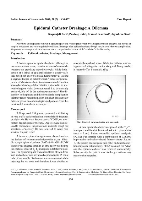
relocate the epidural space. While the catheter was beingremoved<br />
with gentle traction along with Tuohy needle,<br />
it sheared off at 6 cm mark. (Fig.1)<br />
Fi g.1 <strong>Epidural</strong> catheter-broken at 6 cm mark.<br />
A new epidural catheter was placed at the T 11 -T 12<br />
interspace and fixed at 9 cm mark (skin to epidural distance<br />
= 5 cm). Patient controlled epidural analgesia<br />
(PCEA) was initiated with a combination of 0.0625%<br />
bupivacaine hydrochloride and fentanyl citrate (5g.ml -<br />
1 ). The patient had adequate pain relief and chest condition<br />
improved satisfactorily. PCEAwas used for 7 days<br />
and the epidural catheter was removed uneventfully.<br />
Subsequently the patient was discharged without any<br />
neurological sequelae.
Deepanjali Pant et al. <strong>Epidural</strong> catheter breakage<br />
After informing the surgeon and the patient, an<br />
MRI and CT scan were done. Sagittal 3-mm (with 1mm<br />
gap) and axial 5-mm (with 1.5-mm gap) T 1 -<br />
weighted spin-echo (TR 500 ms/TE 16 ms/ 2 excitation)<br />
and proton density and T 2- weighted (TR 2600 ms/TE<br />
16,96 ms/2 excitation) fast spin-echo images were obtained<br />
in a 1.5T MRI scanner (General electric signal)<br />
(matrix 256x256, field of view 20 cm axial, 28 cm sagittal).<br />
Axial and sagittal T 1 -weighted images after IV<br />
gadolinium DTPA were obtained. CT scan (4-mm images<br />
obtained at 3-mm interval) with sagittal and coronal<br />
reconstruction was done. But the severed epidural<br />
catheter was not visualized.<br />
Thepatientwascounselled thatthis eventhad occurred<br />
andwas advised to reportin caseof anyadversesymptoms.<br />
Discussion<br />
Not many cases have been reported and there is<br />
always a dilemma in the mind of all – doctor to patient,<br />
regarding the sequence of leaving the catheter fragment<br />
in situ. So we thought of briefly reviewing the literature<br />
about various possible causes, ways to prevent, diagnose<br />
and manage such a case.<br />
Causes of severed epidural catheter<br />
1. Application of undue force in removing a catheter<br />
trapped between vertebral spinous processes or in<br />
ligamentum flavum or knotted, kinked or curled catheter<br />
in epidural space causes the catheter to stretch<br />
3, 4 or tear.<br />
2. Shearing of catheter by needle when attempts are<br />
made to withdraw the catheter through the Tuohy<br />
needle.<br />
3. Nicking of a catheter by a barb on the bevel of the<br />
needle.<br />
4. Shredding of catheter if the needle is advanced over<br />
the catheter after the catheter has been placed.<br />
5. Weakness of the catheter by imperfect manufacturing.<br />
6. Damage to a catheter occurring after placement<br />
i.e. fraying by pinching between two vertebral pro-<br />
5, 6 cesses.<br />
7. The proposed mechanism for catheter that were<br />
severed at time of insertion is to break or severely<br />
damage an epidural catheter by heavy contact between<br />
tip of theepidural needle and a bony surface,<br />
435<br />
if a length of the catheter was protruding from the<br />
tip. 7<br />
8. <strong>Catheter</strong> damage is often related to excessive in-<br />
8, 9<br />
sertion into the epidural space.<br />
Prevention of catheter breakage – recommendations<br />
1. Lateral decubitus position when removing an epidural<br />
catheter as this results in least force of extraction.<br />
10<br />
2. The force required to remove a catheter should be<br />
minimal. If resistance is encountered, a number of<br />
simple maneuvers may help to enable removal of<br />
catheter without stretching or tearing. These include<br />
(stepwise): -<br />
a. Maximal flexion of back in lateral decubitus position<br />
b. Rotation of spine<br />
c. Returningthe patientto theposition used at time<br />
of insertion e.g. sitting position with legs extended<br />
or kneeling position with hands down<br />
and back flexed<br />
d. Allowing tissues to soften for 15- 30 minutes<br />
before reattempting 11<br />
e. Fillingthe catheter with a rapid injection of saline<br />
to increase the turgor of the catheter and<br />
to lubricate it.<br />
f. Complete relaxation with GA with muscle relaxation<br />
g. Surgical removal<br />
3. The needle should be checked for barbs on bevel<br />
and the catheter for manufacturing defects before<br />
insertion.<br />
4. No more than 4-5 cm of catheter should be advanced<br />
into the epiduralspace to reduce risk of kinking<br />
/curling /knotting. 8,12,13<br />
5. <strong>Catheter</strong> should never be withdrawn through the<br />
metal needle.<br />
6. <strong>Catheter</strong>s of high breaking strain (tensile strength)<br />
and of a sufficient diameter (16/18G) should be obtained<br />
from a reputable, reliable manufacturer.<br />
In our case, there was no obvious cause for breakage.<br />
Most probably it was kinked or curled, as there<br />
was resistance during drug injection.
Diagnosis<br />
Attempts to locate the torn catheter ultrasonically<br />
are usually futile but xeroradiography, CT scanning or<br />
MRI may prove more fruitful.<br />
Radio opaque epidural catheters are easier to locate<br />
radiologically than non-radio opaque ones, but paradoxically,<br />
they have a lower tensile strength than standard<br />
clear catheters. In fact, a radioopaque fragment<br />
may be impossible to locate radiologically because the<br />
surrounding structures are radio-dense.<br />
MRI scanning is a non-invasive means of diagnosing<br />
the complication of spinal stenosis secondary to epidural<br />
fibrosis/scar formation and assessing the extent of<br />
spinal stenosis.<br />
However, CT scanning through level of interest is<br />
more sensitive than MRI in detecting the high attenuation<br />
catheter fragment within the epidural space and is<br />
more sensitivethan plain radiography, especiallyfor small<br />
retained fragments.<br />
In our case CT / MRI did not help to locate the<br />
fragment – since they are helpful once there is a reactive<br />
mass around the catheter fragment.<br />
Management<br />
Sequestered temporary epidural catheter pieces are<br />
generally considered to beinert and should not produce a<br />
foreign bodyreaction. Experiments with cats have shown<br />
that a broken catheter becomes walled off by fibrous tissue<br />
after about3 weeks –remaining innocuous within the<br />
epidural space. 13 Foreign body in epidural space is not<br />
likely to migrate (although this is not impossible).<br />
However, Staats et al reported the formation of a<br />
reactive epidural mass (1.5cm) around the catheter fragment<br />
resulting in lumbar spinal stenosis, patientbeing asymptomatic<br />
until 18 months of the incident and got relieved<br />
with removal of catheter and reactive scar tissue. 14<br />
In contrast, the continued presence of indwelling<br />
catheter has resulted in complications. Chronic, implanted<br />
intrathecal infusion catheters have been associated with<br />
granuloma formation resulting in spinal cord compression.<br />
The changes may occur, rather quickly, as noted<br />
by Durant and Yatish, who reported that catheters can<br />
be walled off by tissue reaction after 72 hours. 15 This<br />
local reaction was reported by Coombs et al, who found<br />
“cocoon” formation with dural thickening around implanted<br />
catheters in post-mortem examination. 16<br />
Therefore, in most cases the current standard of<br />
care application to the retained segments of a tempo-<br />
436<br />
Indian Journal of Anaesthesia, October 2007<br />
rary epiduralcatheter is to leavethem aloneunless symptomatic<br />
because surgical removal can produce more<br />
harm than good. 2 However, there are 3 situations where<br />
apolicy of non-interferenceor reassurancedoes not apply.<br />
1. Where infection or symptoms supervene, a careful<br />
historyand physicalexamination should help determine<br />
the spinallevel involved.<br />
2. If the spinal catheter fragment is sitting partially<br />
intrathecally and is acting as a wick which allows<br />
persistent CSF leakage. 12 If a continuous spinal<br />
micro-catheter becomes separated within the intrathecal<br />
space, appropriate imaging, a neurosurgical<br />
consultation and aggressivesurgical exploration<br />
to retrieve the broken piece are warranted, even in<br />
the asymptomatic patient. 17<br />
3. If the proximal end of the segment is located at or<br />
just beneath the skin such that it can be retrieved<br />
simple through a superficialincision made under local<br />
anaesthesia. The broken distal piece is grasped<br />
with a curved haemostat and drawn out by firm,<br />
gentle traction. 6 Surgical removal is mandatory in<br />
such a situation as bacteria can readily track along<br />
the catheter remnant.<br />
On rare occasions, surgical exploration may be<br />
needed to remove lost catheter fragments and associated<br />
reactive scar tissue and relieve spinal stenosis.<br />
Successfullocalization ofcatheter fragmentby medical<br />
imaging is no guarantee that task of finding the missing<br />
segmentat subsequentsurgery willbe madeany easier.<br />
If pain is caused by traction on catheter, the<br />
anaesthesiologist should suspect that a loop may have<br />
become curled around a nerve root. So removal of a<br />
catheter under anaesthesia may not help to alert the<br />
medical team in such a case. Given the possibility that<br />
avulsion might occur, it would probably be wise to extract<br />
the catheter under direct vision by open surgical<br />
laminectomy. 18 Sidhu et al described a parturient having<br />
epidural catheter coiled around L 2 -L 3 nerve root, causing<br />
severe pain and paresthesia on traction, but producing<br />
no sensory or motor defect. Since the patient refused<br />
a surgical procedure, catheter was removed without<br />
sequelae by gentle traction in various positions. 19<br />
Any kind of trauma, like practice of securing the<br />
catheter bya sutureat skin level, may cause microlesions<br />
and deteriorates the energy adsorbing capacity of catheter<br />
considerably – therefore, it should be practiced only<br />
when absolutely indicated. 20<br />
Our patient has not reported any adverse symptoms<br />
so far till the writing of this article – a time period<br />
of roughly two years.
Deepanjali Pant et al. <strong>Epidural</strong> catheter breakage<br />
Newer inventions<br />
More recently, it has been proposed to provide an<br />
improved epidural catheter which maintains its structural<br />
integrityfor intraoperativeand postoperativeperiod and<br />
eliminates the necessity for an additional surgical procedure<br />
or any outside intervention in order to remove a<br />
portion of the catheter should breakage occur.It exhibits<br />
good handling properties, has adequate tensile strength,<br />
is sterilizable and can be uniformly manufactured using<br />
conventional techniques. It is made up of biodegradable<br />
material (synthetic polymers of absorbable material)<br />
which dissolves with time upon contact with moisture<br />
found in bodyfluids with no undesirabledegradation substance<br />
released into body. 21 Since it is adversely affected<br />
by moisture it is preferably packaged in a substantially<br />
moisture-freeenvironment prior to use and in sealed sterile<br />
packages.<br />
In general, the material should generally maintain its<br />
original integrity for at least 3 days and should be completely<br />
absorbed in living tissue in a period of time from<br />
approximately 20 to 120 days. The degradation time of<br />
the biodegradable material can be selectively altered by<br />
adjusting the molecular weight or chemical make-up of<br />
the syntheticpolymers or through irradiation with gamma<br />
rays thatsimultaneously sterilizes the catheter without any<br />
significant loss of other desirable properties.<br />
In conclusion, inspite of thebest intentions and exercise<br />
of utmost care it can still result in tearing of an<br />
epidural catheter. Butfortunately in only a small proportion<br />
of these cases, it is prudent to attempt to remove the<br />
offending retained portion of the catheter.<br />
The usual guidelines for insertion and removal of<br />
catheter should be strictly followed on a routinebasis to<br />
prevent the occurrence.<br />
The presence of a retained epidural catheter fragment<br />
should be documented and communicated to the<br />
patient, surgeon and primary care physician because the<br />
development of symptoms related to the catheter may<br />
occur months or years later and the patient should be<br />
reviewed periodically to ensure that there is no discomfort,<br />
infection or radiculopathy. If symptoms develop,<br />
spine imaging to find out the level of involvement and<br />
surgery is advocated.<br />
437<br />
References<br />
1. Tio T, Macmurdo S, McKenzie R. Mishap with an epidural<br />
catheter. Anesthesiology 1979; 50:260-62.<br />
2. DeVera H, Ries M. Complication of continuous spinal<br />
microcatheter: should we seek their removal if sheared? Anesthesiology<br />
1991; 74:794.<br />
3. Gough JD,Johnston KR,Harmer M. Kinking of epidural catheters.<br />
Anaesthesia 1989; 40:1060.<br />
4. Jongleux EF,Miller R,Freeman A. An entrapped epidural catheter<br />
in a postpartum patient.Reg Anesth Pain Med 1998;23:615-17.<br />
5. Simpson P. Defective epidural cannulae. Anaesthesia 1981;<br />
36:72.<br />
6. DeArmendi A,Ryan J,Chang H, et al. Retained caudal catheter<br />
in a paediatric patient. Paed Anaes 1992; 2:325-27.<br />
7. Collier C. <strong>Epidural</strong> catheter breakage: a possible mechanism.<br />
Int J Obstet Anesth 2000; 9: 87 –93.<br />
8. Dawkins M. An analysis of the complications of extradural<br />
and caudal block. Anaesthesia 1969; 24:554-63.<br />
9. Dounas M,Peillon P, Lebonhomme JJ, et al .Difficulties in the<br />
removal and rupture of a peridural catheter.Ann Fr Anesth<br />
Reanim 2002;21:600-2.<br />
10. Morris GN,Warren BB,Hanson EW,et al.Influence of patient<br />
position on withdrawal forces during removal of lumbar extradural<br />
catheters. Anesthesiology 1997; 86:778-84.<br />
11. Demiraran Y, Yucel I, Erdogmus B. Subcutaneous effusion resulting<br />
from an epidural catheter fragment. Br J Anaesth 2006;<br />
96:508-9.<br />
12. Pasquariello C,Betz R. A case for the removal of the retained<br />
intrathecal catheter. Anesth Analg 1991;72:562.<br />
13. Bromage PR.<strong>Epidural</strong> Analgesia. Philadelphia,WB Saunders.<br />
1978 pp 664-66.<br />
14. Staats PS,Stinson MS,Lee R.Lumbar stenosis complicating retained<br />
epidural catheter tip.Anesthesiology 1995;83:1115-18.<br />
15. Durant PA,Yatish JL. <strong>Epidural</strong> injection of bupivacaine, morphine,<br />
fentanyl, lofentanil and DADL in chronically implanted<br />
rats: a pharmacologic and pathologic study. Anesthesiology<br />
1986; 64:43-53.<br />
16. Coombs DW,Franklin JD,Meier FA,et al.Neuropathologic lesions<br />
and CSF morphine during chronic continuous intraspinal<br />
morphine infusion: a clinical & post mortem study.Pain<br />
1985;22:337-51.<br />
17. Ugboma S, Au – Truong X, Kranzler LI, et al. The breaking of<br />
an intrathecally placed epidural catheter during extraction.<br />
Anesth Analg 2002; 95: 1087 – 9.<br />
18. Bromage PR.<strong>Epidural</strong> Analgesia.Philadelphia.WB Saunders<br />
1978,pp240.<br />
19. Sidhu MS,Asrani RV,Bassell GM. An unusual complication of<br />
extradural catheterization in obstetric anaesthesia. Br J Anaesth<br />
1983; 55:473-75.<br />
20. Schummer W, Schummer C. Another cause of epidural catheter<br />
breakage ? Anesth Analg 2002;94:233.<br />
21. http://www.freepatentsonline .com/5129889.html. (US Patent<br />
no 5129889)