Kidney Notes - BiologyMad A-Level Biology
Kidney Notes - BiologyMad A-Level Biology
Kidney Notes - BiologyMad A-Level Biology

Create successful ePaper yourself
Turn your PDF publications into a flip-book with our unique Google optimized e-Paper software.
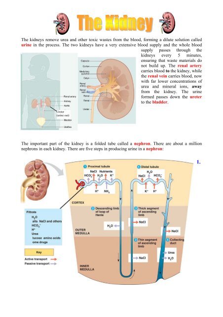
The kidneys remove urea and other toxic wastes from the blood, forming a dilute solution called<br />
urine in the process. The two kidneys have a very extensive blood supply and the whole blood<br />
supply passes through the<br />
kidneys every 5 minutes,<br />
ensuring that waste materials do<br />
not build up. The renal artery<br />
carries blood to the kidney, while<br />
the renal vein carries blood, now<br />
with far lower concentrations of<br />
urea and mineral ions, away<br />
from the kidney. The urine<br />
formed passes down the ureter<br />
to the bladder.<br />
Ureter<br />
(urine out)<br />
The important part of the kidney is a folded tube called a nephron. There are about a million<br />
nephrons in each kidney. There are five steps in producing urine in a nephron:<br />
1.
Renal capsule – Ultrafiltration<br />
The renal artery splits into numerous arterioles, each<br />
feeding a nephron. The arteriole splits into numerous<br />
capillaries, which form a knot called a glomerulus. The<br />
glomerulus is enclosed by the renal capsule (or Bowman’s<br />
capsule)- the first part of the nephron. The arteriole leading<br />
into the glomerulus is wider than the one leading out, so<br />
there is high blood pressure in the capillaries of the<br />
glomerulus. This pressure forces plasma out of the blood by<br />
ultrafiltration. Both the capillary walls and the capsule<br />
walls are formed from a single layer of flattened cells with gaps between them, so that all molecules<br />
with a molecular mass of < 68,000 are squeezed out of the blood to form a filtrate in the renal<br />
capsule. Only blood cells and large proteins (e.g. antibodies and albumin) remain in the blood.<br />
2. First (proximal) Convoluted Tubule – Reabsorption.<br />
The proximal convoluted tubule is the longest (14mm) and<br />
widest (60µm) part of the nephron. It is lined with epithelial cells<br />
containing microvilli and numerous mitochondria. In this part of<br />
the nephron over 80% of the filtrate is reabsorbed into the<br />
tissue fluid and then to the blood. This ensures that all the “useful”<br />
materials that were filtered out of the blood (such as glucose and<br />
amino acids) are now returned to the blood. These cells have<br />
microvilli, to ensure a large surface area (Fick’s Law again!)<br />
• All glucose, all amino acids and 85% of mineral ions<br />
are reabsorbed by active transport from the filtrate to the tissue fluid. They then diffuse<br />
into the blood capillaries.<br />
• Small proteins are reabsorbed by pinocytosis, digested, and the amino acids diffuse into the<br />
blood.<br />
• 80% of the water is reabsorbed to the blood by osmosis.<br />
• Surprisingly, some urea is reabsorbed to the blood by diffusion. Urea is a small, uncharged<br />
molecule, so it can pass through membranes by lipid diffusion and there isn’t much the<br />
kidney can do about it. Since this is a passive process, urea diffuses down its concentration<br />
gradient until the concentrations of urea in the filtrate and blood are equal. So in each pass<br />
through the kidneys half the urea is removed from the blood and half remains in the blood.<br />
3. Loop of Henlé – Formation of a Salt Bath.<br />
The job of the loop of Henlé is to make the tissue fluid in the<br />
medulla hypertonic (i.e. lower ψ) compared to the filtrate in the<br />
nephron. The purpose of this “salt bath” is to reabsorb water as<br />
explained in step 5. The loop of Henlé does this by pumping<br />
sodium and chloride ions out of the filtrate into the tissue<br />
fluid.
The first part of the loop (the descending limb) is impermeable to ions, but some water leaves by<br />
osmosis. This makes the filtrate more concentrated as it descends. The second part of the loop (the<br />
ascending limb) contains a Na + and a Cl - pump, so these ions are actively transported out of the<br />
filtrate into the surrounding tissue fluid. Water would follow by osmosis, but it can’t, because<br />
the ascending limb is impermeable to water. So the water potential of the tissue fluid becomes<br />
lower (more salty - lower ψ) and the water potential of the filtrate becomes higher (less salty –<br />
higher ψ). Since the filtrate is most concentrated at the base of the loop, the tissue fluid is also<br />
more concentrated at the base of the medulla, where it is three times more concentrated than<br />
seawater.<br />
4. Distal Convoluted tubule – Homeostasis and Secretion<br />
In the distal convoluted tubule certain substances are actively transported from the blood into the<br />
filtrate, in other words they are secreted. It is relatively short and has microvilli with numerous<br />
membrane pumps for active transport. The important point about this secretion is that it is regulated<br />
by hormones, so this is the homeostatic part of the kidney. Substances secreted include H + (for pH<br />
homeostasis), K + (for salt homeostasis), ethanol, toxins, drugs and other “foreign” substances.<br />
5. Collecting Duct – Concentration<br />
As the collecting duct passes through the<br />
hypertonic salt bath in the medulla, water leaves<br />
the filtrate by osmosis, so concentrating the urine<br />
and conserving water. The water leaves through<br />
special water channels in the cell membrane called<br />
aquaporins. These aquaporin channels can be<br />
controlled by the hormone ADH (= Anti<br />
Diuretic Hormone) , so allowing the amount of<br />
water in the urine to be controlled. More ADH<br />
opens the channels, so more water is conserved<br />
in the body, and more concentrated urine is<br />
produced. This is described in more detail in water<br />
homeostasis later.
The Bladder<br />
The collecting ducts all join together in the pelvis of the kidney to form the ureter, which leads to<br />
the bladder. The filtrate, only now called urine, is produced continually by each kidney and drips<br />
into the bladder for storage. The bladder is an expandable bag, and when it is full, stretch receptors<br />
in the elastic walls send impulses to the medulla, which causes the sphincter muscles to relax,<br />
causing urination. This is an involuntary reflex response that we can learn to control to a certain<br />
extent when we are young.<br />
© IHW March 2006