Alginate microspheres of Bacillus subtilis - Facultad de Farmacia
Alginate microspheres of Bacillus subtilis - Facultad de Farmacia
Alginate microspheres of Bacillus subtilis - Facultad de Farmacia

You also want an ePaper? Increase the reach of your titles
YUMPU automatically turns print PDFs into web optimized ePapers that Google loves.
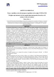
ALGINATE MOCROSPHERES OF BACILLUS SUBTILIS<br />
<strong>Alginate</strong> <strong>microspheres</strong> <strong>of</strong> <strong>Bacillus</strong> <strong>subtilis</strong><br />
Microesferas <strong>de</strong> alginato con <strong>Bacillus</strong> <strong>subtilis</strong><br />
BREGNI, C.*; DEGROSSI, J.*; GARCÍA, R.*; LAMAS, M. C.**; FIRENSTEIN, R.** Y D'AQUINO, M.*<br />
* Faculty <strong>of</strong> Pharmacy and Biochemistry. University <strong>of</strong> Buenos Aires. Junin 956. Piso 6º.<br />
(1113) Buenos Aires. Argentina. E-mail: cbregni@ciudad.com.ar<br />
** Faculty <strong>of</strong> Biochemistry and Pharmaceutical Sciences. University <strong>of</strong> Rosario. Argentina.<br />
RESUMEN<br />
La microencapsulación <strong>de</strong> organismos ha sido consi<strong>de</strong>rada como una alternativa <strong>de</strong> inmovilización <strong>de</strong> células, a fin<br />
<strong>de</strong> que éstas puedan ejercer sus funciones en forma gradual. El objetivo <strong>de</strong>l presente estudio fue elaborar microesferas<br />
<strong>de</strong> <strong>Bacillus</strong> <strong>subtilis</strong> ya sea en forma esporulada como vegetativa.<br />
Microesferas <strong>de</strong> <strong>Bacillus</strong> <strong>subtilis</strong> son preparadas utilizando alginato <strong>de</strong> sodio. Algunas propieda<strong>de</strong>s típicas <strong>de</strong>l sistema<br />
microencapsulado, tales como contenido <strong>de</strong> microorganismos, tamaño <strong>de</strong> partícula y tiempo <strong>de</strong> germinación han sido<br />
estudiados. Las microesferas se prepararon mediante el método <strong>de</strong> coaservación-separación <strong>de</strong> fases, utilizando una<br />
etapa intermedia <strong>de</strong> emulsión múltiple. Las condiciones <strong>de</strong> preparación han sido lo suficientemente benignas para<br />
no producir cambios en las propieda<strong>de</strong>s biológicas generales <strong>de</strong>l sistema, pero con la protección que le otorga la<br />
matriz <strong>de</strong>l hidrogel, la cual evita la directa comunicación con el medio externo.<br />
La viabilidad <strong>de</strong>mostrada por las microesferas con las formas esporuladas fue significativamente superior a las <strong>de</strong><br />
las formas vegetativas.<br />
PALABRAS CLAVE: Alginato. <strong>Bacillus</strong> <strong>subtilis</strong>. Inmovilización. Microesferas.<br />
ABSTRACT<br />
<strong>Bacillus</strong> <strong>subtilis</strong> <strong>microspheres</strong> were prepared by using sodium alginate. Some typical properties <strong>of</strong> microencapsulated<br />
systems such as content <strong>of</strong> microorganisms, particle size and germination time were studied. Microspheres are prepared<br />
by the coacervation phase separation method, mainly based on an intermediate stage <strong>of</strong> primary emulsion. The preparation<br />
conditions were very s<strong>of</strong>t to avoid changes in general biological products <strong>of</strong> calcium alginate <strong>microspheres</strong> containing<br />
cells, but giving them protection with a hydrogel matrix, without preventing the communication with the surrounding<br />
medium. The encapsulation <strong>of</strong> microbial spores and viable cells <strong>of</strong> a mo<strong>de</strong>l microorganism (<strong>Bacillus</strong> <strong>subtilis</strong>) can be<br />
achieved by using sodium alginate in a satisfactory manner. Spore <strong>microspheres</strong> showed higher viability comparated to<br />
vegetative <strong>microspheres</strong>.<br />
KEY WORDS: <strong>Alginate</strong>. <strong>Bacillus</strong> <strong>subtilis</strong>. Immobilization. Microspheres.<br />
Microencapsulation has been suggested as an<br />
alternative method for entrapment and immobilization<br />
<strong>of</strong> whole cells or their extracts. Currently<br />
there is a limited number <strong>of</strong> reports <strong>de</strong>scribing<br />
the microencapsulation <strong>of</strong> microbial cells.<br />
The microencapsulation method in alginate gel<br />
is carried out in a single step process un<strong>de</strong>r very<br />
mild conditions and should be compatible with<br />
most living cells.<br />
INTRODUCTION<br />
245<br />
<strong>Alginate</strong>s are a family <strong>of</strong> polysacchari<strong>de</strong>s<br />
compose <strong>of</strong> (-L-glucuronic acid (G) and (-D-mannuronic<br />
acid (M) residues, arranged in homopolymeric<br />
blocks <strong>of</strong> each type (MM, GG) and in<br />
heteropolymeric blocks which are reported to have<br />
a major impact on the properties <strong>of</strong> the different<br />
systems.<br />
Some hydrophilic polymers have ion binding<br />
properties. Among these are alginates which belong<br />
Ars Pharmaceutica, 41:3; 245-248, 2000
246<br />
to a family <strong>of</strong> unbranched binary copolymers <strong>of</strong><br />
linked acid residues.<br />
<strong>Alginate</strong>s have the ability to bind multivalent<br />
cations being the basis <strong>of</strong> their gelling properties,<br />
leading to the formation <strong>of</strong> covalent bonds<br />
yielding insoluble hydrogels.<br />
This anionic polysacchari<strong>de</strong> forms strong gels<br />
with divalent cations like Ca 2+ , giving both strength<br />
and flexibility. Such crosslinking process stiffens<br />
and roughens the polymer and reduces the swelling<br />
in solvents.<br />
The soluble sodium alginate was crosslinked<br />
with calcium chlori<strong>de</strong> resulting in the formation<br />
<strong>of</strong> the insoluble calcium alginate. Natural poly-<br />
Sodium alginate low viscosity (Sigma Chemical),<br />
Ether sulphuric HPLC gra<strong>de</strong> (Merck) and<br />
Calcium chlori<strong>de</strong> (Carlo Erba, Milan, Italy).<br />
The non-ionic surfactants used were Sorbitan<br />
monooleate (Span 80) and Polyoxyethylene sorbitan<br />
monooleate (Tween 80), manufactured by Atlas Co.<br />
Preparation <strong>of</strong> w/o primary emulsions<br />
The primary emulsions were prepared by onestage<br />
emulsification procedure. The oil external<br />
phase, containing 70 mL <strong>of</strong> liquid vaseline, 30<br />
mL <strong>of</strong> ether sulfuric, and sorbitan monooleate<br />
(3%; w/w) was emulsified in an equal volume <strong>of</strong><br />
sodium alginate solution (4%; w/v) containing<br />
the suspension cells, by means <strong>of</strong> a small vortex<br />
mixer (Whirlimixer) to produce the primary water<br />
in oil emulsion.<br />
After preparation, the nature <strong>of</strong> the emulsions<br />
was confirmed by microscopic examination.<br />
Preparation <strong>of</strong> the <strong>microspheres</strong><br />
Microspheres are prepared by the coacervation<br />
phase separation method, mainly based on<br />
an intermediate stage <strong>of</strong> primary emulsion.<br />
<strong>Alginate</strong> gel microparticles are prepared by<br />
the crosslinking <strong>of</strong> sodium alginate, participating<br />
in the aqueous internal phase <strong>of</strong> the primary<br />
Ars Pharmaceutica, 41:3; 245-248, 2000<br />
BREGNI, C.; DEGROSSI, J.; GARCÍA, R.; LAMAS, M. C.; FIRENSTEIN, R. y D'AQUINO, M.<br />
MATERIALS<br />
METHODS<br />
mers are used both as carriers and <strong>de</strong>terminants<br />
<strong>of</strong> the release rate in controlled release systems<br />
(Magee 1981).<br />
The main advantages <strong>of</strong> natural polymers lie in<br />
their biocompatibility and bio<strong>de</strong>gradability, without<br />
producing systemic toxicity on administration (Pepeljnjak<br />
1988; Takka and Acarturk 1999a).<br />
The objectives <strong>of</strong> the present study were to<br />
investigate the possibility <strong>of</strong> microencapsulation<br />
<strong>of</strong> a mo<strong>de</strong>l (non-pathogenic) microorganism (<strong>Bacillus</strong><br />
<strong>subtilis</strong>) with sodium alginate and to study<br />
the viability <strong>of</strong> the microorganism after the preparation<br />
step (Pepeljnjak 1994; Al Musa 1999;<br />
Takka and Acarturk 1999b).<br />
Bacterial spores and viable cells <strong>of</strong> mo<strong>de</strong>l<br />
microorganism (<strong>Bacillus</strong> <strong>subtilis</strong>, strain 6633) were<br />
used as biological materials. Samples were cultivated<br />
on neutral nutrient broth at 37 °C. The<br />
other chemicals used are <strong>of</strong> analytical gra<strong>de</strong>. All<br />
materials were used as received.<br />
emulsion with calcium ions (CaCl 2 solution 10<br />
% w/w).<br />
The supernatant is <strong>de</strong>canted, the sediment is<br />
washed with distilled water (200 mL) and the<br />
system is filtered and dried at 37 ºC, until they<br />
reach a constant weight.<br />
The <strong>microspheres</strong> are prepared as follows: type<br />
I (spores) and type II (viable cells) (Lim and<br />
Sum 1980).<br />
Morphology<br />
The formation <strong>of</strong> <strong>microspheres</strong> was monitored<br />
by optical microscopy (Optiphot, Nikon)<br />
Particle size distribution<br />
United States standard sieves ranging from<br />
50 - 700 micra were used to <strong>de</strong>termine the particle<br />
size <strong>of</strong> 10g <strong>microspheres</strong> (USP 1999). This<br />
procedure was repeated three times for different<br />
batches (Figures 1 and 2).
ALGINATE MOCROSPHERES OF BACILLUS SUBTILIS<br />
FIGURE 1. Particle size distribution <strong>of</strong> <strong>microspheres</strong> with viable cells. Data are expressed as the mean<br />
for three to five experiments.<br />
% retained<br />
50<br />
40<br />
30<br />
20<br />
10<br />
0<br />
0 100 200 300 500 700<br />
RESULTS AND DISCUSSION<br />
Particle size (micra)<br />
FIGURE 2. Particle size distribution <strong>of</strong> <strong>microspheres</strong> with spores. Data are expressed as the mean<br />
for three to five experiments.<br />
% retained<br />
50<br />
40<br />
30<br />
20<br />
10<br />
0<br />
Two types <strong>of</strong> <strong>microspheres</strong> were prepared as<br />
follows:<br />
— Type I: spores (alginate concentration 2%<br />
w/w).<br />
— Type II: viable cells (alginate concentration<br />
2% w/w).<br />
The <strong>microspheres</strong> obtained were polydisperse<br />
systems with particle size distribution conforming<br />
to a normal distribution and with mean<br />
diameters between 50 and 300 micra.<br />
The <strong>microspheres</strong> were well formed and in<br />
0 100 200 300 500 700<br />
Particle size (micra)<br />
247<br />
spite <strong>of</strong> their s<strong>of</strong>t hydrogel nature they resisted<br />
all the manipulations in liquid media during<br />
experimental work. However, an increase in<br />
microsphere concentration in aqueous media was<br />
obtained, but it was not possible to filter the<br />
microspherical product.<br />
The difference between <strong>microspheres</strong> prepared<br />
with microbial spores and with viable cells<br />
is evi<strong>de</strong>nt from the data for the average number<br />
<strong>of</strong> cells per microsphere and the germination time<br />
(Table I).<br />
Ars Pharmaceutica, 41:3; 245-248, 2000
248<br />
* CFU/g : colony-forming units/g.<br />
Ars Pharmaceutica, 41:3; 245-248, 2000<br />
BREGNI, C.; DEGROSSI, J.; GARCÍA, R.; LAMAS, M. C.; FIRENSTEIN, R. y D'AQUINO, M.<br />
TABLE I. <strong>Alginate</strong> microsphere germination properties including <strong>Bacillus</strong> <strong>subtilis</strong> (colony-forming units/g).<br />
Time<br />
(days)<br />
Type I<br />
Spores (CFU/g)*<br />
0 3.1 x 10 9<br />
30 3.0 x 10 9<br />
90 1.2 x10 8<br />
150 1.0 x10 8<br />
The germination time <strong>of</strong> encapsulated cells is<br />
significantly higher than that <strong>of</strong> the cells that are<br />
not encapsulated (control), giving a relative indication<br />
<strong>of</strong> protection achieved by the process <strong>of</strong><br />
microencapsulation. After the lag-time due to<br />
encapsulation, cell growth was uninhibited and<br />
there was no difference between encapsulated and<br />
The results <strong>of</strong> this study showed that the<br />
encapsulation <strong>of</strong> microbial spores and viable<br />
cells <strong>of</strong> a mo<strong>de</strong>l microorganism can be satisfactorily<br />
achieved by using sodium alginate.<br />
Very mild conditions during the preparation<br />
step allow the production <strong>of</strong> <strong>microspheres</strong> con-<br />
CONCLUSIONS<br />
ACKNOWLEDGEMENTS<br />
Type II<br />
Viable cells<br />
(CFU/g)*<br />
2.8 x 10 9<br />
8.9 x 10 7<br />
4.3 x 10 7<br />
3.4 x 10 7<br />
free cells. Viability <strong>of</strong> encapsulated cells was<br />
kept unchanged during <strong>of</strong> the experiments and<br />
since no difference was noticed, it can be presumed<br />
that it can be kept for much longer.<br />
Polymerization conditions allow obtaining<br />
to obtain microencapsulated cells during 150<br />
days.<br />
taining cells with seemingly no changes in their<br />
general biological properties but gives them<br />
protection with a s<strong>of</strong>t hydrogel matrix while at<br />
the same time it does not prevent completely<br />
the communication with the surrounding medium.<br />
The authors wish to acknowledge the support <strong>of</strong> this work provi<strong>de</strong>d UBACyT (01/TB12).<br />
REFERENCES<br />
Al Musa, S., D. Abu Fara and A. A. Badwan (1999). Evaluation <strong>of</strong> parameters involved in preparation and release <strong>of</strong> drug<br />
loa<strong>de</strong>d in crosslinked matrices <strong>of</strong> alginate. J. Controlled Release, 57: 223-232.<br />
Lim F. and Sum A. M. (1980). Chitosan and <strong>de</strong>rivatives as biomaterials. Formulation <strong>of</strong> polymer membranes and <strong>microspheres</strong>.<br />
Science, 21: 908-912.<br />
Magee E. L., Olson N. F. and Lindsay R. C. (1981). Rheology <strong>of</strong> milk gels formed by milkclotting enzymes. J. Dairy Sci.,<br />
64: 616-621.<br />
Pepeljnjak S., S. Penovski and D. Jalsenjak (1988). Influence <strong>of</strong> microencapsulated propolis extract on <strong>Bacillus</strong> <strong>subtilis</strong><br />
strein IP-5832. Pharmazie, 43: 728-732.<br />
Pepeljnjak, S., J. Filipovic-Grcic and V. Jalsenjak (1994). <strong>Alginate</strong> <strong>microspheres</strong> <strong>of</strong> microbial spores and viable cells <strong>of</strong><br />
<strong>Bacillus</strong> <strong>subtilis</strong>. Pharmazie, 49: 436-437.<br />
Takka, S. and F. Acarturk (1999a). Calcium alginate microparticles for oral administration; I: effect <strong>of</strong> sodium alginate type<br />
on drug release and drug entrapment efficiency. J. Microencapsulation, 16: 275-290.<br />
Takka, S. and F. Acarturk (1999b). Calcium alginate microparticles for oral administration; II: effect <strong>of</strong> formulation factors<br />
on drug release and drug entrapment efficiency. J. Microencapsulation, 16: 291-301.<br />
U.S.Pharmacopeia 24 Ed./National Formulary 19 (1999). Rockville, MD.