Dairy Sheep Symposium - the Department of Animal Sciences ...
Dairy Sheep Symposium - the Department of Animal Sciences ...
Dairy Sheep Symposium - the Department of Animal Sciences ...

Create successful ePaper yourself
Turn your PDF publications into a flip-book with our unique Google optimized e-Paper software.
From <strong>the</strong>se results <strong>the</strong> authors concluded that 1) <strong>the</strong> allometric phase <strong>of</strong> prepubertal<br />
mammogenesis is primarily a function <strong>of</strong> age and largely reverts to isometric growth before<br />
normal puberty; 2) after 20 weeks, <strong>the</strong> size <strong>of</strong> available fat pad may limit fur<strong>the</strong>r lateral expansion<br />
in restricted-gain lambs; and 3) precocious puberty attained within <strong>the</strong> major phase <strong>of</strong><br />
allometric growth can lead to marked decrease in mammary gland size.<br />
The effect <strong>of</strong> nutritional plane on prepubertal mammogenesis is similar across breeds in<br />
sheep and cattle. Hohenboken et al. (1995) reported that <strong>the</strong>re was no difference in sensitivity <strong>of</strong><br />
subsequent milk yield to high prepubertal gain across four Danish dairy or dual-purpose cattle<br />
breeds. In sheep, this relationship has been demonstrated in trials with Dorset-, Suffolk-, Hampshire-,<br />
and Finn-cross ewe lambs (Umberger et al., 1985; McCann et al., 1989; Johnsson and<br />
Hart, 1985; McFadden et al., 1990).<br />
Hormonal Influences on Prepubertal Mammogenesis<br />
Researchers now recognize that <strong>the</strong> impaired mammary development <strong>of</strong> rapidly-reared<br />
prepubertal ruminants is caused by altered secretions <strong>of</strong> hormones, growth factors, and binding<br />
proteins, all <strong>of</strong> which regulate mammary development (Sejrsen et al., 1997). Although <strong>the</strong> exact<br />
physiological mechanisms are not understood, it is agreed that high feeding levels inhibit circulating<br />
growth hormone (GH) concentrations in <strong>the</strong> blood, and that <strong>the</strong>re is a positive correlation<br />
between serum GH and prepubertal mammary growth (Sejrsen et al., 1983, Johnsson et al.,<br />
1985). This has also been confirmed by trials showing that exogenous administration <strong>of</strong> GH<br />
produces increased parenchymal tissue growth in lambs (McFadden et al., 1990; Johnsson et al.,<br />
1986) and heifers (Sejrsen et al., 1997).<br />
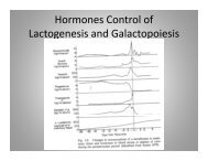
Johnsson et al. (1985) examined <strong>the</strong> effects <strong>of</strong> daily nutrition on GH plasma concentrations at<br />
various ages before puberty and <strong>the</strong> associated mammogenesis. Blood samples were taken from<br />
ewe lambs at 10, 14, 18, 26, and 36 weeks <strong>of</strong> age. As described earlier, lambs were fed to ei<strong>the</strong>r<br />
a high (H) or low (L) daily gain from 4 to 36 weeks. Results showed that GH response to fresh<br />
daily feed was significantly higher in L lambs than H lambs at 10, 14, and 18 weeks, and that GH<br />
concentrations and mammary development were positively correlated. As <strong>the</strong> lambs’ ages increased<br />
after 20 weeks, both <strong>the</strong> mean level <strong>of</strong> serum GH and <strong>the</strong> sensitivity <strong>of</strong> GH concentrations<br />
to feed intake declined (Figure 3).<br />
Growth hormone levels have also been positively correlated with ductal growth during <strong>the</strong><br />
allometric period in heifers (Sejrsen et al., 1983). Serum GH concentrations were depressed in<br />
prepubertal heifers on ad-lib feed, but were not affected by rearing rate after puberty. And, as<br />
with lambs, serum GH concentration was negatively correlated with extraparenchmal adipose<br />
tissue mass, indicating a lipolytic role <strong>of</strong> GH.<br />
It is believed that <strong>the</strong> mammogenic actions <strong>of</strong> GH may be mediated by <strong>the</strong> stromal cells <strong>of</strong><br />
<strong>the</strong> fat pad. In <strong>the</strong> prepubertal ewe, adipose tissue binds GH, and probably stimulates <strong>the</strong> production<br />
and secretion <strong>of</strong> insulin-like growth factor-1 (Hovey et al., 1999). IGF-1 is a direct and<br />
potent mitogen for undifferentiated ruminant epi<strong>the</strong>lial cells (Weber et al., 1999), and also, in<br />
gestation, for differentiated cells, stimulating DNA syn<strong>the</strong>sis in ductal epi<strong>the</strong>lial cells, secretory<br />
alveolar cells, and myoepi<strong>the</strong>lial cells (Forsyth, 1995).<br />
It is important to note that IGF-1 is highly mitogenic for undifferentiated mammary epi<strong>the</strong>lial<br />
cells at low concentrations. Weber et al. (1999) reported that in vitro, additional IGF-1 caused