Dairy Sheep Symposium - the Department of Animal Sciences ...
Dairy Sheep Symposium - the Department of Animal Sciences ...
Dairy Sheep Symposium - the Department of Animal Sciences ...

Create successful ePaper yourself
Turn your PDF publications into a flip-book with our unique Google optimized e-Paper software.
After puberty, <strong>the</strong> mammary gland reverts to isometric growth, with growth again paralleling<br />
that <strong>of</strong> <strong>the</strong> whole body.<br />
Pregnancy causes <strong>the</strong> mammary gland to undergo ano<strong>the</strong>r period <strong>of</strong> extensive development<br />
andallometric growth (Sejrsen et al., 2000). In <strong>the</strong> ewe, mammary growth in early pregnancy<br />
consists <strong>of</strong> rapid ductal growth. In late pregnancy, differentiated epi<strong>the</strong>lial cells form alveoli,<br />
which are anchored to collagen. DNA analysis <strong>of</strong> cell numbers <strong>of</strong> epi<strong>the</strong>lial and connective<br />
tissue in Romney sheep showed that 20% <strong>of</strong> gland growth occurred between birth and puberty,<br />
78% during pregnancy, and 2% during lactation (Anderson, 1975).<br />
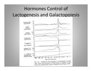
Total milk yield is proportional to total epi<strong>the</strong>lial cell numbers (Hovey et al., 1999). As<br />
revealed by <strong>the</strong> Romney ewe study above, 98% <strong>of</strong> epi<strong>the</strong>lial cell numbers are established by<br />
parturition. Between parturition and peak lactation, secretory cells hypertrophy and become<br />
more fully differentiated. Forsyth (1995) studied <strong>the</strong> relationship between milk yield and alveolar<br />
cell activity and persistence in dairy cattle. She found that after peak lactation, activity per<br />
individual cell is maintained, and that alveolar cell loss is <strong>the</strong> primary cause <strong>of</strong> milk yield decline.<br />
In <strong>the</strong> modern dairy cow, peak yield accounts for 66 to 80% <strong>of</strong> <strong>the</strong> variance in total yield,<br />
and persistency (days in milk) accounts for only 8 to 12 % <strong>of</strong> <strong>the</strong> variance. Peak yield is primarily<br />
determined by secretory cell numbers. Therefore any process that negatively impacts epi<strong>the</strong>lial<br />
cell numbers will negatively impact total milk yield.<br />
The Fat Pad<br />
The mammary fat pad is a matrix <strong>of</strong> connective and adipose tissue that also houses <strong>the</strong><br />
gland’s vascular and lymphatic systems. It has three major functions critical to mammogenesis:<br />
first, its connective tissue network provides <strong>the</strong> structural support for <strong>the</strong> parenchyma; second, it<br />
serves as an active lipid store for <strong>the</strong> growing and/or differentiating epi<strong>the</strong>lia; and third, in a<br />
function only now being understood, it locally regulates mammary development by mediating<br />
hormone action and syn<strong>the</strong>sizing growth factors.<br />
As shown in <strong>the</strong> recent work by Hovey et al. (1999), <strong>the</strong> fat pad’s adipose tissue is extensively<br />
interlaced with rope-like cords <strong>of</strong> connective tissue fibroblasts. In <strong>the</strong> lactating adult,<br />
<strong>the</strong>se cords will give structural support to <strong>the</strong> fluid-filled udder. In <strong>the</strong> developing animal, <strong>the</strong><br />
connective tissue meshwork, interspersed between adipocytes, directs <strong>the</strong> spread <strong>of</strong> parenchymal<br />
growth: ductules are seen lying embedded within veins <strong>of</strong> connective tissue (Figure 2). As <strong>the</strong><br />
growing ducts advance through <strong>the</strong> adipose/collagen matrix, fibroblasts proliferate in response to<br />
<strong>the</strong> oncoming ductules, continuously ensheathing <strong>the</strong> ductules in multiple layers <strong>of</strong> fibrous tissue.<br />
Although <strong>the</strong> proliferating epi<strong>the</strong>lial cells do not directly abut <strong>the</strong> fat pad’s adipocytes, <strong>the</strong><br />
adipose tissue acts as a lipid depot for <strong>the</strong> dividing cells. Lipid-depleted adipocytes are seen in<br />
<strong>the</strong> area <strong>of</strong> ductal infiltration. Fur<strong>the</strong>rmore, Hovey et al. (1999) found that adipocyte-derived<br />
fatty acids markedly increased <strong>the</strong> response <strong>of</strong> mammary epi<strong>the</strong>lial cells to growth factors in<br />
vitro.The actions <strong>of</strong> many hormones are mediated by <strong>the</strong> mammary fat pad, and <strong>the</strong> fat pad is<br />
also a major site for <strong>the</strong> syn<strong>the</strong>sis <strong>of</strong> local growth factors. Growth hormone (GH), a dominant<br />
mammogenic hormone, has binding sites in adipose tissue in <strong>the</strong> prepubertal ewe lamb. Insulinlike<br />
growth factor-1 is stroma-derived in <strong>the</strong> mammary gland, and <strong>the</strong>re appear to be epidermal<br />
growth factor receptors within <strong>the</strong> fat pad as well as on epi<strong>the</strong>lial cells (Hovey et al., 1999).<br />
The mammary fat pad is <strong>the</strong>refore not simply an inert supporting material. It plays an integral<br />
and critical role in directing, stimulating, and regulating mammogenesis.